The Holographic Principle and the Emergence of Spacetime ...
Upload
learn-uconnCategory
view
0download
0
Photochromic Bacteriorhodopsin Mutant with High HolographicEfficiency and Enhanced Stability via a Putative Self-RepairMechanismMatthew J. Ranaghan,† Jordan A. Greco,‡ Nicole L. Wagner,† Rickinder Grewal,† Rekha Rangarajan,†
Jeremy F. Koscielecki,‡ Kevin J. Wise,† and Robert R. Birge*,†,‡
†Department of Molecular and Cell Biology, University of Connecticut, 91 North Eagleville Road, Storrs, Connecticut 06269, UnitedStates‡Department of Chemistry, University of Connecticut, 55 North Eagleville Road, Storrs, Connecticut 06269, United States
*S Supporting Information
ABSTRACT: The Q photoproduct of bacteriorhodopsin (BR) is thebasis of several biophotonic technologies that employ BR as thephotoactive element. Several blue BR (bBR) mutants, generated byusing directed evolution, were investigated with respect to thephotochemical formation of the Q state. We report here a new bBRmutant, D85E/D96Q, which is capable of efficiently converting theentire sample to and from the Q photoproduct. At pH 8.5, where Qformation is optimal, the Q photoproduct requires 65 kJ mol‑1 of amberlight irradiation (590 nm) for formation and 5 kJ mol‑1 of blue light (450 nm) for reversion, respectively. The meltingtemperature of the resting state and Q photoproduct, measured via differential scanning calorimetry, is observed at 100 °C and89 °C at pH 8.5 or 91 °C and 82 °C at pH 9.5, respectively. We hypothesize that the protein stability of D85E/D96Q comparedto other blue mutants is associated with a rapid equilibrium between the blue form E85(H) and the purple form E85(−) of theprotein, the latter providing enhanced structural stability. Additionally, the protein is shown to be stable and functional whensuspended in an acrylamide matrix at alkaline pH. Real-time photoconversion to and from the Q state is also demonstrated withthe immobilized protein. Finally, the holographic efficiency of an ideal thin film using the Q state of D85E/D96Q is calculated tobe 16.7%, which is significantly better than that provided by native BR (6−8%) and presents the highest efficiency of any BRmutant to date.
KEYWORDS: blue bacteriorhodopsin, Q-state, stability, bionanotechnology, electro-optical materials, directed evolution
Bacteriorhodopsin (BR) is the light-harvesting proteinexpressed by the salt marsh archaeon, Halobacterium
salinarum, when the environment lacks sufficient free oxygento permit oxidative phosphorylation as a source of energy.1,2
This transmembrane protein contains a covalently linkedchromophore, all-trans retinal, and is arranged in trimerswithin a semicrystalline lattice.3−5 The ensemble is referred toas the purple membrane. Following the absorption of light byretinal, the light-adapted form of the protein pumps a protonacross the cell membrane, thereby creating a pH gradient thatthen drives ATPase to synthesize ATP.2,6 Proton pumping isachieved by a photocycle that returns to the resting state (bR)in roughly 10 ms at ambient temperature.7−9 While chemicaland genetic modifications of BR have been used to extend thelifetime of various intermediates in the main photocycle,10−12
these methods have only recently produced highly stableintermediates for use of the protein in long-term datastorage13−15 and associative processors.10,16−19 The ability toefficiently convert this photochromic protein from bR (λmax =570 nm) to an inactive state, known as the Q photoproduct(λmax = 390 nm), is of interest for the successfulimplementation of BR into these optical devices.
The Q state is a photoproduct of the BR branchedphotocycle and remains stable for many years at ambienttemperature.20,21 The chromophore in Q is in a 9-cisconfiguration, which makes it an untenable candidate forbinding to Lys-216 due to steric interactions with variousbinding site residues.18,20,21 Bacteriorhodopsin does not form Qduring the native photocycle, however, and requires thesequential absorption of a green photon to photoexcite thebR state and a red photon during the population of the O state(λmax = 610 nm) in order to produce Q (Figure 1).21
Photochemical formation of Q occurs with a lower quantumefficiency than the initial photoexcitation event (ϕ ∼ 0.65),7 asmeasured for a deionized form of the native protein (ϕQ ≤ 1.2× 10‑4).18 Hence, BR must be genetically modified to efficientlyproduce Q before the photoproduct can be utilized in protein-based technologies.13,15−17,22
Blue BR (bBR) membranes contain modified proteins thatexhibit an O-like resting state (bRB; λmax ∼ 603 nm) and are
Received: November 25, 2013Accepted: February 5, 2014Published: February 5, 2014
Research Article
www.acsami.org
© 2014 American Chemical Society 2799 dx.doi.org/10.1021/am405363z | ACS Appl. Mater. Interfaces 2014, 6, 2799−2808

produced by either mutagenesis or low pH-induced proto-nation of the Asp-85 residue.18,23−28 The bBR form of theprotein is a bistable photochrome that is long known toefficiently produce the pink membrane (λmax ∼ 490 nm), a 9-cisprecursor to the Q photoproduct.25,29 Chemical bBRmembranes are generated by acidification with a strong acidor deionization of the bulk protein;30−32 however, suchchemical modification affects the entire protein structure andcan significantly destabilize the protein conformation.33−35 Thephototransformations of these bBR membranes to the Qphotoproduct are also characterized by poor quantumefficiencies (ϕQ ∼ 2 × 10⁻⁴ to 7 × 10⁻³).18,36 Conversely,mutational manipulation of BR results in a more localizedperturbation of the protein structure and provides a moreadvantageous method for producing stable bBR membranes.Novel methods in genetic engineering, including directedevolution, allow the photophysical properties of BR to betailored for specific applications. Directed evolution was used toenhance the ability of BR to form the Q photoproduct for useas a molecular medium in optical devices.22 Of the 1,604unique BR mutants characterized in the directed evolutionsearch, eleven qualify as bBR membranes, and one mutant(D85E/D96Q) exhibits both high stability and the ability tofully switch between the bRB and Q photostates. Below, wedescribe the development of novel bBR mutants via directedevolution, the biophysical characterization of the D85E/D96Qmutant to optimize Q formation, and demonstrate the ability tocycle the immobilized D85E/D96Q protein between the bRBand Q states in real-time. Calculation of the holographicefficiency of the bRB/Q pair in an ideal thin film shows a
marked improvement over similar films that use the bR and M(λmax ∼ 410 nm) photostates of native BR37,38 or the bR/Qpair in the high Q-forming BR mutant,V49A.22
We note that the letter Q is used in several contextsthroughout this paper and define them here for clarity: Qrepresents the Q photoproduct of the branched BR photocycle,Qtotal represents a quality score used to rank BR mutantscreated during directed evolution, and Q in D85E/D96Qrepresents the amino acid glutamic acid.
■ METHODS AND MATERIALSChemicals and Buffers. All chemicals were purchased from
Thermo Fisher Scientific, Inc. (Pittsburg, PA) or Sigma Aldrich (St.Louis, MO). Buffers used for pH investigations were 50 mMphosphate for pH 6.5 to 7.5, 50 mM tris(hydroxymethyl)methyl-3-aminopropanesulfonic acid (TAPS) for pH 8.0 and 8.5, 50 mM glycinefor pH 9.0 and 9.5, or 50 mM 3-(cyclohexylamino)propanesulfonicacid (CAPS) for pH 10.0 and 10.5.
Strain Generation, Library Construction, and ProteinPreparation. The methods for strain generation, construction ofthe mutant library, and the large-scale preparation and purification ofbBR mutants are described in the Supporting Information. Allexperiments used only the high-density form of bBR proteins (see theSupporting Information methods and Figures S1 and S2), unless notedotherwise.
Calculation of the D85E/D96Q Molar Extinction Coefficient.The molar extinction coefficient (ε) of the bRB and Q states of bBRmutant D85E/D96Q was experimentally determined at alkaline pH.This characterization was necessary for experiments that wereconducted at pH 8.5, 9.5, and pH 10.5 to correct for the blue-shiftedλmax at alkaline pH (Figure S3). Buffer exchange was done three timesby using ultracentrifugation (50,000 rpm for 20 min at 4 °C), withresuspension of the protein in the appropriate buffer and equilibrationfor 30 min at ambient temperature after each exchange. The ε wasexperimentally determined by manipulation of the Beer-Lambert lawusing spectra of the bRB and Q states at each pH. Light-adaptation ofeither the bRB or Q states was done using either white light (300 W)for 1 h at ambient temperature for the bRB state or red light (>640nm; 100 mW cm‑2) overnight at 30 °C induce the Q photoproduct.
Preparation of the Q Photoproduct. A 1 mg mL‑1 sample ofbBR membrane was prepared at an alkaline pH in a clearmicrocentrifuge tube (Thermo Fisher Scientific, Inc.). The samplewas then placed under red LED irradiation (100 mW cm‑2; >640 nm)overnight at 30 °C. Upon completion, an aliquot was taken for analysisbefore the sample was wrapped in foil and stored at 4 °C until use.
Absorption Spectroscopy. Spectroscopic investigations wereconducted with a Varian Cary 50 UV-visible spectrophotometer (PaloAlta, CA). Measurements were collected at ambient temperature,unless noted otherwise, in either distilled water (Millipore, Billerica,MA) or buffer.
Differential Scanning Calorimetry. All calorimetric experimentswere done with 1 mg mL‑1 protein using a Microcal VP-DSC(Amherst, MA). Dialysis of all samples versus three one-liter volumesof the appropriate buffer was determined to generate the mostreproducible data. The Q photoproduct was produced as describedabove, and, because it is light sensitive, all manipulation was conductedunder dim red light.
Kinetic DSC experiments were conducted at various scanning rates(30 to 90 K h‑1) based on experimental design. The melting temperature(TM) of transitions within DSC thermograms were extracted and fit tothe kinetic equation
= −⎛⎝⎜
⎞⎠⎟
vT
constERT
lnM
APP
M2
(1)
where v is the scan rate (K min‑1), TM is the melting temperature (K)from DSC thermograms, EAPP is the apparent energy of thermaldenaturation (kJ mol‑1), and R is the gas constant (8.314 J mol‑1
K‑1).39,40
Figure 1. Main and branched photocycles of native BR. The kineticlifetimes of the BR photostates, with the respective absorption maxima(in nanometers) in parentheses, are from refs 20 and 67.
ACS Applied Materials & Interfaces Research Article
dx.doi.org/10.1021/am405363z | ACS Appl. Mater. Interfaces 2014, 6, 2799−28082800

Determining the Activation Energy for Q Formation andReversion. Specialized equipment was designed to measure the abilityof the D85E/D96Q mutant to switch between the bRB and Qphotostates at alkaline pH. The experimental setup suspends a set offour amber Luxeon III Lambertian LEDs (590 nm, 0.3 mW cm‑2) orthree UV mcd LEDs (390 nm, 0.02 mW cm‑2) above the sampleholder of a Cary 50 UV-visible spectrophotometer with a peltiersample cell. The experiment began by collecting an initial set of spectraof D85E/D96Q in either the bRB or Q state (OD ∼ 0.1) at t = 0 minbefore turning on the LED device and monitoring light-inducedchanges by collecting spectra in three stages (1 min intervals to 10min; 5 min intervals to 50 min; 30 min intervals to 300 min). Thekinetic data were then fit to a first order exponential decay rate
= + −∞ ∞−A A A A e( )t
kt0 (2)
where At is the absorbance at time t, A∞ is the absorbance at timeinfinity, A0 is the absorbance at time zero, k is the decay or rise rate(min‑1), and t is the time (min). Experiments were done in triplicate attemperatures between 36 and 50 °C and then fit to an Arrhenius plotto approximate the activation energy for the transition.Preparation of Hydrated Polymer/bBR Cuvettes. Bacterio-
rhodopsin was immobilized within a 5% polyacrylamide matrix thatwas buffered with 50 mM TAPS (pH 8.5). The optical density (OD)of each sample was approximately 1 at the absorption maximum of the
bRB state. To prepare the polymer-based cuvettes, the BR solution wasfirst sonicated on ice for 60 seconds using 10-second intervals, toensure homogeneity, and was then filtered using 5 μm filter paper intoa sterile falcon tube. Ammonium persulfate (400 μL) was added, andthe solution was degassed for 30 minutes. The protein solution wastransferred equally into either 4.0 mL or 1.5 mL methacrylate cuvettes(Plastibrand Cuvettes, Fisher Scientific, Inc.) that were opticallytransparent on all sides. Next, 0.5% (v v‑1) tetramethylethylenediamine(TEMED; Fisher Bioreagents, electrophoresis grade, assay 97%) wasadded to each cuvette, mixed well, and allowed to polymerize atambient temperature. A 15% (w v‑1) polyvinyl alcohol (PVA; Aldrich,99+% hydrolyzed, avg. MW 89-98,000) solution, which was degassedand buffered with TAPS, was applied to fill the remaining headspacewithin the cuvette. The cap was sealed using a chemically inertadhesive and was wrapped with Parafilm to prevent condensationwithin the cuvette.
Real-Time Q Formation Experiments. Specialized equipmentwas designed to measure the ability of the D85E/D96Q mutant toswitch between the bRB and Q photostates. The experimental setup iscomprised of a Cary 50 UV-visible spectrophotometer, a modifiedsample cell holder with holes for LEDs on either side of the cuvette,one royal blue (0.3 mW cm‑2; 450 nm) and one amber (0.2 mW cm‑2;590 nm) Luxeon III LED, and a 1014 Phidget (Alberta, Canada)board. The on/off times of the LEDs were computer controlled via a
Figure 2. (A) Two-dimensional map of native BR. The color-coded divisions of the protein represent the target regions used for mutagenesis. Theseregions contain the following sets of residues (numerically): 1-16; 17-32; 33-47; 48-61; 62-76; 77-90; 91-104; 105-118; 119-133; 134-148; 149-163;164-178; 179-193; 194-208; 209-223; 224-238. (B) Histogram of bacteriorhodopsin mutants created throughout six rounds of directed evolution toenhance the accessibility of the Q photoproduct. The Qtotal values of each bBR mutant are shown in parentheses.
ACS Applied Materials & Interfaces Research Article
dx.doi.org/10.1021/am405363z | ACS Appl. Mater. Interfaces 2014, 6, 2799−28082801

USB connection to the Phidget board, which uses electrical relays. ThePhidget board was driven by in-house software written forMathScriptor (www.mathscriptor.org). The LEDs were powered at75% to stabilize the illumination and prevent overdriving of the LEDs.Holographic Efficiency Calculation. The holographic perform-
ance of BR is associated with the large change in refractive index,which accompanies the population of spectrally shifted intermediates.A theoretical analysis of the holographic efficiency associated with thephotoconversion between the bRB and Q states in an ideal, field-oriented film was done based on the solution spectra of D85E/D96Qat pH 8.5. A Kramers-Kronig transformation of the spectra yields therelationship between the absorption spectrum and refractive indexchanges of a modulated BR film. Kogelnik’s coupled wave theory forthick holographic gratings was used to predict the holographicdiffraction efficiency.41 Hydrated, polymer-based BR films (20−100μm) have been previously developed and implemented as holographicmedia in biophotonic devices.10,11,15,19,37,42−44 Films based on thenative protein function as volume transmission holograms due to thesmall size of the protein (50 nm diameter) relative to the activatinglight wavelength (570 nm) and have been experimentally shown toperform at a resolution that approaches 5000 lines/mm.45 Thediffraction efficiency for a film of native BR (OD = 6) with a 1:1mixture of the bR and M states was measured as ∼8% at 640 nm,which is close to the ∼10% efficiency predicted by using the Kramers-Kronig relationship and Kogelnik approximation.46 When theobserved and calculated data are compared, the Kogelnik theory isoften found to overestimate the actual performance by roughly20%.46,47
While the maximum diffraction efficiency of an absorption grating is3.7%,41 mixed absorption and refraction holograms facilitated by thephotophysical properties of BR can have diffraction efficiencies thatapproach 100%.38 The equations used for the Kramers-Kronigrelationship and Kogelnik approximation are provided in theSupporting Information and have been previously used to describethe bR→M photoreaction of BR.19,47,48 In addition to the bRB and Qstate analysis of D85E/D96Q, we performed the calculation for thebR/Q photochromic pair of the high Q-forming mutant, V49A,22
using solution spectra collected at pH 8.5. All samples were prepared,and all spectra were collected as described above.
■ RESULTS AND DISCUSSIONDirected Evolution of BR. Many of the current
biophotonic technologies that utilize BR as a photochromicmaterial use the transient M photostate (see ref 49 andreferences therein). The Q photoproduct can substitute for Min many of these applications with the added advantage ofbeing a stable photoproduct with a more blue-sifted λmax thatyields better separation of the photochromic pair. However,formation of the Q photoproduct is not beneficial to H.salinarum and thus is produced in minimal amounts within thenative protein. Six rounds of directed evolution were performedto enhance the Q formation properties of BR.22 A quality score(Qtotal) was experimentally measured to assess the ability ofeach mutant to form and revert from the Q photoproduct.Native BR exhibits a Qtotal of ∼15 (pH 7), and higher valuesindicate an improvement in bR→Q and/or Q→bR photo-reactions, with the highest Qtotal score being 977 for a quadruplemutant of BR. Of the 1604 unique mutants characterizedthroughout the six rounds of mutagenesis, eleven mutants withred-shifted absorption maxima (λmax = 585−610 nm) wereidentified (Figure 2). These bBR mutants all contain either oneor two mutations to the native protein sequence. Attempts tointroduce additional mutations to double bBR mutants resultedin either no or significantly low yields of protein.Two bBR mutants had Qtotal values greater than 850: V49A/
D85N and D85E/D96Q (Figure 2). The D85E/D96Q mutantexhibited the greatest Q formation ability of any bBR mutant
and expressed well (2-4 mg per liter of culture) underpreparation methods described in the Supporting Information.While the conditions particular to the formation of Q arecharacterized below, we note that D85E/D96Q is capable ofconverting the entire sample (>99% efficiency) to the Q state atslightly alkaline pH (Figure 3, circles). We also note that the
enhanced sensitivity and holographic properties of V49A/D85N are already known,28,50 but this mutant is reported tohave <1% conversion to the Q state.28 Conversely, we find thatV49A/D85N can produce Q with 30−60% efficiency, depend-ing on the pH, when immobilized in a polyacrylamide matrix(Figure 3, inverted triangles). However, the V49A/D85Nmutant is not capable of full conversion to a stable Qphotoproduct, and the subsequent sections will thus character-ize Q using D85E/D96Q.
Spectral Titration of D85E/D96Q. Interactions within theactive site of BR mutants are conventionally characterized viathe purple-to-blue transition. This transition represents thephysical conversion of the protein pigmentation (i.e., λmax), as isdefined by the protonation state of the D85 counterion.27,51,52
The native protein exhibits a pKA of ∼3 for this shift,53 wheremutational bBR membranes exhibit a blue pigmentation atneutral pH and undergo a blue-to-purple transition with a pKA >7.26,54,55
Titration of D85E/D96Q reveals a pKA at approximately pH9.7 (Figure S3). This value is close to the value reported for theD85E mutant (9.4) and also represents the titration of theSchiff base counterion at position 85.51,56 Perturbation of theactive site geometry is well documented to control the pKA ofthe Schiff base and thus the λmax of the protein.26,27
Nevertheless, we find that titration of D85E/D96Q caused ananalogous blue-shift of the λmax to that of the D85E mutant,51
where both mutants absorb around 530 nm at pH 11. Thisblue-shifted species, which is the ‘purple’ form of D85E/D96Q,forms above the pKA and indicates the formation andstabilization of a deprotonated counterion residue E85(-).Moreover, pH values below the pKA stabilize the protonatedform of E85, E85(H), which is the red-shifted species that isconsistent with the acid blue membrane.32,35 Hence, theobserved broad absorbance of D85E/D96Q likely represents adynamic equilibrium between E85(H) and E85(-), which hasimplications on both protein stability and Q formation andreversion (see below). The D85E/D96Q mutant irreversiblydenatured at highly alkaline pH (i.e., >11), as evidenced by ayellow hue of the sample (λmax ≈ 360 nm) and inability torevert the protein with the addition of acid. Alkaline
Figure 3. The pH dependence of Q formation for three bBR mutantswith the greatest Qtotal values from Figure 2.
ACS Applied Materials & Interfaces Research Article
dx.doi.org/10.1021/am405363z | ACS Appl. Mater. Interfaces 2014, 6, 2799−28082802

denaturation of D85E/D96Q is consistent with earlier reportsof other mutant bBR membranes.51,56
Thermal Stability of D85E/D96Q. The thermal stability ofnative BR is conventionally quantified by measuring twothermal transitions, which are centered at 80 °C and 100 °C, inDSC thermograms.57 In native BR, these transitions represent areversible thermal relaxation of the protein and irreversibledenaturation, respectively.58−60 The irreversible thermaltransition is observed to be within 7 °C of native BR for alltested bBR mutants (Figure S4A), indicating that there isminimal perturbation of the protein structure. These TM datacan be fit to eq 1 by collecting thermograms with varied heatingrates, which estimates the apparent energy (EAPP) required forthermal denaturation of the protein. This method removes thescan rate dependence of the TM, because BR is a kineticallystabilized protein,40 and is a better metric of stability than asingle TM value. The EAPP of both D85E and D85E/D96Q arecomparably stable to native BR, where both D85N and V49A/D85N are not (Figures S4, B and C).We hypothesize that the native-like stability of D85E and
D85E/D96Q results, at least in part, via a self-repair mechanismthat is related to the protonation state of the counterion residue(see above). While this mechanism remains to be fullyunderstood, it is clear that D85E and D85E/D96Q are morestable than chemically modified bBR membranes34,61 and otherbBR mutants (Figure S4 and ref 62). This observation supportsa role for the counterion residue being transiently chargedversus completely neutral, which is the case for the D85Nmutants. We cannot say whether this stability results from arearrangement of the hydrogen-bonded network within BR orfrom proton transfer to a nearby acceptor at this time; however,the key is likely a fast equilibrium of the photochemically activeblue form E85(H) with a highly stable purple form E85(−) thatis accessed on a millisecond time scale to repair any thermalpredenaturation of the blue form. The relative amount ofpurple to blue form is controlled via pH (Figure 3), but thisprotein appears to be very stable in the range 6 < pH < 10,which suggests that the purple form need not be dominant toprovide the desired thermal and photochemical stability.Optimal Q Formation Occurs at Alkaline pH. The active
site of native BR is not amenable to the formation of the 9-cisretinal configuration.18 Under sufficient red-light illumination,however, D85E/D96Q produces significant amounts of the Qphotoproduct at alkaline pH. Figure 3, in combination withFigure S3, illustrates the importance of the protonation state ofthe residues that direct Q formation within the active site ofBR. Alkaline pH, especially pH 8.5 (see below), is optimal forhydrolyzing the 9-cis retinal within the D85E/D96Q active site.Water is essential for the hydrolysis event during the P→Qtransition, and, if the protein is dehydrated, formation of the Pstate is favored with little or no Q production.20
Formation of Q coincides with the spectral shift of the bRBλmax at alkaline pH. The spectral shift of the D85E/D96Q λmaxspans approximately 80 nm between pH 8.5 and 10.5 anddetermining the ε for these experimental conditions wasnecessary to characterize the Q state. Hence, the ε wasestablished at three pH values (8.5, 9.5, and 10.5) to properlyinvestigate the photophysical properties of Q formation (Table1). We note that the ε values determined for the D85E/D96QQ photoproduct at alkaline pH differ from the published value,which use native BR, of 33,000 M‑1 cm‑1.20 This difference notonly is most likely from alterations within the binding site ofthe D85E/D96Q mutant but also may result from the varied
hydration, which is vital for forming the Q photoproduct, in thecited work.Real-time spectral analysis of Q formation at alkaline pH was
conducted under continuous illumination with amber light(Figure 4). These data show the conversion of the O-like bRB
state to a blue-shifted photoproduct that is centered at ∼390nm. To confirm that the blue-shifted absorbance is the Q state,and not a denatured form of the protein, the sample wassubjected to continuous blue light irradiation.63 Thisillumination converts the Q photoproduct back to the O-likebRB state with a rate that is influenced by the solution pH andtemperature (data not shown). The latter dependence allowsfor determination of the activation energy barrier of thermaldenaturation by Arrhenius treatment of the kinetic data (FigureS5). Energies were determined at pH 8.5 and 9.5 because theseconditions are the most favorable for Q formation (Figure 3).Amber (590 nm) LEDs, rather than red (>640 nm), were usedfor better coupling with the protein at both pH values. For thebRB→Q and Q→bRB photoconversions, these values are 65 kJmol‑1 and 5 kJ mol‑1 for pH 8.5 and 80 kJ mol‑1 and 40 kJ mol‑1
for pH 9.5, respectively. Activation energies were not
Table 1. Summary of the Spectral Properties of bBR MutantD85E/D96Q
photostate pH λmax (nm) ε (M‑1 cm‑1)
resting state (bRB) water 608 43,000 ± 2,0008.5 608 34,000 ± 5509.5 565 28,500 ± 1,90010.5 530 27,000 ± 1,200
Q photoproduct 8.5 390 40,000 ± 6509.5 385 40,000 ± 70010.5 385 37,000 ± 1,100
Figure 4. (A) Representative spectra for illuminating D85E/D96Qwith continuous amber light at pH 8.5. These data were collected at 58°C and used for determining the activation energy of the bRB→Qphotoreaction (see Figure S5). (B) Difference spectra of Figure 4A.Kinetic traces of photoconversion are shown in the inset image andwere fit to eq 2.
ACS Applied Materials & Interfaces Research Article
dx.doi.org/10.1021/am405363z | ACS Appl. Mater. Interfaces 2014, 6, 2799−28082803

investigated at pH 10.5 because the protein often denaturedduring the course of the experiment.The thermal denaturation of native BR is sensitive to alkaline
pH.64 Because formation of Q required such an environment(Figure 3), understanding how alkaline pH affected the thermalstability of D85E/D96Q was essential before any practicalapplication of the protein. We find that the irreversible TM ofthe bRB state (Figure S6A) and Q photoproduct (Figure S6B)decrease with increasing pH. This sensitivity is similar to that ofnative BR.64 Optimal stability of D85E/D96Q was observed atpH 8.5, in which the thermogram in the bRB state is similar tothat of native BR. Despite an 11 °C decrease in the TM, the Qphotoproduct also exhibited a high level of stability at this pH.The EAPP was determined for D85E/D96Q at pH 8.5 and 9.5(Figure S7). For the bRB and Q conformations, these values are770 and 520 kJ mol‑1 for pH 8.5 and 100 and 150 kJ mol‑1 forpH 9.5, respectively. These data show that D85E/D96Q issignificantly destabilized above pH 8.5, despite an increasedenergy barrier for the Q→bRB from 5 to 40 kJ mol‑1. Hence,optimal stability of and photoconversion between the bRB andQ states is observed around pH 8.5 (Table 2). Mostimportantly, however, is that the Q photoproduct iscomparably stable to the bRB state and itself does not havedeleterious effects on the stability of the protein.
Application in Biophotonic Devices. Optical memoriescan use either dry or hydrated suspensions of theprotein.10,15,18,28 Formation of the Q photoproduct requireshydrolysis of the Schiff base linkage between the protein andthe 9-cis retinal.20,21 We therefore investigated whether D85E/D96Q was useful in devices by suspending the protein in ahydrated polyacrylamide matrix. Such devices have beenproposed for the storage and manipulation of data and offera simple assessment of this quality.13,15
The left inset image of Figure 5 shows hydrated suspensionswith the mutant protein in either the bRB or Q photostate.Both samples were prepared at pH 8.5, which is the best pairfor bRB/Q cyclicity and stability, with an OD of ∼1 at 600 nm.One sample was then illuminated with continuous red light for16 h to ensure complete formation of the Q photoproduct.Spectral analysis of these samples detected the photochemicaltransition to the Q photoproduct. Optical clarity was alsomaintained, as evidenced by the minimal absorbance at highwavelengths (>700 nm) during the photoconversion and by nodenaturation of the protein following immobilization within thepolyacrylamide matrix.Real-time cycling between the bRB and Q states was also
demonstrated by subjecting the sample to pulses of amber (590nm) and royal blue (450 nm) light (Figure 6). Illumination
using the amber or royal blue LEDs resulted in a decrease orincrease of the baseline at 640 nm, respectively. Thiswavelength is used to monitor the red-shifted component ofthe bRB state as the protein converted to and from the Qphotoproduct. Formation of Q was confirmed by the lastingblue-shifted absorbance at ∼390 nm after the protein relaxesduring the dark period that follows illumination (Figure 6B).Blue irradiation reset the baseline at 640 nm by driving the Q→bRB photoreaction (Figure 6, A and C). While reversion of theprotein to the bRB state initially produced an increase in thepostillumination baseline, this absorbance slowly decayed to thepre-experiment baseline within several minutes. This slowdecay corresponds to the formation of a long-lived, red-shiftedphotointermediate (λmax = 620 nm), presumably an O state,that then relaxed to the bRB resting state of D85E/D96Q. Thephotoresponse of the immobilized protein to variable pulses oflight over the course of several continuous experiments isfurther demonstrated in Figure S8.
Holographic Efficiency of Q. The blue-shift associatedwith Q also has application in protein-based associativememories, which traditionally use the refractive index changeof the bR→M photoreaction for real-time holography.11,15
Specifically, Q has two advantages over M in such applications.First, the Q photoproduct is stable for years,20 while the longestlived M state exists for ∼750 ms in the D96N mutant.65
Second, Q is more blue-shifted than M by ∼20 nm, whichincreases the spectral separation of the photochromic pair.However, the efficient formation of Q has only recently beenrealized through significant mutagenesis of BR.22 This achieve-ment now allows us to calculate the holographic efficiency ofBR mutants using experimental data.The holographic efficiency of D85E/D96Q was calculated
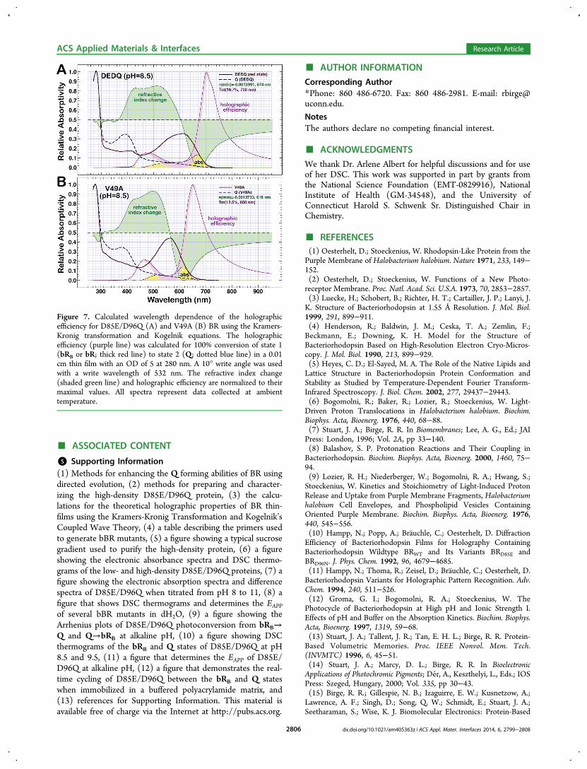
from solution spectra for an ideal film with an OD of ∼5 at 280nm (OD603 ∼ 1.9) (Figure 7A). The maximal diffractionefficiency of such a film is 16.7% at 700 nm, which is notablyimproved over the 6−8% observed for native BR.15,37,38 It isalso improved over the 4% efficiency of V49A/D85N bBR,which uses P (λmax = 480 nm) as the blue-shifted species.28
This observation means the bRB/Q states of D85E/D96Q canimprove the signal-to-noise of a holographic film 2−3 timesover the bR/M pair of a native BR film and 3−4 times over thebRB/P pair of V49A/D85N.
Table 2. Summary of the Physical Properties of bBR MutantD85E/D96Q at Neutral and Alkaline pH
photostate pH EA (kJ mol‑1)a TM (°C)b EAPP (kJ mol‑1)
resting state (bRB) water ndc 98 10508.5 65 100 7709.5 80 91 10010.5 ndc 78 ndc
Q photoproduct 8.5 5 89 5209.5 40 82 15010.5 ndc 65 ndc
aActivation energy for the bRB→Q or Q→bRB photoconversion.bThe TMs represent values collected at 90 K h‑1. cnd = not determined.
Figure 5. Absorbance spectra of the acrylamide cubes containingD85E/D96Q in the bRB state (solid line) or Q state (dashed line). Theleft inset image is a photograph of cubes containing either the (1) bRBstate or (2) Q photoproduct of D85E/D96Q in a buffered (pH 8.5)polyacrylamide gel. The right inset image shows the difference spectraof D85E/D96Q spectra after illumination with (>640 nm; ∼100 mWcm‑2) light for complete conversion after 16 h.
ACS Applied Materials & Interfaces Research Article
dx.doi.org/10.1021/am405363z | ACS Appl. Mater. Interfaces 2014, 6, 2799−28082804

We next calculated the holographic efficiency for the bR/Qstates of V49A, a ‘purple’ BR mutant capable of fully formingQ,22 in order to compare it with that of D85E/D96Q. An idealfilm with an OD280 of ∼5 (OD570 of ∼2.4) will yield adiffraction efficiency of 12.5% at 650 nm (Figure 7B), whichimproves the diffraction efficiency 1.5−2 times over the bR/Mpair of a native BR. This improvement, while significant, is lessthan that of D85E/D96Q due to the increased spectralseparation between the bRB and Q states. While both proteinsoffer an improved holographic efficiency, D85E/D96Q exhibitsthe highest efficiency of any BR mutant explored todate.10,28,37,50,66
■ COMMENTS AND CONCLUSIONS
We report here the first bBR mutant to exhibit efficientconversion of the entire sample to and from the Qphotoproduct. The thermal and photochemical stability ofnative BR makes it well-suited for application in biophotonictechnologies, and this stability is not lost in the D85E/D96Qmutant. This mutant, in particular, also exhibits the highestconversion levels to the Q photoproduct of any bBR mutantcreated to date. The protein structure is partially weakened,however, under the alkaline conditions required for theformation of the Q photoproduct. Optimal stability andformation of Q is determined to occur around pH 8.5 in anaqueous environment. We hypothesize that the general stabilityof D85E/D96Q derives from the rapid interconversion of theprotonated blue form, E85(H), and the deprotonated purple
form, E85(−), of the mutant. The barrier to proton transfer isvery small, and thus this equilibrium is established withinseconds, if not milliseconds. Rapid equilibration between thesespecies serves to stabilize the blue membrane form, E85(H),and is likely responsible for stabilizing the ensemble.Suspension of D85E/D96Q in a polyacrylamide matrix,
buffered at pH 8.5, does not inhibit the formation of Q underthe conditions optimized for the aqueous sample. Furthermore,the Q state is the only true photoproduct of the BR photocycleand, because it is infinitely longer-lived than the M65 or P18
photostates, it is ideal for long-term storage of binary data orholographic associative processing.15 Real-time cycling of thebRB and Q states of immobilized D85E/D96Q with amber andblue light demonstrates this feature. The increased spectralseparation between the photochromic pair, bR/Q, and a highholographic efficiency of D85E/D96Q also surpasses theefficiency of all protein-based holographic media investigatedto date. The coupling of this efficient photochromism with theuniquely enhanced thermal stability of D85E/D96Q dramati-cally improves the viability of using bBR mutants in protein-based optical computing (e.g., permanent data storage, real-time holographic data processing). Following the success ofimmobilizing the protein within a hydrated polymer matrix andthe demonstration of repeatable photoconversion in thisenvironment, we are now eager to investigate the applicationof D85E/D96Q as a photochromic material and furtherestablish the optical and hardware requirements to harnessthis efficiency in various biophotonic architectures.
Figure 6. Illumination of D85E/D96Q, which is suspended in a buffered (pH 8.5) polyacrylamide gel, with amber (590 nm) or royal blue (450 nm)light demonstrates how the protein responds to LED illumination. (A) Real-time changes in the red-shifted absorbance band (640 nm) of theimmobilized protein. Amber illumination consists of two sets of five 15-second pulses with each pulse followed by 5 seconds of dark time. Each set ofpulses is followed by one minute of dark time to allow relaxation of the protein to the bRB state if unconverted to the Q photoproduct. Two minutesof blue illumination are included to drive the Q→bRB photoconversion and reset the A640 baseline. (B) Difference spectra for the bRB→Qphotoconversion, which are subtracted from spectra of the light-adapted protein. All spectra were collected during the long dark period followingeach set of five amber illuminations. The number of 15-second flashes required to produce each spectra are shown on the right side of the image. Thekinetics of photoconversion are shown in the inset image for the absorption maxima and minima. (C) Data are of difference spectra for the Q→bRBphotoconversion and are subtracted from the spectra of D85E/D96Q after 385 15-second amber pulses. All spectra were collected during a 30second dark period following each one second blue illumination. The number of flashes required to produce each spectra are denoted on the rightside of the image. The kinetics of photoconversion are shown in the inset image for the absorption maxima and minima.
ACS Applied Materials & Interfaces Research Article
dx.doi.org/10.1021/am405363z | ACS Appl. Mater. Interfaces 2014, 6, 2799−28082805
■ ASSOCIATED CONTENT
*S Supporting Information(1) Methods for enhancing the Q forming abilities of BR usingdirected evolution, (2) methods for preparing and character-izing the high-density D85E/D96Q protein, (3) the calcu-lations for the theoretical holographic properties of BR thin-films using the Kramers-Kronig Transformation and Kogelnik’sCoupled Wave Theory, (4) a table describing the primers usedto generate bBR mutants, (5) a figure showing a typical sucrosegradient used to purify the high-density protein, (6) a figureshowing the electronic absorbance spectra and DSC thermo-grams of the low- and high-density D85E/D96Q proteins, (7) afigure showing the electronic absorption spectra and differencespectra of D85E/D96Q when titrated from pH 8 to 11, (8) afigure that shows DSC thermograms and determines the EAPP
of several bBR mutants in dH2O, (9) a figure showing theArrhenius plots of D85E/D96Q photoconversion from bRB→Q and Q→bRB at alkaline pH, (10) a figure showing DSCthermograms of the bRB and Q states of D85E/D96Q at pH8.5 and 9.5, (11) a figure that determines the EAPP of D85E/D96Q at alkaline pH, (12) a figure that demonstrates the real-time cycling of D85E/D96Q between the bRB and Q stateswhen immobilized in a buffered polyacrylamide matrix, and(13) references for Supporting Information. This material isavailable free of charge via the Internet at http://pubs.acs.org.
■ AUTHOR INFORMATION
Corresponding Author*Phone: 860 486-6720. Fax: 860 486-2981. E-mail: [email protected].
NotesThe authors declare no competing financial interest.
■ ACKNOWLEDGMENTS
We thank Dr. Arlene Albert for helpful discussions and for useof her DSC. This work was supported in part by grants fromthe National Science Foundation (EMT-0829916), NationalInstitute of Health (GM-34548), and the University ofConnecticut Harold S. Schwenk Sr. Distinguished Chair inChemistry.
■ REFERENCES(1) Oesterhelt, D.; Stoeckenius, W. Rhodopsin-Like Protein from thePurple Membrane of Halobacterium halobium. Nature 1971, 233, 149−152.(2) Oesterhelt, D.; Stoeckenius, W. Functions of a New Photo-receptor Membrane. Proc. Natl. Acad. Sci. U.S.A. 1973, 70, 2853−2857.(3) Luecke, H.; Schobert, B.; Richter, H. T.; Cartailler, J. P.; Lanyi, J.K. Structure of Bacteriorhodopsin at 1.55 Å Resolution. J. Mol. Biol.1999, 291, 899−911.(4) Henderson, R.; Baldwin, J. M.; Ceska, T. A.; Zemlin, F.;Beckmann, E.; Downing, K. H. Model for the Structure ofBacteriorhodopsin Based on High-Resolution Electron Cryo-Micros-copy. J. Mol. Biol. 1990, 213, 899−929.(5) Heyes, C. D.; El-Sayed, M. A. The Role of the Native Lipids andLattice Structure in Bacteriorhodopsin Protein Conformation andStability as Studied by Temperature-Dependent Fourier Transform-Infrared Spectroscopy. J. Biol. Chem. 2002, 277, 29437−29443.(6) Bogomolni, R.; Baker, R.; Lozier, R.; Stoeckenius, W. Light-Driven Proton Translocations in Halobacterium halobium. Biochim.Biophys. Acta, Bioenerg. 1976, 440, 68−88.(7) Stuart, J. A.; Birge, R. R. In Biomembranes; Lee, A. G., Ed.; JAIPress: London, 1996; Vol. 2A, pp 33−140.(8) Balashov, S. P. Protonation Reactions and Their Coupling inBacteriorhodopsin. Biochim. Biophys. Acta, Bioenerg. 2000, 1460, 75−94.(9) Lozier, R. H.; Niederberger, W.; Bogomolni, R. A.; Hwang, S.;Stoeckenius, W. Kinetics and Stoichiometry of Light-Induced ProtonRelease and Uptake from Purple Membrane Fragments, Halobacteriumhalobium Cell Envelopes, and Phospholipid Vesicles ContainingOriented Purple Membrane. Biochim. Biophys. Acta, Bioenerg. 1976,440, 545−556.(10) Hampp, N.; Popp, A.; Brauchle, C.; Oesterhelt, D. DiffractionEfficiency of Bacteriorhodopsin Films for Holography ContainingBacteriorhodopsin Wildtype BRWT and Its Variants BRD85E andBRD96N. J. Phys. Chem. 1992, 96, 4679−4685.(11) Hampp, N.; Thoma, R.; Zeisel, D.; Brauchle, C.; Oesterhelt, D.Bacteriorhodopsin Variants for Holographic Pattern Recognition. Adv.Chem. 1994, 240, 511−526.(12) Groma, G. I.; Bogomolni, R. A.; Stoeckenius, W. ThePhotocycle of Bacteriorhodopsin at High pH and Ionic Strength I.Effects of pH and Buffer on the Absorption Kinetics. Biochim. Biophys.Acta, Bioenerg. 1997, 1319, 59−68.(13) Stuart, J. A.; Tallent, J. R.; Tan, E. H. L.; Birge, R. R. Protein-Based Volumetric Memories. Proc. IEEE Nonvol. Mem. Tech.(INVMTC) 1996, 6, 45−51.(14) Stuart, J. A.; Marcy, D. L.; Birge, R. R. In BioelectronicApplications of Photochromic Pigments; Der, A., Keszthelyi, L., Eds.; IOSPress: Szeged, Hungary, 2000; Vol. 335, pp 30−43.(15) Birge, R. R.; Gillespie, N. B.; Izaguirre, E. W.; Kusnetzow, A.;Lawrence, A. F.; Singh, D.; Song, Q. W.; Schmidt, E.; Stuart, J. A.;Seetharaman, S.; Wise, K. J. Biomolecular Electronics: Protein-Based
Figure 7. Calculated wavelength dependence of the holographicefficiency for D85E/D96Q (A) and V49A (B) BR using the Kramers-Kronig transformation and Kogelnik equations. The holographicefficiency (purple line) was calculated for 100% conversion of state 1(bRB or bR; thick red line) to state 2 (Q; dotted blue line) in a 0.01cm thin film with an OD of 5 at 280 nm. A 10° write angle was usedwith a write wavelength of 532 nm. The refractive index change(shaded green line) and holographic efficiency are normalized to theirmaximal values. All spectra represent data collected at ambienttemperature.
ACS Applied Materials & Interfaces Research Article
dx.doi.org/10.1021/am405363z | ACS Appl. Mater. Interfaces 2014, 6, 2799−28082806
Associative Processors and Volumetric Memories. J. Phys. Chem. B1999, 103, 10746−10766.(16) Hampp, N. Bacteriorhodopsin: Mutating a Biomaterial into anOptoelectronic Material. Appl. Microbiol. Biotechnol. 2000, 53, 633−639.(17) Hampp, N. Bacteriorhodopsin as a Photochromic RetinalProtein for Optical Memories. Chem. Rev. 2000, 100, 1755−1776.(18) Tallent, J. R.; Stuart, J. A.; Song, Q. W.; Schmidt, E. J.; Martin,C. H.; Birge, R. R. Photochemistry in Dried Polymer FilmsIncorporating the Deionized Blue Membrane Form of Bacteriorho-dopsin. Biophys. J. 1998, 75, 1619−1634.(19) Greco, J. A.; Wagner, N. L.; Birge, R. R. Fourier TransformHolographic Associative Processors Based on Bacteriorhodopsin. Int. J.Unconventional Comput. 2012, 8, 433−457.(20) Gillespie, N. B.; Wise, K. J.; Ren, L.; Stuart, J. A.; Marcy, D. L.;Hillebrecht, J.; Li, Q.; Ramos, L.; Jordan, K.; Fyvie, S.; Birge, R. R.Characterization of the Branched-Photocycle Intermediates P and Q ofBacteriorhodopsin. J. Phys. Chem. B 2002, 106, 13352−13361.(21) Popp, A.; Wolperdinger, M.; Hampp, N.; Brauchle, C.;Oesterhelt, D. Photochemical Conversion of the O-intermediate to9-cis-Retinal-Containing Products in Bacteriorhodopsin Films. Biophys.J. 1993, 65, 1449−1459.(22) Wagner, N. L.; Greco, J. A.; Ranaghan, M. J.; Birge, R. R.Directed Evolution of Bacteriorhodopsin for Applications inBioelectronics. J. R. Soc., Interface 2013, 10, 20130197.(23) Fischer, U. C.; Towner, P.; Oesterhelt, D. Light InducedIsomerization, at Acidic pH, Initiates Hydrolysis of Bacteriorhodopsinto Bacterio-Opsin and 9-cis-Retinal. Photochem. Photobiol. 1981, 33,529−537.(24) Turner, G. J.; Miercke, L. J. W.; Thorgeirsson, T. E.; Kliger, D.S.; Betlach, M. C.; Stroud, R. M. Bacteriorhodopsin D85N: ThreeSpectroscopic Species in Equilibrium. Biochemistry 1993, 32, 1332−1337.(25) Tallent, J.; Song, Q. W.; Li, Z.; Stuart, J.; Birge, R. R. EffectivePhotochromic Nonlinearity of Dried Blue-Membrane Bacteriorhodop-sin Films. Opt. Lett. 1996, 21, 1339−1341.(26) Tittor, J.; Schweiger, U.; Oesterhelt, D.; Bamberg, E. Inversionof Proton Translocation in Bacteriorhodopsin Mutants D85N, D85T,and D85,96N. Biophys. J. 1994, 67, 1682−1690.(27) Balashov, S. P.; Imasheva, E. S.; Govindjee, R.; Ebrey, T. G.Titration of Aspartate-85 in Bacteriorhodopsin: What it Says AboutChromophore Isomerization and Proton Release. Biophys. J. 1996, 70,473−481.(28) Millerd, J. E.; Rohrbacher, A.; Brock, N. J.; Chau, C.-K.; Smith,P.; Needleman, R. Improved Sensitivity in Blue-Membrane Bacterio-rhodopsin Films. Opt. Lett. 1999, 24, 1355−1357.(29) Chang, C.-H.; Liu, S. Y.; Jonas, R.; Govindjee, R. The PinkMembrane: The Stable Photoproduct of Deionized Purple Membrane.Biophys. J. 1987, 52, 617−623.(30) Chronister, E. L.; Corcoran, T. C.; Song, L.; El-Sayed, M. A. Onthe Molecular Mechanisms of the Schiff Base Deprotonation Duringthe Bacteriorhodopsin Photocycle. Proc. Natl. Acad. Sci. U.S.A. 1986,83, 8580−8584.(31) Varo, G.; Lanyi, J. K. Photoreactions of Bacteriorhodopsin atAcid pH. Biophys. J. 1989, 56, 1143−1151.(32) Moore, T.; Edgerton, M.; Parr, G.; Greenwood, C.; Perham, R.Studies of an Acid-Induced Species of Purple Membrane fromHalobacterium halobium. Biochem. J. 1978, 171, 469−476.(33) Cladera, J.; Galisteo, M. L.; Sabes, M.; Mateo, P. L.; Padros, E.The Role of Retinal in the Thermal Stability of the Purple Membrane.Eur. J. Biochem. 1992, 207, 581−585.(34) Kresheck, G.; Lin, C.; Williamson, L.; Mason, W.; Jang, D.; El-Sayed, M. The Thermal Stability of Native, Delipidated, Deionized andRegenerated Bacteriorhodopsin. J. Photochem. Photobiol., B 1990, 7,289−302.(35) Okumura, H.; Murakami, M.; Kouyama, T. Crystal Structures ofAcid Blue and Alkaline Purple Forms of Bacteriorhodopsin. J. Mol.Biol. 2005, 351, 481−495.
(36) Liu, S. Y.; Ebrey, T. G. The Quantum Efficiency for theInterphotoconversion of the Blue and Pink Forms of PurpleMembrane. Photochem. Photobiol. 1987, 46, 263−267.(37) Bazhenov, V. Y.; Soskin, M. S.; Taranenko, V. B. HolographicRecording by Continuous Illumination in a Suspension of PurpleMembranes of Halobacteria. Sov. Tech. Phys. Lett. 1987, 13, 382−384.(38) Gross, R. B.; Izgi, K. C.; Birge, R. R. Holographic Thin Films,Spatial Light Modulators, and Optical Associative Memories Based onBacteriorhodopsin. Proc. SPIE 1992, 1662, 186−196.(39) Landin, J. S.; Katragadda, M.; Albert, A. D. ThermalDestabilization of Rhodopsin and Opsin by Proteolytic Cleavage inBovine Rod Outer Segment Disk Membranes. Biochemistry 2001, 40,11176−11183.(40) Galisteo, M.; Sanchez-Ruiz, J. Kinetic Study into the IrreversibleThermal Denaturation of Bacteriorhodopsin. Eur. Biophys. J. 1993, 22,25−30.(41) Kogelnik, H. Coupled Wave Theory for Thick HologramGratings. Bell Syst. Tech. J. 1969, 48, 2909−2947.(42) Birge, R. R.; Fleitz, P. A.; Gross, R. B.; Izgi, J. C.; Lawrence, A.F.; Stuart, J. A.; Tallent, J. R. Spatial Light Modulators and OpticalAssociative Memories Based on Bacteriorhodopsin. Proc. IEEE EMBS1990, 12, 1788−1789.(43) Bunkin, F. V.; Vsevolodov, N. N.; Druzhko, A. B.; Mitsner, B. I.;Prokhorov, A. M.; Savranskii, V. V.; Tkachenko, N. W.; Shevchenko,T. B. Diffraction Efficiency of Bacteriorhodopsin and Its Analogs. Sov.Tech. Phys. Lett. 1981, 7, 630−631.(44) Thoma, R.; Hampp, N.; Bra uchle, C.; Oesterhelt, D.Bacteriorhodopsin Films as Spatial Light Modulators for Nonlinear-Optical Filtering. Opt. Lett. 1991, 16, 651−653.(45) Renner, T.; Hampp, N. Bacteriorhodopsin-Films for DynamicTime Average Interferometry. Opt. Commun. 1993, 96, 142−149.(46) Birge, R. R. Protein-Based Optical Computing and Memories.Computer 1992, 25, 56−67.(47) Xi, B.; Tetley, W. C.; Marcy, D. L.; Zhong, C.; Whited, G.;Birge, R. R.; Stuart, J. A. Evaluation of Blue and Green AbsorbingProteorhodopsins as Holographic Materials. J. Phys. Chem. B 2008,112, 2524−2532.(48) Birge, R. R.; Izgi, K. C.; Stuart, J. A.; Tallent, J. R. WavelengthDependence of the Photorefractive and Photodiffractive Properties ofHolographic Thin Films Based on Bacteriorhodopsin. Proc. Mater. Res.Soc. 1991, 218, 131−140.(49) Ranaghan, M. J.; Wagner, N. L.; Sandberg, M. N.; Birge, R. R. InOptical Biomimetics: Materials and Applications; Large, M., Ed.;Woodhead Publishing: Cambridge, UK, 2012, pp 20−78.(50) Downie, J. D.; Timucin, D. A.; Smithey, D. T.; Crew, M. LongHolographic Lifetimes in Bacteriorhodopsin Films. Opt. Lett. 1998, 23,730−732.(51) Lanyi, J. K.; Tittor, J.; Varo, G.; Krippahl, G.; Oesterhelt, D.Influence of the Size and Protonation State of Acidic Residue 85 onthe Absorption Spectrum and Photoreaction of the BacteriorhodopsinChromophore. Biochim. Biophys. Acta, Bioenerg. 1992, 1099, 102−110.(52) Mogi, T.; Stern, L. J.; Marti, T.; Chao, B. H.; Khorana, H. G.Aspartic Acid Substitutions Affect Proton Translocation by Bacterio-rhodopsin. Proc. Natl. Acad. Sci. U.S.A. 1988, 85, 4148−4152.(53) Mowery, P. C.; Lozier, R. H.; Chae, Q.; Tseng, Y. W.; Taylor,M.; Stoeckenius, W. Effect of Acid pH on the Absorption Spectra andPhotoreactions of Bacteriorhodopsin. Biochemistry 1979, 18, 4100−4107.(54) Richter, H.-T.; Brown, L. S.; Needleman, R.; Lanyi, J. K. ALinkage of the pKa’s of Asp-85 and Glu-204 Forms Part of theReprotonation Switch of Bacteriorhodopsin. Biochemistry 1996, 35,4054−4062.(55) Subramaniam, S.; Marti, T.; Khorana, H. Protonation State ofAsp (Glu)-85 Regulates the Purple-to-Blue Transition in Bacterio-rhodopsin Mutants Arg 82 to Ala and Asp 85 to Glu: The Blue FormIs Inactive in Proton Translocation. Proc. Natl. Acad. Sci. U.S.A. 1990,87, 1013−1017.
ACS Applied Materials & Interfaces Research Article
dx.doi.org/10.1021/am405363z | ACS Appl. Mater. Interfaces 2014, 6, 2799−28082807

(56) Richter, H. T.; Needleman, R.; Lanyi, J. K. Perturbed InteractionBetween Residues 85 and 204 in Tyr185–>Phe and Asp85–>GluBacteriorhodopsins. Biophys. J. 1996, 71, 3392−3398.(57) Jackson, M. B.; Sturtevant, J. M. Phase Transitions of the PurpleMembranes of Halobacterium halobium. Biochemistry 1978, 17, 911−915.(58) Muller, J.; Munster, C.; Salditt, T. Thermal Denaturing ofBacteriorhodopsin by X-Ray Scattering from Oriented PurpleMembranes. Biophys. J. 2000, 78, 3208−3217.(59) Wang, J.; El-Sayed, M. Temperature Jump-Induced SecondaryStructural Change of the Membrane Protein Bacteriorhodopsin in thePremelting Temperature Region: A Nanosecond Time-ResolvedFourier Transform Infrared Study. Biophys. J. 1999, 76, 2777−2783.(60) Wang, J.; El-Sayed, M. A. The Effect of Protein ConformationChange from αII to αI on the Bacteriorhodopsin Photocycle. Biophys.J. 2000, 78, 2031−2036.(61) Cladera, J.; Galisteo, M.; Dunach, M.; Mateo, P.; Padros, E.Thermal Denaturation of Deionized and Native Purple Membranes.Biochim. Biophys. Acta, Biomembr. 1988, 943, 148−156.(62) Taneva, S. G.; Goni, F. M.; Tuparev, N. P.; Petkanchin, I.; Der,A.; Muga, A. Effect of Asp85 Replacement by Thr on theConformation, Surface Electric Properties and Stability of Bacterio-rhodopsin. Colloids Surf., A 2002, 209, 193−200.(63) Dancshazy, Z.; Tokaji, Z. Blue Light Regeneration ofBacteriorhodopsin Bleached by Continuous Light. FEBS Lett. 2000,476, 171−173.(64) Brouillette, C.; Muccio, D.; Finney, T. pH Dependence ofBacteriorhodopsin Thermal Unfolding. Biochemistry 1987, 26, 7431−7438.(65) Pandey, P. C.; Upadhyay, B. C.; Pandey, C. M. D.; Pathak, H. C.Electrochemical Studies on D96N Bacteriorhodopsin and itsApplication in the Development of Photosensors. Sens. Actuators, B1999, 56, 112−120.(66) Hampp, N.; Brauchle, C.; Oesterhelt, D. BacteriorhodopsinWildtype and Variant Aspartate-96 to Asparagine as ReversibleHolographic Media. Biophys. J. 1990, 58, 83−93.(67) Varo, G.; Lanyi, J. K. Kinetic and Spectroscopic Evidence for anIrreversible Step Between Deprotonation and Reprotonation of theSchiff Base in the Bacteriorhodopsin Photocycle. Biochemistry 1991,30, 5008−5015.
ACS Applied Materials & Interfaces Research Article
dx.doi.org/10.1021/am405363z | ACS Appl. Mater. Interfaces 2014, 6, 2799−28082808
Copyright © 2022 FDOKUMEN


![Integrated optical devices using bacteriorhodopsin as active nonlinear optical material [6331-49]](https://static.fdokumen.com/doc/165x107/633478bc7a687b71aa08b32f/integrated-optical-devices-using-bacteriorhodopsin-as-active-nonlinear-optical-material.jpg)