Photocatalytic antibacterial performance of TiO 2 and Ag-doped TiO 2 against S. aureus . P....
8
Journal of Hazardous Materials 162 (2009) 1309–1316 Contents lists available at ScienceDirect Journal of Hazardous Materials journal homepage: www.elsevier.com/locate/jhazmat Photocatalytic antibacterial performance of Sn 4+ -doped TiO 2 thin films on glass substrate Funda Sayılkan a,∗ , Meltem Asiltürk a , Nadir Kiraz b , Esin Burunkaya b , Ertu˘ grul Arpac ¸ b , Hikmet Sayılkan a,1 a Prof.Dr.Hikmet Sayılkan Research & Development Laboratory for Advanced Materials, ˙ Inönü University, 44280 Malatya, Turkey b Akdeniz University, Faculty of Arts and Science, Department of Chemistry, 07100 Antalya, Turkey article info Article history: Received 27 January 2008 Received in revised form 9 June 2008 Accepted 9 June 2008 Available online 20 June 2008 Keywords: Sn 4+ -doped TiO2 Antibacterial surface coating Escherichia coli Staphylococcus aureus abstract Pure anatase, nanosized and Sn 4+ ion doped titanium dioxide (TiO 2 ) particulates (TiO 2 –Sn 4+ ) were synthe- sized by hydrothermal process. TiO 2 –Sn 4+ was used to coat glass surfaces to investigate the photocatalytic antibacterial effect of Sn 4+ doping to TiO 2 against gram negative Escherichia coli (E. coli) and gram pos- itive Staphylococcus aureus (S. aureus). Relationship between solid ratio of TiO 2 –Sn 4+ in coatings and antibacterial activity was reported. The particulates and the films were characterized using particle size analyzer, zeta potential analyzer, Brunauer–Emmett–Teller (BET), X-ray diffractometer (XRD), SEM, AAS and UV/VIS/NIR techniques. The results showed that TiO 2 –Sn 4+ is fully anatase crystalline form and easily dispersed in water. Increasing the solid ratio of TiO 2 –Sn 4+ from 10 to 50% in the coating solution increased antibacterial effect. © 2008 Elsevier B.V. All rights reserved. 1. Introduction Titanium dioxide (TiO 2 ) mediated heterogeneous photocataly- sis has become an innovative technology with attractive application potentials such as self-cleaning and environmental pollution reme- diation, mainly due to the hydrophilic property of TiO 2 [1,2] and its ability to degrade a wide range of inorganic and organic compounds in both aqueous and gaseous phase [3–5]. Many organic compounds can be decomposed in aqueous solution in the presence of TiO 2 powders, sols or films on the different substrates illuminated with UV- or VIS-lights [6–8]. However, the application of powdered TiO 2 as a photocatalyst in wastewater treatment has the drawback of post-separation in a slurry system after photoreaction. Therefore, attempts have been made to immobilize the TiO 2 in the form of thin films on different rigid substrates, mostly on glass and ceramics [9–16]. Recent studies have shown that TiO 2 coated materials like glass or ceramics possess some deodorizing, antibacterial [17–20] and self-cleaning [21–29] character under weak, ultra violet light in living environments. Since TiO 2 has extremely strong oxidation capability, it can decompose various types of organic matter [30,31]. Surfaces exhibit self-cleaning function when they are contaminated by organic ∗ Corresponding author. Fax: +90 422 341 0042. E-mail address: [email protected] (F. Sayılkan). 1 Deceased. stains which are comprised of many types of organic matter [32]. If bacteria come into contact with a TiO 2 coated surface, TiO 2 exhibits an antibacterial function; i.e. it soon becomes sterilized by damage of the cell membranes [33]. TiO 2 has anatase and rutile polymorphs. The band gap of anatase is 3.2 eV and that of rutile is 3.0 eV [34]. The wavelength of light corresponding to the band gaps of anatase and rutile are ∼380 nm which corresponds to near-UV and ∼410nm which corresponds to visible light, respectively. Because UV-light is necessary to excite TiO 2 , little sterilization occurs in the dark when pure TiO 2 is used. On the other hand, metal ions, such as silver, copper, and zinc have also antibacterial capabilities [35]. If these metal ions are combined with a TiO 2 as thin film on the glaze of sanitary ware, it is possi- ble to develop sanitary ware in which the photocatalysts produce a sterilizing effect under UV-light and the metal ions provide an antibacterial effect, even in the dark. In terms of the antibacterial effect of photocatalysts, Matsunaga et al. [36] first reported in 1985 that microbial cells in water could be killed by contact with a TiO 2 /Pt powder catalyst upon irradia- tion with near-UV light for 60–120 min. The same group of workers successfully constructed a practical photochemical device in which TiO 2 powder was immobilized on an acetylcellulose membrane. An Esherichia coli (E. coli) suspension flowing through this device was completely killed [37]. Since then, much research has been per- formed on the antibacterial effects of TiO 2 thin films, as well as TiO 2 powders. For example, Kikuchi et al. [19] studied on the role of active oxygen species in the photocatalytic bactericidal effect 0304-3894/$ – see front matter © 2008 Elsevier B.V. All rights reserved. doi:10.1016/j.jhazmat.2008.06.043
-
Upload
independent -
Category
Documents
-
view
0 -
download
0
Transcript of Photocatalytic antibacterial performance of TiO 2 and Ag-doped TiO 2 against S. aureus . P....
Pt
FEa
b
a
ARRAA
KSAES
1
spdaicpUapafi[gai
ds
0d
Journal of Hazardous Materials 162 (2009) 1309–1316
Contents lists available at ScienceDirect
Journal of Hazardous Materials
journa l homepage: www.e lsev ier .com/ locate / jhazmat
hotocatalytic antibacterial performance of Sn4+-doped TiO2
hin films on glass substrate
unda Sayılkana,∗, Meltem Asiltürka, Nadir Kirazb, Esin Burunkayab,rtugrul Arpacb, Hikmet Sayılkana,1
Prof.Dr.Hikmet Sayılkan Research & Development Laboratory for Advanced Materials, Inönü University, 44280 Malatya, TurkeyAkdeniz University, Faculty of Arts and Science, Department of Chemistry, 07100 Antalya, Turkey
r t i c l e i n f o
rticle history:eceived 27 January 2008eceived in revised form 9 June 2008
a b s t r a c t
Pure anatase, nanosized and Sn4+ ion doped titanium dioxide (TiO2) particulates (TiO2–Sn4+) were synthe-sized by hydrothermal process. TiO2–Sn4+ was used to coat glass surfaces to investigate the photocatalyticantibacterial effect of Sn4+ doping to TiO2 against gram negative Escherichia coli (E. coli) and gram pos-
ccepted 9 June 2008vailable online 20 June 2008
eywords:n4+-doped TiO2
ntibacterial surface coating
itive Staphylococcus aureus (S. aureus). Relationship between solid ratio of TiO2–Sn4+ in coatings andantibacterial activity was reported. The particulates and the films were characterized using particle sizeanalyzer, zeta potential analyzer, Brunauer–Emmett–Teller (BET), X-ray diffractometer (XRD), SEM, AASand UV/VIS/NIR techniques. The results showed that TiO2–Sn4+ is fully anatase crystalline form and easilydispersed in water. Increasing the solid ratio of TiO2–Sn4+ from 10 to 50% in the coating solution increased
sbao
icwvTOawbaa
e
scherichia colitaphylococcus aureus
antibacterial effect.
. Introduction
Titanium dioxide (TiO2) mediated heterogeneous photocataly-is has become an innovative technology with attractive applicationotentials such as self-cleaning and environmental pollution reme-iation, mainly due to the hydrophilic property of TiO2 [1,2] and itsbility to degrade a wide range of inorganic and organic compoundsn both aqueous and gaseous phase [3–5]. Many organic compoundsan be decomposed in aqueous solution in the presence of TiO2owders, sols or films on the different substrates illuminated withV- or VIS-lights [6–8]. However, the application of powdered TiO2s a photocatalyst in wastewater treatment has the drawback ofost-separation in a slurry system after photoreaction. Therefore,ttempts have been made to immobilize the TiO2 in the form of thinlms on different rigid substrates, mostly on glass and ceramics9–16]. Recent studies have shown that TiO2 coated materials likelass or ceramics possess some deodorizing, antibacterial [17–20]nd self-cleaning [21–29] character under weak, ultra violet light
n living environments.Since TiO2 has extremely strong oxidation capability, it canecompose various types of organic matter [30,31]. Surfaces exhibitelf-cleaning function when they are contaminated by organic
∗ Corresponding author. Fax: +90 422 341 0042.E-mail address: [email protected] (F. Sayılkan).
1 Deceased.
btsTEcfTo
304-3894/$ – see front matter © 2008 Elsevier B.V. All rights reserved.oi:10.1016/j.jhazmat.2008.06.043
© 2008 Elsevier B.V. All rights reserved.
tains which are comprised of many types of organic matter [32]. Ifacteria come into contact with a TiO2 coated surface, TiO2 exhibitsn antibacterial function; i.e. it soon becomes sterilized by damagef the cell membranes [33].
TiO2 has anatase and rutile polymorphs. The band gap of anatases 3.2 eV and that of rutile is 3.0 eV [34]. The wavelength of lightorresponding to the band gaps of anatase and rutile are ∼380 nmhich corresponds to near-UV and ∼410 nm which corresponds to
isible light, respectively. Because UV-light is necessary to exciteiO2, little sterilization occurs in the dark when pure TiO2 is used.n the other hand, metal ions, such as silver, copper, and zinc havelso antibacterial capabilities [35]. If these metal ions are combinedith a TiO2 as thin film on the glaze of sanitary ware, it is possi-
le to develop sanitary ware in which the photocatalysts producesterilizing effect under UV-light and the metal ions provide an
ntibacterial effect, even in the dark.In terms of the antibacterial effect of photocatalysts, Matsunaga
t al. [36] first reported in 1985 that microbial cells in water coulde killed by contact with a TiO2/Pt powder catalyst upon irradia-ion with near-UV light for 60–120 min. The same group of workersuccessfully constructed a practical photochemical device in whichiO2 powder was immobilized on an acetylcellulose membrane. An
sherichia coli (E. coli) suspension flowing through this device wasompletely killed [37]. Since then, much research has been per-ormed on the antibacterial effects of TiO2 thin films, as well asiO2 powders. For example, Kikuchi et al. [19] studied on the rolef active oxygen species in the photocatalytic bactericidal effect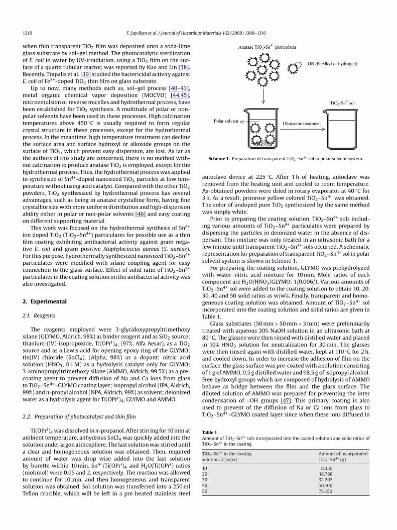
1 rdous Materials 162 (2009) 1309–1316
wgofRE
mmbptcptstohtppacao
ifitFpcpa
2
2
ststs3ct9w
2
asaab(tsT
arA3Tw
idpfrs
wcT3giT
t8iwasoFbdiluted solution of AMMO was prepared for preventing the intercondensation of –OH groups [47]. This primary coating is alsoused to prevent of the diffusion of Na or Ca ions from glass toTiO2–Sn4+–GLYMO coated layer since when these ions diffused in
Table 1Amount of TiO2–Sn4+ sols incorporated into the coated solution and solid ratios ofTiO2–Sn4+ in the coating
TiO2–Sn4+ in the coatingsolution, % (w/w)
Amount of incorporatedTiO2–Sn4+ (g)
310 F. Sayılkan et al. / Journal of Haza
hen thin transparent TiO2 film was deposited onto a soda-limelass substrate by sol–gel method. The photocatalytic sterilizationf E. coli in water by UV-irradiation, using a TiO2 film on the sur-ace of a quartz tubular reactor, was reported by Kuo and Lin [38].ecently, Trapalis et al. [39] studied the bactericidal activity against. coli of Fe3+-doped TiO2 thin film on glass substrate.
Up to now, many methods such as, sol–gel process [40–43],etal organic chemical vapor deposition (MOCVD) [44,45],icroemulsion or reverse micelles and hydrothermal process, have
een established for TiO2 synthesis. A multitude of polar or non-olar solvents have been used in these processes. High calcinationemperatures above 450 ◦C is usually required to form regularrystal structure in these processes, except for the hydrothermalrocess. In the meantime, high temperature treatment can declinehe surface area and surface hydroxyl or alkoxide groups on theurface of TiO2, which prevent easy dispersion, are lost. As far ashe authors of this study are concerned, there is no method with-ut calcination to produce anatase TiO2 is employed, except for theydrothermal process. Thus, the hydrothermal process was appliedo synthesize of Sn4+-doped nanosized TiO2 particles at low tem-erature without using acid catalyst. Compared with the other TiO2owders, TiO2 synthesized by hydrothermal process has severaldvantages, such as being in anatase crystalline form, having finerystallite size with more uniform distribution and high-dispersionbility either in polar or non-polar solvents [46] and easy coatingn different supporting material.
This work was focused on the hydrothermal synthesis of Sn4+
on doped TiO2 (TiO2–Sn4+) particulates for possible use as a thinlm coating exhibiting antibacterial activity against gram nega-ive E. coli and gram positive Staphylococcus aureus (S. aureus).or this purpose, hydrothermally synthesized nanosized TiO2–Sn4+
articulates were modified with silane coupling agent for easyonnection to the glass surface. Effect of solid ratio of TiO2–Sn4+
articulates in the coating solution on the antibacterial activity waslso investigated.
. Experimental
.1. Reagents
The reagents employed were 3-glycidoxypropyltrimethoxyilane (GLYMO, Aldrich, 98%) as binder reagent and as SiO2 source;itanium-(IV)-isopropoxide, Ti(OPri)4, (97%, Alfa Aesar), as a TiO2ource and as a Lewis acid for opening epoxy ring of the GLYMO;in(IV) chloride (SnCl4), (Alpha, 98%) as a dopant; nitric acidolution (HNO3, 0.1 M) as a hydrolysis catalyst only for GLYMO;-aminopropyltrimethoxy silane (AMMO, Aldrich, 99.5%) as a pre-oating agent to prevent diffusion of Na and Ca ions from glasso TiO2–Sn4+–GLYMO coating layer; isopropyl alcohol (IPA, Aldrich,9%) and n-propyl alcohol (NPA, Aldrich, 99%) as solvent; deionizedater as a hydrolysis agent for Ti(OPri)4, GLYMO and AMMO.
.2. Preparation of photocatalyst and thin film
Ti(OPri)4 was dissolved in n-propanol. After stirring for 10 min atmbient temperature, anhydrous SnCl4 was quickly added into theolution under argon atmosphere. The last solution was stirred untilclear and homogeneous solution was obtained. Then, required
mount of water was drop wise added into the last solution
y burette within 10 min. Sn4+/Ti(OPri)4 and H2O/Ti(OPri) ratiosmol/mol) were 0.05 and 2, respectively. The reaction was allowedo continue for 10 min, and then homogeneous and transparentolution was obtained. Sol-solution was transferred into a 250 mleflon crucible, which will be left in a pre-heated stainless steel12345
Scheme 1. Preparation of transparent TiO2–Sn4+ sol in polar solvent system.
utoclave device at 225 ◦C. After 1 h of heating, autoclave wasemoved from the heating unit and cooled to room temperature.s-obtained powders were dried in rotary evaporator at 40 ◦C forh. As a result, primrose yellow colored TiO2–Sn4+ was obtained.he color of undoped pure TiO2 synthesized by the same methodas simply white.
Prior to preparing the coating solution, TiO2–Sn4+ sols includ-ng various amounts of TiO2–Sn4+ particulates were prepared byispersing the particles in deionized water in the absence of dis-ersant. This mixture was only treated in an ultrasonic bath for a
ew minute until transparent TiO2–Sn4+ sols occurred. A schematicepresentation for preparation of transparent TiO2–Sn4+ sol in polarolvent system is shown in Scheme 1.
For preparing the coating solution, GLYMO was prehydrolyzedith water–nitric acid mixture for 10 min. Mole ratios of each
omponent are H2O/HNO3/GLYMO: 1/0.006/1. Various amounts ofiO2–Sn4+ sol were added to the coating solution to obtain 10, 20,0, 40 and 50 solid ratios as w/w%. Finally, transparent and homo-eneous coating solution was obtained. Amount of TiO2–Sn4+ solncorporated into the coating solution and solid ratios are given inable 1.
Glass substrates (50 mm × 50 mm × 3 mm) were preliminarilyreated with aqueous 30% NaOH solution in an ultrasonic bath at0 ◦C. The glasses were then rinsed with distilled water and placed
n 10% HNO3 solution for neutralization for 30 min. The glassesere then rinsed again with distilled water, kept at 110 ◦C for 2 h,
nd cooled down. In order to increase the adhesion of film on theurface, the glass surface was pre-coated with a solution consistingf 1 g of AMMO, 0.5 g distilled water and 98.5 g of isopropyl alcohol.ree hydroxyl groups which are composed of hydrolysis of AMMOehave as bridge between the film and the glass surface. The
0 8.3500 18.7880 32.2070 50.1000 75.150
rdous
ndtGfi1
2
tsRCsm
d
woaoc
dmFpwdpZtmutMAst2
uonai(ibr
cma
dsmscabt
atdaasA
2
TwlottgfTg
2
b4aonntatptotis
tuas
Etocpaast
tdao
F. Sayılkan et al. / Journal of Haza
etwork, the photocatalytic activity of TiO2 decreases [48]. Afterrying in an oven at 110 ◦C and then cooling to room temperature,he pre-coated surface was secondarily coated with TiO2–Sn4+–LYMO solution. To obtain an uniform, crack-free and transparentlm, the double coated surface was dried in a vacuum sterilizer at10 ◦C. After that, transparent coating film was obtained.
.3. Characterization of the photocatalyst
Berghoff model heating unit interfaced with temperature con-roller (up to 240 ◦C) and timer unit was used for hydrothermalynthesis of TiO2 and TiO2–Sn4+. To determine the crystal phase,igaku Geigerflex D Max/B model X-ray diffractometer (XRD) withuK� radiation (� = 0.15418 nm) in the region 2� = 10–70◦ with atep size of 0.04◦ was used. The average crystallite size was esti-ated according to the following Scherrer’s equation,
hkl = k�
ˇ cos(2�)(1)
here dhkl is the average crystallite size (nm), � is the wavelengthf the Cu K� radiation applied (� = 0.154056 nm), � is the Bragg’sngle of diffraction, ˇ is the full-width at half maximum intensityf the peak observed at 2� = 25.24 (converted to Radian) and k is aonstant usually applied as ∼0.9.
The Brunauer–Emmett–Teller (BET) surface area, average poreiameter and micropore volume of the particulates were deter-ined by nitrogen adsorption using ASAP 2000 model BET analyzer.
or BET analysis, powder samples were degassed at 130 ◦C for 4 hrior to nitrogen adsorption measurements. The BET surface areaas determined by multipoint BET method using the adsorptionata in the relative pressure (P/P0) range 0.02–0.3 at liquid N2 tem-erature. Pore size distribution was computed by DFT+ method.eta-nanosizer (Malvern, Nano-ZS model) was used to determinehe stability of the sol solution and particle size distribution. Surface
orphologies of the particles and coated surfaces were investigatedsing scanning electron microscopy, SEM (LEO EVO 40 model). Filmhickness of coated surfaces was measured by Perthometer (MAHR-
1 model). The amount of Sn in TiO2–Sn4+ was determined usingAS analysis (Perkin Elmer, AAnalyst 800 model) in centrifugedolution obtained after the hydrothermal treatment. Adhesion ofhe films was carried out by using multi-cross cutter (Erichsen Type95 model).
UV-Vis spectrophotometer (Varian Cary 100 Bio model) wassed to measure optical densities of incubated bacteria and tobtain bacterial growth curves. Orbital shaking incubator (Inter-ational Inc. 311 DS model) was used for incubation of bacteriat constant rotation rate and temperature. The antibacterial activ-ties of the coatings were investigated against gram negative E. coliAmerican Type Culture Collection, ATCC No: 23282) and gram pos-tive S. aureus (ATCC No.: 35696). Lactose broth (LB) and lactoseroth-agar are also used as incubation mediums of the bacte-ia.
For measuring of the film thickness, half of the plate area wasoated by spin coating technique. Film thickness was automaticallyeasured from the difference between the thicknesses of coated
nd uncoated surfaces sides.The adhesion of the films on glass surface is based on the con-
ensation of hydroxyl groups between the surface and the coatingolution. The adhesion of a coating on a base material is not only aechanical property defining the bond between coating and sub-
trate, but it also has particular importance with regard to theorrosion protection, since areas of poor adhesion can easily beffected by corrosion. The coating on the glass surface was testedy the Erichsen Scratch–Adhesion Test; the standardized cross-cutest provides a very straight forward method of establishing the
pftw1
Materials 162 (2009) 1309–1316 1311
dhesion quality. The basic principle of this test method is to cuthrough the coating with a series of several cuts at right angles in aefined manner. The square pattern that is obtained can be evalu-ted visually by examining the way in which the coating has brokenway from the base material (along the cut edges and/or completequares). The results of the tests were evaluated according to theSTM D 3359 protocol [49].
.4. Irradiation of the coated glass surfaces
UV-irradiation induces the photochemical reactions to proceed.herefore, prior to bacterial treatment, the surfaces with TiO2–Sn4+
as irradiated in a radiation unit (Erichsen 1500 Model) with Xe-amp operated at constant intensity of 1100 W/m2 for excitationf TiO2–Sn4+. After sterilizing the coated glasses in an autoclave,hey were immediately placed in the radiation unit and horizon-ally irradiated for 1 h. The distance between UV lamp and thelass surface was 20 cm. After irradiation, the glasses were removedrom the radiation unit and kept in an incubator at 37 ◦C for 1 h.hen, antibacterial activity tests were started using these irradiatedlasses.
.5. Antibacterial activity tests
Under favorable conditions, a growing bacterial population dou-les at regular intervals. Growth is by geometric progression: 1, 2,, 8, etc. or 20, 21, 22, 23, . . ., 2n, where, n is the number of gener-tions called exponential growth. In reality, exponential growth isnly part of the bacterial life cycle, and not representative of theormal pattern of the bacteria growth in nature. The rate of expo-ential growth of a bacterial culture is expressed as the generationime or the doubling time of bacterial population. Microorganismsre divided at a constant rate depending upon the composition ofhe growth medium and the conditions of incubation such as tem-erature and rotation rate. If rotation rate increase, surface area ofhe liquid inoculation medium of the bacteria increases becausef the centripetal force. This effect results high oxygen supply tohe bacterial species. At the high rotation rate protein denaturations occurred so rotation rate must be optimized for each bacterialpecies [50].
Agar diffusion test method was used to investigate the antibac-erial activity. Antibacterial effect of the coatings was investigatedsing facultative gram negative E. coli and aerobic gram positive S.ureus. Uncoated surface was used as control group for a compari-on [51].
Before bacterial treatment of the surfaces, growth curve for. coli and S. aureus were obtained by measuring optical densi-ies (O.D.) of incubation mediums with UV–Vis spectrophotometerperated at 450 nm. Each O.D. measurement evaluated after aertain incubation time period. In general, S. aureus grows at tem-eratures between 7 ◦C and 47 ◦C, with an optimum of 30–37 ◦Cnd E. coli grows between 5 ◦C and 46 ◦C with an optimum of 35 ◦Cnd 40 ◦C. So that, temperature and rotation rate of incubation wereelected 37 ◦C and 200 rpm, respectively, for evaluation of the bac-erial growth curves.
Inoculation medium was kept stable at 37 ◦C and 200 rpm rota-ion rate up to the determined optical density. All inoculums wereiluted to 10−6. 100 �l portions of these diluted inoculums werepplied on both the coated and uncoated surfaces. After completionf the irradiation procedure, uncoated and coated surfaces were
laced on the LB-agar medium and kept at 37 ◦C for about 30 minor the diffusion of the survived inoculated bacteria from surfaceo the LB-agar medium. Thereafter, coated and uncoated surfacesere taken from the LB-agar medium and incubated at 37 ◦C for3 h. Viable colony formation units of all samples belonging to the
1312 F. Sayılkan et al. / Journal of Hazardous
Table 2Some physicochemical characteristics of TiO2–Sn4+
Property TiO2–Sn4+
Crystal type AnataseCrystallite size (nm)a 9.24Particle size (nm)b 9.62BET surface area (m2 g−1) 100Micropore area (m2 g−1) 52Micropore volume (cm3 g−1) 0.027A
cp
3
3
tai
ltwrldltpaStttwtl
ttThrcw
oTafttcpi
3
TAstrttFa
3
sft
dsorption average pore diameter (Å) 19
a Calculated from Scherrer’s equation.b Measured by particle size analyzer.
oated or uncoated surfaces were counted at the end of incubationeriod.
. Results and discussion
.1. Physicochemical characteristics
XRD, UV/VIS/NIR, BET and SEM analysis results are similar tohose results reported elsewhere [52]. Some physicochemical char-cteristics obtained from these analyses for TiO2–Sn4+ are shownn Table 2.
One interesting result obtained in this work is that the crystal-ite size and the particle size (Table 1) of TiO2–Sn4+ were smallerhan those of TiO2 synthesized by the way reported in the previousorks [53,54], which can signify that the presence of Sn4+ in the
eaction media might be used to control the particle and crystal-ite sizes of the oxides. Moreover, increasing Sn4+ ion concentrationecreases particle sizes. Usually, the smaller the crystallite size, the
arger the specific surface area. Oliviera et al. [55] reported thathe particle size of (Ti,Sn)O2 mixed oxides prepared by the sol–gelrocess is lower than the pure TiO2. Letie et al. [56] also reportednovel approach to control particle size of Nb2O5-doped (5 mol%)nO2 prepared by the polymeric precursors method. They observedhat Nb2O5–SnO2 mixed oxide is formed with lower crystallite size
han the pure SnO2, with a Nb2O5–SnO2 solid solution restricted tohe surface of particles. The lower crystallite size of doped particlesas attributed to this surface effect, with the Nb2O5 preventinghe formation of necks between particles and the process of coa-escence. Similar effect might occur in TiO2–Sn4+ synthesized in
cltab
Fig. 1. SEM-EDX analysis for th
Materials 162 (2009) 1309–1316
his work. There are noteworthy differences between TiO2–Sn4+ ofhis present study and the other oxides. For instance, in this work,iO2 and Sn4+ ion doped TiO2 particulates are synthesized by theydrothermal process at the temperature as low as 200 ◦C in 1 h ofeaction time, which is very short. The particulates are fully anataserystalline form. Additionally, they can easily be dispersed in waterithout using dispersant.
Sayılkan et al. [57] have studied the photocatalytic performancef Sn4+ ion doped TiO2 particles for degradation of malachite green.hey found that doping Sn4+ results in a sharp increase in thebsorption of TiO2 photocatalyst in visible region, leaving unaf-ected intrinsic band gap of anatase TiO2. According to the authors,he greatly red-shift (380–560 nm) can be attributed to the charge-ransfer transitions between doped Sn4+ electrons and the TiO2onduction band. The extended absorbance of Sn4+-doped TiO2hotocatalyst in the visible region provides a possibility for enhanc-
ng the photocatalytic performance of TiO2.
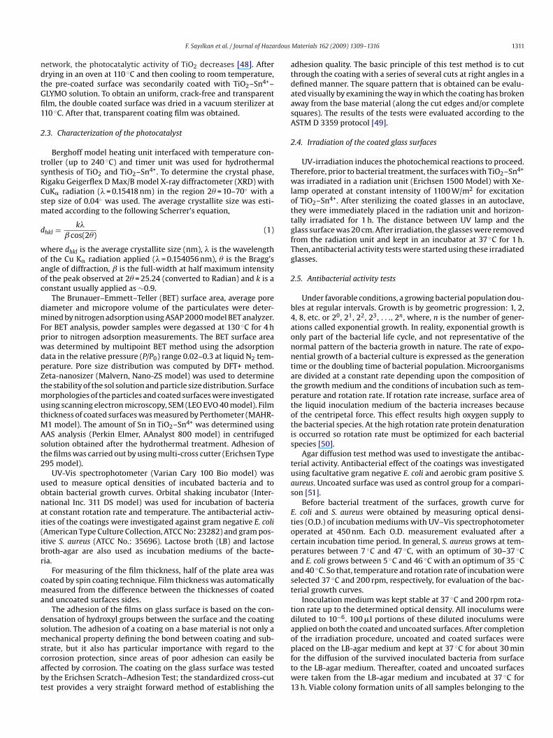
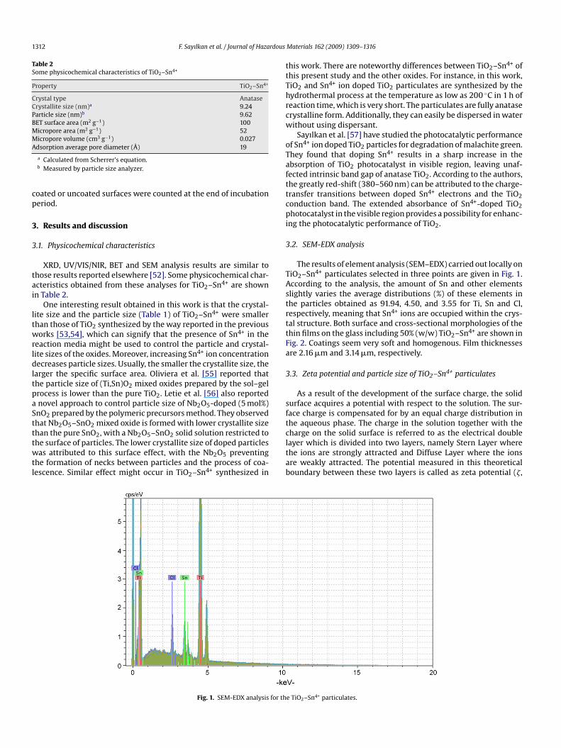
.2. SEM-EDX analysis
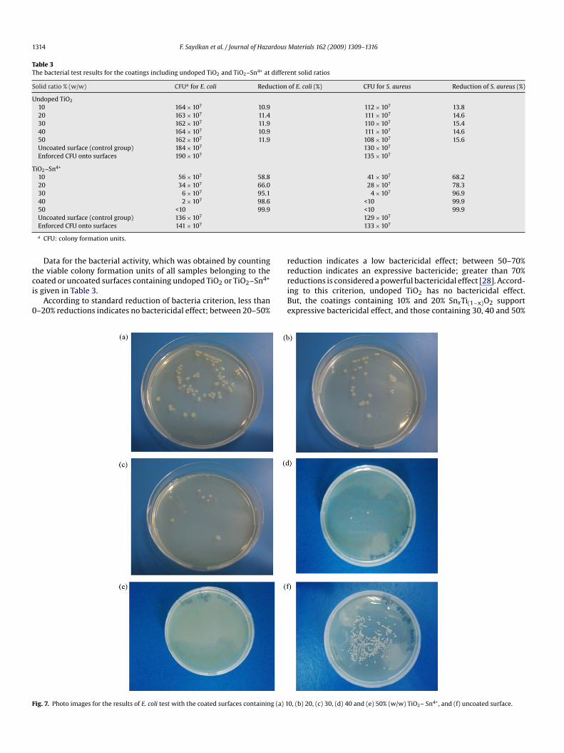
The results of element analysis (SEM–EDX) carried out locally oniO2–Sn4+ particulates selected in three points are given in Fig. 1.ccording to the analysis, the amount of Sn and other elementslightly varies the average distributions (%) of these elements inhe particles obtained as 91.94, 4.50, and 3.55 for Ti, Sn and Cl,espectively, meaning that Sn4+ ions are occupied within the crys-al structure. Both surface and cross-sectional morphologies of thehin films on the glass including 50% (w/w) TiO2–Sn4+ are shown inig. 2. Coatings seem very soft and homogenous. Film thicknessesre 2.16 �m and 3.14 �m, respectively.
.3. Zeta potential and particle size of TiO2–Sn4+ particulates
As a result of the development of the surface charge, the solidurface acquires a potential with respect to the solution. The sur-ace charge is compensated for by an equal charge distribution inhe aqueous phase. The charge in the solution together with the
harge on the solid surface is referred to as the electrical doubleayer which is divided into two layers, namely Stern Layer wherehe ions are strongly attracted and Diffuse Layer where the ionsre weakly attracted. The potential measured in this theoreticaloundary between these two layers is called as zeta potential (�,e TiO2–Sn4+ particulates.
F. Sayılkan et al. / Journal of Hazardous Materials 162 (2009) 1309–1316 1313
Ft
Vpbiuiiho
3
c
Fw
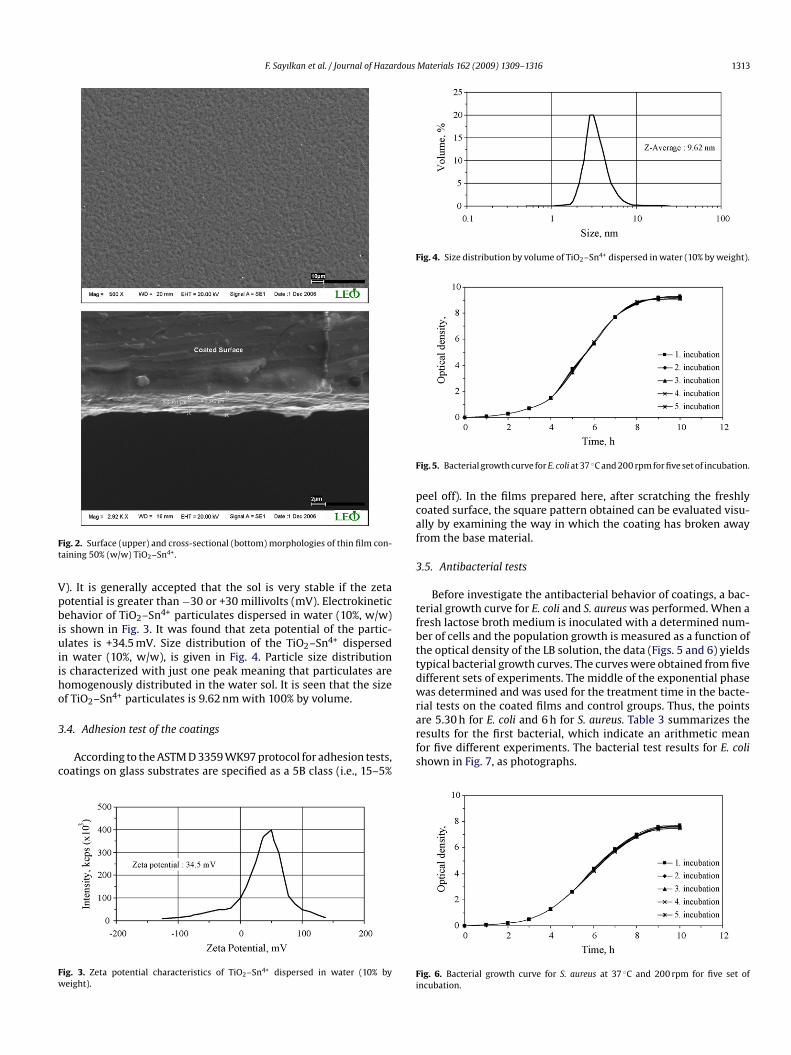
Fig. 4. Size distribution by volume of TiO2–Sn4+ dispersed in water (10% by weight).
F
pcaf
3
tfbttdwrial tests on the coated films and control groups. Thus, the points
ig. 2. Surface (upper) and cross-sectional (bottom) morphologies of thin film con-aining 50% (w/w) TiO2–Sn4+.
). It is generally accepted that the sol is very stable if the zetaotential is greater than −30 or +30 millivolts (mV). Electrokineticehavior of TiO2–Sn4+ particulates dispersed in water (10%, w/w)
s shown in Fig. 3. It was found that zeta potential of the partic-lates is +34.5 mV. Size distribution of the TiO2–Sn4+ dispersed
n water (10%, w/w), is given in Fig. 4. Particle size distributions characterized with just one peak meaning that particulates areomogenously distributed in the water sol. It is seen that the sizef TiO2–Sn4+ particulates is 9.62 nm with 100% by volume.
.4. Adhesion test of the coatings
According to the ASTM D 3359 WK97 protocol for adhesion tests,oatings on glass substrates are specified as a 5B class (i.e., 15–5%
ig. 3. Zeta potential characteristics of TiO2–Sn4+ dispersed in water (10% byeight).
arfs
Fi
ig. 5. Bacterial growth curve for E. coli at 37 ◦C and 200 rpm for five set of incubation.
eel off). In the films prepared here, after scratching the freshlyoated surface, the square pattern obtained can be evaluated visu-lly by examining the way in which the coating has broken awayrom the base material.
.5. Antibacterial tests
Before investigate the antibacterial behavior of coatings, a bac-erial growth curve for E. coli and S. aureus was performed. When aresh lactose broth medium is inoculated with a determined num-er of cells and the population growth is measured as a function ofhe optical density of the LB solution, the data (Figs. 5 and 6) yieldsypical bacterial growth curves. The curves were obtained from fiveifferent sets of experiments. The middle of the exponential phaseas determined and was used for the treatment time in the bacte-
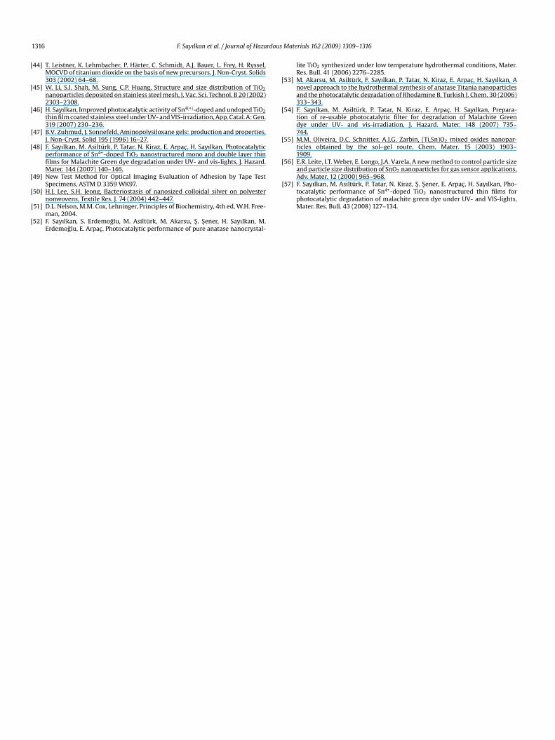
re 5.30 h for E. coli and 6 h for S. aureus. Table 3 summarizes theesults for the first bacterial, which indicate an arithmetic meanor five different experiments. The bacterial test results for E. colihown in Fig. 7, as photographs.
ig. 6. Bacterial growth curve for S. aureus at 37 ◦C and 200 rpm for five set ofncubation.
1314 F. Sayılkan et al. / Journal of Hazardous Materials 162 (2009) 1309–1316
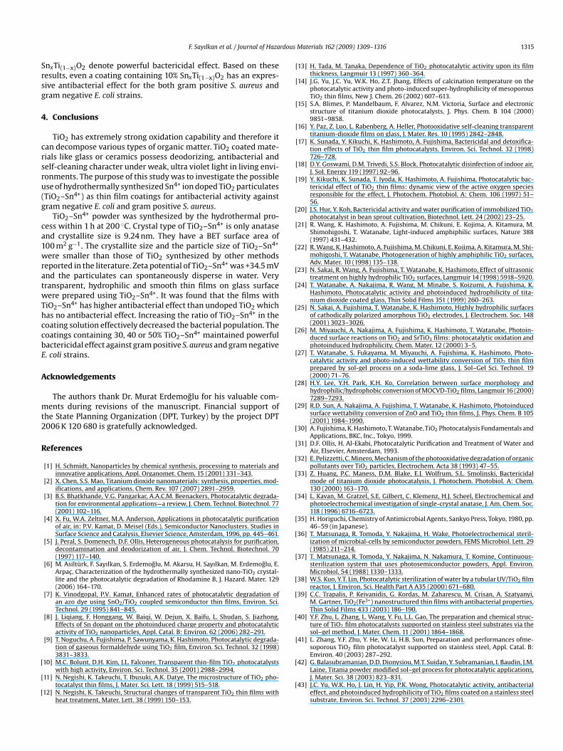
Table 3The bacterial test results for the coatings including undoped TiO2 and TiO2–Sn4+ at different solid ratios
Solid ratio % (w/w) CFUa for E. coli Reduction of E. coli (%) CFU for S. aureus Reduction of S. aureus (%)
Undoped TiO2
10 164 × 107 10.9 112 × 107 13.820 163 × 107 11.4 111 × 107 14.630 162 × 107 11.9 110 × 107 15.440 164 × 107 10.9 111 × 107 14.650 162 × 107 11.9 108 × 107 15.6Uncoated surface (control group) 184 × 107 130 × 107
Enforced CFU onto surfaces 190 × 107 135 × 107
TiO2–Sn4+
10 56 × 107 58.8 41 × 107 68.220 34 × 107 66.0 28 × 107 78.330 6 × 10⁷ 95.1 4 × 10⁷ 96.940 2 × 10⁷ 98.6 <10 99.950 <10 99.9 <10 99.9
tci
0
r
F
Uncoated surface (control group) 136 × 107
Enforced CFU onto surfaces 141 × 107
a CFU: colony formation units.
Data for the bacterial activity, which was obtained by counting
he viable colony formation units of all samples belonging to theoated or uncoated surfaces containing undoped TiO2 or TiO2–Sn4+s given in Table 3.According to standard reduction of bacteria criterion, less than
–20% reductions indicates no bactericidal effect; between 20–50%
rriBe
ig. 7. Photo images for the results of E. coli test with the coated surfaces containing (a) 1
129 × 107
133 × 107
eduction indicates a low bactericidal effect; between 50–70%
eduction indicates an expressive bactericide; greater than 70%eductions is considered a powerful bactericidal effect [28]. Accord-ng to this criterion, undoped TiO2 has no bactericidal effect.ut, the coatings containing 10% and 20% SnxTi(1−x)O2 supportxpressive bactericidal effect, and those containing 30, 40 and 50%0, (b) 20, (c) 30, (d) 40 and (e) 50% (w/w) TiO2– Sn4+, and (f) uncoated surface.
rdous
Srsg
4
crsru(g
ca1wratwThccbE
A
mt2
R
[
[
[
[
[
[
[
[
[
[
[
[
[
[
[
[
[
[
[
[
[
[
[
[
[
[
[
[
[
[
[
[
F. Sayılkan et al. / Journal of Haza
nxTi(1−x)O2 denote powerful bactericidal effect. Based on theseesults, even a coating containing 10% SnxTi(1−x)O2 has an expres-ive antibacterial effect for the both gram positive S. aureus andram negative E. coli strains.
. Conclusions
TiO2 has extremely strong oxidation capability and therefore itan decompose various types of organic matter. TiO2 coated mate-ials like glass or ceramics possess deodorizing, antibacterial andelf-cleaning character under weak, ultra violet light in living envi-onments. The purpose of this study was to investigate the possiblese of hydrothermally synthesized Sn4+ ion doped TiO2 particulatesTiO2–Sn4+) as thin film coatings for antibacterial activity againstram negative E. coli and gram positive S. aureus.
TiO2–Sn4+ powder was synthesized by the hydrothermal pro-ess within 1 h at 200 ◦C. Crystal type of TiO2–Sn4+ is only anatasend crystallite size is 9.24 nm. They have a BET surface area of00 m2 g−1. The crystallite size and the particle size of TiO2–Sn4+
ere smaller than those of TiO2 synthesized by other methodseported in the literature. Zeta potential of TiO2–Sn4+ was +34.5 mVnd the particulates can spontaneously disperse in water. Veryransparent, hydrophilic and smooth thin films on glass surfaceere prepared using TiO2–Sn4+. It was found that the films with
iO2–Sn4+ has higher antibacterial effect than undoped TiO2 whichas no antibacterial effect. Increasing the ratio of TiO2–Sn4+ in theoating solution effectively decreased the bacterial population. Theoatings containing 30, 40 or 50% TiO2–Sn4+ maintained powerfulactericidal effect against gram positive S. aureus and gram negative. coli strains.
cknowledgements
The authors thank Dr. Murat Erdemoglu for his valuable com-ents during revisions of the manuscript. Financial support of
he State Planning Organization (DPT, Turkey) by the project DPT006 K 120 680 is gratefully acknowledged.
eferences
[1] H. Schmidt, Nanoparticles by chemical synthesis, processing to materials andinnovative applications, Appl. Organomet. Chem. 15 (2001) 331–343.
[2] X. Chen, S.S. Mao, Titanium dioxide nanomaterials: synthesis, properties, mod-ifications, and applications, Chem. Rev. 107 (2007) 2891–2959.
[3] B.S. Bhatkhande, V.G. Pangarkar, A.A.C.M. Beenackers, Photocatalytic degrada-tion for environmental applications—a review, J. Chem. Technol. Biotechnol. 77(2001) 102–116.
[4] X. Fu, W.A. Zeltner, M.A. Anderson, Applications in photocatalytic purificationof air, in: P.V. Kamat, D. Meisel (Eds.), Semiconductor Nanoclusters. Studies inSurface Science and Catalysis, Elsevier Science, Amsterdam, 1996, pp. 445–461.
[5] J. Peral, S. Domenech, D.F. Ollis, Heterogeneous photocatalysis for purification,decontamination and deodorization of air, J. Chem. Technol. Biotechnol. 70(1997) 117–140.
[6] M. Asiltürk, F. Sayılkan, S. Erdemoglu, M. Akarsu, H. Sayılkan, M. Erdemoglu, E.Arpac, Characterization of the hydrothermally synthesized nano-TiO2 crystal-lite and the photocatalytic degradation of Rhodamine B, J. Hazard. Mater. 129(2006) 164–170.
[7] K. Vinodgopal, P.V. Kamat, Enhanced rates of photocatalytic degradation ofan azo dye using SnO2/TiO2 coupled semiconductor thin films, Environ. Sci.Technol. 29 (1995) 841–845.
[8] J. Liqiang, F. Honggang, W. Baiqi, W. Dejun, X. Baifu, L. Shudan, S. Jiazhong,Effects of Sn dopant on the photoinduced charge property and photocatalyticactivity of TiO2 nanoparticles, Appl. Catal. B: Environ. 62 (2006) 282–291.
[9] T. Noguchu, A. Fujishima, P. Sawunyama, K. Hashimoto, Photocatalytic degrada-tion of gaseous formaldehyde using TiO2 film, Environ. Sci. Technol. 32 (1998)3831–3833.
10] M.C. Bolunt, D.H. Kim, J.L. Falconer, Transparent thin-film TiO2 photocatalystswith high activity, Environ. Sci. Technol. 35 (2001) 2988–2994.
11] N. Negishi, K. Takeuchi, T. Ibusuki, A.K. Datye, The microstructure of TiO2 pho-tocatalyst thin films, J. Mater. Sci. Lett. 18 (1999) 515–518.
12] N. Negishi, K. Takeuchi, Structural changes of transparent TiO2 thin films withheat treatment, Mater. Lett. 38 (1999) 150–153.
[
[
Materials 162 (2009) 1309–1316 1315
13] H. Tada, M. Tanaka, Dependence of TiO2 photocatalytic activity upon its filmthickness, Langmuir 13 (1997) 360–364.
14] J.G. Yu, J.C. Yu, W.K. Ho, Z.T. Jhang, Effects of calcination temperature on thephotocatalytic activity and photo-induced super-hydrophilicity of mesoporousTiO2 thin films, New J. Chem. 26 (2002) 607–613.
15] S.A. Blimes, P. Mandelbaum, F. Alvarez, N.M. Victoria, Surface and electronicstructure of titanium dioxide photocatalysts, J. Phys. Chem. B 104 (2000)9851–9858.
16] Y. Paz, Z. Luo, L. Rabenberg, A. Heller, Photooxidative self-cleaning transparenttitanium-dioxide films on glass, J. Mater. Res. 10 (1995) 2842–2848.
17] K. Sunada, Y. Kikuchi, K. Hashimoto, A. Fujishima, Bactericidal and detoxifica-tion effects of TiO2 thin film photocatalysts, Environ. Sci. Technol. 32 (1998)726–728.
18] D.Y. Goswami, D.M. Trivedi, S.S. Block, Photocatalytic disinfection of indoor air,J. Sol. Energy 119 (1997) 92–96.
19] Y. Kikuchi, K. Sunada, T. Iyoda, K. Hashimoto, A. Fujishima, Photocatalytic bac-tericidal effect of TiO2 thin films: dynamic view of the active oxygen speciesresponsible for the effect, J. Photochem. Photobiol. A: Chem. 106 (1997) 51–56.
20] J.S. Hur, Y. Koh, Bactericidal activity and water purification of immobilized TiO2
photocatalyst in bean sprout cultivation, Biotechnol. Lett. 24 (2002) 23–25.21] R. Wang, K. Hashimoto, A. Fujishima, M. Chikuni, E. Kojima, A. Kitamura, M.
Shimohigoshi, T. Watanabe, Light-induced amphiphilic surfaces, Nature 388(1997) 431–432.
22] R. Wang, K. Hashimoto, A. Fujishima, M. Chikuni, E. Kojima, A. Kitamura, M. Shi-mohigoshi, T. Watanabe, Photogeneration of highly amphiphilic TiO2 surfaces,Adv. Mater. 10 (1998) 135–138.
23] N. Sakai, R. Wang, A. Fujishima, T. Watanabe, K. Hashimoto, Effect of ultrasonictreatment on highly hydrophilic TiO2 surfaces, Langmuir 14 (1998) 5918–5920.
24] T. Watanabe, A. Nakajima, R. Wang, M. Minabe, S. Koizumi, A. Fujishima, K.Hashimoto, Photocatalytic activity and photoinduced hydrophilicity of tita-nium dioxide coated glass, Thin Solid Films 351 (1999) 260–263.
25] N. Sakai, A. Fujishima, T. Watanabe, K. Hashimoto, Highly hydrophilic surfacesof cathodically polarized amorphous TiO2 electrodes, J. Electrochem. Soc. 148(2001) 3023–3026.
26] M. Miyauchi, A. Nakajima, A. Fujishima, K. Hashimoto, T. Watanabe, Photoin-duced surface reactions on TiO2 and SrTiO3 films: photocatalytic oxidation andphotoinduced hydrophilicity, Chem. Mater. 12 (2000) 3–5.
27] T. Watanabe, S. Fukayama, M. Miyauchi, A. Fujishima, K. Hashimoto, Photo-catalytic activity and photo-induced wettability conversion of TiO2 thin filmprepared by sol-gel process on a soda-lime glass, J. Sol–Gel Sci. Technol. 19(2000) 71–76.
28] H.Y. Lee, Y.H. Park, K.H. Ko, Correlation between surface morphology andhydrophilic/hydrophobic conversion of MOCVD-TiO2 films, Langmuir 16 (2000)7289–7293.
29] R.D. Sun, A. Nakajima, A. Fujishima, T. Watanabe, K. Hashimoto, Photoinducedsurface wettability conversion of ZnO and TiO2 thin films, J. Phys. Chem. B 105(2001) 1984–1990.
30] A. Fujishima, K. Hashimoto, T. Watanabe, TiO2 Photocatalysis Fundamentals andApplications, BKC, Inc., Tokyo, 1999.
31] D.F. Ollis, H. Al-Ekabi, Photocatalytic Purification and Treatment of Water andAir, Elsevier, Amsterdam, 1993.
32] E. Pelizzetti, C. Minero, Mechanism of the photooxidative degradation of organicpollutants over TiO2 particles, Electrochem. Acta 38 (1993) 47–55.
33] Z. Huang, P.C. Maness, D.M. Blake, E.J. Wolfrum, S.L. Smolinski, Bactericidalmode of titanium dioxide photocatalysis, J. Photochem. Photobiol. A: Chem.130 (2000) 163–170.
34] L. Kavan, M. Gratzel, S.E. Gilbert, C. Klemenz, H.J. Scheel, Electrochemical andphotoelectrochemical investigation of single-crystal anatase, J. Am. Chem. Soc.118 (1996) 6716–6723.
35] H. Horiguchi, Chemistry of Antimicrobial Agents, Sankyo Press, Tokyo, 1980, pp.46–59 (in Japanese).
36] T. Matsunaga, R. Tomoda, Y. Nakajima, H. Wake, Photoelectrochemical steril-ization of microbial-cells by semiconductor powders, FEMS Microbiol. Lett. 29(1985) 211–214.
37] T. Matsunaga, R. Tomoda, Y. Nakajima, N. Nakamura, T. Komine, Continuous-sterilization system that uses photosemiconductor powders, Appl. Environ.Microbiol. 54 (1988) 1330–1333.
38] W.S. Kuo, Y.T. Lin, Photocatalytic sterilization of water by a tubular UV/TiO2 filmreactor, J. Environ. Sci. Health Part A A35 (2000) 671–680.
39] C.C. Trapalis, P. Keivanidis, G. Kordas, M. Zaharescu, M. Crisan, A. Szatyanyi,M. Gartner, TiO2(Fe3+) nanostructured thin films with antibacterial properties,Thin Solid Films 433 (2003) 186–190.
40] Y.F. Zhu, L. Zhang, L. Wang, Y. Fu, L.L. Gao, The preparation and chemical struc-ture of TiO2 film photocatalysts supported on stainless steel substrates via thesol–gel method, J. Mater. Chem. 11 (2001) 1864–1868.
41] L. Zhang, Y.F. Zhu, Y. He, W. Li, H.B. Sun, Preparation and performances ofme-soporous TiO2 film photocatalyst supported on stainless steel, Appl. Catal. B:Environ. 40 (2003) 287–292.
42] G. Balasubramanian, D.D. Dionysiou, M.T. Suidan, Y. Subramanian, I. Baudin, J.M.Laine, Titania powder modified sol–gel process for photocatalytic applications,J. Mater. Sci. 38 (2003) 823–831.
43] J.C. Yu, W.K. Ho, J. Lin, H. Yip, P.K. Wong, Photocatalytic activity, antibacterialeffect, and photoinduced hydrophilicity of TiO2 films coated on a stainless steelsubstrate, Environ. Sci. Technol. 37 (2003) 2296–2301.
1 rdous
[
[
[
[
[
[
[
[
[
[
[
[
[and particle size distribution of SnO2 nanoparticles for gas sensor applications,Adv. Mater. 12 (2000) 965–968.
[57] F. Sayılkan, M. Asiltürk, P. Tatar, N. Kiraz, S. Sener, E. Arpac, H. Sayılkan, Pho-
316 F. Sayılkan et al. / Journal of Haza
44] T. Leistner, K. Lehmbacher, P. Härter, C. Schmidt, A.J. Bauer, L. Frey, H. Ryssel,MOCVD of titanium dioxide on the basis of new precursors, J. Non-Cryst. Solids303 (2002) 64–68.
45] W. Li, S.I. Shah, M. Sung, C.P. Huang, Structure and size distribution of TiO2
nanoparticles deposited on stainless steel mesh, J. Vac. Sci. Technol. B 20 (2002)2303–2308.
46] H. Sayılkan, Improved photocatalytic activity of Sn4(+)-doped and undoped TiO2
thin film coated stainless steel under UV- and VIS-irradiation, App. Catal. A: Gen.319 (2007) 230–236.
47] B.V. Zuhmud, J. Sonnefeld, Aminopolysiloxane gels: production and properties,J. Non-Cryst. Solid 195 (1996) 16–27.
48] F. Sayılkan, M. Asiltürk, P. Tatar, N. Kiraz, E. Arpac, H. Sayılkan, Photocatalyticperformance of Sn4+-doped TiO2 nanostructured mono and double layer thinfilms for Malachite Green dye degradation under UV- and vis-lights, J. Hazard.Mater. 144 (2007) 140–146.
49] New Test Method for Optical Imaging Evaluation of Adhesion by Tape TestSpecimens, ASTM D 3359 WK97.
50] H.J. Lee, S.H. Jeong, Bacteriostasis of nanosized colloidal silver on polyesternonwovens, Textile Res. J. 74 (2004) 442–447.
51] D.L. Nelson, M.M. Cox, Lehninger, Principles of Biochemistry, 4th ed, W.H. Free-man, 2004.
52] F. Sayılkan, S. Erdemoglu, M. Asiltürk, M. Akarsu, S. Sener, H. Sayılkan, M.Erdemoglu, E. Arpac, Photocatalytic performance of pure anatase nanocrystal-
Materials 162 (2009) 1309–1316
lite TiO2 synthesized under low temperature hydrothermal conditions, Mater.Res. Bull. 41 (2006) 2276–2285.
53] M. Akarsu, M. Asiltürk, F. Sayılkan, P. Tatar, N. Kiraz, E. Arpac, H. Sayılkan, Anovel approach to the hydrothermal synthesis of anatase Titania nanoparticlesand the photocatalytic degradation of Rhodamine B, Turkish J. Chem. 30 (2006)333–343.
54] F. Sayılkan, M. Asiltürk, P. Tatar, N. Kiraz, E. Arpac, H. Sayılkan, Prepara-tion of re-usable photocatalytic filter for degradation of Malachite Greendye under UV- and vis-irradiation, J. Hazard. Mater. 148 (2007) 735–744.
55] M.M. Oliveira, D.C. Schnitter, A.J.G. Zarbin, (Ti,Sn)O2 mixed oxides nanopar-ticles obtained by the sol–gel route, Chem. Mater. 15 (2003) 1903–1909.
56] E.R. Leite, I.T. Weber, E. Longo, J.A. Varela, A new method to control particle size
tocatalytic performance of Sn4+-doped TiO2 nanostructured thin films forphotocatalytic degradation of malachite green dye under UV- and VIS-lights,Mater. Res. Bull. 43 (2008) 127–134.