Population pharmacokinetics of intravenous caffeine in neonates with apnea of prematurity
Upload
xn--universitdannunzio-eubCategory
view
5download
0
1990;50:7272-7278. Cancer Res D. J. Hnatowich, M. Rusckowski, A. B. Brill, et al. Diethylenetriamine Pentaacetic Acid ChelatorAntigen Antibody Radiolabeled with Indium-111 Using a Novel Pharmacokinetics in Patients of an Anti-Carcinoembryonic
Updated version
http://cancerres.aacrjournals.org/content/50/22/7272
Access the most recent version of this article at:
E-mail alerts related to this article or journal.Sign up to receive free email-alerts
Subscriptions
Reprints and
To order reprints of this article or to subscribe to the journal, contact the AACR Publications
Permissions
To request permission to re-use all or part of this article, contact the AACR Publications
Research. on January 28, 2014. © 1990 American Association for Cancercancerres.aacrjournals.org Downloaded from
Research. on January 28, 2014. © 1990 American Association for Cancercancerres.aacrjournals.org Downloaded from

[CANCER RESEARCH 50, 7272-7278. November 15. 1990]
Pharmacokinetics in Patients of an Anti-Carcinoembryonic Antigen AntibodyRadiolabeled with Indium-Ill Using a Novel DiethylenetriaminePentaacetic Acid Chelator1
D. J. Hnatowich,2 M. Rusckowski, A. B. Brill, D. A. Siebecker, H. Misra, G. Mardirossian, H. Bushe, A. Rescigno,
S. Stevens, D. K. Johnson, and T. W. GriffinUniversity of Massachusetts Medical Center [D. J. //., M. R., A. B. B., D. A. S., H. M., (i. M., H. B.. A. R.. S. S., T. W. G.] Worcester, Massachusetts 01655, anil AbbottLaboratories [D. K. J.], Abbott Park, Illinois 60064
ABSTRACT
The pharmacokinetics of the Cl 10 anti-carcinoembryonic antigen antibody radiolabeled with '"In via a novel benzylisothiocyanate derivative
of diethylenetriamine pentaacetic acid have been determined in 12 patients. The chelator was attached to the protein via a thiourea bond andin such a way that all 5 carboxymethyl arms were presumably able toparticipate in chelation. Patients with known or suspected colorectalcarcinoma received between 5 and 20 mg of the IgG antibody labeledwith5 niCi of '"In. Individual organ radioactivity levels werequantitated,
and serum and urine samples were analyzed, principally by size exclusionhigh-performance liquid chromatography (HPLC). Total urinary excretion averaged 0.18% of the injected dose/h with large patient to patientvariation. At early times postadministration (<8 h) the predominantradiolabeled species in urine was free diethylenetriamine pentaacetic acidmost probably administered as a small radiocontaminant in the injectate.Thereafter, radioactivity in urine was primarily present as a low molecularweight catabolic product. Analysis of serum by size exclusion HPLCoccasionally showed 3 radioactivity peaks, 2 of which are due to circulating immune complexes and labeled antibody. The third peak is of lowmolecular weight and is due to one or more products of antibody catab-olism. Transchelation of '"In to circulating transferrin was observed but
at modest levels. Quantitation of organ radioactivity showed that 18 ±4(SI ))'/•;of the injected dose was in the liver at 1 day postadministration
and 1.4 ±1.1 and 1.2 ±0.9% was in the spleen and in both kidneys,respectively, at this time. The mean half-life for clearance of total injectedradioactivity was fitted to a single exponential and was found to be 34 h(SD, 14 li: .V= M )and that for antibody alone, assessed by size exclusionHPLC analysis of serum samples, was calculated to be 22 h (SD, X li; V= 10). Neither of these values nor organ radioactivity levels were affectedby antibody-loading dose.
INTRODUCTION
A derivative of the bifunctional chelator DTPA1 has recently
been synthesized in which a p-isothiocyanatobenzyl moiety isattached to a méthylènecarbon atom on one of the carboxymethyl arms (1). This chelator was intended to be covalentlyconjugated to antibodies as a means of radiolabeling theseproteins with '"In (2). At present, the method most often used
for this purpose attaches DTPA through either its mixed (3) orcyclic anhydrides (4). There are at least 2 major differences inthis new approach to labeling antibodies: (a) whereas bothanhydrides conjugate DTPA via amide bonds, the new chelator
Received 4/9/90; accepted 8/20/90.The costs of publication of this article were defrayed in part by the payment
of page charges. This article must therefore be hereby marked advertisement inaccordance with 18 U.S.C. Section 1734 solely to indicate this fact.
1This investigation was funded in part by Abbott Laboratories and the NIH
(CA 33029 and CA 39748).2To whom requests for reprints should be addressed, at the Department of
Nuclear Medicine, University of Massachusetts Medical Center, 55 Lake AvenueNorth, Worcester. MA 01655.
3The abbreviations used are: DTPA, diethylenetriamine pentaacetic acid;CEA, carcinoembryonic antigen; HPLC. high performance liquid chromatography; ID. injected dose; BSA. bovine serum albumin; PBS. phosphate-bufferedsaline; HAMA, human anti-mouse antibody.
is conjugated via a thiourea bond, and (b) when attached via itsanhydrides, DTPA is conjugated through one of its carboxymethyl arms which may not then be available to participate inchelation of indium. By contrast, conjugation of the new chelator preserves all 5 carboxymethyl arms. It has been suggestedthat all 5 arms are necessary for the stable chelation of indium(5, 6).
The object of this investigation was to evaluate the pharmacokinetics in patients of one antibody conjugated with the newchelator and radiolabeled with '"In. The antibody selected forthis study was the anti-CEA Cl 10 developed by Sumerdon etal. (2). For this investigation, the intact IgG antibody wasconjugated with an average of 6 chelators/protein molecule andradiolabeled with '"In. "'In, 5 mCi, and the antibody, between
5 and 20 mg, were administered to 12 patients with documentedor suspected colorectal carcinoma. In this report we describethe pharmacokinetic behavior of the label; a detailed descriptionof the imaging results obtained in these patients will appearelsewhere.4
MATERIALS AND METHODS
The C110-DTPA antibody-chelator conjugate, prepared and characterized as previously described (2), was supplied by Abbott Laboratories as a sterile, apyrogenic solution in 0.05 M citrate buffer, pH 6, ata concentration of 5 mg/ml.
The conjugated antibody was radiolabeled with carrier-free "'In
(NEN Dupont, Billerica, MA) by adding 5 mCi of this activity to thedesired weight of antibody. The labeled antibody preparation wasassayed by size exclusion HPLC using a single 7.5 x 300-mm TSK 250column (BioRad Laboratories, Richmond, CA) or a single 10 x 300-mm Superóse 12 column (Pharmacia, Piscataway, NJ) and 0.05 Mphosphate buffer, pH 7.0, eluant. The output of an in-line radioactivitydetector was digitized and stored in a multichannel analyzer to facilitatethe calculation of peak areas (7). Recovery was determined by countingthe effluent against a standard of the injectate.
Patients enrolled in this investigation gave informed consent andwere studied with the approval of the Food and Drug Administration(IND BB 2732) and the appropriate institutional review committees.Each patient received 5 mCi and either 5, 10, or 20 mg of the labeledantibody in 180 ml of normal saline for injection by slow infusion (10ml/min) into a peripheral vein. One preadministration and severalpostadministration blood samples were collected over 3-4 days in red-top Vacutainers so that serum would be available after clotting. Acomplete urine collection over the same period was also obtained.Regular whole body and spot images were obtained for patients 1-9 onan Ohio-Nuclear LFOV gamma camera and for subsequent patients ona Seimens Body Scan. Attenuation correction was achieved as previously described (8).
Several blood samples were collected in heparinized tubes so that thepercentage of radioactivity bound to formed elements could be determined.
4 T. W. Griffin, A. B. Brill, J. A. Collins, et al. Initial clinical study of indium-111 labeled clone 110 anti-CEA antibody in patients with colorectal cancer. J.Clin. Oncol.. submitted for publication.
7272
Research. on January 28, 2014. © 1990 American Association for Cancercancerres.aacrjournals.org Downloaded from

ANTl-CEA ANTIBODY PHARMACOKINETICS
For most patients, 3-5 serum samples were analyzed by size exclusion HPLC using a single 7.5 x 300-mm TSK 400 column (BioRad) ora single 10 x 300-mm Superóse 12 column (Pharmacia) with 0.05 Mphosphate buffer, pH 7.0, eluant. Serum samples were analyzed sem-iautomatically using a WISP injector (Waters Associates, Milford, MA)and an automatic fraction collector. Fractions were then counted in anautomatic well gamma counter. In addition to the analysis of serumsamples by size exclusion, sera from the first 4 patients were alsoanalyzed by cation exchange HPLC using a single 7.5 x 75-mm SP
5PW column (Waters) with gradient elution from 0.02 M sodiumacetate, pH 5.0, to 0.5 M sodium sulfate, 0.02 M Tris hydrochloride,pH 8.0, in 30 min. Urine samples from the first 5 patients were analyzedboth by open column Sephadex G25 chromatography and by sizeexclusion HPLC using a single 7.5 x 300-mm 1-60 column (Waters)and 0.05 M phosphate buffer, pH 7.O. Urine samples from subsequentpatients were analyzed by size exclusion HPLC using the Superóse 12column and by anión exchange HPLC using a 7.5 x 75-mm DEAEcolumn (Waters) with gradient elution from 0.02 MTris buffer, pH 8.5,to 0.02 M Tris buffer, 0.5 M sodium chloride, pH 7.0. in 30 min.Regardless of the method of analysis for both serum and urine samples,the in-line radiation detectors were too insensitive to record radioactivity levels so that fractions (0.2-0.8 ml) were collected for counting in awell gamma counter.
Radioactivity levels in all serum and urine samples were also determined by counting an aliquot of each sample in a well gamma counter.In a limited number of analyses, the percentage of serum radioactivitypresent as labeled antibody, immune complex, and occasionally ascatabolic product(s) could be determined. In these cases, the SAAM29computer program (9) was used to estimate serum clearance curves foreach species separately. Because of the larger number of reliable measurements of antibody concentration, this analysis was most successfulwhen applied to this species.
The determination of the rate and extent of transcomplexation of'"In from antibody in circulation to transferrin was achieved by goatanti-human transferrin affinity chromatography as described previously(10). The percentage of serum activity binding to the column is proportional to the concentration of '"In-labeled transferrin in the sample.
Serum samples obtained at between 14 and 210 days after antibodyadministration were also analyzed for HAMA. To each of several testtubes, 1 polyethylene bead (Precision Plastic Ball Co.. Chicago. IL)previously coated with a mouse antibody (19-9; Centocor. Mähern,PA) was added along with 300 p\ of bicarbonate buffer, pH 9.6.containing \c/c human serum albumin. The beads were incubated for 1h at 37°Cand were then washed 2 times with PBS containing 1%human serum albumin and 0.05/c Tween 20. A sample of a patient's
serum with a known titer of anti-mouse antibody was used as a standard.Three hundred n\ of the control sera (1:5 serial dilutions in PBS) andthe sera under investigation (diluted 1:4 in PBS) were added to separatetest tubes containing the washed beads. The test tubes containing thebeads were agitated on a laboratory rocker at room temperature for 1.5h. The beads were then washed with PBS/Tween 20, and the tracer."'In-DTPA-OC-125 F(ab')2, at 50 ng/ml, was added. The beads were
incubated for 16 h at room temperature and were then washed as beforeand counted in a well gamma counter.
RESULTS
The results of all tests to which the antibody was subjected,both before and after coupling and labeling, demonstrated thatthe injectate was safe for human use. Thus, the preparation wasshown to be sterile, apyrogenic, and free of murine retroviruses,Mycoplasma, and potential toxic substances (Quality Biologies,Camden, NJ). Two preparations of conjugated Cl 10 antibodieswere used in this investigation; patients 1-9 received antibodyfrom the first lot, while the remaining patients received antibodyfrom the second. The average number of chelator groups perantibody molecule was 6 for both lots. Despite this relatively
high degree of conjugation, the immunoreactivity of the antibody was unaffected (2).
Following radiolabeling, analysis by size exclusion HPLCshowed that 96% (SD, 4%; N = 13) of the label was bound toantibody. The recovery during these analyses averaged 93%(SD, 4%;7V = 13).
Twelve patients were enrolled in this study, each with documented colorectal carcinoma. The median age was 54 years,with a range of 36-71 years. Six patients received 5 mg, 5received 10 mg, and 2 received 20 mg. The first patient received5 mg of the labeled antibody preparation and 15 months laterreceived a second administration of 10 mg. Table 1 identifiespatients by number and lists the preinjection circulating CEAlevels, antibody dose administered, the percentage of administered dose in the liver at 1 day, and the percentage of radioactivity in serum present as immune complex at approximately 1day postadministration.
Organ Quantitation. The time-dependent radioactivity levelsin liver are plotted in Fig. 1 with each patient identified by-
number. In most cases, liver accumulation of label is consistentwith that observed in this laboratory for 2 other antibodieslabeled with "'In (8, 10). Maximum values are reached imme
diately, with blood pool activity contributing significantly tothe earliest measurement. Thereafter, high values persistthroughout each investigation (not shown in Fig. 1 is a valuefor patient 3 of 16% ID in the liver at 142 h). Excluding patients4 and 1*(second study of patient 1), the mean liver radioactivity
level at 1 day postadministration is 18% (4% SD, N= 9).Mean values for spleen uptake of radiolabel at approximately
1 day postadministration was 1.2% (SD, 1.1%; N = 13) and forboth kidneys was 1.4% (SD, 0.9%; N = 13). As in the liver,spleen and kidney radioactivity levels were generally unchangedthroughout each investigation.
Urine Analysis. Release of radioactivity to urine was slow butsteady in all patients, averaging 0.18% ID/h (SD, 0.08%; N =13), a value which is less than that observed by us for 2 otherantibodies (8, 10). Fig. 2 presents urine radioactivity in percentage of injected dose (corrected for decay) present in eachcollection plotted at the middle of the collection period. Presented in this manner, it is clear that release of radioactivityinto urine goes through a pronounced minimum at about 8 hpostadministration.
Chromatography by G25 open column and 1-60 HPLCshowed that radioactivity in urine élûtespredominantly in onepeak. Size exclusion HPLC analysis using the Superóse 12
Table t Antibody uose, preadministration circulating CEA antigen let-els,percentage of serum activity present as immune complex, and percentage of
administered activity in liver, at approximately I day postadministration
Listed separately by patient.
Patient123456789101112,.»PrcadministrationDose CEA levels(mg)(ng/ml)555101010102020555102411145254620565328499303150.6684Immune
complexat 20-29 h(%)9(I266.1(16Id8231334770Liver
activityat 19-29h
(%ID)18142070Mm22141412262l61
' Not available.' 1*. second administration to patient
7273
Research. on January 28, 2014. © 1990 American Association for Cancercancerres.aacrjournals.org Downloaded from

ANTI-CEA ANTIBODY PHARMAC'OKINETICS
10 20 30 40 50 60
Time (hrs)70 80
Fig. 1. Radioactivity in liver presented as percentage of administered '"In
(corrected for decay) compared with time after administration of labeled antibody.Patients identified by number.
column showed a slight difference in retention time of thisprominent peak between early and late urine collections. Theresolution was improved by anión exchange DEAE HPLCanalysis, and radiochromatograms obtained of a mixture ofurine collected from one patient at 0-4 and 24-48 h showed 2distinct peaks. The first was exclusively present in an identicalanalysis of the 0- to 4-h urine collection, while the second wasalone in the 24- to 48-h urine collection. This behavior is similarto that observed by us previously in clinical trials with the 19-9 and OC-125 antibodies radiolabeled with '"In via the cyclic
anhydride of DTPA. At early times, radioactivity in urine wasprimarily due to radiolabeled free DTPA clearing to urinefollowing its introduction into serum as a small radiocontami-nant of the injectate. At later times, however, this chelate wasno longer observed in urine. Instead, the predominant speciesin late urines, while also of low molecular weight, no longercoeluted with labeled DTPA and was a product of antibodycatabolism (8, 10).
Accumulations of '"In activity in the gastrointestinal tract
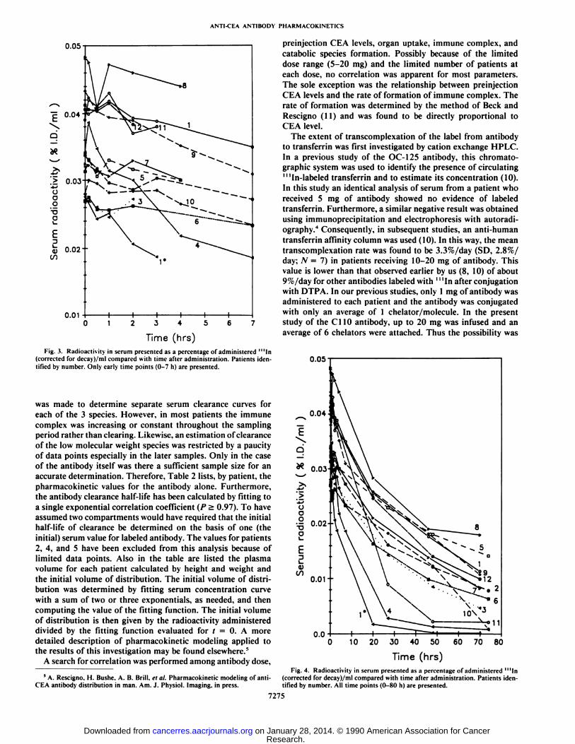
was occasionally seen in late images.Serum Analysis. Figs. 3 and 4 show the individual serum
levels of total radioactivity for each patient identified by number. As seen best in Fig. 3, in almost every case a transientincrease in serum radioactivity occurred at 15 min to 2 hpostadministration. Since the initial time point in these figuresis 15-30 min after the start of antibody infusion, the maximumis unlikely to be due simply to buildup of serum activity duringa slow infusion. Furthermore, serum samples were removedfrom the arm contralateral to that receiving the infúsate; thus
this behavior cannot be attributed simply to contamination.Size exclusion HPLC was performed on a total of 67 serum
samples from the 12 patients. Serum samples from the first 10patients were analyzed on a TSK 250 column (BioRad), whileserum from later patients was analyzed on a Superóse 12column (Pharmacia). The radiochromatograms most oftenshow a prominent peak corresponding to that of labeled antibody. However, species with both shorter and longer retentiontimes were occasionally observed. Fig. 5 shows the most pronounced example of a radiochromatogram in which all 3 speciesare apparent. The radiochromatogram was obtained by analysisof the 24-h serum from patient 4. In this case the first peak(shortest retention time) predominates and is due to immunecomplexes resulting from high circulating CEA levels (Table1). The second peak coelutes with the labeled antibody and istherefore at least largely due to radiolabeled antibody stillpresent in serum. The third peak (longest retention time) coelutes in this analysis with radiolabeled DTPA. However, radiolabeled DTPA. administered as a small radiocontaminant inthe injectate, may be expected to clear rapidly into urine fromserum by glomerular filtration. Since this peak was observedonly in sera collected after about 20 h postadministration, it islikely that the species responsible is not labeled DTPA but is acatabolic product with a similar retention time by size exclusionHPLC.
The analysis of each serum sample by size exclusion HPLCand under the assumption that each peak was due to a singleradiolabeled species resulted in estimates for the fraction ofradioactivity in serum present in each of 3 species. An attempt
10 60 7020 30 40 50
Time (hrs)Fig. 2. Radioactivity in urine presented as a percentage of administered 'In
(corrected for decay) compared «ith time after administration and plotted at themean of each collection period. Patients identified by number.
7274
Research. on January 28, 2014. © 1990 American Association for Cancercancerres.aacrjournals.org Downloaded from
ANTI-CEA ANTIBODY PHARMAC OKINETICS
0.05
üoo'•5o
E5 0.02--
co
0.01
\
Time (hrs)Fig. 3. Radioactivity in serum presented as a percentage of administered '"In
(corrected for decay)/ml compared with time after administration. Patients identified by number. Only early time points (0-7 h) are presented.
was made to determine separate serum clearance curves foreach of the 3 species. However, in most patients the immunecomplex was increasing or constant throughout the samplingperiod rather than clearing. Likewise, an estimation of clearanceof the low molecular weight species was restricted by a paucityof data points especially in the later samples. Only in the caseof the antibody itself was there a sufficient sample size for anaccurate determination. Therefore, Table 2 lists, by patient, thepharmacokinetic values for the antibody alone. Furthermore,the antibody clearance half-life has been calculated by fitting toa single exponential correlation coefficient (P > 0.97). To haveassumed two compartments would have required that the initialhalf-life of clearance be determined on the basis of one (theinitial) serum value for labeled antibody. The values for patients2, 4, and 5 have been excluded from this analysis because oflimited data points. Also in the table are listed the plasmavolume for each patient calculated by height and weight andthe initial volume of distribution. The initial volume of distribution was determined by fitting serum concentration curvewith a sum of two or three exponentials, as needed, and thencomputing the value of the fitting function. The initial volumeof distribution is then given by the radioactivity administereddivided by the fitting function evaluated for t = 0. A moredetailed description of pharmacokinetic modeling applied tothe results of this investigation may be found elsewhere.5
A search for correlation was performed among antibody dose,
preinjection CEA levels, organ uptake, immune complex, andcatabolic species formation. Possibly because of the limiteddose range (5-20 mg) and the limited number of patients ateach dose, no correlation was apparent for most parameters.The sole exception was the relationship between preinjectionCEA levels and the rate of formation of immune complex. Therate of formation was determined by the method of Beck andRescigno (11) and was found to be directly proportional toCEA level.
The extent of transcomplexation of the label from antibodyto transferrin was first investigated by cation exchange HPLC.In a previous study of the OC-125 antibody, this Chromatographie system was used to identify the presence of circulating"'In-labeled transferrin and to estimate its concentration (10).
In this study an identical analysis of serum from a patient whoreceived 5 mg of antibody showed no evidence of labeledtransferrin. Furthermore, a similar negative result was obtainedusing immunoprecipitation and electrophoresis with autoradi-ography.4 Consequently, in subsequent studies, an anti-human
transferrin affinity column was used (10). In this way, the meantranscomplexation rate was found to be 3.3%/day (SD, 2.8%/day; N = 7) in patients receiving 10-20 mg of antibody. Thisvalue is lower than that observed earlier by us (8, 10) of about9%/day for other antibodies labeled with '"In after conjugation
with DTPA. In our previous studies, only 1 mg of antibody wasadministered to each patient and the antibody was conjugatedwith only an average of 1 chelator/molecule. In the presentstudy of the Cl 10 antibody, up to 20 mg was infused and anaverage of 6 chelators were attached. Thus the possibility was
0.05
0.050 60 70 80
Time (hrs)Fig. 4. Radioactivity in serum presented as a percentage of administered '"In
5A. Rescigno. H. Bushe. A. B. Brill, el al. Pharmacokinetic modeling of ami- (corrected for dccay)/ml compared with time after administration. Patients iden-CEA antibody distribution in man. Am. J. Physiol. Imaging, in press. tified by number. All time points (0-80 h) are presented.
7275
Research. on January 28, 2014. © 1990 American Association for Cancercancerres.aacrjournals.org Downloaded from

ANTI-CEA ANTIBODY PHARMACOKINETICS
considered that the transchelation rate may depend on chelatorconcentration. Accordingly, in vitro 37°Cserum incubations
were performed at two different concentrations of labeled Cl 10antibody. The results are shown in Table 3. When the chelatorconcentration in serum was adjusted to be roughly equal to thatof our previous patient studies (5 ng/ml), the transchelationrate was found to be approximately 5%/day. However, whenthe concentration was increased a factor of 25, the rate droppedto approximately 2%/day. The 5%/day value for transchelationobtained by in vitro incubation may be compared to the 9%/day value observed by us previously. The fact that the formerrate is lower indicates that the chelator used in this study formsa chelate which is more stable to transchelation of '"In to
transferrin.
IO^->C
oo
0°OoI>o<>ooo(>0<>o'>oOOo000oooooooo000ooOo
10 20 50 60 70 8030 40
FRACTIONFig. 5. Radiochromatogram obtained by size exclusion HPLC analysis of
serum collected at 1 day postadministration of '"In-labeled C110 antibody to
patient 4. The 3 peaks have been identified as immune complexes (shortestretention time), labeled antibody, and catabolic product(s).
Table 2 Antibody and total radioactivity terminal serum half-lives assuming asingle compartment clearance, the initial volume of distribution of antibody and
of total radioactivity, and the plasma volume of distribution based on heightand »'eight
Listed separately by patient.
Patient123456789101112,.»MeanSDAntibody
terminalhalf-life
(h)31.9*19.4a*27.122.618.724.035.09.625.48.222.28.2Total
radioactivityterminal
half-life(h)36.837.028.826.025.634.835.634.237.646.611.137.08.333.813.8Antibody
initialvolume(liters)2.2•2.5*•2.63.23.82.49.62.02.01.4Total
radioactivityvolume(liters)2.33.23.82.72.93.73.02.12.43.32.22.63.6Plasma
volume(liters)2.33.23.32.12.13.23.62.22.43.33.32.72.4
°Excluded.'' 1*. second administration to patient 1.
Table 3 Rates of transchelalion of'"In from CI 10 antibody to transferrin, N = 5
Relative chelatorconcentration125T=01.6
±0.4°
1.1 ±0.2Not significant7"=48h11.0
±3.94.7±1.2
P < 0.01'% of activity ±SD.
With the exception of the second administration to patient1, all preinjection serums showed no evidence of HAMA activity (i.e., HAMA levels of <30 ng/ml). PostadministrationHAMA levels were determined once for each patient (with theexception of patients 9 and 11) on sera collected between 14and 205 days. Patients 2, 4, 5, 7, 10, and 12 were negative forHAMA (i.e., titers of <80 ng/ml). The remaining patientsshowed HAMA titers of 140-8250 ng/ml and are therefore allconsidered positive. These results are in general agreement witha more complete analysis of anti-isotype and anti-idiotypeHAMA titers.6
Formed Element Binding. The binding of radioactivity toformed elements in blood was determined at several time pointsin 5 patients and in in vitro incubations in which the labeledantibody was added to normal blood at a concentration of 1Mg/ml. Analysis was by centrifugation and counting of thepacked cells and platelets after multiple rinses with cold saline.At approximately 2 days postinjection 4.7% (SD, 4.3%; N = 5)of radioactivity in blood was bound to formed elements. Usingdifferential centrifugation (12), we determined that almost allthis radioactivity was on RBCs.
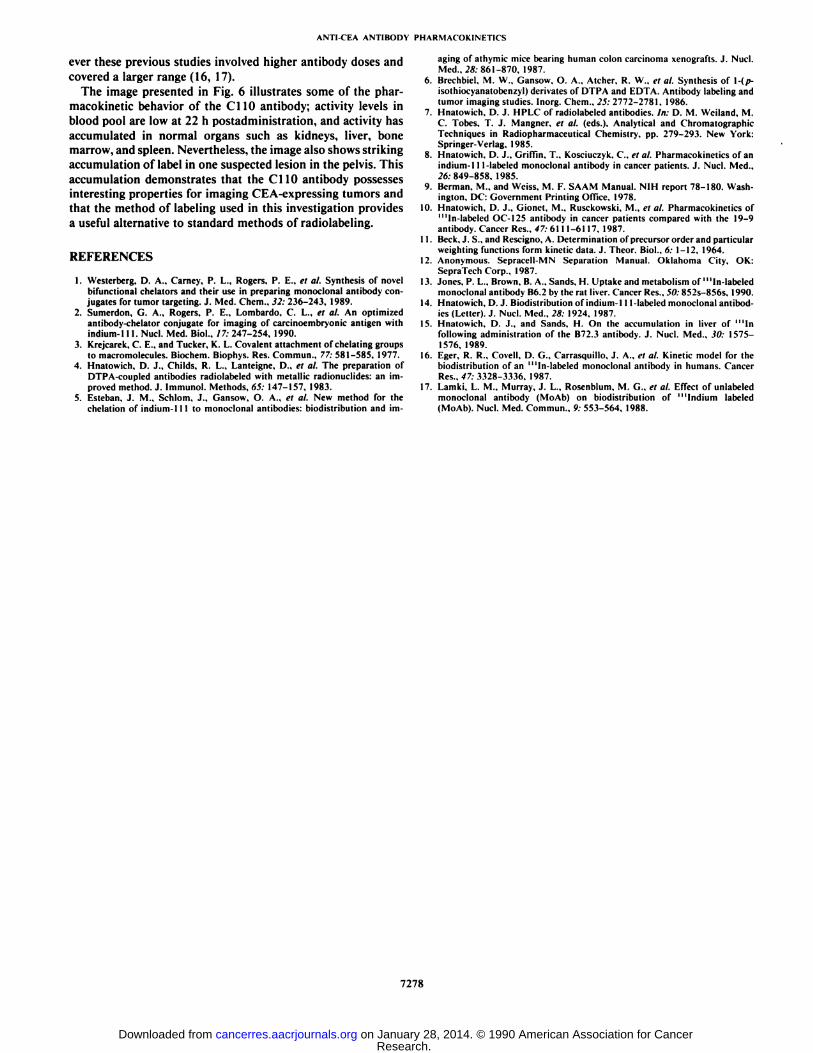
Patient Imaging. The image presented in Fig. 6 is typical ofthose obtained in this investigation. Shown is a posterior wholebody image obtained 22 h postadministration of 20 mg oflabeled antibody (patient 8). Activity is present in kidneys andliver and, to a lesser extent, in bone marrow. An intense focallesion appears in the pelvis that is suspected of being due tometastatic colorectal cancer from a primary removed surgically3 months previously. A detailed description of the imagingresults obtained in these patients will appear elsewhere.4
DISCUSSION
This laboratory has conducted 2 previous pharmacokineticinvestigations of antibodies in patients with the 19-9 (8) andthe OC-125 antibodies (10) radiolabeled with '"In. A compar
ison of these results with that obtained in the present study isinexact even though all 3 antibodies are of the IgGl isotypebecause the 19-9 and OC-125 antibodies were administered asthe F(ab')2 fragment, while the C110 was used as the intact
IgG. Furthermore, only 1 mg of 19-9 and OC-125 antibodywas administered, whereas between 5 and 20 mg of Cl 10 wasused. A final significant difference is the chelator used. Whereasthe 19-9 and OC-125 antibodies were both conjugated withDTPA using the cyclic anhydride, in the present study a derivative of DTPA was attached. Nevertheless, some similaritiesare apparent.
The level of radioactivity in the liver was similar in all 3studies (18, 20, and 12% ID for the Cl 10, 19-9, and OC-125,respectively, at 24 h). However, a higher liver level may beexpected for the intact Cl 10 antibody over that of the F(ab')2
fragments since Fc receptors in the liver are thought to contribute to the accumulation of IgG antibodies in this organ (13).Furthermore, it may have been anticipated that liver levels inthis investigation would have been low (and possibly decreasing)judging from the favorable results obtained in mice with antibodies radiolabeled with '"In via an isothiocyanatobenzyl
DTPA chelator (5). It has been suggested that the decreasedliver levels observed in animals may be due to dissociation of
* R. J. Kinders, S. Stevens, G. M. Hass, J. Daufeldt. J. Jennings. J. Pennoyer.A. Boghosian. and T. W. Griffin. Human immune response to the anti-CEAmurine monoclonal antibody C'110 in a phase I clinical trial, manuscript in
preparation.
7276
Research. on January 28, 2014. © 1990 American Association for Cancercancerres.aacrjournals.org Downloaded from

ANTI-CEA ANTIBODY PHARMAC OKINET1CS
Fig. 6. A posterior whole body image obtained at 22 h after the administrationof'"In-labeled Cl 10 antibody to patient 8. In addition to activity in the kidneys,liver, and bone marrow, an intense focal lesion is evident in suspected metastaticcolorectal cancer in the pelvis.
the thiourea bond with the release from the liver of the intactchelate (14). If so, it is likely that the extent of this phenomenamay be limited in patients such that liver levels are not greatly-
affected (15).Particularly high liver radioactivity levels were observed in
patients 4 and 1' (Table 1). The exceptionally high liver levels
observed in the case of patient 4 are most likely due to antigen-antibody immune complex formation resulting from a highcirculating CEA level with clearance into the liver. Size exclusion HPLC radiochromatograms show that a high molecularweight species formed in serum immediately upon antibodyadministration and persisted as the predominant speciesthroughout this investigation. It is interesting that spleen andkidney levels were unremarkable in this case (0.6 and 0.4%,respectively, at 24 h). The explanation for the high liver levelsobtained in the second administration to patient 1 may also be
the elevated CEA levels, although, in this case, the high liverlevels (and the rapidly clearing serum radioactivity) may be dueto a HAMA response resulting from the first injection of theC110 antibody to this patient 15 months previously. A serumsample from this patient obtained 7 months after the firstantibody administration showed mildly elevated HAMA levelsby two assays. The size exclusion HPLC radiochromatogramsshow that a high molecular weight peak appeared in serumwithin l h of the second antibody administration and becamethe predominant species at 20 h. These 2 cases illustrate theprofound effect that immune complex formation can have onbiodistribution.
The analysis of serum and urine samples in this investigationshowed patterns which had been previously observed in the caseof the 19-9 and OC-125 antibodies. A catabolic radiolabeledspecies with a molecular weight similar to that of labeled freeDTPA appears in urine after approximately 8 h. It is likely thatthis species is generated in the liver since patient 4 exhibitedlevels among the highest urine levels and also had the highestlevel of radioactivity in liver (Fig. 1).
An important feature of this investigation was the determination of the number and concentrations of radiolabeled speciesin a large number of patient sera. As a result, it was possible todetermine the half-life for clearance and the volume of distribution for the radiolabeled antibody itself rather than for themixture of radiolabeled species which makes up the total serumradioactivity. Since these analyses showed that radiolabeledantibody was often the predominant species, the concentrationof this species was determined with more certainty than that ofthe immune complex or catabolite especially in late samples.Because of physical decay and biological clearance, the countingrates of fractions obtained by HPLC analysis of these latesamples were often close to the limit of detection. As such onlyvalues for the radiolabeled antibody are presented in Table 2.It is apparent from the table that, under the assumption ofsingle compartment clearance, the terminal clearance half-timevaried over a considerable range from patient to patient.
The volumes of distribution at steady state are not reportedbecause of uncertainties in these values even in the case ofradiolabeled antibodies. The error inherent in estimating thearea under the clearance curve out to steady state becameunmanageable due to low radioactivity levels in late serumsamples. The initial volume of distribution for the antibody aremore useful, although also subject to uncertainty, in this casebecause the antibody was administered by slow infusion, andthus the initial collection times are not well defined.
Based on the results presented in Table 2, the terminalclearance half-time for the antibody is 22 h. When the identicalanalysis is performed on the total serum radioactivity, this valuebecomes 34 h. The significant difference (P < 0.02) in thesevalues may reflect the contribution to total radioactivity clearance of other species, especially immune complexes. The longerclearance half-life found for total radioactivity over that ofantibody itself reflects the fact that immune complexes andcatabolites were accumulating in blood during the observationperiod. As expected, the plasma volumes calculated by heightand weight and the initial volumes of distribution are in goodagreement, especially for total radioactivity.
One surprising result of this investigation is the lack ofinfluence of antibody dose on pharmacokinetics. Other investigators have reported decreasing liver accumulation and decreased serum clearance with increasing antibody dose. How-
7277
Research. on January 28, 2014. © 1990 American Association for Cancercancerres.aacrjournals.org Downloaded from
ANTI-CEA ANTIBODY PHARMACOKINETICS
ever these previous studies involved higher antibody doses andcovered a larger range (16, 17).
The image presented in Fig. 6 illustrates some of the phar-macokinetic behavior of the C110 antibody; activity levels inblood pool are low at 22 h postadministration, and activity hasaccumulated in normal organs such as kidneys, liver, bonemarrow, and spleen. Nevertheless, the image also shows strikingaccumulation of label in one suspected lesion in the pelvis. Thisaccumulation demonstrates that the C110 antibody possessesinteresting properties for imaging CEA-expressing tumors andthat the method of labeling used in this investigation providesa useful alternative to standard methods of radiolabeling.
REFERENCES
1. \\eslerberg. D. A., Carney, P. L.. Rogers, P. E., el al. Synthesis of novel(»functionalchelators and their use in preparing monoclonal antibody conjugates for tumor targeting. J. Med. Chem., 32: 236-243, 1989.
2. Sumerdon. G. A.. Rogers, P. E., Lombardo. C. L., et al. An optimizedantibody-chelalor conjugate for imaging of carcinoembryonic antigen withindium-l 11. NucÃ.Med. Biol.. 17: 247-254, 1990.
3. Krejcarek. C. E.. and Tucker. K. L. Covalent attachment of chelating groupsto macromolcculcs. Biochem. Biophys. Res. Commun., 77: 581-585, 1977.
4. Hnatowich, D. J., Childs, R. L., Lanteigne, D.. el al. The preparation ofDTPA-coupled antibodies radiolabeled with metallic radionuclides: an improved method. J. Immunol. Methods. 65: 147-157, 1983.
5. Esteban, J. M., Schlom, J., Ganso», O. A., el al. New method for thechelation of indium-l 11 to monoclonal antibodies: biodistribution and im
aging of athymic mice bearing human colon carcinoma xenografts. J. NucÃ.Med.. 28: 861-870. 1987.
6. Brechbiel. M. W., Gansow, O. A., Atcher. R. W., et al. Synthesis of l-(p-isothiocyanatobenzyl) dérivâtesof DTPA and EDTA. Antibody labeling andtumor imaging studies. Inorg. Chem.. 25: 2772-2781. 1986.
7. Hnatowich. D. J. HPLC of radiolabeled antibodies. In: D. M. Weiland. M.C. Tobes, T. J. Mangner. el al. (eds.). Analytical and ChromatographieTechniques in Radiopharmaceutieal Chemistry, pp. 279-293. New York:Springer-Verlag. 1985.
8. Hnatowich, D. J., Griffin, T., Kosciuc/yk, C".,et al. Pharmacokinetics of anindium-l 11-labeled monoclonal antibody in cancer patients. J. NucÃ.Med..-'6:849-858. 1985.
9. Berman. M.. and Weiss. M. F. SAAM Manual. NIH report 78-180. Washington, DC': Government Printing Office. 1978.
10. Hnatowich. D. J., Gionet. M.. Rusckowski. M., el al. Pharmacokinctics of'"In-labeled OC-125 antibody in cancer patients compared with the 19-9antibody. Cancer Res.. 47:6111-6117. 1987.
11. Beck. J. S.. and Rcscigno. A. Determination of precursor order and particularweighting functions form kinetic data. J. Theor. Biol., 6: 1-12, 1964.
12. Anonymous. Sepracell-MN Separation Manual. Oklahoma City. OK:SepraTech Corp.. 1987.
13. Jones. P. L.. Brown. B. A.. Sands. H. Uptake and metabolism of '"In-labeledmonoclonal antibody B6.2 by the rat liver. Cancer Res.. 50: 852s-856s. 1990.
14. Hnatowich. D. J. Biodistribution of indium-l 11-labeled monoclonal antibodies (Letter). J. NucÃ.Med.. 28: 1924. 1987.
15. Hnatowich. D. J.. and Sands. H. On the accumulation in liver of "'Infollowing administration of the B72.3 antibody. J. NucÃ.Med.. 30: 1575-1576. 1989.
16. Eger. R. R.. Covell, D. G.. Carrasquillo, J. A., et al. Kinetic model for thebiodistribution of an '"In-labeled monoclonal antibody in humans. CancerRes.. 47: 3328-3336. 1987.
17. Lamki, L. M., Murray, J. L.. Roscnblum, M. G., et al. Effect of unlabeledmonoclonal antibody (MoAb) on biodistribution of "'Indium labeled(MoAb). NucÃ.Med. Commun., 9: 553-564, 1988.
7278
Research. on January 28, 2014. © 1990 American Association for Cancercancerres.aacrjournals.org Downloaded from
Copyright © 2022 FDOKUMEN