Pharmacokinetic and metabolic effects of American ginseng (Panax quinquefolius) in healthy...
10
BioMed Central Page 1 of 10 (page number not for citation purposes) BMC Complementary and Alternative Medicine Open Access Research article Pharmacokinetic and metabolic effects of American ginseng (Panax quinquefolius) in healthy volunteers receiving the HIV protease inhibitor indinavir Adriana SA Andrade* 1 , Craig Hendrix 2 , Teresa L Parsons 2 , Benjamin Caballero 3 , Chun-Su Yuan 4 , Charles W Flexner 2 , Adrian S Dobs 5 and Todd T Brown 5 Address: 1 Division of Infectious Diseases, The Johns Hopkins University, Baltimore, MD 21287, USA, 2 Division of Clinical Pharmacology, The Johns Hopkins University, Baltimore, MD 21287, USA, 3 Department of Human Nutrition, The Johns Hopkins Bloomberg School of Public Health, Baltimore, MD 21205, USA, 4 Tang Center for Herbal Medicine Research, Department of Anesthesia and Critical Care, Pritzker School of Medicine, University of Chicago, Chicago, IL 60637, USA and 5 Division of Endocrinology and Metabolism, Department of Medicine, Johns Hopkins University, Baltimore, MD 21287, USA Email: Adriana SA Andrade* - [email protected]; Craig Hendrix - [email protected]; Teresa L Parsons - [email protected]; Benjamin Caballero - [email protected]; Chun-Su Yuan - [email protected]; Charles W Flexner - [email protected]; Adrian S Dobs - [email protected]; Todd T Brown - [email protected] * Corresponding author Abstract Background: Complementary and alternative medicine (CAM) use is prevalent among HIV- infected patients to reduce the toxicity of antiretroviral therapy. Ginseng has been used for treatment of hyperglycemia and insulin resistance, a common side effect of some HIV-1 protease inhibitors (PI). However, it is unknown whether American ginseng (AG) can reverse insulin resistance induced by the PI indinavir (IDV), and whether these two agents interact pharmacologically. We evaluated potential pharmacokinetic interactions between IDV and AG, and assessed whether AG improves IDV-induced insulin resistance. Methods: After baseline assessment of insulin sensitivity using the insulin clamp technique, healthy volunteers received IDV 800 mg q8 h for 3 days and then IDV and AG 1g q8h for 14 days. IDV pharmacokinetics and insulin sensitivity were assessed before and after AG co-administration. Results: There was no difference in the area-under the plasma-concentration-time curve after the co-administration of AG, compared to IDV alone (n = 13). Although insulin-stimulated glucose disposal per unit of insulin (M/I) decreased by an average of 14.8 ± 5.9% after 3 days of IDV (from 0.113 ± 0.012 to 0.096 ± 0.014 mg/kgFFM/min per μU/ml of insulin, p = 0.03, n = 11), M/I remained unchanged after co-administration of IDV and AG. Conclusion: IDV decreases insulin sensitivity, which is unaltered by AG co-administration. AG does not significantly affect IDV pharmacokinetics. Published: 19 August 2008 BMC Complementary and Alternative Medicine 2008, 8:50 doi:10.1186/1472-6882-8-50 Received: 8 May 2008 Accepted: 19 August 2008 This article is available from: http://www.biomedcentral.com/1472-6882/8/50 © 2008 Andrade et al; licensee BioMed Central Ltd. This is an Open Access article distributed under the terms of the Creative Commons Attribution License (http://creativecommons.org/licenses/by/2.0 ), which permits unrestricted use, distribution, and reproduction in any medium, provided the original work is properly cited.
-
Upload
independent -
Category
Documents
-
view
0 -
download
0
Transcript of Pharmacokinetic and metabolic effects of American ginseng (Panax quinquefolius) in healthy...
BioMed Central
BMC Complementary and Alternative Medicine
ss
Open AcceResearch articlePharmacokinetic and metabolic effects of American ginseng (Panax quinquefolius) in healthy volunteers receiving the HIV protease inhibitor indinavirAdriana SA Andrade*1, Craig Hendrix2, Teresa L Parsons2, Benjamin Caballero3, Chun-Su Yuan4, Charles W Flexner2, Adrian S Dobs5 and Todd T Brown5Address: 1Division of Infectious Diseases, The Johns Hopkins University, Baltimore, MD 21287, USA, 2Division of Clinical Pharmacology, The Johns Hopkins University, Baltimore, MD 21287, USA, 3Department of Human Nutrition, The Johns Hopkins Bloomberg School of Public Health, Baltimore, MD 21205, USA, 4Tang Center for Herbal Medicine Research, Department of Anesthesia and Critical Care, Pritzker School of Medicine, University of Chicago, Chicago, IL 60637, USA and 5Division of Endocrinology and Metabolism, Department of Medicine, Johns Hopkins University, Baltimore, MD 21287, USA
Email: Adriana SA Andrade* - [email protected]; Craig Hendrix - [email protected]; Teresa L Parsons - [email protected]; Benjamin Caballero - [email protected]; Chun-Su Yuan - [email protected]; Charles W Flexner - [email protected]; Adrian S Dobs - [email protected]; Todd T Brown - [email protected]
* Corresponding author
AbstractBackground: Complementary and alternative medicine (CAM) use is prevalent among HIV-infected patients to reduce the toxicity of antiretroviral therapy. Ginseng has been used fortreatment of hyperglycemia and insulin resistance, a common side effect of some HIV-1 proteaseinhibitors (PI). However, it is unknown whether American ginseng (AG) can reverse insulinresistance induced by the PI indinavir (IDV), and whether these two agents interactpharmacologically. We evaluated potential pharmacokinetic interactions between IDV and AG, andassessed whether AG improves IDV-induced insulin resistance.
Methods: After baseline assessment of insulin sensitivity using the insulin clamp technique, healthyvolunteers received IDV 800 mg q8 h for 3 days and then IDV and AG 1g q8h for 14 days. IDVpharmacokinetics and insulin sensitivity were assessed before and after AG co-administration.
Results: There was no difference in the area-under the plasma-concentration-time curve after theco-administration of AG, compared to IDV alone (n = 13). Although insulin-stimulated glucosedisposal per unit of insulin (M/I) decreased by an average of 14.8 ± 5.9% after 3 days of IDV (from0.113 ± 0.012 to 0.096 ± 0.014 mg/kgFFM/min per μU/ml of insulin, p = 0.03, n = 11), M/I remainedunchanged after co-administration of IDV and AG.
Conclusion: IDV decreases insulin sensitivity, which is unaltered by AG co-administration. AGdoes not significantly affect IDV pharmacokinetics.
Published: 19 August 2008
BMC Complementary and Alternative Medicine 2008, 8:50 doi:10.1186/1472-6882-8-50
Received: 8 May 2008Accepted: 19 August 2008
This article is available from: http://www.biomedcentral.com/1472-6882/8/50
© 2008 Andrade et al; licensee BioMed Central Ltd. This is an Open Access article distributed under the terms of the Creative Commons Attribution License (http://creativecommons.org/licenses/by/2.0), which permits unrestricted use, distribution, and reproduction in any medium, provided the original work is properly cited.
Page 1 of 10(page number not for citation purposes)
BMC Complementary and Alternative Medicine 2008, 8:50 http://www.biomedcentral.com/1472-6882/8/50
BackgroundDietary supplements, herbs, and vitamins are used widelyamong HIV-infected patients [1]. However, the safety orefficacy of many of these therapies has not been formallyevaluated.
These agents are often used among HIV-infected patientsto prevent or treat the adverse effects of antiretroviral ther-apy [2]. Among these adverse effects are disorders of glu-cose metabolism, including insulin resistance[3], glucoseintolerance [4], and diabetes mellitus[3], which weredescribed soon after the introduction of highly activeantiretroviral therapy (HAART)[5]. Although the etiologyof these problems is multifactorial [6], exposure to pro-tease inhibitors (PIs) likely contributes directly. Indinavir(IDV), for example, can worsen insulin sensitivity evenafter a single dose in healthy volunteers[7].
Ginseng is one of the most commonly used herbs in theUnited States [8] and has long been used for the treatmentof hyperglycemia in Traditional Chinese Medicine [9].Experimental evidence for its efficacy comes from severalstudies in both animals and humans [10]. In two smallclinical trials, Panax ginseng [11] and AG [12] given dailyover 8 weeks significantly reduced fasting blood glucoseand hemoglobin A1c in patients with type 2 diabetes.While the mechanisms by which ginseng affects glucosemetabolism have not been fully elucidated, most recentevidence from animal models suggests that improvementin insulin sensitivity may underlie its hypoglycemic effects[13]. It is not known whether ginseng can reverse insulinresistance induced by PIs.
Herbals treatments, like ginseng, are perceived to be safeby patients, but pharmacologic interactions with concom-itant conventional drugs are a major concern. Ginsengwas recently found to reduce the anticoagulant effect ofwarfarin [14]. However, its potential interaction withantiretrovirals has not been investigated. Other herbs,such as St. Johns' wort [15] and garlic [16] dramaticallyreduce the concentrations of PIs in healthy volunteers, inthe case of St. John's wort, through the induction of theircommon metabolic pathway, cytochrome P450 3A4(CYP450 3A4). This could lead to a decrease in effective-ness and potentially result in treatment failure.
The goals of this study were to evaluate potential PK inter-actions between IDV and AG, and assess whether AGimproves IDV-induced insulin resistance.
MethodsStudy ParticipantsHealthy volunteers, male and female, 18–64 years of age,were recruited through newspaper advertisement, flyersand word of mouth. Exclusion criteria included a history
of nephrolithiasis, use of any prescription medications,over the counter medications, or dietary supplementwithin 14 days of enrollment, body mass index (BMI,weight (kg)/height (m2)) ≥ 30 kg/m2, or blood donationwithin 30 days of study enrollment. Prior to enrollment,subjects were required to have a negative HIV-1 antibodytest, normal values for serum creatinine, aspartate ami-notransferase (AST), alanine aminotransferase (ALT), andhemoglobin, and a normal physical examination. Thestudy was approved by the Institutional Review Board atJohns Hopkins University School of Medicine and all par-ticipants signed informed consent.
Study DesignAfter initial screening, participants were admitted to theJohns Hopkins inpatient General Clinical Research Center(GCRC) on three separate occasions over a 21 day periodfor PK and metabolic studies. During the first inpatientvisit, baseline insulin sensitivity was assessed. Subjectswere then discharged with instructions to take IDV 800mg by mouth (Crixivan, Merck and Company, Rahway,New Jersey, USA) every 8 hours (0600, 1400, 2200) on anempty stomach beginning three days before the subse-quent inpatient visit. During the second inpatient visit,insulin sensitivity was reassessed and an 8-hour PK IDVsampling was obtained. Subjects were then instructed totake IDV, as taken previously, and to co-administer encap-sulated AG ground root, 1 gram by mouth every 8 hours.Outpatient safety visits with laboratory assessmentsoccurred 7 days before and 7 days after the final inpatientvisit.
Dried whole-root AG was obtained from a single batchthrough the Wisconsin Ginseng Board (Wausau, Wiscon-sin). The identity of the dried whole root of AG was veri-fied by the Wisconsin ginseng Board prior toencapsulation. Following verification the dried whole-root AG was ground and encapsulated in 500 mg capsulesby the American Pharmaceutical Nutraceutical Laborato-ries (Wausau, Wisconsin) and dispensed by the JohnsHopkins Hospital Investigational Drug service. AG wasadministered under Investigational New Drug Applica-tion # 69,866 granted by the Food and Drug Administra-tion. The dose of AG was selected based on previousstudies investigating the effect of AG on glucose tolerancein humans[12,17]. The dose of IDV was selected accord-ing to its package insert. Following 14 days of IDV andAG, subjects returned to the inpatient GCRC for reassess-ment of insulin sensitivity and IDV PK.
Ginseng AnalysesThe concentrations of six common ginsenosides (Rg1, Re,Rb1, Rc, Rb2, and Rd) were measured, following proce-dures described by Chuang [18], in the Analytical Labora-tory of the Johns Hopkins University Division of Clinical
Page 2 of 10(page number not for citation purposes)
BMC Complementary and Alternative Medicine 2008, 8:50 http://www.biomedcentral.com/1472-6882/8/50
Pharmacology. Briefly, five different capsules were ran-domly selected from five separate bottles. The content ofeach capsule was extracted with 3 mL of ethanol:water(50:50, vol:vol), sonicated, and centrifuged. The pelletwas extracted two more times and the resulting liquid wasfiltered. Samples and ginsenoside standards (Sigma-Aldrich, St. Louis, Missouri) were analyzed by High Per-formance Liquid Chromatography (HPLC) using Beck-man Ultrasphere 5 μm, 250 × 4.6 mm column. Theamount of each ginsenoside assayed in the capsules was asfollows (expressed as percent per gram of ginseng): Rg10.11, Re 1.06, Rb1 0.91, Rc 0.08, Rb2 0.19, Rd 0.08, Totalginsenosides 2.43. The intra- and interassay coefficients ofvariation (CV) were < 10.5% and < 8.5%, respectivelywith > 85% accuracy.
Bioassay for Hypoglycemic ActivityThe hypoglycemic activity of the batch of AG used in thestudy was assessed in diabetic mice prior to initiation ofthe clinical studies, as previously described [13]. Briefly,male ob/ob mice (n = 6) received AG extract dissolved indistilled water by daily intraperitoneal injection at a doseof 250 mg/kg. Fasting serum glucose was measured fromblood obtained from tail samples at baseline, day 5 andday 12. The hypoglycemic response in AG-treated micewas compared to that observed in ob/ob mice who receivedvehicle following the same protocol. Animals were fedrodent chow and housed in environmentally stable condi-tions in metabolic cages. Food consumption and weightwere monitored daily. All animal experiments were car-ried out at the Tang Center for Herbal Medicine Research,University of Chicago in the laboratory of Chun-Su Yuan,MD, PhD.
PK Sampling and AnalysisDuring the second and third inpatient GCRC visits, nineblood samples were collected immediately before admin-istration of the morning dose of IDV and at 0.5,1,2,3,4,5,6, and 8 hours post-dose after an overnight fast.The previous observed dose was given at 2200. PK datawere analyzed using non-compartmental methods usingWinNonlin® (Pharsight, Cary, NC). IDV PK parameterestimates included area under the plasma-concentration-time curve from time 0 to 8 hours after the dose, (AUC0–8
hrs), maximum plasma concentration (Cmax), minimumplasma concentration (Cmin), and time to maximumplasma concentration (Tmax).
Metabolic AssessmentsBody composition, including fat-free mass (FFM), wasassessed at baseline using dual x-ray absorptiometry(Hologic-QDR 4500W, Hologic Co, Bedford, MA, intra-subject CV < 2%). BMI was assessed during each inpatientadmission and was calculated as the weight in kilograms/height in meters squared.
Peripheral sensitivity to glucose was assessed by thehyperinsulinemic euglycemic clamp technique [19]. Sub-jects were admitted to the GCRC in the afternoon prior tothe clamp procedure and maintained on a eucaloric diet(50% carbohydrate, 30% fat, and 20% protein). After anovernight fast, an intravenous catheter was placed in theantecubital vein for infusion of glucose and insulin. A sec-ond catheter for blood sampling was inserted in a dorsalvein of the hand in a retrograde fashion. The hand wasplaced into a 50°C warming box to "arterialize" the sam-ples [20]. After baseline blood samples were obtained, aprimed, continuous infusion of regular insulin (40 mU/m2/min) was started, followed by a constant infusion of20% dextrose at a variable rate, based on 5-minute meas-urements of blood glucose, in order to maintain bloodglucose concentrations at each subject's baseline level ±5%. Infusion continued for a total of 180 minutes.
Glucose utilization (M) was calculated as the average glu-cose infusion rate (milligrams of glucose/kilograms ofFFM/minute) during the last 60 minutes of the infusion(i.e. steady state portion). FFM was derived from the base-line evaluation by dual energy X-ray absorptiometry(DXA).
During the final 60 minutes of the insulin clamp proce-dure, samples were obtained at 10 minutes intervals todetermine insulin concentrations. M divided by the aver-age insulin concentration in the final 60 minutes (I),which represents the amount of glucose metabolized perunit of insulin, was used as the primary measure of insulinsensitivity. During the second and third inpatient visits,the morning dose of IDV (and AG for the third inpatientvisit) was given immediately prior to the beginning of theinsulin clamp procedure. Fasting serum glucose andhomeostasis model assessment of insulin resistance(HOMA-IR), a widely used marker of insulin sensitivity[21], were assessed prior to the insulin clamp procedure ateach of the inpatient study visits.
The serum glucose concentration during the final 60 min-utes of the clamp was 89.8 ± 1.0 mg/dL which was 97.8 ±0.3% of the targeted glucose concentration with a coeffi-cient of variation of 4.2 ± 0.3% (n = 32 clamp studies).The average insulin concentration during the final 60minutes of the infusion was 69.2 ± 1.7 μU/mL.
MeasurementsSerum glucose was measured using the glucose oxidasemethod (Beckman Instruments, Fullerton, CA). Seruminsulin concentrations were determined by enzymeimmunoassay, using a TOSOH 1800 (Tosoh Corporation,Tokyo, Japan). Intra-assay and inter-assay CVs range from1.4–2.3% and 5–6%, respectively. IDV concentrationswere measured by HPLC-mass spectrometry. The intra-
Page 3 of 10(page number not for citation purposes)
BMC Complementary and Alternative Medicine 2008, 8:50 http://www.biomedcentral.com/1472-6882/8/50
and inter-assay CVs were < 9% and < 8%, respectively with> 94% accuracy.
Statistical CalculationsPaired comparisons between outcome measures obtainedduring the first and second inpatients visits (i.e. Baselinevs. IDV Condition) and during the second and third inpa-tient visits (IDV vs IDV + AG) were made using a paired t-test. Univariate relationships between continuous varia-bles were assessed with linear regression. All data are pre-sented as mean ± standard error of the mean (SEM).Differences were regarded as statistically significant if p <0.05. All data analysis was performed using Stata (Version8.1, Stata Corporation, College Station, TX). Pharmacoki-netic parameter estimates were summarized using geo-metric means with 95% confidence intervals; valuesobtained with and without AG were compared using ageometric mean ratio and 90% confidence interval.
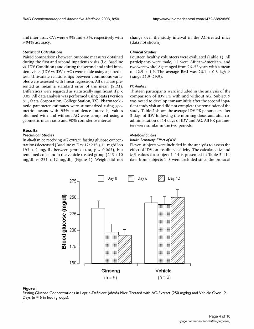
ResultsPreclinical StudiesIn ob/ob mice receiving AG extract, fasting glucose concen-trations decreased (Baseline vs Day 12; 235 ± 11 mg/dL vs193 ± 9 mg/dL, between group t-test, p = 0.003), butremained constant in the vehicle-treated group (243 ± 10mg/dL vs 251 ± 12 mg/dL) (Figure 1). Weight did not
change over the study interval in the AG-treated mice(data not shown).
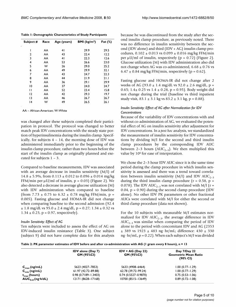
Clinical StudiesFourteen healthy volunteers were evaluated (Table 1). Allparticipants were male, 12 were African-American, andtwo were white. Age ranged from 26–53 years with a meanof 42.9 ± 1.9. The average BMI was 26.1 ± 0.8 kg/m2
(range 21.9–29.9).
PK AnalysisThirteen participants were included in the analysis of thecomparison of IDV PK with and without AG. Subject 9was noted to develop transaminitis after the second inpa-tient study visit and did not complete the remainder of thestudy. Table 2 shows the average IDV PK parameters after3 days of IDV following the morning dose, and after co-administration of 14 days of IDV and AG. All PK parame-ters were similar in the two periods.
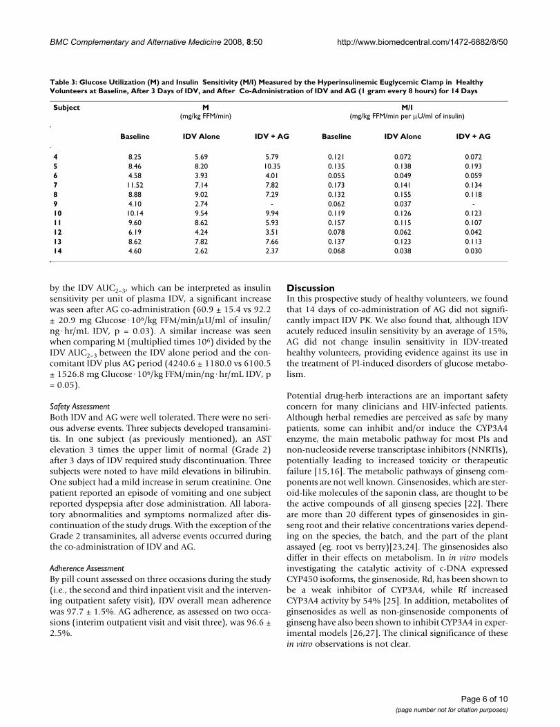
Metabolic StudiesInsulin Sensitivity: Effect of IDVEleven subjects were included in the analysis to assess theeffect of IDV on insulin sensitivity. The calculated M andM/I values for subject 4–14 is presented in Table 3. Thedata from subjects 1–3 were excluded since the protocol
Fasting Glucose Concentrations in Leptin-Deficient (ob/ob) Mice Treated with AG-Extract (250 mg/kg) and Vehicle Over 12 Days (n = 6 in both groups)Figure 1Fasting Glucose Concentrations in Leptin-Deficient (ob/ob) Mice Treated with AG-Extract (250 mg/kg) and Vehicle Over 12 Days (n = 6 in both groups).
Page 4 of 10(page number not for citation purposes)
BMC Complementary and Alternative Medicine 2008, 8:50 http://www.biomedcentral.com/1472-6882/8/50
was changed after these subjects completed their partici-pation in protocol. The protocol was changed to bettermatch peak IDV concentrations with the steady state por-tion of hyperinsulinemia during the insulin clamp. Specif-ically, for subjects 4 – 14, the morning dose of IDV wasadministered immediately prior to the beginning of theinsulin clamp procedure, rather than two hours before thestart of the insulin clamp as originally planned and exe-cuted for subjects 1 – 3.
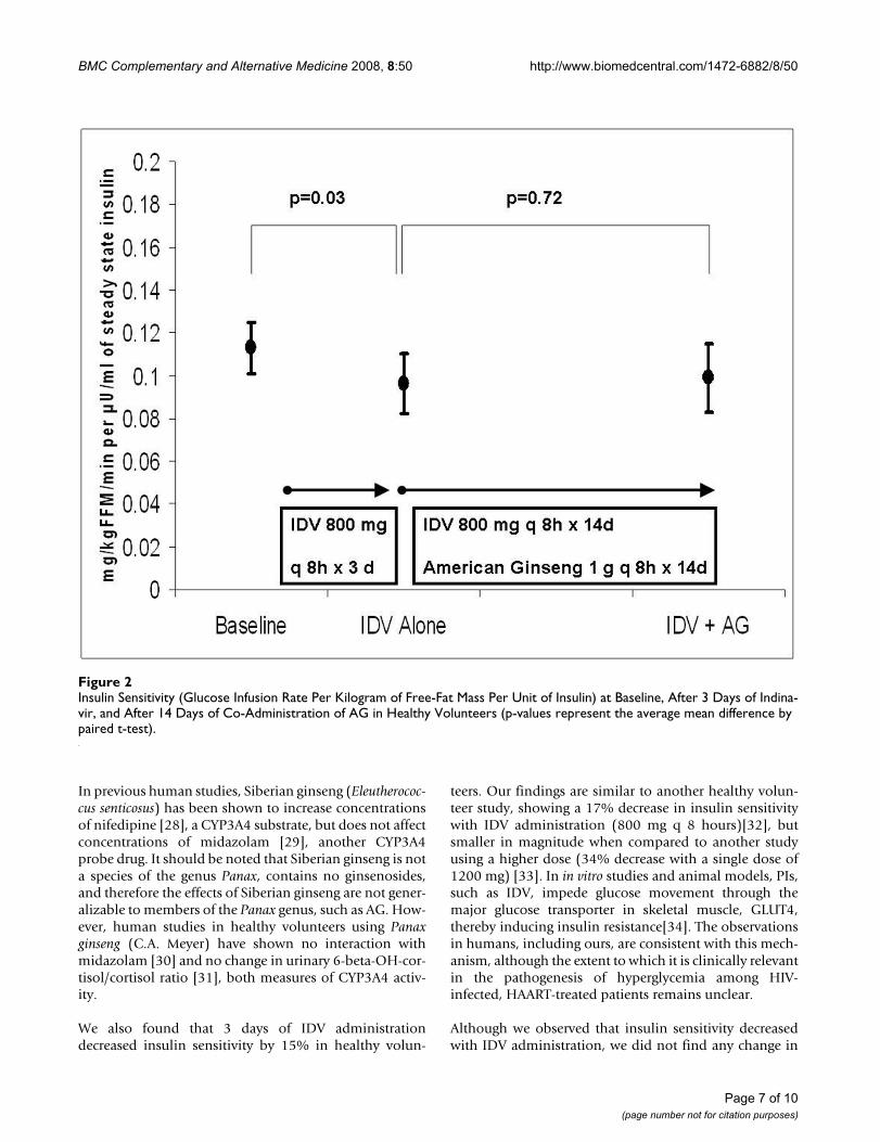
Compared to baseline measurements, IDV was associatedwith an average decrease in insulin sensitivity (M/I) of14.8 ± 5.9%, from 0.113 ± 0.012 to 0.096 ± 0.014 mg/kgFFM/min per μU/ml of insulin, p = 0.03) (Figure 2). Wealso detected a decrease in average glucose utilization (M)with IDV administration when compared to baseline(from 7.73 ± 0.75 to 6.32 ± 0.78 mg/kg FFM/min, p =0.005). Fasting glucose and HOMA-IR did not changewhen comparing baseline to the second admission (92.4± 1.8 mg/dL vs 95.0 ± 2.4 mg/dL, p = 0.27; 1.34 ± 0.32 vs1.34 ± 0.23, p = 0.97, respectively).
Insulin Sensitivity: Effect of AGTen subjects were included to assess the effect of AG onIDV-induced insulin resistance (Table 3). One subject(subject 9) did not have complete data for this analysis
because he was discontinued from the study after the sec-ond insulin clamp procedure, as previously noted. Therewas no difference in insulin sensitivity between the sec-ond (IDV alone) and third (IDV + AG) insulin clamp pro-cedures, 0.102 ± 0.013 vs 0.099 ± 0.016 mg/kg FFM/minper μU/ml of insulin, respectively (p = 0.72) (Figure 2).Glucose utilization (M) with IDV administration also didnot change when AG was co-administered, 6.68 ± 0.76 vs6.47 ± 0.84 mg/kg FFM/min, respectively (p = 0.62).
Fasting glucose and HOMA-IR did not change after 2weeks of AG (93.0 ± 1.4 mg/dL vs 92.0 ± 2.6 mg/dL, p =0.65; 1.4± 0.25 vs 1.4 ± 0.28, p = 0.95). Body weight didnot change during the trial (baseline vs third inpatientstudy visit, 83.1 ± 3.1 kg vs 83.2 ± 3.1 kg, p = 0.88).
Insulin Sensitivity: Effect of AG after Normalization for IDV ConcentrationsBecause of the variability of IDV concentrations with andwithout co-administration of AG, we evaluated the poten-tial effect of AG on insulin sensitivity after adjustment forIDV concentrations. In a post hoc analysis, we standardizedthe measurement of insulin sensitivity for IDV concentra-tions by dividing M/I for the second and third insulinclamp procedures by the corresponding IDV AUCbetween 2–3 hours (AUC2–3). We then multiplied thisvalue by 106 for ease of interpretation.
We chose the 2–3 hour IDV AUC since it is the same timeperiod during the clamp procedure in which insulin sen-sitivity is assessed and there was a trend toward correla-tion between insulin sensitivity (M/I) and IDV AUC2–3during the third insulin clamp procedure (r = 0.58, p =0.078). The IDV AUC2–3 was not correlated with M/I (r =0.04, p = 0.90) during the second clamp procedure (IDValone). No other IDV PK parameters or other fractionalAUCs were correlated with M/I for either the second orthird clamp procedure (data not shown).
For the 10 subjects with measurable M/I estimates nor-malized for IDV AUC2–3, the average difference in IDVAUC2–3 was similar when comparing the period of IDValone to the period with concomitant IDV and AG (2353± 389 vs 1923 ± 483 ng·hr/mL; difference: 430 ± 330ng·hr/mL, p = 0.22). When each subject's M/I was divided
Table 2: PK parameter estimates of IDV before and after co-administration with AG (1 gram every 8 hours), n = 13
IDV alone (Day 7) GM (95%CI)
IDV + AG (Day 22) GM (95%CI)
Day 7/Day 22 Geometric Mean Ratio
(90% CI)
Cmax (ng/mL) 5623 (4027–7853) 5633 (4908–6464) 1.00 (0.77–1.29)Cmin (ng/mL) 61.97 (42.72–89.88) 62.78 (39.72–99.24) 1.00 (0.77–1.29)tmax (hours) 0.98 (0.7189–1.3403) 0.74 (0.5527–0.9870) 0.75 (0.53–1.06)AUC0–8 (ng·h/dL) 12171 (8628–17168) 10700 (8515–13649) 0.89 (0.72–1.08)
Table 1: Demographic Characteristics of Study Participants
Subject # Race Age (years) BMI (kg/m2) Fat (%)
1 AA 41 29.9 29.52 AA 43 22.4 12.23 AA 41 22.3 12.64 AA 53 26.6 23.05 W 26 29.0 25.26 AA 49 29.9 32.17 AA 43 24.7 22.38 AA 44 21.9 21.19 AA 36 29.1 29.910 AA 37 24.0 24.711 AA 52 23.4 15.812 AA 42 29.3 19.713 AA 46 26.7 26.714 W 49 26.5 26.1
AA – African-American; W-White
Page 5 of 10(page number not for citation purposes)
BMC Complementary and Alternative Medicine 2008, 8:50 http://www.biomedcentral.com/1472-6882/8/50
by the IDV AUC2–3, which can be interpreted as insulinsensitivity per unit of plasma IDV, a significant increasewas seen after AG co-administration (60.9 ± 15.4 vs 92.2± 20.9 mg Glucose·106/kg FFM/min/μU/ml of insulin/ng·hr/mL IDV, p = 0.03). A similar increase was seenwhen comparing M (multiplied times 106) divided by theIDV AUC2–3 between the IDV alone period and the con-comitant IDV plus AG period (4240.6 ± 1180.0 vs 6100.5± 1526.8 mg Glucose·106/kg FFM/min/ng·hr/mL IDV, p= 0.05).
Safety AssessmentBoth IDV and AG were well tolerated. There were no seri-ous adverse events. Three subjects developed transamini-tis. In one subject (as previously mentioned), an ASTelevation 3 times the upper limit of normal (Grade 2)after 3 days of IDV required study discontinuation. Threesubjects were noted to have mild elevations in bilirubin.One subject had a mild increase in serum creatinine. Onepatient reported an episode of vomiting and one subjectreported dyspepsia after dose administration. All labora-tory abnormalities and symptoms normalized after dis-continuation of the study drugs. With the exception of theGrade 2 transaminites, all adverse events occurred duringthe co-administration of IDV and AG.
Adherence AssessmentBy pill count assessed on three occasions during the study(i.e., the second and third inpatient visit and the interven-ing outpatient safety visit), IDV overall mean adherencewas 97.7 ± 1.5%. AG adherence, as assessed on two occa-sions (interim outpatient visit and visit three), was 96.6 ±2.5%.
DiscussionIn this prospective study of healthy volunteers, we foundthat 14 days of co-administration of AG did not signifi-cantly impact IDV PK. We also found that, although IDVacutely reduced insulin sensitivity by an average of 15%,AG did not change insulin sensitivity in IDV-treatedhealthy volunteers, providing evidence against its use inthe treatment of PI-induced disorders of glucose metabo-lism.
Potential drug-herb interactions are an important safetyconcern for many clinicians and HIV-infected patients.Although herbal remedies are perceived as safe by manypatients, some can inhibit and/or induce the CYP3A4enzyme, the main metabolic pathway for most PIs andnon-nucleoside reverse transcriptase inhibitors (NNRTIs),potentially leading to increased toxicity or therapeuticfailure [15,16]. The metabolic pathways of ginseng com-ponents are not well known. Ginsenosides, which are ster-oid-like molecules of the saponin class, are thought to bethe active compounds of all ginseng species [22]. Thereare more than 20 different types of ginsenosides in gin-seng root and their relative concentrations varies depend-ing on the species, the batch, and the part of the plantassayed (eg. root vs berry)[23,24]. The ginsenosides alsodiffer in their effects on metabolism. In in vitro modelsinvestigating the catalytic activity of c-DNA expressedCYP450 isoforms, the ginsenoside, Rd, has been shown tobe a weak inhibitor of CYP3A4, while Rf increasedCYP3A4 activity by 54% [25]. In addition, metabolites ofginsenosides as well as non-ginsenoside components ofginseng have also been shown to inhibit CYP3A4 in exper-imental models [26,27]. The clinical significance of thesein vitro observations is not clear.
Table 3: Glucose Utilization (M) and Insulin Sensitivity (M/I) Measured by the Hyperinsulinemic Euglycemic Clamp in Healthy Volunteers at Baseline, After 3 Days of IDV, and After Co-Administration of IDV and AG (1 gram every 8 hours) for 14 Days
Subject M (mg/kg FFM/min)
M/I (mg/kg FFM/min per μU/ml of insulin)
Baseline IDV Alone IDV + AG Baseline IDV Alone IDV + AG
4 8.25 5.69 5.79 0.121 0.072 0.0725 8.46 8.20 10.35 0.135 0.138 0.1936 4.58 3.93 4.01 0.055 0.049 0.0597 11.52 7.14 7.82 0.173 0.141 0.1348 8.88 9.02 7.29 0.132 0.155 0.1189 4.10 2.74 - 0.062 0.037 -10 10.14 9.54 9.94 0.119 0.126 0.12311 9.60 8.62 5.93 0.157 0.115 0.10712 6.19 4.24 3.51 0.078 0.062 0.04213 8.62 7.82 7.66 0.137 0.123 0.11314 4.60 2.62 2.37 0.068 0.038 0.030
Page 6 of 10(page number not for citation purposes)
BMC Complementary and Alternative Medicine 2008, 8:50 http://www.biomedcentral.com/1472-6882/8/50
In previous human studies, Siberian ginseng (Eleutherococ-cus senticosus) has been shown to increase concentrationsof nifedipine [28], a CYP3A4 substrate, but does not affectconcentrations of midazolam [29], another CYP3A4probe drug. It should be noted that Siberian ginseng is nota species of the genus Panax, contains no ginsenosides,and therefore the effects of Siberian ginseng are not gener-alizable to members of the Panax genus, such as AG. How-ever, human studies in healthy volunteers using Panaxginseng (C.A. Meyer) have shown no interaction withmidazolam [30] and no change in urinary 6-beta-OH-cor-tisol/cortisol ratio [31], both measures of CYP3A4 activ-ity.
We also found that 3 days of IDV administrationdecreased insulin sensitivity by 15% in healthy volun-
teers. Our findings are similar to another healthy volun-teer study, showing a 17% decrease in insulin sensitivitywith IDV administration (800 mg q 8 hours)[32], butsmaller in magnitude when compared to another studyusing a higher dose (34% decrease with a single dose of1200 mg) [33]. In in vitro studies and animal models, PIs,such as IDV, impede glucose movement through themajor glucose transporter in skeletal muscle, GLUT4,thereby inducing insulin resistance[34]. The observationsin humans, including ours, are consistent with this mech-anism, although the extent to which it is clinically relevantin the pathogenesis of hyperglycemia among HIV-infected, HAART-treated patients remains unclear.
Although we observed that insulin sensitivity decreasedwith IDV administration, we did not find any change in
Insulin Sensitivity (Glucose Infusion Rate Per Kilogram of Free-Fat Mass Per Unit of Insulin) at Baseline, AfterFigure 2Insulin Sensitivity (Glucose Infusion Rate Per Kilogram of Free-Fat Mass Per Unit of Insulin) at Baseline, After 3 Days of Indina-vir, and After 14 Days of Co-Administration of AG in Healthy Volunteers (p-values represent the average mean difference by paired t-test).
Page 7 of 10(page number not for citation purposes)
BMC Complementary and Alternative Medicine 2008, 8:50 http://www.biomedcentral.com/1472-6882/8/50
insulin sensitivity with co-administration of AG. In previ-ous studies, Vuksan, et al. showed that AG administrationimmediately prior to an oral glucose tolerance test wasassociated with 20% decrease in glucose AUC in bothpatients with type 2 diabetes mellitus and healthy volun-teers [35], but this effect appears to be dependent on thebatch of ginseng used [23]. To rule out an inert batch ofAG, we conducted a bioassay of our batch using an ob/obmouse model and a hypoglycemic effect was observed,suggesting that our batch possessed some active compo-nents.
The mechanism underlying the previously observedeffects of AG on glucose metabolism remains unknownand may be multifactorial. The modulation of digestionand enhancement of insulin secretion have been pro-posed based on findings in some animals models [36,37],but ginseng administration has also been shown toimprove insulin sensitivity in ob/ob mice by more thantwo-fold [13]. It has been postulated that ginsenosidesmay intercalate into the cellular plasma membrane, thusmodulating the cell signaling, electrolyte transport, andreceptor binding [22]. It is not known whether this effectcould also modulate the effect of IDV on the glucose fluxthrough the GLUT4 transporter.
Another factor that may have contributed to the lack ofeffect is the variability of IDV plasma concentrationsbetween the two clamps, which has been previouslyobserved [38]. The effect of AG on IDV-induced insulinresistance would be best determined if IDV plasma con-centrations were the same when it was given alone andwhen it was co-administered with AG. For this reason, wenormalized the measure of insulin sensitivity for drugconcentration in a post hoc analysis, by dividing M/I by theIDV concentration during the steady state portion of theclamp and found that this ratio was significantly higher inthe IDV + AG condition.
Although the interpretation of ratios can be challenging[39] and should be done with caution, one explanation ofthis finding is that insulin sensitivity per unit of IDV con-centration modestly improved with the administration ofAG. Given the known effect of IDV on GLUT4 blockade,AG components may work directly at the plasma mem-brane to allow glucose to enter cells, either by improvingglucose movement through GLUT4, increasing the con-centration of GLUT4 in the plasma membrane, or facili-tating glucose entry through alternate pathways. Anotherpossible mechanism of AGs effect may be through theenhancement of local blood flow. Increased capillaryrecruitment mediated though nitric oxide is an importantmechanism of increasing glucose and insulin delivery toskeletal muscle [40,41]. IDV has been shown to causeendothelial dysfunction in healthy volunteers, likely by
reducing nitric oxide production [42]. Ginseng specieshave been shown in experimental models to increasenitric oxide production [43,44] and therefore, may effectinsulin sensitivity by this mechanism. This hypothesisrequires further investigation.
Our study had additional limitations which may haveimplications for the generalizability of its findings.Although both men and women were eligible for partici-pation, only men enrolled. In previous studies, inducibil-ity of CYP3A4 by herbal compounds has shown aninteraction by sex, whereby women showed a 74%increased effect of St. John's wort on CYP3A4 activityusing a midazolam probe compared to men [45]. In thesame study, however, Panax ginseng showed no effect onthe midazolam metabolism in either men or women. Fur-ther PK interaction studies in women may be necessary. Inaddition, our population was mostly African-American.Although there is evidence for genotypic differences inCYP3A4 between Caucasians and African-Americans, phe-notypic differences in CYP3A4 activity have not beenfound [46]. Finally, although we quantified the amount ofcommon ginsenosides, there is substantial variability ofcomposition even within species. As a result, as is the casein all botanical research using non-standardized complexcompounds whose active component is unknown, theextent to which our findings are generalizable to other AGproducts is not clear.
In conclusion, this study in healthy volunteers found noevidence of a significant PK interaction between AG andIDV. Because CYP3A4 is the principal metabolic pathwayof most HIV PIs and NNRTIs, significant drug-herb inter-actions with these antiretrovirals are unlikely. The meta-bolic effects of AG are more difficult to interpret.Although IDV significantly reduced insulin sensitivity,there was no change in insulin sensitivity with AG admin-istration. However, after normalization for IDV concen-trations, insulin sensitivity improved in the AG condition,leaving open the possibility of a modest effect on glucosemetabolism. Further studies to clarify this questionshould attempt to reduce variability in IDV plasma con-centrations, either by giving multiple doses of IDV atshorter intervals to maintain plasma IDV concentrationsconstant while insulin sensitivity is assessed, or using anIDV regimen boosted with ritonavir. Until these issues areresolved, there is no clear scientific basis to recommendAG for the treatment of glucose abnormalities in HIV-infected patients.
Competing interestsDrs. Dobs, Parsons, Caballero, and Yuan declared thatthey have no competing interests. Dr. Hendrix receivedresearch support from Merck and Company. Dr. Andradeserved as a consultant for Abbott Laboratories. Dr. Brown
Page 8 of 10(page number not for citation purposes)
BMC Complementary and Alternative Medicine 2008, 8:50 http://www.biomedcentral.com/1472-6882/8/50
served as a consultant or has received research supportfrom Merck and Company, Abbott Laboratories, ReliantPharmaceuticals, Glaxo Smith Kline, EMD-Serono, andTheratechnologies. Dr. Flexner served on a Scientific Advi-sory Board for Merck and Company.
Authors' contributionsTTB performed the insulin clamps. C–SY carried out theanimal studies. TLP carried out the measurements of gin-senoside and IDV plasma concentrations. TTB and ASAAdrafted the manuscript. ASAA, TTB, CWF, CH C–SY, ASDand BC participated in the design of the study. TTB, ASAA,and CH performed the statistical analysis. ASAA, TTB,ASD, CH and BC participated in the data interpretation.TTB and ASAA conceived of the study and participated inits coordination. All authors read and approved the finalmanuscript.
AcknowledgementsWe would like to thank Chryssanthi Stylianopoulos, PhD and Margia Arguello for their technical expertise in measuring glucose during the insu-lin clamp procedure and Alice Ryan, PhD for her guidance with the insulin clamp procedure. We are thankful to Andrea Weiss for dispensing study drugs and preparation of solutions used in the insulin clamp procedure. Finally, we also would like to acknowledge Dr Angela Kashuba for the anal-ysis of the indinavir assays. Her work is supported by #9P30 AI 50410-04, University of North Carolina at Chapel Hill Center for AIDS Research (CFAR). This work was supported by a National Center for Complemen-tary and Alternative Medicine supplement to the National Cancer Institute Grant # P30CA06973, the National Center for Complementary and Alter-native Medicine K23 AT002862-01 (TTB), and the Johns Hopkins Univer-sity School of Medicine General Clinical Research Grant M01-RR00052 from the National Center for Research Resources/National Institutes of Health.
References1. Standish LJ, Greene KB, Bain S, Reeves C, Sanders F, Wines RC, Turet
P, Kim JG, Calabrese C: Alternative medicine use in HIV-posi-tive men and women: demographics, utilization patternsand health status. AIDS Care 2001, 13:197-208.
2. Furler MD, Einarson TR, Walmsley S, Millson M, Bendayan R: Use ofcomplementary and alternative medicine by HIV-infectedoutpatients in Ontario, Canada. AIDS Patient Care STDS 2003,17:155-168.
3. Brown TT, Cole SR, Li X, Kingsley LA, Palella FJ, Riddler SA, VisscherBR, Margolick JB, Dobs AS: Antiretroviral therapy and the prev-alence and incidence of diabetes mellitus in the multicenterAIDS cohort study. Arch Intern Med 2005, 165:1179-1184.
4. Hadigan C, Meigs JB, Corcoran C, Rietschel P, Piecuch S, Basgoz N,Davis B, Sax P, Stanley T, Wilson PW, D'Agostino RB, Grinspoon S:Metabolic abnormalities and cardiovascular disease risk fac-tors in adults with human immunodeficiency virus infectionand lipodystrophy. Clin Infect Dis 2001, 32:130-139.
5. Nightingale SL: From the Food and Drug Administration. JAMA1997, 278:379.
6. Brown TT, Cofrancesco J: Metabolic Abnormalities in HIV-infected Patients: An Update. Curr Infect Dis Rep 2006,8:497-504.
7. Noor MA, Seneviratne T, Aweeka FT, Lo JC, Schwarz JM, Mulligan K,Schambelan M, Grunfeld C: Indinavir acutely inhibits insulin-stimulated glucose disposal in humans: a randomized, pla-cebo-controlled study. AIDS 2002, 16:F1-F8.
8. Bressler R: Herb-drug interactions: interactions between gin-seng and prescription medications. Geriatrics 2005, 60:16-17.
9. Yanchi L: The Essential Book of Traditional Chinese Medicine New York,Columbia University Press; 1988:127.
10. Xie JT, Mchendale S, Yuan CS: Ginseng and diabetes. Am J ChinMed 2005, 33:397-404.
11. Sotaniemi EA, Haapakoski E, Rautio A: Ginseng therapy in non-insulin-dependent diabetic patients. Diabetes Care 1995,18:1373-1375.
12. Vuksan V, Sievenpiper JL, Xu Z, Wong EY, Jenkins AL, Beljan-Zdravk-ovic U, Leiter LA, Josse RG, Stavro MP: Konjac-Mannan andAmerican ginsing: emerging alternative therapies for type 2diabetes mellitus. J Am Coll Nutr 2001, 20:370S-380S.
13. Attele AS, Zhou YP, Xie JT, Wu JA, Zhang L, Dey L, Pugh W, Rue PA,Polonsky KS, Yuan CS: Antidiabetic effects of Panax ginsengberry extract and the identification of an effective compo-nent. Diabetes 2002, 51:1851-1858.
14. Yuan CS, Wei G, Dey L, Karrison T, Nahlik L, Maleckar S, Kasza K,Ang-Lee M, Moss J: Brief communication: American ginsengreduces warfarin's effect in healthy patients: a randomized,controlled Trial. Ann Intern Med 2004, 141:23-27.
15. Piscitelli SC, Burstein AH, Chaitt D, Alfaro RM, Falloon J: Indinavirconcentrations and St John's wort. Lancet 2000, 355:547-548.
16. Piscitelli SC, Burstein AH, Welden N, Gallicano KD, Falloon J: Theeffect of garlic supplements on the pharmacokinetics ofsaquinavir. Clin Infect Dis 2002, 34:234-238.
17. Vuksan V, Sievenpiper JL, Wong J, Xu Z, Beljan-Zdravkovic U, Arna-son JT, Assinewe V, Stavro MP, Jenkins AL, Leiter LA, Francis T:American ginseng (Panax quinquefolius L.) attenuates post-prandial glycemia in a time-dependent but not dose-depend-ent manner in healthy individuals. Am J Clin Nutr 2001,73:753-758.
18. Chuang WC, Wu HK, Sheu SJ, Chiou SH, Chang HC, Chen YP: Acomparative study on commercial samples of ginseng radix.Planta Med 1995, 61:459-465.
19. DeFronzo RA, Tobin JD, Andres R: Glucose clamp technique: amethod for quantifying insulin secretion and resistance. AmJ Physiol 1979, 237:E214-E223.
20. McGuire EA, Helderman JH, Tobin JD, Andres R, Berman M: Effectsof arterial versus venous sampling on analysis of glucosekinetics in man. J Appl Physiol 1976, 41:565-573.
21. Matthews DR, Hosker JP, Rudenski AS, Naylor BA, Treacher DF,Turner RC: Homeostasis model assessment: insulin resistanceand beta-cell function from fasting plasma glucose and insu-lin concentrations in man. Diabetologia 1985, 28:412-419.
22. Attele AS, Wu JA, Yuan CS: Ginseng pharmacology: multipleconstituents and multiple actions. Biochem Pharmacol 1999,58:1685-1693.
23. Sievenpiper JL, Arnason JT, Leiter LA, Vuksan V: Variable effects ofAmerican ginseng: a batch of American ginseng (Panax quin-quefolius L.) with a depressed ginsenoside profile does notaffect postprandial glycemia. Eur J Clin Nutr 2003, 57:243-248.
24. Dey L, Xie JT, Wang A, Wu J, Maleckar SA, Yuan CS: Anti-hyperg-lycemic effects of ginseng: comparison between root andberry. Phytomedicine 2003, 10:600-605.
25. Henderson GL, Harkey MR, Gershwin ME, Hackman RM, Stern JS,Stresser DM: Effects of ginseng components on c-DNA-expressed cytochrome P450 enzyme catalytic activity. LifeSci 1999, 65:L209-L214.
26. Liu Y, Zhang JW, Li W, Ma H, Sun J, Deng MC, Yang L: Ginsenosidemetabolites, rather than naturally occurring ginsenosides,lead to inhibition of human cytochrome P450 enzymes. Tox-icol Sci 2006, 91:356-364.
27. Patel J, Buddha B, Dey S, Pal D, Mitra AK: In vitro interaction of theHIV protease inhibitor ritonavir with herbal constituents:changes in P-gp and CYP3A4 activity. Am J Ther 2004,11:262-277.
28. Smith M, Lin KM, Zheng YP: An open trial of nifedipine-herbinteractions: nifedipine with St. John's wort, ginseng orginkgo biloba. Clin Pharm Therap 2001, 9:86.
29. Donovan JL, DeVane CL, Chavin KD, Taylor RM, Markowitz JS: Sibe-rian ginseng (Eleutheroccus senticosus) effects on CYP2D6and CYP3A4 activity in normal volunteers. Drug Metab Dispos2003, 31:519-522.
30. Gurley BJ, Gardner SF, Hubbard MA, Williams DK, Gentry WB, CuiY, Ang CY: Clinical assessment of effects of botanical supple-mentation on cytochrome P450 phenotypes in the elderly:
Page 9 of 10(page number not for citation purposes)
http://www.ncbi.nlm.nih.gov/entrez/query.fcgi?cmd=Retrieve&db=PubMed&dopt=Abstract&list_uids=9244318
http://www.ncbi.nlm.nih.gov/entrez/query.fcgi?cmd=Retrieve&db=PubMed&dopt=Abstract&list_uids=8721940
http://www.ncbi.nlm.nih.gov/entrez/query.fcgi?cmd=Retrieve&db=PubMed&dopt=Abstract&list_uids=8721940
http://www.ncbi.nlm.nih.gov/entrez/query.fcgi?cmd=Retrieve&db=PubMed&dopt=Abstract&list_uids=3899825
http://www.ncbi.nlm.nih.gov/entrez/query.fcgi?cmd=Retrieve&db=PubMed&dopt=Abstract&list_uids=3899825
BMC Complementary and Alternative Medicine 2008, 8:50 http://www.biomedcentral.com/1472-6882/8/50
Publish with BioMed Central and every scientist can read your work free of charge
"BioMed Central will be the most significant development for disseminating the results of biomedical research in our lifetime."
Sir Paul Nurse, Cancer Research UK
Your research papers will be:
available free of charge to the entire biomedical community
peer reviewed and published immediately upon acceptance
cited in PubMed and archived on PubMed Central
yours — you keep the copyright
Submit your manuscript here:http://www.biomedcentral.com/info/publishing_adv.asp
BioMedcentral
St John's wort, garlic oil, Panax ginseng and Ginkgo biloba.Drugs Aging 2005, 22:525-539.
31. Anderson GD, Rosito G, Mohustsy MA, Elmer GW: Drug interac-tion potential of soy extract and Panax ginseng. J Clin Pharma-col 2003, 43:643-648.
32. Noor MA, Lo JC, Mulligan K, Schwarz JM, Halvorsen RA, SchambelanM, Grunfeld C: Metabolic effects of indinavir in healthy HIV-seronegative men. AIDS 2001, 15:F11-F18.
33. Noor MA, Park SY, Lee GA, Schwarz JM, Lee J, Wen M, Lo JC, Mulli-gan K, Schambelan M, Grunfeld C: Indinavir increases hepaticglucose production in healthy HIV-negative men. Antivir Ther2003, 8:L9.
34. Hruz PW: Molecular Mechanisms for Altered Glucose Home-ostasis in HIV Infection. Am J Infect Dis 2006, 2:187-192.
35. Vuksan V, Stavro MP, Sievenpiper JL, Beljan-Zdravkovic U, Leiter LA,Josse RG, Xu Z: Similar postprandial glycemic reductions withescalation of dose and administration time of American gin-seng in type 2 diabetes. Diabetes Care 2000, 23:1221-1226.
36. Waki I, Kyo H, Yasuda M, Kimura M: Effects of a hypoglycemiccomponent of ginseng radix on insulin biosynthesis in normaland diabetic animals. J Pharmacobiodyn 1982, 5:547-554.
37. Chung SH, Choi CG, Park SH: Comparisons between white gin-seng radix and rootlet for antidiabetic activity and mecha-nism in KKAy mice. Arch Pharm Res 2001, 24:214-218.
38. Flexner C: HIV-protease inhibitors. N Engl J Med 1998,338:1281-1292.
39. Allison DB, Paultre F, Goran MI, Poehlman ET, Heymsfield SB: Sta-tistical considerations regarding the use of ratios to adjustdata. Int J Obes Relat Metab Disord 1995, 19:644-652.
40. Vincent MA, Barrett EJ, Lindner JR, Clark MG, Rattigan S: InhibitingNOS blocks microvascular recruitment and blunts muscleglucose uptake in response to insulin. Am J Physiol EndocrinolMetab 2003, 285:E123-E129.
41. Vincent MA, Clerk LH, Lindner JR, Price WJ, Jahn LA, Leong-Poi H,Barrett EJ: Mixed meal and light exercise each recruit musclecapillaries in healthy humans. Am J Physiol Endocrinol Metab 2006,290:E1191-E1197.
42. Shankar SS, Dube MP, Gorski JC, Klaunig JE, Steinberg HO: Indinavirimpairs endothelial function in healthy HIV-negative men.Am Heart J 2005, 150:933.
43. Achike FI, Kwan CY: Nitric oxide, human diseases and theherbal products that affect the nitric oxide signalling path-way. Clin Exp Pharmacol Physiol 2003, 30:605-615.
44. Gillis CN: Panax ginseng pharmacology: a nitric oxide link?Biochem Pharmacol 1997, 54:1-8.
45. Gurley BJ, Gardner SF, Hubbard MA, Williams DK, Gentry WB, CuiY, Ang CY: Cytochrome P450 phenotypic ratios for predictingherb-drug interactions in humans. Clin Pharmacol Ther 2002,72:276-287.
46. Floyd MD, Gervasini G, Masica AL, Mayo G, George AL Jr., Bhat K,Kim RB, Wilkinson GR: Genotype-phenotype associations forcommon CYP3A4 and CYP3A5 variants in the basal andinduced metabolism of midazolam in European- and Afri-can-American men and women. Pharmacogenetics 2003,13:595-606.
Pre-publication historyThe pre-publication history for this paper can be accessedhere:
http://www.biomedcentral.com/1472-6882/8/50/prepub
Page 10 of 10(page number not for citation purposes)
http://www.ncbi.nlm.nih.gov/entrez/query.fcgi?cmd=Retrieve&db=PubMed&dopt=Abstract&list_uids=6759629
http://www.ncbi.nlm.nih.gov/entrez/query.fcgi?cmd=Retrieve&db=PubMed&dopt=Abstract&list_uids=6759629
http://www.ncbi.nlm.nih.gov/entrez/query.fcgi?cmd=Retrieve&db=PubMed&dopt=Abstract&list_uids=6759629
http://www.ncbi.nlm.nih.gov/entrez/query.fcgi?cmd=Retrieve&db=PubMed&dopt=Abstract&list_uids=9562584
http://www.ncbi.nlm.nih.gov/entrez/query.fcgi?cmd=Retrieve&db=PubMed&dopt=Abstract&list_uids=8574275
http://www.ncbi.nlm.nih.gov/entrez/query.fcgi?cmd=Retrieve&db=PubMed&dopt=Abstract&list_uids=8574275
http://www.ncbi.nlm.nih.gov/entrez/query.fcgi?cmd=Retrieve&db=PubMed&dopt=Abstract&list_uids=8574275