
Using Pharmacokinetic modeling to enhance Mn risk assessment
17
Hindawi Publishing Corporation Journal of Toxicology Volume 2012, Article ID 791431, 17 pages doi:10.1155/2012/791431 Research Article Update on a Pharmacokinetic-Centric Alternative Tier II Program for MMT—Part II: Physiologically Based Pharmacokinetic Modeling and Manganese Risk Assessment Michael D. Taylor, 1 Harvey J. Clewell III, 2 Melvin E. Andersen, 2 Jeffry D. Schroeter, 2 Miyoung Yoon, 2 Athena M. Keene, 1 and David C. Dorman 3 1 Health, Safety, Environment, and Security, Afton Chemical Corp., Richmond, VA 23219, USA 2 Institute for Chemical Safety Sciences, The Hamner Institutes for Health Sciences, Research Triangle Park, NC 27709, USA 3 College of Veterinary Medicine, North Carolina State University, Raleigh, NC 27606, USA Correspondence should be addressed to Michael D. Taylor, [email protected] Received 22 October 2011; Accepted 25 January 2012 Academic Editor: Kannan Krishnan Copyright © 2012 Michael D. Taylor et al. This is an open access article distributed under the Creative Commons Attribution License, which permits unrestricted use, distribution, and reproduction in any medium, provided the original work is properly cited. Recently, a variety of physiologically based pharmacokinetic (PBPK) models have been developed for the essential element manganese. This paper reviews the development of PBPK models (e.g., adult, pregnant, lactating, and neonatal rats, nonhuman primates, and adult, pregnant, lactating, and neonatal humans) and relevant risk assessment applications. Each PBPK model incorporates critical features including dose-dependent saturable tissue capacities and asymmetrical diffusional flux of manganese into brain and other tissues. Varied influx and efflux diffusion rate and binding constants for different brain regions account for the differential increases in regional brain manganese concentrations observed experimentally. We also present novel PBPK simulations to predict manganese tissue concentrations in fetal, neonatal, pregnant, or aged individuals, as well as individuals with liver disease or chronic manganese inhalation. The results of these simulations could help guide risk assessors in the application of uncertainty factors as they establish exposure guidelines for the general public or workers. 1. Introduction As an essential element, manganese (Mn) is required for normal function of the central nervous system (CNS) and other tissues [1]. As with all other metals, manganese toxicity can occur with excessive exposure. A variety of clinical effects are associated with manganese toxicity, including manganism, a parkinsonian movement disorder that primarily affects dopaminergic and γ-aminobutyric acid- (GABA-) containing mid-brain structures that control motor functions [2]. More subtle effects can also occur. For example, workers exposed chronically to manganese can develop changes in visual reaction time, hand steadiness, and eye-hand coordination [3]. These neurotoxic syn- dromes develop when either manganese intake is excessive (e.g., following high-dose oral, inhalation, or parenteral manganese exposure) or when hepatobiliary clearance of this metal is impaired. This observation suggests that the dose of manganese delivered to target regions within the CNS is the primary determinant for manganese neurotoxicity. The U.S. Environmental Protection Agency’s (USEPA) list of hazardous air pollutants includes manganese com- pounds. The USEPA and health agencies in other countries have raised concerns that chronic inhalation of low levels of manganese in ambient air may pose a risk to public health due to the possible accumulation of manganese in target tissues [4]. These concerns prompted the USEPA to call for a series of pharmacokinetic studies, as well as the devel- opment of physiologically based pharmacokinetic (PBPK) models for manganese as part of the testing requirements
-
Upload
independent -
Category
Documents
-
view
0 -
download
0
Transcript of Using Pharmacokinetic modeling to enhance Mn risk assessment
Hindawi Publishing CorporationJournal of ToxicologyVolume 2012, Article ID 791431, 17 pagesdoi:10.1155/2012/791431
Research Article
Update on a Pharmacokinetic-Centric AlternativeTier II Program for MMT—Part II: Physiologically BasedPharmacokinetic Modeling andManganese Risk Assessment
Michael D. Taylor,1 Harvey J. Clewell III,2 Melvin E. Andersen,2 Jeffry D. Schroeter,2
Miyoung Yoon,2 Athena M. Keene,1 and David C. Dorman3
1 Health, Safety, Environment, and Security, Afton Chemical Corp., Richmond, VA 23219, USA2 Institute for Chemical Safety Sciences, The Hamner Institutes for Health Sciences, Research Triangle Park, NC 27709, USA3 College of Veterinary Medicine, North Carolina State University, Raleigh, NC 27606, USA
Correspondence should be addressed to Michael D. Taylor, [email protected]
Received 22 October 2011; Accepted 25 January 2012
Academic Editor: Kannan Krishnan
Copyright © 2012 Michael D. Taylor et al. This is an open access article distributed under the Creative Commons AttributionLicense, which permits unrestricted use, distribution, and reproduction in any medium, provided the original work is properlycited.
Recently, a variety of physiologically based pharmacokinetic (PBPK) models have been developed for the essential elementmanganese. This paper reviews the development of PBPK models (e.g., adult, pregnant, lactating, and neonatal rats, nonhumanprimates, and adult, pregnant, lactating, and neonatal humans) and relevant risk assessment applications. Each PBPK modelincorporates critical features including dose-dependent saturable tissue capacities and asymmetrical diffusional flux of manganeseinto brain and other tissues. Varied influx and efflux diffusion rate and binding constants for different brain regions accountfor the differential increases in regional brain manganese concentrations observed experimentally. We also present novel PBPKsimulations to predict manganese tissue concentrations in fetal, neonatal, pregnant, or aged individuals, as well as individuals withliver disease or chronic manganese inhalation. The results of these simulations could help guide risk assessors in the application ofuncertainty factors as they establish exposure guidelines for the general public or workers.
1. Introduction
As an essential element, manganese (Mn) is required fornormal function of the central nervous system (CNS) andother tissues [1]. As with all other metals, manganesetoxicity can occur with excessive exposure. A variety ofclinical effects are associated with manganese toxicity,including manganism, a parkinsonian movement disorderthat primarily affects dopaminergic and γ-aminobutyricacid- (GABA-) containing mid-brain structures that controlmotor functions [2]. More subtle effects can also occur.For example, workers exposed chronically to manganese candevelop changes in visual reaction time, hand steadiness,and eye-hand coordination [3]. These neurotoxic syn-dromes develop when either manganese intake is excessive
(e.g., following high-dose oral, inhalation, or parenteralmanganese exposure) or when hepatobiliary clearance ofthis metal is impaired. This observation suggests that thedose of manganese delivered to target regions within the CNSis the primary determinant for manganese neurotoxicity.
The U.S. Environmental Protection Agency’s (USEPA)list of hazardous air pollutants includes manganese com-pounds. The USEPA and health agencies in other countrieshave raised concerns that chronic inhalation of low levels ofmanganese in ambient air may pose a risk to public healthdue to the possible accumulation of manganese in targettissues [4]. These concerns prompted the USEPA to call fora series of pharmacokinetic studies, as well as the devel-opment of physiologically based pharmacokinetic (PBPK)models for manganese as part of the testing requirements
2 Journal of Toxicology
for the organometallic fuel additive methylcyclopentadienylmanganese tricarbonyl (MMT�, a registered trademark ofAfton Chemical Corporation) [5]. Part I of this two partseries discussed the development of the USEPA’s AlternativeTier II testing program for MMT that collected criticalpharmacokinetic data for manganese in rodents and non-human primates [5]. All test reports and correspondencerelated to the Alternative Tier 2 Testing for MMT can befound in the Federal Docket Management System (FDMS)at http://www.regulations.gov/ identified by docket numberEPA-HQ-OAR-2004-0074.
One objective of the MMT Alternative Tier 2 programwas to generate data to support the development of PBPKmodels for manganese [5, 6]. Development of these modelsrepresents an effort that spans more than a decade. Keypharmacokinetic data needed to support PBPK modeldevelopment and a paradigm for a tissue-dose-based healthrisk assessment for manganese were initially described byAndersen and coworkers [7] in 1999 and helped guide futurestudies. Numerous animal experiments have subsequentlyaddressed many of the data gaps raised by Andersen andcoworkers [7] (reviewed in [5, 6, 8]). This manuscriptdescribes the development of a series of PBPK models formanganese. Moreover, we provide a framework for theirapplication to risk assessment.
2. Manganese PBPK Models: Developmentand Status
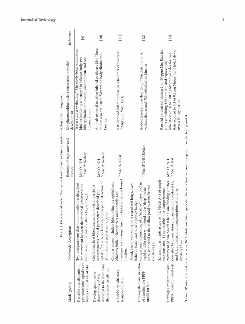
The development of the PBPK models proceeded in a step-wise, iterative fashion with increasing model complexitybeing added at each step. Table 1 provides an overview of theinitial “first generation” models developed for this researchprogram. The earliest dosimetry models were adapted frompharmacokinetic models developed for zinc, copper, andother essential metals that focused on dietary intake anddeficiency. Features of these models that were deemed impor-tant for manganese include features of these models thatwere deemed important for manganese include the ability todescribe homeostatic control of an essential element undernormal and deficient dietary conditions, and the use ofcompartmental and linear exchange rates to distribute theessential element into tissues and cellular compartments.The earliest manganese models were used to quantitativelytest assumptions regarding the movement of manganesefrom the rodent gastrointestinal tract (GIT) and liver [9]and to ascertain the degree to which systemic and orallyderived manganese are handled similarly in the liver [10].The resulting pharmacokinetic models accurately describedthe decreased gastrointestinal (GI) manganese uptake andincreased hepatobiliary elimination that is seen with risinglevels of manganese in the diet.
Early efforts also developed an initial framework for amulticompartment PBPK model. These models evaluatedthe kinetic behaviors of manganese in the brain, liver, andrespiratory tract during and after manganese inhalation [12,13]. Several model structures were considered during this
developmental phase (Table 1). Ultimately, manganese kinet-ics were best described using a model that included dose-dependent saturable tissue binding as well as free and boundmanganese [13]. In this context, bound manganese wasconfined to tissues and reflected basal manganese concentra-tions. Free manganese circulates in the blood and increasingconcentrations resulted during manganese inhalation. Freemanganese was rapidly cleared following exposure, therebyreturning tissue manganese concentrations to their originalbasal levels. This rise of free brain manganese concentrationwas described with diffusion rate constants (kin and kout).Peak tissue manganese concentrations were constrained bythe tissue maximal binding capacity (Bmax). Importantly,dose dependencies predicted by the Nong model [13] wereconsistent with the total manganese tissue levels measured inrats following manganese inhalation. The model also repli-cated the rapid increases in tissue manganese concentrationsseen at the highest inhaled manganese concentrations, as wellas the rapid return to baseline after exposure ceased. Themodel developed by Nong and coworkers [13] for the adultrat incorporated these and other features and was used as thebasis for all subsequent “second generation” animal PBPKmodels (Table 2).
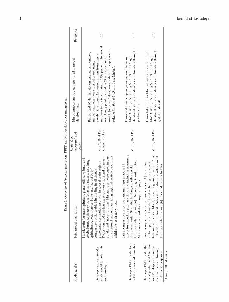
Starting in 2009, the focus of the modeling effortbegan to shift to the development of more complete PBPKmodels for animals (Table 2). These models retained manyof the features found in the Nong model [13], includingdose-dependent saturable tissue capacities and asymmetricaldiffusional flux of manganese into various tissue compart-ments. The second generation models also used airwaydeposition models based on particulate aerodynamics todescribe manganese delivery to the respiratory tract [17].Descriptions of the upper airways were broadened to includedescriptions of the nasal cavity and olfactory epitheliumusing data published by Schroeter et al. [18]. Regardingthe CNS, separate compartments for the olfactory bulb,striatum, pituitary gland, and cerebellum were developed.Specific influx and efflux diffusion rate constants (kin, kout)and binding constants (Bmax, ka, kd) for different brainregions were used to account for the differential increasesin regional brain manganese concentrations seen undervarious experimental conditions. These modifications ledto the publication of the revised adult rat model depictedin Figure 1 [14]. Additional models were subsequentlydeveloped to describe lactational [15] and gestational [16]transfer of manganese in rats. In all cases, model outputwas compared to inhalation data obtained under this testprogram and that from the available literature.
In 2009, Nong and coworkers also described the devel-opment of a PBPK model for nonhuman primates fromthe revised adult rat model [14]. The monkey PBPK modelwas viewed as a critical step in the evolution of appropriatehuman models (Figure 2). One goal of the modeling effortwas to retain as many features present in the rat model aspossible. Body weight, tissue volumes, olfactory and respira-tory tissues surface areas, ventilation rates, blood flows, andcertain other model parameters were adjusted to describemonkey physiology while others (biliary clearance and braindiffusional fluxes) were allometrically scaled based on body
Journal of Toxicology 3
Ta
ble
1:O
verv
iew
ofin
itia
l“fi
rst
gen
erat
ion”
phar
mac
okin
etic
mod
els
deve
lop
edfo
rm
anga
nes
e.
Mod
elgo
al(s
)B
rief
mod
elde
scri
ptio
nR
oute
(s)
ofex
posu
re‡
and
spec
ies
Mn
phar
mac
okin
etic
data
set(
s)u
sed
inm
odel
deve
lopm
ent
Ref
eren
ce
Des
crib
edo
sede
pen
den
tga
stro
inte
stin
alu
ptak
ean
dbi
liary
elim
inat
ion
ofM
n
Two-
com
part
men
tdi
stri
buti
onm
odel
that
desc
ribe
dM
nm
ovem
ent
betw
een
the
inte
stin
allu
men
and
the
liver
usi
ng
sim
ple
rate
con
stan
ts(k
inan
dk o
ut)
.
Mn
:O,I
NH
54M
n:I
VR
oden
t
Trac
erst
udi
esev
alu
atin
g54
Mn
wh
ole-
body
elim
inat
ion
kin
etic
sin
clu
din
ga
diet
ary
Mn
bala
nce
stu
dy,t
wo
bilia
ryel
imin
atio
nst
udi
es,a
nd
one
acu
tean
don
ech
ron
icst
udy
.
[9]
Dev
elop
quan
tita
tive
desc
ript
ion
sof
Mn
deliv
ered
toth
eliv
erfr
omth
esy
stem
icci
rcu
lati
on.
Gu
tlu
men
,liv
erbl
ood,
syst
emic
bloo
d,an
da
tiss
ue
com
part
men
ts.M
odel
para
met
ers
desc
ribe
dgu
tu
ptak
e,54
Mn
trac
erki
net
ics,
and
hep
atic
extr
acti
onof
Mn
from
oral
and
syst
emic
pool
s.
Mn
:O,I
NH
54M
n:I
VR
oden
t
An
imal
sex
pose
dto
eith
erin
hal
edor
diet
ary
Mn
.Th
ese
stu
dies
also
eval
uat
ed54
Mn
wh
ole-
body
elim
inat
ion
kin
etic
s.[1
0]
Des
crib
eth
eol
fact
ory
tran
spor
tof
Mn
.
Com
part
men
tsin
clu
ded:
bloo
d,ol
fact
ory
epit
hel
ium
,ol
fact
ory
bulb
,olf
acto
rytr
act
and
tube
rcle
,an
dst
riat
um
.Eac
hco
mpa
rtm
ent
incl
ude
da
free
and
bou
nd
frac
tion
.
54M
n:I
NH
Rat
Rat
sex
pose
d(9
0m
in)
nos
e-on
lyto
eith
erex
posu
reto
54M
nC
l 2or
54M
nH
PO
4.
[11]
Dev
elop
the
basi
cst
ruct
ure
ofa
mu
ltir
oute
PB
PK
mod
elfo
rM
n.
Blo
od,b
rain
,res
pira
tory
trac
t(n
asal
and
lun
g),l
iver
,ki
dney
s,bo
ne,
and
mu
scle
(res
tof
body
)co
mpa
rtm
ents
con
sist
ing
ofa
“sh
allo
w”
tiss
ue
pool
inra
pid
equ
ilibr
atio
nw
ith
bloo
dan
da
“dee
p”ti
ssu
est
ore,
con
nec
ted
toth
esh
allo
wpo
olby
tran
sfer
rate
con
stan
ts[1
].
54M
n:I
P,IN
HR
oden
tR
oden
ttr
acer
stu
dies
desc
ribi
ng
54M
ndi
stri
buti
onto
vari
ous
tiss
ues
and
54M
nel
imin
atio
nki
net
ics.
[12]
Dev
elop
am
ult
irou
teM
nP
BP
Km
odel
for
adu
ltra
ts.
Sam
eco
mpa
rtm
ents
asab
ove
[4].
Mod
elA
use
dsi
mpl
era
teco
nst
ants
[1]
tode
scri
bein
ter-
com
part
men
tal
mov
emen
tof
Mn
.Mod
elB
had
tiss
ue
bin
din
gki
net
ics
desc
ribe
dby
diss
ocia
tion
and
asso
ciat
ion
con
stan
ts(k
d
andk a
),an
dm
axim
um
con
cen
trat
ion
ofbi
ndi
ng
capa
city
(Bm
ax).
Mn
:O,I
NH
54M
n:I
VR
at
Rat
sfe
don
diet
sco
nta
inin
g2
to10
0pp
mM
n,R
ats
fed
adi
etco
nta
inin
g12
5pp
mM
nan
dex
pose
dvi
ain
hal
atio
nat
0.0
to3.
00m
gM
n/m
3ea
chda
yfo
r14
d.R
ats
expo
sed
to0.
1or
0.5
mg
Mn
/m3
for
6h
/d,5
d/w
kov
era
90-d
ayp
erio
d.
[13]
‡ O:o
ral;
IP:i
ntr
aper
iton
eal;
IV:i
ntr
aven
ous;
INH
:in
hal
atio
n.W
her
eap
plic
able
,Mn
trac
erfo
rman
dro
ute
ofex
posu
reh
ave
also
been
prov
ided
.
4 Journal of Toxicology
Ta
ble
2:O
verv
iew
of“s
econ
dge
ner
atio
n”P
BP
Km
odel
sde
velo
ped
for
man
gan
ese.
Mod
elgo
al(s
)B
rief
mod
elde
scri
ptio
nR
oute
(s)
ofex
posu
re‡
and
spec
ies
Mn
phar
mac
okin
etic
data
set(
s)u
sed
inm
odel
deve
lopm
ent
Ref
eren
ce
Dev
elop
am
ult
irou
teM
nP
BP
Km
odel
for
adu
ltra
tsan
dm
onke
ys.
Blo
od,b
rain
(str
iatu
m,p
itu
itar
ygl
and,
olfa
ctor
ybu
lb,a
nd
cere
bellu
m),
resp
irat
ory
trac
t(o
lfac
tory
mu
cosa
and
lun
gep
ith
eliu
m),
liver
,kid
ney
s,bo
ne,
and
“res
tof
body
”co
mpa
rtm
ents
.Sat
ura
ble
Mn
bin
din
gin
allt
issu
es,
pref
eren
tial
accu
mu
lati
onof
Mn
inse
vera
lbra
inre
gion
s.D
epos
itio
nof
Mn
wit
hin
the
resp
irat
ory
trac
tan
dol
fact
ory
upt
ake
and
“nos
e-to
-bra
in”
Mn
tran
spor
tw
ere
base
din
part
onad
diti
onal
mod
els
desc
ribi
ng
regi
onal
part
icle
depo
siti
onw
ith
inth
ere
spir
ator
ytr
act.
Mn
:O,I
NH
Rat
Rh
esu
sm
onke
y
Rat
14-
and
90-d
ayin
hal
atio
nst
udi
es.I
nm
onke
ys,
mod
elpa
ram
eter
sw
ere
firs
tca
libra
ted
usi
ng
stea
dy-s
tate
tiss
ue
Mn
con
cen
trat
ion
sfr
omrh
esu
sm
onke
ysfe
da
diet
con
tain
ing
133
ppm
Mn
.Th
em
odel
was
then
appl
ied
tosi
mu
late
65ex
posu
reda
ysof
wee
kly
(6h
/day
;5da
ys/w
eek)
inh
alat
ion
expo
sure
sto
solu
ble
Mn
SO4
at0.
03to
1.5
mg
Mn
/m3.
[14]
Dev
elop
aP
BP
Km
odel
for
lact
atin
gda
man
dn
eon
ates
.
Sam
eco
mpa
rtm
ents
for
the
dam
and
pups
asab
ove
[6]
exce
ptfo
rex
clu
din
gpi
tuit
ary
glan
dan
din
clu
din
gm
amm
ary
glan
d(d
amon
ly).
Satu
rabl
ebi
ndi
ng
and
oth
erm
odel
feat
ure
ssi
mila
rto
abov
e[6
].D
ieta
ry(e
.g.,
tran
sfer
offr
eeM
nin
milk
)an
din
hal
atio
nin
puts
topu
ps.
Mn
:O,I
NH
Rat
Dam
san
dth
eir
offsp
rin
gw
ere
expo
sed
toai
ror
Mn
SO4
(0.0
5,0.
5,or
1m
gM
n/m
3)
for
6h
/day
,7da
ys/w
eek
star
tin
g28
days
prio
rto
bree
din
gth
rou
ghpo
stn
atal
day
18.
[15]
Dev
elop
aP
BP
Km
odel
that
cou
ldpr
edic
tfe
talM
ndo
sean
dM
ndi
spos
itio
nin
the
dam
and
fetu
sfo
llow
ing
mat
ern
alM
nex
posu
re.
Sam
eco
mpa
rtm
ents
for
the
dam
asab
ove
[6]
exce
ptfo
rex
clu
din
gth
epi
tuit
ary
glan
dan
din
clu
din
gth
epl
acen
ta.
Feta
lmod
elin
clu
ded
bloo
d,br
ain
,lu
ng,
bon
e,liv
er,a
nd
“res
tof
body
”co
mpa
rtm
ents
.Sat
ura
ble
bin
din
gan
dot
her
mod
elfe
atu
res
sim
ilar
toab
ove
[6].
Pla
cen
talt
ran
sfer
tofe
tus.
Mn
:O,I
NH
Rat
Dam
sfe
da
10-p
pmM
ndi
etw
ere
expo
sed
toai
ror
Mn
SO4
(0.0
5,0.
5,or
1m
gM
n/m
3)
for
6h
/day
,7da
ys/w
eek
star
tin
g28
days
prio
rto
bree
din
gth
rou
ghge
stat
ion
day
20.
[16]
‡ O:o
ral;
INH
:in
hal
atio
n.
Journal of Toxicology 5
BileDiet
Lung and Nose
Inhaled Mn
Bone
Liver
Ven
ous
bloo
d
Art
eria
l blo
od
Brain blood
Rest of body
Olfactory
kd
ka
kd
kaka ka
kdkd kd
ka
kd
ka
kd
Cerebellum Pituitary
kin
kin
kout
koutkin koutkin kout
B + Mn f
B + Mn f
B + Mn f B + Mn f B + Mn f
B + Mn f
B + Mn f
Mnb
Mnb MnbMnb
Mnb
Mnb
Mnb
Striatum-globuspallidus
QBrn
Qp
Qc
ka
Qbone
Qbody
Qliv
(a)
Venous blood
Inhaled Mn
Olfactory bulb
Nasal respiratoryNasal olfactory
Lung tissue
Lung respiratory
B + Mn f Mnbka
kd
(b)
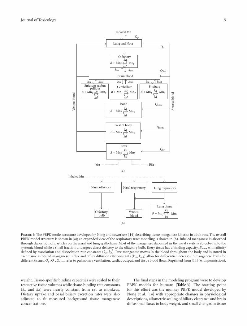
Figure 1: The PBPK model structure developed by Nong and coworkers [14] describing tissue manganese kinetics in adult rats. The overallPBPK model structure is shown in (a); an expanded view of the respiratory tract modeling is shown in (b). Inhaled manganese is absorbedthrough deposition of particles on the nasal and lung epithelium. Most of the manganese deposited in the nasal cavity is absorbed into thesystemic blood while a small fraction undergoes direct delivery to the olfactory bulb. Every tissue has a binding capacity, Bmax, with affinitydefined by association and dissociation rate constants (ka, kd). Free manganese moves in the blood throughout the body and is stored ineach tissue as bound manganese. Influx and efflux diffusion rate constants (kin, kout) allow for differential increases in manganese levels fordifferent tissues. Qp, Qc, Qtissue refer to pulmonary ventilation, cardiac output, and tissue blood flows. Reprinted from [14] (with permission).
weight. Tissue-specific binding capacities were scaled to theirrespective tissue volumes while tissue-binding rate constants(ka and kd) were nearly constant from rat to monkeys.Dietary uptake and basal biliary excretion rates were alsoadjusted to fit measured background tissue manganeseconcentrations.
The final steps in the modeling program were to developPBPK models for humans (Table 3). The starting pointfor this effort was the monkey PBPK model developed byNong et al. [14] with appropriate changes in physiologicaldescriptions, allometric scaling of biliary clearance and braindiffusional fluxes to body weight, and small changes in tissue
6 Journal of Toxicology
Adult ratmodel
model
model
Adult monkey
Adult human
Human
gestation and
lactation
models
Rat
gestation and
lactation
models
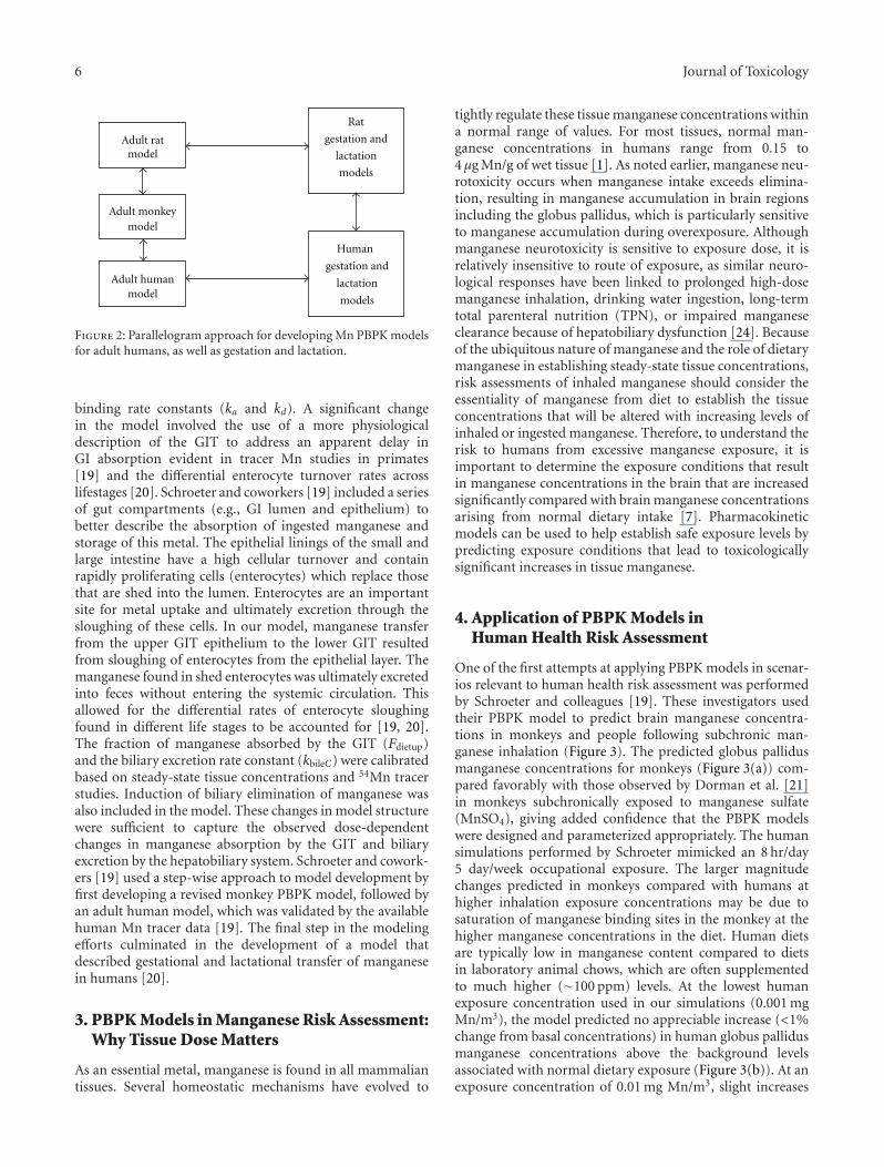
Figure 2: Parallelogram approach for developing Mn PBPK modelsfor adult humans, as well as gestation and lactation.
binding rate constants (ka and kd). A significant changein the model involved the use of a more physiologicaldescription of the GIT to address an apparent delay inGI absorption evident in tracer Mn studies in primates[19] and the differential enterocyte turnover rates acrosslifestages [20]. Schroeter and coworkers [19] included a seriesof gut compartments (e.g., GI lumen and epithelium) tobetter describe the absorption of ingested manganese andstorage of this metal. The epithelial linings of the small andlarge intestine have a high cellular turnover and containrapidly proliferating cells (enterocytes) which replace thosethat are shed into the lumen. Enterocytes are an importantsite for metal uptake and ultimately excretion through thesloughing of these cells. In our model, manganese transferfrom the upper GIT epithelium to the lower GIT resultedfrom sloughing of enterocytes from the epithelial layer. Themanganese found in shed enterocytes was ultimately excretedinto feces without entering the systemic circulation. Thisallowed for the differential rates of enterocyte sloughingfound in different life stages to be accounted for [19, 20].The fraction of manganese absorbed by the GIT (Fdietup)and the biliary excretion rate constant (kbileC) were calibratedbased on steady-state tissue concentrations and 54Mn tracerstudies. Induction of biliary elimination of manganese wasalso included in the model. These changes in model structurewere sufficient to capture the observed dose-dependentchanges in manganese absorption by the GIT and biliaryexcretion by the hepatobiliary system. Schroeter and cowork-ers [19] used a step-wise approach to model development byfirst developing a revised monkey PBPK model, followed byan adult human model, which was validated by the availablehuman Mn tracer data [19]. The final step in the modelingefforts culminated in the development of a model thatdescribed gestational and lactational transfer of manganesein humans [20].
3. PBPK Models in Manganese Risk Assessment:Why Tissue Dose Matters
As an essential metal, manganese is found in all mammaliantissues. Several homeostatic mechanisms have evolved to
tightly regulate these tissue manganese concentrations withina normal range of values. For most tissues, normal man-ganese concentrations in humans range from 0.15 to4 μg Mn/g of wet tissue [1]. As noted earlier, manganese neu-rotoxicity occurs when manganese intake exceeds elimina-tion, resulting in manganese accumulation in brain regionsincluding the globus pallidus, which is particularly sensitiveto manganese accumulation during overexposure. Althoughmanganese neurotoxicity is sensitive to exposure dose, it isrelatively insensitive to route of exposure, as similar neuro-logical responses have been linked to prolonged high-dosemanganese inhalation, drinking water ingestion, long-termtotal parenteral nutrition (TPN), or impaired manganeseclearance because of hepatobiliary dysfunction [24]. Becauseof the ubiquitous nature of manganese and the role of dietarymanganese in establishing steady-state tissue concentrations,risk assessments of inhaled manganese should consider theessentiality of manganese from diet to establish the tissueconcentrations that will be altered with increasing levels ofinhaled or ingested manganese. Therefore, to understand therisk to humans from excessive manganese exposure, it isimportant to determine the exposure conditions that resultin manganese concentrations in the brain that are increasedsignificantly compared with brain manganese concentrationsarising from normal dietary intake [7]. Pharmacokineticmodels can be used to help establish safe exposure levels bypredicting exposure conditions that lead to toxicologicallysignificant increases in tissue manganese.
4. Application of PBPK Models inHuman Health Risk Assessment
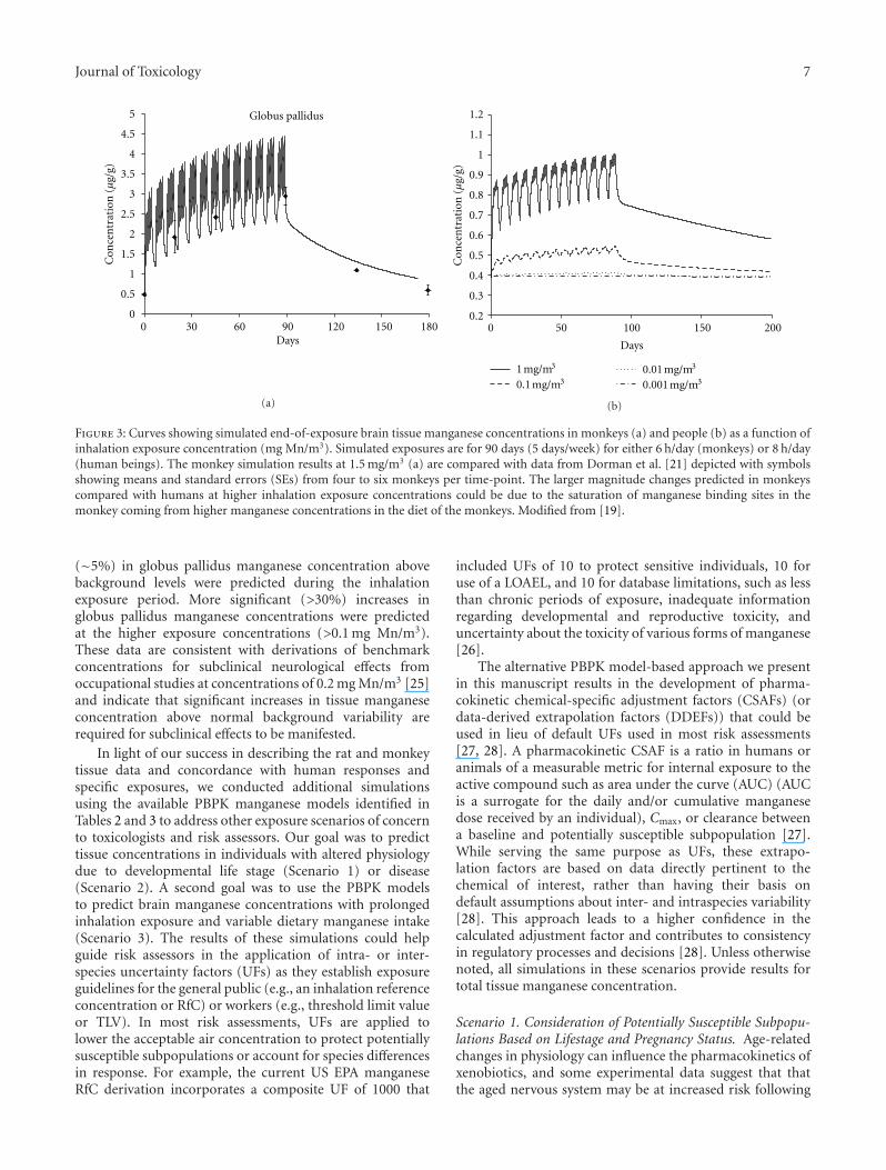
One of the first attempts at applying PBPK models in scenar-ios relevant to human health risk assessment was performedby Schroeter and colleagues [19]. These investigators usedtheir PBPK model to predict brain manganese concentra-tions in monkeys and people following subchronic man-ganese inhalation (Figure 3). The predicted globus pallidusmanganese concentrations for monkeys (Figure 3(a)) com-pared favorably with those observed by Dorman et al. [21]in monkeys subchronically exposed to manganese sulfate(MnSO4), giving added confidence that the PBPK modelswere designed and parameterized appropriately. The humansimulations performed by Schroeter mimicked an 8 hr/day5 day/week occupational exposure. The larger magnitudechanges predicted in monkeys compared with humans athigher inhalation exposure concentrations may be due tosaturation of manganese binding sites in the monkey at thehigher manganese concentrations in the diet. Human dietsare typically low in manganese content compared to dietsin laboratory animal chows, which are often supplementedto much higher (∼100 ppm) levels. At the lowest humanexposure concentration used in our simulations (0.001 mgMn/m3), the model predicted no appreciable increase (<1%change from basal concentrations) in human globus pallidusmanganese concentrations above the background levelsassociated with normal dietary exposure (Figure 3(b)). At anexposure concentration of 0.01 mg Mn/m3, slight increases
Journal of Toxicology 7
Globus pallidus
0
0.5
1
1.5
2
2.5
3
3.5
4
4.5
5
0 30 60 90 120 150 180Days
Con
cen
trat
ion
(µ
g/g)
(a)
0.2
0.3
0.4
0.5
0.6
0.7
0.8
0.9
1
1.1
1.2
0 50 100 150 200
Days
Con
cen
trat
ion
(µ
g/g)
1 mg/m3
0.1 mg/m30.01 mg/m3
0.001 mg/m3
(b)
Figure 3: Curves showing simulated end-of-exposure brain tissue manganese concentrations in monkeys (a) and people (b) as a function ofinhalation exposure concentration (mg Mn/m3). Simulated exposures are for 90 days (5 days/week) for either 6 h/day (monkeys) or 8 h/day(human beings). The monkey simulation results at 1.5 mg/m3 (a) are compared with data from Dorman et al. [21] depicted with symbolsshowing means and standard errors (SEs) from four to six monkeys per time-point. The larger magnitude changes predicted in monkeyscompared with humans at higher inhalation exposure concentrations could be due to the saturation of manganese binding sites in themonkey coming from higher manganese concentrations in the diet of the monkeys. Modified from [19].
(∼5%) in globus pallidus manganese concentration abovebackground levels were predicted during the inhalationexposure period. More significant (>30%) increases inglobus pallidus manganese concentrations were predictedat the higher exposure concentrations (>0.1 mg Mn/m3).These data are consistent with derivations of benchmarkconcentrations for subclinical neurological effects fromoccupational studies at concentrations of 0.2 mg Mn/m3 [25]and indicate that significant increases in tissue manganeseconcentration above normal background variability arerequired for subclinical effects to be manifested.
In light of our success in describing the rat and monkeytissue data and concordance with human responses andspecific exposures, we conducted additional simulationsusing the available PBPK manganese models identified inTables 2 and 3 to address other exposure scenarios of concernto toxicologists and risk assessors. Our goal was to predicttissue concentrations in individuals with altered physiologydue to developmental life stage (Scenario 1) or disease(Scenario 2). A second goal was to use the PBPK modelsto predict brain manganese concentrations with prolongedinhalation exposure and variable dietary manganese intake(Scenario 3). The results of these simulations could helpguide risk assessors in the application of intra- or inter-species uncertainty factors (UFs) as they establish exposureguidelines for the general public (e.g., an inhalation referenceconcentration or RfC) or workers (e.g., threshold limit valueor TLV). In most risk assessments, UFs are applied tolower the acceptable air concentration to protect potentiallysusceptible subpopulations or account for species differencesin response. For example, the current US EPA manganeseRfC derivation incorporates a composite UF of 1000 that
included UFs of 10 to protect sensitive individuals, 10 foruse of a LOAEL, and 10 for database limitations, such as lessthan chronic periods of exposure, inadequate informationregarding developmental and reproductive toxicity, anduncertainty about the toxicity of various forms of manganese[26].
The alternative PBPK model-based approach we presentin this manuscript results in the development of pharma-cokinetic chemical-specific adjustment factors (CSAFs) (ordata-derived extrapolation factors (DDEFs)) that could beused in lieu of default UFs used in most risk assessments[27, 28]. A pharmacokinetic CSAF is a ratio in humans oranimals of a measurable metric for internal exposure to theactive compound such as area under the curve (AUC) (AUCis a surrogate for the daily and/or cumulative manganesedose received by an individual), Cmax, or clearance betweena baseline and potentially susceptible subpopulation [27].While serving the same purpose as UFs, these extrapo-lation factors are based on data directly pertinent to thechemical of interest, rather than having their basis ondefault assumptions about inter- and intraspecies variability[28]. This approach leads to a higher confidence in thecalculated adjustment factor and contributes to consistencyin regulatory processes and decisions [28]. Unless otherwisenoted, all simulations in these scenarios provide results fortotal tissue manganese concentration.
Scenario 1. Consideration of Potentially Susceptible Subpopu-lations Based on Lifestage and Pregnancy Status. Age-relatedchanges in physiology can influence the pharmacokinetics ofxenobiotics, and some experimental data suggest that thatthe aged nervous system may be at increased risk following
8 Journal of Toxicology
00 50 100 150 200 250
Days
Olfactory bulb
NormalAged
Con
cen
trat
ion
(µ
g/g)
0.2
0.4
0.6
0.8
1
1.2
1.4
1.6
(a)
Striatum
00 50 100 150 200 250
Days
NormalAged
Con
cen
trat
ion
(µ
g/g)
0.2
0.4
0.6
0.8
1
1.2
(b)
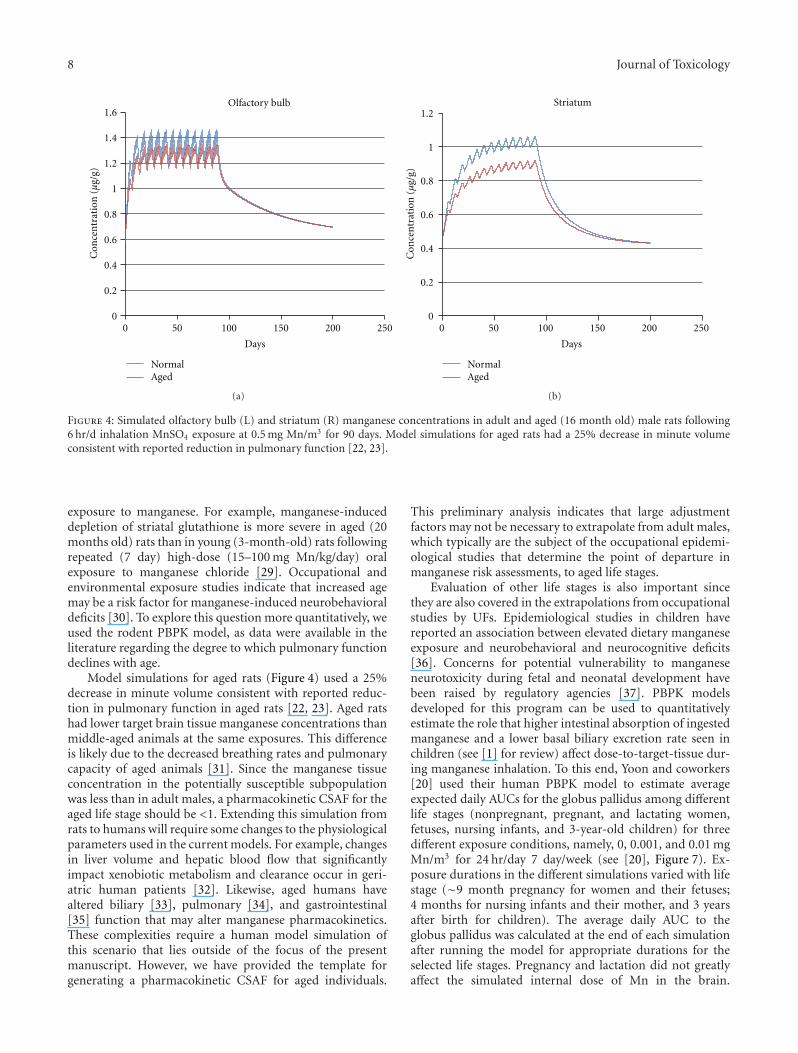
Figure 4: Simulated olfactory bulb (L) and striatum (R) manganese concentrations in adult and aged (16 month old) male rats following6 hr/d inhalation MnSO4 exposure at 0.5 mg Mn/m3 for 90 days. Model simulations for aged rats had a 25% decrease in minute volumeconsistent with reported reduction in pulmonary function [22, 23].
exposure to manganese. For example, manganese-induceddepletion of striatal glutathione is more severe in aged (20months old) rats than in young (3-month-old) rats followingrepeated (7 day) high-dose (15–100 mg Mn/kg/day) oralexposure to manganese chloride [29]. Occupational andenvironmental exposure studies indicate that increased agemay be a risk factor for manganese-induced neurobehavioraldeficits [30]. To explore this question more quantitatively, weused the rodent PBPK model, as data were available in theliterature regarding the degree to which pulmonary functiondeclines with age.
Model simulations for aged rats (Figure 4) used a 25%decrease in minute volume consistent with reported reduc-tion in pulmonary function in aged rats [22, 23]. Aged ratshad lower target brain tissue manganese concentrations thanmiddle-aged animals at the same exposures. This differenceis likely due to the decreased breathing rates and pulmonarycapacity of aged animals [31]. Since the manganese tissueconcentration in the potentially susceptible subpopulationwas less than in adult males, a pharmacokinetic CSAF for theaged life stage should be <1. Extending this simulation fromrats to humans will require some changes to the physiologicalparameters used in the current models. For example, changesin liver volume and hepatic blood flow that significantlyimpact xenobiotic metabolism and clearance occur in geri-atric human patients [32]. Likewise, aged humans havealtered biliary [33], pulmonary [34], and gastrointestinal[35] function that may alter manganese pharmacokinetics.These complexities require a human model simulation ofthis scenario that lies outside of the focus of the presentmanuscript. However, we have provided the template forgenerating a pharmacokinetic CSAF for aged individuals.
This preliminary analysis indicates that large adjustmentfactors may not be necessary to extrapolate from adult males,which typically are the subject of the occupational epidemi-ological studies that determine the point of departure inmanganese risk assessments, to aged life stages.
Evaluation of other life stages is also important sincethey are also covered in the extrapolations from occupationalstudies by UFs. Epidemiological studies in children havereported an association between elevated dietary manganeseexposure and neurobehavioral and neurocognitive deficits[36]. Concerns for potential vulnerability to manganeseneurotoxicity during fetal and neonatal development havebeen raised by regulatory agencies [37]. PBPK modelsdeveloped for this program can be used to quantitativelyestimate the role that higher intestinal absorption of ingestedmanganese and a lower basal biliary excretion rate seen inchildren (see [1] for review) affect dose-to-target-tissue dur-ing manganese inhalation. To this end, Yoon and coworkers[20] used their human PBPK model to estimate averageexpected daily AUCs for the globus pallidus among differentlife stages (nonpregnant, pregnant, and lactating women,fetuses, nursing infants, and 3-year-old children) for threedifferent exposure conditions, namely, 0, 0.001, and 0.01 mgMn/m3 for 24 hr/day 7 day/week (see [20], Figure 7). Ex-posure durations in the different simulations varied with lifestage (∼9 month pregnancy for women and their fetuses;4 months for nursing infants and their mother, and 3 yearsafter birth for children). The average daily AUC to theglobus pallidus was calculated at the end of each simulationafter running the model for appropriate durations for theselected life stages. Pregnancy and lactation did not greatlyaffect the simulated internal dose of Mn in the brain.
Journal of Toxicology 9
Ta
ble
3:O
verv
iew
ofhu
man
PB
PK
mod
els
deve
lop
edfo
rm
anga
nes
e.
Mod
elgo
al(s
)B
rief
mod
elde
scri
ptio
nR
oute
(s)
ofex
posu
re‡
and
spec
ies
Mn
phar
mac
okin
etic
data
set(
s)u
sed
inm
odel
deve
lopm
ent
Ref
eren
ce
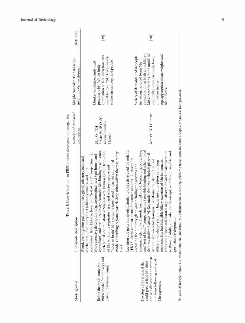
Refi
ne
the
mu
lti-
rou
teM
nP
BP
Km
odel
for
mon
keys
and
exte
nd
tohu
man
bein
gs.
Blo
od,b
rain
(glo
bus
palli
dus,
pitu
itar
ygl
and,
olfa
ctor
ybu
lb,a
nd
cere
bellu
m),
resp
irat
ory
trac
t(o
lfac
tory
mu
cosa
and
lun
gep
ith
eliu
m),
liver
,kid
ney
s,bo
ne,
and
“res
tof
body
”co
mpa
rtm
ents
.M
ore
exte
nsi
vede
scri
ptio
nof
gast
roin
test
inal
trac
t(g
ut
lum
enan
dep
ith
eliu
m)
and
peri
ton
ealc
avit
y.Sa
tura
ble
Mn
bin
din
gin
allt
issu
es.
Pre
fere
nti
alac
cum
ula
tion
ofM
nin
seve
ralb
rain
regi
ons.
Dep
osit
ion
ofM
nw
ith
inth
ere
spir
ator
ytr
act
and
olfa
ctor
yu
ptak
ean
d“n
ose-
to-b
rain
”M
ntr
ansp
ort
wer
eba
sed
inpa
rton
addi
tion
alm
odel
sde
scri
bin
gre
gion
alpa
rtic
lede
posi
tion
wit
hin
the
resp
irat
ory
trac
t.
Mn
:O,I
NH
54M
n:I
V,I
P,O
,SC
Rh
esu
sm
onke
yH
um
an
Mon
key
inh
alat
ion
stu
dyu
sed
prev
iou
sly
[6].
Wh
ole-
body
elim
inat
ion
orfe
cale
xcre
tion
data
avai
labl
efr
om54
Mn
trac
erki
net
icst
udi
esin
mon
keys
and
peo
ple
[19]
Dev
elop
aP
BP
Km
odel
that
cou
ldpr
edic
tfe
talM
ndo
sean
dM
ndi
spos
itio
nin
wom
enan
dfe
tus
follo
win
gm
ater
nal
Mn
expo
sure
.
Lact
atio
nan
dge
stat
ion
mod
els
sim
ilar
toth
ose
deve
lop
edfo
rro
den
ts[1
5,16
].Sa
me
com
part
men
tsfo
rw
omen
asab
ove
[9]
exce
ptfo
rex
clu
din
gth
epi
tuit
ary
glan
dan
din
clu
din
gth
epl
acen
taan
dm
amm
ary
glan
d.Fe
talm
odel
incl
ude
dbl
ood,
brai
n,l
un
g,bo
ne,
liver
,an
d“r
est
ofbo
dy”
com
part
men
ts.S
atu
rabl
ebi
ndi
ng
and
oth
erm
odel
feat
ure
ssi
mila
rto
abov
e[9
].K
eym
odel
feat
ure
sin
clu
ded:
plac
enta
lM
ntr
ansf
ervi
aac
tive
tran
spor
t,la
ctat
ion
altr
ansf
erof
Mn
use
ddi
ffu
sion
-med
iate
dse
cret
ion
,hig
her
gut
abso
rpti
onin
nu
rsin
gn
eon
ates
,low
but
indu
cibl
ebi
liary
excr
etio
nof
Mn
inn
eon
ates
,tr
ansi
tion
ofn
eon
atal
feat
ure
sof
gut
abso
rpti
onan
dbi
liary
excr
etio
nto
thos
eof
adu
lts,
and
enh
ance
dbr
ain
upt
ake
ofM
ndu
rin
gfe
tala
nd
post
nat
alde
velo
pmen
t.
Mn
:O,I
NH
Hu
man
Var
iety
ofda
taob
tain
edin
peo
ple
incl
udi
ng:
repo
rted
brai
nM
nco
nce
ntr
atio
nat
birt
han
dch
ildre
n,
Mn
con
cen
trat
ion
sin
the
um
bilic
alco
rd,m
ilk,n
ewbo
rnbl
ood,
bon
e,an
dot
her
tiss
ues
.A
geap
prop
riat
eti
ssu
ew
eigh
tsan
dbl
ood
flow
s.
[20]
‡ O:o
ral;
IP:i
ntr
aper
iton
eal;
IV:i
ntr
aven
ous;
INH
:in
hal
atio
n;S
C:s
ubc
uta
neo
us.
Wh
ere
appl
icab
le,M
ntr
acer
form
and
rou
teof
expo
sure
hav
eal
sobe
enpr
ovid
ed.
10 Journal of Toxicology
Liver impairment
00 10050 200150 300250 400350
Time (days)
Human
Control
0.2
0.4
0.6
0.8
1
1.2exposure0.00005 mg/m3 inhalation
Glo
bus
palli
dus
con
cen
trat
ion
(µg/
g)
(a)
0 10050 200150 300250 4003500
Time (days)
Human
0.2
0.4
0.6
0.8
1
1.2exposure0.2 mg/m3 inhalation
Glo
bus
palli
dus
con
cen
trat
ion
(µg/
g)
Liver impairmentControl
(b)
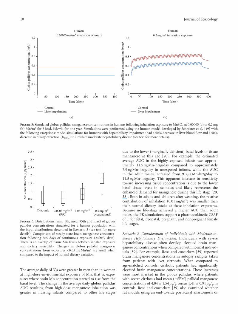
Figure 5: Simulated globus pallidus manganese concentrations in humans following inhalation exposure to MnSO4 at 0.00005 (a) or 0.2 mg(b) Mn/m3 for 8 hr/d, 5 d/wk, for one year. Simulations were performed using the human model developed by Schroeter et al. [19] withthe following exceptions: model simulations for humans with hepatobiliary impairment had a 50% decrease in liver blood flow and a 50%decrease in biliary excretion (KbileC) to simulate moderate hepatobiliary disease (see text for more details).
0Diet only
0.5
1
1.5
2
2.5
3
3.5
(RfC)0.05 mg/m3 0.5 mg/m3
(occupational)0.0005 mg/m3
Mn
con
cen
trat
ion
t
issu
e)(µ
g/g
Figure 6: Distributions (min, 5th, med, 95th and max) of globuspallidus concentrations simulated for a human population withthe input distributions described in Scenario 3 (see text for moredetails). Comparison of steady-state brain manganese concentra-tion following 365 days of continuous exposure (24 hr/7 days).There is an overlap of tissue Mn levels between inhaled exposureand dietary variability. Changes in globus pallidal manganeseconcentrations from exposures <0.05 mg Mn/m3 are small whencompared to the impact of normal dietary variation.
The average daily AUCs were greater in men than in womenat high-dose environmental exposure of Mn, that is, expo-sures where brain Mn concentration started to rise from thebasal level. The change in the average daily globus pallidusAUC resulting from high-dose manganese inhalation wasgreater in nursing infants compared to other life stages
due to the lower (marginally deficient) basal levels of tissuemanganese at this age [20]. For example, the estimatedaverage AUC in the highly exposed infants was approx-imately 11.5 μg Mn·hr/g/day compared to approximately7.9 μg Mn·hr/g/day in unexposed infants, while the AUCin the adult males increased from 9.3 μg Mn·hr/g/day to11.5 μg Mn·hr/g/day. This apparent increase in sensitivitytoward increasing tissue concentration is due to the lowerbasal tissue levels in neonates and likely represents theenhanced demand for manganese during this life stage [20,38]. Both in adults and children after weaning, the relativecontribution of inhalation (0.01 mg/m3) was smaller thantheir normal dietary intake at these inhalation exposures.Because no life-stage achieved a higher AUC than adultmales, the PK simulations support a pharmacokinetic CSAFof 1 for fetal, neonatal, pregnant, and nonpregnant femalelife-stages.
Scenario 2. Consideration of Individuals with Moderate-to-Severe Hepatobiliary Dysfunction. Individuals with severehepatobiliary disease often develop elevated brain man-ganese concentrations when compared with normal individ-uals [39]. For example, Rose and coworkers [39] reportedbrain manganese concentrations in autopsy samples takenfrom patients with liver cirrhosis. When compared toage-matched controls, cirrhotic patients had significantlyelevated brain manganese concentrations. These increaseswere most marked in the globus pallidus, where patientswith severe cirrhosis had mean (±SEM) pallidal manganeseconcentrations of 4.04 ± 1.54μg/g versus 1.41 ± 0.91μg/g incontrols. Rose and coworkers [39] also examined whetherrat models using an end-to-side portacaval anastomosis or

Journal of Toxicology 11
0
1
2
3
4
5
0.00001 0.0001 0.001 0.01 0.1 1
Glo
bus
palli
dus
Mn
con
cen
trat
ion
(µ
g/g)
Inhaled Mn concentration (mg/m3)
90-day, 24 hours/day, 7 days/week2-year, 24 hours/day, 7 days/week
Figure 7: Simulated end-of-exposure nonhuman primate globuspallidus manganese concentrations following a 24 h/d, 7 d/wkinhalation for either 90 days (subchronic) or 2 yr (chronic)exposure to MnSO4. These simulations indicate that globuspallidus manganese concentrations are expected to rapidly reachpseudosteady-state levels during high dose manganese exposure,and that duration of exposure has a minimal effect. Its contributiononly occurs once exposures reach the threshold to cause tissueaccumulation.
an intracholedochal injection of formalin and concomitantligation of the bile duct to create an experimental model ofbiliary cirrhosis could mimic these findings. As expected, ratswith biliary cirrhosis or a portacaval-shunt had increasedpallidal (increased by 27% to 57%) and caudate/putamen(increased by 57 to 67%) manganese concentrations whencompared with sham operated or normal control groups. Itis important to note that the humans and animals studiedby Rose had significant clinical liver disease. For example,the cirrhotic patients studied by Rose et al. [39] died of anextreme form of hepatic encephalopathy (i.e., hepatic coma).
Individuals with liver or biliary cirrhosis can alsodevelop significant changes in hepatic blood flow that mayaffect manganese pharmacokinetics. Annet and cowork-ers [40] reported that patients with chronic liver diseaseof various grades (i.e., Child-Pugh classes A, B, andC) had mean (±SD) apparent liver perfusion rates of36.29 ± 17.96 mL/min/100 mL liver volume versus 65.22 ±24.73 mL/min/100 mL liver volume in patients without livercirrhosis (as measured using magnetic resonance imaging).An approximately 50% reduction in total hepatic bloodflow has been seen in dogs with side-to-side or end-to-side portacaval anastomoses [41]. Other investigators havereported significant (∼50%) reductions in hepatic bloodflow in rats with common bile duct ligation [42]. A similarobservation is also seen in people with hepatic cirrhosis [43].These studies indicate that there should be a 50% reductionin hepatic blood flow in the PBPK model parameters forconsistency with the observations of experimentally inducedhepatobiliary dysfunction.
Impaired secretion of bile acids, bilirubin, and otherorganic anions, consistent with reduced biliary function, isalso observed in liver disease [44]. For example, rats withmild hepatic stenosis have an approximate 15% reductionin basal bile flow when compared with control animals[45]. One animal model in which biliary function hasbeen quantitatively examined is liver dysfunction inducedby the subchronic to chronic administration of TPN, anintravenous diet given in severe cases of GI disorders. Daset al. [46] reported that rabbits receiving TPN will developqualitatively similar decreases in bile flow (reduced by 60%),bile acid secretion (52%), and sulfobromophthalein (BSP)excretion (38%) when compared with control animals. Theseanimals also developed hepatocellular degeneration andportal tract inflammation. Thus, the available data supportusing a 50% reduction in bile flow in the PBPK modelsimulation.
In this study, we used the Schroeter et al. [19] modelto simulate globus pallidus manganese concentrations inhumans following a one year inhalation MnSO4 expo-sure to either 0.00005 (the USEPA RfC) or 0.2 mg (thecurrent ACGIH TLV) Mn/m3 for 8 hr/d, 5 d/wk. Modelsimulations for people with hepatobiliary impairment hada 50% decrease in liver blood flow and a concomitant50% decrease in biliary excretion (KbileC) consistent withchanges reported in people and/or animals with liver diseaseas discussed above. The simulations show that, regardlessof inhalation concentration, the model predicted higherpallidal brain manganese concentrations due to hepaticdysfunction alone (Figure 5), which was expected based onavailable data. Inhalation at the RfC had no significanteffect on pallidal concentrations, regardless of hepatobiliaryfunction (Figure 5(a)). Inhalation concentrations at the TLVproduced an increase in end-of-exposure pallidal manganeseconcentrations that were in addition to the increase fromhepatobiliary disease (approx. 0.85 μg/g versus 0.68 μg/gin controls, Figure 5(b)). A CSAF of ∼1.25 (0.85/0.68)is supported by the PBPK modeling for this extremelysensitive subgroup. Since this CSAF value was determined atoccupational exposure levels, and no changes were observedat the RfC, this disease-related CSAF is likely conservativefor environmental exposures which do not cause tissueaccumulation.
Scenario 3. Consideration of Dietary Mn Variability andChronic Manganese Inhalation. The strengths of using PBPKmodels in risk assessment include the ability to use themodels to examine both dietary and inhaled intakes andsupport extrapolations from high to low doses, across routes,for different animal species, and for durations of exposurelonger than those used in the studies that the models werebased on [47]. To date, the most complete pharmacokineticdatasets for inhaled manganese available for PBPK modeldevelopment and validation were developed for rats andmonkeys using exposure durations of up to 90 exposuredays (typically 6 hr/day, 5 days/week, reviewed in [5]). Themanganese PBPK models described in this manuscript canbe used to extrapolate beyond these exposure conditions.

12 Journal of Toxicology
This scenario demonstrates this ability by predicting globuspallidus manganese concentrations in people following acontinuous (24 hr/day) chronic (1 year) manganese exposure(Figure 6).
The monkey PBPK model developed by Nong et al. [14]scaled to humans was used to simulate globus pallidus man-ganese concentrations in people while varying the dietaryintake. Normal variation of manganese concentration inglobus pallidus due to the fluctuation in daily dietary expo-sure was simulated in an adult human population of 10,000using Monte Carlo techniques. These simulations variedthe daily dietary intake of manganese using published data(mean [±SD]: 2.43 ± 1.8 mg/day; range 0.07–6.2 mg/day)[48]. Published distribution values (mean) were used forbody weight (70 kg), tissue volumes (as % body weight) forblood (8%), bone (12%), brain (2%), liver (3%), lung (1%),and the remainder of the body (0.74%) [49, 50]. Distributionvalues (mean) for tissue blood flow (as % cardiac output)for bone (4%), brain (11%), liver (23%), and nose (1%)were also used [49, 50]. The coefficient of variation usedfor body weight, tissue volume, and blood flow parameterswas 0.30. Mean values for cardiac output and pulmonaryventilation were set at 13 L/hr/kg and 20 L/hr/kg, respectively.A coefficient of variation used for these parameters was 0.50.All parameter distributions were truncated by two standarddeviations and statistical correlations of parameters werenot included in our analysis. Air manganese concentrationsranged from current USEPA RfC (0.00005 mg Mn/m3) to0.5 mg Mn/m3, a concentration that represents potentialoccupational exposure levels, although with continuousexposure in this case.
A second question that we wanted to explore is the effectof exposure duration on the rate at which globus pallidusmanganese concentrations change following manganeseinhalation. Here, we compared the end-of-exposure tissueconcentrations of a subchronic (90-day) versus 2-year expo-sure duration with the nonhuman primate model (Figure 7).Globus pallidus manganese concentrations rapidly reachpseudosteady state levels during high dose manganese expo-sure. These simulations are in accord with observationsreported by Dorman and coworkers [21] who reportedthat rhesus monkeys exposed (5 d/week) for 15, 33, or65 exposure days to MnSO4 at 1.5 mg Mn/m3 developedmean (±SEM) globus pallidus manganese concentrations of1.92 ± 0.40, 2.41 ± 0.29, and 2.94 ± 0.23μg Mn/g tissuewet weight, respectively, all of which were significantlydifferent (P < 0.05) than background tissue levels of 0.48 ±0.04μg Mn/g tissue wet weight. Extending the simulation to2 years produced a very slight leftward shift of the exposure-accumulation curve, but it did not change the thresholdfor tissue accumulation. For example, at 0.2 mg MnSO4/m3
(the current ACGIH TLV), a pharmacokinetic CSAF forsubchronic to chronic duration is 1.06. Once exposurepasses the threshold for tissue accumulation, a PK CSAF forduration of exposure is maximally 1.1.
Results of these simulations show several importantfindings. Brain manganese concentrations are controlledover a wide range of low-to-moderate exposure conditionsat and above typical environmental exposures. Due to
homeostatic controls, changes in globus pallidal manganeseconcentrations from exposures that exceed the current RfCeven by several orders of magnitude (0.05 mg Mn/m3) aresmall when compared to those seen as a result of normalvariation in the dietary intake of manganese as demon-strated by the Monte Carlo analysis of dietary variation(Figure 6). However, once these homeostatic mechanism(s)are overwhelmed, pallidal manganese concentrations riserapidly. The threshold for this response appears to occur atapproximately 0.001–0.01 mg Mn/m3 (Figure 7).
5. Future Applications of PBPK Models
The scenarios that we have explored here can easily bebroadened to address other concerns raised in relation tothe human health risk assessment of manganese. Anotherscenario that may prove useful to risk assessors is a consider-ation of the effect of altered iron homeostasis on manganesepharmacokinetics, since iron deficiency and iron-deficientanemia exist worldwide [51]. Inadequate tissue iron statusresulting from dietary iron deficiency or anemia can lead toaltered brain manganese deposition in animals [52–54]. It isunknown whether interactions between iron and manganesefor divalent metal transporter 1 (DMT1) and other sharedcellular membrane metal transporters account for this effect[55, 56]. Once these pathways and interactions are more fullyelucidated, especially with quantitative measurements, thesefeatures can be incorporated into the existing PBPK models.
Although the PBPK models were originally created tosupport the risk assessment of combustion products ofthe fuel additive MMT (see [5]), they have much broaderapplication to toxicologists and risk assessors. PBPK modelscan consider the impact of particle size and solubility onmanganese dosimetry, especially as it relates to nanoma-terials. Manganese nanoparticle exposure may occur dur-ing occupational exposure scenarios, potentially includingwelding. Nanoparticles display several curious inhalationpharmacokinetic behaviors that may be independent ofchemical form [57]. For example, inhaled nanomaterials aredeposited extensively in the nasal cavity [58]; in addition,a large percentage (∼75%) of the nanomaterials that reachthe alveolar region remain at that site, with less than 5%of inhaled nanoparticles translocating out of the lungs [59];charged nanoparticles are more likely to travel to the brainvia axonal transport within the olfactory nerve than areneutral nanoparticles [60]; and nanoparticle size also influ-ences organ distribution and renal excretion [61, 62]. SeveralPBPK models have been developed for nanoparticles [11, 62–64]. These models were parameterized and validated usingexperimental pharmacokinetic data collected for differentnanomaterials (e.g., iridium, silver, or technetium-labeledcarbon nanoparticles) using data obtained from rats orhumans. There is only sparse data on the pharmacokineticsof manganese-based nanoparticles. Some work examinedtranslocation of manganese oxide (MnO2) nanoparticlesfrom the nasal cavity to the brain [65]; however, theseinvestigators relied on the use of nasal instillation ratherthan inhalation. Elder and coworkers [66] exposed rats to

Journal of Toxicology 13
MnO2 nanoparticles with individual aerodynamic diametersof 3–8 nm (note these particles form ∼30 nm agglomeratesin the exposure system) for 6 hr/day, 5 days/wk, for a totalof 12 inhalation exposure days. In this study, the olfactorybulb showed the greatest changes in proinflammatory geneexpression when compared to the midbrain, striatum, andother brain regions. This finding supports the conclusionthat inhaled manganese nanoparticles, like larger particles,can undergo olfactory transport from the nasal cavity tothe olfactory bulb. However, PBPK modeling in rodentsand MRI analysis in primates have demonstrated that theolfactory pathway does not appear to significantly impactmanganese delivery to tissues outside of the olfactory path-way [67, 68]. While research on manganese nanoparticles isstill limited, there is some evidence that soluble manganesemay be more bioavailable and cause more effects relativeto equivalent amounts of nanoparticle manganese. Whereasmost of the deposited nanomaterial appears to stay in thelung, soluble manganese is readily bioavailable [14, 59,69]. Furthermore, manganese nanoparticles appear to beless toxic than an equivalent dose of soluble Mn2+. Dailyintratracheal instillation of soluble MnCl2 in rats for 3–6weeks led to increased brain manganese levels, a reductionin body weight gain, and a decrease of open field motilitywhen compared to controls, whereas the equivalent dose ofMnO2 nanoparticles had no significant effects [65]. Also,MnO2 nanoparticles were less toxic to PC-12 cells in vitro bythe MTT assay than an equivalent dose (in ug/mL) of solubleMn acetate [70]. Thus, the current PBPK modeling, basedon soluble manganese (MnSO4), may represent a worst-casescenario relative to nano-manganese after accounting fordifferences in pulmonary deposition. This expectation willbe further clarified as more data become available comparingthe pharmacokinetics and pharmacodynamics of nano- andsoluble manganese.
Another potential application of the manganese PBPKmodels for risk assessment is the evaluation of the literatureon the neurological outcomes of manganese exposure inprimates, potentially identifying tissue concentrations thatlead to adverse effects. This assessment would allow themodels to explore the pharmacodynamic aspects of Mnexposure. The models could then be used to select themost appropriate dose metric for establishing a point ofdeparture in future risk assessments. A previous attempt toevaluate dose response for the effects of Mn in experimentalanimals [71] relied on estimated cumulative intake of Mnas the only measure for comparison across studies withdifferent doses, durations, and exposure routes. Alternativetoxicologically relevant dose metrics, including estimatedpeak concentration, average concentration, and cumulativedose (i.e., AUC) during the Mn exposure period could beestimated using a PBPK model known to accurately accountfor dose dependencies of Mn distribution in the monkeyfor combined inhalation and dietary exposures. A largenonhuman primate response literature exists for analysis,including exposures by inhalation, oral, intraperitoneal,and subcutaneous dose routes, and spanned durationsup to 2 years [71]. This type of analysis using PBPKmodels is currently underway and will make it possible
to provide a consistent description of the dose responserelationship for the effects of Mn independent of exposureroute.
Finally, PBPK models may be used in an alternativedosimetric-based risk assessment strategy for essential ele-ments considering dietary intake, natural tissue backgroundlevels, and dietary and population variability. An upper safeexposure value could be based on an air concentration thatchanges brain tissue levels by no more than some fractionof the normal variability within a healthy population [7,72]. The relationship between exposure levels and target-tissue levels would be determined by the use of PBPKmodels, which would account for the existence of the dose-dependent transition (i.e., threshold level) for accumulation.This methodology is inherently conservative with respectto neurological outcome, as the air guideline would be setto prevent only tissue accumulation. Potentially sensitivesubpopulations as described in the scenarios here can bequantitatively taken into account with PBPK modelinginstead of the application of UFs as described in the scenarioshere. Another key advantage of a pharmacokinetic approachfor risk assessment is that it is not reliant upon existingoccupational studies, which have limitations with respectto exposure assessment, evaluation of adverse effects, andestablishing causation (reviewed in [30]), to establish a pointof departure [72]. Similar PBPK model-based dosimetryapproaches should also be considered for risk assessmentswith other essential metals, such as copper and zinc.However, the development of a comprehensive PBPK modelfor any essential element depends on the availability of asufficiently diverse and robust data set to enable modelconstruction and validation. These data now exist for Mn,due in large part to the Alternative Tier 2 testing program forMMT [5].
6. Conclusions
The development of PBPK models facilitates more rigorousquantitative analyses of the available pharmacokinetic dataand allows the comparison and consideration of dose totarget tissue in risk assessment decisions. While PBPKmodeling for many exogenous compounds has becomeroutine, there are significantly more challenges in under-standing the full set of biological factors that control uptake,distribution, and clearance of manganese and other essentialnutrients that exert toxicity at high doses. An overarchinggoal of our modeling efforts was to evaluate situations thatmay lead to increased brain accumulation due to alteredmanganese regulation in healthy and potentially susceptiblehuman populations. These subpopulations, identified aspart of the Alternative Tier 2 testing program for MMT,include adult males, females, the aged, fetuses, neonates,and pregnant women, as well as those with high or lowdietary manganese intake. Pharmacokinetic CSAFs werecalculated to extrapolate from adult males, which arethe typical subjects evaluated in occupational manganesestudies, to these other life stages. These values were all ≤1,indicating that no pharmacokinetic adjustment is needed
14 Journal of Toxicology
to account for these populations. Regarding duration ofexposure, a CSAF of 1 to 1.1 is calculated depending onthe inhalation concentration once exposure levels increaseabove the threshold for tissue accumulation (i.e., 0.001–0.01 ug/m3). In addition, while diseased individuals are nottypically included in the extrapolations done in typical healthrisk assessments of environmental exposure, we have nowextended the simulations to include those with moderate tosevere hepatobiliary insufficiency to represent a populationat a higher risk of manganese effects. The simulationssuggest that an impaired individual may have elevatedbrain manganese concentrations regardless of manganeseinhalation levels, and typical environmental levels do notincrease this burden. At higher exposure levels, a CSAF of1.25 is derived for the extremely sensitive subgroup. In total,the pharmacokinetic CSAFs developed here are less thanthe pharmacokinetic portion of the typical UF of 10 forhuman variability incorporated in current risk assessmentsof ambient manganese exposure. Future efforts can refinethe scenarios presented here in humans, examine the effectof iron homeostasis, and evaluate the effects of particle sizeand solubility, including manganese nanoparticles.
A second outcome of these efforts is the increasedconfidence in the quantitative predictions of elevated man-ganese levels that might serve as a basis for a dosimetry-based risk assessment. An underlying assumption to thisrisk assessment approach is that elevated brain manganeseconcentration is a prerequisite for the development ofmanganese neurotoxicity. From a risk assessor’s perspective,an upper safe exposure value could be based on an airconcentration that changes brain tissue levels by no morethan some fraction of the normal variability within apopulation [7, 72]. While some data gaps still exist regard-ing the biology of manganese transport and storage, themodels described in this manuscript capture the main dose-dependent characteristics of manganese disposition. Using aMonte Carlo analysis to simulate population variability ontarget tissue manganese levels, modeling simulations indicatethat air manganese concentrations of 0.001–0.01 mg/m3
are required to begin to influence natural backgroundtissue concentrations in adult males, which are the mostsensitive subgroup regarding manganese tissue accumulationcurrently examined. With the forthcoming evaluation ofmonkey studies of Mn toxicity using the model to assessdosimetry and with applications to human datasets, thecurrent PBPK model should be an important compo-nent of future tissue-dose-based approaches for Mn riskassessment.
In summary, data collection efforts associated with theAlternative Tier 2 testing program for MMT over the past10 to 15 years on tissue Mn after inhalation exposuresat different dietary levels and associated PBPK modelinghave greatly improved understanding of the integration ofmultiple processes that collectively control Mn concentra-tions in various tissues. These efforts produced a multidoseroute, multispecies PBPK model that recapitulates dose-dependent brain accumulation on excessive exposures. Thekey biological characteristics required in fitting the modelto the rat and monkey tissue time course data were finite
capacities for tissue binding, slow dissociation of bound Mn,dose-dependent elimination from liver and dose-dependentuptake from the diet. As expected with control process for anessential metal with high-exposure toxicity, the physiologicalprocesses preserve body stores of Mn at low intakes accelerateexcretion and reduce oral absorption at higher intakes. Thesedose-dependent processes are well known in a general man-ner, but not in terms of every biochemical detail. Biologicaldeterminants for tissue binding, membrane transport, Mnretention in enterocyte and sloughing of these cells intothe intestinal lumen are under investigation. Further detailregarding these steps will refine specific parameters in thecurrent PBPK model. One area ripe for inclusion in the next-generation PBPK model is increased knowledge of metaltransporters [73–75]. More detailed dose-dependencies forthese transporters would elaborate the asymmetric, dose-dependent uptake processes into brain in the present model[76]. However, the relative rate constants cannot varysignificantly from the current ones since the simulations withthe current model show very good correspondence with allavailable tissue time-course data. Thus, despite some gaps inunderstanding the underlying biology, the PBPK modelingwith Mn shows clearly how similar processes work to controlbasal Mn levels to a remarkably common concentrationacross species and how they accomplish control of tissue Mnin the face of widely different dietary intake. Controls forMn after inhalation include enhanced elimination but lackthe ability to restrict absorption through the lung epitheliumafforded by the gut epithelium for oral ingestion of themetal. Clearly, the current human PBPK model stands poisedto assist in further integration of emerging knowledge intoa more quantitative and biologically complete descriptionof regulation of Mn in the body. Future work, potentiallyincluding uncertainty and sensitivity analyses, will examinethe inherent limitations of modeling and scaling to helpdetermine the precise level of confidence that can beascribed to the current predictions of human globus pallidusmanganese concentrations. However, in its present form, thehuman PBPK model for Mn already provides a solid foun-dation for improving risk assessment for this essential metalthat also causes neurotoxicity in humans with higher doseexposures.
Acknowledgments
The invaluable contributions of Dr. Andy Nong in thedevelopment of the manganese PBPK models and thegeneration of Figure 6 are gratefully acknowledged. Theauthors would like to thank the members of the mod-eling Technical Advisory Panel, including Drs. DanielKrewski (chair), Jeff Fisher, and Michele Medinsky fortheir helpful contributions and review of the manganesePBPK models. This publication and work are based onstudies sponsored and funded by Afton Chemical Cor-poration in satisfaction of registration requirements aris-ing under Section 211(a) and (b) of the Clean Air Actand corresponding regulations at 40 CFR Substance 79.50et seq.

Journal of Toxicology 15
References
[1] M. Aschner, K. M. Erikson, and D. C. Dorman, “Manganesedosimetry: species differences and implications for neurotox-icity,” Critical Reviews in Toxicology, vol. 35, no. 1, pp. 1–32,2005.
[2] T. R. Guilarte, “Manganese and Parkinson’s disease: a criticalreview and new findings,” Environmental Health Perspectives,vol. 118, no. 8, pp. 1071–1080, 2010.
[3] Agency for Toxic Substances and Disease Registry (ATSDR),“Toxicological Profile for Manganese,” U.S. Department ofHealth and Human Services, Atlanta, Ga, USA, 2008, http://www.atsdr.cdc.gov/toxprofiles/tp.asp?id=102&tid=23.
[4] U.S. Environmental Protection Agency, “Reevaluation ofinhalation risks associated with methylcyclopentadienyl man-ganese tricarbonyl (MMT) in gasoline,” EPA Office ofResearch and Development. EPA Air Docket A-93-26 No. II-A-12, 1994.
[5] D. C. Dorman, M. E. Andersen, and M. D. Taylor, “Updateon a pharmacokinetic-centric Alternative Tier II Program forMMT. Part I. Program implementation and lessons learned,”Journal of Toxicology, vol. 2012, Article ID 946742, 10 pages,2012.
[6] W. K. Boyes, “Essentiality, toxicity, and uncertainty in therisk assessment of manganese,” Journal of Toxicology andEnvironmental Health A, vol. 73, no. 2-3, pp. 159–165, 2010.
[7] M. E. Andersen, J. M. Gearhart, and H. J. Clewell, “Pharma-cokinetic data needs to support risk assessments for inhaledand ingested manganese,” NeuroToxicology, vol. 20, no. 2-3,pp. 161–172, 1999.
[8] D. C. Dorman, M. F. Struve, H. J. Clewell, and M. E. Andersen,“Application of pharmacokinetic data to the risk assessment ofinhaled manganese,” NeuroToxicology, vol. 27, no. 5, pp. 752–764, 2006.
[9] J. G. Teeguarden, D. C. Dorman, T. R. Covington, H. J.Clewell, and M. E. Andersen, “Pharmacokinetic modeling ofmanganese. I. Dose dependencies of uptake and elimination,”Journal of Toxicology and Environmental Health A, vol. 70, no.18, pp. 1493–1504, 2007.
[10] J. G. Teeguarden, D. C. Dorman, A. Nong, T. R. Covington, H.J. Clewell, and M. E. Andersen, “Pharmacokinetic modelingof manganese. II. Hepatic processing after ingestion andinhalation,” Journal of Toxicology and Environmental Health A,vol. 70, no. 18, pp. 1505–1514, 2007.
[11] L. MacCalman, C. L. Tran, and E. Kuempel, “Development ofa bio-mathematical model in rats to describe clearance, reten-tion and translocation of inhaled nano particles throughoutthe body,” Journal of Physics: Conference Series, vol. 151, ArticleID 012028, 2009.
[12] J. G. Teeguarden, J. Gearhart, H. J. Clewell, T. R. Covington,A. Nong, and M. E. Andersen, “Pharmacokinetic modelingof manganese. III. Physiological approaches accounting forbackground and tracer kinetics,” Journal of Toxicology andEnvironmental Health A, vol. 70, no. 18, pp. 1515–1526, 2007.
[13] A. Nong, J. G. Teeguarden, H. J. Clewell, D. C. Dorman, andM. E. Andersen, “Pharmacokinetic modeling of manganese inthe rat IV: assessing factors that contribute to brain accumu-lation during inhalation exposure,” Journal of Toxicology andEnvironmental Health A, vol. 71, no. 7, pp. 413–426, 2008.
[14] A. Nong, M. D. Taylor, H. J. Clewell, D. C. Dorman, and M. E.Andersen, “Manganese tissue dosimetry in rats and monkeys:accounting for dietary and inhaled Mn with physiologicallybased pharmacokinetic modeling,” Toxicological Sciences, vol.108, no. 1, pp. 22–34, 2009.
[15] M. Yoon, A. Nong, H. J. Clewell, M. D. Taylor, D. C. Dorman,and M. E. Andersen, “Lactational transfer of manganese inrats: predicting manganese tissue concentration in the damand pups from inhalation exposure with a pharmacokineticmodel,” Toxicological Sciences, vol. 112, no. 1, pp. 23–43, 2009.
[16] M. Yoon, A. Nong, H. J. Clewell, M. D. Taylor, D. C. Dorman,and M. E. Andersen, “Evaluating placental transfer and tissueconcentrations of manganese in the pregnant rat and fetusesafter inhalation exposures with a PBPK model,” ToxicologicalSciences, vol. 112, no. 1, pp. 44–58, 2009.
[17] S. Anjilvel and B. Asgharian, “A multiple-path model ofparticle deposition in the rat lung,” Fundamental and AppliedToxicology, vol. 28, no. 1, pp. 41–50, 1995.
[18] J. D. Schroeter, J. S. Kimbell, E. A. Gross et al., “Application ofphysiological computational fluid dynamics models to predictinterspecies nasal dosimetry of inhaled acrolein,” InhalationToxicology, vol. 20, no. 3, pp. 227–243, 2008.
[19] J. D. Schroeter, A. Nong, M. Yoon et al., “Analysis ofmanganese tracer kinetics and target tissue dosimetry inmonkeys and humans with multi-route physiologically basedpharmacokinetic models,” Toxicological Sciences, vol. 120, no.2, pp. 481–498, 2011.
[20] M. Yoon, J. D. Schroeter, A. Nong et al., “Physiologically basedpharmacokinetic modeling of fetal and neonatal manganeseexposure in humans: describing manganese homeostasis dur-ing development,” Toxicological Sciences, vol. 122, no. 2, pp.297–316, 2011.
[21] D. C. Dorman, M. F. Struve, M. W. Marshall, C. U. Parkinson,R. A. James, and B. A. Wong, “Tissue manganese concentra-tions in young male rhesus monkeys following subchronicmanganese sulfate inhalation,” Toxicological Sciences, vol. 92,no. 1, pp. 201–210, 2006.
[22] T. Nagase, Y. Fukuchi, S. Teramoto, T. Matsuse, and H. Orimo,“Mechanical interdependence in relation to age: effects oflung volume on airway resistance in rats,” Journal of AppliedPhysiology, vol. 77, no. 3, pp. 1172–1177, 1994.
[23] P. H. Saldiva, M. P. Caldeira, and W. A. Zin, “Respiratorymechanics in the aging rat,” Brazilian Journal of Medical andBiological Research, vol. 21, no. 4, pp. 863–868, 1988.
[24] J. L. Aschner and M. Aschner, “Nutritional aspects of man-ganese homeostasis,” Molecular Aspects of Medicine, vol. 26, no.4-5, pp. 353–362, 2005.
[25] H. J. Clewell III, G. A. Lawrence, D. B. Calne, and K. S. Crump,“Determination of an occupational exposure guideline formanganese using the benchmark method,” Risk Analysis, vol.23, no. 5, pp. 1031–1046, 2003.
[26] U.S. Environmental Protection Agency (USEPA), “IntegratedRisk Information System (IRIS) Manganese (CASRN 7439-96-5) Reference Concentration for Chronic Inhalation Expo-sure (RfC),” 1993, http://www.epa.gov/iris/subst/0373.htm.
[27] M. E. Meek, A. Renwick, E. Ohanian et al., “Guide-lines for application of chemical-specific adjustment factorsin dose/concentration-response assessment,” Toxicology, vol.181-182, pp. 115–120, 2002.
[28] U.S. Environmental Protection Agency Office of the Sci-ence Advisor, “Guidance for Applying Quantitative Data toDevelop Data-Derived Extrapolation Factors for Interspeciesand Intraspecies Extrapolation,” 2011, http://www.epa.gov/raf/DDEF/index.htm.
[29] M. S. Desole, G. Esposito, R. Migheli et al., “Cellulardefence mechanisms in the striatum of young and aged ratssubchronically exposed to manganese,” Neuropharmacology,vol. 34, no. 3, pp. 289–295, 1995.
16 Journal of Toxicology
[30] A. B. Santamaria, C. A. Cushing, J. M. Antonini, B. L. Finley,and F. S. Mowat, “State-of-the-science review: does manganeseexposure during welding pose a neurological risk?” Journal ofToxicology and Environmental Health B, vol. 10, no. 6, pp. 417–465, 2007.
[31] D. C. Dorman, B. E. McManus, M. W. Marshall, R. A. James,and M. F. Struve, “Old age and gender influence the phar-macokinetics of inhaled manganese sulfate and manganesephosphate in rats,” Toxicology and Applied Pharmacology, vol.197, no. 2, pp. 113–124, 2004.
[32] D. L. Schmucker, “Aging and the liver: an update,” Journals ofGerontology A, vol. 53, no. 5, pp. B315–B320, 1998.
[33] S. O. Ross and C. E. Forsmark, “Pancreatic and biliary disor-ders in the elderly,” Gastroenterology Clinics of North America,vol. 30, no. 2, pp. 531–545, 2001.
[34] L. Wang, F. H. Y. Green, S. M. Smiley-Jewell, and K. E.Pinkerton, “Susceptibility of the aging lung to environmentalinjury,” Seminars in Respiratory and Critical Care Medicine,vol. 31, no. 5, pp. 539–553, 2010.
[35] A. Bhutto and J. E. Morley, “The clinical significance of gas-trointestinal changes with aging,” Current Opinion in ClinicalNutrition and Metabolic Care, vol. 11, no. 5, pp. 651–660, 2008.
[36] J. A. Menezes-Filho, M. Bouchard, P. De, and J. C. Moreira,“Manganese exposure and the neuropsychological effect onchildren and adolescents: a review,” Revista Panamericana deSalud Publica, vol. 26, no. 6, pp. 541–548, 2009.
[37] B. S. Winder, A. G. Salmon, and M. A. Marty, “Inhalation ofan essential metal: development of reference exposure levelsfor manganese,” Regulatory Toxicology and Pharmacology, vol.57, no. 2-3, pp. 195–199, 2010.
[38] M. Aschner, “Manganese: Brain transport and emergingresearch needs,” Environmental Health Perspectives, vol. 108,no. 3, pp. 429–432, 2000.
[39] C. Rose, R. F. Butterworth, J. Zayed et al., “Manganese depo-sition in basal ganglia structures results from both portal-systemic shunting and liver dysfunction,” Gastroenterology,vol. 117, no. 3, pp. 640–644, 1999.
[40] L. Annet, R. Materne, E. Danse, J. Jamart, Y. Horsmans, andB. E. Van Beers, “Hepatic flow parameters measured with MRimaging and Doppler US: correlations with degree of cirrhosisand portal hypertension,” Radiology, vol. 229, no. 2, pp. 409–414, 2003.
[41] J. E. Restrepo and W. D. Warren, “Total liver blood flow afterportacaval shunts, hepatic artery ligation and 70 per centhepatectomy,” Annals of surgery, vol. 156, pp. 719–726, 1962.
[42] D. R. Hunt, “Changes in liver flow with development of biliaryobstruction in the rat,” Australian and New Zealand Journal ofSurgery, vol. 49, no. 6, pp. 733–737, 1979.
[43] M. H. F. El-Shabrawi, M. El-Raziky, M. Sheiba et al., “Value ofduplex doppler ultrasonography in non-invasive assessmentof children with chronic liver disease,” World Journal ofGastroenterology, vol. 16, no. 48, pp. 6139–6144, 2010.
[44] M. Trauner, P. J. Meier, and J. L. Boyer, “Molecular regulationof hepatocellular transport systems in cholestasis,” Journal ofHepatology, vol. 31, no. 1, pp. 165–178, 1999.
[45] E. A. Rodriguez-Garay, C. Larocca, G. Pisani, M. Del LujanAlvarez, and G. P. Rodriguez, “Adaptive hepatic changes inmild stenosis of the common bile duct in the rat,” Researchin Experimental Medicine, vol. 198, no. 6, pp. 307–323, 1999.
[46] J. B. Das, I. L. Uzoaru, and G. G. Ansari, “Biliary lithocholateand cholestasis during and after total parenteral nutrition: anexperimental study,” Proceedings of the Society for ExperimentalBiology and Medicine, vol. 210, no. 3, pp. 253–259, 1995.
[47] M. B. Reddy, R. S. H. Yang, M. E. Andersen, and H. J. ClewellIII, Physiologically Based Pharmacokinetic Modeling: Scienceand Applications, Wiley, Hoboken, NJ, USA, 2005.
[48] Institute of Medicine (IOM) The National Academy ofSciences, Dietary Reference Intakes for Vitamin A, Vitamin K,Arsenic, Boron, Chromium, Copper, Iodine, Iron, Manganese,Molybdenum, Nickel, Silicon, Vanadium, and Zinc, NationalAcademy Press, Washington, DC, USA, 2001.
[49] C. E. Hack, T. R. Covington, G. Lawrence et al., “A phar-macokinetic model of the intracellular dosimetry of inhalednickel,” Journal of Toxicology and Environmental Health A, vol.70, no. 5, pp. 445–464, 2007.
[50] P. S. Price, R. B. Conolly, C. F. Chaisson et al., “Modelinginterindividual variation in physiological factors used in PBPKmodels of humans,” Critical Reviews in Toxicology, vol. 33, no.5, pp. 469–503, 2003.
[51] S. F. Clark, “Iron deficiency anemia,” Nutrition in ClinicalPractice, vol. 23, no. 2, pp. 128–141, 2008.
[52] K. M. Erikson, T. Syversen, E. Steinnes, and M. Aschner,“Globus pallidus: a target brain region for divalent metalaccumulation associated with dietary iron deficiency,” Journalof Nutritional Biochemistry, vol. 15, no. 6, pp. 335–341, 2004.
[53] V. A. Fitsanakis, N. Zhang, M. J. Avison, K. M. Erikson, J. C.Gore, and M. Aschner, “Changes in dietary iron exacerbateregional brain manganese accumulation as determined bymagnetic resonance imaging,” Toxicological Sciences, vol. 120,no. 1, pp. 146–153, 2011.
[54] J. D. Park, K. Y. Kim, D. W. Kim et al., “Tissue distribution ofmanganese in iron-sufficient or iron-deficient rats after stain-less steel welding-fume exposure,” Inhalation Toxicology, vol.19, no. 6-7, pp. 563–572, 2007.
[55] K. M. Erikson and M. Aschner, “Increased manganese uptakeby primary astrocyte cultures with altered iron status ismediated primarily by divalent metal transporter,” NeuroTox-icology, vol. 27, no. 1, pp. 125–130, 2006.
[56] B. B. Williams, G. F. Kwakye, M. Wegrzynowicz et al., “Alteredmanganese homeostasis and manganese toxicity in a hunting-ton’s disease striatal cell model are not explained by defects inthe iron transport system,” Toxicological Sciences, vol. 117, no.1, pp. 169–179, 2010.
[57] R. S. H. Yang, L. W. Chang, C. S. Yang, and P. Lin, “Pharma-cokinetics and physiologically-based pharmacokinetic model-ing of nanoparticles,” Journal of Nanoscience and Nanotechnol-ogy, vol. 10, no. 12, pp. 8482–8490, 2010.
[58] Y. S. Cheng, H. C. Yeh, R. A. Guilmette, S. Q. Simpson, K. H.Cheng, and D. L. Swift, “Nasal deposition of ultrafine particlesin human volunteers and its relationship to airway geometry,”Aerosol Science and Technology, vol. 25, no. 3, pp. 274–291,1996.
[59] M. Simko and M. O. Mattsson, “Risks from accidentalexposures to engineered nanoparticles and neurological healtheffects: a critical review,” Particle and Fibre Toxicology, vol. 7,article no. 42, 2010.
[60] W. Yang, J. I. Peters, and R. O. Williams, “Inhaled nano-particles—a current review,” International Journal of Pharma-ceutics, vol. 356, no. 1-2, pp. 239–247, 2008.
[61] W. I. Hagens, A. G. Oomen, W. H. de Jong, F. R. Cassee, andA. J. A. M. Sips, “What do we (need to) know about the kineticproperties of nanoparticles in the body?” Regulatory Toxicologyand Pharmacology, vol. 49, no. 3, pp. 217–229, 2007.
[62] M. Li, K. T. Al-Jamal, K. Kostarelos, and J. Reineke, “Physi-ologically based pharmacokinetic modeling of nanoparticles,”ACS Nano, vol. 4, no. 11, pp. 6303–6317, 2010.

Journal of Toxicology 17
[63] A. R. R. Pery, C. Brochot, P. H. M. Hoet, A. Nemmar, and F. Y.Bois, “Development of a physiologically based kinetic modelfor 99m-Technetium-labelled carbon nanoparticles inhaledby humans Human PBPK model for carbon nanoparticles,”Inhalation Toxicology, vol. 21, no. 13, pp. 1099–1107, 2009.
[64] D. P. K. Lankveld, A. G. Oomen, P. Krystek et al., “The kineticsof the tissue distribution of silver nanoparticles of differentsizes,” Biomaterials, vol. 31, no. 32, pp. 8350–8361, 2010.
[65] G. Oszlanczi, T. Vezer, L. Sarkozi, E. Horvath, Z. Konya,and A. Papp, “Functional neurotoxicity of Mn-containingnanoparticles in rats,” Ecotoxicology and Environmental Safety,vol. 73, no. 8, pp. 2004–2009, 2010.
[66] A. Elder, R. Gelein, V. Silva et al., “Translocation of inhaledultrafine manganese oxide particles to the central nervoussystem,” Environmental Health Perspectives, vol. 114, no. 8, pp.1172–1178, 2006.
[67] T. L. Leavens, D. Rao, M. E. Andersen, and D. C. Dorman,“Evaluating transport of manganese from olfactory mucosato striatum by pharmacokinetic modeling,” Toxicological Sci-ences, vol. 97, no. 2, pp. 265–278, 2007.
[68] D. C. Dorman, M. F. Struve, B. A. Wong, J. A. Dye, andI. D. Robertson, “Correlation of brain magnetic resonanceimaging changes with pallidal manganese concentrations inrhesus monkeys following subchronic manganese inhalation,”Toxicological Sciences, vol. 92, no. 1, pp. 219–227, 2006.
[69] D. Vitarella, O. Moss, and D. C. Dorman, “Pulmonary clear-ance of manganese phosphate, manganese sulfate, and man-ganese tetraoxide by CD rats following intratracheal instilla-tion,” Inhalation Toxicology, vol. 12, no. 10, pp. 941–957, 2000.
[70] S. M. Hussain, A. K. Javorina, A. M. Schrand, H. M. H. M.Duhart, S. F. Ali, and J. J. Schlager, “The interaction of man-ganese nanoparticles with PC-12 cells induces dopaminedepletion,” Toxicological Sciences, vol. 92, no. 2, pp. 456–463,2006.
[71] R. Gwiazda, R. Lucchini, and D. Smith, “Adequacy andconsistency of animal studies to evaluate the neurotoxicity ofchronic low-level manganese exposure in humans,” Journal ofToxicology and Environmental Health A, vol. 70, no. 7, pp. 594–605, 2007.
[72] M. E. Andersen, D. C. Dorman, H. J. Clewell III, M. D. Taylor,and A. Nong, “Multi-dose-route, Multi-Species pharmacoki-netic models for manganese and their use in risk assessment,”Journal of Toxicology and Environmental Health A, vol. 73, no.2-3, pp. 217–234, 2010.
[73] M. Lu and D. Fu, “Structure of the zinc transporter YiiP,”Science, vol. 317, no. 5845, pp. 1746–1748, 2007.
[74] B. R. Chandra, M. Yogavel, and A. Sharma, “Structural analysisof ABC-family periplasmic zinc binding protein provides newinsights into mechanism of ligand uptake and release,” Journalof Molecular Biology, vol. 367, no. 4, pp. 970–982, 2007.
[75] R. J. McMahon and R. J. Cousins, “Mammalian zinc trans-porters,” Journal of Nutrition, vol. 128, no. 4, pp. 667–670,1998.
[76] D. H. Nies, “How cells control zinc homeostasis,” Science, vol.317, no. 5845, pp. 1695–1696, 2007.