pH-sensitive liposomes as a carrier for oligonucleotides: a physico-chemical study of the...
10
pH-sensitive liposomes as a carrier for oligonucleotides : a physico-chemical study of the interaction between DOPE and a 15-mer oligonucleotide in quasi-anhydrous samples Mo “ nica Cristina De Oliveira a;b , Elias Fattal a , Patrick Couvreur a , Pierre Lesieur c , Claudie Bourgaux c , Michel Ollivon a , Catherine Dubernet a; * a URA CNRS 1218, Physico-chimie-Pharmacotechnie-Biopharmacie, Universite ¤ Paris 11, Centre d’Etudes Pharmaceutiques, 5 rue Jean Baptiste Cle ¤ment, 92296 Cha “tenay-Malabry Cedex, France b Faculdade de Farma ¤cia da Universidade Federal de Minas Gerais, Avenida Olega ¤rio Maciel, 2360, 30180-112 Belo Horizonte/MG, Brazil c LURE, Laboratoire pour l’Utilisation du Rayonnement Electromagne ¤tique, Universite ¤ Paris Sud, 91405 Orsay, France Received 7 January 1998; revised 6 April 1998; accepted 16 April 1998 Abstract pH-sensitive liposomes made of dioleoylphosphatidylethanolamine (DOPE)/oleic acid (OA)/cholesterol (CHOL) mixtures were shown to be very promising carriers for oligonucleotides (ON). However, it appeared necessary to clarify the structural consequence of the interactions of ON with the liposome, and especially on DOPE, the lipid responsible for the pH sensitivity. The present study was carried out by differential scanning calorimetry and X-ray diffraction, at low hydration. In such a case, DOPE generally adopt a hexagonal phase. It could be shown that ON increased DOPE transition temperature and increased v/al, as a result of electrostatic interactions between ON and DOPE headgroups. OA was found to have exactly opposite effects, its presence between DOPE molecules inhibiting the formation of hydrogen bonds. The presence of both ON and OA allowed the system to organize in a lamellar phase below the solid/liquid transition, whereas above this temperature ON preferably interacted with DOPE in a hexagonal phase and led OA to separate. ß 1998 Elsevier Science B.V. All rights reserved. Keywords : pH sensitive liposome ; Antisense oligonucleotide ; Dioleoylphosphatidylethanolamine ; Oleic acid ; Di¡erential scanning cal- orimetry ; X-Ray di¡raction 1. Introduction Liposomes are generally taken up by the cells through an endocytosis process which leads to the release of the encapsulated drug in the lysosomes. This compartment, particularly rich in enzymes, may, however, be aggressive against sensitive drugs such as proteins, or nucleic acids. To circumvent this problem, it is necessary to deliver the encapsulated drug into the cell cytoplasm before the formation of phagolysosomes. pH-sensitive liposomes, which have been designed for this purpose, can be considered e/cient cytoplasmic delivery systems for a variety of drugs [1^7]. The concept of pH-sensitive liposomes is based on the destabilization of liposomal membranes in an acidic environment, such as the endosomes, with a 0005-2736 / 98 / $19.00 ß 1998 Elsevier Science B.V. All rights reserved. PII S0005-2736(98)00067-4 * Corresponding author. Fax: +33 (1) 46619334; E-mail : [email protected] Biochimica et Biophysica Acta 1372 (1998) 301^310
-
Upload
independent -
Category
Documents
-
view
2 -
download
0
Transcript of pH-sensitive liposomes as a carrier for oligonucleotides: a physico-chemical study of the...

pH-sensitive liposomes as a carrier for oligonucleotides:a physico-chemical study of the interaction between DOPE and
a 15-mer oligonucleotide in quasi-anhydrous samples
Moªnica Cristina De Oliveira a;b, Elias Fattal a, Patrick Couvreur a, Pierre Lesieur c,Claudie Bourgaux c, Michel Ollivon a, Catherine Dubernet a;*
a URA CNRS 1218, Physico-chimie-Pharmacotechnie-Biopharmacie, Universite Paris 11, Centre d'Etudes Pharmaceutiques,5 rue Jean Baptiste Clement, 92296 Chaªtenay-Malabry Cedex, France
b Faculdade de Farmacia da Universidade Federal de Minas Gerais, Avenida Olegario Maciel, 2360, 30180-112 Belo Horizonte/MG, Brazilc LURE, Laboratoire pour l'Utilisation du Rayonnement Electromagnetique, Universite Paris Sud, 91405 Orsay, France
Received 7 January 1998; revised 6 April 1998; accepted 16 April 1998
Abstract
pH-sensitive liposomes made of dioleoylphosphatidylethanolamine (DOPE)/oleic acid (OA)/cholesterol (CHOL) mixtureswere shown to be very promising carriers for oligonucleotides (ON). However, it appeared necessary to clarify the structuralconsequence of the interactions of ON with the liposome, and especially on DOPE, the lipid responsible for the pHsensitivity. The present study was carried out by differential scanning calorimetry and X-ray diffraction, at low hydration. Insuch a case, DOPE generally adopt a hexagonal phase. It could be shown that ON increased DOPE transition temperatureand increased v/al, as a result of electrostatic interactions between ON and DOPE headgroups. OA was found to have exactlyopposite effects, its presence between DOPE molecules inhibiting the formation of hydrogen bonds. The presence of both ONand OA allowed the system to organize in a lamellar phase below the solid/liquid transition, whereas above this temperatureON preferably interacted with DOPE in a hexagonal phase and led OA to separate. ß 1998 Elsevier Science B.V. All rightsreserved.
Keywords: pH sensitive liposome; Antisense oligonucleotide; Dioleoylphosphatidylethanolamine; Oleic acid; Di¡erential scanning cal-orimetry; X-Ray di¡raction
1. Introduction
Liposomes are generally taken up by the cellsthrough an endocytosis process which leads to therelease of the encapsulated drug in the lysosomes.This compartment, particularly rich in enzymes,
may, however, be aggressive against sensitive drugssuch as proteins, or nucleic acids. To circumvent thisproblem, it is necessary to deliver the encapsulateddrug into the cell cytoplasm before the formation ofphagolysosomes. pH-sensitive liposomes, which havebeen designed for this purpose, can be considerede¤cient cytoplasmic delivery systems for a varietyof drugs [1^7].
The concept of pH-sensitive liposomes is based onthe destabilization of liposomal membranes in anacidic environment, such as the endosomes, with a
0005-2736 / 98 / $19.00 ß 1998 Elsevier Science B.V. All rights reserved.PII S 0 0 0 5 - 2 7 3 6 ( 9 8 ) 0 0 0 6 7 - 4
* Corresponding author. Fax: +33 (1) 46619334;E-mail : [email protected]
BBAMEM 77392 10-7-98
Biochimica et Biophysica Acta 1372 (1998) 301^310

subsequent release of trapped molecules. The con-struction of pH-sensitive liposomes takes advantageof the polymorphic phase behaviour of unsaturatedphosphatidylethanolamine (PE) which forms invertedhexagonal phase (HII) rather than bilayers underphysiological conditions of pH and temperature. Lip-osome stabilization into bilayers at neutral pH canbe achieved using several titratable acid lipids, suchas cholesterylhemisuccinate (CHEMS) [8], N-palmi-toylhomocysteine (PHC) [9], oleic acid (OA) [10] ordiacylsuccinylglycerols [11], which are negativelycharged at neutral pH. These lipids, homogeneouslydistributed among PE molecules, provide electro-static repulsions which decrease PE intermolecularinteractions, thus preventing HII phase formationunder physiological conditions. The protonation ofthese acid lipids in the acidic endosomal compart-ment neutralizes their negative charges, and thevesicles become destabilized as the PE componentreverts to HII phase. It has been evidenced that theformation of this non-bilayer phase (HII) plays animportant role in the leakage of liposome content[12].
As oligonucleotides are very sensitive to degrada-tion in biological medium, especially by nucleases, itis particularly interesting to deliver them encapsu-lated within pH-sensitive liposomes [13^15]. Biolog-ical results, in terms of inhibition of the Friend ret-rovirus, con¢rmed the superiority of pH-sensitiveliposomes against conventional ones. However, insome conditions it was suspected that oligonucleo-tides might interfere with the organization of thelipids, as attested by scanning electron microscopy[14]. If oligonucleotide molecules modify the LK-HII phase transition, the pH sensitivity of the lip-osomes might be altered.
This paper investigates the e¡ects of the additionof oligonucleotide molecules on the phase behaviourof dioleoylphosphatidylethanolamine (DOPE), in thepresence or absence of OA and cholesterol (CHOL).Thermal behaviour of DOPE was examined by dif-ferential scanning calorimetry (DSC), and X-ray dif-fraction measurements recorded at di¡erent temper-atures were used to characterize the phases involved.Small angle (SAXS) and wide angle (WAXS) X-rayscatterings were both performed, but only SAXS dif-fraction patterns gave remarkable information andhence are discussed in this paper. The study was
performed with quasi-anhydrous preparations, in or-der to be able to identify the lipid comportment atlow temperatures in the absence of ice formation.Such a study represents a prerequisite for the exami-nation of more diluted systems since the presence (orthe absence) of interactions between oligonucleotidemolecules and the lipid components of pH-sensitiveliposomes in such concentrated and con¢ned systemswill shed some light on the behaviour of more dilutedpreparations.
2. Materials and methods
2.1. Materials
1,2-Dioleoyl-sn-glycero-3-phosphatidylethanol-amine (DOPE), OA and CHOL as well as ethylene-diaminetetraacetic acid (EDTA) and tris(hydroxy-methyl)aminomethane (Tris) were purchased fromSigma (St. Louis, MO, USA). Lipids were used with-out further puri¢cation. The 15-mer phosphorylatedoligonucleotide (ON), 5P-TGAACACGCCATGTC-3P (complementary sequence to the region of the startcodon AUG of Friend virus env gene), was obtainedfrom Eurogentec (France).
2.2. Sample preparation
DOPE, OA and CHOL were dissolved in chloro-form and mixed in proportion to achieve the desiredmolar fractions (1:0:0, 1:0.4:0, 1:1.3:0, 1:2.4:0 and1:1.3:0.4). The resulting mixtures were then evapo-rated to dryness under reduced pressure. The dryresidue was hydrated at 30³C with 50 mM Tris con-taining 5 mM EDTA (pH 8.5). Liposomes were thencalibrated by successive extrusion through 0.4 and0.2 mm polycarbonate membranes, except for pureDOPE liposomes that were simply hydrated at 4³C,below the lamellar to hexagonal phase transitiontemperature. When needed, oligonucleotide wasadded at an ON/DOPE molar ratio of 0.02. Lipo-somes were then submitted to three cycles of freeze-thawing. Non-entrapped oligonucleotide was elimi-nated by ultracentrifugation (3 times, 150 000Ug,40 min each, 4³C). Samples puri¢ed in such a way,which displayed an ON/DOPE molar ratio 10 timeslower than unpuri¢ed preparations, were then
BBAMEM 77392 10-7-98
M.C. De Oliveira et al. / Biochimica et Biophysica Acta 1372 (1998) 301^310302

lyophilized. Aliquots containing 2^5 mg of DOPEwere introduced in pre-weighed DSC aluminiumpans and hydrated over saturated solution ofMg(NO3)2, at 4³C (58.8% relative humidity), untilconstant weight. The water weight fraction for thedi¡erent samples ranged between 0.064 and 0.094(Table 1). At equilibrium, the di¡erent pans wereweighed, sealed and analysed by DSC. For X-raydi¡raction analysis, the hydrated samples were trans-ferred to a X-ray capillary.
2.3. Di¡erential scanning calorimetry
Calorimetric analyses were performed on a Perkin-Elmer DSC 7 instrument (Norwalk, CT, USA) at aheating rate of 2.5³C/min. Indium and n-decane wereused for temperature and enthalpy calibration. Pre-cisely weighted samples (corresponding to 2^5 mgDOPE) were scanned from 345 to 110³C. Data ac-quisition and analysis were performed on a micro-computer using a DSC 7 Isothermal Software Kitprovided by Perkin-Elmer. The transition tempera-tures were de¢ned by the position of the peak max-imum.
2.4. X-Ray di¡raction
As mentioned above, the X-ray specimens wereadded to thin glass capillaries (GLAS, Muller, Ber-lin, Germany) (0.01 mm wall thickness, diameter6 1.5 mm), by means of centrifugation (5500 rpm).Samples were kept under argon and the capillarieswere sealed with a drop of melted para¤n, andmaintained at 4³C until analysis. Laboratory-madesample holders allowing X-ray di¡raction examina-tion in the 330³C/+130³C temperature range wereused. Sample temperature was measured by a thinthermocouple placed next to the capillary on thesame axis. The thermocouple was placed in a closedloop together with a temperature controller (Euro-therm 902P) and sample holder heater. This set upallowed 0.1³C accuracy. The D22 and D24 benchesof the DCI synchrotron of LURE were used alter-natively (V= 1.377 Aî and 1.489 Aî , respectively) [16].Di¡raction patterns were obtained after thermalequilibration and X-ray exposure of 200 s. The struc-tural dimensions of each phase observed were calcu-lated from X-ray di¡raction measurements as ex-plained in Fig. 6, considering as dlam or dhex thedi¡raction line of ¢rst order.
Table 1Structural parameters of DOPEa and DOPE based mixtures as a function of temperature and composition (meaning of parameters isshown Fig. 6)
Sample Water weightfraction
Temperature(³C)
dhex=cub
(Aî )Rw
(Aî )dl
(Aî )dm
(Aî )dlam
(Aî )db
(Aî )dw
(Aî )
DOPE 0.087 325.0 58.05 10.38 46.27 56.64 ^ ^ ^DOPE 0.087 36.5 56.15 10.04 44.75 54.75 ^ ^ ^DOPE+ONb 0.094 328.0 56.15 10.43 43.98 54.01 ^ ^ ^DOPE+ONb 0.094 30.6 51.53 9.58 40.34 49.54 ^ ^ ^DOPE-OAc 0.075 328.0 61.24 10.17 50.37 61.31 ^ ^ ^DOPE-OAc 0.075 35.4 57.80 9.59 47.56 57.88 ^ ^ ^DOPE-OA+ONc 0.064 328.0 ^ ^ ^ ^ 53.13 49.73 3.40DOPE-OA+ONc 0.064 36.5 64.24 9.85 54.48 66.06 ^ ^ ^DOPE-OA-CHOLd 0.084 36.0 (66.14)e ^ ^ ^ ^ ^ ^DOPE-OA-CHOLd 0.084 43.0 54.17 9.52 43.51 53.18 ^ ^ ^DOPE-OA-CHOL+ONb;d 0.080 329.0 ^ ^ ^ ^ 50.27 46.24 4.02DOPE-OA-CHOL+ONb;d 0.080 315.0 ^ ^ ^ ^ 48.33 44.46 3.87aVolume of one molecule of anhydrous DOPE is equivalent to 1228 Aî c [18].bThese samples refer to puri¢ed preparations (low oligonucleotide concentration).cDOPE/OA molar ratio equivalent to 1.0:1.3.dDOPE/OA/CHOL molar ratio equivalent to 1.0:1.3:0.4.eNumber in parentheses concerns cubic phase.
BBAMEM 77392 10-7-98
M.C. De Oliveira et al. / Biochimica et Biophysica Acta 1372 (1998) 301^310 303
3. Results
3.1. DOPE-OA mixtures
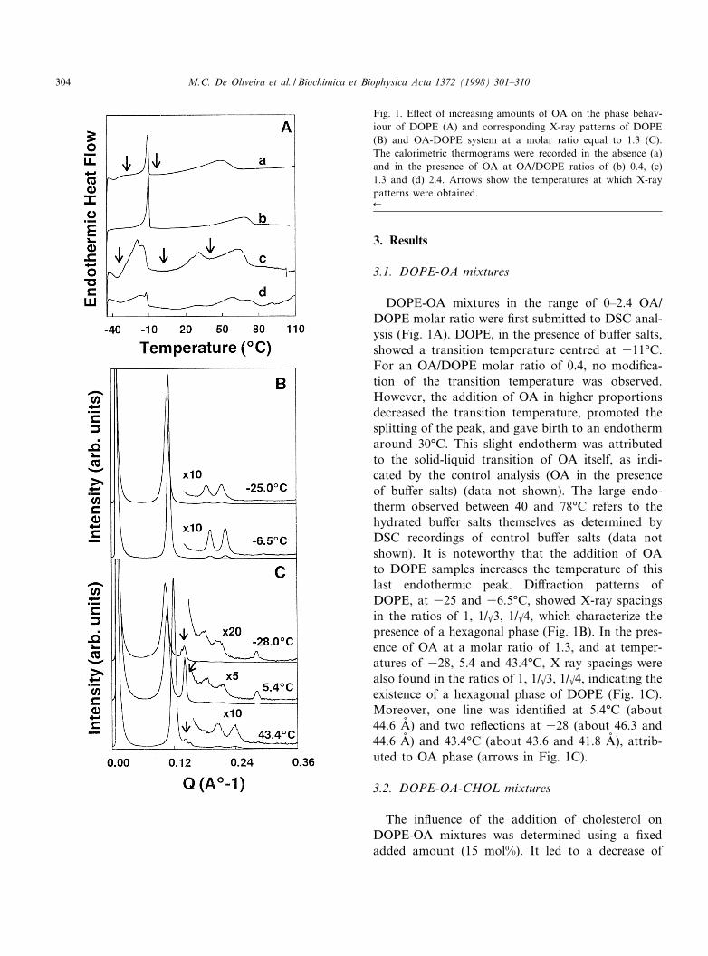
DOPE-OA mixtures in the range of 0^2.4 OA/DOPE molar ratio were ¢rst submitted to DSC anal-ysis (Fig. 1A). DOPE, in the presence of bu¡er salts,showed a transition temperature centred at 311³C.For an OA/DOPE molar ratio of 0.4, no modi¢ca-tion of the transition temperature was observed.However, the addition of OA in higher proportionsdecreased the transition temperature, promoted thesplitting of the peak, and gave birth to an endothermaround 30³C. This slight endotherm was attributedto the solid-liquid transition of OA itself, as indi-cated by the control analysis (OA in the presenceof bu¡er salts) (data not shown). The large endo-therm observed between 40 and 78³C refers to thehydrated bu¡er salts themselves as determined byDSC recordings of control bu¡er salts (data notshown). It is noteworthy that the addition of OAto DOPE samples increases the temperature of thislast endothermic peak. Di¡raction patterns ofDOPE, at 325 and 36.5³C, showed X-ray spacingsin the ratios of 1, 1/k3, 1/k4, which characterize thepresence of a hexagonal phase (Fig. 1B). In the pres-ence of OA at a molar ratio of 1.3, and at temper-atures of 328, 5.4 and 43.4³C, X-ray spacings werealso found in the ratios of 1, 1/k3, 1/k4, indicating theexistence of a hexagonal phase of DOPE (Fig. 1C).Moreover, one line was identi¢ed at 5.4³C (about44.6 Aî ) and two re£ections at 328 (about 46.3 and44.6 Aî ) and 43.4³C (about 43.6 and 41.8 Aî ), attrib-uted to OA phase (arrows in Fig. 1C).
3.2. DOPE-OA-CHOL mixtures
The in£uence of the addition of cholesterol onDOPE-OA mixtures was determined using a ¢xedadded amount (15 mol%). It led to a decrease of
Fig. 1. E¡ect of increasing amounts of OA on the phase behav-iour of DOPE (A) and corresponding X-ray patterns of DOPE(B) and OA-DOPE system at a molar ratio equal to 1.3 (C).The calorimetric thermograms were recorded in the absence (a)and in the presence of OA at OA/DOPE ratios of (b) 0.4, (c)1.3 and (d) 2.4. Arrows show the temperatures at which X-raypatterns were obtained.6
BBAMEM 77392 10-7-98
M.C. De Oliveira et al. / Biochimica et Biophysica Acta 1372 (1998) 301^310304

the lower transition temperature and abolished thesplitting of the peak in the case of OA/DOPE molarratios equal to 1.3 and 2.4 (Fig. 2). On the otherhand, in the case of lower OA/DOPE molar ratio(0.4), addition of cholesterol slightly increased thewidth of the transition peak of DOPE. Fig. 2 alsoshows that the introduction of cholesterol in the OA-DOPE mixture, 1.3 molar ratio, abolished the endo-therm around 30³C, and broadened this transition inthe case of 2.4 OA/DOPE molar ratio.
3.3. DOPE-oligonucleotide mixtures
Fig. 3A depicts the in£uence of two oligonucleo-tide concentrations on the phase behaviour ofDOPE. Preparations submitted to the puri¢cationstep by ultracentrifugation showed an endothermictransition at 37³C, which was increased by 4³C incomparison with DOPE phase transition. Such puri-¢ed samples can be considered as low concentratedin ON (DOPE:ON 1:0.002). In the case of unpuri-¢ed samples (DOPE:ON 1:0.02), the shift of thispeak was even more pronounced (6^7³C). As previ-ously observed with OA, the addition of ON mole-cules to DOPE samples (DOPE:ON 1:0.002) leads tothe shift of the peak corresponding to the bu¡er saltstowards higher temperatures (around 70³C). Con-
cerning X-ray analysis and as described above, hex-agonal phase was found in DOPE samples at both325 and 36.5³C (Fig. 1B). In the case of a lowconcentrated oligonucleotide-DOPE mixture, thesmall-angle re£ections, at 328 and 30.6³C, werealso in the ratio of 1, 1/k3, 1/k4 and demonstratedtherefore the existence of a hexagonal phase (Fig.3B).
Fig. 2. In£uence of cholesterol (15 mol%) on the phase transi-tion of di¡erent mixtures of DOPE and OA. Calorimetric tracesare shown in the absence of cholesterol (a,b,c) and in its pres-ence (aP,bP,cP) in the case of OA/DOPE molar ratios of0.4 (a,aP), 1.3 (b,bP) and 2.4 (c,cP).
Fig. 3. E¡ect of two concentrations of ON on the phase behav-iour of DOPE (A) and corresponding X-ray patterns of a puri-¢ed DOPE-ON preparation (B). The calorimetric traces wererecorded in the absence of ON (a), in the case of a low ONconcentration (puri¢ed liposomes) (b) and a high ON concen-tration (unpuri¢ed liposomes) (c). Arrows show the tempera-tures at which X-ray patterns were obtained.
BBAMEM 77392 10-7-98
M.C. De Oliveira et al. / Biochimica et Biophysica Acta 1372 (1998) 301^310 305

3.4. DOPE-OA-ON mixtures
The e¡ect of ON (low concentration) on the ther-mal events of DOPE:OA 1:1.3 is shown in Fig. 4A.These ratios were chosen with regard to the ¢nallipids to ON concentrations in pH-sensitive lipo-somes. The main transition temperature appeared
shifted upwards in the presence of oligonucleotidesand the splitting of the peak seemed to be abolished.Concomitantly, the enthalpy of the endothermaround 30³C was increased. X-Ray measurementsat 328³C showed the presence of both hexagonaland lamellar phases in the OA-DOPE system (1.3:1molar ratio), corresponding to DOPE and OA, re-spectively, as described above (Fig. 1C). In contrast,for the OA-DOPE-ON-containing system (Fig. 4B),two series of two X-ray line spacings in the ratios ofthe unit-cell dimension, dlam, 1/1, 1/2, were observedat 328³C. At 36.5³C, however, re£ections were dif-ferent, in the ratios of 1, 1/k3, 1/k4 and 1/1, 1/2,indicating the coexistence of hexagonal and lamellarphases. Lastly, at 42³C, the X-ray patterns showedline spacings in the ratios of 1, 1/k3, 1/k4 and a short-er re£ection at 41.1 Aî (Fig. 4B). Thus, X-ray di¡rac-tion allowed to attribute the ¢rst endotherm (around315³C) to LK-HII DOPE phase transition and thesecond one (around +30³C) to solid-liquid OA phasetransition.
3.5. DOPE-OA-CHOL-ON mixtures
Finally, the e¡ect of oligonucleotides on the ther-mal phase behaviour of a DOPE-OA-CHOL system(1:1.3:0.4 molar ratio) was investigated. The pres-ence of a high concentration of oligonucleotide(0.02 ON/DOPE molar ratio) induced a high increasein DOPE transition temperature and a decrease inthe transition enthalpy (Fig. 5A). In the case of alower oligonucleotide concentration, the modi¢ca-tions were less pronounced: appearance of a smallendotherm at 313.6³C and a slight decrease in totalenthalpy corresponding to DOPE phase transitionregions.
In the absence of ON, the X-ray patterns ofDOPE-OA-CHOL, at 36³C, showed re£ectionsin the ratios 1 (66.14), 1/k2 (46.19), 1/k3 (38.08),1/2 (32.72), 1/k8 (23.71), indicating the presence ofa cubic phase (in parentheses, measured values)(Fig. 5B). In contrast, the sample containing the low-est oligonucleotide concentration (0.002 ON/DOPEmolar ratio) revealed the presence of two series oftwo X-ray spacings in the ratios of 1/1, 1/2, at thetemperatures of 329, 315 and 36³C (Fig. 5C).These X-ray patterns suggested the existence of geland liquid-crystalline lamellar phases of DOPE. At
Fig. 4. Thermal phase behaviour of an oligonucleotide-contain-ing OA-DOPE system (1.3 molar ratio) (A). The calorimetrictraces are shown in the absence (a) and in the presence of alow ON concentration (0.002 ON/DOPE molar ratio) (b). Ar-rows show the temperatures at which X-ray patterns were ob-tained. X-Ray di¡raction lines (B) are those of an OA-DOPEsystem (1.3 molar ratio) containing oligonucleotide (0.002 ON/DOPE molar ratio).
BBAMEM 77392 10-7-98
M.C. De Oliveira et al. / Biochimica et Biophysica Acta 1372 (1998) 301^310306

43³C, however, X-ray lines which were observed inthe ratios of 1, 1/k3, 1/k4 for both samples, character-ize a hexagonal phase.
4. Discussion
PE are known to have a strong tendency to adoptthe inverted hexagonal phase. At low water contents,this has been explained by the formation of phos-phate-ammonium hydrogen bonds resulting in a de-crease in the e¡ective hydrophilicity of phosphatidyl-ethanolamine headgroups [15]. Such intermolecularbonds are all the more favoured as the water contentis low. Indeed, when hydrogen bonds can no more besatis¢ed with water, intermolecular bondings appear.The presence of a hexagonal phase in the case of ourDOPE samples even at low temperatures, is then ingood agreement with those reported in other studies[17^19], describing hexagonal phases in low hydratedDOPE samples.
The structural dimensions of the phase observedbefore and after phase transition by X-ray di¡ractionallowed to determine their crystalline parameters.According to Rand and Fuller [18], the water coreprisms can be assumed either as circular in cross-section and of radius Rw, or as hexagonal in cross-section and of side Hw (Fig. 6). The reductions of the`interaxial' `bilayer' thickness, dl, and of the intersti-tial length, dm, observed for DOPE after the maintransition at 311³C (Table 1) are consistent with thechain-ordered (or solid) hexagonal to chain-disor-dered (or liquid) hexagonal transition.
The presence of OA induced the appearance of aphase separation, resulting in domains with hetero-geneous distribution of DOPE and OA, which isidenti¢ed by the splitting of the DOPE transitionpeak in Fig. 1A. Furthermore, hydrogen bondingbetween PE molecules must be prevented by the pres-ence of OA, and the interactions between hydrocar-bon chains must be lowered. This can explain thatDOPE transition shifted to lower temperatures in the
Fig. 5. Thermal phase behaviour of an oligonucleotide-contain-ing DOPE-OA-CHOL system (1:1.3:0.4 molar ratio) (A). Thecalorimetric traces are shown in the absence (a) and in the pres-ence of low ON concentration (0.002 ON/DOPE molar ratio)(b) and high ON concentration (0.02 ON/DOPE molar ratio)(c). Arrows show the temperatures at which X-ray patterns(B,C) were obtained. X-Ray di¡raction patterns are those of aDOPE-OA-CHOL system (1:1.3:0.4 molar ratio) in the absence(B) and in the presence (C) of oligonucleotide (0.002 ON/DOPE molar ratio).6
BBAMEM 77392 10-7-98
M.C. De Oliveira et al. / Biochimica et Biophysica Acta 1372 (1998) 301^310 307

presence of OA. The calculated dimensions dl and dm
(Table 1) support this interpretation since they areboth increased by the presence of OA. The hetero-geneous distribution of OA in itself can be attestedby the presence of a lamellar OA phase meltingaround 30³C as indicated by both DSC and X-raypatterns (endotherm at 30³C and decrease in thespacing lines between 5.4 and 43.4³C).
The addition of ON molecules to DOPE, on thecontrary, seemed to increase intermolecular interac-tions as evidenced by the increase in DOPE phasetransition temperature. We can reasonably assumethat phosphate groups brought by ON moleculesparticipate in electrostatic interactions with PEamine headgroups, competing with PE phosphate'sown groups. As regards structural dimension calcu-lations (Table 1), ON favoured the ordered hexago-nal phase by decreasing dl and dm, thus bringing PEmolecules closer to each other. This can be explainedby charge screening or intermolecular interactionwhich change the apparent v/al of DOPE as observedwith ionic surfactants species [20]. Indeed, a curva-ture increase, corresponding to an increase in v/al,
induces a decrease in both rod diameters and distan-ces. Similar observations were found with DOPE/DOTAP cationic liposomes [21] in which DNA wasshown to favour highly ordered structures.
Although bearing the same electric charge, it mustbe noticed, at this time, that OA and ON have op-posite e¡ects on v/al (likely due to the fact that one isamphiphilic and the other is not), which might com-pete when OA and ON would both be mixed withDOPE. Results described just before clearly showedthat ON displaced OA from the lipid phase, as at-tested by the disappearance of peak splitting ofDOPE transition and the increase in the enthalpymeasured for the endotherm around 30³C, attributedto OA phase. These observations suggest that ON-DOPE interactions are stronger than OA-DOPEones. This can easily be understood if we considerthe length of ON molecules (15-mer) including15 phosphate groups able to interact with amineDOPE headgroups. As proposed in Fig. 7, ON mol-ecules might organize perpendicular to DOPE mole-cules, modifying the surface pressure. This modelcould explain the appearance of a lamellar phase
Fig. 6. Structural parameters describing molecular packing of the hexagonal and lamellar phases (according to [18]).
BBAMEM 77392 10-7-98
M.C. De Oliveira et al. / Biochimica et Biophysica Acta 1372 (1998) 301^310308

for DOPE in the presence of both OA and ON atvery low temperatures (Fig. 4B), considering thatON might have increased lateral pressure in theDOPE headgroup region. Above the melting temper-ature (at 313.4³C), £uidity allows OA to leave theDOPE region and to gather in an extra lamellarphase. The absence of OA between DOPE moleculeswould then allow DOPE molecules to be broughtcloser to each other, favouring hexagonal phase for-mation (as illustrated in Fig. 7).
Concerning the endothermic event correspondingto bu¡er salts, OA and ON both tend to increase thetemperature of this peak as shown for OA in Fig.1A. This e¡ect probably results from electrostaticinteractions between OA or ON and the bu¡er com-ponents (carboxylic and phosphate groups on onehand, and Tris on the other hand).
Cholesterol is known to modify lipid £uidity andto decrease transition enthalpy of phosphatidylcho-line derivatives. With both saturated or unsaturatedphosphatidylethanolamines, a decrease in both melt-ing enthalpies and temperature is observed as foundby Van Dijck et al. [22], Takahashi et al. [23] andBlume [24]. In our study, cholesterol induced a tem-perature decrease in DOPE transition which is ingood agreement with the literature. However, whenOA is mixed with DOPE samples (1.3 and 2.4 molarratio), it is interesting to note that the addition ofcholesterol led to the co-crystallization of the lipids,
which was seen from the disappearance of the split-ting of the transition peak of DOPE. As discussedabove, this broad transition at lower temperature canbe explained by taking into account DOPE intermo-lecular hydrogen bonds. Cholesterol, as OA, verylikely interferes with the formation of these hydrogenbonds. Concerning samples containing both choles-terol and ON, it is interesting to point out that la-mellar phases could be observed in a larger range oftemperatures than in the absence of cholesterol. Asdescribed above, cholesterol may help the co-crystal-lization of lipids, and at least part of OA will be ableto stay in DOPE regions above the solid/liquid tran-sition. At higher temperatures, hexagonal transitionoccurs, leading to the clear appearance of OA lamel-lar phases (Fig. 5C, 43³C).
This study clearly demonstrated that interactionsbetween ON and DOPE occur when molecules are inclose contact. Because of the number of electrostaticinteractions able to form between one ON moleculeand DOPE, the interactions of ON with liposomalmembrane are rather strong (able to displace OAmolecules). In the presence of OA and cholesterol,they are strong enough to favour a lamellar phaseover a large range of temperatures. In our study,whereas OA alone could not allow the formationof lamellar phase, ON supplied the default of waterand satis¢ed hydrogen bonds with DOPE. Such aresult would let think that the phase behaviour ofpH-sensitive liposomes in the endosomal compart-ment could be altered by the presence of ON. How-ever, in these conditions, water molecules will be inlarge excess, and will compete with ON. In orderto clarify the comportment of pH-sensitive formula-tions in a biological medium, studies in fully hy-drated systems are currently in progress in our labo-ratory.
Acknowledgements
We thank the Fundac,a¬o Coordenac,a¬o de Aperfei-c,oamento de Pessoal de N|vel Superior-CAPES, Bra-zil, for supporting Moªnica Cristina De Oliveira witha scholarship. We also thank the GDR from theCNRS No. 1207 for ¢nancial support. We thankG. Keller (URA CNRS 1218) for his technical sup-port.
Fig. 7. Schematic representation of the interactions likely to oc-cur between oligonucleotides and DOPE in the presence of OA.
BBAMEM 77392 10-7-98
M.C. De Oliveira et al. / Biochimica et Biophysica Acta 1372 (1998) 301^310 309

References
[1] C.-Y. Wang, L. Huang, Biochemistry 28 (1989) 9508^9514.[2] C.-J. Chu, J. Dijkstra, M.-Z. Lai, K. Hong, F.C. Szoka,
Pharm. Res. 7 (1990) 824^834.[3] R. Reedy, F. Zhou, L. Huang, F. Carbone, M. Bevan, B.T.
Rouse, J. Immunol. Methods 141 (1991) 157^163.[4] M.-J. Choi, H.-S. Han, H. Kim, J. Biochem. 112 (1992) 694^
699.[5] D.C. Litzingzer, L. Huang, Biochim. Biophys. Acta 1113
(1992) 201^227.[6] K. Kono, K.-i. Zenitani, T. Takagishi, Biochim. Biophys.
Acta 1193 (1994) 1^9.[7] X. Zhou, A.L. Klibanov, L. Huang, J. Liposome Res. 2
(1992) 125^139.[8] H. Ellens, J. Bentz, F.C. Szoka, Biochemistry 23 (1984)
1532^1538.[9] M.B. Yatvin, W. Krentz, M. Horwitz, M. Shinitzky, Science
210 (1980) 1253^1255.[10] D. Liu, L. Huang, Biochim. Biophys. Acta 1022 (1990) 348^
354.[11] D. Collins, D.C. Litzinger, L. Huang, Biochim. Biophys.
Acta 1025 (1990) 234^242.[12] J.M. Seddon, Biochim. Biophys. Acta 1031 (1990) 1^69.
[13] C. Ropert, M. Lavignon, C. Dubernet, P. Couvreur, C.Malvy, Biochem. Biophys. Res. Commun. 183 (1992) 879^885.
[14] C. Ropert, C. Malvy, P. Couvreur, Pharm. Res. 10 (1993)1427^1433.
[15] C. Ropert, Z. Mishal, J.M. Rodrigues, C. Malvy, P. Couv-reur, Biochim. Biophys. Acta 1310 (1996) 53^59.
[16] G. Keller, F. Lavigne, C. Loisel, M. Ollivon, C. Bourgaux,J. Therm. Anal. 47 (1996) 1545^1565.
[17] K. Gawrisch, V.A. Parsegian, Biochemistry 31 (1992) 2856^2864.
[18] R.P. Rand, N.L. Fuller, Biophys. J. 66 (1994) 2127^2138.[19] M.M. Kozlov, S. Leikin, R.P. Rand, Biophys. J. 67 (1994)
1603^1611.[20] J.N. Israelachvili, D.J. Mitchell, B.N. Ninham, J. Chem.
Soc. Faraday Trans. 2 (1976) 1525^1567.[21] J.O. Ra«dler, J. Koltover, T. Salditt, C.R. Sa¢nya, Science
275 (1997) 810^814.[22] P.W.M Van Dijck, B. De Kruij¡, L.L.M. Van Deenen, J. De
Gier, R.A. Demel, Biochim. Biophys. Acta 455 (1976) 576^587.
[23] H. Takahashi, K. Sinoda, I. Hatta, Biochim. Biophys. Acta1289 (1996) 209^216.
[24] A. Blume, Biochemistry 19 (1980) 4908^4913.
BBAMEM 77392 10-7-98
M.C. De Oliveira et al. / Biochimica et Biophysica Acta 1372 (1998) 301^310310