Antimalarial drug discovery: efficacy models for compound screening
Upload
drbabasahebambedkarmarathwadaCategory
view
3download
0
Send Orders for Reprints to [email protected]
214 Current Medicinal Chemistry, 2015, 22, 214-236
Peptide Deformylase: A New Target in Antibacterial, Antimalarial and Anticancer Drug Discovery
Jaiprakash N. Sangshetti1,*, Firoz A. Kalam Khan1 and Devanand B. Shinde2
1Y. B. Chavan College of Pharmacy, Dr. Rafiq Zakaria Campus, Rauza Baugh, Aurangabad-431001; 2Department of Chemical Technology, Dr. B. A. M. University, Aurangabad-431004, India
Abstract Peptide deformylase (PDF) is a class of metalloenzyme responsible for catalyzing the removal of the N-formyl group from N-terminal methionine following translation. PDF inhibitors are moving into new phase of drug development. Initially, PDF was considered as an important target in antibacterial drug dis-covery; however genome database searches have revealed PDF-like sequences in parasites (P. falciparum)and human, widening the utility of this target in antimalarial and anticancer drug discovery along with anti-bacterial. Using structural and mechanistic information together with high throughput screening, several types of chemical classes of PDF inhibitors with improved efficacy and specificity have been identified. Various drugs like, GSK-1322322(Phase II), BB-83698 (Phase I), and LBM-415 (Phase I) have entered into clinical developments. Developments in the field have prompted us to review the current aspects of PDFs, especially their structures, different classes of PDF inhibi-tors, and molecular modeling studies. In nut shell, this review enlightens PDF as a versatile target along with its inhibitors and future perspectives of different PDF inhibitors.
Keywords: Antibacterial, anticancer, antimalarial, clinical developments, peptide deformylase.
1. INTRODUCTION
The successful development of antibacterial drugs gener-ated a misconception in the late 1960s and early 1970s that infectious diseases had been conquered, resulting in a de-crease in academic and industrial research in antibacterial area. However, 40 years later, infectious diseases remain the second-leading cause of death worldwide [1]. During the past decades, many epidemiologists have reported that pathogenic bacteria and parasites rapidly develop resistance to medicines currently used for treatment in human infec-tions [2]. The emergence of multi-drug resistance is of great concern and has created a situation in which there are few or no treatment options for infections with certain microorgan-isms [3]. Gram-negative bacteria like P. aeruginosa, K.pneumoniae, S. maltophila and A. baumannii are posing the most serious treatment challenges with increasing incidence of drug resistant isolates, and also there are fewer active an-tibiotics available to treat infections caused by such organ-isms [4, 5]. Among Gram-positive pathogens, the drug resis-tant pathogens of greatest concern are S. aureus, S. epider-midis, E. faecium and E. faecalis [6, 7]. Also, the emergence of multi-drug resistant of M. tuberculosis strains has made many of the currently available antitubercular drugs (ATDs) ineffective [8].
Due to the current global resistance crisis, search for new antibacterial agents with activity against drug-resistant pathogens should be a high priority for the academic and industrial researchers. It is now widely accepted that the tra-ditional screening methods are unlikely to generate new
*Address correspondence to this author at the Y. B. Chavan College of Pharmacy, Dr. Rafiq Zakaria Campus, Rauza Baugh, Aurangabad-431001, India; Tel:/Fax: +91-240-23801129; E-mail: [email protected]
promising molecules. Alternative strategies must therefore be developed to find new drugs. One possible strategy is to identify a molecular target at the outset and then to screen the available libraries of chemical compounds, looking for “hits” with potent inhibitory capacities in-vitro. In order to address this strategy, the identification of good and novel target is vital [2]. Therefore, identification of a new target which is previously untapped will play a critical role in the development of new antibacterial agents active against resis-tant pathogens [9]. It is important that such new target should: (i) be present in most human pathogens (i.e., the in-hibitors should have broad spectrum activity); (ii) be absent in human cells; (iii) be part of an essential pathway in the pathogens; (iv) not be inhibited by widely used anti-infective agents; (v) be easy to assay in-vitro and in-vivo; (vi) be highly specific for the pathogens and be non toxic to human being; and (vii) not be result in the rapid acquisition of resis-tance [10]. The analysis of microbial genomes has revealed an abundance of novel and potentially useful targets, but, so far, little has been achieved from these efforts [11]. One tar-get that has not received much attention until recently is pep-tide deformylase (PDF). PDF has been a possible target that may fulfill all the above criteria essential for good target to develop new antibacterial agents with novel mechanism of action.
Recently, genome database searches have revealed eu-karyotic PDF-like sequences in parasites, plants, and mam-mals [12]. Malaria is the world’s most serious tropical para-sitic disease and accounts for 1-3 million deaths each year [13]. The increasing incidences of malaria reflect the devel-opment of drug resistant strains of Plasmodium and justify referring to malaria as a re-emerging disease [14, 15]. Rec-ognizing the potential importance of P. falciparum peptide
1875-533X/15 $58.00+.00 © 2015 Bentham Science Publishers

Peptide Deformylase: A New Target Current Medicinal Chemistry, 2015, Vol. 22, No. 2 215
deformylase (PfPDF) as a drug target, new antimalarial agents with novel mechanism of action can be developed. More recently, the human (H. sapiens) mitochondrial peptide deformylase (HsPDF) has been cloned and characterized [16, 17]. The discovery of HsPDF has raised the possibility for the development of new cancer cell target. Advancements and findings of these PDFs in bacteria, parasites and human provide tremendous opportunities for discovery of new anti-bacterial, antimalarial and anticancer as peptide deformylase inhibitors.
There are very few reports published regarding PDF bi-ology and its suitability as drug target [18-21]. To the best our knowledge, there is no comprehensive review with me-dicinal chemistry view and covering all current aspects of peptide deformylase. In this review, the authors have dis-cussed the potential of PDF inhibitors as a new class of anti-bacterial agents. This article also summarizes the potential of PDF inhibitors as antimalarial and anticancer agents. We have discussed all the recent PDF inhibitors reported and also that are in preclinical and clinical phases.
2. PEPTIDE DEFORMYLASE
Peptide deformylase (PDF) is a class of metalloenzyme and responsible for catalyzing the removal of the N-formyl group from N-terminal methionine following translation. Adam [22] was the first to demonstrate the presence of PDF (EC 3.5.1.27) in crude bacterial extracts more than four dec-ades ago. The process for bacterial protein synthesis (Fig. 1)is initiated with N-formylmethionine (f-Met-tRNAi), which is generated through enzymatic transformylation of me-thionyl-tRNA (Met-tRNAi) by formyl methionyl transferase (f-Mett). The N-formyl group of the polypeptide (emerges from ribosome after completion of elongation process) is removed by the sequential action of peptide deformylase [23, 24]. Methionine amino peptidase (MAP; EC 3.4.11.18) then removes the N-terminal methionine depending on the nature of the second amino acid in the peptide chain [25]. Thus, deformylation plays an important role in bacterial protein maturation.
The molecular catalytic cycle of PDF proceeds as shown in (Fig. 2). The step 1 represents the initial state of PDF as found in the structures. In the next step the formylated pep-tide binds with enzyme, thereby replacing water (W2) by
the carbonyl oxygen of the formyl group. The carbonyl oxygen is polarized by hydrogen bonds to the amide of Leu-91 and the side chain of Gln-50. This supports the nucleo-philic attack of water (W1) (probably a deprotonated water) on the carbonyl carbon of the formyl group that leads to the transition state of the reaction as depicted in step 3. The carbonyl oxygen is tetrahedrally ligated by the metal, car-bonyl carbon, side chain amide of Gln-50, and main chain amide of Leu-91. This arrangement of amide hydrogen bond donors resembles the oxyanion hole in serine and cysteine proteases for stabilizing the transition state. The absence of ionizable groups in the oxyanion hole is well in line with the broad pH-activity profile of PDF-Fe which is nearly con-stant between pH 6.1 and 11.2. The electronic state of the attacked carbonyl carbon changes from sp2 to sp3 accompa-nied by a transition from the tetrahedral to a five-coordinate metal center.
The proton of W1 is transferred with the help of Glu-133 to the amide at the N-terminus of the peptide, the added posi-tive charge making the nitrogen suitable as a leaving group. Subsequent bond cleavage leads to the ternary enzyme-formate-peptide complex shown in step 4. Here, the formate is bound to the five-coordinated metal center and the free N-terminus of the peptide is hydrogen bonded to Glu-133. The reaction proceeds by dissociation of the peptide which leaves an activated enzyme-formate complex as shown in step 5. The existence of such a complex is consistent with bio-chemical data which show that PDF can transfer the formyl group from one formyl peptide to another peptide by a “ping-pong” mechanism. The reaction cycle closes with the release of formate and the uptake of two water molecules, W1 and W2. It is very likely that formate release proceeds by attack of a water molecule on the metal rather than on the formate carbon atom. Evidence comes from the lack of 17Oexchange during incubation of 17O formate in the presence of PDF [26].
Removal of the formyl group from polypeptide by PDF is a necessary activity for prokaryotic cell viability [27]. This activity was not believed to be important in eukaryotic cells until recently, because nuclear encoded proteins are not N-formylated [17]. However, in eukaryotes, mitochondrial pro-tein synthesis may also involve the formylation and de-formylation process. This is evidenced by the presence of the enzyme machinery to perform these activities in mammals
Fig. (1). Initiation of protein synthesis by methionine and role of PDF. Abbreviation: Met, Methionine; tRNAi, Initiator transfer RNA; f-Mett, Formyl methionyl transferase; aa-tRNAe, Amino acid elongation transfer RNA.

216 Current Medicinal Chemistry, 2015, Vol. 22, No. 2 Sangshetti et al.
Fig. (2). Proposed molecular catalytic mechanism of PDF. Abbreviation: W1 and W2, Water molecules.
and among other eukaryotes [28, 29]. PfPDF is found in the apicoplast, an essential multimembrane-surrounded organ-elle. Bracchi-Ricchard et al. [30] have cloned the nuclear encoded gene of the apicoplast-localized PfPDF and over expressed this in E. coli. The recombinant PfPDF is catalyti-cally active, suggesting that formylation and deformylation process occurs in the apicoplast of the malaria parasite and play important role in protein synthesis [31].
HsPDF is recently shown to be present in mitochondria [16, 32]. The physiological role of HsPDF in cells is unclear. The mitochondrial localization of HsPDF, and N-formylation of human mitochondrial translation products for translation initiation point at the 13 proteins encoded by the mitochon-drial genome as putative substrates of HsPDF [33]. The de-crease in human cell growth resulting from PDF inhibitors actinonin and its analogues suggest that HsPDF is functional in the mitochondria of human [34].
3. CLASSIFICATION OF PDFs
Since, Ni2+ or Co2+ substituted PDFs have activity equivalent to that of the Fe2+, various laboratories have in-volved such substitutions for further overproduction and purification of PDFs. Based on published genome sequences and crystal structure data, three types of PDF proteins were defined (Fig. 3). The def genes from E. coli and P. aerugi-nosa, which encode “Type I” PDFs, and those from S. aureus and B. stearothermophilus, which encode “Type II”
PDFs, have been cloned and over expressed. PDFs of all Gram-negative bacteria, some Gram-positive bacteria, and all eukaryotes fall systematically into “Type I” class. The “Type II” PDFs are found in Gram-positive bacteria (with low C+G content) and mycoplasma. However, if two PDFs occur in Gram-positive bacteria, the second is often a “Type I” enzyme [35]. The third class, i.e., “Type III” represents newly identified archaeal, kinetoplastids, leishmanial and trypanosomal homologs [36]. Due to different sequences in highly conserved motifs of “Type III” PDFs, in the absence of experimental evidences, they are unlikely to function as PDF enzyme.
4. STRUCTURE OF PDFs
Peptide deformylase was discovered 40 years ago, but as a result of its unusually unstable activity, it was not fully characterized until very recently when the PDF-encoding gene, def, was cloned [25, 37]. Bacterial PDF utilizes a Fe2+
ion as the catalytic metal ion [38, 39]. However, the Fe2+ ion in PDF is very unstable, and is rapidly and irreversibly oxi-dized to the Fe3+ ion through contact with atmospheric oxy-gen, resulting in an inactive enzyme [40]. Fortunately, it was discovered that addition of other divalent cations in vitro,such as Ni2+ or Co2+ resulted in better enzyme stability with very little loss of enzyme activity [38]. Currently, crystal PDF structures of various prokaryotes and eukaryotes have been reported.

Peptide Deformylase: A New Target Current Medicinal Chemistry, 2015, Vol. 22, No. 2 217
Bacillus subtilis, Mycoplasma pneunomiae, Staphylococcus aureus,Bacillus stearotherophilus, Staphylococcus pyrogenes, Enterococcus faecalis,Lactococcus lactis.
Type of PDFs
Type I Type II Type III
Escherichia coli, Haemophilus influenza, Hellobacter pyroli, Legionella pneumonia, Chlamydia trachomatis, Pseudomonas aeruginosa, Mycobacteriam tuberculosis, Thermus thermophilus, Deinococcus radiodurans, Treponema pallidum, Borrella burdorferi, Prochlorococcus marinus, Streptomyces coelicolor, Arabidopsis thaliana, Aquifex aeolicuc, Rickettsia prowazekii, Vibrio cholerae, Neisseria gonorrhoeae, Clostridium acetobutylicum, Thermotoga maritima, Plasmodium falciparum, Homo sapiens.
Leishmania major,Trypanosoma cruzi, Trypanosoma brucei, Methanothermus fervidus, Archeal PDF.
Fig. (3). A phylogenetic tree for three distinct classes of PDFs.
4.1. Three-Dimensional Structure of Bacterial PDFs
Three-dimensional structure data are available for nu-merous PDFs. The PDFs are small monomers composed of about 160- 200 residues. Very few variations are observed in the lengths of their N- and C-terminal extremities. It has been reported that the N-terminal is essential for in vitro ac-tivity whereas the C-terminal is not required for deformylase action [41]. The conserved residues are in three short stretches of amino acids, motifs 1 {G�G�AAXQ}, 2 {EGC�S}, and 3 {HE�DH} (where � is a hydrophobic amino acid and X is any amino acid). These three motifs have been described as an important criterion for member-ship of the PDF family and for deformylase activity [42]. As these motifs are physically close to each other and also building the three sides of the active site crevice, the three-dimensional structure of the active site of PDFs is conserved. The amino acids cysteine of motif 2 and the two histidines of motif 3 are involved in metal cation binding [43].
The stability of PDFs structure depends upon hydrogen bondings between (i) side chain of the serine of motif 2 and the glutamine side chain of motif 1 and (ii) the carboxylate of the glutamate of motif 2 and the aspartate of motif 3 with ar-ginine located between motifs 2 and 3 [44]. The last conserved region residue is an asparagine, located between motifs 1 and 2. Its side-chain forms hydrogen bond with the backbone of an amino acid located at the N-terminal of the protein. The altera-tion of hydrophobic and hydrophilic residues in motif 1 is responsible for the formation of the �1 strand, along which the peptide substrate aligns. Other less well-conserved residues are involved in the optimization of the packing of the three-dimensional structure [45]. Due to these networks of hydrogen bondings and hydrophobic interactions, the overall structure of PDF is very compact and solid. This makes the PDF to resist proteolytic attacks and folding-unfolding of structure at freez-ing and high temperature.
The sequence similarity between “Types I and II”classes of PDF is low, for example, the homology between the E.
coli and S. aureus proteins is only 23% [35]. Generally, “Type II” PDFs are larger in size due to differences at either the N- or C-termini, as well as internal insertions. The C-terminal of “Type I” PDFs (Fig. 4a) is helical, but in “Type II” PDFs (Fig. 4b), the C-terminal consists of a �-strand that can fold back against the enzyme to form a �-sheet. Also, C-terminal domains of “Type II” PDFs contain many hydro-phobic amino acids, whereas “Type I” PDFs do not. There-fore, the C-terminal domains of “Type II” PDFs may not fold into an �-helix. Compared with “Type I”, “Type II” PDFs have two sequence insertions at the N-terminal, just up-stream from the �1 helix (Insertion I1) and �1 strand in motif 1 (Insertion I2). However, these differences do not signifi-cantly affect the active site regions, which are structurally quite similar between “Types I and II” PDFs [42].
4.2. Three-Dimensional Structure of P. falciparum PDF
The subunit structure of PfPDF consists of a mixed �-�topology, with three anti-parallel �-sheets, three major �-helices, and one small helix near the N terminus (Fig. 4c). The overall dimensions of a subunit are 58�44�39 Å. The enzyme structure has shown a deep pocket, which functions as the substrate binding region. The metal ion is positioned at the bottom of the pocket to enable the enzyme to carry out its catalytic function. The metal ion is tetrahedrally coordinated by three amino acid residues (Cys-155, His-196, and His-200, respectively) and a water molecule.
The sequence identity between the structure of E. coliPDF (PDB ID: 1DFF) and P. falciparum PDF (PDB ID: 1BS8) is about 33%. There are three insertions in the loop connecting � strands, �3 and �4 in the PfPDF that result in an altered loop structure from that in the E. coli PDF en-zyme. The most significant insertion is of Tyr-125, which packs against �2 helix, forcing the latter to shift along the helix axis toward the active site in the PfPDF enzyme. An additional difference pertains to the last helix, comprising amino acid residues 142-145, in the E. coli PDF enzyme.

218 Current Medicinal Chemistry, 2015, Vol. 22, No. 2 Sangshetti et al.
Fig. (4). Comparison of PDF structures (a) E. coli (PDB ID: 1BS7); (b) S. aureus (PDBID: 2AI9); (c) P. falciparum (PBD ID: 1BS8); and (d) H. sapiens (PBD ID: 3G5K); M1: motif 1, M2: motif 2, and M3: motif 3.
The corresponding residues form a coil in the PfPDF struc-ture. The structural similarity is closely conserved in the metal binding region: 44 main chain and five side chain resi-dues (Gln-111, Cys-155, His-196, Glu-197, and His-200) superimposed within 0.58 Å.
There are several important structural differences be-tween the PDF of E. coli and P. falciparum in the substrate binding region. First, the conserved Ile-105 in PfPDF lies closer to the active site cavity than the equivalent Ile-44 in E. coli PDF, making a ridge on the active site floor and decreas-ing the cavity volume. Specifically, the inserted residue Tyr-125 in the PfPDF enzyme leans against the end of helix �2, where the crucial Ile-105 is located, causing this helix to move closer toward the active site. Second, the side chain of Arg-97 in E. coli PDF acts as a lid over the active site region. The corresponding Glu-161 in PfPDF is too short to provide any additional interaction. Third, the Arg-97-Glu-42 pair in E. coli PDF is replaced by the Glu-161-Lys-103 pair in PfPDF. The change of this pair does not cause any change in the overall electrostatic charge lining the active site cavity, but it leads to important consequences with respect to bind-ing of inhibitors. The fourth and last important difference, Cys-129 in the E. coli PDF is replaced by Ile-193 in PfPDF. This leads to decrease in volume of active site, but this dif-ference is relatively small [31].
4.3. Three-Dimensional Structure of Human PDF The crystal structure of human mitochondrial PDF
(HsPDF) was expressed and purified as Co2+ enzyme be-cause this was the only metal that allowed reconstitution of
its enzymatic activity [16]. The approximately 30% sequence similarity is observed between HsPDF and other non-mammalian PDFs, such as Gram-positive and Gram-negative bacteria, and plants. In HsPDF (Fig. 4d), an antiparallel �-sheet is formed by �1 (Gly-52 to Ser-54), �2 (Val-64 to Leu-67), and �3 (Arg-93 to Val-96), while a second antiparallel �-sheet is formed by �4 (Ser-99 to Leu-103), �5 (Leu-107 to Glu-112), �6 (Ala-122 to Gly-127), �7 (Ala-128 to Leu-135), and �8 (Gly-139 to Ser-147). The helices consist of �1(Pro-32 to Arg-48), �2 (Glu71 to Glu76), �3 (Pro-79 to Arg-85), and �4 (Trp-149 to Gln-162). The geometry of the metal ion (Co2+) in HsPDF is close to tetrahedral. The Co2+ metal ion is kept at the active site by coordination to the side chain N atoms of His-156 and His-160, the side chain sulfur atom of Cys-114, and a fourth unexpected ligand (PO4
3- ). The topology of the metal and coordinating atoms and the metal in HsPDF is comparable between HsPDF and other non-mammalian PDFs.
The HsPDF shares the � hairpin loop anchoring points that shape the entrance to the active site in the PDF family. However, the conformation of the C-terminus in HsPDF, in combination with the presence of the �2/�3 helical loop cre-ates a characteristic entrance to its active site compared to non-mammalian PDFs. The sequence identity between the structure of HsPDF and E. coli PDF is low (~21%). The to-pology of amino acid residues involved in the reaction mechanism and metal coordination is conserved; this sug-gests that the mechanism of catalysis of HsPDF is also con-served. The substrate binding S1’ pocket is conserved in HsPDF, but lacking the S2’and S3’ pockets as observed in

Peptide Deformylase: A New Target Current Medicinal Chemistry, 2015, Vol. 22, No. 2 219
other PDFs. Despite HsPDF lacking true S2’and S3’ substrate binding pockets, the residue surrounding the S2’and S3’pockets such as Arg-48, Met-87, Arg-85, Pro-111, Glu-115, Leu-121, and Trp-149, which create a hydrophobic depres-sion, could contribute to binding specificity.
The HsPDF enzyme shares only 28% amino acid se-quence identity with malarial enzyme (PfPDF). The differ-ence in the substrate binding pocket between HsPDF and PfPDF include: (i) Arg-107 in HsPDF verses Lys-103 in PfPDF, (ii) Val-109 in HsPDF verses Ile-105 in PfPDF, (iii) Leu-179 in HsPDF verses Glu-161 in PfPDF, and (iv)Pro-169 in HsPDF verses Ile-152 in PfPDF [30]. The amino acid sequences of PfPDF, EcPDF and HsPDF are shown in (Fig. 5).
4.4. Substrate-Binding Pockets
The active site of PDF proteins contains three substrate-binding pockets along with the metal binding site (Fig. 6). These pockets are referred to as S1
’, S2’ and S3
’ pockets and corresponding positions on substrate or inhibitors are re-ferred to as P1
’, P2’ and P3
’.
4.4.1. S1’ Pocket
The S1’ pocket of PDF is mostly hydrophobic pocket and
conserved among all of the bacterial PDF [35]. This pocket is the binding site for the methionine side chain, which is required to remove the formyl group from the N-formylmethionine peptide. This pocket remains very similar
in size and shape among PDFs from various Gram-positive and Gram-negative organisms. The S1
’ pocket is formed by Ile-44, Ile-86, Glu-88, Leu-125, Ile-128, Cys-129, and His-132. The only non-conservative amino acid residue in this region lies on the border between the S1
’ and S3’ pockets. The
residues are Leu-125 in E. coli, Leu-125 in H. influenza,Leu-127 in P. aeruginosa, Leu-146 in B. stearothermophi-lus, Tyr-166 in S. pneumoniae, Tyr-147 in S. aureus, and Tyr-122 in T. marimata. A good enzyme inhibition can be obtained with inhibitors that bind only to the metal and into the S1
’ pocket in both Gram-positive and Gram-negative PDFs [46, 47].
4.4.2. S2’ Pocket
The S2’ pocket is an open tunnel-like space pointing out
into solvent. The amino acid residues lining the S2’ pocket
differ in different species, but overall chemical nature re-mains the same for all PDFs [35]. This region is able to ac-commodate a large variety of side chains presented by the various formylated substrates to be processed by the PDF proteins [48].
4.4.3. S3’ Pocket
In contrast, the S3’ pocket of PDFs is the least conserved
region and showed greater variation among the species that alter both the shape and chemical nature of the pocket [35]. Similar to S2
’ pocket, this region is also solvent exposed and more of a surface depression [46].
Fig. (5). An alignment of amino acid sequences in P. falciparum (PfPDF), E. coli (EcPDF), and H. sapiens (HsPDF) [31].

220 Current Medicinal Chemistry, 2015, Vol. 22, No. 2 Sangshetti et al.
Fig. (6). Schematic representation of the PDF active site.
4.5. Metal-Binding Site
PDFs are metalloprotease enzyme. The crystal structures of PDF with Fe2+, Ni2+, Co2+, and Zn2+ have been solved and no significant structural differences are observed among the various metal forms. Metal ion in the active site of PDF is tetrahedrally ligated and bound to the two histidines from the HE�DH motif, as well to a cysteine and a water molecule (Fig. 5) [26]. The four ligands are N atom in His-132, His-136 of the highly conserved HE�DH sequence, S atom of Cys-90 in the EGC�S sequence and O atom in water mole-cule. All of these ligands are aligned precisely within an ex-tended H network involving most highly conserved residues as: 1) His-132 is held in position by hydrogen bonding with
Glu-88, which in turn forms hydrogen bonding with Arg-102. The Arg-102 also forms hydrogen bonding with highly conserved Asp-135.
2) His-136 is held in place by two hydrogen bonds with a water molecule which in turn forms hydrogen bond with the backbone of Leu-13.
3) The ligated water molecule forms two hydrogen bonds with highly conserved Glu-133 and one hydrogen bond with another highly conserved residue Gln-50. The Gln-50 forms hydrogen bonds to conserved Ser-92, Ala-47, and water molecule. Ser-92 forms hydrogen bond with the backbone of Leu-6 [44].
5. DISCOVERY OF PDF INHIBITORS
A rational, mechanism-based approach has been success-fully used to design the several therapeutically important metalloprotease inhibitors. The metalloproteases such as angiotensin converting enzyme (ACE) and matrix metallo-proteases (MMPs) are among the best studied of enzyme class and they are excellent precedents for the mechanism-based design of their inhibitors [49]. The fact that PDF is also metalloprotease enzyme, gives it an added attractiveness as a target for drug discovery, since it permits the rational design of inhibitors. So similar strategy like, ACE inhibitors and MMP inhibitors can be adopted to discover therapeuti-cally important PDF inhibitors.
The successful development of E. coli F-PDF [34] led the several groups to investigate and design the substrate analogues as PDF inhibitors. Hu and co-workers [48] pre-pared a combinatorial library of resin-bound formylated tetrapeptide substrates and evaluated which sequentially and
preferably deformylated by E. coli PDF. The study revealed that the optimal residue at P1
’ (Fig. 6) was methionine with glycine and aromatic amines (histidines, phenylalanine, and tyrosine) following at a distant second and third. No selectiv-ity was observed at P2’ residues as all amino acids were se-lected at similar frequencies except for glycine, aspartate, and glutamate, which were not selected by the enzyme as PDF substrate. The P3
’ residue showed a strong preferences for an aromatic residue as over 80% of the selected beads either had tyrosine, phenylalanine or histidine at this posi-tion.
Actinonin is the first reported naturally occurring PDF inhibitors obtained from Streptomyces species [50]. Al-though actinonin is too weak as PDF inhibitor due to its poor bioavailability, it did form the framework for the synthesis, purification, and evaluation of more potent PDF inhibitors. Based on the mechanistic and structural information, to-gether with understanding of the general principles of inhib-iting metalloproteases, a generic PDF inhibitor structure was proposed [23] (Fig. 7). In this structure, (i) X represents a metal cation-chelator that will be responsible for providing binding energy, (ii) the n-butyl group is used to mimic the methionine side chain of the substrate at P1, and (iii) side chain at P2 and P3 regions of the inhibitor can provide addi-tional binding energy, selectivity, and favorable pharma-cokinetic and toxicities properties. All PDF inhibitors re-ported to date contain a functional moiety that can chelate the metal ion of the enzyme. Others, while they may not fit the generic inhibitor structure directly, can still be viewed as structures with characteristics of metal cation-chelator [11].
X NN
P3
P1
P2O
OH
H
Fig. (7). Proposed general structure for PDF inhibitors.
6. PDF INHIBITORS
The PDF inhibitors demonstrate potent activity against a wide spectrum of bacterial species, including but not limited to, E. coli, B. sublitis, S. aureus, S. pneumoniae, S. pyogenes,S. agalactiae, V. streptococci, H. influenzae, M. catarrhalis,M. pneumoniae, C. pneumoniae, B. fragilis, M. tuberculosis,H. pylori, P. aerogenosa, Enterococcus spp., Fusobacterium spp., and anaerobes, such as Clostridium spp. Some reports suggest that the in vitro growth of P. falciparum could be inhibited by PDF inhibitors, thus further supporting the po-tential use of PDF inhibitors as anti-parasitic agents. Some PDF inhibitors have also been reported to inhibit tumor growth, both in vitro and in vivo. Using rational design, ran-dom screening or the screening of rationally designed, fo-cused chelator-based libraries, many structural different classes of PDF inhibitors have been identified. The classifi-cation of PDF inhibitors are presented in (Fig. 8).
6.1. PDF Inhibitor as Antibacterial Agents
The PDF inhibitors with antibacterial activity inhibit the enzyme activity very effectively. The PDF inhibitors may also work via stimulation of the innate immune system.

Peptide Deformylase: A New Target Current Medicinal Chemistry, 2015, Vol. 22, No. 2 221
Fig. (8). Classification of PDF inhibitors. In this system, activation of formyl peptide receptors (FPR) mediates phagocyte (neutrophil) migration and release of free radicals and other antibacterial substances from phago-cytes. Theoretically, PDF inhibitors should enhance this re-sponse and, hence, innate immunity. This has been demon-strated with actinonin in animal models and results showed that actinonin enhances the production and secretion of neu-trophil-activating peptides that work via FPR [51]. The exis-tence of HsPDF undoubtedly raises the issue of potential mechanism-based toxicities. However, the lead PDF inhibi-tors exhibit activity against HsPDF enzyme only at ex-tremely high concentrations and have excellent safety pro-files in the preclinical development [52]. In the past few years, several classes of PDF inhibitor as antibacterial agents have been reported.
6.1.1. Peptidic Inhibitor as Antibacterial Agents
6.1.1.1. Thiol Derivatives
Various metal-binding groups such as thiols, carboxy-lates, hydroxamates, and phosphonates have been used to replace the scissible bond of substrates processed by thermo-lysin. Yaoming et al. explored thiol derivatives for their abil-ity to form tight-binding complexes with ferrous and nickel ions [53]. The synthesized compounds exhibited competitive inhibition against E. coli PDF enzyme. The structure-activity relationship studies revealed that replacement of anthracene moiety with naphthalene ring had improved the potency by 3-5 folds. Further reduction of size aromatic ring to just one ring led to slightly decrease in the inhibitory potency. There-fore, an aromatic amine containing two rings appeared to be best accommodated by the PDF active site. The most potent compound (1) has inhibition constant (KI) 0.015 ± 0.002 �M against E. coli PDF.
SH
HN
O
HN
NH
NH2
O
(1)
The same group has also designed and synthesized a se-ries of peptide thiols that act as potent, reversible inhibitors of purified recombinant PDF from E. coli and B. sabtilis [54]. The most potent inhibitor (2) has a KI value of 0.011 �M toward the B. subtilis Co-PDF enzyme. These inhibitors showed antibacterial activity against both Gram-positive and Gram-negative bacteria, with MIC (Minimum inhibitory concentration) value as low as 2 �g/mL. These PDF inhibi-tors had induced bacterial cell lysis and showed bactericidal activity towards all four bacterial strains that have been tested, B. subtilis, S. epidermidis, E. faecalis, and E. coli.
HSHN
ONH
ONO2
(2)
6.1.1.2. H-Phosphonate Derivatives
Similarly, Yun-Jin et al. explored the feasibility of using an H-phosphonate as metal-binding group to inhibit the PDF activity [55]. The structure of H-phosphonate compounds

222 Current Medicinal Chemistry, 2015, Vol. 22, No. 2 Sangshetti et al.
revealed that H-phosphonate group could potentially resem-ble the tetrahedral intermediate or the transition states for the formation and/ or breakdown of the intermediate and, there-fore, act as a deformylase inhibitor. The inhibitor (3) was not highly potent (KI = 37 �M versus E. coli Fe-PDF and KI = 76 �M versus E. coli Zn-PDF).
OO
NH O
HN
(3)
PO
OHH
NO2
6.1.1.3. Aldehyde Derivatives
Researchers at Merck Research Laboratory investigated a small set of peptide aldehyde inhibitors, postulating that the aldehyde might bind to the metal centre in the form of a hy-drate, thus serving as a transition state mimetic [56]. The most potent inhibitor was calpeptin (4) and showed good inhibitory effect against the zinc-containing metalloenzymes, E. coli and B. subtilis PDF with KI values of 26 and 55.6 �M, respectively. Cobalt-substituted E. coli and B. sabtilis PDFs were also inhibited by these aldehydes with KI values for calpeptin of 9.5 and 12.4 �M, respectively. Calpeptin has a similar potency and mechanism of inhibition with PDFs from both Gram-negative (E. coli) and Gram-positive (B. subtilis) bacterial sources. This gives added support to the proposal that a single PDF inhibitor might have broad-spectrum activity.
O
O
NH O
HN
O HCalpeptin(4)
6.1.2. Pseudopeptidic Inhibitors as Antibacterial Agents
6.1.2.1. Hydroxamic Acid or N-formyl-N-hydroxylamine Derivatives
Actinonin (5) is a peptidic hydroxamic acid has been known as moderate antibacterial agents. Despite minor dif-ferences in the PDF active sites and secondary structures, actinonin adopts a similar binding conformation in all of the PDF enzymes for which co-crystallization data are available. The conserved residues involved in binding interactions with actinonin are concentrated in the S1
’ pocket, related to or needed for metal coordination, or involved in H-bonding to the amide bonds [34]. It has never been developed as thera-peutic agent due to lack of in vivo efficacy in animal models of infection, due to its known instability. It also lacks the selectivity against human secondary targets, as it binds other classes of metalloproteases [57].
However, actinonin as a potent inhibitor of PDF was a key discovery in the development of newer PDF inhibitors with superior enzyme binding through a metal-chelator. Al-though compounds with many different chelators can inhibit the enzyme, only compounds containing hydroxamic acid or
N-formyl-N-hydroxylamine exhibit appreciable antibacterial activity. Most PDF inhibitors identified to date share a com-mon structural feature of a “chelator + peptidomimetic” scaf-fold [20]. Researchers of British Biotech reported the N-formyl hydroxylamine containing compound, BB-3497 (6) as an effective inhibitor (IC50= 0.007 �M) of the E. coli Ni-PDF enzyme, exhibiting potent antibacterial activity both invitro and in vivo [58].
NHN
OH
OO
HN
O OH
Actinonin(5)
NO
H
OHHN
ON
O
BB-3497(6)
6.1.2.2. P2-Pyrrolidine Containing Inhibitors
Using a combination of iterative parallel synthesis and traditional medicinal chemistry, researchers at Versicor and Novartis have identified a new potent class of PDF inhibitors with N-alkyl urea at P1 site [59]. These compounds have shown MICs of �4 �g/mL against Gram-positive and Gram-negative pathogens, including S. aureus, S. pneumoniae, and H. influenza. The IC50 values for these compounds for E. coli Ni-PDF and S. pneumoniae Zn-PDF enzymes were �0.1 �M. In addition, these compounds were very selective for PDF, with IC50 values >200 �M for matrilysin (MMP-7). Molecu-lar modeling information indicated that the urea compounds adopt a binding position similar to that for succinate hy-droxymates. The most potent compound (VRC4307) (7) has displayed in vivo efficacy in a mouse protection assay, with 50% protective dose of 17.9 mg/kg.
NH
NHOO
N
OO N
HN
S
VRC4307(7)
Researchers at Vicuron (formerly Versicor) and Novartis
further investigated the analogues of 2-R-butyl succinic acid and explored the structure-activity relationship (SAR) of various chelator groups, �-substituents, P2 and P3 substituent in order to achieve optimal antibacterial activity with mini-mal toxicity liability [60]. In vitro study indicated that hy-droxamate group provided that best enzyme inhibition and antibacterial activity among various chelators tested. At P2 position, cyclic 4-6 member amino acids were incorporated and found that proline group could be preferred. The struc-ture-activity relationship of P3 position revealed that a proper hydrophilic and hydrophobic would be required for maximal antibacterial activities with reduced toxicity. Pyrrolidine at P3 position showed the best combination of in vitro potency, cytotoxicity and selectivity. The most potent compound (8) has MIC values 1-2, 0.25-1, and 0.25-1�g/mL for S. pneu-moniae, S. aureus, and H. influenza, respectively, and IC50 value 0.02 �M for E. coli Ni-PDF.

Peptide Deformylase: A New Target Current Medicinal Chemistry, 2015, Vol. 22, No. 2 223
NH
HOO
N
OO N
H(8)
Scientists at Novartis also designed and synthesized the highly potent PDF inhibitor of M. tuberculosis [61]. In this study, they have introduced the benzimidazoles and benzox-azoles moieties in peptidomimetic chain to reduce the pep-tidic character. The two most potent compounds, one from each series, (9) and (10) showed IC50 value 0.01 and 0.013 �M, respectively, for M. tuberculosis Ni-PDF. These com-pounds have also shown excellent activity against single and multi-drug resistant M. tuberculosis strains. The compound (9) showed MIC values of 0.1, 0.03, 0.2, 0.125, 0.25, and 0.03 �g/mL and compound (10) 0.15, 0.06, 0.5, 0.6, 0.25, and 0.06 �g/mL against M. tuberculosis strain H37Rv, isoni-azid single-drug resistant strain, streptomycin-resistant strain, rifampicin-resistant strain, pyrazinamide-resistant strain, and Beijing W multi-drug-resistant strain, respec-tively.
NOH
O
N
O NHN
(9)
NOH
O
N
O NO
(10)
In search of potent antibacterial agents against Gram-positive pathogens, Zhenyu et al. have synthesized novel (2S)-N-(substitutedphenyl)-1-[2R)-2-[(formylhyroxyamino) methyl]-1-oxohexyl]-2-pyrrolidinecarboxamide analogues of PDF inhibitor [62]. Modification at P3 position in PDF in-hibitors with hydrophobic group have been shown to an im-provement of both enzymatic and whole-cell activities. One of the most important features of the fluoroquinolone anti-bacterials is the presence of a nitrogen-containing aliphatic heterocycles at position 7 in the quinolone scaffold, with the effect of improving antibacterial activity and/ or pharma-cokinetic properties. Based on these, they investigated the PDF inhibitors with the nitrogen-containing aliphatic hetero-cycles at the P3 position. The most potent compound (11) has shown MIC values 0.139, 0.049, 0.195, 0.049, and 0.39 �g/mL against S. aureus, S. pneumoniae, S. albus, S. en-teridis, and S. non-hemolyticus, respectively.
HONCHO
N
OO N
H
FN
(11)
Scientists at GlaxoSmithKline have reported acylproli-namide derivatives as potent, broad-spectrum PDF inhibitors
[63]. In this study, they have replaced the n-butyl with cy-clopentyl methyl group at P1 position and synthesized the three series as imide, acylcarbamate and N-hyrdoxyformamide derivatives. Among the three series, N-hyrdoxyformamide derivatives have shown better potency in the enzyme assay as well as antibacterial activities, particu-larly against S. pneumoniae. The most potent compound (12) has IC50 values 0.0009, 0.0003, and 0.0018 �M (PDF inhibi-tory activity) and MIC values 0.125, 0.125, and 0.5 �g/mL against S. aureus, S. pneumoniae, and H. influenza, respec-tively.
HON
CHO
N
OO N
H
O
NH
(12) 6.1.2.3. P2-Dihropyrrole Containing Inhibitors
Wei et al. have reported 2,5-dihyropyrrole formyl hyr-doxyamine derivatives as novel and potent PDF inhibitor and screened against both Gram-positive and Gram-negative pathogens [64]. The replacement of pyrrolidine with 2,5-dihyropyrrole group at the P2 position and introduction of heterocyclic amine at P3 position in peptidomimetic chain resulted in good to excellent antibacterial activity. The most potent compound (13), which contains thiadiazolyl function-ality, exhibited MIC values 0.0625 to 0.25 �g/mL against Gram-positive bacterial strains including S. aureus, methicil-lin-susceptible S. aureus (MSSA), methicillin-resistant S. aureus (MRSA), and S. epidermis and 16 to 32 �g/mL against Gram-negative bacterial strains including E. coli 1 (ATCC25922) and E. coli 2 (ATCC35218).
N NHO
OO N
HS
NN CF3
OH
(13) 6.1.2.4. P2-Methylenepyrrolidine Containing Inhibitors
In search of new PDF inhibitors Wei et al. have reported 3-methylenepyrrolidine formyl hydroxyamino derivatives and screened in vitro antibacterial activities against Gram-positive pathogens [65]. The most potent compound (14), which contains thiazoly functionality at P3 position and cy-clopentylmethyl group at P1, exhibited MIC values 0.0625 to 0.5 �g/mL against Gram-positive bacterial strains including S. aureus, methicillin-susceptible S. aureus, methicillin-resistant S. aureus, and S. epidermis. 6.1.2.5. P2-Oxazolidine Containing Inhibitors
Oxazolidine moiety is an important building block for many biologically active agents. It is called pseudoproline and mimics the proline skeleton of pseudopeptidic inhibitors [66-68]. Linliang et al. have reported the substitution of oxa-zolidine moiety at P2 position in peptidomimetic chain and

224 Current Medicinal Chemistry, 2015, Vol. 22, No. 2 Sangshetti et al.
synthesized a series of (2S)-N-substituted-1-[(formyhydroxy-amino) methyl]-1-oxohexyl]-2-oxazolidinecarboxamide de-rivatives as antibacterial agents [69]. These compounds have shown significantly better antibacterial activity against Gram-positive bacteria than Gram-negative bacteria. The most potent compound (15) with 3-chloro-4-morpholinylphenyl substitution at P3-position has MIC val-ues 0.39, 0.098 and 0.39 �g/mL against Gram-positive or-ganisms including S. aureus, S. pneumoniae, and S. albus, respectively, and 6.25 �g/mL against both Gram-negative bacteria including S. boydii and S. flexneri.
N NHO
OO N
HS
N
O H
(14)
N NHO
O
O
O NH
OH
Cl
N
O(15) 6.1.2.6. P2-Azaamino Containing Inhibitors
Bioisosteric replacement of amino acids with azaamino acids is a peptidomimetic strategy which has been applied to a number of protease inhibitors [70]. Also, azaamino acids are more stable, particularly to hydrolytic cleavage by prote-olytic enzymes. Researchers at Vernalis have investigated the N-formyl hydroxylamines containing azaproline and azapicolinic acid as E. coli Ni-PDF enzyme inhibitor [71]. The five-membered furan containing compounds at P3 posi-tion provided the best potent compounds with IC50 value ranging from 0.005-0.03 �M. The most potent compound (16) has IC50 value 0.005 �M for E. coli Ni-PDF enzyme and MIC values 0.5-2 �g/mL against S. pneumoniae (Gram-positive bacteria) and <0.125-0.25, and <0.125 �g/mL against H. influenza, and M. catarrhalis, respectively (Gram-negative bacteria).
N NHO
O
OH
N
OO(16)
6.1.3. Non-Peptidic Inhibitors as Antibacterial Agents
6.1.3.1. Thyropropic Acid Derivatives
Researchers at Pfizer Global Research reported some thy-ropropic acid derivative as a novel, non-peptidic inhibitors of E. coli PDF [72]. SAR study demonstrated that the carboxy-late group is required for activity as ester and amide ana-logues are less potent. The best compound (17) has IC50 value 0.94 ± 0.05 �M. This compound inhibited E. coli PDF similar to actinonin, but lacked antibacterial activity. These compounds were the first non-peptidic inhibitors disclosed and used as template to discover better PDF inhibitors. GlaxoSmithKline has got a patent for hydroxamic acid (18) and N-formyl-N-hydroxylamine (19) analogues of a thyro-propic acid core as PDF inhibitors [73, 74].
O
HO
I
I OH
O
(17) NH
OOH
I
I
O
H
(18)
NOH
I
I
O
H
H
O(19) 6.1.3.2. Bicyclic Hydroxamic Acid Derivatives
Scientists at Hoffmann-La Roche Ltd. have developed and synthesized structurally related bicyclic hydroxamic acid derivative as potent and selective new E. coli PDF inhibitors [75]. They synthesized two series of related hydroxamic acid analogues, quinazoline hydroxyl-acetamides, and thiadiazine hydroxyl-acetamides. The two most potent compounds, one from each series, (20) and (21) showed IC50 value 0.31 and 0.12 �M, respectively. However, these compounds showed weak antibacterial activity.
NH
NHN
OOH
O(20)
NH
SN
HN
OOH
OO
(21)
The group at Novartis has identified and synthesized new thiazepine derivative as very potent inhibitors of S. aureus Ni-PDF with an IC50 in the low nanomolar range [76]. Many of the synthesized compounds proved to be excellent S. aureus Ni-PDF inhibitors, but lacked the antibacterial activ-ity. In order to improve antibacterial activity, they prepared the corresponding sulfone and carbon analogues. These ana-logues showed lower potency than thiazepine series. Further study revealed that these molecules are unable to penetrate the outer cell membrane of E. coli and may bind to the cell membrane of S. aureus. The most potent compound (22) hasshown IC50 value <0.005 �M.
NH
NS
HN
OOH
O(22)
6.1.3.3. �-Sulfonyl- and �-sulfinylhyrdoxamic Acid Deriva-tives
Scientists at Hoffman-La Roche Ltd. have reported �-sulfonyl- and �-sulfinylhyrdoxamic acid derivatives as new class of potent PDF inhibitor [77]. In this, they replaced am-ide group with sulfonyl (23) and sulfinyl (24) group and evaluated for PDF enzyme activity. The sulfonyl-containing compounds generally demonstrated better enzyme inhibitory activity than the sulfinyl compounds. This trend was re-versed with respect to antibacterial activity in spite of their weaker enzymatic activity. However, the antibacterial activ-ity against Gram-positive bacteria was poor. 6.1.3.4. Biaryl Acid Derivatives
The biaryl acid analogues were developed by Merck Re-search Laboratories and evaluated as PDF inhibitor against

Peptide Deformylase: A New Target Current Medicinal Chemistry, 2015, Vol. 22, No. 2 225
E. coli PDF [78]. All the inhibitors contain acidic pharma-cophore, including tetrazole, acyl sulphonamide and car-boxylate. Structure-activity relationship studies of biaryl acid analogues revealed that substitution at the head group, biaryl group, and the nature of acidic group all contributed to the inhibitory activity of the these compounds against PDF. Tetrazole and acyl sulphonamide analogues appeared to pro-vide the best potency. The acidic group of these compounds may bind to the metal ion and instead interact with an amino acid residue within PDF active site much like the binding of the angiotensin II receptor. The most potent compounds, one from each acidic pharmacophoric series (25), (26), and (27) showed IC50 value 3.9, 10, and 22.8 �M, respectively. The antibacterial activity of these compounds was not reported.
SHN
OOH
O
(23)
SHN
OOH
OO
(24)
OHON
NN
(25)
NHNN
NN
N
N
Cl(26)
NN
NS
O OHN O
(27) 6.1.3.5. Benzamide and Aryl Ether Derivatives
The group at GlaxoSmithKline has published a patent application covering the series of aryl ether (28) and ben-zamide (29) derivative as novel PDF inhibitors [79, 80]. These inhibitors were designed to explore the feasibility of preparing small molecular weight, non-peptidic compounds that would bind only into well conserved S1’ pocket. The reported compounds have shown potent enzyme inhibitory activity (<0.1 �M). The benzamide series has shown excel-lent oral bioavailability (100%) in a H. influenza respiratory tract infected rat model (in vivo model).
O
OHH
O
(28)
ClCl
HN
ONOH
H
O
(29) 6.1.3.6. Benzothiazole Derivatives
Wataru et al. have reported a novel series of benzothia-zole derivatives as potent non-peptidic PDF inhibitor [81]. The newly synthesized compounds were evaluated for their in vitro inhibitory effect on E. coli Ni-PDF enzyme and ex-hibited micromolar order enzyme inhibitory activity. The
oxidation of sulfur of benzthiazole ring to sulfone resulted in complete loss of PDF inhibitory activity. SAR studies re-vealed that increase in the length of N-alkyl chain enhanced the PDF inhibitory activity. The compounds showed antibac-terial activity against S. aureus and P. aerogenosa, but they were inactive against E. coli. The most potent compound (30) has IC50 value 1.04 �M for PDF enzyme inhibition and MIC value 10 �g/mL against S. aureus.
N
S NHO OH
(30) 6.1.3.7. Isoxazole Derivatives
Fieulaine et al. have designed and developed a series of isoxazole-3-hydroxamic acid derivatives as potential inhibi-tor of E. coli and S. aureus PDF enzymes [82]. Molecular modeling studies predict that the aryl substituent of isoxazole binds into S1’ pocket and that the oxygen atom of the isoxa-zole is involved in an H-bonding interaction with Ile-44 in E. coli PDF, similar to the P1 carbonyl of actinonin (5). The compounds showed better enzyme inhibitory activity for S. aureus as compared to E. coli. The oxidation of thioether derivatives to sulfoxide and sulfone has resulted in slightly improved PDF enzyme inhibitory activity with decrease in IC50 value 1.5-3 times than the corresponding un-oxidized compounds. The most potent compound (31) has IC50 value 3.4 �M for E. coli PDF enzyme and the compound (32) has 0.8 �M for S. aureus PDF enzyme. The poor antibacterial activity of these compounds may be due to poor penetration of the bacterial cell wall.
NO
S
O
NH
OH
S
(31) N O
S
O
NH
HO Cl
(32) 6.1.3.8. Macrocyclic Derivatives
A macrocyclic PDF inhibitors comprise of peptide mi-metic chain having three residues, P1, P2, and P3, wherein P2 connects with P1 and P3, wherein P1 and P3 each have a side chain, and wherein the side chains on P1 and P3 are cross-linked to complete the cycle. Since most of the PDF inhibitors have significant peptide characteristics, there are some con-cerns about their selectivity (e.g., inhibition of matrix metallo-proteases) and in vivo stability (e.g., proteolysis of peptide bond). The best approach to improve both selectivity and sta-bility of PDF inhibitors is to form cyclic peptides or dipeptides [59]. Xubu et al. have designed and reported the macrocyclic PDF inhibitor of E. coli Co-PDF enzyme [83]. The macrocy-cle containing N-formylhydroxylamine side chain as metal-chelating was synthesized from a diene precursor via olefin metathesis using Grubb’s catalyst. The cyclic inhibitor (33) showed potent inhibitory activity toward E. coli PDF enzyme (KI= 0.00067 �M) and antibacterial activity against both Gram-positive and Gram-negative bacteria (MIC= 0.7-12

226 Current Medicinal Chemistry, 2015, Vol. 22, No. 2 Sangshetti et al.
g/mL). The group has further studied the structure-activity relationship on the size of the macrocycle and found that 15-17-membered macrocycles are optimal for binding to the PDF active site. Unlike the acyclic compounds, which are simple competitive inhibitors, the cyclic compounds all act as slow-binding inhibitors. As compared to their acyclic counterparts, the cyclic inhibitors displayed 20-50-fold higher potency against the PDF active site, improved selectivity toward PDF, and improved the metabolic stability in rat plasma. Some of the macrocyclic inhibitors had potent, broad spectrum antibac-terial activity against clinically significant Gram-positive and Gram-negative pathogens.
NOH
O
H
O
NH
O
HN
(33)
Gang et al. have reported a novel class of macrocyclic peptidyl hydroxamates from commercially available 5-hexenoic acid and evaluated as E. coli Co-PDF inhibitors [84]. In this work, they have replaced the N-formylhydroxylamine moiety with another metal chelating group, the synthetically more accessible hydroxamate. The most potent compound (34) exhibited tight, slow-binding inhibition of E. coli PDF (KI = 0.0044 �M) and had potent antibacterial activity against Gram-positive bacterium B. subtilis (MIC = 2-4 g/mL).
HN
HO
O
NH
O
HNO
(34) 6.1.3.9. Benzimidazole Derivatives
Ling et al. have reported benzimidazole derivative as new class of PDF inhibitors [85]. The molecular docking study of synthesized compounds against the crystallographic structure of E. coli Ni-PDF enzyme revealed that N atom of benzimidazole ring formed the hydrogen bond with amine hydrogen of Gly-89. The functional group HCO-NOH group chelated the Ni2+ ion and formed the hydrogen bonds with Gln-50, Leu-91, and Glu-133. All the synthesized com-pounds were screened for in vitro antibacterial activities against S. aureus and K. pneumoniae. All the target com-pounds exhibited weak inhibitory activity against K. pneu-moniae. The compounds possessing electron-withdrawing groups on benzimidazole ring have shown increased antibac-terial activity against S. aureus. The most potent compound (35) has MIC value < 1 g/mL against S. aureus. 6.1.3.10. Indole Derivatives
The binding affinity of PDF inhibitors depends essen-tially on the additive effects of two chemical groups: (i) a
metal-binding group and (ii) the P1 group, which binds the S1’ pocket of PDF [86]. The increase in entropy due to the binding of either of these low-affinity binding groups results in the molecule being a potent PDF inhibitor, with inhibition constants in the nanomolar range. The S1’ pocket of bacterial PDFs of both types (I and II), accepts n-butyl, n-pentyl, n-hexyl, n-phenyl, and other cyclic side chains with low levels of selectivity. The HsPDF has a modified S1’ pocket that cannot tolerate bulky chains such as phenyl chains [87, 88]. Based on these facts, Adrien et al. have discovered and re-ported indole (bulky and cyclic ring) derivatives as a new class of potent and selective bacterial PDF inhibitor [89]. They developed a dual-screening strategy for selecting highly effective compounds with low inhibition effect against HsPDF. The most potent compound (36) has IC50 values 360 M for HsPDF enzyme and 0.013 M for E. coli PDF enzyme. This compound has also shown potent broad spectrum antibacterial activity with MIC value 6 g/mL against E. coli and 3.1 g/mL against B. subtilis bacterium.
HN
NNH
N
O
OHO
OCH3
(35)
The same group has further studied the compound (36) to investigate the effect of substituent at position 5 and opti-mize position 1 to improve the both potency and antibacterial activity [90]. For this purpose, they synthesized the mor-phomimetic series, termed “reverse indole”. The PDF inhibi-tory data of indole derivatives revealed the selectivity for bacterial PDF enzyme. The structure-activity relationship has suggested that bromine is the best group at position 5 and cannot be replaced by bulkier substituents. The substitution of N-benzyl group at position 1 has resulted in most potent compound with improved the potency relative to compound (37). This potent compound (37) has IC50 value 0.008 ±0.001 �M for E. coli PDF enzyme and MIC value 1-2 g/mL against B. subtilis.
NH
Br NH
O
OH
(36)
N
Br HN
O
OH
(37) 6.1.3.11. Dithiazole Derivatives
Alexander et al. have unexpectedly discovered that the solution of 2-amino-5-mercapto-1,3,4-thiadiazole (AMT) (38) in dimethylformamide solvent (but not in any other sol-vents) served as a slow-binding inhibitor of E. coli Ni-PDF inhibitor upon aging [91]. The DMF-mediated activation involves the dimerization of two 2-amino-5-mercapto-1,3,4-thiadiazole molecules via the dithiol linkage forming bis-AMT (39), which serves as a slow-binding inhibitor of en-zyme. The magnitudes of KI for AMT and bis-AMT as being equal to 129 and 3.7 M, respectively, it is significant that the slow-binding step enhances the apparent binding affinity

Peptide Deformylase: A New Target Current Medicinal Chemistry, 2015, Vol. 22, No. 2 227
of AMT for the enzyme by about 30 fold. The molecular modeling study revealed that metal ion of enzyme coordi-nated with the 2-amino group of bis-AMT as well as the ad-jacent ring nitrogen of the thiadiazole ring at the distance of 2.55 and 2.81 Å, respectively.
N N
S SHH2N
(38) N N
S SH2N
S
NN
SNH2
(39) 6.1.4. Naturally Occurring PDF Inhibitor as Antibacterial Agents
Actinonin (5) was the first reported as naturally occurring PDF inhibitor with a metal ion-binding hydroxamate moiety and a tripeptide binding domain. Yoo et al. have reported a new 24-membered ring lactone compound named macrolac-tin N (40) as potent PDF inhibitor [92]. This compound was isolated from a culture broth of B. sabtilis. The compound has IC50 value of 7.5 M for S. aureus PDF and inhibited bacterial growth against E. coli with a MIC of 100 g/mL, while inhibiting weaker bacterial growth against S. aureus and B. subtilis with a MIC50 of 100 g/mL, respectively.
Fumimycin (41) was isolated from the fermentation broth of A. fumisynnematus F746 [93, 94]. It possess a skeleton with a six-membered ring fused with the similar five-membered lactone and an �,�-disubstituted amino acid moi-ety linked to a fumaric acid residue. Besides exhibiting anti-bacterial activity against S. aureus, MRSA (methicillin resis-tant S. aureus) and QRSA (quinolone resistant S. aureus), the compound has promising inhibitory activity towards S. aureus PDF with an IC50 of 4.1 M. The same group has also isolated a hydroxylated 1,3-dihydroisobenzofuran derivatives FR198248 (42) and FR202306 (43) as potent PDF inhibitor from A. flavipes. The compounds (42) and (43) inhibited S. aureus PDF with IC50 values of 3.6 and 2.5 M, respectively, and also showed antibacterial activity with an MIC value of 25 g/mL against S. aureus [95].
O
O
CH3
O
OH
Macrolectin N(40)
OHO
HO
OHN
OHO
O
Fumimycin(41)
O
OH
HO
HOOH
FR198248(42)
O
OH
HO
HOOCH3
FR202306(43)
Recently, Kunqiang et al. have reported caffeic acid
phenethyl ester (CAPE) (44), an active component of propo-lis, as inhibitor of H. pylori PDF (HpPDF) [96]. Propolis, a natural antibiotic from honey bees, is reported to have an invitro inhibitory effect on the growth of H. pylori. CAPE, one of the main medicinal components of propolis, is a competi-
tive inhibitor against HpPDF, with an IC50 value of 4.02 M. Furthermore, absorption spectra and crystal structural char-acterization revealed that CAPE did not chelate HpPDF and did not disrupt the metal-dependent catalysis.
HO
HO
O
O
CAPE(44)
6.2. PDF Inhibitor as Antimalarial Agents
The finding of PDF in eukaryotic parasites such as PfPDF suggests that it might be a good target for new anti-malarial drugs. Several reports suggest that the in vitro growth of P. falciparum could be inhibited by PDF inhibi-tors, thus further supporting the potential use of PDF inhibi-tors as antimalarial agents. Actinonin (5) inhibits the growth of P. falciparum with an IC50 of 3 M. However, actinonin has no effect against malaria in vivo in a rodent model of malaria [30, 97, 98].
Abhinav et al. [31] have studied the crystal structure of PfPDF (PDBID: 1JYM) and suggested regarding the design of improved inhibitors of PfPDF. They have suggested that replacing the lysyl side chain of inhibitor (45) by a negative charged side chain of similar length is likely to remove the repulsive interactions between inhibitor (45) and the PfPDF enzyme. The same research group [99] has further improved crystal structutre of PfPDF (PDBID: 1RL4). They added a synthesized potent PfPDF inhibitor (46) that had an IC50 of 0.13 �M against PfPDF to crystallization screens, in order to structurally characterize the interaction between PfPDF and inhibitor. The P1, P2 and P3 components of inhibitor (46) have corresponded very well with the destiny PfPDF enzyme.
HSHN
O
NH2
NH
ONO2
(45)
NO
H
NHOH
O
NH2
HN
O
(46)
Amina et al. [100] have analyzed the interactions be-tween PfPDF and actinonin (5) to explore their binding modes with view to identify novel and more efficient antima-larial drugs. Replacement of the hydroxymethyl and n-pentyl groups of the actinonin with hydroxyl and cyclopentyl-ethyl, respectively, has resulted in most active compound (47) with enhancement of binding energy from -24.73 to -35.11 kJ/mol.

228 Current Medicinal Chemistry, 2015, Vol. 22, No. 2 Sangshetti et al.
NH
HOO
HN
ON
O OH
(47) 6.3. PDF Inhibitor as Anticancer Agents
HsPDF has been proposed as a novel cancer therapeu-tic target [101]. Despite the slow kinetic properties of HsPDF in an in vitro deformylation assay, it has been re-ported that small interfering RNA (siRNA) interference of HsPDF decreases human cancer cell proliferation. Simi-larly, pharmacological inhibition with the PDF antibiotic inhibitor actinonin (5) and its analogues results in mito-chondrial membrane depolarization and promotes cell death or proliferation arrest in a wide variety of cancer cells [102]. Actinonin (5) has been reported to inhibit the growth of HL60 and other human cell lines in vitro with IC50 val-ues ranging from 5.4 to 13.5 �M. In a leukemia cell-induced mouse tumor model (in vivo), mice were treated with actinonin 100 �g/mL/mice/day for first 3 days after transplantation followed by three additional injections (in-jected every other day) at the same dose. On day 17, no tumor growth was found in the actinonin-treated mice [103]. Mona et al. [104] have investigated the anticancer activity of actinonin (5) against Daudi and HL60 human cancer cell lines and found the activity with an LC50 of 5.3 and 8.8 �M, respectively.
6.3.1. Peptidic Inhibitor as Anticancer Agents
Mona et al. [105] has explored the potential of some actinonin-based compounds as anticancer agents through the inhibition of HsPDF activity. The compounds selectively inhibited the 16 tumor cell lines (prostate cancer and lung cancer) but not normal cells. The most potent compounds of the series (48) and (49) have IC50 values ranging from 1 to 11 �M against cell lines. The IC50 value for the same com-pounds against HsPDF assay was 0.069 �M and 0.115 �M, respectively. Thus, suggesting that HsPDF enzyme may pro-vide a novel selective target for anticancer therapy.
NH
HOO
HN
ON
O
O
O
(48)
NH
HOO
HN
ON
O
(49)
Sang et al. have identified the four new potent HsPDF inhibitors for the development of anticancer agents focused
on breast cancer [106]. These inhibitors were previously reported to show strong inhibitory activities against patho-genic bacteria. The KI values for the most potent compound PMT497 (50) inhibiting the HsPDF was measured as 0.0295 ±0.0913 �M. They also performed a cytotoxicity test for the chemicals using two kinds of breast cancer cell lines (MDA-MB231 and MDA-MB468). The cytotoxic IC50 values for compound PMT497 (50) was 20.9± 5.3 �M against MDAMB231 and 21.2± 6.4 �M against MDA-MB468 cell lines.
HN
HN
NHN
OHO
O
O
F
F PMT497(50)
6.3.2. Non-Peptidic Inhibitor as Anticancer Agents
In an effort to develop novel non-peptidomimetic and non-hydroxamic acid-based inhibitors of HsPDF, Christophe et al. [107] have developed a high-throughput screening (HTS) strategy using a fluorescence polarization (FP)-based binding assay as the primary assay for screening chemical libraries, followed by an enzymatic-based assay to confirm hits. The cytotoxicity studies revealed that most of the con-firmed hits have antiproliferative activity. The IC50 value in the FP binding assay for most active hits hematoxylin (51), theaflavin (52) and puppurogallin (53) have been found to be 2.7 �M, 6.1 �M, and 8.8 �M, respectively.
The same research group [108] has also synthesized benzofuran-4,5-diones as novel and selective non-peptidomimetic based inhibitor of HsPDF a new class of antitumor agents. They investigated the cytotoxicity study in a panel of 9 cancer cell lines, and assessed in vivo effi-cacy in a mouse xenograft model. The most active com-pounds (54) and (55) of the series were reported to have IC50 value 6.1 �M and 5.2 �M, respectively, for HsPDF enzyme. Thus, the derivatives of benzofuran-4,5-dione scaffold may constitute a new class of potent antitumor agents selective for HsPDF.
HO
HO
OHOOH
OH
Hematoxylin(51) HO
HO
O
O
O
HO
OH
OH
O
HOHO
OH
Theaflavin(52)
HO
HOOH O O
Puppurogallin(53)
O
OO
O
ClCl
OCH3
OCH3
(54)O
OO
BrBr
OCH3
OCH3
CH3
(55)

Peptide Deformylase: A New Target Current Medicinal Chemistry, 2015, Vol. 22, No. 2 229
7. IN-SILICO STUDIES OF PDF INHIBITORS
In-silico drug design is a rapidly growing tool in the area of medicinal chemistry and proved its utility in recent years. Crystal structures of proteins are the most common source of structural information for drug design since high resolution protein structures are available. The several crystal structures for PDFs, co-crystallized with different inhibitors have been reported. The various recent crystal structures of PDFs avail-able in PDB databank are summarized in Table 1.
Drug-receptor studies have depicted that actinonin (5)can bind into the active site of various PDFs, such as E. coli,S. aureus, and P. aeruginosa. Despite minor differences in the active site of PDFs and secondary structures, actinonin (5) adopts the similar binding conformation in all of the PDF enzymes for which co-crystallization data are available. The interactions of actinonin (5) with E. coli Ni-PDF is shown in (Fig. 9a). The metal-binding ion (Ni2+) is pentacoordinated as two O atoms of the hydroxamate moiety form two bonds with metal ion. The oxygen-nickel binding distances are 2.1 Å (the nitrogen-bound oxygen atom of the hydroxamate) and 2.3 Å (the carbonyl oxygen atom of the hydroxamate). Hy-drogen bonds are made to the hydroxamate by the side chains of His-132 and Glu-133. The hydrogen bonds are also made between the main-chain NH of Ile-44 and the P1 car-bonyl and also between the main-chain carbonyl oxygen and NH group of Gly-89. The NH group of Gln-50 and Leu-91 form hydrogen bonds with P2 carbonyl group. The hydro-phobic S1’ pocket is delineated by the residues Ile-44 and
His-132 and is occupied by the n-pentyl side chain of acti-nonin (5). Similarly, inhibitor atoms at the P3 position are solvent accessible, with one face of the pyrrolidine ring in actinonin (5) packing against the side chain of Ile-44. In the PDF-actinonin complex, a final hydrogen bond is made be-tween the terminal alcohol group and the main chain car-bonyl oxygen of Glu-87 [58].
7.1. Drug-Receptor Interactions for Antibacterial Agents
There are number of reports published on drug-receptor interactions for PDF inhibitors as antibacterial agents. Here, we have discussed the binding interaction of BB-3497 (6)with E. coli Ni-PDF as a representative example. Research-ers at British Biotech have elucidated the binding interac-tions of BB-3497 (6) to E. coli Ni-PDF (Fig. 9b). The metal-binding ion (Ni2+) is pentacoordinated as two O atoms of the N-formyl-hydroxylamine form two bonds with metal ion. The bond lengths between oxygen-nickel are 2.1Å (the car-bonyl oxygen atom of the N-formyl-hydroxylamine) and 2.3 Å (the nitrogen-bound oxygen atom of the N-formyl-hydroxylamine). The side chains of Glu-133 and Gln-50 and the main-chain NH of Leu-91 have formed the hydrogen bonding with N-formyl-hydroxylamine. The hydrogen bonds are also made between the main-chain NH of Ile-44 and the P1 carbonyl and also between the main-chain carbonyl oxy-gen and NH groups of Gly-89 and the P2 NH and carbonyl groups. The hydrophobic S1’ pocket is delineated by the residues Ile-44 and His-132 and is occupied by the n-butyl
Table 1. List of some crystal structures available in PDB for PDF, co-crystallized with various ligands.
Sr. No.
PDB Code Organism Metal Ion Co-crystallized ligand Release date Ref.
1 1BSJ E. coli Co2+ (S)-2-(Phosphonoxy)caproyl-l-leucyl-p-nitro anilide 2000-04-15 [109]
2 1BSK E. coli Zn2+ (S)-2-(Phosphonoxy)caproyl-l-leucyl-p-nitro anilide 2000-04-15 [109]
3 1G27 E. coli Ni2+ 2-[(Formyl-hydroxy-amino)-methyl]-hexanoicacid(1-dimethylcarbamoyl-2,2-dimethyl-propyl)-amide
2001-10-17 [58]
4 1G2A E. coli Ni2+ Actinonin 2001-10-17 [58]
5 1LRY P. aerogenosa Zn2+ Actinonin 2002-07-24 [39]
6 1S17 P. aerogenosa Ni2+ 2-(3,4-Dihydro-3-oxo-2H-benzo[b][1,4] thiazin-2-yl)-n-hydroxyacetamide
2004-03-30 [110]
7 1Q1Y S. aureus Zn2+ Actinonin 2004-07-23 [111]
8 1SZZ L. interrogans Zn2+ Actinonin 2005-08-16 [112]
9 2AI7 S. pneumoniae Ni2+ Hydroxy-(3-phenylpropyl)amino methanol 2005-09-06 [46]
10 2EW5 H. pyroli Co2+ 4-{(1E)-3-Oxo-3-[(2-phenylethyl) amino]prop-1-en-1-yl}-1,2-phenylene diacetate
2006-10-24 [113]
11 3E3U M. tuberculosis Ni2+ N-[(2R)-2-{[(2S)-2-(1,3-benzoxazol-2-yl) pyrrolidin-1-yl]carbonyl}hexyl]-N-hydroxy formamide
2009-01-20 [61]
12 3U7N S. aureus Zn2+ N-((2R,4S)-2-butyl-5-methyl-4-(3-(5-methyl pyridin-2-yl)ureido)-3-oxohexyl)-N-hydroxy formamide
2012-06-27 [114]
13 4E9A H. pyroli Co2+ 2-Phenylethyl-(2E)-3-(3,4-dihydroxyphenyl) prop- 2-enoate 2013-04-24 [96]
14 1RL4 P. falciparum Co2+ (2R)-2-{[Formyl(hydroxy)amino]methyl} hexanoic acid [99]
15 3G5K H. sapiens Co2+ Actinonin 2009-04-07 [101]
230 Current Medicinal Chemistry, 2015, Vol. 22, No. 2 Sangshetti et al.
side chain of BB-3497. These findings suggest that binding interaction E. coli Ni-PDF-BB-3497 complex is very similar to that of E. coli Ni-PDF-actinonin complex [58].
Fig. (9). (a) Actinonin bound to the active site of E. coli Ni-PDF (PDB ID: 1G2A); (b) BB-3497 bound to the active site of E. coli Ni-PDF (PDB ID: 1G27); (c) Actinonin bound to the active site of P. falciparum Co-PDF (PDB ID: 1RQC); (d) Actinonin bound to the active site of H. sapiens Co-PDF (PDB ID: 3G5K).
7.2. Drug-Receptor Interaction for Antimalarial Agents
The identification of P. falciparum PDF has suggested the new target for antimalarial therapy. Amina et al. have analyzed the interactions between the P. falciparum Co-PDF and actinonin (5) to explore the binding modes with a view to identify more efficient antimalarial drugs using molecular docking software FlexX. The actinonin (5) forms several hydrogen bonds with amino acid residues of the binding pockets. The metal-ion of P. falciparum PDF (Co2+) is pen-tacoordinated as two O atoms of the hydroxamate moiety form two bonds with metal ion. The hydroxamate moiety also forms three hydrogen bonds with amino acid residues like Gln-112, His-198, and Glu-199. The actinonin (5) back-bone is kept in place by forming five hydrogen bonds with amino acid residues like Ile-106, Ile-153, Gly-155, and Leu-157 (Fig. 9c). The actinonin (5) is also stabilized by hydro-phobic interactions with S1’ residues which are the binding site for the methionine side chain of the substrate Gly-155, Leu-157, Ile-195, and His-198 [100]. The result confirms the previous reports by Guilloteau et al. [35] where the interac-tions between the actinonin and four PDFs were studied to determine their binding mode.
7.3. Drug-Receptor Interaction for Anticancer Agents HsPDF is capable of removing formyl groups from N-
terminal methionine of newly synthesized mitochondrial proteins. Sindy et al. have reported the binding interactions of HsPDF with actinonin (5) in order to explore potential of HsPDF as a novel target for cancer (Fig. 9d). In the presence of actinonin (5), Co2+ metal ion of HsPDF is pentacoordi-nated as it has formed two bonds with two O atoms of the hydroxamate group. The hydroxamate moiety of actinonin (5) also forms four hydrogen bonds with amino acid residues
like Gly-52, Gln-57, His-156, and Glu-157. Actinonin (5)fits on the HsPDF active site in a linear conformation, with its backbone kept in place through hydrogen bonds with four hydrogen donors/ acceptors in the main chain of HsPDF (Val-51, Pro-111, Gly-113, and Glu-115) and hydrophobic interactions (Glu-112, Ile-153, and His-156 amino acid resi-dues). Comparison of actinonin-bound HsPDF to number of PDFs has been elucidated and showed a similar conforma-tion for actinonin (5), except for that in S. aureus [101].
7.4. QSAR Studies of PDF Inhibitors
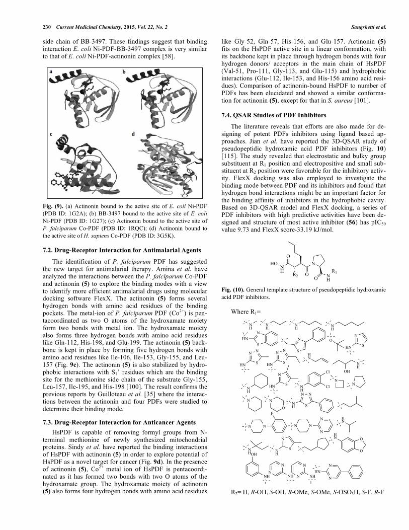
The literature reveals that efforts are also made for de-signing of potent PDFs inhibitors using ligand based ap-proaches. Jian et al. have reported the 3D-QSAR study of pseudopeptidic hydroxamic acid PDF inhibitors (Fig. 10)[115]. The study revealed that electrostatic and bulky group substituent at R1 position and electropositive and small sub-stituent at R2 position were favorable for the inhibitory activ-ity. FlexX docking was also employed to investigate the binding mode between PDF and its inhibitors and found that hydrogen bond interactions might be an important factor for the binding affinity of inhibitors in the hydrophobic cavity. Based on 3D-QSAR model and FlexX docking, a series of PDF inhibitors with high predictive activities have been de-signed and structure of most active inhibitor (56) has pIC50value 9.73 and FlexX score-33.19 kJ/mol.
NH
HOO
N
OO N
H
R1R2
Fig. (10). General template structure of pseudopeptidic hydroxamic acid PDF inhibitors.
Where R1=
NH
NH
NH N
HN
SHN N
S
ON
SHNN
S
N
HN
N
S
N
HN
HN
NH
OH
NH
N
SHN NH
Cl
ClN
N
N N
O
NN
SNH
NN
F
NNN
N HN
NH
N
SNH
O
OHN
OH
NHN
N
NH N
N
NH N
NHN
R2= H, R-OH, S-OH, R-OMe, S-OMe, S-OSO3H, S-F, R-F

Peptide Deformylase: A New Target Current Medicinal Chemistry, 2015, Vol. 22, No. 2 231
NH
HOO
N
OO N
HS
OH
CH3
Cl
CH3(56)
Ji et al. have performed the 2D and 3D-QSAR studies of a series of published (British Biotech Pharmaceuticals, Ox-ford, UK) reverse hydroxamate derivatives (Fig. 11) having antibacterial activity against E. coli PDF [116]. This study yielded some important structural information that can be used for the design of novel PDF inhibitors. The study re-vealed that sterically bulky substituents like n-butyl or cy-clopentyl-methyl group at R1 position, sterically moderately bulky groups at R2 position and para-substituted phenyl group at R3 position were beneficial for activity.
NOHN
O
R1OH
H
O
R3R2
Fig. (11). General template structure of reverse hydroxamate de-rivatives PDF inhibitors.
When
N
R2=
R3=
R1= n-Bu, Me, Et, n-Pr, (S) n-Bu, n-Pentyl, n-Hexyl, n-Heptyl, n-Octyl, i-Pr, i-Bu, i-Pentyl, c-Pentylmethyl, c-Pentyl, c-Hexylmethyl, Allyl, But-3-enyl, But-2-ynyl, EtSCH2, Bn, Ph(4-Cl), (4-MeO)PhCH2, 1-Piperidylmethyl
When
R1=
NR3=
R2= Gly, Ala, Val, Leu, Cha, Ile, (R)t-Leu, Pen(SMe), Cys(Bn), Ser, Val(�-OH), Val(�-OMe), Asp(�-Bn), Glu(�-Bn), Lys, Lys ( -NMe2), Arg, Phe, Phe (4-Cl), Tyr, L-Tic, Pro
When
R1=
R2=
R3= OMe, OH, Me, Pyrrolidin-1-yl, Mor-pholin-4-yl, 4-Me-piperazin-1-yl, 4-Me-piperidin-1-yl, 4-Ac-piperidin-1-yl, 4-EtO2C-piperidin-1-yl, 4-Bn-piperidin-1-yl, N(Me)c-Hexyl, Decahydroquinolin-1-yl, N(Bn)CH2CH2Ph, Ph, 2-Pyridyl, 2-Furyl
Manish et al. have reported the 2D-QSAR study of �-
sulfonyl and �-sulfinyl hydroxamic acid derivatives and demonstrated that the PDF inhibitory activity in cell free and the whole cell system was increased with increase in molar refractivity and hydrophobicity [117]. Itishree et al. have performed comparative molecular field analysis (CoMFA) analysis of �-sulfonyl and �-sulfinyl hydroxamic acid deriva-tives to figure out the structural requirements of PDF inhibi-tors belonging to non-peptidic class and to optimize their E. coli peptide deformylase inhibitory activity [118]. On the basis of spatial arrangement of field contribution, three novel molecules were designed and predicted for PDF inhibitory activity. The most active designed compound (57) has pIC50 value 1.3061 and predicted IC50 value 0.0494 �M for E. coli PDF.
HN
O
OHNH
OHO
(57)
Very few reports are available on the pharmacophore de-velopment of PDF inhibitors. There is an urgent need for developing common pharmacophore so that the designing of various chemical classes of PDF inhibitors could be rational-ized. Molecular docking and QSAR studies of existing PDF inhibitors could help in developing a more potent and selec-tive PDF inhibitor with lesser side effects and better pharma-cokinetic properties.
8. CURRENT CLINICAL CANDIDATES
After initial validation of PDF enzyme as an antibacterial drug target, tremendous progress has been made in the de-velopment of PDF inhibitors. Extensive preclinical studies have been carried out for theses classes of compounds. Three compounds, GSK-1322322, BB-83698, and LBM-415 have been entered into clinical trials.
8.1. GSK-1322322
GSK-1322322 (58) (discovered by GlaxoSmithKline) is novel PDF inhibitors of the pseudopeptidic class, which has shown good safety and pharmacokinetic properties in a Phase I clinical trial and promising proof of-concept results in a Phase IIa study [119]. GSK-1322322 (58) is currently being developed for the oral and intravenous treatment of acute bacterial skin and skin structure infections and hospi-talized patients with community-acquired pneumonia. The activity of GSK-1322322 (58) was tested against a global collection of clinical isolates of H. influenzae (n= 2,370), M. catarrhalis (n= 115), S. pneumoniae (n= 947), S. pyogenes (n= 617), and S. aureus (n= 940), including strains resistant to one or more marketed antibiotics. GSK-1322322 (58) had an MIC90 of 1 �g/mL against M. catarrhalis and 4 �g/mL against H. influenzae, with 88.8% of �-lactamase-positive strains showing growth inhibition at that concentration. All S. pneumoniae strains were inhibited by <4 �g/mL of GSK-1322322 (58), with an MIC90 of 2 �g/mL. Pre-existing resis-tance mechanisms did not affect its potency, as evidenced by the MIC90 of 1 �g/mL for penicillin, levofloxacin, and mac-rolide-resistant S. pneumoniae. GSK-1322322 (58) was very potent against S. pyogenes strains, with an MIC90 of 0.5 �g/mL, irrespective of their macrolide resistance phenotype. This PDF inhibitor was also active against S. aureus strains regardless of their susceptibility to methicillin, macrolides, or levofloxacin, with an MIC90 of 4 �g/mL in all cases. Time-kill studies showed that GSK-1322322 (58) had bacte-ricidal activity against S. pneumoniae, H. influenzae, S. pyo-genes, and S. aureus, demonstrating a >3-log10 decrease in the number of CFU/mL at 4 �MIC within 24 h in 29 of the 33 strains tested.
In summary, GSK-1322322 (58) has the spectrum of ac-tivity and in vitro potency necessary for therapeutic use against respiratory tract and skin and soft tissue infections. Furthermore, its unique mode of action provides a clear ad-

232 Current Medicinal Chemistry, 2015, Vol. 22, No. 2 Sangshetti et al.
vantage in the treatment of multidrug-resistant pathogens and ensures lack of cross-resistance with any other marketed antibiotics. This PDF inhibitor has completed early clinical trials and has shown pharmacokinetic and safety parameterswarranting progression to Phase IIb studies. The antibacterial potency of GSK-1322322 (58) suggests a valuable alterna-tive therapy for the treatment of infectious diseases caused by drug-resistant pathogens [120].
N
O
N
N N
HN
NH
O
N
O
H
HF
OH
GSK-1322322 (58)
8.2. BB-83698
BB-83698 (59) (discovered by British Biotech, in col-laboration with Genesoft) has been investigated in Phase I clinical trial for intravenous treatment of various bacterial infections. This pseudopeptidic inhibitor showed potent invitro activity against S. pneumoniae with an MIC90= 0.5 �g/mL, but less potent activity against S. aureus (MIC90= 8 �g/mL) and H. influenza (MIC90= 16 �g/mL). The subcuta-neous administration to mice, the compound showed rapid distribution to the lung, with 4-fold higher concentrations than that in plasma [121]. The pharmacokinetic studies with dose ranging from 10 to 50 mg/kg in mice, rats and dogs exhibited biphasic pattern with moderate volumes of distri-bution in all species. The pharmacokinetic parameters were consistent, predictable, and exhibited good allometric scaling among all species (R2 > 0.98).
The Phase I study of BB-83698 (59) was explored at eight dose levels ranging from 10 to 475 mg and was admin-istered via 15 min intravenous infusion. The overall mean clearance rate (CL) was approximately 238 ± 130 mL/min, with CL values ranging from 189 mL /min at a dose of 475 mg to 521 mL/min at the lowest dose. At the highest dose of 475 mg, an AUC/MIC ratio of 184 was achieved, based on S. pneumoniae MIC of 0.25 �g/mL, which is predicted to be within the efficacious range (AUC/MIC� 133) for once-daily dosing derived from PK/PD studies [122]. The compound was terminated from the study when British Biotech over-took by Vernalis (Financial Times, September 30, 2003). Oscient has now purchased this compound but further clini-cal study is still considered to be discontinued [18].
O
O NN
ONH
O
N
O
HOH
BB-83698(59)
8.3. LBM-415
LBM-415 (60) (discovered Vicuron Pharmaceuticals, in collaboration with Novartis, also called VIC-104959 or
NVP-PDF-713) is also a pseudopeptidic class of novel PDF inhibitor targeting a large number of bacterial species. This compound has been investigated in Phase I clinical trial as oral administration. LBM-415 (60) has excellent in vitro antibacterial activity against S. aureus (MIC90 = 2 �g/mL), S. pneumoniae (MIC90 = 1 �g/mL), and E. faecalis (MIC90 = 4 �g/mL). In pharmacokinetic studies, LBM-415 (60) showed 62% and 22-101% absolute oral bioavailability in mice and rats, respectively, with no indication of absorption saturation at higher doses. Oral administration of LBM-415 (60) exhib-ited similar efficacy in comparison to linezolid and clarithromycin in S. aureus and S. pneumoniae systemic in-fection model. The pharmacodynamic study of LBM-415 (60) using a neutropenic mouse thigh model of S. pneumo-niae showed a prolonged post-antibiotic effect (4.1-11.6 h).
The Phase I clinical study was performed to explore LBM-415 (60) as a potential oral agent for community respi-ratory tract infections. Single oral doses ranging from 100 mg to 3000 mg were given in an ascending order, and the compound was rapidly absorbed with Tmax (time of maximum observed concentration) � 1 h across all dose groups. Linear pharmacokinetics were observed with dose -proportional increase of maximum observed concentration (Cmax) and mean terminal half life (T1/2) was ranged from 2 to 4.2 h. The pharmacokinetic studies of LBM-415 (60) after food intake revealed that while the Tmax was delayed (from 0.5 to 2 h) and lower (from 15.5 to 6.7 �g/mL), with no change in over-all systemic exposure. A multiple dose study (250, 500, and 100 mg twice daily for 11 days) showed no accumulation after 11 days with no significant differences in pharmacoki-netic parameters. From the Phase I clinical trial study, the overall profile of LBM-415 (60) supported a twice-daily dosing regimen [11]. Recently, pharmacokinetics and unex-pected safety issues of LBM-415 study have been reported and revealed that the compound was well tolerated at low doses, but at the highest dose, 1000 mg t.i.d. (target thera-peutic dose), reversible cyanosis and low oxygen saturation, attributable to methemoglobinemia, were detected on day 11. Oxygen saturation was as low as 88% in one subject on day 11 [123].
N
F
HN
ON
N
O
H
O
O OH
LBM-415(60)
9. RESISTANCE ISSUE TO PDF INHIBITORS
PDF is now recognized as novel antibacterial target for new generation, broad spectrum antibiotics. However, some issues, including possible resistance mechanisms and antici-pation of toxicity to human have to be addressed. PDF in-hibitors have exerted bacteriostatic, not bacteriocidal activ-ity, thus reducing their potential usefulness in the manage-ment of serious infections. The relative ease with which mi-croorganisms have been able to develop resistance and the multiple available mechanisms of resistance (mutations in formyl transferase (fmt), tetrahydrofolate dehydrogenase (folD) genes; and AcrAB/ToIC efflux pump) are worrisome

Peptide Deformylase: A New Target Current Medicinal Chemistry, 2015, Vol. 22, No. 2 233
[124-127]. Despite these current liabilities, PDF is still con-sidered as an extremely attractive target for antibacterial agents as the resistance mechanisms are newer one. For ex-ample, fmt mutants have shown systematically reduced growth rate compared to the wild type.
The most serious reservation for the use of PDF inhibi-tors as antibacterial could come from the recent identifica-tion of an HsPDF. The existence of this enzyme undoubtedly raises the issue that treatment with such compounds could result in toxicity. However, the PDF inhibitors inhibit the growth of human cell lines at high concentrations and have excellent safety profiles in the preclinical development. The clinical candidates GSK-1322322 (58), BB-83698 (59), and LBM-415 (60), appear well tolerated in a Phase I study. This selectivity suggests strongly that PDF inhibitors will not be toxic to humans at its clinical dose [128].
CONCLUSION
In last few years, PDF enzyme has been emerged as an important target in antibacterial drug discovery. Different types of PDF have also been identified. The three-dimensional structures and co-crystal structures of PDF en-zyme have been well characterized and established. The ac-tive binding sites in the PDF enzyme for inhibitors have also been well identified. These results led to the development of PDF inhibitors as a new class of antibacterial agents. Three PDF inhibitors as antibacterial agents, GSK-1322322 (Phase II), BB-83698 (Phase I), and LBM-415 (Phase I) have en-tered into clinical developments. The further clinical studies of BB-83698 and LBM-415 have been stopped due to un-known reasons. Thus, only in case of positive results of Phase II clinical study of GSK-1322322 will validate the PDF as a target for antibacterial drug discovery.
Some recent reports about the involvement of PDF in human cancer and malaria parasites have gained a great deal of attention from scientific community. The active binding sites of human PDF and plasmodium PDF have also been well identified and established. Some chemical classes of PDF inhibitors as antimalarial and anticancer are developed based on drug-receptor interactions. However, the clinical utility of these PDF inhibitors need to be established in the future. In short, PDF has been extensively explored as an attractive target for antibacterial, antimalarial and anticancer drug discovery. Further results from clinical trials will ulti-mately decide the fate of PDF as a versatile drug target in drug discovery.
CONFLICT OF INTEREST
The authors confirm that this article content has no con-flict of interest.
ACKNOWLEDGEMENTS
The author JNS is grateful to Department of Science and Technology (DST), New Delhi, India for Fast Track Project (SR/FT/LS119/2012). The authors are also thankful to the Mrs. Fatma Rafiq Zakaria, Chairman, Maulana Azad Educa-tional Trust and Dr. Zahid Zaheer, Principal, Y.B. Chavan College of Pharmacy, Dr. Rafiq Zakaria Campus, Auran-gabad 431 001 (M.S.), India for constant support and provid-ing necessary facilities.
REFERENCES [1] World Health Organization. Deaths by cause, sex and mortality
stratum in WHO regions, estimates for 2001. World Health Report. Geneva, 2002.
[2] Carmela, G.; Michele, P.; Thierry, M. Peptide deformylase as tar-get for new generation broad spectrum antimicrobial agents. Mol. Microbiol., 2000, 36 (6), 1197-1205.
[3] Wenzel, R.P.; Edmond, M.B. Managing antibiotic resistance. N. Engl. J. Med., 2000, 343 (3), 1961-1963.
[4] Gaynes, R.; Edwards, J.R.; National Nosocomial Infections Sur-veillance System. Overview of nosocomial infections caused by gram-negative bacilli. Clin. Infect. Dis., 2005, 41(6), 848-854.
[5] D’Agata, E.M.C. Rapidly rising prevalence of nosocomial multidrug-resistant gram-negative bacilli: a 9-year surveillance study. Infect. Control. Hosp. Epidemiol., 2004, 25 (10), 842-846.
[6] Rice, L. B. Antimicrobial resistance in gram-positive bacteria. Am. J. Infect. Control., 2006, 34 (5 Suppl. 1), S11-S19.
[7] Aziz, M.A.; Wright, A.; Laszlo, A.; De Muynck, A.; Portaels, F.; Van Deun, A.; Wells, C.; Nunn, P.; Blanc, L.; Raviglione, M. Epi-demiology of antituberculosis drug resistance (the global project on anti-tuberculosis drug resistance surveillance): an updated analysis.Lancet, 2006, 368 (9553), 2142-2154.
[8] Wallis, R.S.; Patil, S.; Cheon, S.H.; Edmonds, K.; Phillips, M.; Perkins, M.D.; Joloba M.; Namale, A.; Johnson, J.L.; Teixeira, L.; Dietze, R.; Siddiqi, S.; Mugerwa, R.D.; Eisenach, K.; Ellner, J.J. Drug tolerance in Mycobacterium tuberculosis. Antimicrob. Agents Chemother., 1999, 43 (11), 2600-2606.
[9] Kelly, A.; Magdalena, Z. In: Peptide deformylase inhibitors; King and Lawton, Ed.; Elsevier Science B. V: Progress in medicinal chemistry, 2006; Vol. 44, pp. 110-143.
[10] Meinnel, T.; Patiny, L.; Ragusa, S.; Blanquet, S. Design and syn-thesis of substrate analogue inhibitors of peptide deformylase. Bio-chemistry, 1999, 38 (14), 4287-4295.
[11] Jain, R.; Chen, D.; White, R.J.; Patel, D.V.; Yuan, Z. Bacterial peptide deformylase inhibitors: a new class of antibacterial agents. Curr. Med. Chem., 2005, 12, 1607-1621.
[12] Giglione, C.; Sereo, A.; Pierre, M.; Boisson, B.; Meinnel T. Identi-fication of eukaryotic peptide deformylases reveals universality of N-terminal protein processing mechanisms. EMBO J., 2000, 19(21), 5916-5929.
[13] World Health Organization. WHO Expert Committee on Malaria (20th Report). WHO Tech. Rep. Ser., 2000, 892, 1-83.
[14] Sachs, J.; Malaney, P. The economic and social burden of malaria. Nature, 2002, 415 (6872), 680-685.
[15] Kevin, B.J. Chloroquine resistance in Plasmodium vivax. Antimi-crob. Agents Chemother., 2004, 98(41), 4075-4083.
[16] Lee, M.D.; Antczak, C.; Li, Y.; Sirotnak, F.M.; Bornmann. W.G.; Scheinberg, D.A. A new human peptide deformylase inhibitable by actinonin. Biochem. Biophy. Res. Commun., 2003, 312 (2), 306-315.
[17] Serero, A.; Giglione, C.; Sardini, A.; Martinez-Sanz, J.; Meinnel, T. An unusual peptide deformylase features in the human mito-chondrial N-terminal methoinine excision pathway. J. Biol. Chem., 2003, 278 (52), 52953-52963.
[18] David R P Guay. Drug forecast- the peptide deformylase inhibitors as antibacterial agents. Ther. Clin. Risk Manag., 2007, 3 (4), 513-525.
[19] Jennifer, A.L.; Charles, R.D. Peptide deformylase as an antibacte-rial target: a critical assessments. Curr. Opin. Pharmacol., 2006, 6(5), 445-452.
[20] Zhengyu, Y.; Joaquim, T.; Richard, J.W. Deformylase as a novel antibacterial target. Drug Discov. Today, 2001, 6 (18), 954-961.
[21] Carmela, G.; Thierry, M. Peptide deformylase as an emerging target for antiparasitic agents. Expert Opin. Ther. Targets, 2001, 5(1), 41-57.
[22] Adams, J.M. On the release of the formyl group from nascent pro-tein. J. Mol. Biol., 1968, 33 (3), 571-589.
[23] Adams, J.M. Capecchi, M.R. N-formylmethionyl-sRNA as the initiator of protein synthesis. Proc. Natl. Acad. Sci. USA., 1966, 55(1), 147-155.
[24] Lucchini, G.; Bianchetti, R. Initiation of protein synthesis in iso-lated mitochondria and chloroplasts. Biochim. Biophys. Acta, 1980,608 (1), 54-61.
[25] Meinnel, T.; Mechulam, Y.; Blanquet, S. Methionine as translation start signal: a review of the enzymes of the pathway in Escherichia coli. Biochimie, 1993, 75 (12), 1061-1075.

234 Current Medicinal Chemistry, 2015, Vol. 22, No. 2 Sangshetti et al.
[26] Becker, A.; Schlichting, I.; Kabsch, W.; Groche, D.; Schultz, S.; Wagner, A.F. Iron center, substrate recognition and mechanism ofpeptide deformylase. Nature Struct. Biol., 1998, 5 (12), 1053-1058.
[27] Mazel, D.; Pochet, S.; Marliere, P. Genetic characterization of polypeptide deformylase, a distinctive enzyme of eubacterial trans-lation. EMBO J., 1994, 13 (4), 914-923.
[28] Takeuchi, N.; Kawakami, M.; Omori, A.; Ueda, T.; Spremulli, L.L.; Watanabe, K. Mammalian mitochondrial methionyl-tRNA transformylase from bovine liver: purification, characterization, and gene structure. J. Biol. Chem., 1998, 273 (24), 15085-15090.
[29] Takeuchi, N.; Vial, L.; Panvert, M.; Schmitt, E.; Watanabe, K.; Mechulam, Y.; Blanquet, S. Recognition of tRNAs by methionyl-tRNA transformylase from mammalian mitochondria. J. Biol. Chem., 2001, 276 (23), 20064-20068.
[30] Bracchi-Richard, V.; Nguyen, K.; Zhou, Y.; Rajagopalan, P.T.R.; Chakrabarti, D.; Pei, D. Characterization of an eukaryotic peptide deformylase from Plasmodium falciparum. Arch. Biochem. Bio-phys., 2001, 396 (2), 162-170.
[31] Abhinav, K.; Kiet, T.N.; Sumant, S.; Brad, O.; Stewart, T.; Irwin, H.; Dehua, P.; Wim, G.J.H. Crystals of peptide deformylase from Plasmodium falciparum reveal critical characteristics of the active site for drug design. Structure, 2002, 10 (3), 357-367.
[32] Lee, M.D.; She, Y.; Soskis, M.J.; Borella, C.P.; Gardner, J.R.; Hayes, P.A.; Dy, B.M.; Heaney, M.L.; Philips, M.R.; Bornmann, W.G.; Sirotnak, F.M.; Scheinberg, DA. Human mitochondrial pep-tide deformylase, a new anticancer target of actinonin-based antibi-otics. J. Clin. Invest., 2004, 114 (8), 1107-1116.
[33] Spencer, A.C.; Spremulli, L.L. Interaction of mitochondrial initia-tion factor 2 with mitochondrial fMet-tRNA. Nucleic Acids Res.,2004, 32 (18), 5464-5470.
[34] Giglione, C.; Vallon, O.; Meinnel, T. Control of protein life-span by N-terminal methionine excision. EMBO J., 2003, 22 (1), 13-23.
[35] Guilloteau, J.P.; Mathieu, M.; Giglione, C.; Blanc, V.; Dupuy, A.; Chevrier, M.; Gil, P.; Famechon, A.; Meinnel, T. The crystal struc-tures of four peptide deformylases bound to the antibiotic actinonin reveal two distinct types: a platform for the structure-based design of antibacterial agents. J. Mol. Biol., 2002, 320 (5), 951-962.
[36] Meinnel, T. Peptide deformylase of eukaryotic protists: a target for new antiparasitic agents. Parasitol. Today, 2000, 16 (4), 165-168.
[37] Mazel, D.; Pochet, S.; Marliere, P. Genetic characterization of polypeptide deformylase, a distinctive enzyme of eubacterial trans-lation. EMBO J., 1994, 13 (4), 914-923.
[38] Groche, D.; Becker, A.; Schlichting, I.; Kabsch, W.; Schultz, S.; Wagner, A.F. Isolation and crystallization of functionally compe-tent Escherichia coli peptide deformylase forms containing either iron or nickel in the active site. Biochem. Biophys. Res. Comm., 1998, 246 (2), 342-346.
[39] Rajagopalan, P.T.R.; Yu, X.C.; Pei, D. Peptide deformylase, a new type of maononuclear iron protein. J. Am. Chem. Soc., 1997, 119(50), 12418-12419.
[40] Rajagopalan, P.T.; Pei, D. Oxygen mediated inactivation of peptide deformylase. J. Biol. Chem., 1998, 273 (35), 22305-22310.
[41] Meinnel, T.; Lazennec, C.; Dardel, F.; Schmitter, J.M.; Blanquet, S. The C-terminal domain of peptide deformylase is disordered and dispensable for activity. FEBS Lett., 1996, 385 (1-2), 91-95.
[42] Meinnel, T.; Lazennec, C.; Dardel, F.; Viloing, S.; Blanquet, S. Structure-function relationships within the peptide deformylase family. Evidence for a conserved architecture of the active site in-volving three conserved motifs and metal ion. J. Mol. Biol., 1997,267 (3), 749-761.
[43] Chang, M.K.; Gong, W.; Rajagopalan, P.T.; Hao, B.; Tsai, C.M.; Pei, D. Crystal structure of the Escherichia coli peptide deformy-lase. Biochemistry, 1997, 36 (45), 13904-13909.
[44] Becker, A.; Schlichting, I.; Kabsch, W.; Schultz, S.; Wagner, A.F. Structure of peptide deformylase and identification of the substrate binding site. J. Biol. Chem., 1998, 273 (19), 11413-11416.
[45] Dardel, F.; Ragusa, S.; Lazennec, C.; Blanquet, S.; Meinnel, T. Solution structure of nickel-peptide deformylase. J. Mol. Biol., 1998, 280 (3), 501-513.
[46] Smith, K.J.; Petti, C.M.; Aubart, K.; Symth, M.; McManus, E.; Jones, J.; Fosberry, A.; Lewis, C.; Lonetto, M.; Christensen, S.B. Structural variation and inhibitor binding in polypeptide deformy-lase from four different bacterial species. Protein Sci., 2003, 12 (2), 349-360.
[47] Kreusch, A.; Spraggon, G.; Lee, C.C.; Klock, H.; McMullan, D.; Ng, K.; Shin, T.; Vincent, J.; Warner, I.; Ericson, C.; Lesley, S.A.
Structure analysis of peptide deformylases from Streptococcus pneumoniae, Staphylococcus aureus, Thermotoga maritima and Pseudomonas aeruginosa: snapshots of the oxygen sensitivity of peptide deformylase. J. Mol. Biol., 2003, 330 (2), 309-321.
[48] Hu, Y.; Wei, Y.; Zhou, Y.; Rajagopalan, P.T.; Pei, D. Determina-tion of substrate specificity for peptide deformylase through the screening of a combinatorial peptide library. Biochemistry, 1999,38 (2), 643-650.
[49] Leung, D.; Abbenante, G.; Fairlie, D.P. Protease inhibitors: current status and future prospects. J. Med. Chem., 2000, 43 (3), 305-341.
[50] Chen, D.Z.; Patel, D.V.; Hackbarth, C.J.; Wang, W.; Dreyer, G.; Young, D.C.; Margolis, P.S.; Wu, C.; Ni, Z.J.; Trias, J.; White, R.J.; Yuan, Z. Actinonin, a naturally occurring antibacterial agent, is a potent deformylase inhibitor. Biochemistry, 2000, 39 (6), 1256-1262.
[51] Fu, H.; Dahlgren, C.; Bylund, J. Subinhibitory concentrations of the deformylase inhibitor actinonin increase bacterial release of neutrophil-activation peptides: a new approach to antimicrobial chemotherapy. Antimicrob. Agents Chemother., 2003, 47 (8), 2545-2550.
[52] Gross, M.; Clements, J.; Beckett, R.P.; Thomas, W.; Taylor, S.; Lofland, D.; Ramanathan-Girish, S.; Garcia, M.; Difuntorum, S.; Hoch, U.; Chen, H.; Johnson, K.W. Oral antipneumococcal activity and pharmacokinetic profiling of a novel peptide deformylase in-hibitor. J. Antimicrob. Chemother., 2004, 53 (3), 487-493.
[53] Yaoming, W.; Tian, Y.; Kristi, M.H.; Chaity, C.; Dehua, P. Identi-fication of a potent peptide deformylase inhibitor from a rationally designed combinatorial library. J. Comb. Chem., 2000, 2 (6), 650-657.
[54] Kristi, M.H.; Tian, Y.; Yaoming, W.; Dehua, P. Synthesis and Antibacterial Activity of Peptide Deformylase Inhibitors. Biochem-istry, 2000, 39 (15), 4543-4551.
[55] Yun-Jin, H.; Rajagopalan, P.T.R.; Dehua, P. H-phosphonate de-rivatives as novel peptide deformylase inhibitors. Bioorg. Med. Chem. Lett., 1998, 8 (18), 2479-2482.
[56] Daniel, J.D.; Barbara, G.G.; John, F.O.; Stephan, K.G. Peptide aldehyde inhibitors of bacterial peptide deformylases. Arch. Bio-chem. Biophys., 1999, 367 (2), 297-302.
[57] Smith, H.K.; Beckett, R.P.; Clements, J.M.; Doel, S.; East, S.P.; Launchbury, S.B.; Pratt, L.M.; Spavold, Z.M.; Thomas, W.; Todd, R.S.; Whittaker, M. Structure-activity relationships of the peptide deformylase inhibitor BB-3497: modification of the metal binding group. Bioorg. Med. Chem. Lett., 2002, 12 (24), 3595-3599.
[58] Clements, J.M.; Beckett, R.P.; Brown, A.; Catllin, G.; Lobell, M.; Palan, S.; Thomas, W.; Whittaker, M.; Wood, S.; Salama, S.; Baker, P.J.; Rodgers, H.F.; Barynin, V.; Rice, D.W.; Hunter, M.G. Antibiotic activity and characterization of BB-3497, a novel pep-tide deformylase Inhibitor. Antimicrob. Agents Chemother., 2001,45 (2) 563-570.
[59] Corinne, J.H.; Dawn, Z.C.; Jason, G.L.; Kirk, C.; James, B.M.; Jeffrey, A.C.; Peter, S.M.; Wen, W.; Jim, K.; Charlotte, W.; Lopez, S.; George, W.I.; Helen, G.; Elina, D.; Kulathia, R.; Shi-Hao, P.; Wilma, L.P.; Jeff, J.; Joaquim, T.; Dinesh, V.P.; Beat, W.; Richard, J.W.; Zhengyu, Y. N-alkyl urea hydroxamic acids as a new class of peptide deformylase inhibitors with antibacterial activity. Antimi-crob. Agents Chemother., 2002, 46 (9), 2752-2764.
[60] Jain, R.; Sundram, A.; Lopez, S.; Neckermann, G.; Wu, C.; Hack-barth, C.; Chen, D.; Wang, W.; Ryder, N.S.; Weidmann, B.; Patel, D.; Trias, J.; White, R.; Yuan, Z. �-Substituted hydroxamic acids as novel bacterial deformylase inhibitor-based antibacterial agents. Bioorg. Med. Chem. Lett., 2003, 13 (23), 4223-4228.
[61] Arkadius, P.; Jeyaraj, D.; Zheng, Y.; Thomas, H.K.; Jenefer, A.; Sarah, L.; Gladys, L.; Mei, D.; Gang, W.; Wai, L.C.; Mark, S.; Ida, M.; David, B.; Xinyi, N.; Kakoli, M.; Mahesh, N.; Jeanette, W.P.T.; Pamela, T.; Amelia, Y.; Thomas, D.; Wuyi, M.; Mei, X.; James K.; Shi-Hao P.; Kirk C.; Xiaoling X.; Carolyn S.; Michael C. Peptide deformylase inhibitors of Mycobacterium tuberculosis:synthesis, structural investigations, and biological results. Bioorg. Med. Chem. Lett., 2008, 18 (24), 6568-6572.
[62] Zhenyu, W.; Weicheng, Z. Syntheis and in vitro antibacterial activ-ity of (2S)-N-(substitutedphenyl)-1-[2R)-2-[(formylhyroxy-amino)methyl]-1-oxohexyl]-2-pyrrolidine carboxamides as poten-tial peptide deformylase inhibitors. Chem. Biol. Drug Des., 2009,73 (1), 142-146.
[63] Jeffrey, M.A.; Jesus, R.M.; Charles, W.B.; Celine, D.; Seth, W.G.; Mark, A.B.; Tony, P.; William, H.M.; Theresa, P.; Timothy, F.G.;

Peptide Deformylase: A New Target Current Medicinal Chemistry, 2015, Vol. 22, No. 2 235
Swarupa, K.; Thomas, L.; Glenn, S.V.A.; Rimma, Z.; Paris, W.; Nino, C. Acylprolinamides: a new class of peptide deformylase in-hibitors with in vivo antibacterial activity. Bioorg. Med. Chem. Lett., 2012, 22 (12), 4028-4032.
[64] Wei, S.; Yuejiao, D.; Yu, Q.; Ming, L.; Liping, Y.; Wenhao, H. Design, synthesis and antibacterial activity of 2,5-dihydropyrrole formyl hydroxyamino derivatives as novel peptide deformylase in-hibitors. Bioorg. Med. Chem. Lett., 2010, 20 (12), 3592-3595.
[65] Wei, S.; Haikun, M.; Yuejiao, D.; Kelly, A.; Yuhong, F.; Rimma, Z.; Liping, Y.; Wenhao, H. Design, synthesis and antibacterial ac-tivity of 3-methylenepyrrolidine formyl hydroxyamino derivatives as novel peptide deformylase inhibitors. Bioorg. Med. Chem. Lett., 2011, 21 (3), 1060-1063.
[66] Ii, K.; Ichikawa, S.; Al-Dabhagh, B.; Bouhss, A.; Matsuda, A. Function-oriented synthesis of simplified caprazamycins: discovery of oxazolidine-containing uridine derivatives as antibacterial agents against drug-resistant bacteria. J. Med. Chem., 2010, 53 (9), 3793-3813.
[67] Keller, M.; Boissard, C.; Patiny, L.; Chung, N. N.; Lemieux, C.; Mutter, M.; Schiller, P.W. Pseudoproline-Ccontaining analogues of morphiceptin and endomorphin-2: evidence for a Cis Tyr�Pro am-ide bond in the bioactive conformation. J. Med Chem., 2001, 44(23), 3896-3903.
[68] Tuchscherer, G.; Grell, D.; Tatsu, Y.; Durieux, P.; Fernandez-Carneado, J.; Hengst, B.; Kardinal, C.; Feller, S. Targeting molecu-lar recognition: exploring the dual role of functional pseudoprolines in the design of SH3 ligands. Angew. Chem., Int. Ed., 2001, 40(15), 2844.
[69] Linliang, Y.; Weicheng, Z.; Zhenyu, W. Synthesis and in vitroantibacterial activity of oxazolidine LBM-415 analogsas peptide deformylase inhibitors. Bioorg. Med. Chem. Lett., 2011, 21 (5) 1541-1544.
[70] Zega, A. Azapeptides as pharmacological agents. Curr. Med. Chem., 2005, 12 (5), 589-597.
[71] Stephen P. E.; Andrew A.; Ian T.; Steven T.; Wayne T. Pepti-domimetic inhibitors of bacterial peptide deformylase. Bioorg. Med. Chem. Lett., 2011, 21 (13), 4032-4035.
[72] Maithri, M.K.J.; Ann, K.; Ramy, S.; Michael, D.; Mathew, T.; Martin, A.S.; Tod, P.H. Novel nonpeptidic inhibitors of peptide de-formylase. Arch. Biochem. Biophys., 2000, 381 (2), 313-316.
[73] Bhat, A.; Christensen, S.B.; Frazee, J.S.; Head, M.S.; Leber, J.D.; Li, M. Peptide deformylase inhibitors. PCT Int. Appl. WO 02,081,426. Chem. Abstr., 2002, 137, 310696.
[74] Bhat, A.; Christensen, S.B.; Frazee, J.S.; Head, M.S.; Leber, J.D.; Li, M. Peptide deformylase inhibitors. PCT Int. Appl. WO 03,002,522, Chem. Abstr., 2003, 138, 89584.
[75] Christian A.; David W. B.; Daniel B.; Michel D.; Christian H.; Hans L.; Frederic M.; Raffaello M.; Wolfgang P.; Henri, S. 2-(2-Oxo-1,4-dihydro-2H-quinazolin-3-yl)- and 2-(2,2-Dioxo-1,4-dihydro-2H-2�6-benzo[1,2,6]thiadiazin-3-yl)-N-hydroxy-acetamides as potent and selective peptide deformylase inhibitors. J. Med. Chem. 2001, 44 (12), 1847-1852.
[76] Valentina, M.; Xiaohui, H.; Juliet, N.; Kunyong, Y.; Andreas, K.; Perry, G.; Badry, B.; Ian, W.; Tanya, S.; Tanya, B.; Neil, S.R.; Ron, G.; John, D.; Yun, H. Identification of novel potent bicyclic peptide deformylase inhibitors. Bioorg. Med. Chem. Lett., 2004, 14(6) 1477-1481.
[77] Apfel, C.; Banner, D.W.; Bur, D.; Dietz, M.; Hirata, T.; Hubschwelen, C.; Locher, H.; Page, M.G.P.; Pirson, W.; Rosse, G.; Specklin, J. Hydroxamic Acid Derivatives as Potent Peptide De-formylase Inhibitors and Antibacterial Agents. J. Med. Chem., 2000, 43 (12), 2324-2331.
[78] Barbara, G.G.; Jeffrey, H.T.; John, W.K.; Stephan, K.G. Inhibition of bacterial peptide deformylase by biaryl acid analogs. Arch. Bio-chem. Biophys., 2000, 375 (2), 355-358.
[79] Aubart, K.M.; Christensen, S.B.; Briand, J.; Cummings, M.D. Peptide deformylase inhibitors. U.S. Patent 6,806,369, October 19, 2004.
[80] Christensen, S.B.; Cummings, M.D.; Karpinski, J.M. Peptide de-formylase inhibitors. U.S. Patent 6,852,882, February 8, 2005.
[81] Wataru, T.; Yoshihisa, S.; Yusuke, S.; Emi, N.; Shuhei, F.; Ka-nako, S.; Jun, I. Synthesis and PDF inhibitory avtivities of novel benzothiazolylidenehydroxamic acid derivatives. Bioorg. Med. Chem. Lett., 2003, 13 (19), 3273-3276.
[82] Patrizia, C.; Lars, N.; Seema, M.; Anders, H. Isoxazole-3-hydroxamic acid derivatives as peptide deformylase inhibitors and
potential antibacterial agents. Bioorg. Med. Chem. Lett., 2004, 14(24), 5997-6000.
[83] Xubo H.; Kiet T. N.; Christophe L. M. J. V.; Wim G. J. H.; Dehua P. Structure-based design of a macrocyclic inhibitor for peptide de-formylase. J. Med. Chem. 2003, 46 (18), 3771-3774.
[84] Gang, S.; Jinge, Z.; Anthony, M.S.; Dehua, P. Design and synthesis of macrocyclic peptidyl hydroxamates as peptide deformylase in-hibitors. Bioorg. Med. Chem. Lett. 2008, 18 (10), 3060-3063.
[85] Ling, Y.; Wei-Ren, X.; Zhi-Guo, W.; Da-Tong, Z.; Jiong, J.; Yan-Qing, G.; Yan, L.; Jian-Wu, W. Synthesis and antimicrobial activi-ties of novel peptide deformylase inhibitors. ARKIVOC, 2010, 9(9), 196-205.
[86] Boularot, A.; Giglione, C.; Artaud, I.; Meinnel, T. Structure-activity relationship and therapeutic potential of peptide deformy-lase inhibitors. Curr. Opin. Invest. Drug., 2004, 5 (8), 809-822.
[87] Fieulaine, S.; Juillan-Binard, C.; Serero, A.; Dardel, F.; Giglione, C.; Meinnel, T.; Ferrer, J.L. The crystal structure of mitochondrial (type IA) peptide deformylase provides clear guidelines for the de-sign of inhibitors specific for the bacterial forms. J. Biol. Chem., 2005, 280 (51), 42315-42324.
[88] Serero, A.; Giglione, C.; Meinnel, T. Distinctive features of the two classes of eukaryotic peptide deformylases. J. Mol. Biol., 2001, 314(4), 695-708.
[89] Adrein, B.; Carmela, G.; Sylvain, P.; Yann, D.; Rodolphe, A.S.; Valery, L.; Thierry, C.; Frederic, D.; Isabelle, A.; Thierry, M. Dis-covery and refinement of a new structural class of potent peptide deformylase inhibitors. J. Med. Chem., 2007, 50 (1), 10-20.
[90] Sylvain, P.; Yann, D.; Valery, L.; Carmela, G.; Carole, L.; Coralie, S.; Alexis, D.; Frederic, D.; Thierry, M.; Isabelle, A. Structure-activity relationship analysis of the peptide deformylase inhibitor 5-bromo-1H-indole-3-acetohydroxamic acid. ChemMedChem, 2009,4 (2), 261-275.
[91] Alexander, K.B.; Qingfeng, Y.; Steven, Y.Q.; Manas, K.H.; Srivas-tava, D.K. Solvent-assisted slow conversion of a dithiazole deriva-tive produces a competitive inhibitor of peptide deformylase. Bio-chim. Biophys. Acta, 2010, 1804 (4), 704-713.
[92] Jung-Sung, Y.; Chang-Ji, Z.; Sangku, L.; Jin-Hwan, K.; Won-Gon, K. Macrolactin N, a new peptide deformylase inhibitor produced by Bacillus sabtilis. Bioorg. Med. Chem. Lett., 2006, 16 (18), 4889-4892.
[93] Kwon, Y.J.; Sohn, M.J.; Zheng, C.J.; kim, W.G. Fumimycin: a peptide deformylase inhibitor with an unusual skeletal produced by Aspergillus fumisynnematus. Org. Lett., 2007, 9 (13), 2449-2451.
[94] Zhi-Wang, Z.; Wei-Chao, L.; Yu, H.; Bin, W.; Gang, R.; Li-Hua, F. Synthesis of the intermediate for fumimycin: a natural peptide deformylase inhibitor. Res. Chem. Intermediat., 2013, 39 (7), 3049-3054.
[95] Kwon, Y.J.; Sohn, M.J.; Zheng, C.J.; kim, W.G. Isolation and identification of FR198248, a hydroxylated 1,3-dihydroisobenzofuran Asperrgillus flavipes as an inhibitor of pep-tide deformylase. Biosci. Biotechnol. Biochem., 2010, 74 (2), 90565-90568.
[96] Kunqiang, C.; Weiqiang, L.; Lili, Z.; Xu, S.; Jin, H. Caffeic acid phenethyl ester (CAPE), an active component of propolis, inhibits Helicobacter pylori peptide deformylase activity. Biochem. Bio-phys. Res., 2013, 435 (2), 289-294.
[97] Wiesner, J.; Sanderbrand, S.; Altincicek, B.; Beck, E.; Jomaa, H. Seeking new targets for antiparasitic agents. Trends Parasitol., 2001, 17(1), 7-8.
[98] Wiesner, J.; Seeber, F. The plastid derived organelle of protozoan human parasites as a target of established and emerging drugs. Ex-pert Opin. Ther. Targets, 2005, 9 (1), 23-44.
[99] Mark, A.R.; Kiet, T.N.; Abhinav, K.; Irwin, H.; Stewart, T.; Dehua, P.; Wim, G.J.H. An improved crystal form of Plasmodium falcipa-rum peptide deformylase. Protein Sci., 2004, 13 (4), 1155-1163.
[100] Amina, M.; Abdelouahab, C.; Abderrahmane, B.; Khadidja, S.H.; Hanane, B.; El-Hassen, M.; Soumia, T. Study on the antimalarial activity of actinonin derivatives my molecular modeling. Trends Bioinform., 2013, 6 (1), 17-24.
[101] Sindy, E.; Yehuda, G.; Guangli, Y.; Ouathek, O.; Yueming, L.; David, A.S. Structure and activity of human mitochondrial peptide deformylase, a novel cancer target. J. Mol. Biol., 2009, 387 (5), 1211-1228.
[102] Nguyen, K.T.; Hu, X.C.; Colton, R.; Chakrabarti, M.; Zhu, X.; Pei, D. Characterization of a human peptide deformylase: implications

236 Current Medicinal Chemistry, 2015, Vol. 22, No. 2 Sangshetti et al.
for antibacterial drug design. Biochemistry, 2003, 42 (33), 9952-9958.
[103] Xu, Y.; Lai, L.T.; Gabrilove, J.L.; Schienberg, D.A. Antitumor activity of actinonin in vitro and in vivo. Clin. Cancer Res., 1998, 4(1), 23-44.
[104] Mona, D.L.; Chritophe, A.; Yueming, L.; Francis, M.S.; William, G.B.; David, A.S. A new human peptide deformylase inhibitable by actinonin. Biochem. Biophys. Res., 2003, 312 (2), 309-315.
[105] Mona, D.L.; Yuhong, S.; Michael, J.S.; Christopher, P.B.; Jeffrey, R.G.; Paula, A.H.; Benzon, M.D.; Mark, L.H.; Mark, R.P.; Wil-lium, G.B.; Francis, M.S.; David, A.S. Human mitochondrial pep-tide deformylase, a new anticancer target of actinonin-based antibi-otics. J. Clin. Invest., 2004, 114 (8), 1107-1116.
[106] Sang, J.L.; Ok, S.J.; Bong-Jin, L.; Kwang-Hwi, C.; Byung, I.L. Identification of potent inhibitors against human peptide deformy-lase as anticancer agents. Bull. Korean Chem. Soc., 2013, 34 (1), 1-3.
[107] Christophe, A.; David, S.; Sindy, E.; Bhiramdeo, B.; Earl, K.; Venkatraman, E.S.; Nian, W.; Guangli, Y.; Ouathek, O.; Yue-Ming, L.; David, A.S.; Hakim, D. High-throughput identification of inhibitors of human mitochondrial peptide deformylase. J. Biomol. Screen, 2007, 12 (4), 521-535.
[108] Christophe, A.; David, S.; Bhiramdeo, B.; Mark, G.F.; Yue-Ming, L.; Elisa, S.; David, A.S.; Hakim, D. Identification of benzofuran-4,5-diones as novel and selective non-hydroxamic acid, non-peptidomimetic based inhibitors of human peptide deformylase. Bioorg. Med. Chem. Lett., 2011, 21 (15), 4528-4532.
[109] Hao, B.; Gong, W.; Rajagopalan, P.T.; Zhou, Y.; Pei, D.; Chan, M.K. Structural basis for the design of antibiotics targeting peptide deformylase. Biochemistry, 1999, 38 (15) 4712-4719.
[110] Molteni, V.; He, X.; Nabakka, J.; Yang, K.; Kreusch, A.; Gordon, P.; Bursulaya, B.; Warner, I.; Shin, T.; Biorac, T.; Ryder, N.S.; Goldberg, R.; Doughty, J.; He, Y. Identification of novel potent bi-cyclic peptide deformylase inhibitors. Bioorg.Med.Chem.Lett., 2004, 14 (6), 1477-1481.
[111] Yoon, H.J.; Kim, H.L.; Lee, S.K.; Kim, H.W.; Kim, H.W.; Lee, J.Y.; Mikami, B.; Suh, S.W. Crystal structure of peptide deformy-lase from Staphylococcus aureus in complex with actinonin, a natu-rally occurring antibacterial agent. Proteins, 2004, 57 (3), 639-642.
[112] Zhou, Z.; Song, X.; Gong, W. Novel conformational states of pep-tide deformylase from pathogenic bacterium Leptospira interro-gans: implications for population shift. J. Biol. Chem., 2005, 280(51), 42391-42396.
[113] Cai, J.; Han, C.; Hu, T.; Zhang, J.; Wu, D.; Wang, F.; Liu, Y.; Ding, J.; Chen, K.; Yue, J.; Shen, X.; Jiang, H. Peptide deformylase is a potential target for anti-Helicobacter pylori drugs: reverse docking, enzymatic assay, and X-ray crystallography validation. Protein Sci. 2006, 15 (9), 2071-2081.
[114] Lee, S.J.; Lee, S.J.; Lee, S.K.; Yoon, H.J.; Lee, H.H.; Kim, K.K.; Lee, B.J.; Lee, B.I.; Suh, S.W. Structures of Staphylococcus aureuspeptide deformylase in complex with two classes of new inhibitors. Acta Cryst., 2012, D68 (7) 784-793.
[115] Jian, G.; Yuanhua, C.; Wei, C.; Quan, C.; Fushi, Z.; Yuguo, D.; Mingjuan, J. 3D-QSAR and molecular docking studies of hydrox-
amic acids as peptide deformylase inhibitors. Med. Chem. Res., 2012, 21 (8), 1597-1610.
[116] Ji, Y.L.; Munikumar, R.D.; Yong, S.C.; Hyunah, C.; Hun, Y.K.; Jae-Hoon, K.; Kyoung, T.N.; Ae, N.P. Comparative QSAR studies on peptide deformylase inhibitors. J. Mol. Model., 2007, 13 (12), 543-558.
[117] Manish, K.G.; Pradeep, M.; Philip, P.; Anil, K.S. 2D-QSAR in hydroxamic acid derivatives as peptide deformylase inhibitors and antibacterial agents. Bioorg. Med. Chem., 2002, 10 (12), 3713-3716.
[118] Itishree, S.V.; Kaumudi, S.P.; Krishnacharya, A. Designing of novel peptide deformylase inhibitors by a comparative molecular field analysis. IJAPR., 2011, 2 (7), 405-411.
[119] http://www.clinicaltrials.gov dated 20/12/2013. [120] Karen, O.; Meredith, H.; Sarah, H.; Daryl, H.; Samuel, B.;
Donghui, Q.; Kelly, A.; Magdalena, Z.; Deborah, B. Comparative analysis of the antibacterial activity of a novel peptide deformylase inhibitor, GSK1322322. Antimicrob. Agents Chemother., 2013, 57(5), 2333-2342.
[121] Azoulay-Dupuis, E.; Mohler, J.; Bedos, J. P. Efficacy of BB-83698, a novel peptide deformylase inhibitor, in a mouse model of pneumococcal pneumonia. Antimicrob. Agents Chemother., 2004,48 (1), 80-85.
[122] Ramanathan-Girish, S.; McColm, J.; Clements, J. M.; Taupin, P.; Barowcliffe, S.; Hevizi, J.; Safrin, S.; Moore, C.; Patou, G.; Moser, H.; Gadd, A.; Hoch, U.; Jiang, V.; Lofland, D.; Johnson, K.W. An-timicrob. Agents Chemother., 2004, 48 (12), 4835-4842.
[123] Rolan, P.; Sun, H.; MacLeod, C.; Bracken, K.; Evans, T.G. Phar-macokinetics and unexpected safety issues of LBM415, a novel oral peptide deformylase inhibitor. Clin. Pharmacol. Ther., 2011,90 (2), 256-262.
[124] Teo, J.W.P.; Thayalan, P.; Beer, D.; Yap, A.S.L.; Nanjundappa, M.; Ngew, X.; Duraiswamy, J.; Liung, S.; Dartois, V.; Schreiber, M.; Hasan, S.; Cynamon, M.; Ryder, N.S.; Yang, X.; Weidmann, B.; Bracken, K.; Dick, T.; Mukherjee, K. Peptide deformylase in-hibitors as potent antimicrobial agents. Antimicrob. Agents Che-mother., 2006, 50 (11), 3665-3673.
[125] Christian, M.A.; Hans, L.; Stefan, E.; Bela, T.; Christain, H.; Wolf-gang, P.; Malcolm, G.P.P.; Wolfgang, K. Peptide deformylase as an antibacterial drug target: target validation and resistance devel-opment. Antimicrob. Agents Chemother., 2001, 45 (4), 1058-1064.
[126] Dean, C.R.; Narayan, S.; Daigle, D.M.; Dzink-Fox, J.L.; Puyang X.; Bracken, K.R.; Dean, K.E.; Weidmann, B.; Yuan Z.; Jain R.; Ryder, N.S. Role of the AcrAB-TolC efflux pump in determining susceptibility of Haemiphilus influenza to the novel peptide deformylase inhibitors LB415. Antimicrob. Agents Chemother.,2005, 49 (8), 3129-3135.
[127] Nilsson, A.I.; Zorzet, A.; Kanth, A.; Dahlstrom, S.; Berg, O.G.; Andersson, D.I. Reducing the fitness costs of antibiotic resistance by the amplification of initiator tRNA genes. Proc. Natl. Acad. Sci. USA., 2006, 103 (18), 6976-6981.
[128] Carmela, G.; Thierry, M. Resistance to anti-peptide deformylase drugs. Expert Opin. Ther. Targets, 2001, 5 (3), 415-418.
Received: January 09, 2014 Revised: June 28, 2014 Accepted: August 22, 2014
Copyright © 2022 FDOKUMEN