Pax 3an dDach 2Positiv eRegulatio ni nthe Developin gSomite
6
BRIEF COMMUNICATIONS Pax3 and Dach2 Positive Regulation in the Developing Somite G. KARDON, T.A. HEANUE, AND C.J. TABIN * Department of Genetics, Harvard Medical School, Boston, Massachusetts ABSTRACT In vertebrates, skeletal muscles of the body arise from cells of somitic origin. Recently, somite culture experiments have iden- tified a set of genes, including Pax3, Six1, Eya2, and Dach2, that appear to play an important role in early myogenesis during somite development (Heanue et al. [1999] Genes Dev. 13:3231–3243). In somite culture Pax3, Six1, Eya2, and Dach2 not only function to activate myogenesis, but they form a complex network regulating each other’s transcription. We sought to examine whether this putative Pax3/Six1/Eya2/Dach2 network of regulation actually functions in vivo. In particu- lar, we tested whether Pax3 and Dach2 partici- pate in a positive regulatory feedback loop in vivo as they do in culture. To test in vivo Pax3/ Dach2 interregulation, we took advantage of the known dependence of both factors on ectodermal signals. Somites isolated from the overlying ecto- derm lose expression of Pax3 and Dach2. There- fore, we attempted to rescue Pax3 or Dach2 ex- pression in somites isolated from the ectoderm by retroviral misexpression of the complemen- tary factor. Indeed misexpression of Pax3 or Dach2 resulted in rescue of Dach2 or Pax3, re- spectively. These rescue experiments demon- strate that Pax3 and Dach2 positively regulate each other’s expression in vivo and support the validity of the Pax3/Six1/Eya2/Dach 2 network in regulating myogenesis. © 2002 Wiley-Liss, Inc. Key words: Pax3; Dach2; somite; myogenesis INTRODUCTION In vertebrates, all skeletal muscles of the body (trunk and limb) as well as some head muscles arise from somites, segmentally organized structures flank- ing the neural tube (reviewed in Brand-Saberi and Christ, 2000). During development, somites bud off from the cranial end of the presomitic mesoderm (psm) to form epithelial balls of tissue. In response to signals from the adjacent notochord and neural tube and over- lying ectoderm, the somites become regionalized to form dorsal dermomyotome and ventral sclerotome (re- viewed in Borycki and Emerson, 2000). The dermomyo- tome, in turn, gives rise to skeletal muscle. Cells in the medial dermomytome give rise to the epaxial (deep back) muscles, and lateral cells give rise to the hypax- ial muscles, which form the body wall and limb muscles (Ordahl and Le Douarin, 1992). In both epaxial and hypaxial muscle, the expression of the muscle-specific helix-loop-helix transcription factors Myf5 and MyoD marks the initiation of the myogenic differentiation program (reviewed in Buckingham, 2001). Recently, several transcriptional regulators, includ- ing members of the Pax, Six, Eya, and Dach gene families, have been shown to play important roles in early myogenesis during somite development (re- viewed in Relaix and Buckingham, 1999). Important clues to how these factors function together came from Drosophila, in which their homologues, eyeless (ey), sine oculis (so), eyes absent (eya), and dachshund (dac), are critical for eye development. A large body of work has now established that these genes form a complex regulatory network, including positive feedback regu- lation of each others’ expression (reviewed in Wawersik and Maas, 2000). Therefore, when their respective ho- mologues Pax, Six, Eya, and Dach genes were found expressed in overlapping patterns in vertebrates, this finding suggested that these factors might operate in a similar regulatory network. The expression of several gene family members in the somite suggested that these genes might function in vertebrate myogenesis. In particular, Pax3 and 7; Six1 and 4; Eya1, 2, and 4; and Dach1 and 2 are all expressed in the dorsal somite before the expression of Myf5 and MyoD (Jostes et al., 1990; Williams and Ordahl, 1994; Oliver et al., 1995; Xu et al., 1997; Mishima and Tomarev, 1998; Borsani et al., 1999; Esteve and Bovolenta, 1999; Heanue et al., Grant sponsor: National Institutes of Health; Grant number: HD32443. Drs. Kardon and Heanue contributed equally to this work. Dr. Heanue’s present address is Department of Molecular Biology, National Institute of Medical Research, NW7 1AA London, England. *Correspondence to: C.J. Tabin, Department of Genetics, Harvard Medical School, 200 Longwood Avenue, Boston, MA 02115. E-mail: [email protected] Received 31 December 2001; Accepted 21 March 2002 DOI 10.1002/dvdy.10107 Published online 15 May 2002 in Wiley InterScience (www. interscience.wiley.com). DEVELOPMENTAL DYNAMICS 224:350 –355 (2002) © 2002 WILEY-LISS, INC.
-
Upload
independent -
Category
Documents
-
view
0 -
download
0
Transcript of Pax 3an dDach 2Positiv eRegulatio ni nthe Developin gSomite
BRIEF COMMUNICATIONS
Pax3 and Dach2 Positive Regulation in theDeveloping SomiteG. KARDON, T.A. HEANUE, AND C.J. TABIN*Department of Genetics, Harvard Medical School, Boston, Massachusetts
ABSTRACT In vertebrates, skeletal musclesof the body arise from cells of somitic origin.Recently, somite culture experiments have iden-tified a set of genes, including Pax3, Six1, Eya2,and Dach2, that appear to play an important rolein early myogenesis during somite development(Heanue et al. [1999] Genes Dev. 13:3231–3243). Insomite culture Pax3, Six1, Eya2, and Dach2 notonly function to activate myogenesis, but theyform a complex network regulating each other’stranscription. We sought to examine whetherthis putative Pax3/Six1/Eya2/Dach2 network ofregulation actually functions in vivo. In particu-lar, we tested whether Pax3 and Dach2 partici-pate in a positive regulatory feedback loop invivo as they do in culture. To test in vivo Pax3/Dach2 interregulation, we took advantage of theknown dependence of both factors on ectodermalsignals. Somites isolated from the overlying ecto-derm lose expression of Pax3 and Dach2. There-fore, we attempted to rescue Pax3 or Dach2 ex-pression in somites isolated from the ectodermby retroviral misexpression of the complemen-tary factor. Indeed misexpression of Pax3 orDach2 resulted in rescue of Dach2 or Pax3, re-spectively. These rescue experiments demon-strate that Pax3 and Dach2 positively regulateeach other’s expression in vivo and support thevalidity of the Pax3/Six1/Eya2/Dach 2 network inregulating myogenesis. © 2002 Wiley-Liss, Inc.
Key words: Pax3; Dach2; somite; myogenesis
INTRODUCTION
In vertebrates, all skeletal muscles of the body(trunk and limb) as well as some head muscles arisefrom somites, segmentally organized structures flank-ing the neural tube (reviewed in Brand-Saberi andChrist, 2000). During development, somites bud offfrom the cranial end of the presomitic mesoderm (psm)to form epithelial balls of tissue. In response to signalsfrom the adjacent notochord and neural tube and over-lying ectoderm, the somites become regionalized toform dorsal dermomyotome and ventral sclerotome (re-viewed in Borycki and Emerson, 2000). The dermomyo-
tome, in turn, gives rise to skeletal muscle. Cells in themedial dermomytome give rise to the epaxial (deepback) muscles, and lateral cells give rise to the hypax-ial muscles, which form the body wall and limb muscles(Ordahl and Le Douarin, 1992). In both epaxial andhypaxial muscle, the expression of the muscle-specifichelix-loop-helix transcription factors Myf5 and MyoDmarks the initiation of the myogenic differentiationprogram (reviewed in Buckingham, 2001).
Recently, several transcriptional regulators, includ-ing members of the Pax, Six, Eya, and Dach genefamilies, have been shown to play important roles inearly myogenesis during somite development (re-viewed in Relaix and Buckingham, 1999). Importantclues to how these factors function together came fromDrosophila, in which their homologues, eyeless (ey),sine oculis (so), eyes absent (eya), and dachshund (dac),are critical for eye development. A large body of workhas now established that these genes form a complexregulatory network, including positive feedback regu-lation of each others’ expression (reviewed in Wawersikand Maas, 2000). Therefore, when their respective ho-mologues Pax, Six, Eya, and Dach genes were foundexpressed in overlapping patterns in vertebrates, thisfinding suggested that these factors might operate in asimilar regulatory network. The expression of severalgene family members in the somite suggested thatthese genes might function in vertebrate myogenesis.In particular, Pax3 and 7; Six1 and 4; Eya1, 2, and 4;and Dach1 and 2 are all expressed in the dorsal somitebefore the expression of Myf5 and MyoD (Jostes et al.,1990; Williams and Ordahl, 1994; Oliver et al., 1995;Xu et al., 1997; Mishima and Tomarev, 1998; Borsaniet al., 1999; Esteve and Bovolenta, 1999; Heanue et al.,
Grant sponsor: National Institutes of Health; Grant number:HD32443.
Drs. Kardon and Heanue contributed equally to this work.Dr. Heanue’s present address is Department of Molecular Biology,
National Institute of Medical Research, NW7 1AA London, England.*Correspondence to: C.J. Tabin, Department of Genetics, Harvard
Medical School, 200 Longwood Avenue, Boston, MA 02115.E-mail: [email protected]
Received 31 December 2001; Accepted 21 March 2002DOI 10.1002/dvdy.10107Published online 15 May 2002 in Wiley InterScience (www.
interscience.wiley.com).
DEVELOPMENTAL DYNAMICS 224:350–355 (2002)
© 2002 WILEY-LISS, INC.
1999). Preliminary studies indicate that these genesmay indeed form a regulatory network similar to thatfound in Drosophila (Heanue et al., 1999).
The best understood of these factors is Pax3. Pax3 isnecessary for the migration of muscle precursor cellsfrom the somites. Homozygous splotch mice, whichcarry a mutation in Pax3, lack migrating muscle pop-ulations, resulting in the absence of limb, diaphragm,tongue, ventral body wall, and some shoulder muscles(Franz et al., 1993; Bober et al., 1994; Goulding et al.,1994; Tajbakhsh et al., 1997). Moreover, analysis ofmice doubly homozygous mutant for Pax3 and Myf5suggests that Pax3 is necessary for the activation ofMyoD (Tajbakhsh et al. 1997; but see Mankoo et al.,1999). Not only is Pax3 necessary but it is sufficient toinduce myogenesis when ectopically expressed in sev-eral explanted tissues, including somites (Maroto et al.,1997).
Recent experiments have revealed that Six1, Eya2,and Dach2 synergistically regulate myogenesis. Insomite cultures, ectopic expression of Dach2 with Eya2or Eya2 with Six1 is able to induce MyoD and MyosinHeavy Chain, whereas ectopic expression of Dach2,Eya2, or Six1 alone is unable to induce these myogenicmarkers (Heanue et al., 1999). This synergistic regula-tion of myogenesis by Dach2 with Eya2 and by Eya2with Six1 is underlain by protein-protein interactions(Heanue et al., 1999). Furthermore, particular combi-nations of Six and Eya proteins have been found tosynergistically activate another myogenic regulatorygene, Myogenin, by means of binding to its MEF3 bind-ing site (Spitz et al., 1998; Ohto et al., 1999).
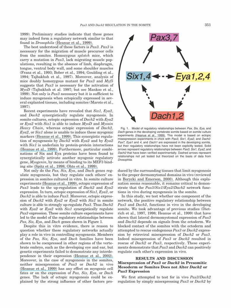
Not only do the Pax, Six, Eya, and Dach genes reg-ulate myogenesis, but they regulate each others’ ex-pression in somites cultured in vitro. In somite cultureexperiments (Heanue et al., 1999), ectopic expression ofPax3 leads to the up-regulation of Dach2 and Eya2expression. In turn, ectopic expression of Six1, Eya2, orDach2 is able to induce Pax3. Moreover, ectopic expres-sion of Dach2 with Eya2 or Eya2 with Six1 in somiteculture is able to strongly up-regulate Pax3. Thus Dach2with Eya2 or Eya2 with Six1 synergistically regulatePax3 expression. These somite culture experiments haveled to the model of the regulatory relationships betweenPax, Six, Eya, and Dach genes shown in Figure 1.
Despite this in vitro evidence, there is reason toquestion whether these regulatory networks actuallyplay a role in vivo in controlling myogenesis. Membersof the Pax, Six, Eya, and Dach families have beenshown to be coexpressed in other regions of the verte-brate embryo, such as the developing eye and ear, butgenetic experiments failed to demonstrate any interde-pendence in their expression (Heanue et al., 2002).Moreover, in the case of myogenesis in the somites,neither misexpression of Pax3 or Dach2 in vivo(Heanue et al., 1999) has any effect on myogenic cellfates or on the expression of Pax, Six, Eya, or Dachgenes. The lack of ectopic myogenesis could be ex-plained by the strong influence of other factors pro-
duced by the surrounding tissues that limit myogenesisto the proper dermomytomal domains in vivo (reviewedin Borycki and Emerson, 2000). Although this expla-nation seems reasonable, it remains critical to demon-strate that the Pax3/Six1/Eya2/Dach2 network func-tions in vivo during myogenesis in the somite.
In this study, we test whether one component of thenetwork, the positive regulatory relationship betweenPax3 and Dach2, functions in vivo in the developingsomite. We took advantage of previous studies (Diet-rich et al., 1997, 1998; Heanue et al., 1999) that haveshown that lateral dermomyotomal expression of Pax3and Dach2 depends on signals from the ectoderm. Weblocked contact of the somites with the ectoderm andattempted to rescue endogenous Pax3 or Dach2 expres-sion by retroviral misexpression of Dach2 or Pax3.Indeed misexpression of Pax3 or Dach2 resulted inrescue of Dach2 or Pax3, respectively. These experi-ments demonstrate that Pax3 and Dach2 can positivelyregulate each other’s expression in vivo.
RESULTS AND DISCUSSIONMisexpression of Pax3 or Dach2 in PresomiticMesoderm or Somites Does not Alter Dach2 orPax3 Expression
We first attempted to test for in vivo Pax3/Dach2regulation by simply misexpressing Pax3 or Dach2 by
Fig. 1. Model of regulatory relationships between Pax, Six, Eya, andDach genes in the developing vertebrate somite based on somite cultureexperiments (Heanue et al., 1999). This model is based on ectopicmisexpression experiments in chick with Pax3, Six1, Eya2, and Dach2.Pax7, Eya1 and 4, and Dach1 are expressed in the developing somite,but their regulatory relationships have not been explicitly tested. Solidarrows represent regulatory relationships between Pax3, Six1, Eya2, andDach2 that have been verified experimentally. Dashed arrows representrelationships not yet tested but theorized on the basis of data fromDrosophila.
351Pax3 AND Dach2 REGULATION IN THE SOMITE
means of injection of replication-competent retroviralvectors (RCAS) transducing these genes into psm orrecently formed somites. Embryos harvested 36 hr af-ter injection and analyzed by in situ hybridizationshowed no alteration in Dach2 or Pax3 expression (Fig.2A,B). The lack of Pax3 or Dach2 up-regulation in theventral result is agreement with previous observations(Heanue et al., 1999). Such a result may be due to thestrong influence of Shh from the notochord and floor-plate that inhibits myogenesis in the ventral somite(reviewed in Borycki and Emerson, 2000).
Signals from the Ectoderm Are Required forNormal Pax3 and Dach2 Expression in theDermomyotome
One successful experimental approach to avoidingthe normally tight regulation of somite development isto manipulate somites in culture; however, this strat-egy leaves open the question of in vivo relevance. Here,we present an in vivo approach to dissecting this com-plex system that takes advantage of knowledge of thefactors influencing somite differentiation. The ecto-derm overlying the developing somite has been shownto regulate patterning of the dorsal somite (reviewed inBorycki and Emerson, 2000). In the absence of ectoder-mal signals (perhaps members of the Wnt family; seeFan et al., 1997, and review in Borycki and Emerson,2000), Pax3 and Dach2 are down-regulated in the der-momyotome (Fan and Tessier-Lavigne, 1994; Dietrichet al., 1997, 1998; Heanue et al., 1999). In agreementwith these previous studies, we found that dermomyo-tomal expression of Pax3 and Dach2 depends on sig-nals from the ectoderm. A barrier inserted between theectoderm and the psm or recently formed somites had nogross morphologic effect on somite growth. Indeed whenharvested, the somites of these embryos appear indistin-guishable on the experimental and control sides. How-ever, in situ hybridization revealed that this manipula-tion results in down-regulation of both Pax3 and Dach2(Fig. 2C,D), although somite growth was otherwise nor-mal. In our experiments, we frequently found that bothlateral and medial Pax3 and Dach2 expression were lostdue the barrier inhibition of signals from both the ecto-derm (leading to loss of lateral expression, in agreementwith Dietrich et al., 1997, 1998; Heanue et al. 1999) andthe neural tube (leading to loss of medial expression, inagreement with Dietrich et al. 1997).
In the Absence of Ectodermal Signals, EctopicPax3 and Dach2 Can Rescue Each Other’sExpression in Somites
Isolation of the somite from the overlying ectodermand the resulting down-regulation of Pax3 and Dach2gene expression allowed us to test whether misexpres-sion of one gene was capable of rescuing expression ofthe other, by using RCAS vectors to ectopically expressPax3 or Dach2. It proved technically demanding totarget the infections to the regions blocked by the bar-
riers. In experiments where Dach2 was misexpressedbut the infection as visualized by staining by 3C2missed the targeted region (brown staining, Fig. 2C),down-regulation of Pax3 was still observed. However,in cases where the misexpression of Dach2 was suc-cessfully targeted to the barrier regions (n � 4/24),Pax3 expression was observed in the lateral dermo-myotmal region that had been covered by a barrier(Fig. 2E,G). This expression corresponded to the do-mains of the lateral dermomyotome, which were in-fected with the Dach2 retrovirus (compare purple Pax3expression with orange 3C2 stain, Fig. 2E,G). Notethat, because the retrovirus is replication-competentand continues to spread in the injected embryo, tissueoutside the dermomyotome is also infected. These othertissues do not up-regulate Pax3, as expected from thelack of response to virus injected into intact somites(see above). In rescue experiments involving Pax3misexpression, we observed Dach2 expression in thelateral dermomyotomal regions that had been coveredby the barrier (n � 2/11) (Fig. 2F,H). The patches ofPax3 or Dach2 expression observed in these experi-ments were found in dermomytomal regions but clearlyhad irregular borders and did not exhibit a normal,wild-type expression pattern (Fig. 2E–H). Such patcheswere never seen when the barrier was inserted alone.Moreover, because of the irregular expression domainsof Pax3 or Dach2, we could be certain that this expres-sion was due to rescue and not simply due to a failureof barrier inhibition of ectodermal signals. The rescueof dermomyotomal Pax3 or Dach2, down-regulated inthe absence of ectodermal signals, by RCAS Dach2 orPax3 demonstrates that in vivo Pax3 and Dach2 posi-tively regulate each other’s expression. This findingresult strongly suggests that the regulatory gene net-work defined in the somite culture system is importantfor normal myogenic regulation.
Previous somite culture experiments demonstratedthat the ability of Dach2 to up-regulate Pax3 expres-sion was improved when Dach2 was misexpressed incombination with Eya2 (Heanue et al., 1999). We mightexpect such synergistic action of Dach2 and Eya2 to besimilarly observed in vivo. By using the rescue protocoldescribed here, it would be possible to directly test forsynergy of Dach2 and Eya2 to regulate Pax3 expres-sion, by misexpressing Dach2 together with Eya2 (bymeans of RCAS(A) and RCAS(B) retroviral coinjec-tions; Morgan and Fekete, 1996). If Dach2 and Eya2synergistically regulate Pax3 in vivo, a more pro-nounced rescue of Pax3 expression would be expected.In the future, these and other aspects of the Pax3/Six1/Eya2/Dach2 network identified in vitro can be con-firmed in vivo by using the approach presented here. Inaddition, barriers isolating the somite from other invivo influences (e.g., notochord or lateral plate) couldallow other aspects of the regulation of myogenesis tobe examined.
352 KARDON ET AL.
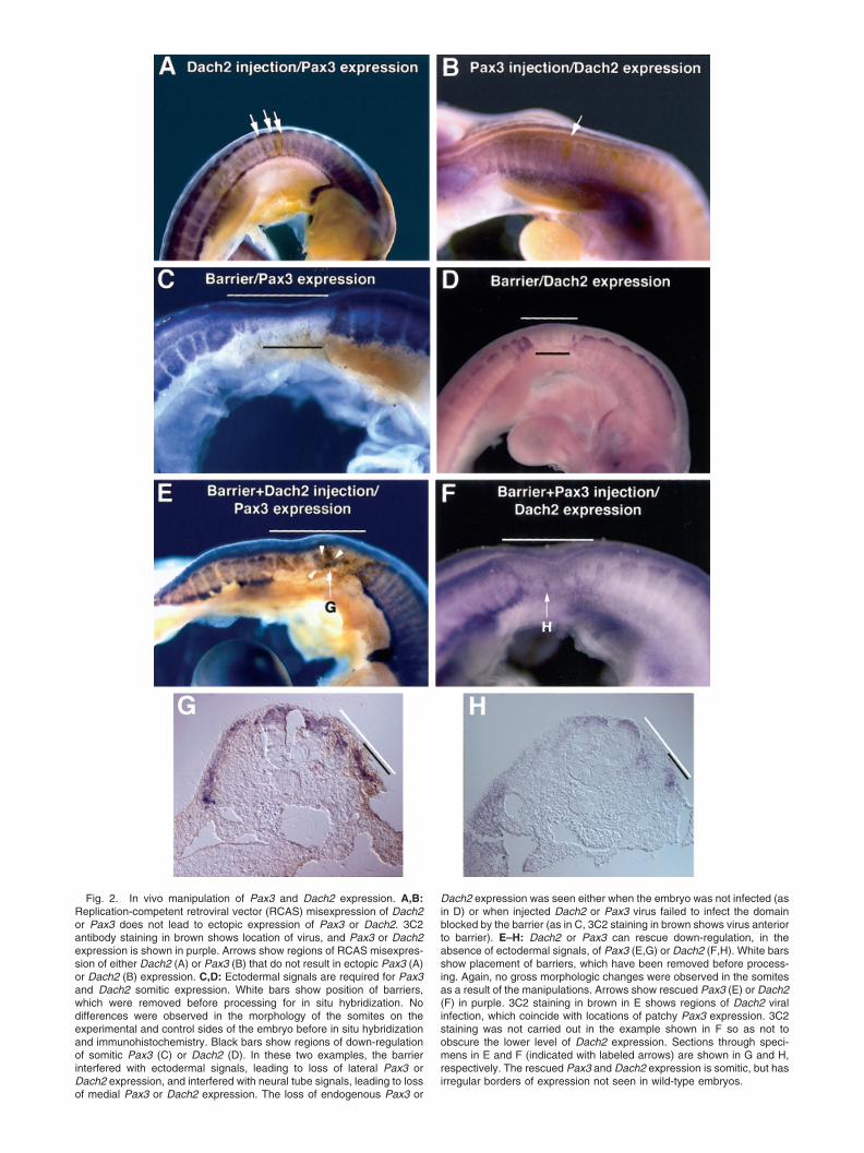
Fig. 2. In vivo manipulation of Pax3 and Dach2 expression. A,B:Replication-competent retroviral vector (RCAS) misexpression of Dach2or Pax3 does not lead to ectopic expression of Pax3 or Dach2. 3C2antibody staining in brown shows location of virus, and Pax3 or Dach2expression is shown in purple. Arrows show regions of RCAS misexpres-sion of either Dach2 (A) or Pax3 (B) that do not result in ectopic Pax3 (A)or Dach2 (B) expression. C,D: Ectodermal signals are required for Pax3and Dach2 somitic expression. White bars show position of barriers,which were removed before processing for in situ hybridization. Nodifferences were observed in the morphology of the somites on theexperimental and control sides of the embryo before in situ hybridizationand immunohistochemistry. Black bars show regions of down-regulationof somitic Pax3 (C) or Dach2 (D). In these two examples, the barrierinterfered with ectodermal signals, leading to loss of lateral Pax3 orDach2 expression, and interfered with neural tube signals, leading to lossof medial Pax3 or Dach2 expression. The loss of endogenous Pax3 or
Dach2 expression was seen either when the embryo was not infected (asin D) or when injected Dach2 or Pax3 virus failed to infect the domainblocked by the barrier (as in C, 3C2 staining in brown shows virus anteriorto barrier). E–H: Dach2 or Pax3 can rescue down-regulation, in theabsence of ectodermal signals, of Pax3 (E,G) or Dach2 (F,H). White barsshow placement of barriers, which have been removed before process-ing. Again, no gross morphologic changes were observed in the somitesas a result of the manipulations. Arrows show rescued Pax3 (E) or Dach2(F) in purple. 3C2 staining in brown in E shows regions of Dach2 viralinfection, which coincide with locations of patchy Pax3 expression. 3C2staining was not carried out in the example shown in F so as not toobscure the lower level of Dach2 expression. Sections through speci-mens in E and F (indicated with labeled arrows) are shown in G and H,respectively. The rescued Pax3 and Dach2 expression is somitic, but hasirregular borders of expression not seen in wild-type embryos.
EXPERIMENTAL PROCEDURESEmbryo Surgery and Virus Injection
Fertilized chick eggs were obtained from SPAFAS(Norwich, CT), incubated to specified stages, and win-dowed before surgery. Three treatments of the psm andsomites I-III (Christ and Ordahl, 1995) on stage 10–12embryos were performed: (1) misexpression of Dach2 orPax3 in somitic cells by means of injection of RCAScontaining the genes of interest, (2) isolation of somitictissue from ectoderm by placement of barriers, and (3)combination of misexpression of Dach2 or Pax3 andisolation from ectoderm of somitic tissue. For misex-pression experiments, RCAS virus was injected intothe somites with a micropipette and Hamilton syringe.For barrier experiments, ectoderm was separated fromthe underlying psm and somites by application of asmall amount of pancreatin to loosen the tissue andteasing away the ectoderm with a sharpened tungstenneedle. Fifteen-micron-thick cellophane barriers werethen placed on top of the psm and somites, and theectoderm was allowed to grow over the barrier. Forcombination experiments, the ectoderm was first re-moved, then virus was injected, and finally the barrierwas placed. In all experiments, embryos were sealedand incubated for an additional 36 hr, fixed in 4%paraformaldehyde, and analyzed by whole-mount RNAin situ hybridization and antibody staining (see below).Before fixation, the position of the barrier and themorphology of the somites were recorded and then thebarrier was removed.
RCAS Virus Construction
Viral constructs were generated and high titer viruswas produced following the protocols of Logan andTabin (1998). Both the Pax3 and Dach2 RCAS con-structs have been described previously (Maroto et al.,1997; Heanue et al., 1999).
Whole-Mount In Situ RNA Hybridization andAntibody Staining
Whole-mount RNA in situ hybridization with nonra-dioactive riboprobes was performed as described previ-ously (Riddle et al., 1993). On a subset of embryos,RCAS viral protein was visualized by staining embryoswith the 3C2 antibody after whole-mount in situ hy-bridization (Logan and Tabin, 1998). After photograph-ing the hybridization and staining results, a few em-bryos were embedded in 7.5% gelatin, cryosectioned (20�m thick), and rephotographed.
ACKNOWLEDGMENTS
G.K. was supported by a NIH postdoctoral grant.C.J.T. received funding from the NIH.
REFERENCES
Bober E, Franz T, Arnold HH, Gruss P, Tremblay P. 1994. Pax-3 isrequired for the development of limb muscles: a possible role for the
migration of dermomyotomal muscle progenitor cells. Development120:603–612.
Borsani G, DeGrandi A, Ballabio A, Bulfone A, Bernard L, Banfi S,Gattuso C, Mariani M, Dixon M, Donnai D, Metcalfe K, Winter R,Robertson M, Axton R, Brown A, van Heyningen V, Hanson I. 1999.EYA4, a novel vertebrate gene related to Drosophila eyes absent.Hum Mol Genet 8:11–23.
Borycki AG, Emerson CP. 2000. Multiple tissue interactions andsignal transduction pathways control somite myogenesis. Curr TopDev Biol 48:165–224.
Brand-Saberi B, Christ B. 2000. Evolution and development of dis-tinct cell lineages derived from somites. Curr Top Dev Biol 48:1–42.
Buckingham M. 2001. Skeletal muscle formation in vertebrates. CurrOpin Genet Dev 11:440–448.
Christ B, Ordahl CP. 1995. Early stages of chick somite development.Anat Embryol (Berl) 191:381–396.
Dietrich S, Schubert FR, Lumsden A. 1997. Control of dorsoventralpattern in the chick paraxial mesoderm. Development 124:3895–3908.
Dietrich S, Schubert FR, Healy C, Sharpe PT, Lumsden A. 1998.Specification of the hypaxial musculature. Development 125:2235–2249.
Esteve P, Bovolenta P. 1999. cSix4, a member of the six gene family oftranscription factors, is expressed during placode and somite devel-opment. Mech Dev 85:161–165.
Fan CM, Tessier-Lavigne M. 1994. Patterning of mammalian somitesby surface ectoderm and notochord: evidence for sclerotome induc-tion by a hedgehog homolog. Cell 79:1175–1186.
Fan CM, Lee CS, Tessier-Lavigne M. 1997. A role for WNT proteins ininduction of dermomyotome. Dev Biol 191:160–165.
Franz T, Kothary R, Surani MA, Halata Z, Grim M. 1993. The splotchmutation interferes with muscle development in the limbs. AnatEmbryol (Berl) 187:153–160.
Goulding M, Lumsden A, Paquette AJ. 1994. Regulation of Pax-3expression in the dermomyotome and its role in muscle develop-ment. Development 120:957–971.
Heanue TA, Reshef R, Davis RJ, Mardon G, Oliver G, Tomarev S,Lassar AB, Tabin CJ. 1999. Synergistic regulation of vertebratemuscle development by Dach2, Eya2, and Six1, homologs ofgenes required for Drosophila eye formation. Genes Dev 13:3231–3243.
Heanue TA, Davis RJ, Rowitch DH, Kispert A, McMahon AP, MardonG, Tabin CJ. 2002. Dach1, a vertebrate homologue of Drosophiladachshund, is expressed in the developing eye and ear of both chickand mouse and is regulated independently of Pax and Eya genes.Mech Dev 111:75–87.
Jostes B, Walther C, Gruss P. 1990. The murine paired box gene,Pax7, is expressed specficiall during the development of the nervousand muscular system. Mech Dev 33:27–37.
Logan M, Tabin C. 1998. Targeted gene misexpression in chick limbbuds using avian replication-competent retroviruses. Methods 14:407–420.
Mankoo BS, Collins NS, Ashby P, Grigoleva E, Pevny LH, Canadia A,Wright CV, Rigby PW, Pachnis V. 1999. Mox2 is a component of thegenetic hierarchy controlling limb muscle development. Nature 122:831–838.
Maroto M, Reshef R, Munsterberg AE, Koester S, Goulding M, LassarAB. 1997. Ectopic Pax-3 activates MyoD and Myf-5 expression inembryonic mesoderm and neural tissue. Cell 89:139–148.
Mishima N, Tomarev S. 1998. Chicken eyes absent 2 gene: isolationand expression pattern during development. Int J Dev Biol 42:1109–1115.
Morgan BA, Fekete DM. 1996. Manipulating gene expression withreplication-competent retroviruses. Methods Cell Biol 51:185–218.
Ohto H, Kamada S, Tago K, Tominaga SI, Ozaki H, Sato S, KawakamiK. 1999. Cooperation of Six and Eya in activation of their target genesthrough nuclear translocation of Eya. Mol Cell Biol 19:6815–6824.
Oliver G, Mailhos A, Wehr R, Copeland NG, Jenkins NA, Gruss P.1995. Six3, a murine homologue of the sine oculis gene, demarcatesthe most anterior border of the developing neural plate and isexpressed during eye development. Development 121:4045–4055.
354 KARDON ET AL.

Ordahl CP, Le Douarin NM. 1992. Two myogenic lineages within thedeveloping somite. Development 114:339–353.
Relaix F, Buckingham M. 1999. From insect eye to vertebratemuscle: redeployment of a regulatory network. Genes Dev 13:3171–3178.
Riddle RD, Johnson RL, Laufer E, Tabin C. 1993. Sonic hedgehogmediates the polarizing activity of the ZPA. Cell 75:1401–1416.
Spitz F, Demignon J, Porteu A, Kahn A, Concordet JP, Daegelen D,Maire P. 1998. Expression of myogenin during embryogenesis iscontrolled by Six/sine oculis homeoproteins through a conservedMEF3 binding site. Proc Natl Acad Sci U S A 95:14220–14225.
Tajbakhsh S, Rocancourt D, Cossu G, Buckingham M. 1997. Redefin-ing the genetic hierarchies controlling skeletal myogenesis: Pax- 3and Myf-5 act upstream of MyoD. Cell 89:127–138.
Wawersik S, Maas RL. 2000. Vertebrate eye development as modeledin Drosophila. Hum Mol Genet 9:917–925.
Williams BA, Ordahl CP. 1994. Pax-3 expression in segmental meso-derm marks early stages in myogenic cell specification. Develop-ment 120:785–796.
Xu PX, Cheng J, Epstein JA, Maas RL. 1997. Mouse Eya genes areexpressed during limb tendon development and encode a transcrip-tional activation function. Proc Natl Acad Sci U S A 94:11974–11979.
355Pax3 AND Dach2 REGULATION IN THE SOMITE