Pattern of Transcription Factor Activation in Helicobacter pylori–Infected Mongolian Gerbils...
15
Pattern of Transcription Factor Activation in Helicobacter pylori–Infected Mongolian Gerbils TAKAHIKO KUDO,* HONG LU,* ,‡ JENG–YIH WU,* ,§ TOMOYUKI OHNO,* MICHAEL J. WU,* ROBERT M. GENTA, DAVID Y. GRAHAM,* and YOSHIO YAMAOKA* *Department of Medicine, Veterans Affairs Medical Center and Baylor College of Medicine, Houston, Texas; ‡ Department of Gastroenterology, Renji Hospital, Shanghai Jiao Tong University School of Medicine, Shanghai Institute of Digestive Disease, Shanghai, China; § Division of Gastroenterology, Department of Internal Medicine, Kaohsiung Medical University Hospital, Kaohsiung, Taiwan; and Pathology and Laboratory Service, Veterans Affairs North Texas Health Care System, Dallas, Texas Background & Aims: Helicobacter pylori interact with epithelial cells resulting in activation of cellular sig- naling pathways leading to an inflammatory re- sponse. The pattern and timing of transcription fac- tor activation in H pylori-infected gastric mucosa remain unclear. We investigated the roles of tran- scription factors in the gastric mucosa of H pylori- infected gerbils over the course of the infection. Methods: Six-week-old male Mongolian gerbils were inoculated orally with H pylori TN2GF4 or isogenic cagE mutants and examined at 1, 3, 9, and 18 months. We examined the expression of 54 transcription fac- tors using DNA/protein arrays and electrophoretic mobility shift assays. Phosphorylation status of mi- togen-activated protein kinases and IB were evalu- ated by immunoblot and immunohistochemistry. Results: Ten transcription factors were up-regulated by H pylori infection. Six of these factors, including activator protein-1 (AP-1) and cAMP responsive ele- ment binding protein (CREB), reached maximal lev- els at 3 months and were strongly correlated with cellular inflammation and ulceration. Phosphoryla- tion of extracellular signal-regulated kinase corre- lated with activation of AP-1 and CREB. Levels of nuclear factor-B and interferon-stimulated respon- sive element (ISRE) peaked at 18 months and corre- lated with the presence of severe atrophy and with phosphorylation of Jun-N-terminal kinase (JNK), p38, and IB. Conclusions: The gastric mucosal transcription factors induced by H pylori infection differed according to the phase and outcome of in- fection; AP-1 and CREB levels were early responders related to inflammation and ulceration, whereas NF-B and ISRE were late responders related to at- rophy. H elicobacter pylori causes chronic gastric inflammation in virtually all infected persons. The cellular inflam- matory response initially consists of infiltration by neu- trophils followed by T and B lymphocytes, plasma cells, and macrophages. Infiltration of the mucosa with in- flammatory cells is associated with epithelial cell damage (reviewed in Suerbaum and Michetti 1 ) and with a marked increase in the expression of proinflammatory cytokines, including interleukin (IL)-1, IL-6, and IL-8, 2,3 which are regulated at the level of transcription. The signal trans- duction pathways involve transcription factors that bind DNA at specific promoter or enhancer regions. Activated transcription factors regulate gene expression by modu- lating the frequency of transcription initiation by inter- acting with specific DNA-binding elements present in promoters. The details and sequence of transcription factor acti- vation in H pylori infection are not well understood (re- viewed in Naumann and Crabtree 4 ). Much of the avail- able data comes from in vitro studies in which H pylori was cocultured with gastric cancer cells. In these experi- ments, transcription factor activation typically occurs within 1 hour, and the relationship between these in vitro observations and the phenomenon that occurs in H py- lori-infected gastric mucosa remains largely unexplored. The Mongolian gerbil (Meriones unguiculatus) has proven to be a convenient animal model for studies of H pylori infection because the gross and histologic findings mimic the lesions induced by H pylori infection in the human gastric mucosa. 5–9 In the present study, we investigated the roles of transcription factors in the gastric mucosa of H pylori-infected gerbils over the course of the infection in vivo. Materials and Methods Animals Six-week-old male Mongolian gerbils (MGS/Sea; Harlan Sprague Dawley, Inc., Indianapolis, IN) were Abbreviations used in this paper: AP-1, activator protein-1; CREB, cAMP responsive element binding protein; EMSA, electrophoretic mo- bility shift assay; ERK, extracellular signal-regulated kinase; IFN, inter- feron; IL, interleukin; IRF, interferon regulatory factor; ISRE, interferon- stimulated responsive element; JNK, Jun-N-terminal kinase; MAPK, mitogen-activated protein kinase; PAI, pathogenicity island; USF, up- stream stimulatory factor; WT, wild-type. © 2007 by the AGA Institute 0016-5085/07/$32.00 doi:10.1053/j.gastro.2007.01.009 BASIC– ALIMENTARY TRACT GASTROENTEROLOGY 2007;132:1024 –1038
-
Upload
utsouthwestern -
Category
Documents
-
view
2 -
download
0
Transcript of Pattern of Transcription Factor Activation in Helicobacter pylori–Infected Mongolian Gerbils...
PM
TD
*SMD
BenstrsiMicWtmtaRbamectlnslpptdfrNr
Hmtafl
BA
SIC–
ALIM
ENTA
RY
TRA
CT
GASTROENTEROLOGY 2007;132:1024 –1038
attern of Transcription Factor Activation in Helicobacter pylori–Infectedongolian Gerbils
AKAHIKO KUDO,* HONG LU,*,‡ JENG–YIH WU,*,§ TOMOYUKI OHNO,* MICHAEL J. WU,* ROBERT M. GENTA,�
AVID Y. GRAHAM,* and YOSHIO YAMAOKA*
Department of Medicine, Veterans Affairs Medical Center and Baylor College of Medicine, Houston, Texas; ‡Department of Gastroenterology, Renji Hospital,hanghai Jiao Tong University School of Medicine, Shanghai Institute of Digestive Disease, Shanghai, China; §Division of Gastroenterology, Department of Internaledicine, Kaohsiung Medical University Hospital, Kaohsiung, Taiwan; and �Pathology and Laboratory Service, Veterans Affairs North Texas Health Care System,
allas, Texas(iirdDtlap
vvawmwolTtitgtHi
H
cbfsms
ackground & Aims: Helicobacter pylori interact withpithelial cells resulting in activation of cellular sig-aling pathways leading to an inflammatory re-ponse. The pattern and timing of transcription fac-or activation in H pylori-infected gastric mucosaemain unclear. We investigated the roles of tran-cription factors in the gastric mucosa of H pylori-nfected gerbils over the course of the infection.ethods: Six-week-old male Mongolian gerbils were
noculated orally with H pylori TN2GF4 or isogenicagE mutants and examined at 1, 3, 9, and 18 months.
e examined the expression of 54 transcription fac-ors using DNA/protein arrays and electrophoretic
obility shift assays. Phosphorylation status of mi-ogen-activated protein kinases and I�B were evalu-ted by immunoblot and immunohistochemistry.esults: Ten transcription factors were up-regulatedy H pylori infection. Six of these factors, includingctivator protein-1 (AP-1) and cAMP responsive ele-ent binding protein (CREB), reached maximal lev-
ls at 3 months and were strongly correlated withellular inflammation and ulceration. Phosphoryla-ion of extracellular signal-regulated kinase corre-ated with activation of AP-1 and CREB. Levels ofuclear factor-�B and interferon-stimulated respon-ive element (ISRE) peaked at 18 months and corre-ated with the presence of severe atrophy and withhosphorylation of Jun-N-terminal kinase (JNK),38, and I�B. Conclusions: The gastric mucosal
ranscription factors induced by H pylori infectioniffered according to the phase and outcome of in-ection; AP-1 and CREB levels were early responderselated to inflammation and ulceration, whereasF-�B and ISRE were late responders related to at-
ophy.
elicobacter pylori causes chronic gastric inflammationin virtually all infected persons. The cellular inflam-
atory response initially consists of infiltration by neu-rophils followed by T and B lymphocytes, plasma cells,nd macrophages. Infiltration of the mucosa with in-
ammatory cells is associated with epithelial cell damagereviewed in Suerbaum and Michetti1) and with a markedncrease in the expression of proinflammatory cytokines,ncluding interleukin (IL)-1�, IL-6, and IL-8,2,3 which areegulated at the level of transcription. The signal trans-uction pathways involve transcription factors that bindNA at specific promoter or enhancer regions. Activated
ranscription factors regulate gene expression by modu-ating the frequency of transcription initiation by inter-cting with specific DNA-binding elements present inromoters.The details and sequence of transcription factor acti-
ation in H pylori infection are not well understood (re-iewed in Naumann and Crabtree4). Much of the avail-ble data comes from in vitro studies in which H pylorias cocultured with gastric cancer cells. In these experi-ents, transcription factor activation typically occursithin 1 hour, and the relationship between these in vitrobservations and the phenomenon that occurs in H py-
ori-infected gastric mucosa remains largely unexplored.he Mongolian gerbil (Meriones unguiculatus) has proven
o be a convenient animal model for studies of H pylorinfection because the gross and histologic findings mimiche lesions induced by H pylori infection in the humanastric mucosa.5–9 In the present study, we investigatedhe roles of transcription factors in the gastric mucosa of
pylori-infected gerbils over the course of the infectionn vivo.
Materials and MethodsAnimalsSix-week-old male Mongolian gerbils (MGS/Sea;
arlan Sprague Dawley, Inc., Indianapolis, IN) were
Abbreviations used in this paper: AP-1, activator protein-1; CREB,AMP responsive element binding protein; EMSA, electrophoretic mo-ility shift assay; ERK, extracellular signal-regulated kinase; IFN, inter-eron; IL, interleukin; IRF, interferon regulatory factor; ISRE, interferon-timulated responsive element; JNK, Jun-N-terminal kinase; MAPK,itogen-activated protein kinase; PAI, pathogenicity island; USF, up-
tream stimulatory factor; WT, wild-type.© 2007 by the AGA Institute
0016-5085/07/$32.00
doi:10.1053/j.gastro.2007.01.009
hfctfCiMH
ngdilb((gas
s2silHBNH
oH9fmt
cOfffgtotgdam
cmc
lospHmticcwpcsd
wsb((TiabnmowafIttdwb
pp(cvp
BA
SIC–
ALI
MEN
TARY
TRA
CT
March 2007 GERBILS AND H PYLORI 1025
oused in an air-conditioned biohazard room designedor infectious animals with a 12-hour light/12-hour darkycle. They were provided rodent diet and water ad libi-um. The animal facility is accredited by the Associationor Assessment and Accreditation of Laboratory Animalare. The experiment protocol was approved by the An-
mal Care Committee of Baylor College of Medicine andichael E. DeBakey Veterans Affairs Medical Center,ouston, TX.
Bacterial Strains and InoculationH pylori strain TN2GF4 was isolated from a Japa-
ese gastric ulcer patient and has been shown to colonizeerbils consistently for at least 1 year and to cause repro-ucible mucosal damage.6 This strain is cag pathogenicity
sland (PAI) positive, vacA s1 (production of the vacuo-ating cytotoxin) and has functional blood group antigeninding adhesin (BabA) and outer inflammatory proteinOipA). Long-term studies (eg, 18 months) used wild-typeWT) strain TN2GF4, whereas the cag PAI mutant (iso-enic cagE mutant) was used for short-term studies (ie, 1nd 3 months). The isogenic cagE mutant was con-tructed previously10 and was chloramphenicol resistant.
H pylori were grown in brain heart infusion (BHI) brothupplemented with 15% fetal bovine serum (FBS) for0 –30 hours at 37°C under microaerobic conditions andaturated humidity, with shaking at 200 rpm. After fast-ng for 12 hours, each animal was orogastrically inocu-ated 3 times (days 0, 1, and 2) with 1.0 mL inoculum of
pylori (108 colony forming unit [CFU]/mL) or sterileHI broth (as a control) using gastric intubation needles.o specific pretreatments were given before orogastricpylori inoculation.
Time Course and DeathInfected gerbils were killed and necropsied 1, 3, 9,
r 18 months after H pylori inoculation. Five or 6pylori-inoculated gerbils were used for the 1-, 3-, and
-month time points, and 18 inoculated gerbils were usedor the 18-month time point. Three to 5 age- and sex-
atched uninfected gerbils were used as controls at eachime point.
At necropsy, stomachs were opened along the greaterurvature and were divided longitudinally into 2 parts.ne half was fixed in 10% phosphate-buffered formalin
or histologic examination. The other half was dividedurther into the pyloric gland mucosa (antrum) and theundic gland mucosa (corpus) and stored at �80°C. Theastric mucosa was separated as much as possible fromhe underlying muscle by sharp dissection. In addition,ne mm2 piece of gastric mucosa from the antrum wasaken to culture H pylori. The culture of H pylori from theastric mucosa of gerbils was performed as previouslyescribed.9,11 The recovered strains were tested for chlor-mphenicol resistance to confirm the stability of the
utation for the cagE mutant-infected cases. Polymerase ehain reaction (PCR) analysis was also performed for cagEutants to confirm the preservation of chloramphenicol
assettes inside the target genes as previously described.12
HistopathologyTissues were fixed in 10% neutral-buffered forma-
in and sliced along the longitudinal axis into 4 to 7 slipsf equal width, embedded in paraffin, and cut into 4-�mections. The sections were stained with H&E for mor-hologic observation and with Genta stain to detect
pylori and mucin-containing cells. Because the inflam-atory component of the mucosa was virtually identical
o that found in human H pylori gastritis,7 the degree ofnflammation and existence of intestinal metaplasiaould be graded blindly by one pathologist (R.M.G) ac-ording to the Updated Sydney System.13 H pylori densityas also evaluated by histology because only one mm2
iece of gastric mucosa from the antrum was taken toulture H pylori, and the number of H pylori by culturehould not be more reliable than scoring the H pyloriensity by histology.
Protein/DNA Array Analysis of SignalTransduction System ComponentsNuclear extracts from gastric mucosal specimens
ere prepared using hypotonic/nonionic detergent ly-is.14 After extraction, nuclear proteins were normalizedy protein assay (Bio-Rad, Hercules, CA). Equal amounts25 �g per sample) were used for the protein/DNA arrayTranSignal Arrays, Panomics, Inc., Redwood City, CA).he protein/DNA array provides a profile of DNA bind-
ng activity for multiple transcription factors in a singlerray experiment and allows 54 transcription factors toe identified simultaneously. For quantitation, pooleduclear extracts were prepared from a mixture of 3 gastricucosal specimens obtained 9 months after inoculation
f WT strains (standard sample). Six array membranesere set up simultaneously, with 1 membrane being useds a control. The density of dots for each transcriptionactor and the standard sample were scanned using Scionmage beta 4.02 software (Scion Corp. Frederick, MA). Inhe standard sample, the density of all dots from 54ranscription factors was summed (� density for all). Theensity of each transcription factor in the test samplesas divided by the “density for all” and multipliedy 100.
Electrophoretic Mobility Shift AssayElectrophoretic mobility shift assay (EMSA) was
erformed using the same nuclear extracts used in therotein/DNA array. Equal amounts of nuclear proteins
10 �g per sample) were used to bind to duplex oligonu-leotides corresponding to nuclear factor (NF)-�B, acti-ator protein-1 (AP-1), cAMP responsive element bindingrotein (CREB), and interferon-stimulated responsive el-
ment (ISRE) binding sites. We used commercial consen-
siapptmsrspasBta
ddl(i(tfTr
ibpfAtimm(p
ml(bFmim(sfl
2tppwc
mcicp(h(S
gfu2efafctptpCfptmaf1suam(ctlm
BA
SIC–
ALIM
ENTA
RY
TRA
CT
1026 KUDO ET AL GASTROENTEROLOGY Vol. 132, No. 3
us probes for these binding sites (Promega Corp, Mad-son, WI). EMSA was performed using standard methodss previously described.15 For semiquantitation, we usedooled nuclear extracts from the mixture of 3 WT Hylori-infected gastric mucosal specimens from the gas-ritis group at 18 months (standard sample). Density was
easured by scanning, using the software Image J 1.36oftware from the National Institutes of Health (http://sbweb.nih.gov/ij/), and the density of samples was pre-ented as a percentage of the standard sample. For com-etition assays, 5 pmol of unlabeled competitor wasdded at the time of probe addition. In the gel mobilityupershift assays, commercial antibodies (Santa Cruziotechnology, Inc., Santa Cruz, CA) against specific
ranscription factors were added to the binding reactionsnd incubated on ice prior to fractionation by 6% PAGE.
Immunoblot Analysis of Mitogen-ActivatedProtein Kinases and I�BImmunoblot analysis was performed using stan-
ard methods. We used phospho-specific antibodies toetect phospho-p38, phospho-extracellular signal-regu-
ated kinase (ERK), phospho-Jun-N-terminal kinaseJNK), phospho-inhibitor of I�B �, and control antibod-es against total p38, ERK, and JNK as well as �-actin1:1500 dilution) (antibodies for mitogen-activated pro-ein kinase [MAPKs] were from Promega, and antibodiesor phospho-I�B� and �-actin were from Cell Signalingechnology, Beverly, MA). These antibodies have been
eported to cross-react with Mongolian gerbils.16 –20
Cytoplasmic extract (10 �g per sample) obtained dur-ng the preparation of nuclear extracts was fractionatedy SDS-PAGE and electrophoretically transferred to aolyvinylidene difluoride membrane. Detection was per-ormed using ECL reagents (Amersham Life Science,rlington Heights, IL). For semiquantitation, pooled cy-
oplasmic extracts from the mixture of 3 WT H pylori-nfected gastric mucosal specimens with gastritis at 18
onths were used as the standard sample. Density waseasured by scanning, using Image J 1.36 software
NIH), and the density of samples was calculated as aercentage of the standard sample.
Isolation of Primary Gastric Epithelial CellsFrom Gerbil Gastric MucosaPrimary gastric epithelial cells were isolated enzy-
atically from gerbil stomachs using previously estab-ished methods.21,22 Briefly, the surface mucosal layercombined antrum and corpus) was removed with a razorlade, immediately minced, and then incubated in Ham’s-12 culture medium containing collagenase type I (0.2g/mL) (Invitrogen) for 10 minutes. Cells from the final
ncubation were washed and cultured in Ham’s F-12edium supplemented with 10% FBS and streptomycin
300 �g/mL) at 37°C in a humidified 5% CO2 atmo-phere. Cultured cells (1 � 106 cells/mL) formed subcon-
uent monolayers within 24 hours of inoculation in a E4-well collagen-coated dish. Approximately 93% of cul-ured cells in the monolayers had periodic acid-Schiff-ositive material in the cytoplasm, confirming that theopulation consisted of mucus-producing epithelial cellsith minimal contamination by other cells. Epithelial
ells could be maintained up to 1 week in culture.
In Vitro Experiments Using PrimaryEpithelial Cells From Gerbil Gastric MucosaH pylori TN2GF4 were suspended in cell culture
edium, and the subconfluent epithelial cells (1 � 106
ells/mL) were cocultured with H pylori (multiplicity ofnfection [MOI] of 100) or kept uninfected in a 24-wellollagen-coated dish for 3 hours. Cell viability was ap-roximately 90% during the coculturing experiments
data not shown). Nuclear extracts were prepared usingypotonic/nonionic detergent lysis, and equal amounts
10 �g per sample) were used for protein/DNA arrays.emiquantitation was performed as described previously.
In some experiments, we performed luciferase reporterene assays using primary gastric epithelial cells isolatedrom gerbil stomachs as described above. Commonlysed nonviral transfection methods (eg, Lipofectamine000 [Invitrogen]) did not succeed using primary gastricpithelial cells. Therefore, we used a novel nonviral trans-ection technology specially designed for primary cellsnd difficult to transfect cell lines, which allows trans-ected DNA to enter directly the nucleus of nondividingells within a few hours (Nucleofector; Amaxa Biosys-ems, Allemagne, Germany). The PathDetect cis-reportinglasmids pNF-�Bluc, pAP-1luc, and pCREluc, which con-ain the luciferase reporter gene driven by the TATA boxlus multiple repeats of consensus NF-�B, AP-1, andRE binding sequences, respectively, were purchased
rom Stratagene (La Jolla, CA). Luciferase assays wereerformed using the Dual-Luciferase reporter assay sys-em according to the manufacturer’s instructions (Pro-
ega). Epithelial cells (1 � 106 cells/mL) were cultured in24-well collagen-coated dish for 24 hours before trans-
ection, and, 6 hours after transfection, H pylori (MOI of00) were added or kept uninfected. Nine hours aftertimulation with H pylori, cells were harvested and lysedsing passive lysis buffer (Promega), and the lysates weressayed for luciferase activity. Cell viability was approxi-ately 85% to 90% during the coculturing experiments
data not shown). Normalized luciferase activity was cal-ulated as firefly luciferase activity/Renilla luciferase ac-ivity. Luciferase activity is reported as fold increase ofuciferase activity in treated cells relative to uninfected or
ock-treated controls.
ImmunohistochemistryImmunohistochemistry was performed for p38,
RK, JNK, and I�B�. We used phospho-specific antibod-

ianhtbatmAss((v
umtrbggqYdsLppoeGGt
f8pmaHtrl
Mpmw
Hfctpcatmipts(arHtpaMogcaipwtgt5utaut
imwitdiTtsss
BA
SIC–
ALI
MEN
TARY
TRA
CT
March 2007 GERBILS AND H PYLORI 1027
es to detect phospho-p38, phospho-ERK, phospho-JNK,nd phospho-I�B� (Promega and Cell Signaling Tech-ology). Tissues were placed in a microwave oven atigh power in 0.1 mol/L citrate buffer, pH 6.5, andreated 4 times for 5 minutes each. This was followedy a 2-hour incubation at room temperature with eachntibody diluted 1:50. The immunoperoxidase reac-ion was developed using the biotin-streptavidin im-
unoperoxidase method (DAKO, Santa Barbara, CA).reas of the surface/foveolar epithelium, glands, and
ubmucosal lymphocytes were estimated, and the re-ults were scored 0 to 3: 3, high level of stainingapproximately 80%–100%); 2, medium level of staining25%– 80%); 1, low level of staining (1%–25%); and 0,irtually no staining (�1%).
Analysis of Cytokine mRNA Expression byReal-Time Quantitative ReverseTranscription-PCRTotal RNA was extracted from the gastric mucosa
sing an RNA extraction kit (Qiagen). After DNase treat-ent, 5 �g total RNA was subjected to reverse transcrip-
ion (RT) using 200 U Moloney murine leukemia-viruseverse transcriptase (Life Technologies, Inc., Gaithers-urg, MD) and 1 �mol/L of oligo(dT)16 primers. We usederbil-specific IL-6 complementary DNA (cDNA) andlyceraldehyde-3-phosphate dehydrogenase (GAPDH) se-uences (GenBank accession number AB164706 andamaoka,10 respectively) and gerbil-specific keratinocyte-erived chemokine (KC) and interferon (IFN)-� cDNAequences (GenBank accession number: AJ877921 and37782, respectively). Specific primers and TaqManrobes for KC, IL-6, IFN-�, and GAPDH were describedreviously.23,24 Real-time PCR was performed as previ-usly described.24 Cytokine messenger RNA (mRNA) lev-ls were expressed as the ratio of cytokine mRNA toAPDH mRNA (100,000 � cytokine mRNA [unit/�L]/APDH mRNA [unit/�L]). Each assay was performed in
riplicate.In some experiments, we used RNA extracted from
ormalin-fixed, paraffin-embedded, tissue blocks. Serial-�m-thick sections of selected tissue blocks werelaced onto glass slides, and the areas of interest (sub-ucosal infiltrating cells) were microdissected visually
fter matching with an adjacent section stained with&E. Microdissected samples were digested with pro-
einase K, and RNA was purified by phenol and chlo-oform extraction and reversed transcribed as pub-ished.25
Statistical AnalysisStatistical analysis was performed using the
ann–Whitney rank sum test and the paired t test de-ending on the data set. The data are presented asean � standard error (SE). A P value of less than .05
as accepted as statistically significant. TResultsEffect of H Pylori Infection on Macroscopicand Histopathologic FindingsMongolian gerbils were inoculated orally with
pylori TN2GF4 or isogenic cagE mutants. H pylori in-ection was confirmed by culture and/or histology in allases. All cagE mutants recovered from gerbils were resis-ant to chloramphenicol, confirming that the chloram-henicol cassette remained functional. The disruption of
agE gene was also confirmed based on the size of themplicons by PCR. One month after inoculation, infil-ration of neutrophils and monocytes into the gastric
ucosa, mainly located in the submucosa, was observedn WT-infected gerbils. Infiltration into the stomach ap-eared to spread from the antrum to the corpus overime, with focal lymphocyte aggregates observed in theubmucosa at the glandular border of the pyloric mucosaantrum) and the fundic mucosa (corpus). Three monthsfter inoculation, inflammation and H pylori densityeached maximal levels (Figure 1). Inflammation and
pylori density were more severe in the antrum than inhe corpus in each time point (P � .01). Macroscopically,yloric channel ulcers were present in 1 of 5 gerbils (20%)t 9 months and in 8 of 18 gerbils (44%) at 18 months.icroscopically, intestinal metaplasia was present in 10
f 18 (56%) infected gerbils at 18 months, including 5erbils with gastric ulcers. The intestinal metaplasia oc-urred frequently in the transitional zone between pyloricnd fundic mucosa, and Paneth cells were rarely found,ndicating that the metaplasia was mostly of the incom-lete type. Eighteen months after inoculation, gerbilsere divided into 3 groups based on the gross and his-
ologic outcomes. Eight gerbils had gastric ulcers (ulcerroup), 5 gerbils had severe atrophic gastritis with intes-inal metaplasia, but without ulcers (atrophy group), and
gerbils had gastritis without intestinal metaplasia orlcers (gastritis group). Cellular infiltration in the an-rum was more severe in the ulcer group than in thetrophy group (Figure 1). In contrast to a previous studysing strain6 TN2GF4, no infected gerbils developed gas-ric cancer.
Infiltration of neutrophils and monocytes was absentn control animals. cagE mutants did not induce inflam-
ation during the period of observation of 3 monthsith the exception of a mild monocytic infiltration seen
n the antrum 3 months after inoculation (score 1) in 2 ofhe 5 gerbils. Importantly, the H pylori density was notifferent between WT-infected gerbils and cagE mutant-
nfected gerbils (data not shown). The WT strainN2GF4 induced an antral-dominant gastritis, whereas
he cagE mutants induced little inflammation. These re-ults are in agreement with previous studies using theame strain11 as well as with strain TN2, which shares theame origin20,27 as TN2GF4 and strain24 ATCC43504.
hese results differ from experiments using strain B128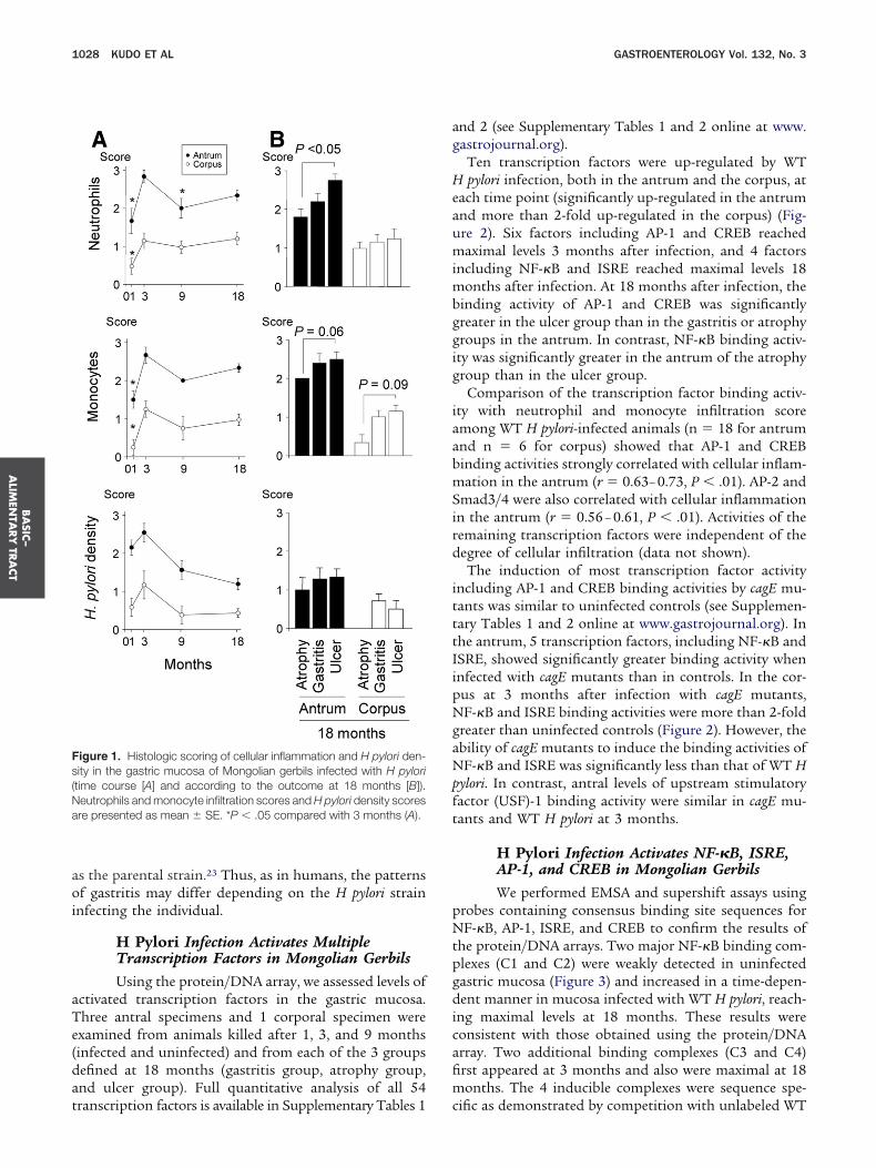
aoi
aTe(dat
ag
Heaumimbggig
iaabmSird
itttIipNgaNpft
pNtpgdicafim
Fs(Na
BA
SIC–
ALIM
ENTA
RY
TRA
CT
1028 KUDO ET AL GASTROENTEROLOGY Vol. 132, No. 3
s the parental strain.23 Thus, as in humans, the patternsf gastritis may differ depending on the H pylori strain
nfecting the individual.
H Pylori Infection Activates MultipleTranscription Factors in Mongolian GerbilsUsing the protein/DNA array, we assessed levels of
ctivated transcription factors in the gastric mucosa.hree antral specimens and 1 corporal specimen werexamined from animals killed after 1, 3, and 9 monthsinfected and uninfected) and from each of the 3 groupsefined at 18 months (gastritis group, atrophy group,nd ulcer group). Full quantitative analysis of all 54
igure 1. Histologic scoring of cellular inflammation and H pylori den-ity in the gastric mucosa of Mongolian gerbils infected with H pyloritime course [A] and according to the outcome at 18 months [B]).eutrophils and monocyte infiltration scores and H pylori density scoresre presented as mean � SE. *P � .05 compared with 3 months (A).
ranscription factors is available in Supplementary Tables 1 c
nd 2 (see Supplementary Tables 1 and 2 online at www.astrojournal.org).
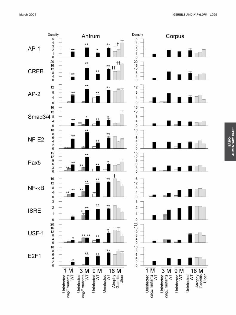
Ten transcription factors were up-regulated by WTpylori infection, both in the antrum and the corpus, at
ach time point (significantly up-regulated in the antrumnd more than 2-fold up-regulated in the corpus) (Fig-re 2). Six factors including AP-1 and CREB reachedaximal levels 3 months after infection, and 4 factors
ncluding NF-�B and ISRE reached maximal levels 18onths after infection. At 18 months after infection, the
inding activity of AP-1 and CREB was significantlyreater in the ulcer group than in the gastritis or atrophyroups in the antrum. In contrast, NF-�B binding activ-ty was significantly greater in the antrum of the atrophyroup than in the ulcer group.
Comparison of the transcription factor binding activ-ty with neutrophil and monocyte infiltration scoremong WT H pylori-infected animals (n � 18 for antrumnd n � 6 for corpus) showed that AP-1 and CREBinding activities strongly correlated with cellular inflam-ation in the antrum (r � 0.63– 0.73, P � .01). AP-2 and
mad3/4 were also correlated with cellular inflammationn the antrum (r � 0.56 – 0.61, P � .01). Activities of theemaining transcription factors were independent of theegree of cellular infiltration (data not shown).The induction of most transcription factor activity
ncluding AP-1 and CREB binding activities by cagE mu-ants was similar to uninfected controls (see Supplemen-ary Tables 1 and 2 online at www.gastrojournal.org). Inhe antrum, 5 transcription factors, including NF-�B andSRE, showed significantly greater binding activity whennfected with cagE mutants than in controls. In the cor-us at 3 months after infection with cagE mutants,F-�B and ISRE binding activities were more than 2-fold
reater than uninfected controls (Figure 2). However, thebility of cagE mutants to induce the binding activities ofF-�B and ISRE was significantly less than that of WT H
ylori. In contrast, antral levels of upstream stimulatoryactor (USF)-1 binding activity were similar in cagE mu-ants and WT H pylori at 3 months.
H Pylori Infection Activates NF-�B, ISRE,AP-1, and CREB in Mongolian GerbilsWe performed EMSA and supershift assays using
robes containing consensus binding site sequences forF-�B, AP-1, ISRE, and CREB to confirm the results of
he protein/DNA arrays. Two major NF-�B binding com-lexes (C1 and C2) were weakly detected in uninfectedastric mucosa (Figure 3) and increased in a time-depen-ent manner in mucosa infected with WT H pylori, reach-
ng maximal levels at 18 months. These results wereonsistent with those obtained using the protein/DNArray. Two additional binding complexes (C3 and C4)rst appeared at 3 months and also were maximal at 18onths. The 4 inducible complexes were sequence spe-
ific as demonstrated by competition with unlabeled WT
BA
SIC–
ALI
MEN
TARY
TRA
CT
March 2007 GERBILS AND H PYLORI 1029
bacomppcbp
dtHaadwojts(Htap
ispHpTlteasc
wi1tt
Fantpcrcm0aiabac
dwaiSomicto
sifpJu
fpdsval
4Fpaoo(pc
BA
SIC–
ALIM
ENTA
RY
TRA
CT
1030 KUDO ET AL GASTROENTEROLOGY Vol. 132, No. 3
ut not mutant oligonucleotides (Figure 3). Supershiftssays demonstrated that p65 was a component of the C1omplex. We were unable to identify components of thether complexes. In previous in vitro studies using hu-an gastric cancer cell lines, we showed that both
50 and p65 were components of NF-�B binding com-lexes.15,22,26 However, because the accuracy of commer-ially available antibodies against p50 in Mongolian ger-ils is unknown, we cannot conclude that p50 was notresent.Two ISRE binding complexes (C1 and C2) were weakly
etected in uninfected gastric mucosa and increased in aime-dependent manner in mucosa infected with WT
pylori, reaching maximal levels at 18 months (Figure 3),gain confirming the results obtained from protein/DNArrays. The inducible complexes were sequence specific asemonstrated by competition with unlabeled WT but notith mutant oligonucleotides. In contrast, strong signalsbserved in both uninfected and infected gerbils were
udged to be nonspecific because there was no competi-ion with unlabeled WT oligonucleotides. Supershift as-ays demonstrated that interferon regulatory factorIRF)-1, IRF-3, and IRF-7 were components of the
pylori-inducible ISRE complex. Of note, the density ofhe C1 and C2 complexes was not decreased by thesentibodies, suggesting that several IRF families might beresent in the complexes.One clear AP-1 binding complex was weakly detected
n the antral mucosa of uninfected gerbils but was noteen clearly in the corporal mucosa (Figure 3). The com-lex increased in the mucosa of gerbils infected with WT
pylori and reached maximal levels at 3 months. Thisattern was similar to the histologic changes (Figure 1).he density of the AP-1 complex was significantly corre-
ated with neutrophil and monocyte infiltration scores inhe antrum (r � 0.61 and 0.63, respectively; P � .01 forach), similar to data obtained using the protein/DNArrays. The inducible complex was sequence specific, andupershift assays demonstrated that c-Jun and c-Fos wereomponents of the AP-1 complex.
At least 3 CREB binding complexes (C1, C2, and C3)ere weakly detectable in uninfected gastric mucosa and
ncreased in mucosa of infected WT H pylori peaking at8 months (Figure 3). Complex C2 was variable at eachime point and among different experiments, althoughhe conditions and buffers were identical (eg, very faint in
™™™™™™™™™™™™™™™™™™™™™™™™™™™™™™™™™™™™igure 2. The binding activity of 10 transcription factors was inducedrotein/DNA array analyses. Three antral specimens and 1 corporal spend uninfected) and from each outcome group at 18 months (gastritis grf each sample was scanned and compared with that of standard nubtained 9 months after inoculation of WT strains. In the standard sa
� density for all). The density of each transcription factor in the test samerform statistical analyses only for the antral samples. Data are prese
ontrol. †P � .05 and ††P � .01 compared with the ulcer group.igures 4 and 5). The 3 complexes were sequence specifics demonstrated by competition with unlabeled WT butot mutant oligonucleotides; however, competition withhe complex C2 was less efficient than the other 2 com-lexes (Figure 3). Therefore, complex C2 was not in-luded in the semiquantitative analyses. Consistent withesults of the protein/DNA array, the density of CREBomplexes significantly correlated with neutrophil andonocyte infiltration scores in the antrum (r � 0.58 and
.61, respectively; P � .01 for each). Use of anti-CREB-1,nti-ATF-2, and anti-c-Jun antibodies in EMSAs resultedn supershifted bands, whereas use of anti-CREB-2 andnti-c-Fos antibodies resulted in weak supershiftedands, suggesting that members of the CREB/ATF familyre major components of the H pylori-inducible CREomplexes in gerbils.
EMSA confirmed the results of protein/DNA arrayata, showing that AP-1 and CREB binding activitiesere greater in the ulcer group than in the gastritis ortrophy groups and that NF-�B and ISRE binding activ-ties were greater in the atrophy group in the antrum (seeupplementary Figure 1 online at www.gastrojournal.rg). EMSA also confirmed that infection with the cagEutant resulted in more NF-�B and ISRE binding activ-
ty than uninfected controls both in the antrum and theorpus, although the degree of activation was less thanhat with the WT strain (see Supplementary Figure 2nline at www.gastrojournal.org).
Immunoblot Analysis of MAPKs and I�BPathways in Gastric Mucosa of GerbilsThe MAPKs and I�B pathways are involved in
ignaling upstream of many transcription factors includ-ng NF-�B, ISRE, AP-1, and CREB. Therefore, we per-ormed immunoblots for MAPKs and I�B using phos-ho-specific antibodies to detect phospho-ERK1/2, p38,
NK, and I�B, as well as control antibodies to detectnphosphorylated forms of ERK, p38, and JNK.Phosphorylation in both pathways was greater in in-
ected tissues than in uninfected controls (Figure 4). ERKhosphorylation reached maximal levels at 3 months,ecreased at 9 months, and increased again at 18 months,imilar to the pattern of cellular inflammation and acti-ation of AP-1 and CREB binding. ERK phosphorylationlmost disappeared at 9 months. ERK phosphorylationevels correlated with cellular inflammation scores and
™™™™™™™™™™™™™™™™™™™™™™™™™™™™™™™™™™™™pylori infection both in the antrum and the corpus as determined by
n were examined from animals killed after 1, 3, and 9 months (infectedtrophy group, and ulcer group). The transcription factor binding activityextracts obtained from the mixture of 3 gastric mucosal specimens, the density of dots from all 54 transcription factors were summeden was divided by the “density for all” and multiplied by 100. We coulds mean � SE. *P � .05 and **P � .01 compared with the uninfected
™™™by Hcime
oup, aclearmpleple th
nted a

F1umsssLa
igure 3. EMSA of NF-�B, ISRE, AP-1, and CREB binding complexes. Four to 5 antral and corporal specimens were used from animals killed at, 3, and 9 months (infected and uninfected). We also examined samples from each of the 3 outcome groups at 18 months (gastritis, atrophy, andlcer groups). Left column: Nuclear extracts were prepared from uninfected control mucosa at 18 months; infected mucosa from 1, 3, 9, and 18onths. Middle column: semiquantitative EMSA analyses. The densities of nuclear extracts from a mixture of 3 WT H pylori-infected gastric mucosal
pecimens from the gastritis outcome group at 18 months (standard sample) are presented as the percentage of the standard samples aftertandardization with free probe. Data are presented as mean � SE. Right column: lanes 1 to 3: competition analysis. Nuclear extracts from infectedamples (18 months) were used to bind probe in the absence (�) or presence of 100-fold excess of unlabeled competitors (WT or mutated [Mut]).anes 4 and later: supershift interference assay. EMSA was performed using nuclear extracts of infected samples (18 months) and commercial
ntibodies against specific transcriptional factors.
ADytwmIftsI
flt
Fptpd
tl3saJtt
F(sf*
BA
SIC–
ALIM
ENTA
RY
TRA
CT
1032 KUDO ET AL GASTROENTEROLOGY Vol. 132, No. 3
P-1 and CREB binding levels measured by both protein/NA array and EMSA (data not shown). p38 Phosphor-
lation reached maximal levels at 3 months and pla-eaued until 18 months. Phosphorylation of JNK and I�Bas induced in a time-dependent manner through 18onths, a pattern similar to the activation of NF-�B and
SRE binding. These data agree with results obtainedrom human gastric cells infected with H pylori, showinghat JNK and I�B are involved in upstream NF-�Bignaling and that p38 and I�B are upstream ofSRE.4,15,22,26
Immunoblots were used to examine mucosal samplesrom the 3 groups at 18 months. Levels of phosphory-ated ERK were highest in the ulcer group, followed by
igure 4. Expression of ERK, p38, JNK, and I�B. Four to 5 antral andinfected and uninfected) and at 18 months (infected only). Semiquantitatandardizing with total MAPK or the phospho-I�B signal after standardrom antral mucosal samples infected with WT H pylori for 18 months (*P � .01 compared with uninfected controls.
he gastritis and the atrophy groups (see Supplementary A
igure 3A online at www.gastrojournal.org). Levels ofhosphorylated I�B were higher in the atrophy grouphan in the ulcer group (P � .07). In contrast, levels ofhosphorylated p38 and JNK were independent of theifferent groups.Infection with cagE mutants induced phosphoryla-
ion of p38, JNK, and I�B, although the levels wereower than with WT strains (see Supplementary FigureB online at www.gastrojournal.org). This pattern wasimilar to the one observed upon NF-�B and/or ISREctivation, confirming the relationship between p38/NK/I�B and NF-�B and/or ISRE activation. In con-rast, cagE mutants did not induce ERK phosphoryla-ion, which is consistent with their inability to activate
ral specimens were analyzed from animals killed at 1, 3, and 9 monthsnalyses were performed by comparing the phospho-MAPK signal afterith �-actin and calculated as a percentage of the standardized signals
itis outcome group). Data are presented as mean � SE. *P � .05 and
corpotive aizing wgastr
P-1 and CREB.
ticcsmwHfwffIbi5TgweucbbcliIwuvectcea
cbitccub
E
Fpepfdvttn9S.05 and ††P � .01 compared with WT H pylori infected samples.
BA
SIC–
ALI
MEN
TARY
TRA
CT
March 2007 GERBILS AND H PYLORI 1033
H Pylori Infection Activates NF-�B andISRE but not AP-1 and CREB in PrimaryEpithelial Cells Isolated From GerbilGastric MucosaOne disadvantage of the above approach was that
he mucosal specimens contained many different cellsncluding epithelial cells, stromal cells, and inflammatoryells infiltrating the mucosa, such that the origin of cellsontaining activated transcription factors could not bepecifically determined. We attempted to establish pri-
ary epithelial cell cultures from the gastric mucosa, bute were unable to isolate pure epithelial cells from
pylori-infected gastric mucosa without contaminationrom nonepithelial cells (unpublished data). Therefore,e isolated epithelial cells from 7-week-old male unin-
ected gerbils (combined antrum and corpus) and per-ormed the protein/DNA array. Five factors (NF-�B,SRE, USF-1, E2F1, and Smad3/4) showed increasedinding activity following WT H pylori infection of ep-
thelial cells (Figure 5). Full quantitative analysis of all4 transcription factors is available in Supplementaryable 3 (see Supplementary Table 3 online at www.astrojournal.org). Importantly, AP-1 binding activityas not detected in either H pylori-infected or uninfected
pithelial cells, and CREB binding activity was similar inninfected and infected epithelial cells. Infection with
agE mutants did not induce NF-�B, ISRE, or Smad3/4inding activity in epithelial cells but did induce USF-1inding activity similar to WT H pylori. Infection withagE mutants also induced E2F1, although the levels wereower than in WT H pylori infection (Figure 5). Finally,nfection with cagE mutants did not induce NF-�B orSRE binding activities, which differed from results ofhole gastric mucosa but agreed with previous reportssing human gastric epithelial cell lines.15 With the ca-eat that experiments in primary gerbil gastric mucosalpithelial cells may differ from stable gastric epithelialell lines in many ways, NF-�B and ISRE binding activi-ies appear to be activated in epithelial and submucosalells. Activation of NF-�B and ISRE binding activities inpithelial cells appears to be cag PAI dependent, whereasctivation in submucosal cells is cag PAI independent.
In agreement with the protein/DNA array results, lu-iferase reporter gene assays showed that AP-1 and CREBinding activities were not up-regulated by WT H pylori
nfection (1.1- � 0.1-fold and 1.0- � 0.1-fold, respec-ively). In contrast, NF-�B binding activity was signifi-antly up-regulated by WT H pylori infection (fold in-rease of luciferase activity in treated cells overninfected, or mock-treated controls � 3.4- � 0.5-fold),ut not by infection with cagE mutants (1.1- � 0.1-fold).
Immunohistochemistry Detection ofPhosphorylation Forms of MAPKs and I�BAs described above, the phosphorylation status of
igure 5. Activation of 5 transcription factors by H pylori infection ofrimary gastric epithelial cells using protein/DNA array. The subconflu-nt primary epithelial cells (1 � 106 cells/mL) were cocultured with Hylori (MOI of 100) or kept uninfected in 24-well collagen-coated dishes
or 3 hours. Nuclear extracts were prepared using hypotonic/nonionicetergent lysis and equal amounts (10 �g per sample) were used. Theertical axis shows the density of each transcription factor adjusted byhe density of all dots from 54 transcription factors that were summed inhe standard sample. The standard sample was prepared from pooleduclear extracts from 3 gastric mucosal specimens from animals killedmonths after inoculation of WT strains. Data are presented as mean �E. *P � .05 and **P � .01 compared with uninfected controls. †P �
RK, p38, JNK, and I�B could be determined by immu-

ne(FiwpHwasmtaw0atg
mp
tsbcwcatcc
tfbt
Fils* comp
BA
SIC–
ALIM
ENTA
RY
TRA
CT
1034 KUDO ET AL GASTROENTEROLOGY Vol. 132, No. 3
oblot. Therefore, immunohistochemistry was used toxamine the source of these factors in the gastric mucosadata for 1 and 3 months after infection are presented inigure 6). In control uninfected gerbils and in gerbils
nfected with cagE mutants, phospho-ERK-positive cellsere generally absent in the antrum and were only weaklyresent in the surface epithelium of the corpus. In WT
pylori-infected gerbils, cells containing phospho-ERKere markedly observed in the surface epithelium as wells in submucosal cells. Because immunoblot analyseshowed that phospho-ERK had largely disappeared at 9
onths after infection, we also performed immunohis-ochemistry for phospho-ERK at 9 months. At 9 monthsfter infection, cells containing phospho-ERK wereeakly observed in the surface epithelium (score, 1.2 �.2 in the antrum and 1.7 � 0.3 in the corpus, P � .01nd P � .05 compared with 3 months in the antrum andhe corpus, respectively) and were not observed in the
igure 6. Immunohistochemical staining for phosphorylated MAPKsnfected with WT strains, cagE mutants, or uninfected controls for 3 mymphocytes were estimated, and the results were scored 0 to 3 wheretaining (25%–80%), 1 � low level of staining (1%–25%), and 0 � virt*P � .01 compared with uninfected controls. †P � .05 and ††P � .01
land and submucosal cells. These results are in agree- m
ent with immunoblot analyses showing that the phos-ho-ERK levels were markedly decreased at 9 months.More phospho-I�B-positive cells were observed in epi-
helial and submucosal cells of gerbils infected with WTtrains than in uninfected gerbils. Importantly, the num-er of phospho-I�B-positive submuocsal cells was in-reased in both WT- and cagE mutant-infected gerbils,hereas the number of phospho-I�B-positive epithelial
ells was similar between cagE mutant-infected gerbilsnd uninfected controls (Figure 6). These data suggestedhat activation of I�B¡NF-�B pathways in epithelialells is cag PAI dependent, whereas activation in submu-osal cells is cag PAI independent.
Phospho-JNK staining was homogenous throughouthe gastric mucosa of both H pylori-infected and unin-ected gerbils (data not shown). Several nonspecific JNKands were seen in the immunoblots, possibly because ofhe fact that the antibodies used to detect phospho-JNK
and p38) and I�B. Bar, 50 �m. Representative sections from gerbilss. Areas of the surface/foveolar epithelium, glands, and submucosalhigh level of staining (approximately 80%–100%), 2 � medium level ofno staining (�1%). Data are presented as mean � SE. *P � .05 andared with WT H pylori infected samples.
(ERKonth3 �
ually
ay not work for immunohistochemistry with gerbils.

Soeipgbicat
eIiAAifrIHtacpmItiIamcg
eelaecwimsfmrfimw
HeA
Fumm
BA
SIC–
ALI
MEN
TARY
TRA
CT
March 2007 GERBILS AND H PYLORI 1035
taining patterns were not different between the sourcesf antibodies (Promega or Cell Signaling Technology),xcept for straining for phospho-p38 (data not shown),ndicating that the antibodies used to detect phospho-38 may not be reliable for immunohistochemistry witherbils. We therefore performed immunoblot using anti-ody to detect phospho-p38 (Cell Signaling Technology)
n randomly selected 30 samples and confirmed the ac-uracy of the immunoblot for phospho-p38 describedbove because the results were identical irrespective ofhe antibodies we used (data not shown).
Expression of IFN-�, IL-6, and KC in GastricMucosa of Mongolian GerbilsIt is well-known that proinflammatory cytokines,
specially the cysteine-X-cysteine CXC chemokines (eg,L-8), IL-6 and IFN-� are involved in gastric mucosalnjury. The IFN-� promoter primarily is regulated byP-1, whereas the IL-6 promoter is regulated by NF-�B,P-1, and CREB. The promoter of KC, a CXC chemokine,
s regulated by NF-�B and CREB. We extracted RNArom frozen gerbil gastric mucosal tissues and performedeal-time RT-PCR. Consistent with previous studies,22,23
FN-�, IL-6, and KC mRNA levels were greater in WTpylori-infected gerbils than in uninfected or cagE mu-
ant-infected gerbils (see Supplementary Figure 4 onlinet www.gastrojournal.org). IFN-� mRNA levels were de-reased at 9 months, which differs from previous re-orts that IFN-� mRNA levels remained high at 9onths.9,28 The reason was unclear; however, the
FN-� mRNA levels correlated well with cellular infil-ration, ERK phosphorylation, and AP-1 binding activ-ty. These results are consistent with the fact that theFN-� promoter is regulated primarily by AP-1. IL-6nd KC mRNA levels were maximal at 3 months. At 18onths, mRNA levels of all 3 cytokines were signifi-
antly higher in the ulcer group than in the otherroups.
We also extracted mRNA from formalin-fixed paraffin-mbedded specimens from the 18-month time point. Therosive regions with infiltrating cells, but without epithe-ial cells, were easily microdissected by sharp dissection,nd RT-PCR was performed. For comparison, mRNA wasxtracted from paraffin-embedded samples of whole mu-osa from the same gerbil, and the same amount of RNAas used as template. Cytokine mRNA was undetectable
n all uninfected controls. In infected samples, all 3RNA levels were significantly higher in whole muco-
al and submucosal infiltrating samples than in unin-ected controls (P � .01 for each) (Figure 7). IFN-�
RNA levels were similar in the whole mucosa and inegions with infiltrating cells, consistent with the in-ltrating cells being the primary origin of IFN-�RNA. IL-6 and KC mRNA levels were lower in regions
ith infiltrating cells, but the signals were still strong. bDiscussionNumerous studies have examined the effect of
pylori cocultivation with gastric cancer cells on thexpression of transcription factors, especially NF-�B andP-1 (reviewed in Naumann and Crabtree4). The applica-
igure 7. Cytokine mRNA levels in Mongolian gerbil gastric mucosasing formalin-fixed paraffin-embedded specimens from samples 18onths postinfection. Cytokine mRNA levels (100,000 � cytokinesRNA/GAPDH mRNA) are presented as mean � SE.
ility and transferability of the findings from these acute
ccstiurecpipfatgtsaicooc
idiAtdaocwcdtacaHrcsfltdtHt
Aim
wpvppccpJblrtoaaKivig
owpvaiygi
abacfad(taemilgr
cbacta
BA
SIC–
ALIM
ENTA
RY
TRA
CT
1036 KUDO ET AL GASTROENTEROLOGY Vol. 132, No. 3
oculture experiments to in vivo conditions remain un-lear. Natural H pylori infections tend to be stable orlowly progressing chronic infections with an inflamma-ory response involving many different cell types includ-ng inflammatory cells that infiltrate into the mucosa. Wesed the Mongolian gerbil model to examine the in vivoesponse. The gerbil model provides an opportunity toxamine responses to H pylori that display different spe-ific virulence factors both over time and with differentatterns of mucosal damage including gastric mucosal
nflammation, ulcers, and intestinal metaplasia. A fewrior studies have investigated the roles of transcriptionactors (AP-1 and NF-�B) in gerbils with brain dam-ge.29 –32 This study focused on effects of H pylori infec-ion on transcription factors in the gastric mucosa oferbils. We also used DNA/protein arrays for 54 differentranscription factors, based on the premise that consen-us transcription factor binding sites are conservedmong species. Although it is possible that failure todentify transcription factor up-regulation may have oc-urred related to differences across species, the possibilityf false-positive results is low considering that our resultsf the arrays for the 4 transcription factors selected wereonfirmed by EMSA and supershift assays.
The pattern of transcription factor binding activitynduced by H pylori infection in the gastric mucosaliffered according to the phase and outcome of the
nfection. In the early phase of infection, activation ofP-1 and CREB was correlated with gastric cellular infil-
ration and activation of AP-1 and CREB was stronglyependent on the presence of the cag PAI as reflected byreduced inflammatory response in the gastric mucosa
f gerbils infected with cagE mutants. In addition, in thehronic phase of infection, activation of AP-1 and CREBas associated with gastric ulcerations. Although we
ould not determine the in vivo source of AP-1 and CREBirectly, coculture experiments using primary gerbil gas-ric epithelial cells, as well as the identification of MAPKss upstream inducers of AP-1 and CREB, suggested thatells infiltrating the submucosa are the main origin ofctivated AP-1 and CREB. Because direct adherence of
pylori to the infiltrating cells should be a rare occur-ence, other factors induced by H pylori-infected epithelialells likely are required to activate AP-1 and CREB inubmucosal cells, which then induce proinflammatoryactors such as IFN-� (whose promoter mainly is regu-ated by AP-1). Although we could not directly measurehe pathways, signaling pathways in human cells are wellescribed and, along with our in vivo data, suggest thathe ERK¡AP-1 and CREB pathways are involved in
pylori-induced gastric cell infiltration and possibly inhe development of gastric ulcers.
The binding activities of NF-�B and ISRE, as well asP-1 and CREB, were induced in the chronic phase of
nfection. NF-�B and ISRE binding activities reached
aximal levels 18 months after infection and correlated eith the presence of gastric atrophy and intestinal meta-lasia but not with severe cellular inflammation or de-elopment of gastric ulcers. H pylori also induced thehosphorylation of p38, JNK, and I�B; however, thesehosphorylations also did not correlate with the severeellular inflammation or the development of gastric ul-ers. Combing these data with the well-known signalingathways in human suggests that the p38¡ISRE,
NK¡NF-�B, and I�B¡NF-�B/ISRE pathways appear toe related to the development of gastric atrophy. It has
ong been suspected that NF-�B signaling plays a pivotalole in malignancies associated with chronic inflamma-ion.33–35 Several reports suggest that the main effectsf NF-�B signaling on tumor development are exertedt the promotion and progression stages by preventingpoptosis of premalignant cells (reviewed in Li et al33 andarin and Greten34). Although no cancers developed dur-
ng the 18-month observation period of our study, acti-ated NF-�B in gastric atrophy and intestinal metaplasias consistent with a role for NF-�B in precursor stages ofastric malignancy.
Immunohistochemistry showed that phosphorylationf I�B in epithelial cells appears to be cag PAI dependent,hereas activation in submucosal cells is cag PAI inde-endent. These data are in agreement with previous initro studies using gastric epithelial gastric cell lines15
nd monocytic cell lines.36 Shibata et al also performedmmunohistochemistry and reported that I�B phosphor-lation levels were similar between cagE mutant-infectederbils and uninfected gerbils20; however, the source ofmmunostaining cells was unclear in their study.
We used a DNA/protein array to examine the status ofnumber of transcription factors. We found that E2F1
inding activity was up-regulated by H pylori infectionnd that it increased in a time-dependent manner, be-oming maximum at 18 months. The E2F transcriptionactor induces coordinated transactivation of genes suchs dihydrofolate reductase, DNA polymerase �, thymi-ine kinase, and proliferating cell nuclear antigen
PCNA), which regulate cell proliferation, particularly athe G1 to S phase transition of the cell cycle.37 E2F haslso been reported to be expressed in the nuclei of gastricpithelial cells within the gastric pit of human gastricucosa, and its expression was significantly up-regulated
n H pylori-infected gastric mucosa.38 In gerbils, similarevels of E2F1 were observed in the ulcer and atrophyroups. Further experiments are needed to determine theole, if any, of E2F family members in gastric injury.
We also found that USF-1 binding activity was in-reased by H pylori infection. USF transcription factorselong to the family of “basic helix-loop-helix” proteinsnd represent ubiquitously expressed mammalian nu-lear proteins.39 Interestingly, infection with cagE mu-ants or WT H pylori resulted in similar levels of USF-1ctivation in both the gastric mucosa and in primary
pithelial cells. This finding agrees with recent in vitro
sgmswdoo
cg
1
1
1
1
1
1
1
1
1
1
2
2
2
2
2
2
2
2
2
2
BA
SIC–
ALI
MEN
TARY
TRA
CT
March 2007 GERBILS AND H PYLORI 1037
tudies showing that USF-1 activation of the cyclooxy-enase-2 promoter during H pylori infection of AGS hu-an gastric epithelial cells was independent of cag PAI
tatus.40 Of interest, among the 54 transcription factorse examined, only USF-1 binding activity was indepen-ent of cag PAI status. It will be of interest to investigatether factors regulated by USF-1 to determine the effectsf H pylori infection independent of cag PAI status.
Appendix
Supplementary Data
Supplementary data associated with this articlean be found, in the online version, at doi:10.1053/j.astro.2007.01.009.
References
1. Suerbaum S, Michetti P. Helicobacter pylori infection. N EnglJ Med 2002;347:1175–1186.
2. Yamaoka Y, Kita M, Kodama T, Sawai N, Imanishi J. Helicobacterpylori cagA gene and expression of cytokine messenger RNA ingastric mucosa. Gastroenterology 1996;110:1744–1752.
3. Yamaoka Y, Kita M, Kodama T, Sawai N, Kashima K, Imanishi J.Induction of various cytokines and development of severe muco-sal inflammation by cagA gene-positive Helicobacter pyloristrains. Gut 1997;41:442–451.
4. Naumann M, Crabtree JE. Helicobacter pylori-induced epithelialcell signaling in gastric carcinogenesis. Trends Microbiol 2004;12:29–36.
5. Hirayama F, Takagi S, Yokoyama Y, Iwao E, Ikeda Y. Establish-ment of gastric Helicobacter pylori infection in Mongolian gerbils.J Gastroenterol 1996;31(Suppl 9):24–28.
6. Watanabe T, Tada M, Nagai H, Sasaki S, Nakao M. Helicobacterpylori infection induces gastric cancer in Mongolian gerbils. Gas-troenterology 1998;115:642–648.
7. Ikeno T, Ota H, Sugiyama A, Ishida K, Katsuyama T, Genta RM,Kawasaki S. Helicobacter pylori-induced chronic active gastritis,intestinal metaplasia, and gastric ulcer in Mongolian gerbils. Am JPathol 1999;154:951–960.
8. Franco AT, Israel DA, Washington MK, Krishna U, Fox JG, RogersAB, Neish AS, Collier-Hyams L, Perez-Perez GI, Hatakeyama M,Whitehead R, Gaus K, O’Brien DP, Romero-Gallo J, Peek RM Jr.Activation of �-catenin by carcinogenic Helicobacter pylori. ProcNatl Acad Sci U S A 2005;102:10646–10651.
9. Yamaoka Y, Yamauchi K, Ota H, Sugiyama A, Ishizone S, GrahamDY, Maruta F, Murakami M, Katsuyama T. Natural history ofgastric mucosal cytokines expression in Helicobacter pylori gas-tritis in Mongolian gerbils. Infect Immun 2005;73:2205–2212.
0. Yamaoka Y, Kwon DH, Graham DY. A Mr 34,000 proinflammatoryouter membrane protein (oipA) of Helicobacter pylori. Proc NatlAcad Sci U S A 2000;97:7533–7538.
1. Sakai T, Fukui H, Franceschi F, Penland R, Sepulveda AR, FujimoriT, Terano A, Genta RM, Graham DY, Yamaoka Y. Cyclooxygenaseexpression during Helicobacter pylori Infection in Mongolian ger-bils. Dig Dis Sci 2003;48:2139–2146.
2. Yamaoka Y, Kita M, Kodama T, Imamura S, Ohno T, Sawai N,Ishimaru A, Imanishi J, Graham DY. Helicobacter pylori infectionin mice: role of outer membrane proteins in colonization andinflammation. Gastroenterology 2002;123:1992–2004.
3. Dixon MF, Genta RM, Yardley JH, Correa P. Classification andgrading of gastritis: the updated Sydney system. Am J Surg
Pathol 1996;20:1161–1181.4. Brasier AR, Jamaluddin M, Casola A, Duan W, Shen Q, GarofaloRP. A promoter recruitment mechanism for tumor necrosis factor-�-induced interleukin-8 transcription in type II pulmonary epithe-lial cells. Dependence on nuclear abundance of RelA, NF-�B1,and c-Rel transcription factors. J Biol Chem 1998;273:3551–3561.
5. Yamaoka Y, Kudo T, Lu H, Casola A, Brasier AR, Graham DY. Roleof interferon-stimulated responsive element-like element in inter-leukin-8 promoter in Helicobacter pylori infection. Gastroenterol-ogy 2004;126:1030–1043.
6. Walton KM, DiRocco R, Bartlett BA, Koury E, Marcy VR, Jarvis B,Schaefer EM, Bhat RV. Activation of p38 MAPK in microglia afterischemia. J Neurochem 1998;70:1764–1767.
7. Sugino T, Nozaki K, Takagi Y, Hattori I, Hashimoto N, Moriguchi T,Nishida E. Activation of mitogen-activated protein kinases aftertransient forebrain ischemia in gerbil hippocampus. J Neurosci2000;20:4506–4514.
8. Kawano T, Fukunaga K, Takeuchi Y, Morioka M, Yano S, HamadaJ, Ushio Y, Miyamoto E. Neuroprotective effect of sodium or-thovanadate on delayed neuronal death after transient forebrainischemia in gerbil hippocampus. J Cereb Blood Flow Metab2001;21:1268–1280.
9. Koyama M, Spicer SS, Schulte BA. Distribution of I�B proteins ingastric mucosa and other organs of mouse and gerbil. J Histo-chem Cytochem 2000;48:191–200.
0. Shibata W, Hirata Y, Maeda S, Ogura K, Ohmae T, Yanai A,Mitsuno Y, Yamaji Y, Okamoto M, Yoshida H, Kawabe T, OmataM. CagA protein secreted by the intact type IV secretion systemleads to gastric epithelial inflammation in the Mongolian gerbilmodel. J Pathol 2006;210:306–314.
1. Tanahashi T, Kita M, Kodama T, Yamaoka Y, Sawai N, Ohno T,Mitsufuji S, Wei YP, Kashima K, Imanishi J. Cytokine expressionand production by purified Helicobacter pylori urease in humangastric epithelial cells. Infect Immun 2000;68:664–671.
2. Lu H, Wu JY, Kudo T, Ohno T, Graham DY, Yamaoka Y. Regulationof interleukin-6 promoter activation in gastric epithelial cellsinfected with Helicobacter pylori. Mol Biol Cell 2005;16:4954–4966.
3. Rieder G, Merchant JL, Haas R. Helicobacter pylori cag-type IVsecretion system facilitates corpus colonization to induce pre-cancerous conditions in Mongolian gerbils. Gastroenterology2005;128:1229–1242.
4. Saito H, Yamaoka Y, Ishizone S, Maruta F, Sugiyama A, GrahamDY, Yamauchi K, Ota H, Miyagawa S. Roles of virD4 and cagGgenes in the cag pathogenicity island of Helicobacter pylori usinga Mongolian gerbil model. Gut 2005;54:584–590.
5. Specht K, Richter T, Muller U, Walch A, Werner M, Hofler H.Quantitative gene expression analysis in microdissected archivalformalin-fixed and paraffin-embedded tumor tissue. Am J Pathol2001;158:419–429.
6. Kudo T, Lu H, Wu JY, Graham DY, Casola A, Yamaoka Y. Regu-lation of RANTES promoter activation in gastric epithelial cellsinfected with Helicobacter pylori. Infect Immun 2005;73:7602–7612.
7. Ogura K, Maeda S, Nakao M, Watanabe T, Tada M, Kyutoku T,Yoshida H, Shiratori Y, Omata M. Virulence factors of Helicobac-ter pylori responsible for gastric diseases in Mongolian gerbil.J Exp Med 2000;192:1601–1610.
8. Crabtree JE, Court M, Aboshkiwa MA, Jeremy AH, Dixon MF,Robinson PA. Gastric mucosal cytokine and epithelial cell re-sponses to Helicobacter pylori infection in Mongolian gerbils.J Pathol 2004;202:197–207.
9. Kuramoto N, Azuma Y, Inoue K, Ogita K, Mitani A, Zhang L,Yanase H, Masuda S, Kataoka K, Yoneda Y. Correlation betweenpotentiation of AP1 DNA binding and expression of c-Fos inassociation with phosphorylation of CREB at serine133 in thala-
mus of gerbils with ischemia. Brain Res 1998;806:152–164.
3
3
3
3
3
3
3
3
3
3
4
EHt
U
D
BA
SIC–
ALIM
ENTA
RY
TRA
CT
1038 KUDO ET AL GASTROENTEROLOGY Vol. 132, No. 3
0. Domanska-Janik K, Bong P, Bronisz-Kowalczyk A, Zajac H,Zablocka B. AP1 transcriptional factor activation and its relationto apoptosis of hippocampal CA1 pyramidal neurons after tran-sient ischemia in gerbils. J Neurosci Res 1999;57:840–846.
1. Kapinya K, Penzel R, Sommer C, Kiessling M. Temporary changesof the AP-1 transcription factor binding activity in the gerbil hip-pocampus after transient global ischemia, and ischemic toler-ance induction. Brain Res 2000;872:282–293.
2. Nurmi A, Vartiainen N, Pihlaja R, Goldsteins G, Yrjanheikki J,Koistinaho J. Pyrrolidine dithiocarbamate inhibits translocation ofnuclear factor �-B in neurons and protects against brain isch-aemia with a wide therapeutic time window. J Neurochem 2004;91:755–765.
3. Li Q, Withoff S, Verma IM. Inflammation-associated cancer: NF-�Bis the lynchpin. Trends Immunol 2005;26:318–325.
4. Karin M, Greten FR. NF-�B: linking inflammation and immunity tocancer development and progression. Nat Rev Immunol 2005;5:749–759.
5. Pikarsky E, Ben-Neriah Y. NF-�B inhibition: a double-edged swordin cancer? Eur J Cancer 2006;42:779–784.
6. Maeda S, Akanuma M, Mitsuno Y, Hirata Y, Ogura K, Yoshida H,Shiratori Y, Omata M. Distinct mechanism of Helicobacter pylori-mediated NF-�B activation between gastric cancer cells andmonocytic cells. J Biol Chem 2001;276:44856–44864.
7. Dyson N. The regulation of E2F by pRB-family proteins. Genes
Dev 1998;12:2245–2262. f8. Isomoto H, Furusu H, Shin M, Ohnita K, Miyazaki M, Omagari K,Mizuta Y, Murase K, Inoue K, Murata I, Koji T, Kohno S. Enhancedexpression of transcription factor E2F in Helicobacter pylori-infected gastric mucosa. Helicobacter 2002;7:152–162.
9. Sirito M, Lin Q, Maity T, Sawadogo M. Ubiquitous expression ofthe 43- and 44-kDa forms of transcription factor USF in mamma-lian cells. Nucleic Acids Res 1994;22:427–433.
0. Jüttner S, Cramer T, Wessler S, Walduck A, Gao F, Schmitz F,Wunder C, Weber M, Fischer SM, Schmidt WE, Wiedenmann B,Meyer TF, Naumann M, Höcker M. Helicobacter pylori stimulateshost cyclooxygenase-2 gene transcription: critical importance ofMEK/ERK-dependent activation of USF1/-2 and CREB transcrip-tion factors. Cell Microbiol 2003;5:821–834.
Received September 16, 2006. Accepted December 7, 2006.Address requests for reprints to: Yoshio Yamaoka, MD, PhD, Michael
. DeBakey Veterans Affairs Medical Center (111D), Rm 3A-320, 2002olcombe Blvd, Houston, Texas 77030. e-mail: yyamaoka@bcm.
mc.edu; fax: (713) 795-4471.T.K.’s current address is Third Department of Internal Medicine,
niversity of Toyama, Toyama, Japan.Supported in part by grants from the National Institutes of Health
K62813 (to Y.Y.) and a Public Health Service grant (DK56338), which
unds the Texas Gulf Coast Digestive Diseases Center.