C1q and tumor necrosis factor superfamily: modularity and versatility
Upload
independentCategory
view
1download
0
June 14, 2005 originally published onlinedoi:10.1182/blood-2004-11-4315
2005 106: 2399-2408
Sonia Lastraioli, Lucia Gargiulo, Lucio Luzzatto and Rosario NotaroAlessandro Poggi, Simone Negrini, Maria Raffaella Zocchi, Anna-Maria Massaro, Lucia Garbarino, inhibiting superfamily receptorsfrequency of peripheral-blood T cells expressing activating isoforms of Patients with paroxysmal nocturnal hemoglobinuria have a high
http://bloodjournal.hematologylibrary.org/content/106/7/2399.full.htmlUpdated information and services can be found at:
(1174 articles)Red Cells (5170 articles)Immunobiology
(3215 articles)Hematopoiesis and Stem Cells Articles on similar topics can be found in the following Blood collections
http://bloodjournal.hematologylibrary.org/site/misc/rights.xhtml#repub_requestsInformation about reproducing this article in parts or in its entirety may be found online at:
http://bloodjournal.hematologylibrary.org/site/misc/rights.xhtml#reprintsInformation about ordering reprints may be found online at:
http://bloodjournal.hematologylibrary.org/site/subscriptions/index.xhtmlInformation about subscriptions and ASH membership may be found online at:
Copyright 2011 by The American Society of Hematology; all rights reserved.of Hematology, 2021 L St, NW, Suite 900, Washington DC 20036.Blood (print ISSN 0006-4971, online ISSN 1528-0020), is published weekly by the American Society
For personal use only.on May 8, 2014. by guest bloodjournal.hematologylibrary.orgFrom For personal use only.on May 8, 2014. by guest bloodjournal.hematologylibrary.orgFrom

IMMUNOBIOLOGY
Patients with paroxysmal nocturnal hemoglobinuria have a high frequencyof peripheral-blood T cells expressing activating isoforms of inhibitingsuperfamily receptorsAlessandro Poggi, Simone Negrini, Maria Raffaella Zocchi, Anna-Maria Massaro, Lucia Garbarino, Sonia Lastraioli, Lucia Gargiulo,Lucio Luzzatto, and Rosario Notaro
Patients with paroxysmal nocturnal hemo-globinuria (PNH) have a large clonal popu-lation of blood cells deriving from hema-topoietic stem cells (HSCs) deficient inglycosylphosphatidylinositol (GPI)–anchored surface molecules. A currentmodel postulates that PNH arises throughnegative selection against normal HSCsexerted by autoreactive T cells, whereasPNH HSCs escape damage. We have in-vestigated the inhibitory receptor super-family (IRS) system in 13 patients withPNH. We found a slight increase in the
proportion of T cells expressing IRS. Incontrast to what applies to healthy do-nors, the engagement of IRS moleculeson T cells from patients with PNH eliciteda powerful cytolytic activity in a redi-rected killing assay, indicating that theseIRSs belong to the activating type. Thiswas confirmed by clonal analysis: 50% ofIRS� T-cell clones in patients with PNHwere of the activating type, while only 5%were of the activating type in healthydonors. Moreover, the ligation of IRS in-duces (1) production of tumor necrosis
factor � (TNF-�) and interferon � (IFN-�)and (2) brisk cytolytic activity againstcells bearing appropriate IRS counter-ligands. In addition, these IRS� T cellsshow natural killer (NK)–like cytolytic ac-tivity to which GPI� cells were less sensi-tive than GPI� cells. Thus, T cells withNK-like features, expressing the activat-ing isoforms of IRS, may include effectorcells involved in the pathogenesis of PNH.(Blood. 2005;106:2399-2408)
© 2005 by The American Society of Hematology
Introduction
Paroxysmal nocturnal hemoglobinuria (PNH) is a clonal disorderof the hematopoietic stem cell (HSC)1 characterized by an acquiredsomatic mutation in the PIG-A gene2,3; this results in a deficiencyon the cell membrane of all proteins anchored by the glycosylphos-phatidylinositol (GPI) molecules.4,5 PNH is closely related toidiopathic aplastic anemia (IAA),6,7 which in turn is thought toresult from an auto immune attack against HSCs.8 Indeed, thoroughanalysis of the T-cell repertoire has revealed an increased frequencyof expanded T-cell clones in both PNH9,10 and IAA.10,11 This set offacts could be interpreted as suggesting that in PNH autoreactive Tcells cause the selective destruction of normal HSCs, whereasHSCs with a PIG-A mutation and consequently with a GPI�
phenotype can escape T-cell–mediated damage, thus being able tosurvive and expand.12-14 The identity of the putative autoreactive Tcells and of their molecular targets remain unknown.
Recent studies have identified a subset of peripheral-blood Tlymphocytes expressing on their surface molecules that are mem-bers of the inhibitory receptor superfamily (IRS), including killerimmunoglobulin (Ig)–like receptor (KIR, CD158) or C-lectin typeinhibitory receptors (CLIRs, CD94/NKG2).15-17 Upon interactionwith HLA-I expressed on autologous cells, these IRSs can deliveran inhibitory signal leading to the reduction of cytolytic activity aswell as to the inhibition of cytokine secretion.18-20 This inhibitory
effect is mediated by the association of IRS with tyrosine phospha-tases, such as Src homology 2–containing protein tyrosine phospha-tase-1 (SHP-1), which in turn block the positive signal delivered byactivating T-cell surface molecules.15-17 In addition to the inhibitoryisoforms of IRS, activating isoforms also exist.15-17 The engage-ment of activating IRS leads to activation of effector-cell functionsincluding cytolytic activity, cytokine production, and prolifera-tion.15-24 In healthy donors, IRS� T lymphocytes express surfacemarkers characteristic of memory T cells.25,26 Thus, it has beenproposed that this T-cell subset may be expanded in the course oflong-term stimulation (eg, in certain viral infections and autoim-mune processes).27 Interestingly, it has been reported that T cellsbearing activating isoforms of IRS are increased in the sinovialfluid of patients with rheumatoid arthritis, suggesting that thesecells may play a role in the pathogenesis of this disease.24 Indeed, Tcells bearing activating IRS may actually recognize self–majorhistocompatibility complex (MHC) alleles and thus activate autoim-mune reaction.
We have characterized IRS� T cells in patients with PNH, andwe have found distinctive qualitative abnormalities. The engage-ment of IRS molecules on cells belonging to this T-cell subsetinduced triggering of cytolytic activity and production of cyto-kines. This indicates that the IRS� T-cell population found in
From the Laboratory of Experimental Oncology and the Laboratory of HumanGenetics, National Institute for Cancer Research, the Laboratory of ClinicalImmunology, Department of Internal Medicine, University of Genoa, and theLaboratory of Hystocompatibility/IBMDR, Galliera Hospital, Genoa, Italy; andthe Laboratory of Tumor Immunology, San Raffaele Scientific Institute, Milan,Italy.
Submitted November 12, 2004; accepted May 30, 2005. Prepublished onlineas Blood First Edition Paper, June 14, 2005; DOI 10.1182/blood-2004-11-4315.
Supported in part by the Associazione Italiana Ricerca Cancro (A.P., M.R.Z., L.L.,
and R.N.) and by the Fondo Investimenti Ricerca di Base-MIUR (L.L. and R.N.).
Reprints: Alessandro Poggi, Laboratory of Experimental Oncology D, Departmentof Translational Oncology, National Institute for Cancer Research, Largo R. Benzi10,16132-Genoa, Italy; e-mail: [email protected].
The publication costs of this article were defrayed in part by page chargepayment. Therefore, and solely to indicate this fact, this article is herebymarked ‘‘advertisement’’ in accordance with 18 U.S.C. section 1734.
© 2005 by The American Society of Hematology
2399BLOOD, 1 OCTOBER 2005 � VOLUME 106, NUMBER 7
For personal use only.on May 8, 2014. by guest bloodjournal.hematologylibrary.orgFrom
patients with PNH, in contrast to healthy donors, tends to bearIRS of activating type, which may be involved in the pathogene-sis of PNH.
Patients, materials, and methods
Subjects
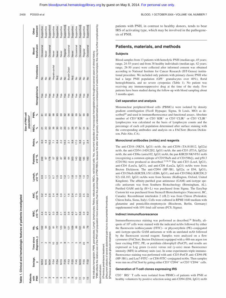
Blood samples from 13 patients with hemolytic PNH (median age, 45 years;range, 24-55 years) and from 38 healthy individuals (median age, 42 years;range, 28-50 years) were collected after informed consent was obtainedaccording to National Institute for Cancer Research (IST-Genoa) institu-tional procedure. We included only patients with primary classic PNH whohad a large PNH population (GPI� granulocytes over 40%), floridhemoglobinuria, and no severe cytopenias (Table 1). No patient wasreceiving any immunosuppressive drug at the time of the study. Fewpatients have been studied during the follow-up with blood sampling about3 months apart.
Cell separation and analysis
Mononuclear peripheral-blood cells (PBMCs) were isolated by densitygradient centrifugation (Ficoll Hypaque; Sigma, St Louis, MO) as de-scribed28 and used in immunofluorescence and functional assays. Absolutenumber of CD3�KIR� or CD3�KIR� or CD3�CLIR� or CD3�CLIR�
lymphocytes was calculated on the basis of lymphocyte counts and thepercentage of each cell population determined after surface staining withthe corresponding antibodies and analysis on a FACSort (Becton Dickin-son, Palo Alto, CA).
Monoclonal antibodies (mAbs) and reagents
The anti-CD16 (NK54, IgG1) mAb, the anti-CD56 (TA181H12, IgG2a)mAb, the anti-CD54 (14D12D2, IgG1) mAb, the anti-CD3 (JT3A, IgG2a)mAb, the anti-CD8� (astra102, IgG1) mAb, the pan KIR2D NKVFS1 mAb(recognizing a common epitope of CD158a/h and of CD158b/j), and p50.3(CD158i) were produced as described.15,28,29 The anti-CD3 (Leu4, IgG1),anti-CD4 (Leu3a, IgG1), and anti-CD8 (Leu2a, IgG1) mAbs were fromBecton Dickinson. The anti-CD94 (HP-3B1, IgG2a; or E94, IgG1),anti-CD158a/h (KIR2DL1/S1) (EB6, IgG1), and anti-CD158b/j (KIR2DL2/S2) (GL183, IgG1) mAbs were from Serotec (Kidlington, Oxford, UnitedKingdom). The affinity-purified goat antimouse (GAM) anti-isotype spe-cific antiserum was from Southern Biotechnology (Birmingham, AL).Purified GAM anti-Ig (H�L) was purchased from Sigma. The EasySepCustom kit was purchased from Stemcell Biotechnologies (Vancouver, BC,Canada). Recombinant interleukin 2 (rIL2) was from Chiron (Proleukin;Chiron Italia, Siena, Italy). Cells were cultured in RPMI 1640 medium withglutamine and pennicillin-streptomycin (Biochrom, Berlin, Germany)supplemented with 10% fetal calf serum (FCS; Sigma).
Indirect immunofluorescence
Immunofluorescence staining was performed as described.28 Briefly, ali-quots of 105 cells were stained with the indicated mAbs followed by eitherthe fluorescein isothiocyanate (FITC)– or phycoerythrin (PE)–conjugatedanti–isotype-specific GAM antiserum or with an unrelated mAb followedby the fluorescent second reagent. Samples were analyzed on a flowcytometer (FACSort; Becton Dickinson) equipped with a 488-nm argon ionlaser exciting FITC, PE, or peridinin chlorophyll (PerCP), and results areexpressed as Log green (x-axis) versus red (y-axis) mean fluorescenceintensity (MFI) in arbitrary units (au). In some experiments triple immuno-fluorescence staining was performed with anti–CD3-PerCP, anti–CD94-PE(HP-3B1), and Leu7-FITC– or CD8-FITC–conjugated mAbs. Then sampleswere run on a FACSort by gating either CD3�CD94� or CD3�CD94� cells.
Generation of T-cell clones expressing IRS
CD3� IRS� T cells were isolated from PBMCs of patients with PNH orhealthy volunteers by positive selection using anti-CD94 (E94, IgG1) mAb
Tab
le1.
Clin
ical
and
hem
ato
log
icfe
atu
res
ofp
atie
nts
inve
stig
ated
Pat
ien
tsw
ith
PN
HS
ex/a
ge,
yT
ime
sin
ced
iag
no
sis,
yH
b,
g/L
WB
C,
x10�
9 /L
AN
C,
x10�
9 /L
Lym
ph
,x1
0�9 /
LP
late
lets
,x1
0�9 /
LR
etic
s,%
Siz
eo
fPN
H-c
ell
po
pu
lati
on
*
Hem
og
lob
inu
ria
Th
rom
bo
sis
Blo
od
tran
sfu
sio
nO
ther
trea
tmen
tR
BC
s,%
PM
N,
%
1M
/53
110
24.
43.
20.
924
96.
021
81Y
esN
oO
ccas
iona
lS
uppo
rtiv
e
2M
/45
294
2.9
1.6
1.1
974.
820
55Y
esN
oN
ever
Sup
port
ive
3M
/34
210
14.
72.
51.
717
810
.250
80Y
esN
oO
ccas
iona
lS
uppo
rtiv
e†
4F
/54
1081
3.3
2.4
0.7
577.
136
98Y
esH
epat
o-po
rtal
Nev
erS
uppo
rtiv
e
5F
/54
1077
3.8
1.8
1.5
193
6.4
3093
Yes
No‡
Nev
erS
uppo
rtiv
e
6M
/33
512
32.
10.
90.
987
2.2
2269
Yes
No
Nev
erS
uppo
rtiv
e
7F
/25
867
2.5
1.1
1.2
8314
.523
82Y
esN
oR
egul
arS
uppo
rtiv
e§
8F
/53
712
13.
61.
61.
764
2.1
5196
Yes
Bud
d-C
hiar
iO
ccas
iona
lS
uppo
rtiv
e
9F
/25
110
94.
52.
41.
832
39.
826
80Y
esN
o‡N
ever
Sup
port
ive
10F
/45
586
8.7
7.5
1.0
143
17.0
7599
Yes
No
Occ
asio
nal
Sup
port
ive
11M
/33
510
15.
63.
31.
826
26.
837
97Y
esN
oO
ccas
iona
lS
uppo
rtiv
e§
12F
/55
2384
3.9
2.5
1.0
185
10.1
5490
Yes
No
Reg
ular
Sup
port
ive
13F
/32
378
4.0
2.2
1.3
193
2.4
9169
Yes
No
Occ
asio
nal
Sup
port
ive§
Hb
indi
cate
she
mog
lobi
n;P
MN
,gra
nulo
cyte
s;Ly
mph
,lym
phoc
ytes
;Ret
ics,
retic
uloc
ytes
;RB
Cs,
red
bloo
dce
lls;Y
es,r
ecur
rent
epis
odes
ofm
acro
scop
iche
mog
lobi
nuria
thro
ugho
utse
vera
lyea
rsof
clin
ical
hist
ory.
*PM
Nre
flect
sth
ere
lativ
esi
zeof
the
PN
H-c
ellp
opul
atio
nm
ore
accu
rate
lyth
anth
epe
rcen
tage
ofP
NH
RB
Cs,
beca
use
the
latte
rwill
begr
ossl
yun
dere
stim
ated
asa
cons
eque
nce
ofse
lect
ive
hem
olys
is;t
his
may
befu
rthe
rcom
plic
ated
bybl
ood
tran
sfus
ion.
†Pre
viou
spr
edni
sone
trea
tmen
t.‡R
ecur
rent
atta
cks
ofse
vere
abdo
min
alpa
inbu
tno
docu
men
ted
thro
mbo
sis.
§Pre
viou
san
tithy
moc
yte
glob
ulin
trea
tmen
t.
2400 POGGI et al BLOOD, 1 OCTOBER 2005 � VOLUME 106, NUMBER 7
For personal use only.on May 8, 2014. by guest bloodjournal.hematologylibrary.orgFrom
and custom preparation of EasySep (Stemcell Biotechnologies). Theresulting cell population was 60% to 90% CD94� and 50% to 95% CD3�
depending on the donor. Purified CD94� cells were stimulated with 10�g/mL phytohemagglutinin (PHA) and cultured in 96-well U-bottomedmicroplates (Becton Dickinson) with complete medium in the presence of100 U/mL rIL2 in a final volume of 200 �L/well in the presence of 105/wellirradiated allogeneic PBMCs and 5 � 103/well 721.221 lymphoblastoidcell line transfected with HLA-G.30 CD3� T-cell clones were obtained byculturing purified CD94� cells under limiting dilution conditions aspreviously reported.29,30 Briefly, CD94� cells were cultured in U-bottomedplates at different numbers of cells per well (25/well, 10/well, 5/well,1/well, and 0.5/well) in the presence of 105 irradiated PBMCs and 103
irradiated 721.221 HLA-I–negative lymphoblastoid cell line with 1 �g/mLPHA and 100 IU/mL IL2. After 5 days medium was changed and cellproliferation was detected from day 10 of culture. Cloning efficiencyranged from 10% to 30%, calculated as described.31 All cell populationswere analyzed for the expression of CD3, CD94/NKG2, CD16, CD56,KIR2DL1/S1 with EB6 mAb, and KIR2DL2/S2 with GL183 mAb. For theexpression of KIR2DS4, cells reacted with NKVFS1 mAb but not with EB6and GL183 mAbs. Each clone was analyzed in a redirected killing assayusing the Fc�R� P815 murine mastocytoma cell line in the presence ofmAbs recognizing either KIR (anti-CD158 mAb, NKVFS1) or CLIR(anti-CD94 mAb, E94) at the effector-to-target (E/T) ratio of 20:1 or 2:1 toidentify clones with inhibiting or activating forms of these HLA-I receptors,respectively.30 Indeed, when the engagement of either KIR or CLIRtriggered cytolysis of P815 at a 2:1 E/T ratio, that clone was assigned to theactivating group. On the other hand, when cytolysis of P815 at a 20:1 E/Tratio was inhibited by the addition of either anti-KIR or anti-CLIR mAb,that clone expressed IRS of the inhibiting type.30
Determination of IFN-� and TNF-� in culture supernatants
Interferon � (IFN-�) and tumor necrosis factor � (TNF-�) released inculture supernatant from T-cell clones derived from PBMCs of patients withPNH upon incubation for 24 hours with medium alone, or in the presence ofanti-IRS or anti-CD3 mAbs followed by GAM to obtain optimal cross-linking of the corresponding surface molecule, as indicated in “Results,”was evaluated by enzyme-linked immunosorbent assay (ELISA; Pepro-Tech, London, United Kingdom).32
Cytolytic assays
Cytolytic activity of either ex vivo–isolated PBMCs from patients withPNH or healthy donors or from T-cell clones was tested in a 4-hour51Cr-release assay as previously described.28,29 T-cell clones were selectedfor the expression of activating forms of either KIR and/or CLIR, and wereused as effector cells with the Fc�R� murine mastocytoma cell line P815 inthe presence of mAb directed against activating receptor for HLA, at an E/Tratio of 2:1, in a final volume of 200 �L RPMI 1640 medium in V-bottomedmicrowells.28,29 In order to confirm that killing of target cells by an IRS�
T-cell population or by a T-cell clone is mediated by the ligation of anactivating type IRS to its cognate HLA ligand, we used the HLA-I–negativelymphoblastoid cell line 721.221. These cells were transfected either withHLA-G (to induce expression of HLA-E with HLA-G peptides, asappropriate for the CD94/NKG2 complex of activating type); or withHLA-Cw3 (appropriate for CD158h [KIR2D2S1]); or with HLA-Cw4(appropriate for CD158j [KIR2DS2]). These cells were then used withoutand with the addition of either anti–HLA-I or anti-IRS mAb (5 �g/mL). Insome experiments, to study the spontaneous cytolytic activity of IRS� Tcells, GPI� K562 erythroleukemia cell line (K562wt) and the parental K562cell line lacking GPI-linked surface molecules (KCRN) generated byFinberg and collaborators33 were used in cytolytic assays with IRS� T cellsfrom patients with PNH. To evaluate the contribution of GPI-linkedmolecules to the interaction between effector and K562 target cells, theexperiments were carried out also in the presence of saturating amounts ofanti-LFA1 mAb (5 �g/mL).
Analysis of KIR genotype and HLA-I typing in patients with PNH
High-molecular-weight DNA was extracted from peripheral-blood samplesby a standard method. KIR genotyping was performed by using a multiplexpolymerase chain reaction (PCR) method developed by Sun et al.34 PCRamplification was performed with hot-start DNA polymerase, AmpliTaqGold (Applied Biosystems), by using the described PCR conditions34 andthe mixtures of KIR-allele–specific primers kindly provided by D. Senitzer(City of Hope National Medical Center, Duarte, CA). The presence ofselected alleles was confirmed by using primers and PCR conditionsdescribed by Hiby et al.35 As a quality control of our KIR genotypingprocedures, we used cell lines with known KIR genotypes (NK/KIR PhaseII QC Reference Panel, International Histocompatibility Working Group,Seattle, WA). HLA-I typing was determined by PCR using the commercialkit from One Lambda (Dynal Biotech, Oslo, Norway).
Statistical analysis
Data are expressed as the mean plus or minus the standard deviation (SD).Nonparametric Mann-Whitney and Fisher exact tests have been used whenappropriate. Statistical significance was accepted for P values below .05.
Results
Immune-phenotypic analysis of circulating T lymphocytesbearing molecules belonging to the IRS in patients with PNH
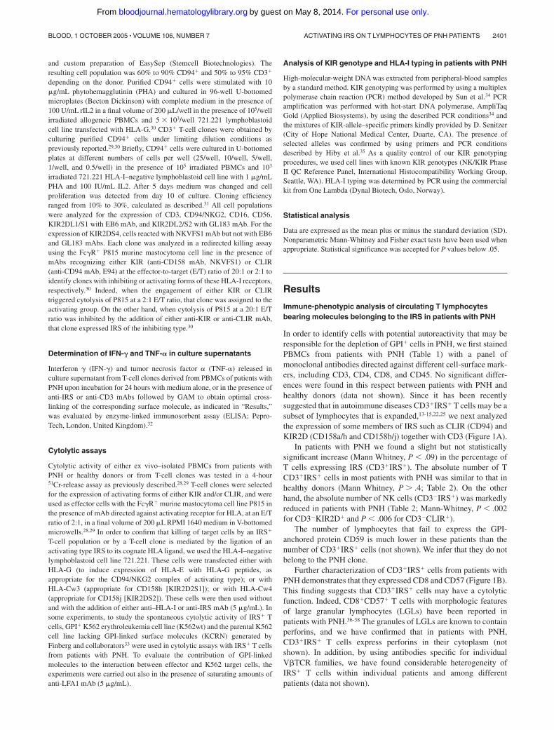
In order to identify cells with potential autoreactivity that may beresponsible for the depletion of GPI� cells in PNH, we first stainedPBMCs from patients with PNH (Table 1) with a panel ofmonoclonal antibodies directed against different cell-surface mark-ers, including CD3, CD4, CD8, and CD45. No significant differ-ences were found in this respect between patients with PNH andhealthy donors (data not shown). Since it has been recentlysuggested that in autoimmune diseases CD3�IRS� T cells may be asubset of lymphocytes that is expanded,13-15,22,25 we next analyzedthe expression of some members of IRS such as CLIR (CD94) andKIR2D (CD158a/h and CD158b/j) together with CD3 (Figure 1A).
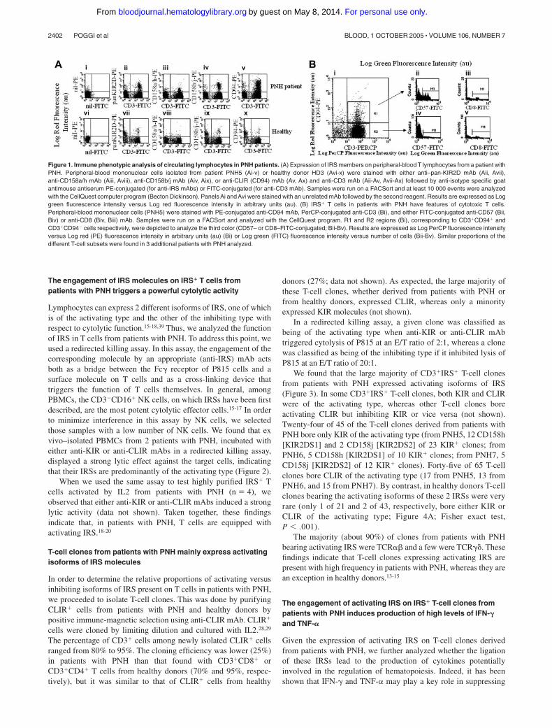
In patients with PNH we found a slight but not statisticallysignificant increase (Mann Whitney, P � .09) in the percentage ofT cells expressing IRS (CD3�IRS�). The absolute number of TCD3�IRS� cells in most patients with PNH was similar to that inhealthy donors (Mann Whitney, P � .4; Table 2). On the otherhand, the absolute number of NK cells (CD3�IRS�) was markedlyreduced in patients with PNH (Table 2; Mann-Whitney, P � .002for CD3�KIR2D� and P � .006 for CD3�CLIR�).
The number of lymphocytes that fail to express the GPI-anchored protein CD59 is much lower in these patients than thenumber of CD3�IRS� cells (not shown). We infer that they do notbelong to the PNH clone.
Further characterization of CD3�IRS� cells from patients withPNH demonstrates that they expressed CD8 and CD57 (Figure 1B).This finding suggests that CD3�IRS� cells may have a cytolyticfunction. Indeed, CD8�CD57� T cells with morphologic featuresof large granular lymphocytes (LGLs) have been reported inpatients with PNH.36-38 The granules of LGLs are known to containperforins, and we have confirmed that in patients with PNH,CD3�IRS� T cells express perforins in their cytoplasm (notshown). In addition, by using antibodies specific for individualVTCR families, we have found considerable heterogeneity ofIRS� T cells within individual patients and among differentpatients (data not shown).
ACTIVATING IRS ON T LYMPHOCYTES OF PNH PATIENTS 2401BLOOD, 1 OCTOBER 2005 � VOLUME 106, NUMBER 7
For personal use only.on May 8, 2014. by guest bloodjournal.hematologylibrary.orgFrom
The engagement of IRS molecules on IRS� T cells frompatients with PNH triggers a powerful cytolytic activity
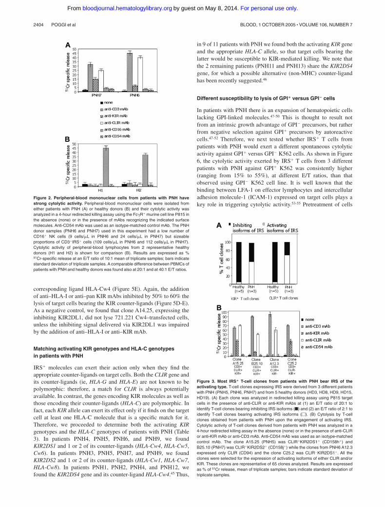
Lymphocytes can express 2 different isoforms of IRS, one of whichis of the activating type and the other of the inhibiting type withrespect to cytolytic function.15-18,39 Thus, we analyzed the functionof IRS in T cells from patients with PNH. To address this point, weused a redirected killing assay. In this assay, the engagement of thecorresponding molecule by an appropriate (anti-IRS) mAb actsboth as a bridge between the Fc� receptor of P815 cells and asurface molecule on T cells and as a cross-linking device thattriggers the function of T cells themselves. In general, amongPBMCs, the CD3�CD16� NK cells, on which IRSs have been firstdescribed, are the most potent cytolytic effector cells.15-17 In orderto minimize interference in this assay by NK cells, we selectedthose samples with a low number of NK cells. We found that exvivo–isolated PBMCs from 2 patients with PNH, incubated witheither anti-KIR or anti-CLIR mAbs in a redirected killing assay,displayed a strong lytic effect against the target cells, indicatingthat their IRSs are predominantly of the activating type (Figure 2).
When we used the same assay to test highly purified IRS� Tcells activated by IL2 from patients with PNH (n 4), weobserved that either anti-KIR or anti-CLIR mAbs induced a stronglytic activity (data not shown). Taken together, these findingsindicate that, in patients with PNH, T cells are equipped withactivating IRS.18-20
T-cell clones from patients with PNH mainly express activatingisoforms of IRS molecules
In order to determine the relative proportions of activating versusinhibiting isoforms of IRS present on T cells in patients with PNH,we proceeded to isolate T-cell clones. This was done by purifyingCLIR� cells from patients with PNH and healthy donors bypositive immune-magnetic selection using anti-CLIR mAb. CLIR�
cells were cloned by limiting dilution and cultured with IL2.28,29
The percentage of CD3� cells among newly isolated CLIR� cellsranged from 80% to 95%. The cloning efficiency was lower (25%)in patients with PNH than that found with CD3�CD8� orCD3�CD4� T cells from healthy donors (70% and 95%, respec-tively), but it was similar to that of CLIR� cells from healthy
donors (27%; data not shown). As expected, the large majority ofthese T-cell clones, whether derived from patients with PNH orfrom healthy donors, expressed CLIR, whereas only a minorityexpressed KIR molecules (not shown).
In a redirected killing assay, a given clone was classified asbeing of the activating type when anti-KIR or anti-CLIR mAbtriggered cytolysis of P815 at an E/T ratio of 2:1, whereas a clonewas classified as being of the inhibiting type if it inhibited lysis ofP815 at an E/T ratio of 20:1.
We found that the large majority of CD3�IRS� T-cell clonesfrom patients with PNH expressed activating isoforms of IRS(Figure 3). In some CD3�IRS� T-cell clones, both KIR and CLIRwere of the activating type, whereas other T-cell clones boreactivating CLIR but inhibiting KIR or vice versa (not shown).Twenty-four of 45 of the T-cell clones derived from patients withPNH bore only KIR of the activating type (from PNH5, 12 CD158h[KIR2DS1] and 2 CD158j [KIR2DS2] of 23 KIR� clones; fromPNH6, 5 CD158h [KIR2DS1] of 10 KIR� clones; from PNH7, 5CD158j [KIR2DS2] of 12 KIR� clones). Forty-five of 65 T-cellclones bore CLIR of the activating type (17 from PNH5, 13 fromPNH6, and 15 from PNH7). By contrast, in healthy donors T-cellclones bearing the activating isoforms of these 2 IRSs were veryrare (only 1 of 21 and 2 of 43, respectively, bore either KIR orCLIR of the activating type; Figure 4A; Fisher exact test,P � .001).
The majority (about 90%) of clones from patients with PNHbearing activating IRS were TCR� and a few were TCR��. Thesefindings indicate that T-cell clones expressing activating IRS arepresent with high frequency in patients with PNH, whereas they arean exception in healthy donors.13-15
The engagement of activating IRS on IRS� T-cell clones frompatients with PNH induces production of high levels of IFN-�and TNF-�
Given the expression of activating IRS on T-cell clones derivedfrom patients with PNH, we further analyzed whether the ligationof these IRSs lead to the production of cytokines potentiallyinvolved in the regulation of hematopoiesis. Indeed, it has beenshown that IFN-� and TNF-� may play a key role in suppressing
Figure 1. Immune phenotypic analysis of circulating lymphocytes in PNH patients. (A) Expression of IRS members on peripheral-blood T lymphocytes from a patient withPNH. Peripheral-blood mononuclear cells isolated from patient PNH5 (Ai-v) or healthy donor HD3 (Avi-x) were stained with either anti–pan-KIR2D mAb (Aii, Avii),anti-CD158a/h mAb (Aiii, Aviii), anti-CD158b/j mAb (Aiv, Aix), or anti-CLIR (CD94) mAb (Av, Ax) and anti-CD3 mAb (Aii-Av, Avii-Ax) followed by anti-isotype specific goatantimouse antiserum PE-conjugated (for anti-IRS mAbs) or FITC-conjugated (for anti-CD3 mAb). Samples were run on a FACSort and at least 10 000 events were analyzedwith the CellQuest computer program (Becton Dickinson). Panels Ai and Avi were stained with an unrelated mAb followed by the second reagent. Results are expressed as Loggreen fluorescence intensity versus Log red fluorescence intensity in arbitrary units (au). (B) IRS� T cells in patients with PNH have features of cytotoxic T cells.Peripheral-blood mononuclear cells (PNH5) were stained with PE-conjugated anti-CD94 mAb, PerCP-conjugated anti-CD3 (Bi), and either FITC-conjugated anti-CD57 (Bii,Biv) or anti-CD8 (Biv, Biii) mAb. Samples were run on a FACSort and analyzed with the CellQuest program. R1 and R2 regions (Bi), corresponding to CD3�CD94� andCD3�CD94� cells respectively, were depicted to analyze the third color (CD57– or CD8–FITC-conjugated; Bii-Bv). Results are expressed as Log PerCP fluorescence intensityversus Log red (PE) fluorescence intensity in arbitrary units (au) (Bi) or Log green (FITC) fluorescence intensity versus number of cells (Bii-Bv). Similar proportions of thedifferent T-cell subsets were found in 3 additional patients with PNH analyzed.
2402 POGGI et al BLOOD, 1 OCTOBER 2005 � VOLUME 106, NUMBER 7
For personal use only.on May 8, 2014. by guest bloodjournal.hematologylibrary.orgFrom
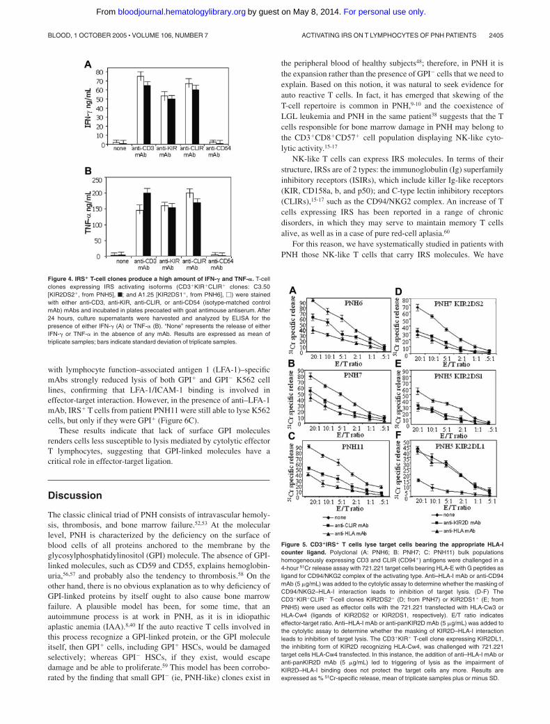
hematopoiesis in AA.40-44 To analyze whether T-cell clones bearingactivating IRS were able to produce IFN-� and TNF-�, we stainedthese clones with either anti-CLIR or anti-KIR mAbs; thus, theseclones were incubated for 24 hours at 37°C on GAM-coated platesto achieve the cross-linking of the corresponding IRS. As shown inFigure 5, both IFN-� and TNF-� were produced by IRS� T-cellclones upon the engagement of either CLIR or KIR on the cellsurface. It is of note that the amount of IFN-� or TNF-� released inculture supernatant was comparable to that elicited by the engage-ment of the CD3/TCR complex achieved by using anti-CD3 mAb,in the same experimental system. T-cell clones that bore only onemember of IRS of the activating type were able to produce theIFN-� and TNF-A cytokines only when the corresponding receptorwas engaged by the specific mAb (data not shown). These findingssuggest that activating IRS, when appropriately engaged by thecorresponding natural ligand, can induce not only activation of thecytolytic machinery, but also the release of cytokines, which mayplay a role in regulating hematopoiesis.
T cells expressing activating IRS lyse target cells bearing thecognate IRS counter-receptors
721.221 cells do not express HLA class I molecules but, oncetransfected with HLA-G, they will express HLA-E, the counter-ligand for CD94/NKG2-presenting HLA-G–derived peptides. Whenwe exposed 721.221 lymphoblastoid cells transfected with HLA-Gto T-cell populations from patients PNH6, PNH7, and PNH11, whoexpressed the CLIR CD94/NKG2 of the activating type, weobtained lysis in all 3 cases (Figure 5). Addition of either anti–HLAclass I or anti-CD94 mAb to the cytolytic assay strongly inhibitedlysis (Figure 5A-C). To determine that also activating KIR-expressing T cells lyse cells bearing KIR ligand, we selected T-cellclones from patients with PNH that express only one activatingKIR. Clone B25.5 CD158j� (KIR2DS2�) from patient PNH7killed 721.221 cells transfected with the counter-ligand HLA-Cw3allele (Figure 5D); and clone C15.10 CD158h� (KIR2DS1�)from patient PNH5 killed 721.221 cells transfected with the
Table 2. Quantitative analysis of the expression of molecules belonging to the IRS on T and NK lymphocytes from patients with PNH
Patient no.CD3� KIR2D�,
cell/�LCD3� KIR2D�,
cell/�LCD3� KIR2D�, %
of T cells CD3�CLIR�, cell/�L CD3�CLIR�, cell/�LCD3�CLIR�, % of
T cells
PNH1
Sample 5 98 91.6 2.9 — — —
Sample 3 — — — 31.3 27.3 3.1
PNH2
Sample 2 195 69 20.6 — — —
PNH3
Sample 5 51 30.6 3.7 — — —
Sample 3 — — — 64 42 4.8
PNH4
Sample 1 55 10 9.2 — — —
PNH5
Sample 2 52.5 206 3.8 — — —
PNH6
Sample 4 70.2 10 8.3 — — —
Sample 2 — — — 115.5 9.5 12.7
PNH7
Sample 4 61.8 80.3 5.8 — — —
Sample 2 — — — 112.5 87 8.4
PNH8
Sample 2 57.5 69.5 3.8 — — —
PNH9
Sample 3 71.3 174.7 5 — — —
Sample 1 — — — 87 204 6.9
PNH10
Sample 1 29 10 2.9 — — —
PNH11
Sample 4 151.3 111.8 15.3 — — —
Sample 3 — — — 177.3 73.0 17.0
PNH12
Sample 1 87 34 9.4 67 24 7.2
PNH13
Sample 1 31 27 2.6 94 56 7.9
Mean � SD (range) 81.8 � 47.0 (31-195) 76.2 � 62.7 (10-206) 7.5 � 5.5 (2.6-20.6) 96.3 � 43.7 (31.3-177.3) 65.4 � 61.7 (9.5-204.0) 8.5 � 4.4 (3.1-17.0)
Healthy controls*
Mean � SD (range) 63 � 36 (19-171) 192 � 155 (8-690) 4.3 � 2.4 (1.3-11.4) 102 � 71 (15-309) 214 � 155 (9-666) 6.8 � 4.7 (1.0-20.6)
Peripheral-blood lymphocytes were isolated from the indicated patients. Samples of blood were collected at about 3 months from one to another during the follow-up; thesample number indicates the number of the blood collection. Samples were stained with anti-CD3 mAb and anti-panKIR2D mAb (NKVFS1) or anti-CLIR mAb followed byisotype-specific antimouse antiserum. At least 10 000 events for each sample were analyzed on a FACSort (Becton Dickinson). Percent of CD3� T lymphocytes that areKIR2D� or CLIR� is also shown.
— indicates that the sample was not stained with the indicated antibody combination.*For healthy controls, n 38 for CD3/KIR2D staining, and n 17 for CD3/CLIR staining.
ACTIVATING IRS ON T LYMPHOCYTES OF PNH PATIENTS 2403BLOOD, 1 OCTOBER 2005 � VOLUME 106, NUMBER 7
For personal use only.on May 8, 2014. by guest bloodjournal.hematologylibrary.orgFrom
corresponding ligand HLA-Cw4 (Figure 5E). Again, the additionof anti–HLA-I or anti–pan KIR mAbs inhibited by 50% to 60% thelysis of target cells bearing the KIR counter-ligands (Figure 5D-E).As a negative control, we found that clone A14.25, expressing theinhibiting KIR2DL1, did not lyse 721.221 Cw4–transfected cells,unless the inhibiting signal delivered via KIR2DL1 was impairedby the addition of anti–HLA-I or anti–KIR mAb.
Matching activating KIR genotypes and HLA-C genotypesin patients with PNH
IRS� molecules can exert their action only when they find theappropriate counter-ligands on target cells. Both the CLIR gene andits counter-ligands (ie, HLA-G and HLA-E) are not known to bepolymorphic: therefore, a match for CLIR is always potentiallyavailable. In contrast, the genes encoding KIR molecules as well asthose encoding their counter-ligands (HLA-C) are polymorphic. Infact, each KIR allele can exert its effect only if it finds on the targetcell at least one HLA-C molecule that is a specific match for it.Therefore, we proceeded to determine both the activating KIRgenotypes and the HLA-C genotypes of patients with PNH (Table3). In patients PNH4, PNH5, PNH6, and PNH9, we foundKIR2DS1 and 1 or 2 of its counter-ligands (HLA-Cw4, HLA-Cw5,Cw6). In patients PNH3, PNH5, PNH7, and PNH9, we foundKIR2DS2 and 1 or 2 of its counter-ligands (HLA-Cw1, HLA-Cw7,HLA-Cw8). In patients PNH1, PNH2, PNH4, and PNH12, wefound the KIR2DS4 gene and its counter-ligand HLA-Cw4.45 Thus,
in 9 of 11 patients with PNH we found both the activating KIR geneand the appropriate HLA-C allele, so that target cells bearing thelatter would be susceptible to KIR-mediated killing. We note thatthe 2 remaining patients (PNH11 and PNH13) share the KIR2DS4gene, for which a possible alternative (non-MHC) counter-ligandhas been recently suggested.46
Different susceptibility to lysis of GPI� versus GPI� cells
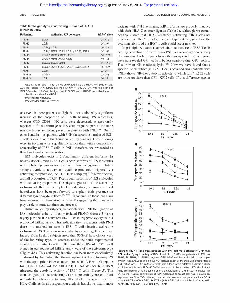
In patients with PNH there is an expansion of hematopoietic cellslacking GPI-linked molecules.47-50 This is thought to result notfrom an intrinsic growth advantage of GPI� precursors, but ratherfrom negative selection against GPI� precursors by autoreactivecells.47-52 Therefore, we next tested whether IRS� T cells frompatients with PNH would exert a different spontaneous cytolyticactivity against GPI� versus GPI� K562 cells. As shown in Figure6, the cytolytic activity exerted by IRS� T cells from 3 differentpatients with PNH against GPI� K562 was consistently higher(ranging from 15% to 55%), at different E/T ratios, than thatobserved using GPI� K562 cell line. It is well known that thebinding between LFA-1 on effector lymphocytes and intercellularadhesion molecule-1 (ICAM-1) expressed on target cells plays akey role in triggering cytolytic activity.53-55 Pretreatment of cells
Figure 2. Peripheral-blood mononuclear cells from patients with PNH havestrong cytolytic activity. Peripheral-blood mononuclear cells were isolated fromeither patients with PNH (A) or healthy donors (B) and their cytolytic activity wasanalyzed in a 4-hour redirected killing assay using the Fc�R� murine cell line P815 inthe absence (none) or in the presence of mAbs recognizing the indicated surfacemolecules. Anti-CD54 mAb was used as an isotype-matched control mAb. The PNHdonor samples (PNH6 and PNH7) used in this experiment had a low number ofCD16� NK cells (9 cells/�L in PNH6 and 24 cells/�L in PNH7) but sizeableproportions of CD3�IRS� cells (109 cells/�L in PNH6 and 112 cells/�L in PNH7).Cytolytic activity of peripheral-blood lymphocytes from 2 representative healthydonors (H1 and H2) is shown for comparison (B). Results are expressed as %51Cr-specific release at an E/T ratio of 10:1 mean of triplicate samples; bars indicatestandard deviation of triplicate samples. A comparable difference between PBMCs ofpatients with PNH and healthy donors was found also at 20:1 and at 40:1 E/T ratios.
Figure 3. Most IRS� T-cell clones from patients with PNH bear IRS of theactivating type. T-cell clones expressing IRS were derived from 3 different patientswith PNH (PNH5, PNH6, PNH7) and from 5 healthy donors (HD3, HD8, HD9, HD15,HD19). (A) Each clone was analyzed in redirected killing assay using P815 targetcells in the presence of anti-CLIR or anti-KIR mAbs at (1) an E/T ratio of 20:1 toidentify T-cell clones bearing inhibiting IRS isoforms (f) and (2) an E/T ratio of 2:1 toidentify T-cell clones bearing activating IRS isoforms (�). (B) Cytolysis by T-cellclones obtained from patients with PNH upon the engagement of activating IRS.Cytolytic activity of T-cell clones derived from patients with PNH was analyzed in a4-hour redirected killing assay in the absence (none) or in the presence of anti-CLIRor anti-KIR mAb or anti-CD3 mAb. Anti-CD54 mAb was used as an isotype-matchedcontrol mAb. The clone A15.25 (PNH5) was CLIR�KIR2DS1� (CD158h�) andB25.10 (PNH7) was CLIR�KIR2DS2� (CD158j�) while the clones from PNH6 A12.3expressed only CLIR (CD94) and the clone C25.2 was CLIR�KIR2DS1�. All theclones were selected for the expression of activating isoforms of either CLIR and/orKIR. These clones are representative of 65 clones analyzed. Results are expressedas % of 51Cr release, mean of triplicate samples; bars indicate standard deviation oftriplicate samples.
2404 POGGI et al BLOOD, 1 OCTOBER 2005 � VOLUME 106, NUMBER 7
For personal use only.on May 8, 2014. by guest bloodjournal.hematologylibrary.orgFrom
with lymphocyte function–associated antigen 1 (LFA-1)–specificmAbs strongly reduced lysis of both GPI� and GPI� K562 celllines, confirming that LFA-1/ICAM-1 binding is involved ineffector-target interaction. However, in the presence of anti–LFA-1mAb, IRS� T cells from patient PNH11 were still able to lyse K562cells, but only if they were GPI� (Figure 6C).
These results indicate that lack of surface GPI moleculesrenders cells less susceptible to lysis mediated by cytolytic effectorT lymphocytes, suggesting that GPI-linked molecules have acritical role in effector-target ligation.
Discussion
The classic clinical triad of PNH consists of intravascular hemoly-sis, thrombosis, and bone marrow failure.52,53 At the molecularlevel, PNH is characterized by the deficiency on the surface ofblood cells of all proteins anchored to the membrane by theglycosylphosphatidylinositol (GPI) molecule. The absence of GPI-linked molecules, such as CD59 and CD55, explains hemoglobin-uria,56,57 and probably also the tendency to thrombosis.58 On theother hand, there is no obvious explanation as to why deficiency ofGPI-linked proteins by itself ought to also cause bone marrowfailure. A plausible model has been, for some time, that anautoimmune process is at work in PNH, as it is in idiopathicaplastic anemia (IAA).8,40 If the auto reactive T cells involved inthis process recognize a GPI-linked protein, or the GPI moleculeitself, then GPI� cells, including GPI� HSCs, would be damagedselectively; whereas GPI� HSCs, if they exist, would escapedamage and be able to proliferate.59 This model has been corrobo-rated by the finding that small GPI� (ie, PNH-like) clones exist in
the peripheral blood of healthy subjects48; therefore, in PNH it isthe expansion rather than the presence of GPI� cells that we need toexplain. Based on this notion, it was natural to seek evidence forauto reactive T cells. In fact, it has emerged that skewing of theT-cell repertoire is common in PNH,9-10 and the coexistence ofLGL leukemia and PNH in the same patient38 suggests that the Tcells responsible for bone marrow damage in PNH may belong tothe CD3�CD8�CD57� cell population displaying NK-like cyto-lytic activity.15-17
NK-like T cells can express IRS molecules. In terms of theirstructure, IRSs are of 2 types: the immunoglobulin (Ig) superfamilyinhibitory receptors (ISIRs), which include killer Ig-like receptors(KIR, CD158a, b, and p50); and C-type lectin inhibitory receptors(CLIRs),15-17 such as the CD94/NKG2 complex. An increase of Tcells expressing IRS has been reported in a range of chronicdisorders, in which they may serve to maintain memory T cellsalive, as well as in a case of pure red-cell aplasia.60
For this reason, we have systematically studied in patients withPNH those NK-like T cells that carry IRS molecules. We have
Figure 4. IRS� T-cell clones produce a high amount of IFN-� and TNF-�. T-cellclones expressing IRS activating isoforms (CD3�KIR�CLIR� clones: C3.50[KIR2DS2�, from PNH5], f; and A1.25 [KIR2DS1�, from PNH6], �) were stainedwith either anti-CD3, anti-KIR, anti-CLIR, or anti-CD54 (isotype-matched controlmAb) mAbs and incubated in plates precoated with goat antimouse antiserum. After24 hours, culture supernatants were harvested and analyzed by ELISA for thepresence of either IFN-� (A) or TNF-� (B). “None” represents the release of eitherIFN-� or TNF-� in the absence of any mAb. Results are expressed as mean oftriplicate samples; bars indicate standard deviation of triplicate samples.
Figure 5. CD3�IRS� T cells lyse target cells bearing the appropriate HLA-Icounter ligand. Polyclonal (A: PNH6; B: PNH7; C: PNH11) bulk populationshomogeneously expressing CD3 and CLIR (CD94�) antigens were challenged in a4-hour 51Cr release assay with 721.221 target cells bearing HLA-E with G peptides asligand for CD94/NKG2 complex of the activating type. Anti–HLA-I mAb or anti-CD94mAb (5 �g/mL) was added to the cytolytic assay to determine whether the masking ofCD94/NKG2–HLA-I interaction leads to inhibition of target lysis. (D-F) TheCD3�KIR�CLIR� T-cell clones KIR2DS2� (D; from PNH7) or KIR2DS1� (E; fromPNH5) were used as effector cells with the 721.221 transfected with HLA-Cw3 orHLA-Cw4 (ligands of KIR2DS2 or KIR2DS1, respectively). E/T ratio indicateseffector-target ratio. Anti–HLA-I mAb or anti-panKIR2D mAb (5 �g/mL) was added tothe cytolytic assay to determine whether the masking of KIR2D–HLA-I interactionleads to inhibition of target lysis. The CD3�KIR� T-cell clone expressing KIR2DL1,the inhibiting form of KIR2D recognizing HLA-Cw4, was challenged with 721.221target cells HLA-Cw4 transfected. In this instance, the addition of anti–HLA-I mAb oranti-panKIR2D mAb (5 �g/mL) led to triggering of lysis as the impairment ofKIR2D–HLA-I binding does not protect the target cells any more. Results areexpressed as % 51Cr-specific release, mean of triplicate samples plus or minus SD.
ACTIVATING IRS ON T LYMPHOCYTES OF PNH PATIENTS 2405BLOOD, 1 OCTOBER 2005 � VOLUME 106, NUMBER 7
For personal use only.on May 8, 2014. by guest bloodjournal.hematologylibrary.orgFrom
observed in these patients a slight but not statistically significantincrease of the proportion of T cells bearing IRS molecules,whereas CD3�CD16� NK cells were decreased, as previouslyreported.61,62 This shortage of NK cells might be part of the bonemarrow failure syndrome present in patients with PNH.63,64 On theother hand, in most patients with PNH the absolute number of IRS�
T cells was similar to that found in healthy controls. These findingswere in keeping with a qualitative rather than with a quantitativeabnormality of IRS� T cells in PNH; therefore, we proceeded totheir functional characterization.
IRS molecules exist in 2 functionally different isoforms. Inhealthy donors, most IRS� T cells bear isoforms of IRS moleculeswith inhibiting properties. In fact, their engagement inhibitsstrongly cytolytic activity and cytokine production triggered viaactivating receptors (ie, the CD3/TCR complex).25,26 Nevertheless,a small proportion of IRS� T cells bear isoforms of IRS moleculeswith activating properties. The physiologic role of the activatingisoforms of IRS is incompletely understood, although severalhypotheses have been put forward to explain their presence ondifferent lymphocyte subsets.15-17,39 Expansion of these cells hasbeen reported in rheumatoid arthritis,24 suggesting that they mayplay a role in some autoimmune process.
Unlike in healthy subjects, in patients with PNH the ligation ofIRS molecules either on freshly isolated PBMCs (Figure 3) or onhighly purified IL2-activated IRS� T cells triggered cytolysis in aredirected killing assay. This indicates that in patients with PNHthere is a marked increase in IRS� T cells bearing activatingisoforms of IRS. This was corroborated by generating T-cell clones.Indeed, from healthy subjects more than 95% of these clones wereof the inhibiting type. In contrast, under the same experimentalconditions, in patients with PNH more than 50% of IRS� T-cellclones in our redirected killing assay were of the activating type(Figure 4A). This activating function has been more conclusivelyconfirmed by the finding that the engagement of the activating IRSwith the appropriate HLA counter-ligands (HLA-E with G peptidefor CLIR; HLA-Cw4 for KIR2DS1; HLA-CW3 for KIR2DS2)triggered the cytolytic activity of IRS� T cells (Figure 5). Thecounter-ligand of the activating CLIR is potentially present in allindividuals, whereas activating KIR alleles recognize specificHLA-C alleles. In this respect, our analysis has shown that in most
patients with PNH, activating KIR isoforms are properly matchedwith their HLA-C counter-ligands (Table 3). Although we cannotpositively state that HLA-C–matched activating KIR alleles areexpressed on IRS� T cells, the genotype data suggest that thecytotoxic ability of the IRS� T cells could occur in vivo.
In principle, we cannot say whether the increase in IRS� T cellsbearing activating IRS isoforms in PNH is a secondary or a primaryphenomenon. Earlier reports from other groups and from our grouphave not revealed GPI� cells to be less sensitive than GPI� cells toT-cell65,66 or NK-mediated lysis.33,66 Now we have found that aspecific T-cell subset (ie, IRS� T cells obtained from patients withPNH) shows NK-like cytolytic activity to which GPI� K562 cellsare more sensitive than GPI� K562 cells. If this difference applies
Figure 6. IRS� T cells from patients with PNH kill more efficiently GPI� thanGPI� cells. Cytolytic activity of IRS� T cells from 3 different patients with PNH (A:PNH6; B: PNH7; C: PNH11) against GPI� K562 cell line or its GPI� counterpart(KCRN) was analyzed in a 4-hour 51Cr release assay at the indicated effector-target(E/T) ratios. Anti–LFA-1 mAb (5 �g/mL) was added to the cytotoxic assay in order toblock the contribution of LFA-1/ICAM-1 interaction to the activation of T cells. As the 2K562 cell lines differ from each other for the expression of GPI-linked molecules, thisshows the relative contribution of GPI molecules to target-cell lysis. Results areexpressed as % of 51Cr release, mean of triplicate samples plus or minus SD.�indicates KCRN (K562 GPI-); F, KCRN (K562 GPI�) plus anti-LFA-1 mAb; Œ, K562(GPI�); f, K562 (GPI�) plus anti-LFA-1 mAb.
Table 3. The genotype of activating KIR and of HLA-Cin PNH patients
Patient no. Activating KIR genotype HLA-C allele
PNH1 2DS4 04,‡ 16
PNH2 2DS4 04,‡ 07
PNH3 2DS2,† 2DS4 08,† 12
PNH4 2DS1,* 2DS2, 2DS3, 2DS4,‡ 2DS5, 3DS1 04,‡ 06
PNH5 2DS1,* 2DS2,† 2DS3, 3DS1 04,* 07†
PNH6 2DS1,* 2DS3, 2DS4, 3DS1 05,* 15
PNH7 2DS2,† 2DS3, 2DS4 01,† 07†
PNH9 2DS1,* 2DS2,† 2DS3, 2DS4, 2DS5, 3DS1 06,* 07†
PNH11 2DS4 07, 07
PNH12 2DS4‡ 03, 04‡
PNH13 2DS4 06, 15
Patients as in Table 1. The ligands of KIR2DS1 are the HLA-CLys80 (w2, w4, w5,w6); the ligands of KIR2DS2 are the HLA-CAsn80 (w1, w3, w7, w8); the ligand ofKIR2DS4 is the HLA-Cw4; the ligands of KIR2DS3 and KIR2DS5 are still unknown.
*Positive matches for KIRDS1.†Matches for KIR2DS2.‡Matches for KIRDS4.15-17,45,46
2406 POGGI et al BLOOD, 1 OCTOBER 2005 � VOLUME 106, NUMBER 7
For personal use only.on May 8, 2014. by guest bloodjournal.hematologylibrary.orgFrom
to HSCs as well, this is one plausible mechanism whereby GPIdeficiency would constitute a selective advantage for GPI� HSCs,explaining the expansion of PNH hematopoiesis in the context ofthe bone marrow failure present in patients with PNH. This bonemarrow failure might be determined also by IFN-� and TNF-�,cytokines that suppress hematopoiesis,40-44 produced by IRS�
T-cell clones. Thus, the IRS� T cells we have characterized aregood candidates for being able to damage GPI� HSCs in PNH.
Acknowledgments
We thank Robert Finberg (Boston, MA) for providing the GPI� andGPI� K562 cell lines and David Sanitzer (Duarte, CA) for providing theprimers for KIR genotyping. We are grateful to Ji-Yao Sun (Duarte, CA)for the helpful technical suggestion for KIR genotyping and MartinaSerra for help in the Laboratory of Human Genetics (LST, Genoa).
References
1. Oni SB, Osunkoya BO, Luzzatto L. Paroxysmalnocturnal hemoglobinuria: evidence for monoclo-nal origin of abnormal red cells. Blood. 1970;36:145-152.
2. Takeda J, Miyata T, Kawagoe K, Iida Y, et al. Defi-ciency of the GPI anchor caused by a somaticmutation of the PIG-A gene in paroxysmal noctur-nal hemoglobinuria. Cell. 1993;73:703-711.
3. Bessler M, Mason PJ, Hillmen P, Miyata et al.Paroxysmal nocturnal haemoglobinuria (PNH) iscaused by somatic mutations in the PIG-A gene.EMBO J. 1994;13:110-117.
4. van der Schoot CE, Huizinga TW, van ’t Veer-Korthof ET, Wijmans R, Pinkster J, von demBorne AE. Deficiency of glycosyl-phosphatidylino-sitol-linked membrane glycoproteins of leuko-cytes in paroxysmal nocturnal hemoglobinuria,description of a new diagnostic cytofluorometricassay. Blood 1990;76:1853-1859.
5. Bessler M, Schaefer A, Keller P. Paroxysmal noc-turnal haemoglobinuria: insight from recent ad-vances in molecular biology. Transfus Med Rev.2001;15:255-267.
6. Dacie JV, Lewis SM. Paroxysmal nocturnal hae-moglobinuria: clinical manifestations, haematol-ogy, and nature of the disease. Series Haemato-logica. 1972;5:3-23.
7. Young NS, Maciejewski JP, Sloand E, et al. Therelationship of aplastic anemia and PNH. IntJ Hematol. 2002;76(suppl 2):168-172.
8. Young NS, Maciejewski J. The pathophysiologyof acquired aplastic anemia. N Engl J Med. 1997;336:1365-1372.
9. Karadimitris A, Manavalan JS, Thaler HT, et al.Abnormal T-cell repertoire is consistent with im-mune process underlying the pathogenesis ofparoxysmal nocturnal hemoglobinuria. Blood.2000;96:2613-2620.
10. Risitano AM, Kook H, Zeng W, et al. Oligoclonaland polyclonal CD4 and CD8 lymphocytes inaplastic anemia and paroxysmal nocturnal hemo-globinuria measured by V beta CDR3 spectratyp-ing and flow cytometry. Blood. 2002;100:178-183.
11. Zeng W, Nakao S, Takamatsu H, et al. Character-ization of T-cell repertoire of the bone marrow inimmune-mediated aplastic anemia: evidence forthe involvement of antigen-driven T-cell responsein cyclosporine-dependent aplastic anemia.Blood. 1999;93:3008-3016.
12. Rotoli B, Luzzatto L. Paroxysmal nocturnal hemo-globinuria. Semin Hematol. 1989;26:201-207.
13. Luzzatto L, Bessler M, Rotoli B. Somatic muta-tions in paroxysmal nocturnal hemoglobinuria: ablessing in disguise? Cell. 1997;88:1-4.
14. Young NS, Maciejewski JP. Genetic and environ-mental effects in paroxysmal nocturnal hemoglo-binuria: this little PIG-A goes “Why? Why? Why?.”J Clin Invest. 2000;106:637-641.
15. Moretta A, Bottino C, Vitale M, et al. Receptors forHLA-class I molecules in human natural killercells. Annu Rev Immunol. 1996;14:619-648.
16. Lanier LL. NK cell receptors. Annu Rev Immunol.1998;16:359-393.
17. Long EO. Regulation of immune responses
through inhibitory receptors. Annu Rev Immunol.1999;17:875-904.
18. Cantoni C, Biassoni R, Pende D, et al. The acti-vating form of CD94 receptor complex: CD94 co-valently associates with the Kp39 protein thatrepresents the product of the NKG2-C gene. EurJ Immunol. 1998;28:327-338.
19. Perez-Villar JJ, Melero I, Rodriguez A, et al.Functional ambivalence of the Kp43 (CD94) NKcell-associated surface antigen. J Immunol. 1995;154:5779-5788.
20. Warren H, Campbell AJ, Waldron JC, Lanier LL.Biphasic response of NK cells expressing bothactivating and inhibitory killer Igh-like receptors.Int Immunol. 2001;13:1043-1052.
21. Poggi A, Tomasello E, Revello V, et al. p40 regu-lates NK cell activation mediated by NK receptorsfor HLA class I antigens and TCR-mediated trig-gering of T lymphocytes. Int Immunol. 1997;9:1271-1279.
22. Mandelboim O, Kent S, Davis DM, et al. Naturalkiller activating receptors trigger interferon gammasecretion from T cells and natural killer cells. ProcNatl Acad Sci U S A. 1998;95:3798-3803.
23. Mandelboim O, Davis DM, Reyburn HT, et al. En-hancement of class-II-restricted T cell responsesby costimulatory NK receptors for class I MHCproteins. Science. 1996;274:2097-2100.
24. Namekawa T, Snyder MR, Yen J-H, et al. Killercell activating receptor function as costimulatorymolecules on CD4�CD28null T cells clonally ex-panded in rheumatoid arthritis. J Immunol. 2000;165:1138-1145.
25. Bertone S, Schiavetti F, Bellomo R, et al. Trans-forming growth factor-beta-induced expression ofCD94/NKG2A inhibitory receptors in human Tlymphocytes.Eur J Immunol. 1999;29:23-29.
26. D’Andrea A, Chang C, Phillips JH, Lanier LL.Regulation of T cell lymphokine production bykiller cell inhibitory receptor recognition of selfHLA class I alleles. J Exp Med. 1996;184:789-794.
27. Young NT, Uhrberg M. KIR expression shapescytotoxic repertoires: a developmental program ofsurvival. Trends Immunol. 2002;23:71-75.
28. Poggi A, Pella N, Morelli L, et al. p40, a novel sur-face molecule involved in the regulation of thenon-major histocompatibility complex-restrictedcytolytic activity in humans. Eur J Immunol. 1995;25:369-376.
29. Moretta A, Poggi A, Pende D, et al. CD69-medi-ated pathway of lymphocyte activation: anti-CD69monoclonal antibodies trigger the cytolytic activityof different lymphoid effector cells with the excep-tion of cytolytic T lymphocytes expressing T cellreceptor �/. J Exp Med. 1991;174:1393-1398.
30. Spaggiari GM, Contini P, Carosio R, et al. SolubleHLA class I molecules induce natural killer cellapoptosis through the engagement of CD8: evi-dence for a negative regulation exerted by CD94/NKG2A complex and KIR2D. Blood. 2002;99:1706-1714.
31. Taswell C. Limiting dilution assay for the determi-nation of immunocompetent cell frequencies, I:data analysis. J Immunol. 1981;126:1614-1619.
32. Zocchi MR, Rubartelli A, Morgavi P, Poggi A.HIV-1 Tat inhibits human natural killer cell functionby blocking L-type calcium channels. J Immunol.1998;161:2938-2943.
33. Finberg RW, White W, Nicholson-Weller A. De-cay-accelerating factor expression on either ef-fector or target cells inhibits cytotoxicity by humannatural killer cells. J Immunol. 1992;149:2055-2060.
34. Sun YJ, Gaidulis L, Miller MM, et al. Developmentof a multiplex PCR-SSP method for killer-cell im-munoglobulin-like receptor genotyping. TissueAntigens. 2004;64:462-468.
35. Hiby SE, Walker JJ, O’Shaughnessy KM, et al.Combinations of maternal KIR and fetal HLA-Cgenes influence the risk of preeclampsia and re-productive success. J Exp Med. 2004;200:957-965.
36. Lamy T, Loughran TP Jr. Current concepts: largegranular lymphocyte leukemia. Blood Rev. 1999;13:230-240.
37. Rose MG, Berliner N. T-cell large granular lym-phocyte leukemia and related disorders. Oncolo-gist. 2004;9:247-258.
38. Karadimitris A, Li K, Notaro R, et al. Associationof clonal T-cell large granular lymphocyte diseaseand paroxysmal nocturnal haemoglobinuria(PNH): further evidence for a pathogenetic linkbetween T cells, aplastic anaemia and PNH. Br JHaematol. 2001;115:1010-1014.
39. Spaggiari GM, Contini P, Dondero A, et al.Soluble HLA class I induces NK cell apoptosisupon the engagement of killer-activating HLAclass I receptors through FasL-Fas interaction.Blood. 2002;100:4098-4107.
40. Zoumbos NC, Gascon P, Djeu JY, Young NS. In-terferon is a mediator of hematopoietic suppres-sion in aplastic anemia in vitro and possibly invivo. Proc Natl Acad Sci U S A. 1985;82:188-192.
41. Miura A, Endo K, Sugawara T, et al. T cell-medi-ated inhibition of erythropoiesis in aplastic anae-mia: the possible role of IFN-gamma and TNF-alpha. Br J Haematol. 1991;78:442-449.
42. Maciejewski JP, Weichold FF, Young NS. HIV-1suppression of hematopoiesis in vitro mediatedby envelope glycoprotein and TNF-alpha. J Im-munol. 1994;153:4303-4310.
43. Selleri C, Maciejewski JP, Sato T, Young NS. In-terferon-gamma constitutively expressed in thestromal microenvironment of human marrow cul-tures mediates potent hematopoietic inhibition.Blood. 1996;87:4149-4157.
44. Binder D, van den Broek MF, Kagi D, et al. Aplas-tic anemia rescued by exhaustion of cytokine-secreting CD8� T cells in persistent infection withlymphocytic choriomeningitis virus. J Exp Med.1998;187:1903-1920.
45. Katz G, Markel G, Mizrahi S, Arnon TI, Mandel-boim O. Recognition of HLA-Cw4 but not HLA-Cw6 by the NK cell receptor killer cell Ig-like re-ceptor two-domain short tail number 4.J Immunol. 2001;166:7260-7267.
46. Katz G, Gazit R, Arnon TI, et al. MHC class I-independent recognition of NK-activating receptorKIR2DS4. J Immunol. 2004;173:1819-1825.
ACTIVATING IRS ON T LYMPHOCYTES OF PNH PATIENTS 2407BLOOD, 1 OCTOBER 2005 � VOLUME 106, NUMBER 7
For personal use only.on May 8, 2014. by guest bloodjournal.hematologylibrary.orgFrom
47. Hillmen P, Lewis SM, Bessler M, Luzzatto L, Da-cie JV. Natural history of paroxysmal nocturnalhemoglobinuria. N Engl J Med. 1995;333:1253-1258.
48. Alfinito F, Del Vecchio L, Rocco S, Boccuni P,Musto P, Rotoli B. Blood cell flow cytometry inparoxysmal nocturnal hemoglobinuria: a tool formeasuring the extent of the PNH clone. Leuke-mia. 1996;10:1326-1330.
49. Rosti V, Tremml G, Soares V, Pandolfi PP, LuzzattoL, Bessler M. Murine embryonic stem cells withoutpig-a gene activity are competent for hematopoiesiswith the PNH phenotype but not for clonal expan-sion. J Clin Invest. 1997;100:1028-1036.
50. Maciejewski JP, Sloand EM, Sato T, Anderson S,Young NS. Impaired hematopoiesis in paroxys-mal nocturnal hemoglobinuria/aplastic anemia isnot associated with a selective proliferative defectin the glycosylphosphatidylinositol-anchored pro-tein-deficient clone. Blood. 1997;89:1173-1181.
51. Murakami Y, Kinoshita T, Maeda Y, Nakano T, Ko-saka H, Takeda J. Different roles of glycosylphos-phatidylinositol in various hematopoietic cells as re-vealed by a mouse model of paroxysmal nocturnalhemoglobinuria. Blood. 1999;94:2963-2970.
52. Araten DJ, Nafa K, Pakdeesuwan K, Luzzatto L.Clonal populations of hematopoietic cells withparoxysmal nocturnal hemoglobinuria genotypeand phenotype are present in normal individuals.Proc Natl Acad Sci U S A. 1999;96:5209-5214.
53. Barber DF, Faure M, Long EO. LFA-1 contributes
an early signal for NK cell cytotoxicity. J Immunol.2004;173:3653-3659.
54. Takagi J, Springer TA. Integrin activation andstructural rearrangement. Immunol Rev. 2002;186:141-163.
55. Poggi A, Pardi R, Pella N, et al. CD45-mediatedregulation of LFA1 function in human natural killercells: anti-CD45 monoclonal antibodies inhibit thecalcium mobilization induced via LFA1 molecules.Eur J Immunol. 1993;23:2454-2463.
56. Holguin MH, Fredrick LR, Bernshaw NJ, WilcoxLA, Parker CJ. Isolation and characterization of amembrane protein from normal human erythro-cytes that inhibits reactive lysis of the erythro-cytes of paroxysmal nocturnal hemoglobinuria.J Clin Invest. 1989;84:7-17.
57. Rosse WF, Dacie JV. Immune lysis of normal hu-man and paroxysmal nocturnal hemoglobinuria(PNH) red blood cells, I: the sensitivity of PNH redcells to lysis by complement and specific anti-body. J Clin Invest. 1966;45:736-748.
58. Shichishima T, Saitoh Y, Terasawa T, Noji H, KaiT, Maruyama Y. Complement sensitivity of eryth-rocytes in a patient with inherited complete defi-ciency of CD59 or with the Inab phenotype. Br JHaematol. 1999;104:303-306.
59. Karadimitris A, Luzzatto L. The cellular pathogen-esis of paroxysmal nocturnal haemoglobinuria.Leukemia. 2001;15:1148-1152.
60. Handgretinger R, Geiselhart A, Moris A, et al.Pure red-cell aplasia associated with clonal ex-pansion of granular lymphocytes expressing
killer-cell inhibitory receptors. N Engl J Med.1999;340:278-284.
61. Schubert J, Uciechowski P, Delany P, Tischler HJ,Kolanus W, Schmidt RE. The PIG-anchoring de-fect in NK lymphocytes of PNH patients. Blood.1990;76:1181-1187.
62. Tseng JE, Hall SE, Howard TA, Ware RE. Pheno-typic and functional analysis of lymphocytes inparoxysmal nocturnal hemoglobinuria. Am J He-matol. 1995;50:244-253.
63. Dacie JV. Paroxysmal nocturnal haemoglobin-uria. In: Dacie JV, ed. The Haemolytic Anaemias:Drug and Chemical Induced Haemolytic Anae-mias, Paroxysmal Nocturnal Haemglobinuria, andHaemolytic Disease of the Newborn. Vol 5. 3rded. London, United Kingdom: Churchill Living-ston; 1999:139-330.
64. Luzzatto L, Notaro R. Paroxysmal nocturnal hae-moglobinuria. In: Handin RI, Lux SE, Stossel TP,eds. Blood, Principles and Practice of Hematol-ogy. 2nd ed. Philadelphia, PA: Lippincott Williams& Wilkins; 2003:319-334.
65. Hollander N, Shin ML, Rosse WE, Springer TA.Distinct restriction of complement- and cell-mediated lysis. J Immunol. 1988;142:3913-3916.
66. Karadimitris A, Notaro R, Koehne G, RobertsIAG, Luzzatto L. PNH cells are sensitive to T-cellmediated lysis as their normal counterparts: impli-cation for the pathogenesis of paroxysmal noctur-nal haemoglobinuria. Br J Haematol. 2000;111:1158-1163.
2408 POGGI et al BLOOD, 1 OCTOBER 2005 � VOLUME 106, NUMBER 7
For personal use only.on May 8, 2014. by guest bloodjournal.hematologylibrary.orgFrom
Copyright © 2022 FDOKUMEN