Passamonti neuropsychologia 09
10
Neuropsychologia 47 (2009) 546–555 Contents lists available at ScienceDirect Neuropsychologia journal homepage: www.elsevier.com/locate/neuropsychologia Audio-visual stimulation improves oculomotor patterns in patients with hemianopia Claudia Passamonti a,b , Caterina Bertini a,b , Elisabetta Làdavas a,b,∗ a Dipartimento di Psicologia, Università di Bologna, Bologna, Italy b CSRNC, Centro Studi e Ricerche in Neuroscienze Cognitive, Polo Didattico e Scientifico di Cesena, Italy article info Article history: Received 5 June 2008 Received in revised form 17 September 2008 Accepted 3 October 2008 Available online 15 October 2008 Keywords: Rehabilitation of visual field defects Audio-visual stimulation Eye movements Visual search Reading abstract Patients with visual field disorders often exhibit impairments in visual exploration and a typical defective oculomotor scanning behaviour. Recent evidence [Bolognini, N., Rasi, F., Coccia, M., & Làdavas, E. (2005b). Visual search improvement in hemianopic patients after audio-visual stimulation. Brain, 128, 2830–2842] suggests that systematic audio-visual stimulation of the blind hemifield can improve accuracy and search times in visual exploration, probably due to the stimulation of Superior Colliculus (SC), an important multisensory structure involved in both the initiation and execution of saccades. The aim of the present study is to verify this hypothesis by studying the effects of multisensory training on oculomotor scanning behaviour. Oculomotor responses during a visual search task and a reading task were studied before and after visual (control) or audio-visual (experimental) training, in a group of 12 patients with chronic visual field defects and 12 controls subjects. Eye movements were recorded using an infra-red technique which measured a range of spatial and temporal variables. Prior to treatment, patients’ performance was signif- icantly different from that of controls in relation to fixations and saccade parameters; after Audio-Visual Training, all patients reported an improvement in ocular exploration characterized by fewer fixations and refixations, quicker and larger saccades, and reduced scanpath length. Overall, these improvements led to a reduction of total exploration time. Similarly, reading parameters were significantly affected by the training, with respect to specific impairments observed in both left- and right-hemianopia readers. Our findings provide evidence that Audio-Visual Training, by stimulating the SC, may induce a more organized pattern of visual exploration due to an implementation of efficient oculomotor strategies. Interestingly, the improvement was found to be stable at a 1 year follow-up control session, indicating a long-term persistence of treatment effects on the oculomotor system. © 2008 Elsevier Ltd. All rights reserved. 1. Introduction Patients with posterior visual pathway lesions may develop homonimous hemianopia (HH), a visual field defect (VFD) in which half of the visual field is blind. In the majority of patients (about 70%), field sparing does not exceed 5 ◦ (Zihl, 1989). As a consequence, patients show difficulties in detecting stimuli and finding objects in the visual space that correspond to the affected field region, often complaining about having a limited overview, bumping into obsta- cles, missing or misreading words, hurting people in busy places. These complaints are partly due to a common underlying defective mechanism, namely a visual scanning deficit. Registration of eye movements has been successfully employed to assess visual scanning deficit (Zangemeister, Meienberg, Stark, ∗ Corresponding author at: Dipartimento di Psicologia, Viale Berti Pichat 5, 40127 Bologna, Italy. Tel.: +39 051 209 1347; fax: +39 051 209 1844. E-mail address: [email protected] (E. Làdavas). & Hoyt, 1982; Zangemeister & Oechsner, 1996; Tant, Cornelissen, Kooijman, & Brouwer, 2002) and reading disabilities in patients with visual field defects (Trauzettel-Klosinski & Brendel, 1998; Behrmann, Shomstein, Black, & Barton, 2001). On the basis of ocular recordings, Zihl (1995) estimated that nearly 60% of HH patients do not show effective compensatory oculomotor behaviour. In a rela- tively simple stimulus display (dot counting task), subjects exhibited longer scanning times and scanpaths, higher numbers of fixations and refixations and, at least in part, longer fixation durations and shorter saccadic amplitudes. Differential hemifield performance has been reported by several studies, revealing a higher number of fixations and hypometric saccades in the hemianopic field (Chedru, Leblanc, & Lhermitte, 1974; Ishiai, Furukawa, & Tsukagoshi, 1987; Meienberg, Zangemeister, Rosenberg, Hoyt, & Stark, 1981; Zihl, 1995, 1999, 2000; Tant et al., 2002). In addition to impairments in visual exploration, reading disabil- ities can constitute a severe handicap in patients with hemianopic field defects, especially when they are asked to read a text. Text reading needs the integration of perceptual and motor processes, 0028-3932/$ – see front matter © 2008 Elsevier Ltd. All rights reserved. doi:10.1016/j.neuropsychologia.2008.10.008
Transcript of Passamonti neuropsychologia 09

Ap
Ca
b
a
ARR1AA
KRAEVR
1
hh7ptccTm
t
B
0d
Neuropsychologia 47 (2009) 546–555
Contents lists available at ScienceDirect
Neuropsychologia
journa l homepage: www.e lsev ier .com/ locate /neuropsychologia
udio-visual stimulation improves oculomotor patterns inatients with hemianopia
laudia Passamontia,b, Caterina Bertinia,b, Elisabetta Làdavas a,b,∗
Dipartimento di Psicologia, Università di Bologna, Bologna, ItalyCSRNC, Centro Studi e Ricerche in Neuroscienze Cognitive, Polo Didattico e Scientifico di Cesena, Italy
r t i c l e i n f o
rticle history:eceived 5 June 2008eceived in revised form7 September 2008ccepted 3 October 2008vailable online 15 October 2008
eywords:ehabilitation of visual field defectsudio-visual stimulationye movementsisual searcheading
a b s t r a c t
Patients with visual field disorders often exhibit impairments in visual exploration and a typical defectiveoculomotor scanning behaviour. Recent evidence [Bolognini, N., Rasi, F., Coccia, M., & Làdavas, E. (2005b).Visual search improvement in hemianopic patients after audio-visual stimulation. Brain, 128, 2830–2842]suggests that systematic audio-visual stimulation of the blind hemifield can improve accuracy and searchtimes in visual exploration, probably due to the stimulation of Superior Colliculus (SC), an importantmultisensory structure involved in both the initiation and execution of saccades. The aim of the presentstudy is to verify this hypothesis by studying the effects of multisensory training on oculomotor scanningbehaviour. Oculomotor responses during a visual search task and a reading task were studied before andafter visual (control) or audio-visual (experimental) training, in a group of 12 patients with chronic visualfield defects and 12 controls subjects. Eye movements were recorded using an infra-red technique whichmeasured a range of spatial and temporal variables. Prior to treatment, patients’ performance was signif-icantly different from that of controls in relation to fixations and saccade parameters; after Audio-VisualTraining, all patients reported an improvement in ocular exploration characterized by fewer fixations and
refixations, quicker and larger saccades, and reduced scanpath length. Overall, these improvements ledto a reduction of total exploration time. Similarly, reading parameters were significantly affected by thetraining, with respect to specific impairments observed in both left- and right-hemianopia readers. Ourfindings provide evidence that Audio-Visual Training, by stimulating the SC, may induce a more organizedpattern of visual exploration due to an implementation of efficient oculomotor strategies. Interestingly,nd toffects
&KwBrntlas
the improvement was foupersistence of treatment e
. Introduction
Patients with posterior visual pathway lesions may developomonimous hemianopia (HH), a visual field defect (VFD) in whichalf of the visual field is blind. In the majority of patients (about0%), field sparing does not exceed 5◦ (Zihl, 1989). As a consequence,atients show difficulties in detecting stimuli and finding objects inhe visual space that correspond to the affected field region, oftenomplaining about having a limited overview, bumping into obsta-les, missing or misreading words, hurting people in busy places.
hese complaints are partly due to a common underlying defectiveechanism, namely a visual scanning deficit.Registration of eye movements has been successfully employedo assess visual scanning deficit (Zangemeister, Meienberg, Stark,
∗ Corresponding author at: Dipartimento di Psicologia, Viale Berti Pichat 5, 40127ologna, Italy. Tel.: +39 051 209 1347; fax: +39 051 209 1844.
E-mail address: [email protected] (E. Làdavas).
hfiLM1
ifir
028-3932/$ – see front matter © 2008 Elsevier Ltd. All rights reserved.oi:10.1016/j.neuropsychologia.2008.10.008
be stable at a 1 year follow-up control session, indicating a long-termon the oculomotor system.
© 2008 Elsevier Ltd. All rights reserved.
Hoyt, 1982; Zangemeister & Oechsner, 1996; Tant, Cornelissen,ooijman, & Brouwer, 2002) and reading disabilities in patientsith visual field defects (Trauzettel-Klosinski & Brendel, 1998;ehrmann, Shomstein, Black, & Barton, 2001). On the basis of ocularecordings, Zihl (1995) estimated that nearly 60% of HH patients doot show effective compensatory oculomotor behaviour. In a rela-ively simple stimulus display (dot counting task), subjects exhibitedonger scanning times and scanpaths, higher numbers of fixationsnd refixations and, at least in part, longer fixation durations andhorter saccadic amplitudes. Differential hemifield performanceas been reported by several studies, revealing a higher number ofxations and hypometric saccades in the hemianopic field (Chedru,eblanc, & Lhermitte, 1974; Ishiai, Furukawa, & Tsukagoshi, 1987;eienberg, Zangemeister, Rosenberg, Hoyt, & Stark, 1981; Zihl,
995, 1999, 2000; Tant et al., 2002).In addition to impairments in visual exploration, reading disabil-
ties can constitute a severe handicap in patients with hemianopiceld defects, especially when they are asked to read a text. Texteading needs the integration of perceptual and motor processes,

psych
rPl(MRLlcamp&2
pstea&SKTctmrmossiamg&ioissstBVan2
fotocltomcpttdf
vtiasmaIlV2
t
obamWtavrr
lTvsutbofViiWvoecp
Vftm
hwiopim
wev
C. Passamonti et al. / Neuro
equiring the reader to direct the eyes along the array of words.atients with hemianopia have reading difficulties that reflect theaterality of the visual field defect; patients with right-hemianopiaRH) are more disabled than those with left-hemianopia (LH) (Eber,
etz-Lutz, Bataillard, & Collard, 1987; Gassel, & Williams, 1963;emond, Lesevre, & Gabersek, 1957; Schoepf & Zangemeister, 1993).eft VFD causes difficulties in finding the beginning of a newine, while right-hemianopia leads to more severe reading diffi-ulties characterized by prolonged fixations, inappropriately smallmplitude saccades to the right, and many saccadic regressions,ainly due to the loss of the anticipatory parafoveal scanning
rocess (De Luca, Spinelli, & Zoccolotti, 1996; Trauzettel-KlosinskiRheinard, 1998; Leff, Scott, Rothwell & Wise, 2001; Wang,
003).The improvement of these disabilities has been, therefore, the
redominant aim in the rehabilitation of these patients. Visualearch and reading impairments have been classically treated byraining patients to enlarge saccadic amplitude and efficientlyxplore their blind area, with the support of search paradigmsnd computerized reading programs (Zihl, 1981, 1995, 2000; Zihl
Kennard, 1996; Kerkhoff, Munssinger, Haaf, Eberle-Strauss, &togerer, 1992; Kerkhoff, Munbinger, & Meier, 1994; Pambakian &ennard, 1997; Pambakian, Mannan, Hodgson, & Kennard, 2004).he common assumption of this kind of methods, sometimesalled “awareness enhancement training”, is that the patient israined to be aware of the deficit and to use compensatory eye
ovements for scanning the blind field. Because these proceduresely on top-down mechanisms, requiring patients to voluntaryaintain attention oriented to the affected hemifield, the amount
f amelioration strongly depends on additional lesions to thetriate cortex, such as injury to the thalamus, parieto-occipitaltructures and white matter (Zihl, 2000).Considering these lim-tations, Bolognini, Rasi, Coccia, & Làdavas (2005b) developed
new approach to compensatory rehabilitation of VFD, basedainly on a bottom-up mechanism involving audio-visual inte-
ration processes. Neurophysiological studies in animals (SteinMeredith, 1993; Stein, Jiang, & Standford, 2004) have shown,
n Superior Colliculus (SC) and regions of cortex, the existencef neurons responding to stimuli from different sensory modal-ties that may play a significant role in spatial orientation andaccadic eye movements (Stein, 1998). Behavioural studies havehown, in humans, the existence of an integrated audio-visualystem that can be successfully activated to enhance visual detec-ion in normal subjects (Frassinetti, Bolognini, & Làdavas, 2002;olognini, Frassinetti, & Làdavas, 2005a) and in patients withFD (Frassinetti, Bolognini, Bottari, Bonora, & Làdavas, 2005), inccordance with the spatial and temporal constraints observed ateural level (Stein and Meredith, 1993) (for a review, see Làdavas,008).
Based on these findings, a recent study (Bolognini et al., 2005b)rom our laboratory has shown that multisensory integration mightffer a unique approach for the stimulation of the SC, leadingo the possibility that systematic bimodal stimulation, affectingrientation towards the blind hemifield and modulating the pro-essing of visual events, might improve visual exploration withong-lasting effects. In other words, we assessed the possibilityhat a crossmodal training regime reinforces the innate abilityf the brain to perceive multisensory events, an ability normallyasked when unimodal processing of sensory events is suffi-
ient for their perception (Frassinetti et al., 2005). In this study,
atients with chronic visual field defects were trained to detecthe presence of visual targets that could be presented alone, orogether with an acoustic stimulus, in various multisensory con-itions. The treatment allowed patients to efficiently compensateor the loss of their vision, by improving visual detection andftEet
ologia 47 (2009) 546–555 547
isual search time. This improvement is probably mediated byhe intact Superior Colliculus, an important oculomotor structurenvolved in both the initiation and execution of saccades as wells in target selection (Krauzlis, Liston, & Carello, 2004). Moreover,ince sensory maps within the SC are in register with premotoraps, multisensory information can be translated directly into
n appropriate orientation response toward the blind hemifield.ndeed, studies of normal subjects suggest that audio-visual stimu-ation improves both saccade accuracy and saccade timing (Corneil,an Wanrooij, Munoz, & Van Opstal, 2002; Colonius & Arndt,001).
Thus, in the light of previous evidence (Bolognini et al., 2005b),he present study was aimed to address four novel issues.
The first aim was to verify the effect of multisensory trainingn patients’ oculomotor scanning, which has never been assessedefore. Thus, oculomotor responses during a visual search task andreading text task were studied both before and after the experi-ental training in a group of 12 patients with chronic hemianopia.e examined the eye movements patterns with particular regard
o the following variables: number and duration of fixations, refix-tions, saccadic amplitude and duration, length of scanpath (for theisual search task); number of progressive and regressive saccades,eturn sweep, duration of fixations and saccadic amplitude (for theeading task).
In the previous study (Bolognini et al., 2005b) a significant ame-ioration of visual exploration was observed after the Audio-Visualraining, compared to the condition in which no training was pro-ided. However, this study did not rule out the possibility that aimilar improvement can be obtained by using a unimodal stim-lation. Thus, the second aim of the present study was to verifyhe specific effects of unimodal stimulation (control training) andimodal stimulation (experimental training) in the ameliorationf patients’ performance. The testing paradigm comprised five dif-erent Sessions (S1, S2, S3, S4, S5). Patients underwent a Controlisual Training, consisting of visual stimulation of the visual field,
n the period occurring between S1 and S2, and Audio-Visual Train-ng, consisting of audio-visual stimulation, between S2 and S3.
e hypothesized that systematic audio-visual stimulation of theisual field, activating multisensory neurons in the SC, might affectrientation toward the blind hemifield and improve oculomotorxploration. No beneficial effects were expected after the visualontrol training, due to the lesion of the retino-genicolo-striateathway.
The third aim was to evaluate the long-lasting effects of Audio-isual Training. Since in the previous study (Bolognini et al., 2005b)
ollow-up assessment was limited to one month after the end ofhe training, in the present study patients were evaluated at three
onths (S4) and 1 year (S5) after the end of the treatment.Finally, the fourth aim was to compare the performance of the
emianopic patients to that of a control group of healthy subjects,ho were not given any training, but did perform the same exper-
mental tasks as patients in five different sessions. The inclusionf healthy individuals allowed us to verify if patients’ oculomotorattern can reach normal levels after training, and to monitor the
nfluence of a “practice effect” due to the repetition of the experi-ental tasks.Moreover, in addition to eye-movement recording, patients
ere given a clinical assessment of VFD at each session (Bologninit al., 2005b) to evaluate the effects of the two training on differentisual abilities. Visual detections were measured under two dif-
erent conditions in order to test visual field size and the abilityo compensate for visual field loss by using eye movements (Fixed-yes Condition and Eye Movements Condition, respectively). Visualxploration and impairments of daily life activities (ADL) wereested as well.
548 C. Passamonti et al. / Neuropsych
FA
2
2
panU
2
dpfiPwiccchomyp
2
md
2
V
lrpfvewtpp0tthvao
2
Da
i1taC
2
iucT2
2
2
twbpveaiT
2
etai
ttr
e
dtib3tdto
3
3
ast
3
ipwbmhe
ig. 1. A schematic bird-view of the training apparatus. V1–V8 = visual stimuli;1–A8 = auditory stimuli.
. Methods
.1. Subjects
A group of hemianopic patients and a control group of healthy subjects tookart in the study. All the subjects gave informed consent to participate in the studyccording to the Declaration of Helsinki (International Committee of Medical Jour-al Editors, 1991) and the Ethical Committee of the Department of Psychology,niversity of Bologna.
.1.1. Patient selectionSelection of patients was based on complete availability of visual perimetry
ata. A total of 12 right-handed patients with chronic visual field deficits due to aostchiasmatic lesion were recruited and trained. Patients were recruited at leastve months after the onset of their hemianopia, when their field defects were stable.atients with coexisting eye movement pathologies or other cognitive impairmentsere excluded. All patients showed a normal hearing, as measured by audiometry
n each ear, with no sign of asymmetry between ears. Moreover, all were able toorrectly localize sounds. The binocular visual acuity of all patients was normal ororrected to normal by contact lenses. The patients’ pathologies varied: seven haderebral infarctions, one had arteriovenous malformations (AVMs) that had bled, onead a temporal lobectomy, and three had traumatic brain injury. The brain lesionsf each patient were confirmed by CT and MRI, and all lesions were coded using theethod introduced by Damasio and Damasio (1989). Details concerning sex, age,
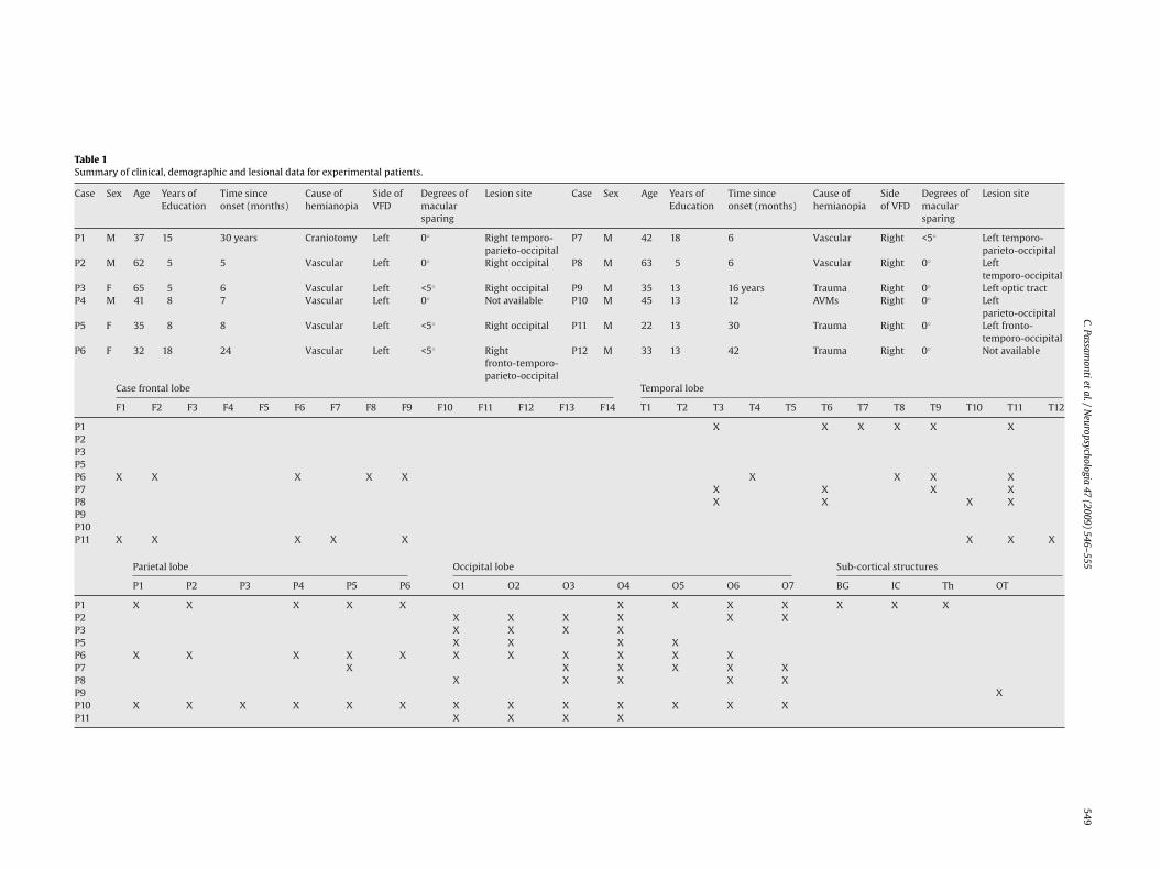
ears of education, length of illness, lesion sites, degrees of macular sparing and theresence of visual field defect are reported in Table 1.
.1.2. Control subjectsThe control group comprised 12 right-handed healthy subjects (7 female, 5 male;
ean age: 40 years; mean years of education: 13) with no history of neurologicalisease.
.2. Training paradigm
All the patients underwent Control Visual Training and subsequently Audio-isual Training.
Both training were performed on a semicircular structure in which LEDs andoudspeakers were mounted at eight locations along the azimuth (8◦ ,24◦ ,40◦ ,56◦
ight and left from the central fixation point) (see Fig. 1). Audio-Visual Training com-rised systematic audio-visual stimulation of the intact and affected visual fields,or 4 h daily over a period of 2 weeks. Patients were asked to detect the presence ofisual targets, which consisted of illumination of a LED for 100 ms, by moving theiryes towards them; these visual targets could be presented either alone or togetherith an acoustic stimulus (a 100-ms white noise burst). In the audio-visual condi-
ions, the two stimuli could be presented at either the same spatial position or atositions with a spatial disparity (16◦ and 32◦ of disparity); furthermore, the tem-oral interval between the sound and the light was gradually reduced from 300 toms over the sessions, as visual detection in the blind field reached 50% accuracy in
he unimodal visual condition (for more details, see Bolognini et al., 2005b). Duringhe training, the blind hemifield was more intensively stimulated than the intactemifield. Control Visual Training consisted of systematic visual stimulation of theisual field; it was also carried out for a similar amount of time. The apparatus, task,nd procedures were the same as during the Audio-Visual Training, but in this casenly visual stimuli were presented.
.3. Testing paradigm
Clinical and eye movements assessments were conducted in five sessions.Session 1 (S1) was the initial evaluation; Session 2 (S2) occurred two weeks later.
uring that two weeks, patients underwent Control Visual Training. After the secondssessment, patients underwent Audio-Visual Training and were then tested again,
cM[Ob
ologia 47 (2009) 546–555
mmediately at the end of the treatment (S3), three months later (S4) and yet againyear later (S5). The assessment at each session was carried out by a different inves-
igator, who was not aware of the type of treatment; moreover, patients were notware of the possible different impact of the two treatments on hemianopic deficits.ontrol Subjects performed the same tasks on the same schedule as the patients.
.4. Clinical assessment
Patients underwent a neuropsychological examination of visual field disordersn five different sessions (see Testing Paradigm). The assessment consisted of eval-ating visual detection ability (Computerized Visual Field Test, performed in twoonditions: Fixed-Eyes and Eye Movements Condition), visual exploration (Triangleest) and impairment in daily life activities (ADL) (for details, see Bolognini et al.,005b).
.5. Assessment of eye movements responses
.5.1. ApparatusEye movements were recorded in a dimly lit room using a Pan/Tilt optic eye-
racker (Eye-Track ASL-6000) which registers real-time gaze at 60 Hz. The recordingas performed in a dimly lit room. The subject’s dominant eye was illuminatedy invisible infra-red light, and the reflections were recorded by a video-cameraositioned 60 cm from the eye. During the tasks, the position of subject’s eye in theisual scene was monitored on-line by the experimenter. Before collecting data fromach subject, the equipment was calibrated using a nine-point grid. Subjects weresked to fixate successively on each of a series of small dots arranged on three linesn the form of a square. Fixation time at each dot position was at least three seconds.o prevent head movements a head stabilization device was used.
.5.2. Stimuli and experimental tasksNumber test (modified from Bolognini et al., 2005b). In this visual search task,
ight stimulus arrays (56◦ × 48◦ , horizontally and vertically respectively) each con-aining 15 numbers (from 1 to 15, stimulus size 2◦ × 2◦) distributed at random overblack background, were presented. The subjects were asked to count the numbers
n an ascending order by moving the eyes on each target.Data from eye movements recordings were quantitatively analyzed with respect
o the following parameters: number of fixations, duration of fixation, saccade dura-ion, saccade amplitude, scanpath length (sum of the saccadic amplitudes), andefixations.
In addition, mean exploration time was taken as behavioural measure of ocularfficacy.
Reading text task. The text, in Italian, was a short story (330 syllables). Fourifferent stories were counterbalanced between subjects and testing sessions. Theexts chosen were equivalent with respect to the graphical and lexical character-stics (font: Arial 40; 6–8 lines for each paragraph; 5–6 words per line; distanceetween lines: 1.5 cm) and were presented on a computer monitor (visual scene:0◦ × 24◦). Subjects were asked to read aloud to obtain both accuracy and readingime. The following variables were evaluated: number of saccades in the readingirection, number of regressions (backward saccades), number of saccades duringhe return sweep (additional to the one necessary to start a new line), mean durationf fixation, and amplitude of reading saccades.
. Results
.1. Clinical assessment
Data obtained from Computerized Visual Field Test, Triangle Testnd ADL were analysed with a series of ANOVAs. Whenever neces-ary, pairwise comparisons were conducted using Newman–Keulsests. Only significant interactions are discussed.
.1.1. Analysis of visual detection (computerized visual field test)The analyses were conducted only for the impaired hemifield
n the two task conditions (Fixed-Eyes and Eye Movements). Bothercentage of correct detections and signal detection measuresere analyzed. This latter analysis was done in order to distinguishetween perceptual (d′) and response bias processes (ˇ). Theain factors were Group (LH: left-hemianopic patients, RH: right-
emianopic patients) and Session (S1, S2, S3, S4, S5). No significantffects of main factors were found in the Fixed-Eyes Condition. In
ontrast, a significant main effect of Session was found for the Eyeovements Condition in both the percentage of visual detectionsF(4, 50) = 6.33, p < .001] and d′ measure [F(4, 50) = 8.05, p < .0001].n the contrary, ˇ measure remained stable along sessions inoth task conditions. Visual detections and perceptual sensitivity
C.Passamontiet
al./Neuropsychologia
47(2009)
546–55554
9
Table 1Summary of clinical, demographic and lesional data for experimental patients.
Case Sex Age Years ofEducation
Time sinceonset (months)
Cause ofhemianopia
Side ofVFD
Degrees ofmacularsparing
Lesion site Case Sex Age Years ofEducation
Time sinceonset (months)
Cause ofhemianopia
Sideof VFD
Degrees ofmacularsparing
Lesion site
P1 M 37 15 30 years Craniotomy Left 0◦ Right temporo-parieto-occipital
P7 M 42 18 6 Vascular Right <5◦ Left temporo-parieto-occipital
P2 M 62 5 5 Vascular Left 0◦ Right occipital P8 M 63 5 6 Vascular Right 0◦ Lefttemporo-occipital
P3 F 65 5 6 Vascular Left <5◦ Right occipital P9 M 35 13 16 years Trauma Right 0◦ Left optic tractP4 M 41 8 7 Vascular Left 0◦ Not available P10 M 45 13 12 AVMs Right 0◦ Left
parieto-occipitalP5 F 35 8 8 Vascular Left <5◦ Right occipital P11 M 22 13 30 Trauma Right 0◦ Left fronto-
temporo-occipitalP6 F 32 18 24 Vascular Left <5◦ Right
fronto-temporo-parieto-occipital
P12 M 33 13 42 Trauma Right 0◦ Not available
Case frontal lobe Temporal lobe
F1 F2 F3 F4 F5 F6 F7 F8 F9 F10 F11 F12 F13 F14 T1 T2 T3 T4 T5 T6 T7 T8 T9 T10 T11 T12
P1 X X X X X XP2P3P5P6 X X X X X X X X XP7 X X X XP8 X X X XP9P10P11 X X X X X X X X
Parietal lobe Occipital lobe Sub-cortical structures
P1 P2 P3 P4 P5 P6 O1 O2 O3 O4 O5 O6 O7 BG IC Th OT
P1 X X X X X X X X X X X XP2 X X X X X XP3 X X X XP5 X X X XP6 X X X X X X X X X X XP7 X X X X X XP8 X X X X XP9 XP10 X X X X X X X X X X X X XP11 X X X X

5 psych
sdcpd
3
pna(
3
5cnb
3
otptwhS
v
3
[ip
ttsscscbcacnrncitafcr
Ftgl
50 C. Passamonti et al. / Neuro
ignificantly increased in S3 (67%; d′: 2.48) compared to S2 (47%;′:1.84; p < .01), while no significant changes were found whenompared S3 to S4 (72%; d′: 2.65; p > .7) and to S5 (71%; d′: 2.48;> .7). Again, no differences were found when comparing S1 (42%;′:1.77) to S2 (p > .7).
.1.2. Analysis of visual exploration (triangle test)The only main effect of Session was significant [F(4, 50) = 12.39,
< .0001]. Compared to the baseline (S1: 71% of correct responses),o significant difference was observed in S2 (70%; p > .8), whileccuracy significantly improved in S3 (93%), S4 (94%), and S5 (93%)p < .003 in all comparisons).
.1.3. Analysis of ADLA significant effect of the main factor Session was found [F(4,
0) = 18.84, p < .0001]. Compared to S1 (score:15), subjective per-eived disability was significantly reduced at S3 (6; p < .001), whileo differences were observed between S1 and S2 (15; p > .9), noretween S3 and S4 (5; p > .5), S3 and S5 (4; p > .7).
.2. Analysis of eye movements
To study differences between control subjects and patients’culomotor behaviour and to investigate the differences betweenhe two stimulation types (visual and audio-visual), we com-ared oculomotor data collected during the visual search task andhe reading task using two MANOVAs (Wilks’ multivariate test),
ith Group (Controls, LH: left-hemianopic patients, RH: right-emianopic patients) as a between-subjects factor and Session (S1,2, S3, S4, S5) as a within-subjects factor.When significant multivariate effects were observed, the uni-ariate effects were inspected with a series of ANOVAs.
ipsoT
ig. 2. Visual search task. Eye movements’ parameters ((A) number of fixations; (B) numbhe five sessions (S1 = baseline; S2 = after visual training; S3 = after Audio-Visual Training; Sraphs (A)–(D): white bars = control subjects; grey bars = left-hemianopic patients; black bines represent saccadic duration (triangles = control subjects; squares = left-hemianopic p
ologia 47 (2009) 546–555
.2.1. Visual search task (number test)The MANOVA showed a significant Group x Session interaction
F(56, 538) = 1.40, p < .03]. Univariate ANOVAs confirmed the samenteraction for each parameter [smallest F-value (8, 105) = 2.12,< .04], except for the duration of fixation.
At S1, patients took more time than control subjects to explorehe visual display, and also produced more fixations and refixa-ions, resulting in a longer scanpath. Moreover, their saccades werelower and shorter in amplitude (p < .05 in all comparisons). Noignificant differences were found between S1 and S2 (p > .4 in allomparisons), suggesting that the Control Visual Training did notignificantly affect the oculomotor responses of patients. On theontrary, comparing the patients’ performance in S2 and S3, i.e.efore and after the Audio-Visual Training, we observed signifi-antly fewer fixations, saccadic duration was markedly reduced,nd mean saccadic amplitude was significantly increased. As aonsequence, visual scanning appeared to be much better orga-ized, due to a significant reduction of the length of scanpath,efixations and search times (p < .05 in all comparisons). No sig-ificant differences were found when results obtained in S3 wereompared to S4 and S5 (p > .2 in all comparisons), thus suggest-ng the stability of the results at three months and 1 year afterhe end of the Audio-Visual Training. Moreover, in the sessionsfter the Audio-Visual Training patients showed the same per-ormance as control subjects on most parameters (p > .2 in allomparisons), except for the number of fixations, the total explo-ation time and the length of scanpath, all of which remained
mpaired (p < .05 in all comparisons), although improved com-ared to baseline. Performance of normal subjects was stable acrossessions, so suggesting that practice did not appreciably affectculomotor responses (p > .8 in all comparisons). (see Fig. 2 andable 2)er of refixations; (C) length of scanpath; (D) amplitude and duration of saccades) in4 = 3 months after Audio-Visual Training; S5 = 1 year after Audio-Visual Training). Forars = right-hemianopic patients. For graph (D): bars represent saccadic amplitude;atients; circles = right-hemianopic patients).

C. Passamonti et al. / Neuropsychologia 47 (2009) 546–555 551
Table 2Visual search task.
S1 S2 S3 S4 S5
Number of fixations C 47 (13) 45 (13) 42 (13) 43 (13) 46 (13)LH 95(24) 95 (19) 65 (15) 60 (16) 71 (18)RH 100 (32) 107 (30) 80 (26) 83 (29) 85 (26)
Duration of fixation (ms) C 185 (31) 188 (36) 178 (31) 188 (31) 181 (31)LH 195 (14) 192 (17) 188 (15) 183 (21) 188 (21)RH 206 (34) 208 (29) 210 (34) 202 (31) 192 (31)
Saccadic amplitude (◦) C 9.50 (.6) 9.50 (.8) 9.33 (.3) 9.32 (.2) 9.53 (.9)LH 7.69 (.7) 7.54 (.8) 8.80 (.5) 8.85 (.3) 8.20 (1.4)RH 7.72 (.4) 7.77 (.5) 8.87 (.5) 8.83 (.4) 8.33 (.5)
Saccadic duration (ms) C 83 (9) 84 (9) 84 (11) 83 (10) 74 (10)LH 130 (30) 131 (30) 89 (13) 84 (15) 76 (17)RH 171 (60) 168 (62) 94 (27) 98 (18) 96 (28)
Length of scanpath (◦) C 428 (123) 400 (129) 408 (118) 404 (123) 415(124)LH 624 (90) 600 (76) 461 (76) 456 (72) 476 (70)RH 746 (165) 756 (169) 648 (153) 587 (198) 675 (183)
Rate of refixations (%) C 5 (2) 6 (1) 4 (2) 4 (2) 4 (3)LH 12 (3) 14 (3) 5 (1) 4 (2) 7 (2)RH 11 (2) 11 (2) 6 (3) 5 (2) 8 (2)
E
M ients (
3
[a1
dpfsiin
aapiso(cFea
wpaisi
4
ddr
tcht&mavtdritp1sFgifimrsipotrtrfidtt
xploration time (s) C 12 (3)LH 28 (5)RH 33 (7)
ean values for each session (S1, S2, S3, S4, S5) in controls (C), left-hemianopic pat
.2.2. Reading taskMANOVA revealed a significant Group × Session interaction
F(56, 538) = 2.00, p < .0001], which was replicated by Univari-te ANOVAs for every variable measured [smallest F-value (8,05) = 2.00, p < .05].
Compared to control subjects at S1, both groups of patientsemonstrated reduced reading speed (LH patients: p < .0001; RHatients: p < .009). RH readers were impaired with respect to theollowing variables: number of errors, progressive saccades, regres-ions per line, duration of fixation and saccadic amplitude (p < .001n all comparisons). In contrast, LH readers were only impairedn the return sweep, producing at least 1 saccade more than thatecessary to find the new line (p < .001).
No significant differences were found in patients between S1nd S2 (p > .5 in all comparisons). In contrast, all patients showedsignificant reduction of reading time between S2 and S3 (LH
atients: p < .01; RH patients: p < .02) and an overall improvementn performance. RH patients made fewer errors, fewer progressiveaccades and fewer regressions; they showed a shorter durationf fixation, and a clear enlargement in mean saccadic amplitudep < .05 in all comparisons). In contrast, only the number of sac-ades during the return sweep decreased in LH patients (p < .02).urthermore, no significant differences were found in any param-ters between S3, S4 and S5 (p > .3 in all comparisons), suggestinglong-lasting effect of the Audio-Visual Training.
As in the Visual Search Task, the ocular responses of controlsere stable across sessions (p > .7 in all comparisons). When com-aring patients to controls at S3, S4 and S5, only LH readers showedcomplete normalisation of all parameters (p > .1 in all compar-
sons); despite their clear improvements, RH patients remainedignificantly different from both controls and LH patients (p < .05n all comparisons) (see Fig. 3 and Table 3)
. Discussion
Unilateral damage to visual cortex of the occipital lobe can ren-er patients to be unaware of contralesional visual informationue to hemianopia. However, it has been shown that they mayetain neuroendocrine, reflexive and other behavioural responses
1
trt
12 (4) 13 (3) 11 (2) 12 (3)27 (4) 20 (3) 20 (3) 20 (4)34 (4) 25 (5) 23 (5) 28 (6)
LH) and right-hemianopic patients (RH). Standard errors between parentheses.
o visual stimuli (Weiskrantz, 1986). Although they do not haveonscious vision, they may accurately localize visual stimuli in theemianopic field with the hand or eye movements, or show mul-isensory localization behaviour (Leo, Bolognini, Passamonti, Stein,
Làdavas, 2008a). In the last study, it was shown that visual infor-ation in the blind field affects auditory localization performance,
lthough patients remained unaware of both the presence of theisual stimulus in their blind field and of its effects on their audi-ory responses. One explanation that has been postulated for theseifferent forms of residual vision is based on the involvement of theetinotectal or secondary visual pathway, which is usually intactn patients with hemianopia. Visual information can be transmit-ed through the retinotectal pathway, or some other subcorticalathway (e.g. retino-pulvinar, see Williams, Azzopardi, & Cowey,995), to extrastirate visual cortex. This information seems to beufficient to drive visually guided behaviour without awareness.urthermore, the SC, in addition to generating accurate visuallyuided saccades or orienting responses to unseen targets, is anmportant neural structure for multisensory integration. Recentndings provided evidence of the pivotal role of SC in mediatingultisensory integration in humans when orienting responses are
equired (Leo, Bertini, di Pellegrino, & Làdavas, 2008b). By usingtimuli visible (red stimuli) and invisible (purple stimuli) to the SC,t has been demonstrated that, when subjects were presented witheripheral visual stimuli, multisensory integration effects occurrednly with stimuli visible to the SC. This important result suggestshat the activity of the SC is necessary when spatial orientingesponses are required. In addition, it seems that the stimula-ion of the temporal hemifield leads to a greater multisensoryesponse enhancement, compared to stimulation of the nasal hemi-eld (Bertini, Leo, & Làdavas, 2008), thus reflecting the anatomicalifferences according to which the nasal hemiretina (processing theemporal hemifield) has a stronger direct input to the SC than theemporal hemiretina (processing the nasal hemifield) (Sherman,
974).Taken together, these studies strongly suggest that intact retino-ectal functioning may be crucial for these different forms ofesidual vision (see also Ro & Rafal, 2006, for a review) and thathe recruitment or training of these retinotectal pathways may

552 C. Passamonti et al. / Neuropsychologia 47 (2009) 546–555
F des; (B( after AA atient
bmcstniivlststadl
ttC
mtattttLIimtcci1
ig. 3. Reading task. Eye movements’ parameters ((A) number of progressive saccaE) return sweep) in the five sessions (S1 = baseline; S2 = after visual training; S3 =udio-Visual Training). White bars = control subjects; grey bars = left-hemianopic p
e advantageous in the restoration of visual function after pri-ary visual cortex damage. Since most of the patients with visual
ortex damage have an intact Superior Colliculus, it might be pos-ible to train them to use their retinotectal functions to activatehe extrastriate cortex processes, thereby enhancing visual aware-ess. Since one of the major functions of the SC is its ability to
ntegrate multisensory information when an orienting responsess required, systematic audio-visual stimulation should improveisual processing in patients with hemianopia. A study from ouraboratory demonstrated the validity of this assumption: inten-ive multisensory stimulation allowed patients to compensate forhe loss of their vision by improving visual detection and visualearch time (Bolognini et al., 2005b). These behavioural modifica-ions are thought to result from the synergistic activation of corticalnd subcortical multisensory areas, which play a pivotal role byetermining oculomotor orienting behaviour towards peripheral
ocations.To verify this hypothesis, the responsiveness of the oculomo-
or system was tested directly in the present study by comparinghe effects of two different kinds of stimulation (Audio-Visual vs.ontrol Visual Training) on oculomotor responses. Based on eye
L
dce
) number of regressive saccades; (C) duration of fixation; (D) saccadic amplitude;udio-Visual Training; S4 = 3 months after Audio-Visual Training; S5 = 1 year after
s; black bars = right-hemianopic patients.
ovement data collected before the training, our results highlighthe clear difficulty patients had organising their ocular scanning insystematic way, both in the visual search and reading tasks. In
he visual search task, both LH and RH patients, compared to con-rols, made a higher number of fixations and refixations, leadingo a longer scanpath and smaller and slower saccades, resulting inime-consuming exploration (Chedru et al.,1974; Jahnke, Denzler, &iebelt, 1995; Zihl, 1995; Pambakian et al., 2000; Tant et al., 2002).n the text reading task, the two groups of patients showed differ-ng oculomotor deficits. Compared to control subjects, RH patients
ade prolonged fixations, inappropriately low amplitude saccadeso the right, and many progressive and regressive saccades; as aonsequence, their reading was markedly inaccurate and slow. Inontrast, LH patients reported difficulties in the return sweep, lead-ng to a slight, but significant, reduction of their reading speed (Zihl,995; De Luca et al., 1996; Trauzettel-Klosinski and Rheinard, 1998;
eff et al., 2000; Leff et al., 2001).The results of the present study showed that these oculomotoreficits were differentially affected by the two training proto-ols. Control Visual Training proved ineffective in amelioratingither the clinical signs of hemianopia or the impaired oculomo-

C. Passamonti et al. / Neuropsychologia 47 (2009) 546–555 553
Table 3Reading task.
S1 S2 S3 S4 S5
Number of progressivesaccades per line
C 4.9 (1.1) 5.1 (1.2) 5.4 (1.0) 5.2 (1.1) 4.9 (1.1)LH 6.1 (.5) 6.2 (.5) 4.7 (1.9) 5.4 (.0) 5.3 (.0)RH 15.6 (.7) 16.0 (.8) 13.6 (.8) 10.7 (.6) 9.8 (2.2)
Number of regressivesaccades per line
C .9 (.4) 1.0 (.5) .8 (.3) .7 (.4) .8 (.4)LH 2.0 (.7) 1.9 (.8) 1.7 (.8) 1.3 (.6) 1.3 (.6)RH 3.1 (1.6) 2.9 (1.4) 2.2 (1.1) 1.4 (.4) 1.4 (.4)
Duration of fixation(ms)
C 205 (18) 205 (23) 202 (18) 202 (48) 204 (29)LH 233 (44) 244 (37) 221 (35) 229 (37) 230 (36)RH 306 (54) 308 (55) 278 (60) 262 (34) 280(34)
Saccadic amplitude (◦) C 4.89 (.6) 4.80 (.7) 4.62 (.4) 4.71 (1.1) 4.80 (1.1)LH 4.65 (.5) 4.61 (.4) 5.16 (.4) 5.01 (.4) 5.00 (.4)RH 1.87 (.2) 1.90 (.3) 2.75 (.4) 2.79 (.2) 2.32 (.2)
Return sweep (n) C .39 (.2) .39 (.7) .39 (.2) .44 (.2) .40 (.1)LH 1.21 (.2) 1.20 (.2) .62 (.2) .66 (.2) .65 (.2)RH .64 (.1) .73 (.2) .66 (.2) .64 (.2) .62 (.2)
Reading speed (syll/s) C 6.0 (.9) 6.3 (1.1) 6.1 (.9) 5.8 (2.0) 5.8 (1.2)LH 4.0 (.7) 4.1 (.8) 5.8 (.4) 4.8 (.6) 4.8 (.5)RH 1.6 (.7) 1.4 (.6) 6.0 (.6) 3.2 (.7) 3.4 (.7)
Errors (n) C .2 (.6) .2 (.6) .2 (.4) .1 (.3) .0 (.2)LH 1.8 (1.3) 1.2 (1.2) .3 (.5) .5 (.5) .5 (.5)
.7 (3.2
M ients (
ts“TfiTmr1tisrggddhawoowyest
oatwommtTw
deras
tSnMadtt(rupcsAopommatsh
as
RH 5.6 (3.2) 4
ean values for each session (S1, S2, S3, S4, S5) in controls (C), left-hemianopic pat
or responses in hemianopic patients, probably because the visualtimulation, presented only for 100 ms, was weakly effective topull” patients attention and eye movements into the scotoma.his evidence suggests that unimodal visual stimulation is insuf-cient to significantly affect the impaired oculomotor responses.his does not imply that other types of “unimodal training” withore salient visual stimuli and with more active visual explo-
ation might be effective in improving visual search strategy (Zihl,995; Pambakian et al., 2004). In contrast to unimodal training,he multisensory stimulation provided by the Audio-Visual Train-ng helped patients on every behavioural dimension tested; theyhowed improvement in the clinical tests as well as in the ocularesponses recorded during visual search. In addition, RH patientsained a more accurate reading performance, producing fewer pro-ressive and regressive saccades, larger in amplitude, and a reduceduration of fixation, while in LH patients the number of saccadesuring the return sweep decreased significantly. After the training,owever, when compared to controls, only LH patients obtainedn almost complete normalization of defective ocular responses,hile in the reading task RH patients still showed an impairment
f the ocular responses, despite the clear benefit gained. More-ver, the improvement observed after the Audio-Visual Trainingas long-lasting, as revealed by the maintenance of the results one
ear after the end of training, and it cannot be ascribed to a practiceffect. In fact, performance of control healthy subjects remainedtable along time, despite the repetition of the experimentalasks.
It is worth noting that the treatment did not improve the fieldf vision, since no relevant change in visual field size was observedfter Audio-Visual Training. In fact, when patients were not allowedo move the eyes (Fixed-Eyes Condition), no significant differenceas observed in perceptual sensitivity. The amelioration was foundnly when patients’ behaviour relied on other visual processing
echanisms, such as the coding within the SC for reflexive eyeovements. The improvement found, therefore, can be ascribedo the implementation of an oculomotor compensatory behaviour.his hypothesis is supported by an increase of perceptual sensitivityhen patients were free to move the eyes (Eye movements Con-
nla(F
) 2.7 (1.9) 1.8 (1.5) 1.8 (1.4)
LH) and right-hemianopic patients (RH). Standard errors between parentheses.
ition) and by the improvement obtained in test assessing visualxploration (“Triangle” Test). These data indicate that the delete-ious physiological consequences of visual cortex lesions can bemeliorated by cross-modal training, which enhances the respon-iveness of the oculomotor system.
Neurophysiological studies in animals and humans have shownhat multisensory neurons form the major output circuitry of theC (see Stein and Meredith, 1993, for a review) and may play a sig-ificant role in orienting behaviour (Stein, Meredith, Huneycut, &cDade, 1989). In fact, since the sensory and premotor maps in SC
re in spatial register, audio-visual information can be translatedirectly into an appropriate orientation response. In particular,he behavioural significance of multisensory integration to facili-ate orienting has been recently addressed in the saccadic systemsee Colonius & Arndt, 2001, for a review). Corneil et al. (2002)eported that saccades generated to low-intensity bimodal stim-li presented in close temporal and spatial proximity combine theroperties of both visual saccades (i.e., accuracy) and auditory sac-ades (i.e., short latency) in an optimal fashion. We may, therefore,uppose that the intensive bimodal stimulation provided by ourudio-Visual Training has affected the spatial and temporal aspectsf saccades generated by patients. This may explain, at least inart, the enlargements of saccadic amplitude and the reductionf saccadic duration observed after the training. The benefit ofultisensory stimulation may be further increased by using tri-odal stimuli. In fact, there is evidence from healthy subjects that
dditional tactile stimulation can result in faster responses to near-hreshold stimuli (Diederich & Colonius, 2004). So far, however, notudy has addressed the effects of a trimodal training paradigm onemianopic patients’s behaviour.
Although the SC has been demonstrated to be crucial in medi-ting multisensory integration, other cortical areas may have apecific influence in modulating its activity. Neuroanatomical and
europhysiological studies in animals have demonstrated stronginkages among the Superior Colliculus, posterior parietal cortex,nd frontal eyes fields (FEF) for the control of eye movementsArikuni, Sakai, Hamada, & Kubota, 1980; Barbas & Mesulam, 1981).urthermore, data from neuroimaging in humans have reported a

5 psych
cP
oarocet
cAsnTtcbtm
A
sM
R
A
B
B
B
B
B
C
C
C
C
D
D
D
E
F
F
G
G
I
J
K
K
K
K
K
L
L
L
L
L
L
M
M
N
N
P
P
P
R
R
S
S
S
SS
S
T
54 C. Passamonti et al. / Neuro
ommon activation of these areas during visual search (Gitelman,arrish, Friston, & Mesulam, 2002).
Thus, Audio-Visual Training might have affected several higherrder cognitive correlates of visual exploration, such as spatialttention and strategic oculomotor planning, as suggested by theeduction of fixation, refixation rates and, consequently, the lengthf scanpath. Training may have enhanced the preparation and exe-ution of eye movements by stimulating the activity of the frontalyes fields (FEF), which represents a descending input pathways tohe SC (for a review, see Liversedge & Findlay, 2000).
Our data, taken together, lead to the conclusion that an effi-ient oculomotor compensation may be induced by this specificudio-Visual Training, with long-lasting effects on the networkubserving oculomotor exploration, as suggested by the mainte-ance of training effects one year later the end of the training.his more organized pattern of ocular exploration may well explainhe improvements observed in other behavioural measures used inlinical evaluation and daily-life activities. Overall, results obtainedy eye movement recordings seem to be promising with respecto the efficacy of Audio-Visual Training as a valid compensatory
ethod for visual field disorders.
cknowledgments
We thank Nancy Stein for helpful comments on an earlier ver-ion of the manuscript. This work was supported by grants fromURST to EL.
eferences
rikuni, T., Sakai, M., Hamada, I., & Kubota, K. (1980). Topographical projections fromthe prefrontal cortex to the post-arcuate area in the Rhesus monkey, studied byretrograde axonal transport of horseradish peroxidase. Neuroscience Letters, 19,155–160.
arbas, H., & Mesulam, M. (1981). Organization of afferent input to subdivisions ofarea 8 in the rhesus monkey. Journal of Comparative Neurology, 200, 407–431.
ehrmann, M., Shomstein, S. S., Black, S. E., & Barton, J. J. (2001). The eye movementsof pure alexic patients during reading and nonreading tasks. Neuropsychologia,39, 983–1002.
ertini, C., Leo, F., & Ladavas, E. (2008). Temporo-nasal asymmetry in multisensoryintegration mediated by the Superior Colliculus. Brain Research, epub ahead ofprint.
olognini, N., Frassinetti, F., & Làdavas, E. (2005). Acoustical vision of below thresh-old stimuli: Interaction among spatially converging stimuli. Experimental BrainResearch, 160, 273–282.
olognini, N., Rasi, F., Coccia, M., & Làdavas, E. (2005). Visual search improvement inhemianopic patients after audio-visual stimulation. Brain, 128, 2830–2842.
hedru, F., Leblanc, M., & Lhermitte, F. (1974). Visual searching in normal and braindamaged subjects (contribution to the study of unilateral inattention). Cortex, 9,94–111.
iuffreda, K. J. (1994). Reading eye movements in patients with oculomotor dis-turbances. In J. Ygge & G. Lennerstrand (Eds.), Eye movements in reading (pp.163–186). Oxford: Alden Press.
olonius, H., & Arndt, P. (2001). A two-stage model for visual-auditory interactionin saccadic latencies. Perception and Psychophysics, 63, 126–147.
orneil, B. D., Van Wanrooij, M., Munoz, D., & Van Opstal, J. (2002). Auditory-visualinteractions subserving goal-directed saccades in complex scene. Journal of Neu-rophysiology, 88, 438–454.
amasio, H., & Damasio, A. R. (1989). Lesion analysis in neuropsychology. New York:Oxford University Press.
e Luca, M., Spinelli, D., & Zoccolotti, P. (1996). Eye movement patterns in reading asa function of visual field defects and contrast sensitivity loss. Cortex, 32, 491–502.
iederich, A., & Colonius, H. (2004). Bimodal and trimodal multisensory enhance-ment: Effects of stimulus onset and intensity on reaction time. Perception andPsychophysics, 66, 1388–1404.
ber, A. M., Metz-Lutz, M. N., Bataillard, M., & Collard, M. (1987). Reading eyemovements of patients with homonymous hemianopia. In K. J. O’Regan & A.Levy-Schoen (Eds.), Eye movements: From physiology to cognition (pp. 544–545).North Holland: Elsevier.
rassinetti, F., Bolognini, N., & Làdavas, E. (2002). Enhancement of visual percep-tion by crossmodal audio-visual interaction. Experimental Brain Research, 147,332–343.
rassinetti, F., Bolognini, N., Bottari, D., Bonora, A., & Làdavas, E. (2005). Audio-visualintegration in patients with visual deficit. Journal of Cognitive Neuroscience, 17,1442–1452.
T
T
ologia 47 (2009) 546–555
assel, M. M., & Williams, D. (1963). Visual function in patients with homonymoushemianopia. II. Oculomotor mechanisms. Brain, 86, 1–36.
itelman, D. R., Parrish, T. B., Friston, K. J., & Mesulam, M. M. (2002). Functionalanatomy of visual search: Regional segregations within the frontal eye fieldsand effective connectivity of the superior colliculus. NeuroImage, 15, 970–982.
shiai, S., Furukawa, T., & Tsukagoshi, H. (1987). Eye-fixation patterns in homonimoushemianopia and unilateral spatial neglect. Neuropsychologia, 25, 675–679.
ahnke, M. T., Denzler, P., Liebelt, B., Reichert, H., & Mauritz, K. H. (1995). Eye move-ments and fixation characteristic in perception of stationary scenes: Normalsubjects as compared to patients with visual neglect or hemianopia. EuropeanJournal of Neuroscience, 2, 275–295.
erkhoff, G., Munssinger, U., Haaf, E., Eberle-Strauss, G., & Stogerer, E. (1992). Reha-bilitation of homonymous scotoma in patients with postgeniculate damage tothe visual system: Saccadic compensation training. Restorative Neurology andNeuroscience, 4, 245–254.
erkhoff, G., Munbinger, U., & Meier, E. K. (1994). Neurovisual rehabilitation in cere-bral blindness. Archives of Neurology, 51, 474–481.
erkhoff, G. (1999). Restorative and compensatory therapy approaches in celebralblindness—a review. Restorative Neurology and Neuroscience, 1, 255–271.
erkhoff, G. (2000). Neurovisual rehabilitation: Recent developments and futuredirections. Journal of Neurology Neurosurgery and Psychiatry, 68, 691–706.
rauzlis, R. J., Liston, D., & Carello, C. D. (2004). Target selection and the superiorcolliculus: Goals, choices and hypothesis. Vision Research, 44, 1445–1451.
àdavas, E. (2008). Multisensory-based approach to the recovery of unisensorydeficit. Annals of the New York Academy of Sciences, 1124, 98–110.
eff, A. P., Scott, S. K., Crewes, H., Hodgson, T. L., Cowey, A., Howard, D., & Wise, R. J. S.(2000). Impaired reading in patients with right hemianopia. Annals of Neurology,47, 171–178.
eff, A. P., Scott, S. K., Rothwell, J. C., & Wise, R. J. (2001). The planning and guiding ofreading saccades: A repetitive transcranial magnetic stimulation study. CerebralCortex, 11, 918–923.
eo, F., Bolognini, N., Passamonti, C., Stein, B. E., & Làdavas, E. (2008). Cross-modallocalization in hemianopia: New insights on multisensory integration. Brain, 131,855–865.
eo, F., Bertini, C., di Pellegrino, G., & Làdavas, E. (2008). Multisensory integration fororienting responses in humans requires the activation of the superior colliculus.Experimental Brain Research, 186(1), 67–77.
iversedge, S. P., & Findlay, J. M. (2000). Saccadic eye movements and cognition.Trends in Cognitive Neuroscience, 4, 6–14.
eienberg, O. (1988). Augenbewegungsmuster bei homonymer Hemianopsie undvisuellem Hemineglekt. Klinische Monatsblatter Augenheilkdunde, 192, 108–112.
eienberg, O., Zangemeister, W. H., Rosenberg, M., Hoyt, W. F., & Stark, L. (1981).Saccadic eye movement strategies in patients with homonymous hemianopia.Annals of Neurology, 9, 537–544.
eetens, A. (1994). Revalidation of homonymous hemianopic patients. In A. C. Kooij-man, P. L. Looijestijn, J. A. Welling, & G. J. Van Der Wildt (Eds.), Low vision. Researchand new developments in rehabilitation (pp. 296–300). Amsterdam: IOS Press.
elles, G., Esser, J., Eckstein, A., Tiede, A., Gerhard, H., & Diener, H. C. (2001).Compensatory visual field training for patients with hemianopia after stroke.Neuroscience Letters, 306, 189–192.
ambakian, A. L., & Kennard, C. (1997). Can visual function be restored inpatients with homonymous hemianopia? British Journal of Ophthalmology, 81,324–328.
ambakian, A. L., Wooding, D. S., Patel, N., Morland, A. B., Kennard, C., & Mannan,S. K. (2000). Scanning the visual world: A study of patients with homonymoushemianopia. Journal of Neurology Neurosurgery and Psychiatry, 69, 751–759.
ambakian, A. L., Mannan, S. K., Hodgson, T. L., & Kennard, C. (2004). Saccadic visualsearch training: A treatment for patients with homonymous hemianopia. Journalof Neurology Neurosurgery and Psychiatry, 75, 1443–1448.
emond, A., Lesevre, N., & Gabersek, V. (1957). Approche d’une semiologie electro-graphique du regard. Revue Neurologique, 96, 536–546.
o, T., & Rafal, R. (2006). Visual restoration in cortical blindness: Insights from naturaland TMS-induced blindsight. Neuropsychological Rehabilitation, 16(4), 377–396.
choepf, D., & Zangemeister, W. H. (1993). Eye and head reading path in hemianopicpatients. In S. E. Wright & R. Groner (Eds.), Facets of dyslexia and its remediation.Amsterdam: Elsevier.
herman, M. S. (1974). Visual fields of cats with cortical and tectal lesions. Science,185(148), 355–357.
tein, B. E., Meredith, M., Huneycut, W. S., & McDade, L. W. (1989). Behavioural indicesof multisensory integration: Orientation to visual cues is affected by auditorystimuli. Journal of Cognitive Neurosciences, 1, 12–24.
tein, B. E., & Meredith, M. A. (1993). Merging of senses. Cambridge, MA: MIT Press.tein, B. E. (1998). Neural mechanism for synthesizing sensory information and
producing adaptive behaviors. Experimental Brain Research, 123, 124–135.tein, B. E., Jiang, W., & Standford, T. R. (2004). Multisensory integration in single
neurons of the midbrain. In G. Calvert, C. Spence, & B. E. Stein (Eds.), The handbookof multisensory processes. Cambridge, MA: MIT Press.
ant, M. L., Cornelissen, F. W., Kooijman, A. C., & Brouwer, W. H. (2002). Hemianopicvisual field defect elicit hemianopic scanning. Vision Research, 42, 1339–1348.
rauzettel-Klosinski, S., & Brendler, K. (1998). Eye movements in reading with hemi-anopic field defects: The significance of clinical parameters. Graefe’s Archive forClinical and Experimental Ophthalmoogy, 236, 91–102.
rauzettel-Klosinski, S., & Rheinard, J. (1998). The vertical field border in hemianopiaand its significance for fixation and reading. Investigative Ophthalmology andVisual Science, 39(11), 2177–2186.

psych
W
W
W
Z
Z
Z
Z
ZNeuropsychologia, 33, 287–303.
C. Passamonti et al. / Neuro
ang, M. K. (2003). Reading with a right homonymous haemianopia. Lancet,361(9363), 1138.
eiskrantz, L. (1986). Blindsight: A case study and implications. Oxford: ClarendonPress; Oxford University Press.
illiams, C., Azzopardi, P., & Cowey, A. (1995). Nasal and temporal retinal ganglioncells projecting to the midbrain: Implications for “blindsight”. Neuroscience,65(2), 577–586.
angemeister, W. H., Meienberg, O., Stark, L., & Hoyt, W. F. (1982). Eye-
head coordination in homonymous hemianopia. Journal of Neurology, 226,243–254.angemeister, W. H., & Oechsner, U. (1996). Evidence for scanpaths in hemianopicpatients show through string editing methods. In W. H. Zangemeister, H. S. Stiehl,& C. Freksa (Eds.), Visual attention and cognition (pp. 197–220). Amsterdam:Elsevier science.
Z
Z
ologia 47 (2009) 546–555 555
ihl, J. (1981). Recovery of visual functions in patients with cerebral blindness. Effectof specific practice with saccadic localization. Experimental Brain Research, 44,159–169.
ihl, J. (1989). Cerebral disturbances of elementary visual function. In J. W. Brown(Ed.), Neuropsychology of visual perception (pp. 35–58). Hillsdale, NJ: LawrenceErlbaum.
ihl, J. (1995). Visual scanning behaviour in patients with homonymous hemianopia.
ihl, J. (2000). Rehabilitation of visual disorders after brain injury. UK: PsychologicalPress.
ihl, J., & Kennard, C. (1996). Disorders of higher visual function. In T. Brandt, L.R. Caplan, J. Dichgans, H. C. Diener, & C. Kennard (Eds.), Neurological disorders:Course and treatment. San Diego: Academic Press.