Paralytic shellfish toxin content is related to genomic sxtA4 copy number in Alexandrium minutum...
10
ORIGINAL RESEARCH published: 01 May 2015 doi: 10.3389/fmicb.2015.00404 Edited by: Senjie Lin, University of Connecticut, USA Reviewed by: Deana Erdner, Uiversity of Texas at Austin, USA Paulo João Vieira Vale, Instituto Português do Mar e da Atmosfera, Portugal *Correspondence: Anke Stüken, Department of Biosciences, University of Oslo, P.O. Box 1066, Blindern, 0316 Oslo, Norway [email protected] Specialty section: This article was submitted to Aquatic Microbiology, a section of the journal Frontiers in Microbiology Received: 03 September 2014 Accepted: 17 April 2015 Published: 01 May 2015 Citation: Stüken A, Riobó P, Franco J, Jakobsen KS, Guillou L and Figueroa RI (2015) Paralytic shellfish toxin content is related to genomic sxtA4 copy number in Alexandrium minutum strains. Front. Microbiol. 6:404. doi: 10.3389/fmicb.2015.00404 Paralytic shellfish toxin content is related to genomic sxtA4 copy number in Alexandrium minutum strains Anke Stüken 1 *, Pilar Riobó 2 , José Franco 2 , Kjetill S. Jakobsen 3 , Laure Guillou 4,5 and Rosa I. Figueroa 6,7 1 Department of Biosciences, University of Oslo, Oslo, Norway, 2 U.A. Microalgas Nocivas (Consejo Superior de Investigaciones Científicas – Instituto Español de Oceanografía), Instituto de Investigaciones Marinas, Vigo, Spain, 3 Centre for Ecological and Evolutionary Synthesis, Department of Biosciences, University of Oslo, Oslo, Norway, 4 Laboratoire Adaptation et Diversité en Milieu Marin, CNRS, UMR 7144, Roscoff, France, 5 Sorbonne Universités – Université Pierre et Marie Curie, UMR 7144, Roscoff, France, 6 Aquatic Ecology, Lund University, Lund, Sweden, 7 U.A. Microalgas Nocivas (Consejo Superior de Investigaciones Científicas – Instituto Español de Oceanografía), Instituto Español de Oceanografía, Vigo, Spain Dinoflagellates are microscopic aquatic eukaryotes with huge genomes and an unusual cell regulation. For example, most genes are present in numerous copies and all copies seem to be obligatorily transcribed. The consequence of the gene copy number (CPN) for final protein synthesis is, however, not clear. One such gene is sxtA, the starting gene of paralytic shellfish toxin (PST) synthesis. PSTs are small neurotoxic compounds that can accumulate in the food chain and cause serious poisoning incidences when ingested. They are produced by dinoflagellates of the genera Alexandrium, Gymnodium, and Pyrodinium. Here we investigated if the genomic CPN of sxtA4 is related to PST content in Alexandrium minutum cells. SxtA4 is the 4th domain of the sxtA gene and its presence is essential for PST synthesis in dinoflagellates. We used PST and genome size measurements as well as quantitative PCR to analyze sxtA4 CPN and toxin content in 15 A. minutum strains. Our results show a strong positive correlation between the sxtA4 CPN and the total amount of PST produced in actively growing A. minutum cells. This correlation was independent of the toxin profile produced, as long as the strain contained the genomic domains sxtA1 and sxtA4. Keywords: Dinoflagellate, Alexandrium, saxitoxin (STX), paralytic shellfish toxin (PST), sxtA, gene dosage, copy number variation, genome size Introduction “From the beginning, the problem of mussel poisoning appeared to be complex,” Dr. R. Stohler, 1937 (Sommer et al., 1937). Over 60 years later, despite many great advances in research on ori- gin and diversity of mussel toxins, the phenomenon of paralytic shellfish poisoning (PSP) is still not fully explained. Stohler was co-author on a series of papers that summarized the knowledge on PSP at the time. Among the main breakthroughs was the identification of the dinoflagellate Alexandrium catenella (Whedon and Kofoid) Balech as the plankton species responsible for PSP outbreaks along the Pacific coast of North America. They showed that the mussels became toxic when they fed on A. catenella and gradually lost their toxicity after the mussels stopped feeding Frontiers in Microbiology | www.frontiersin.org 1 May 2015 | Volume 6 | Article 404
-
Upload
independent -
Category
Documents
-
view
0 -
download
0
Transcript of Paralytic shellfish toxin content is related to genomic sxtA4 copy number in Alexandrium minutum...

ORIGINAL RESEARCHpublished: 01 May 2015
doi: 10.3389/fmicb.2015.00404
Edited by:Senjie Lin,
University of Connecticut, USA
Reviewed by:Deana Erdner,
Uiversity of Texas at Austin, USAPaulo João Vieira Vale,
Instituto Português do Mar e daAtmosfera, Portugal
*Correspondence:Anke Stüken,
Department of Biosciences, Universityof Oslo, P.O. Box 1066, Blindern,
0316 Oslo, [email protected]
Specialty section:This article was submitted to
Aquatic Microbiology,a section of the journal
Frontiers in Microbiology
Received: 03 September 2014Accepted: 17 April 2015Published: 01 May 2015
Citation:Stüken A, Riobó P, Franco J,
Jakobsen KS, Guillou Land Figueroa RI (2015) Paralyticshellfish toxin content is relatedto genomic sxtA4 copy numberin Alexandrium minutum strains.
Front. Microbiol. 6:404.doi: 10.3389/fmicb.2015.00404
Paralytic shellfish toxin content isrelated to genomic sxtA4 copynumber in Alexandrium minutumstrainsAnke Stüken1*, Pilar Riobó2, José Franco2, Kjetill S. Jakobsen3, Laure Guillou4,5 andRosa I. Figueroa6,7
1 Department of Biosciences, University of Oslo, Oslo, Norway, 2 U.A. Microalgas Nocivas (Consejo Superior deInvestigaciones Científicas – Instituto Español de Oceanografía), Instituto de Investigaciones Marinas, Vigo, Spain, 3 Centrefor Ecological and Evolutionary Synthesis, Department of Biosciences, University of Oslo, Oslo, Norway, 4 LaboratoireAdaptation et Diversité en Milieu Marin, CNRS, UMR 7144, Roscoff, France, 5 Sorbonne Universités – Université Pierre etMarie Curie, UMR 7144, Roscoff, France, 6 Aquatic Ecology, Lund University, Lund, Sweden, 7 U.A. Microalgas Nocivas(Consejo Superior de Investigaciones Científicas – Instituto Español de Oceanografía), Instituto Español de Oceanografía,Vigo, Spain
Dinoflagellates are microscopic aquatic eukaryotes with huge genomes and an unusualcell regulation. For example, most genes are present in numerous copies and all copiesseem to be obligatorily transcribed. The consequence of the gene copy number (CPN)for final protein synthesis is, however, not clear. One such gene is sxtA, the startinggene of paralytic shellfish toxin (PST) synthesis. PSTs are small neurotoxic compoundsthat can accumulate in the food chain and cause serious poisoning incidences wheningested. They are produced by dinoflagellates of the genera Alexandrium, Gymnodium,and Pyrodinium. Here we investigated if the genomic CPN of sxtA4 is related to PSTcontent in Alexandrium minutum cells. SxtA4 is the 4th domain of the sxtA gene andits presence is essential for PST synthesis in dinoflagellates. We used PST and genomesize measurements as well as quantitative PCR to analyze sxtA4 CPN and toxin contentin 15 A. minutum strains. Our results show a strong positive correlation between thesxtA4 CPN and the total amount of PST produced in actively growing A. minutum cells.This correlation was independent of the toxin profile produced, as long as the straincontained the genomic domains sxtA1 and sxtA4.
Keywords: Dinoflagellate, Alexandrium, saxitoxin (STX), paralytic shellfish toxin (PST), sxtA, gene dosage, copynumber variation, genome size
Introduction
“From the beginning, the problem of mussel poisoning appeared to be complex,” Dr. R. Stohler,1937 (Sommer et al., 1937). Over 60 years later, despite many great advances in research on ori-gin and diversity of mussel toxins, the phenomenon of paralytic shellfish poisoning (PSP) is stillnot fully explained. Stohler was co-author on a series of papers that summarized the knowledgeon PSP at the time. Among the main breakthroughs was the identification of the dinoflagellateAlexandrium catenella (Whedon and Kofoid) Balech as the plankton species responsible for PSPoutbreaks along the Pacific coast of North America. They showed that the mussels became toxicwhen they fed on A. catenella and gradually lost their toxicity after the mussels stopped feeding
Frontiers in Microbiology | www.frontiersin.org 1 May 2015 | Volume 6 | Article 404
Stüken et al. Genomic sxtA4 copy numbers in Alexandrium
on it. Sommer et al. (1937) also demonstrated that the paralyticshellfish toxins (PSTs) were present in plankton samples con-taining A. catenella, that the PST content in the dinoflagellatesvaried and that it was likely to consist of more than one activesubstance.
Paralytic shellfish toxins are a group of small neurotoxicalkaloids that are among the most potent natural toxinsknown. To date, 57 isoforms of saxitoxin, the parent com-pound of PSTs, have been described (reviewed in Wiese et al.,2010). More PST-producing plankton species have also beenidentified. These include additional Alexandrium species, andthe two dinoflagellates Gymnodinium catenatum Graham andPyrodinium bahamense Plate, but also several species of freshwa-ter cyanobacteria.
On a worldwide basis,Alexandrium species are the most abun-dant and widespread (Anderson et al., 2012) and much researchhas focused on identifying factors that influence PST synthesisin this genus (recent review: Anderson et al., 2012). About onethird of the 31 taxonomically acceptedAlexandrium species todayhave been reported to produce PSTs (Anderson et al., 2012; Guiryand Guiry, 2013). The mix of PST isoforms produced, i.e., thePST profile, appears to be fixed in each strain and is thoughtto be inherited in Mendelian fashion (Sako et al., 1992) but canvary between strains of the same species. The total amount ofPSTs and the relative proportions of the PST isoforms produced,however, can vary in each strain in response to a range of bioticand abiotic factors. These include for example nutrient limita-tions (Boczar et al., 1988; Anderson et al., 1990; John and Flynn,2000), intracellular arginine concentration (Anderson et al., 1990;John and Flynn, 2000), temperature (Anderson et al., 1990), andgrazer presence (Bergkvist et al., 2008). In addition, strains thatdo not produce any detectable amounts of PSTs have also beenreported to occur within otherwise PST-producing Alexandriumspecies.
Despite these advances, it is still not known how PST synthe-sis is regulated at a cellular level in dinoflagellates. This gap ofknowledge is most likely due to the unusual genome organiza-tion of dinoflagellates. For one dinoflagellate genomes are huge.Haploid genome size measurements range from 1.5 to >225 pgcell per cell (Veldhuis et al., 1997; LaJeunesse et al., 2005) and thuscorrespond to 0.5 to >70 times the human haploid genome. Thebiggest part of dinoflagellate genomes is made up of simple andcomplex repeats (Allen et al., 1975; Davies et al., 1988; McEwanet al., 2008; Jaeckisch et al., 2011) and only about 0.2–1.8% ofsequence code for protein coding genes (McEwan et al., 2008;Hou and Lin, 2009; Jaeckisch et al., 2011). These genes frequentlyoccur in multiple copies and are often arranged in tandem arrays(Le et al., 1997; Li and Hastings, 1998; Bachvaroff and Place,2008; Shoguchi et al., 2013), but single-copy-genes may also exist(Bachvaroff and Place, 2008). The different copies of multi-copygenes are often not identical (Lee et al., 1993; Machabée et al.,1994), and it appears as if all gene copies are constantly expressed(Machabée et al., 1994). Further, recent studies using whole tran-scriptome sequencing technology (Moustafa et al., 2010; Yanget al., 2010) or microarray analyses (Yang et al., 2011) suggestthat only 0.35–27% of dinoflagellate genes are transcriptionallyregulated.
The copy numbers (CPNs) of different genes withinone species vary widely. For example, the dinoflagellateLingulodinium polyedra (Stein) Dodge has been reported to con-tain∼30 copies of a protein kinase gene (Salois andMorse, 1997),146 copies of the luciferase gene (Liu and Hastings, 2005),∼1,000copies of the Luciferin-binding Protein genes (Lee et al., 1993)and ∼5,000 copies of the mitotic cyclin gene (Bertomeu andMorse, 2004). The importance of these high gene CPNs for thecellular biology of dinoflagellates is not clear. However, it hasbeen suggested that they may be related to the amount of proteinthat can be synthesized by a dinoflagellate cell (Lee et al., 1993,2014; Moustafa et al., 2010).
Recently, two research groups have identified transcripts andtranscript fragments that are putatively involved in PST syn-thesis in dinoflagellates (Stüken et al., 2011; Hackett et al.,2013; Orr et al., 2013). Both groups have independently iden-tified transcripts that are related to sxtA (Stüken et al., 2011;Hackett et al., 2013) the putative starting gene of PST synthe-sis in cyanobacteria (Kellmann et al., 2008). The cyanobacte-rial sxtA gene contains four catalytic domains, sxtA1 to sxtA4(Kellmann et al., 2008) and dinoflagellate transcripts containingeither sxtA1–A3 or all four catalytic domains have been charac-terized in detail (Stüken et al., 2011). The organization of thefour domains within the dinoflagellate genome, however, is unre-solved: so far, only genomic fragments of sxtA1 and sxtA4 couldbe amplified, not the entire gene (Stüken et al., 2011). However,evidence is accumulating that PST-producing dinoflagellates con-tain genomic copies of sxtA1 and sxtA4, whereas one or bothof these domains are often not detected in dinoflagellates thatdo not produce PSTs (Murray et al., 2011b, 2012; Stüken et al.,2011; Orr et al., 2013; Suikkanen et al., 2013). In addition it hasbeen shown that at least sxtA4 occurs in multiple, non-identicalgenomic copies (Stüken et al., 2011; Wiese et al., 2014). Thesedifferent copies are most likely constitutivly transcribed: diver-gent sxtA1 and sxtA4 transcript families have been detected inAlexandrium transcriptomes (Stüken et al., 2011). Further, spe-cific sxtA4 transcript analyses ofA. catenella strains did not revealany changes in transcript levels throughout the growth cycle,despite significant changes in PST production rates (Wiese et al.,2014).
The aim of this study was to investigate if there is a relation-ship between the genomic CPN of sxtA4 and PST content indinoflagellate cells. We focused on A. minutum, a globally dis-tributed Alexandrium species that contains PST-producing andnon-producing strains. We measured genome size and PST con-tent and estimated the genomic sxtA4 CPN in all strains usingquantitative PCR methods.
Materials and Methods
Strains and Culture ConditionsThe A. minutum strains studied are listed in Table 1. All strainswere grown in L1 medium (Guillard and Hargraves, 1993) with-out added silica, at 30 PSU salinity, 16◦C, a 12:12 h light–darkphotoperiod and a photon irradiance of 90–100 μmol photonsm2 s−1. Strains were xenic.
Frontiers in Microbiology | www.frontiersin.org 2 May 2015 | Volume 6 | Article 404

Stüken et al. Genomic sxtA4 copy numbers in Alexandrium
TABLE 1 | Overview over the Alexandrium minutum strains used in this study, their genome size, genomic sxtA4 CPNs, and average total PST content.
Strain Isolation Genome sxtA4 Total PST Toxin group
(Synonym) Location Year Size [pg] SD CPN cell−1 SD fmol cell−1 SD ∗
AL1V (CCMP113) Ria de Vigo, Spain 1987 22.5 3.8 4.3 1.9 0.442 0.000 1
VGO722 Cambrils, Catalonia, Spain 2003 23.3 1.0 2.2 0.5 2.269 0.292 1
AMP13 Palma de Mallorca, Spain 1995 24.4 2.3 2.3 0.5 6.200 4.111 1
AL10C Estartit, Catalonia, Spain 2002 24.5 2.2 2.4 0.9 0.115 0.026 1
VGO942 Adriatic Sea, Italy 2008 24.9 1.5 1.5 1.0 0.681 0.731 1
AL4V Ria de Vigo, Spain 2000 25.2 2.6 7.0 5.4 2.358 0.360 1
Min3 Arenys, Catalonia, Spain 2002 25.2 2.2 2.0 1.1 0.650 0.439 1
VGO577 Girona, Catalonia, Spain 2002 25.7 3.2 6.4 0.5 6.574 4.336 1
AMP4 Palma de Mallorca, Spain 1995 26.2 3.0 4.0 2.2 8.446 2.762 1
VGO874 Boughrara, Tunesia 2006 29.0 3.4 2.8 0.2 0.531 0.181 1
RCC3227 Rance river, Brittany, France 2011 25.4 1.7 10.8 3.3 3.303 4.689 2
RCC3337 Penzé river, Brittany, France 2011 26.3 2.4 5.8 2.2 1.850 1.108 2
VGO650 Brittany, France 2003 26.5 1.7 6.3 0.7 15.915 3.343 2
VGO651 Brittany, France 2003 26.9 3.5 7.7 1.2 17.773 8.888 2
VGO663 Sardina, Italy 2003 29.6 2.6 N.D. N.D. 3
SD, standard deviation; CPN, copy number per genome; N.D, not detected; ∗group 1, mainly GTX1/4 and GTX2/3; group 2, mainly C1/2, GTX2/3; group 3, no PSTdetected.
Genome Size MeasurementsExponentially growing cultures (6,000–10,000 cells mL−1) wereincubated 48 h in darkness to induce synchronization of celldivision in Alexandrium (Taroncher-Oldenburg et al., 1997;Figueroa et al., 2010). Fifty milliliter of culture were filteredthrough a 5.0 μm isopore membrane filter (Millipore, Ireland),fixed with 1% paraformaldehyde for 10 min, and washed in PBS(pH 7, Sigma–Aldrich, St. Louis, MO, USA; 1200 g × 10 min).The pellet was re-suspended in 2 mL of cold methanol andstored for at least 12 h at 4◦C to allow chlorophyll extraction.The cells were then washed twice in PBS and the pellet was re-suspended in a staining solution (PBS, 0.1 mg propidium iodidemL−1 and 2 μg RNaseA mL−1) for at least 2 h before analy-sis. A Beckman FC500 bench machine with a laser emitting at488 nm was used. Four replicate samples were run at low speed(∼18 μL min−1) and data were acquired in linear and log modesuntil at least 1000 events had been recorded. As DNA standard,10 μl of a triploid trout solution (7.8 pg nucleus−1, Biosure,USA) was added to each sample. Fluorescence emission of pro-pidium iodide was detected at 620 nm. The software FlowJo7.6 (Tree Star, Inc., USA) was used to compute peak numbers,coefficients of variation (CVs), and peak ratios for the DNA flu-orescence distributions in a population. Runs with CVs above 10were discarded from analyses.
PST MeasurementsThe cultures grown for toxin analyses are listed in Table 1. Theywere grown in batch mode in 500 mL Erlenmeyer flasks contain-ing 250 mL of L1 medium (see Strains and Culture Conditionsfor details). The cultures were initiated at a density of 500 cellsmL−1 provided by the inoculation of each flask with acclimatedexponentially growing cells. For growth monitoring 5 mL sam-ples were collected every 3 days, fixed with Lugol’s solutionand cell counts in Sedgewick–Rafter slides. For toxin analy-ses, three 50 ml aliquots were harvested in actively growing
cultures using two different inocula. Cultures were filtered using1.4 μm GF/C glass fiber filters (25 mm θ). Toxins in cells wereextracted with 1000–2500μL acetic acid 0.05M depending on cellconcentration.
The PSP toxins were separated using the method proposed byRourke et al. (2008) Standard solutions of GTX4,1, dcGTX2,3,GTX2,3, STX, neoSTX, and dcSTX were purchased from theInstitute for Marine Bioscience, National Research Council,Certified Reference Material Program (NRC–CRM), Halifax, NS,Canada. Toxin concentrations were determined by comparingthe peak area for each toxin with that of the standard.
DNA Isolation and QuantificationGenomic DNA was isolated using the following Cetyl TrimethylAmmonium Bromide (CTAB) protocol: Alexandrium cultureswere harvested through centrifugation and pellets were stored at−80◦C. To isolate DNA, 700μl CTAB buffer containing 4� (v/v)2-Mercapthoethanol and 0.1 mgml−1 Proteinase K was added toeach thawed pellet. Samples were vortexed, then incubated for 1 hat 65◦C during which they were inverted every 10 min. Sampleswere let to cool, 700 μl chloroform: isoamyl alcohol (24:1) wasadded, samples were mixed by inversion and then shaken hori-zontally for 20 min. Afterward, samples were centrifuged at 4◦C,15,700 × g for 20 min, the upper aqueous phase was transferredto a fresh tube and the chloroform: isoamyl alcohol step wasrepeated on the transferred extract. Then 1 μl RNAse A (10 mgml−1) was added and samples were incubated for 30 min at 37◦C.Then 1.5 volumes 96% EtOH and 0.1 volumes sodium acetate3 M, 5.2 pH were added, and the samples stored at −20◦C overnight. The DNA was recovered by centrifugation (20 min, 4◦C,15,700× g). The supernatant was carefully removed and the DNAwashed with 200 μl of cold 70% EtOH (centrifugation 20 min,4◦C, 15,700 × g). The EtOH was removed and the pellet was airdried, before 25μl TE buffer was added and the DNA re-dissolvedat 65◦C for 1 h.
Frontiers in Microbiology | www.frontiersin.org 3 May 2015 | Volume 6 | Article 404
Stüken et al. Genomic sxtA4 copy numbers in Alexandrium
A subsample of the isolated DNA was run on a 1% agarosegel stained with GelRedTM (Biotium) and photographed toassess the integrity of the genomic DNA. If the DNA wasdegraded or contained many short fragments, then paramag-netic beads (Agencourt AMPure XP system, Beckman CoulterInc.) were used to select for DNA fragments >500 bp. Inshort, the manufacturers protocol for PCR purification wasfollowed, but fewer beads than suggested were used (vol-ume beads used here = 0.5x DNA volume). The elutionbuffer was 25 μl TE. Finally, the genomic DNA was quanti-fied with a Qubit R© Fluorometer using the dsDNA HS Assay(Invitrogen).
SxtA1 and SxtA4 Sequencing and CloningSxtA1 and sxtA4 PCRs were run according to Stüken et al.(2011) using primers sxt001/sxt002 and sxt007/sxt008(Table 2), respectively. Selected products were Sangersequenced using the same primers. SxtA4 productsfrom strains RCC3227 and RCC3337 were cloned andsequenced using the procedure described in Stüken et al.(2011).
Quantitative PCRAll quantitative PCRs (qPCRs) were performed on a RocheLightCycler R© 480 system in white 96-well-plates usingLightCycler R© 480 SYBR Green I Master chemistry (RocheDiagnostics AG, Penzberg, Germany). The sample DNA wasfreshly diluted with PCR-grade water prior to each experiment.All reactions were run in duplicate or triplicate. Two differentqPCRs were run on each DNA, a 5.8S- and a sxtA4-targetedqPCR.
The 5.8S qPCR was based on the qPCR assay developed byGalluzzi et al. (2004) but run with a slightly modified protocol asdescribed in Orr et al. (2013). In short, each reaction contained0.5x SYBR Green I master mix, 150 nM of each primer (5.8S-b5′ed and 5.8S-b3′, Table 2) and between 2, 4, or 8 ng genomicDNA. The cycling protocol was: hot start, one cycle of 95◦C for10 min; amplification, 45 cycles of 95◦C for 15 s and 60◦C for45 s, with single acquisition; followed by the melt curve program,one cycle of 95◦C for 5 s and 65◦C for 1 min, with up to 97◦Ccontinuous measurements; and finally, cooling, one cycle of 40◦Cfor 10 s.
The sxtA qPCRs were run according to the SYBR Green pro-tocol developed by Stüken et al. (2013). In short, each reaction
contained a final concentration of 0.5x SYBR Green I mastermix, 125 nM of each primer (sxt072 and sxt073, Table 2), anda known quantity of genomic DNA (1, 2, or 4 ng). The cyclingprotocol was: hot start, one cycle 95◦C for 10 min; amplifica-tion, 45 cycles 15 s at 95◦C, 15 s at 64◦C, and 30 s at 72◦C;followed by the same melt curve and cooling program as for the5.8S qPCR (see above). A purified sxtA4 PCR product generatedfrom genomic DNA of strain CCMP113 with primers sxt007 andsxt008 (Table 2) according to Stüken et al. (2011) was run on eachsxtA4 qPCR plate as a standard and contained ∼800,000 sxtA4copies.
Quantitative PCR efficiency calculations were based on thekinetics of individual PCR reactions using the algorithm imple-mented in the Real-time PCR Miner (Zhao and Russell, 2005),available online at: http://www.miner.ewindup.info/. To convertthe raw data from the Roche LightCycler R© 480 system into theright format, the program LC480Conversion.exe originally writ-ten for the LinRegPCR program (Ruijter et al., 2009) was used. Itis available at: http://www.hartfaalcentrum.nl/.
SxtA4 Copy Number DeterminationFirst, the mean efficiency (meaneff) of all positive sxtA4 qPCRreactions and the corresponding slope (s) were calculated:s = −1
log10(meaneff+1) . Then to standardize between the qPCRruns, the intercept (iplate) for each plate was calculated:
iplate =(CP1+CP2
2
)− s × log10 STD. CP1 and CP2 were the
crossing points of the standard (STD) on each plate. The stan-dard contained ∼800,000 sxtA4 copies per reaction, see Section“Quantitative PCR” for details. Next, the sxtA4 CPN in each
reaction (CPNR) was calculated: CPNR = 10(CPR−iplate)
s , whereCPR is the crossing point of the individual reaction. Finally, thesxtA4 CPN per genome (CPNG) was estimated by first calcu-lating the sxtA4 CPN per ng (CPNng):CPNng = CPNR
DNAin, where
DNAin is the amount of input DNA in ng, and then per genome:CPNG = CPNng × Gsize
1000 , whereGsize is the measured genome sizein pg.
Correlation AnalysesTo test for associations between genome size, gene CPN, and totalPSTs produced Spearman’s rho was calculated using the cor.testfunction implemented in the R statistical software (package statsversion 2.14.1).
TABLE 2 | Primers used in this study.
Name Use Sequence (5′- 3′) Reference
5.8S-b5′ed 5.8S qPCR GAT GAA GAA TGC AGC AAM ATG Orr et al. (2013)
5.8S-b3′ 5.8S qPCR CAA GCA HAC CTT CAA GMA TAT CC Galluzzi et al. (2004)
sxt072 sxtA4 qPCR CTT GCC CGC CAT ATG TGC TT Stüken et al. (2013)
sxt073 sxtA4 qPCR GCC CGG CGT AGA TGA TGT TG Stüken et al. (2013)
sxt001 sxtA1 PCR TGC AGC GMT GCT ACT CCT ACT AC Stüken et al. (2011)
sxt002 sxtA1 PCR GGT CGT GGT CYA GGA AGG AG Stüken et al. (2011)
sxt007 sxtA4 PCR ATG CTC AAC ATG GGA GTC ATC C Stüken et al. (2011)
sxt008 sxtA4 PCR GGG TCC AGT AGA TGT TGA CGA TG Stüken et al. (2011)
Frontiers in Microbiology | www.frontiersin.org 4 May 2015 | Volume 6 | Article 404
Stüken et al. Genomic sxtA4 copy numbers in Alexandrium
Results
Genome Size MeasurementsThe A. minutum genome sizes are listed in Table 1 and rangedfrom 22.5 to 29.6 pg cell−1, with an average of 25.7 ± 1.9 pgcell−1 (n = 15). The genome sizes of toxin group 1 strains (seePST Measurements for toxin profile details) ranged between 22.5and 29.0 pg and of toxin group 2 strains between 25.4 and 26.9 pg.Strain VGO663 had the biggest genome; a strain in which no PSTtoxins nor sxtA4 copies were detected and the sole member oftoxin group 3.
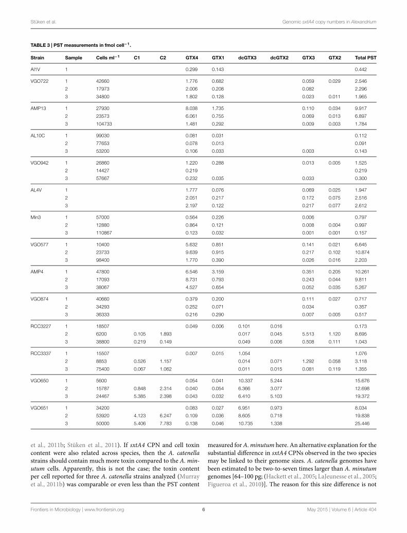
PST MeasurementsThe results of the toxin analyses are listed inTables 1 and 3. Threegroups of PST profiles were observed: (1) GTX1/4 and GTX2/3(strains isolated from different locations in the MediterraneanSea and the Atlantic Ocean); (2) C1/2, GTX2/3, and dcGTX2/3(strains isolated from Brittany, France), and (3) no PST detected(strain VGO663 isolated off Sardinia, Italy).
The total PST content varied considerably between the dif-ferent strains within each toxin group, and in some casesalso between measurements of the same strains. But alto-gether were the mean and median values higher in toxingroup 2 (mean ± SD = 9.710 ± 8.769; median = 8.364;n = 12) than in group 1 (mean ± SD = 2.997 ± 3.506;median = 1.865; n = 29).
Quantitative PCR and sxtA Gene CopyNumbers Per GenomeThe 5.8S qPCR had an amplification efficiency based on all sam-ples of 0.65± 0.05 (mean± SD, n= 124). AllAlexandrium strainsamplified with single, specific meltcurve.
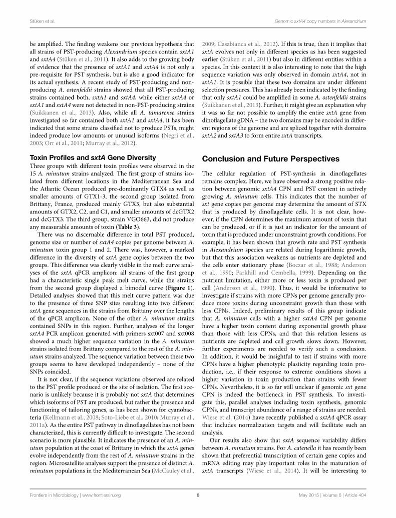
The sxtA qPCR had an amplification efficiency of 0.94 ± 0.06(mean ± SD, n = 141) and amplification was observed forall Alexandrium strains tested, apart from A. affine strainsCCMP112,A. andersoniiCCMP2222, and A. minutum VGO663.The majority of amplified strains showed a specific melt curvewith a single peak. The four A. minutum strains isolated from theChannel, Brittany, France (VGO650, VGO651, RCC3337, andRCC3227), however, had a distinct melt curve with a double peak(Figure 1). This double peak was due to three SNP loci in theqPCR amplicon only present in the Brittany strains.
The sxtA4 CPN per genome varied between 1.5 ± 1.0 and10.8 ± 3.3 (mean ± SD; Table 1) in all A. minutum strains inves-tigated. The CPNs ranged from 1.5 ± 1.0 to 7.0 ± 5.4 in toxingroup 1 and 5.8 ± 2.2 to 10.8 ± 3.3 in toxin group 2 (Table 1).
SxtA1 and SxtA4 Amplification andSequence DiversityThe PCRs for sxtA1 and sxtA4 amplified single products of theright size for all PST producing strains. No amplification wasobserved for A. minutum VGO663, as well as the A. affine andA. andersonii strains.
Direct sequencing of the 670 bp long sxtA4 fragment resultedin identical sequences without SNPS for strains AL1V, AL10C,AL4, and Min3. The sequences of strain VGO577 and VGO847had three and six SNPs over the same length. The SNPs were
located outside the region used for qPCR and also present inthe transcriptome of strain AL1V (Stüken et al., 2011). StrainsVGO650 and VGO651, had 14 and 16 SNPs, respectively, noneof which coincided with SNPs of the AL1V transcriptome. ThesxtA4 amplicons from RCC3337 and RCC3227 were clonedand sequenced. These sequences confirmed the SNP positionsobserved in VGO650 and 651. Three of the SNPs observedin VGO650, VGO651, RCC3337, and RCC3227 were locatedbetween qPCR primers sxt072 and sxt073 and resulted in twodistinct sequences. None of the SNPs observed in any of the A.minutum sequences coincided with SNPs in the A. fundyenseCCMP1719 transcriptome (Stüken et al., 2011), which had 12SNPs over the same length.
Direct sequencing of the sxtA1 amplicons (503 bp) resultedin identical sequences for strains AL1V, AL10C, AL4V, Min3,VGO577, and VGO874, sequences from strains VGO650 andVGO651 were 1 bp different.
Sequences have the GenBank accession numbers KM438016–KM438027.
Correlation AnalysesOnly strains for which sxtA4 could be amplified were includedin the analyses. The Spearman rank order correlation resultsindicated significant positive relations between sxtA4 CPN pergenome and total toxin content per cell (ρ = 0.470, S = 4844,p = 0.003, Figure 2A), and between total toxin content per celland genome size (ρ = 0.355, S = 5896, p = 0.029, Figure 2B). Nosignificant relation was detected between genome size and sxtA4CPN per cell (ρ = 0.473, S = 240, p = 0.088, Figure 2C).
Discussion
Relation between sxtA Gene Copy Number,PST Content and Genome SizeThe results of this study support our previous idea that strainswith low levels of PST have fewer copies of the sxt genes com-pared to those with higher toxin levels (Stüken et al., 2011).Specifically, the results show a strong positive correlation betweenthe genomic CPN of sxtA4 domains and the total cellular contentof PST in actively growing A. minutum cultures (Figure 2). It hasbeen shown that the toxin content and profile of the sameA. min-utum culture throughout the growth curve are generally stableeven under different nutrient conditions. Exceptions are limit-ing conditions, especially the combination of P and N limitations,where toxin content may increase drastically (Flynn et al., 1994).By taking several PST measurements from each strain duringactive growth and pooling the results, we aimed at capturing thetypical variation of toxin production in each strain under stan-dard growth conditions. Thus, our results indicate that strainswith a higher genomic sxtA4 CPN have on average a higher PSTcontent.
Interestingly, the genomic sxtA4CPN reported forA. catenellastrains previously is considerably higher than found for the A.minutum strains here. All A. minutum strains were estimated tohave fewer than 15 copies per genome (Table 1), while 100–280genomic copies have been reported for A. catenella (Murray
Frontiers in Microbiology | www.frontiersin.org 5 May 2015 | Volume 6 | Article 404
Stüken et al. Genomic sxtA4 copy numbers in Alexandrium
TABLE 3 | PST measurements in fmol cell−1.
Strain Sample Cells ml−1 C1 C2 GTX4 GTX1 dcGTX3 dcGTX2 GTX3 GTX2 Total PST
Al1V 1 0.299 0.143 0.442
VGO722 1 42660 1.776 0.682 0.059 0.029 2.546
2 17973 2.006 0.208 0.082 2.296
3 34800 1.802 0.128 0.023 0.011 1.965
AMP13 1 27930 8.038 1.735 0.110 0.034 9.917
2 23573 6.061 0.755 0.069 0.013 6.897
3 104733 1.481 0.292 0.009 0.003 1.784
AL10C 1 99030 0.081 0.031 0.112
2 77653 0.078 0.013 0.091
3 53200 0.106 0.033 0.003 0.143
VGO942 1 26860 1.220 0.288 0.013 0.005 1.525
2 14427 0.219 0.219
3 57667 0.232 0.035 0.033 0.300
AL4V 1 1.777 0.076 0.069 0.025 1.947
2 2.051 0.217 0.172 0.075 2.516
3 2.197 0.122 0.217 0.077 2.612
Min3 1 57000 0.564 0.226 0.006 0.797
2 12880 0.864 0.121 0.008 0.004 0.997
3 110867 0.123 0.032 0.001 0.001 0.157
VGO577 1 10400 5.632 0.851 0.141 0.021 6.645
2 23733 9.639 0.915 0.217 0.102 10.874
3 98400 1.770 0.390 0.026 0.016 2.203
AMP4 1 47800 6.546 3.159 0.351 0.205 10.261
2 17093 8.731 0.793 0.243 0.044 9.811
3 38067 4.527 0.654 0.052 0.035 5.267
VGO874 1 40660 0.379 0.200 0.111 0.027 0.717
2 34293 0.252 0.071 0.034 0.357
3 36333 0.216 0.290 0.007 0.005 0.517
RCC3227 1 18507 0.049 0.006 0.101 0.016 0.173
2 6200 0.105 1.893 0.017 0.045 5.513 1.120 8.695
3 38800 0.219 0.149 0.049 0.006 0.508 0.111 1.043
RCC3337 1 15507 0.007 0.015 1.054 1.076
2 8853 0.526 1.157 0.014 0.071 1.292 0.058 3.118
3 75400 0.067 1.062 0.011 0.015 0.081 0.119 1.355
VGO650 1 5600 0.054 0.041 10.337 5.244 15.676
2 15787 0.848 2.314 0.040 0.054 6.366 3.077 12.698
3 24467 5.385 2.398 0.043 0.032 6.410 5.103 19.372
VGO651 1 34200 0.083 0.027 6.951 0.973 8.034
2 53920 4.123 6.247 0.109 0.036 8.605 0.718 19.838
3 50000 5.406 7.783 0.138 0.046 10.735 1.338 25.446
et al., 2011b; Stüken et al., 2011). If sxtA4 CPN and cell toxincontent were also related across species, then the A. catenellastrains should contain muchmore toxin compared to the A. min-utum cells. Apparently, this is not the case; the toxin contentper cell reported for three A. catenella strains analyzed (Murrayet al., 2011b) was comparable or even less than the PST content
measured forA.minutum here. An alternative explanation for thesubstantial difference in sxtA4 CPNs observed in the two speciesmay be linked to their genome sizes. A. catenella genomes havebeen estimated to be two-to-seven times larger than A. minutumgenomes [64–100 pg; (Hackett et al., 2005; LaJeunesse et al., 2005;Figueroa et al., 2010)]. The reason for this size difference is not
Frontiers in Microbiology | www.frontiersin.org 6 May 2015 | Volume 6 | Article 404

Stüken et al. Genomic sxtA4 copy numbers in Alexandrium
FIGURE 1 | SxtA4 qPCR melt curve comparison. (A) Bimodal melting curve of Alexandrium minutum strains VGO650, VGO651, RCC3227, and RCC3337isolated from Brittany, France, producing PST isoforms C1/2, GTX2/3, and dcGTX2/3. (B) Single peak melting curves of strains AL1V, VGO577, AL10C, and Min3producing PST isoforms GTX1/4 and GTX2/3.
FIGURE 2 | Spearman rank order correlation plots. Scatterplots showing the relation between (A) sxtA4 copy number (CPN) per genome and total toxin contentper cell, (B) total toxin content per cell and genome size, and (C) genome size and sxtA4 CPN per cell. CPN, copy number; significance level: ∗∗p ≤ 0.01, ∗p ≤ 0.05.
clear, but may be related to ploidy differences. In this case, sxtA4CPN would not be a positively selected trait but rather a result ofgenome dynamics.
However, within the species A. minutum sxtA4 CPN did notscale with genome size (Figure 2), even though we observed con-siderable differences in genome size (Table 1). Thus, sxtA4 CPNdoes not appear to be a function of genome size in A. minutum.This is the first time that so many strains of the same dinoflagel-late species have beenmeasured and that such a breath of genomesize within a species has been documented. It is currently unclearwhat causes the genome size diversity but it might be relatedto chromosomal differences, as aneuploidy may be common indinoflagellate cultures (Loper et al., 1980).
We also observed a positive correlation between genome sizeand toxin content (Figure 2; Table 3). It is unclear what causedthis relationship, as toxin content and sxtA4 CPN were positively
correlated, but sxtA4CPN and genome size were not. In dinoflag-ellates DNA content correlates with cell size (LaJeunesse et al.,2005; Dapena et al., 2015) a relation that has been document formost eukaryotic lineages, e.g., (Gregory, 2001; Cavalier-Smith,2005). It is possible that cells with a higher cell volume containmore PSTs. However, studies comparing toxin content of dif-ferent sized cells have, to our knowledge, not been undertakenyet.
Non-PST Producing A. minutum StrainVGO663For one of the A. minutum strains, strain VGO663, neither PSTnor sxtA1, or sxtA4 gene fragments were detected. While it waspreviously known that the species A. minutum contains PST-producing and non-producing strains, e.g., (Touzet et al., 2007),this is the first A. minutum strain for which no sxt genes could
Frontiers in Microbiology | www.frontiersin.org 7 May 2015 | Volume 6 | Article 404
Stüken et al. Genomic sxtA4 copy numbers in Alexandrium
be amplified. The finding weakens our previous hypothesis thatall strains of PST-producing Alexandrium species contain sxtA1and sxtA4 (Stüken et al., 2011). It also adds to the growing bodyof evidence that the presence of sxtA1 and sxtA4 is not only apre-requisite for PST synthesis, but is also a good indicator forits actual synthesis. A recent study of PST-producing and non-producing A. ostenfeldii strains showed that all PST-producingstrains contained both, sxtA1 and sxtA4, while either sxtA4 orsxtA1 and sxtA4 were not detected in non-PST-producing strains(Suikkanen et al., 2013). Also, while all A. tamarense strainsinvestigated so far contained both sxtA1 and sxtA4, it has beenindicated that some strains classified not to produce PSTs, mightindeed produce low amounts or unusual isoforms (Negri et al.,2003; Orr et al., 2011; Murray et al., 2012).
Toxin Profiles and sxtA Gene DiversityThree groups with different toxin profiles were observed in the15 A. minutum strains analyzed. The first group of strains iso-lated from different locations in the Mediterranean Sea andthe Atlantic Ocean produced pre-dominantly GTX4 as well assmaller amounts of GTX1-3, the second group isolated fromBrittany, France, produced mainly GTX3, but also substantialamounts of GTX2, C2, and C1, and smaller amounts of dcGTX2and dcGTX3. The third group, strain VGO663, did not produceany measurable amounts of toxin (Table 3).
There was no discernable difference in total PST produced,genome size or number of sxtA4 copies per genome between A.minutum toxin group 1 and 2. There was, however, a markeddifference in the diversity of sxtA gene copies between the twogroups. This difference was clearly visible in the melt curve anal-yses of the sxtA qPCR amplicon: all strains of the first grouphad a characteristic single peak melt curve, while the strainsfrom the second group displayed a bimodal curve (Figure 1).Detailed analyses showed that this melt curve pattern was dueto the presence of three SNP sites resulting into two differentsxtA gene sequences in the strains from Brittany over the lengthsof the qPCR amplicon. None of the other A. minutum strainscontained SNPs in this region. Further, analyses of the longersxtA4 PCR amplicon generated with primers sxt007 and sxt008showed a much higher sequence variation in the A. minutumstrains isolated from Brittany compared to the rest of the A. min-utum strains analyzed. The sequence variation between these twogroups seems to have developed independently – none of theSNPs coincided.
It is not clear, if the sequence variations observed are relatedto the PST profile produced or the site of isolation. The first sce-nario is unlikely because it is probably not sxtA that determineswhich isoforms of PST are produced, but rather the presence andfunctioning of tailoring genes, as has been shown for cyanobac-teria (Kellmann et al., 2008; Soto-Liebe et al., 2010; Murray et al.,2011a). As the entire PST pathway in dinoflagellates has not beencharacterized, this is currently difficult to investigate. The secondscenario is more plausible. It indicates the presence of an A. min-utum population at the coast of Brittany in which the sxtA genesevolve independently from the rest of A. minutum strains in theregion. Microsatellite analyses support the presence of distinct A.minutum populations in the Mediterranean Sea (McCauley et al.,
2009; Casabianca et al., 2012). If this is true, then it implies thatsxtA evolves not only in different species as has been suggestedearlier (Stüken et al., 2011) but also in different entities within aspecies. In this context it is also interesting to note that the highsequence variation was only observed in domain sxtA4, not insxtA1. It is possible that these two domains are under differentselection pressures. This has already been indicated by the findingthat only sxtA1 could be amplified in some A. ostenfeldii strains(Suikkanen et al., 2013). Further, it might give an explanation whyit was so far not possible to amplify the entire sxtA gene fromdinoflagellate gDNA – the two domains may be encoded in differ-ent regions of the genome and are spliced together with domainssxtA2 and sxtA3 to form entire sxtA transcripts.
Conclusion and Future Perspectives
The cellular regulation of PST-synthesis in dinoflagellatesremains complex. Here, we have observed a strong positive rela-tion between genomic sxtA4 CPN and PST content in activelygrowing A. minutum cells. This indicates that the number ofsxt gene copies per genome may determine the amount of STXthat is produced by dinoflagellate cells. It is not clear, how-ever, if the CPN determines the maximum amount of toxin thatcan be produced, or if it is just an indicator for the amount oftoxin that is produced under unconstraint growth conditions. Forexample, it has been shown that growth rate and PST synthesisin Alexandrium species are related during logarithmic growth,but that this association weakens as nutrients are depleted andthe cells enter stationary phase (Boczar et al., 1988; Andersonet al., 1990; Parkhill and Cembella, 1999). Depending on thenutrient limitation, either more or less toxin is produced percell (Anderson et al., 1990). Thus, it would be informative toinvestigate if strains with more CPNs per genome generally pro-duce more toxins during unconstraint growth than those withless CPNs. Indeed, preliminary results of this group indicatethat A. minutum cells with a higher sxtA4 CPN per genomehave a higher toxin content during exponential growth phasethan those with less CPNs, and that this relation lessens asnutrients are depleted and cell growth slows down. However,further experiments are needed to verify such a conclusion.In addition, it would be insightful to test if strains with moreCPNs have a higher phenotypic plasticity regarding toxin pro-duction, i.e., if their response to extreme conditions shows ahigher variation in toxin production than strains with fewerCPNs. Nevertheless, it is so far still unclear if genomic sxt geneCPN is indeed the bottleneck in PST synthesis. To investi-gate this, parallel analyses including toxin synthesis, genomicCPNs, and transcript abundance of a range of strains are needed.Wiese et al. (2014) have recently published a sxtA4 qPCR assaythat includes normalization targets and will facilitate such ananalysis.
Our results also show that sxtA sequence variability differsbetween A. minutum strains. For A. catenella it has recently beenshown that preferential transcription of certain gene copies andmRNA editing may play important roles in the maturation ofsxtA transcripts (Wiese et al., 2014). It will be interesting to
Frontiers in Microbiology | www.frontiersin.org 8 May 2015 | Volume 6 | Article 404

Stüken et al. Genomic sxtA4 copy numbers in Alexandrium
investigate, if the same is also true for A. minutum and if this mayhave an impact on the total amount of toxin produced.
Finally it remains to be established if the same mechanismsregulate PST-synthesis in all Alexandrium species. Recent anal-yses of the ribosomal DNA in various Alexandrium speciesrevealed distinct chromosomal organization (Figueroa et al.,2014).
Acknowledgments
The work was funded by a grant from Formas (Sweden) to RIF(Formas 215-2010-824) and the Norwegian Research Councilgrant 186292/V40 to KSJ. LG received funding through thePARALEX project by the French research agency’s (ANR) 2009and GDR Phycotox (http://www.phycotox.fr).
References
Allen, J., Roberts, T., Loeblich, A., and Klotz, L. (1975). Characterization ofDNA from dinoflagellate Crypthecodinium cohnii and implications for nuclearorganization. Cell 6, 161–169. doi: 10.1016/0092-8674(75)90006-9
Anderson, D. M., Alpermann, T. J., Cembella, A. D., Collos, Y., Masseret, E.,and Montresor, M. (2012). The globally distributed genus Alexandrium: mul-tifaceted roles in marine ecosystems and impacts on human health. HarmfulAlgae 14, 10–35. doi: 10.1016/j.hal.2011.10.012
Anderson, D. M., Kulis, D. M., Sullivan, J. J., Hall, S., and Lee, C. (1990). Dynamicsand physiology of saxitoxin production by the dinoflagellatesAlexandrium spp.Mar. Biol. 104, 511–524. doi: 10.1007/BF01314358
Bachvaroff, T. R., and Place, A. R. (2008). From stop to start: tandem gene arrange-ment, copy number and trans-splicing sites in the dinoflagellate Amphidiniumcarterae. PLoS ONE 3:e2929. doi: 10.1371/journal.pone.0002929
Bergkvist, J., Selander, E., and Pavia, H. (2008). Induction of toxin productionin dinoflagellates: the grazer makes a difference. Oecologia 156, 147–154. doi:10.1007/s00442-008-0981-6
Bertomeu, T., and Morse, D. (2004). Isolation of a dinoflagellate mitotic cyclinby functional complementation in yeast. Biochem. Biophys. Res. Commun. 323,1172–1183. doi: 10.1016/j.bbrc.2004.09.008
Boczar, B. A., Beitler, M. K., Liston, J., Sullivan, J. J., and Cattolico,R. A. (1988). Paralytic shellfish toxins in Protogonyaulax tamarensis andProtogonyaulax catenella in axenic Culture. Plant Physiol. 88, 1285–1290. doi:10.1104/pp.88.4.1285
Casabianca, S., Penna, A., Pecchioli, E., Jordi, A., Basterretxea, G., and Cristiano, V.(2012). Population genetic structure and connectivity of the harmful dinoflag-ellate Alexandrium minutum in the Mediterranean Sea. Proc. Biol. Sci. 279,129–138. doi: 10.1098/rspb.2011.0708
Cavalier-Smith, T. (2005). Economy, speed and size matter: evolutionary forcesdriving nuclear genome miniaturization and expansion. Ann. Bot. 95, 147–175.doi: 10.1093/aob/mci010
Dapena, C., Bravo, I., Cuadrado, A., and Figueroa, R. I. (2015). Nuclearand cell morphological changes during the cell cycle and growth ofthe toxic dinoflagellate Alexandrium minutum. Protist 166, 146–160. doi:10.1016/j.protis.2015.01.001
Davies, W., Jakobsen, K. S., and Nordby, Ø. (1988). Characterization of DNAfrom the dinoflagellateWoloszynskia bostoniensis. J. Protozool. 35, 418–422. doi:10.1111/j.1550-7408.1988.tb04120.x
Figueroa, R. I., Cuadrado, A., Stüken, A., Rodríguez, F., and Fraga, S.(2014). Ribosomal DNA organization patterns within the dinoflagellate genusAlexandrium as revealed by FISH: life cycle and evolutionary implications.Protist 165, 343–363. doi: 10.1016/j.protis.2014.04.001
Figueroa, R. I., Garces, E., and Bravo, I. (2010). The use of flow cytometry forspecies identification and life-cycle studies in dinoflagellates.Deep Sea Res. PartII Top. Stud. Oceanogr. 57, 301–307. doi: 10.1016/J.Dsr2.2009.09.008
Flynn, K., Franco, J. M., Fernandez, P., Reguera, B., Zapata, M., Wood, G., et al.(1994). Changes in toxin content, biomass and pigments of the dinoflagellateAlexandrium minutum during nitrogen refeeding and growth into nitrogenor phosphorus stress. Mar. Ecol. Prog. Ser. 111, 99–109. doi: 10.3354/meps111099
Galluzzi, L., Penna, A., Bertozzini, E., Vila, M., Garces, E., and Magnani, M. (2004).Development of a real-time PCR assay for rapid detection and quantifica-tion of Alexandrium minutum (a dinoflagellate). Appl. Environ. Microbiol. 70,1199–1206. doi: 10.1128/Aem.70.2.1199-1206.2004
Gregory, R. T. (2001). Coincidence, coevolution, or causation? DNA content,cellsize, and the C-value enigma. Biol. Rev. 76, 65–101. doi: 10.1111/j.1469-185X.2000.tb00059.x
Guillard, R. R. L., and Hargraves, P. E. (1993). Stichochrysis immobilis is a diatom,not a chrysophyte. Phycologia 32, 234–236. doi: 10.2216/i0031-8884-32-3-234.1
Guiry, M. D., and Guiry, G. M. (2013). AlgaeBase. World-Wide ElectronicPublication. National University of Ireland, Galway. Available at: http://www.
algaebase.org [accessed May 20, 2013].Hackett, J. D., Scheetz, T. E., Yoon, H. S., Soares, M. B., Bonaldo, M. F., Casavant,
T. L., et al. (2005). Insights into a dinoflagellate genome through expressedsequence tag analysis. BMC Genomics 6:80. doi: 10.1186/1471-2164-6-80
Hackett, J. D., Wisecaver, J. H., Brosnahan, M. L., Kulis, D. M., Anderson, D. M.,Bhattacharya, D., et al. (2013). Evolution of saxitoxin synthesis in cyanobacteriaand dinoflagellates.Mol. Biol. Evol. 30, 70–78. doi: 10.1093/molbev/mss142
Hou, Y., and Lin, S. (2009). Distinct gene number-genome size relationshipsfor eukaryotes and non-Eukaryotes: gene content estimation for dinoflagellategenomes. PLoS ONE 4:e6978. doi: 10.1371/journal.pone.0006978
Jaeckisch, N., Yang, I., Wohlrab, S., Glöckner, G., Kroymann, J., Vogel, H., et al.(2011). Comparative genomic and transcriptomic characterization of the toxi-genic marine dinoflagellate Alexandrium ostenfeldii. PLoS ONE 6:e28012. doi:10.1371/journal.pone.0028012.t005
John, E., and Flynn, K. (2000). Growth dynamics and toxicity of Alexandriumfundyense (Dinophyceae): the effect of changing N:P supply ratioson internal toxin and nutrient levels. Eur. J. Phycol. 35, 11–23. doi:10.1017/S0967026200002572
Kellmann, R., Mihali, T. K., Jeon, Y. J., Pickford, R., Pomati, F., and Neilan,B. A. (2008). Biosynthetic intermediate analysis and functional homologyreveal a saxitoxin gene cluster in cyanobacteria. Appl. Environ. Microbiol. 74,4044–4053. doi: 10.1128/aem.00353-08
LaJeunesse, T., Lambert, G., Andersen, R., Coffroth, M., and Galbraith, D. (2005).Symbiodinium (Pyrrhophyta) genome sizes (DNA content) are smallest amongdinoflagellates. J. Phycol. 41, 880–886. doi: 10.1111/j.1529-8817.2005.00111.x
Le, Q. H., Markovic, P., Hastings, J., Jovine, R. V., and Morse, D. (1997).Structure and organization of the peridinin-chlorophyll a-binding pro-tein gene in Gonyaulax polyedra. Mol. Gen. Genet. 225, 595–604. doi:10.1007/s004380050533
Lee, D., Mittag, M., Sczekan, S., Morse, D., and Hastings, J. (1993). Molecular-cloning and genomic organization of a gene for Luciferin-binding Protein fromthe dinoflagellate Gonyaulax polyedra. J. Biol. Chem. 268, 8842–8850.
Lee, R., Lai, H., Malik, S. B., Saldarriaga, J. F., Keeling, P. J., and Slamovits, C. H.(2014). Analysis of EST data of the marine protist Oxyrrhis marina, an emerg-ing model for alveolate biology and evolution. BMC Genomics 15:122. doi:10.1186/1471-2164-15-122
Li, L. M., and Hastings, J. W. (1998). The structure and organization of theluciferase gene in the photosynthetic dinoflagellate Gonyaulax polyedra. PlantMol. Biol. 36, 275–284. doi: 10.1023/A:1005941421474
Liu, L. Y., and Hastings, J. W. (2005). Novel and rapidly diverging intergenicsequences between tandem repeats of the luciferase genes in seven dinoflagellatespecies. J. Phycol. 42, 96–103. doi: 10.1111/j.1529-8817.2006.00165.x
Loper, C. L., Steidinger, K. A., and Walker, L. M. (1980). A simple chro-mosome spread technique for unarmoured dinoflagellates and implicationsof polyploidy in algal cultures. Trans. Am. Microsc. Soc. 99, 343–346. doi:10.2307/3226012
Machabée, S., Wall, L., and Morse, D. (1994). Expression and genomic orga-nization of a dinoflagellate gene family. Plant Mol. Biol. 25, 23–31. doi:10.1007/BF00024195
McCauley, L. A. R., Erdner, D. L., Nagai, S., Richlen, M. L., and Anderson,D. M. (2009). Biogeographic analysis of the globally distributed harmfulalgal bloom species Alexandrium minutum (Dinophyceae) based on rRNAgene sequences and microsatellite markers. J. Phycol. 45, 454–463. doi:10.1111/j.1529-8817.2009.00650.x
Frontiers in Microbiology | www.frontiersin.org 9 May 2015 | Volume 6 | Article 404
Stüken et al. Genomic sxtA4 copy numbers in Alexandrium
McEwan, M., Humayun, R., Slamovits, C. H., and Keeling, P. J. (2008). Nucleargenome sequence survey of the dinoflagellateHeterocapsa triquetra. J. Eukaryot.Microbiol. 55, 530–535. doi: 10.1111/j.1550-7408.2008.00357.x
Moustafa, A., Evans, A. N., Kulis, D. M., Hackett, J. D., Erdner, D. L., Anderson,D. M., et al. (2010). Transcriptome profiling of a toxic dinoflagellate reveals agene-rich protist and a potential impact on gene expression due to bacterialpresence. PLoS ONE 5:e9688. doi: 10.1371/journal.pone.0009688
Murray, S. A., Mihali, T. K., and Neilan, B. A. (2011a). Extraordinary conservation,gene loss, and positive selection in the evolution of an ancient neurotoxin.Mol.Biol. Evol. 28, 1173–1182. doi: 10.1093/molbev/msq295
Murray, S. A., Wiese, M., Stüken, A., Brett, S., Kellmann, R., Hallegraeff, G., et al.(2011b). A quantitative molecular assay based on the gene sxtA to identifysaxitoxin-producing harmful algal blooms in marine waters. Appl. Environ.Microbiol. 77, 7050–7057. doi: 10.1128/AEM.05308-11
Murray, S. A., Wiese, M., Neilan, B. A., Orr, R. J. S., De Salas, M., Brett, S.,et al. (2012). A reinvestigation of saxitoxin production and sxtA in the ‘non-toxic’ Alexandrium tamarense Group V clade. Harmful Algae 18, 96–104. doi:10.1016/J.Hal.2012.05.001
Negri, A., Llewellyn, L., Doyle, J., Webster, N., Frampton, D., and Blackburn, S.(2003). Paralytic shellfish toxins are restricted to few species among Australia’staxonomic diversity of cultured microalgae. J. Phycol. 39, 663–667. doi:10.1046/j.1529-8817.2003.02131.x
Orr, R. J., Stüken, A., Murray, S. A., and Jakobsen, K. S. (2013). Evolutionaryacquisition and loss of saxitoxin biosynthesis in dinoflagellates: the second“core” gene, sxtG. Appl. Environ. Microbiol. 79, 2128–2136. doi: 10.1128/AEM.03279-12
Orr, R. J. S., Stüken, A., Rundberget, T., Eikrem, W., and Jakobsen, K. S.(2011). Improved phylogenetic resolution of toxic and non-toxic Alexandriumstrains using a concatenated rDNA approach. Harmful Algae 10, 676–688. doi:10.1016/J.Hal.2011.05.003
Parkhill, J., and Cembella, A. (1999). Effects of salinity, light and inorganic nitro-gen on growth and toxigenicity of the marine dinoflagellate Alexandriumtamarense from northeastern Canada. J. Plankton Res. 21, 939–955. doi:10.1093/plankt/21.5.939
Rourke, W. A., Murphy, C. J., Pitcher, G., Van De Riet, J. M., Burns, B. G.,Thomas, K.M., et al. (2008). Rapid postcolumnmethodology for determinationof paralytic shellfish toxins in shellfish tissue. J. AOAC Int. 91, 589–597.
Ruijter, J. M., Ramakers, C., Hoogaars, W. M., Karlen, Y., Bakker, O., vanden Hoff, M. J., et al. (2009). Amplification efficiency: linking baseline andbias in the analysis of quantitative PCR data. Nucleic Acids Res. 37:e45. doi:10.1093/nar/gkp045
Sako, Y., Kim, C., and Ishida, Y. (1992).Mendelian inheritance of Paralytic ShellfishPoisoning Toxin in the marine dinoflagellate Alexandrium catenella. Biosci.Biotechnol. Biochem. 56, 692–694. doi: 10.1271/bbb.56.692
Salois, P., and Morse, D. (1997). Characterization and molecular phylogeny ofa protein kinase cDNA from the dinoflagellate Gonyaulax (Dinophyceae).J. Phycol. 33, 1063–1072. doi: 10.1111/j.0022-3646.1997.01063.x
Shoguchi, E., Shinzato, C., Kawashima, T., Gyoja, F., Mungpakdee, S., Koyanagi, R.,et al. (2013). Draft assembly of the Symbiodinium minutum nucleargenome reveals dinoflagellate gene structure. Curr. Biol. 23, 1399–1408. doi:10.1016/j.cub.2013.05.062
Sommer, H., Whedon, W. F., Kofoid, C. A., and Stohler, R. (1937). Relation of par-alytic shellfish poison to certain plankton organisms of the genus Gonyaulax.Arch. Pathol. 24, 537–559.
Soto-Liebe, K., Murillo, A. A., Krock, B., Stucken, K., Fuentes-Valdes,J. J., Trefault, N., et al. (2010). Reassessment of the toxin profile ofCylindrospermopsis raciborskii T3 and function of putative sulfotransferases insynthesis of sulfated and sulfonated PSP toxins. Toxicon 56, 1350–1361. doi:10.1016/J.Toxicon.2010.07.022
Stüken, A., Dittami, S. M., Eikrem, W., Mcnamee, S., Campbell, K., Jakobsen, K. S.,et al. (2013). Novel hydrolysis-probe based qPCR assay to detect saxitoxin tran-scripts of dinoflagellates in environmental samples.Harmful Algae 28, 108–117.doi: 10.1016/J.Hal.2013.06.003
Stüken, A., Orr, R. J., Kellmann, R., Murray, S. A., Neilan, B. A., and Jakobsen,K. S. (2011). Discovery of nuclear-encoded genes for the neurotoxin sax-itoxin in dinoflagellates. PLoS ONE 6:e20096. doi: 10.1371/journal.pone.0020096
Suikkanen, S., Kremp, A., Hautala, H., and Krock, B. (2013). Paralytic shellfishtoxins or spirolides? The role of environmental and genetic factors in toxin pro-duction of the Alexandrium ostenfeldii complex. Harmful Algae 26, 52–59. doi:10.1016/j.hal.2013.04.001
Taroncher-Oldenburg, G., Kulis, D. M., and Anderson, D. M. (1997). Toxin vari-ability during the cell cycle of the dinoflagellateAlexandrium fundyense. Limnol.Oceanogr. 42, 1178–1188. doi: 10.4319/lo.1997.42.5_part_2.1178
Touzet, N., Franco, J. M., and Raine, R. (2007). Characterization of non-toxic and toxin-producing strains of Alexandrium minutum (Dinophyceae)in Irish coastal waters. Appl. Environ. Microbiol. 73, 3333–3342. doi:10.1128/Aem.02161-06
Veldhuis, M., Cucci, T., and Sieracki, M. (1997). Cellular DNA content of marinephytoplankton using two new fluorochromes: taxonomic and ecological impli-cations. J. Phycol. 33, 527–541. doi: 10.1111/j.0022-3646.1997.00527.x
Wiese,M., D’Agostino, P. M., Mihali, T. K., Moffitt, M. C., and Neilan, B. A. (2010).Neurotoxic alkaloids: saxitoxin and its analogs. Mar. Drugs 8, 2185–2211. doi:10.3390/Md8072185
Wiese, M., Murray, S. A., Alvin, A., and Neilan, B. A. (2014). Gene expres-sion and molecular evolution of sxtA4 in a saxitoxin producing dinoflagellateAlexandrium catenella.Toxicon 92, 102–112. doi: 10.1016/j.toxicon.2014.09.015
Yang, I., John, U., Beszteri, S., Glöckner, G., Krock, B., Goesmann, A., et al.(2010). Comparative gene expression in toxic versus non-toxic strains ofthe marine dinoflagellate Alexandrium minutum. BMC Genomics 11:248. doi:10.1186/1471-2164-11-248
Yang, I., Selander, E., Pavia, H., and John, U. (2011). Grazer-induced toxin forma-tion in dinoflagellates: a transcriptomic model study. Eur. J. Phycol. 46, 66–73.doi: 10.1080/09670262.2011.552194
Zhao, S., and Russell, D. F. (2005). Comprehensive algorithm for quantitativereal-time polymerase chain reaction. J. Comput. Biol. 12, 1047–1064. doi:10.1089/cmb.2005.12.1047
Conflict of Interest Statement: The authors declare that the research was con-ducted in the absence of any commercial or financial relationships that could beconstrued as a potential conflict of interest.
Copyright © 2015 Stüken, Riobó, Franco, Jakobsen, Guillou and Figueroa. This is anopen-access article distributed under the terms of the Creative Commons AttributionLicense (CC BY). The use, distribution or reproduction in other forums is permitted,provided the original author(s) or licensor are credited and that the original publica-tion in this journal is cited, in accordance with accepted academic practice. No use,distribution or reproduction is permitted which does not comply with these terms.
Frontiers in Microbiology | www.frontiersin.org 10 May 2015 | Volume 6 | Article 404