Fate of paralytic shellfish poisoning toxins ingested by the copepod Acartia clausi
11
MARINE ECOLOGY PROGRESS SERIES Mar Ecol Prog Ser Vol. 240: 105–115, 2002 Published September 12 INTRODUCTION Smayda (1997) suggested that toxin production by some dinoflagellates species could be an adaptation evolved to offset the ecological disadvantage of those dinoflagellates with low nutrient affinity. He showed that the nutrient uptake affinity for NH 4 – , NO 3 – and PO 4 3– in dinoflagellates is lower than in diatoms, and hence diatoms would be expected to outcompete dinoflagel- lates under low nutrient conditions. Other studies have also shown that toxic dinoflagellates are poor competi- tors compared to Prymnesiophyceae (Riegman et al. 1996) and even to non-toxic dinoflagellates (Cannon 1996). Guisande et al. (2002) showed that the copepod Acartia clausi fed mainly on Alexandrium minutum when the toxin content per cell of the latter was low, but with increasing toxin content per cell A. minutum (with decreasing PO 4 3– concentrations), feeding pres- sure was redirected to the non-toxic dinoflagellate Pro- rocentrum micans. Therefore, in addition to feeding avoidance, an ecological advantage of toxin produc- tion by toxic dinoflagellates is probably to offset inter- specific competition by redirecting grazing pressure onto non-toxic phytoplankton species that are poten- tial competitors (Smayda 1997, Guisande et al. 2002). Both feeding avoidance and the strategy of produc- ing toxins as a mechanism to enhance interspecific competition under nutrient limitation implies that tox- ins must act as feeding deterrents. However, many studies have shown that grazers ingest toxic algae (see © Inter-Research 2002 · www.int-res.com * *E-mail: [email protected] **Present address: Stazione Zoologica ‘A. Dohrn’, Villa Co- munale, 80121 Naples, Italy Fate of paralytic shellfish poisoning toxins ingested by the copepod Acartia clausi Cástor Guisande*, Máximo Frangópulos, Ylenia Carotenuto **, Isabel Maneiro, Isabel Riveiro, Alba Ruth Vergara Facultad de Ciencias del Mar, Universidad de Vigo, Lagoas-Marcosende, 36200 Vigo, Spain ABSTRACT: The fate of paralytic shellfish poisoning (PSP) toxins ingested by the copepod Acartia clausi was studied in unialgal and mixed cultures of the toxic dinoflagellate Alexandrium minutum and the non-toxic dinoflagellate Prorocentrum micans. Acartia clausi fed actively on Alexandrium minutum, but feeding pressure diminished over time. This reduced feeding upon toxic phytoplank- ton seems to be due to behavioural rejection, since feeding pressure on the non-toxic dinoflagellate did not diminish over time. The assimilation efficiency of toxins ingested by copepods was 3.8%. Some of these toxins assimilated by copepods were redirected to the eggs, but the daily total toxin output in the eggs was only 0.98% of the daily toxins assimilated by the copepods. This small amount of toxins in the eggs had no effect on the fate of the toxins in the copepods, but did affect copepod reproductive success, since reduced egg hatching was observed with increasing toxin accumulation in the copepod tissues. The amount of toxins daily excreted in the pellets was only 2.26% of the daily amount of toxins assimilated by the copepods. However, the detoxification rate of PSP toxins by the copepods was 0.586 d –1 . Therefore, toxins were either transformed and excreted as other compounds in faecal pellets and/or were eliminated through excretion in dissolved form. A model showed that the copepods accumulated PSP toxins through dietary incorporation, but excreted them after several days. Copepods accumulate toxins up to a threshold without any negative effect on fecundity, but above this threshold, they require a higher amount of food to achieve the same egg production rate. KEY WORDS: Copepods · Toxins · PSP · Toxin fate · Ingestion · Toxin accumulation · Detoxification Resale or republication not permitted without written consent of the publisher
Transcript of Fate of paralytic shellfish poisoning toxins ingested by the copepod Acartia clausi

MARINE ECOLOGY PROGRESS SERIESMar Ecol Prog Ser
Vol. 240: 105–115, 2002 Published September 12
INTRODUCTION
Smayda (1997) suggested that toxin production bysome dinoflagellates species could be an adaptationevolved to offset the ecological disadvantage of thosedinoflagellates with low nutrient affinity. He showedthat the nutrient uptake affinity for NH4
–, NO3– and PO4
3–
in dinoflagellates is lower than in diatoms, and hencediatoms would be expected to outcompete dinoflagel-lates under low nutrient conditions. Other studies havealso shown that toxic dinoflagellates are poor competi-tors compared to Prymnesiophyceae (Riegman et al.1996) and even to non-toxic dinoflagellates (Cannon
1996). Guisande et al. (2002) showed that the copepodAcartia clausi fed mainly on Alexandrium minutumwhen the toxin content per cell of the latter was low,but with increasing toxin content per cell A. minutum(with decreasing PO4
3– concentrations), feeding pres-sure was redirected to the non-toxic dinoflagellate Pro-rocentrum micans. Therefore, in addition to feedingavoidance, an ecological advantage of toxin produc-tion by toxic dinoflagellates is probably to offset inter-specific competition by redirecting grazing pressureonto non-toxic phytoplankton species that are poten-tial competitors (Smayda 1997, Guisande et al. 2002).
Both feeding avoidance and the strategy of produc-ing toxins as a mechanism to enhance interspecificcompetition under nutrient limitation implies that tox-ins must act as feeding deterrents. However, manystudies have shown that grazers ingest toxic algae (see
© Inter-Research 2002 · www.int-res.com
**E-mail: [email protected]**Present address: Stazione Zoologica ‘A. Dohrn’, Villa Co-
munale, 80121 Naples, Italy
Fate of paralytic shellfish poisoning toxins ingestedby the copepod Acartia clausi
Cástor Guisande*, Máximo Frangópulos, Ylenia Carotenuto**, Isabel Maneiro, Isabel Riveiro, Alba Ruth Vergara
Facultad de Ciencias del Mar, Universidad de Vigo, Lagoas-Marcosende, 36200 Vigo, Spain
ABSTRACT: The fate of paralytic shellfish poisoning (PSP) toxins ingested by the copepod Acartiaclausi was studied in unialgal and mixed cultures of the toxic dinoflagellate Alexandrium minutumand the non-toxic dinoflagellate Prorocentrum micans. Acartia clausi fed actively on Alexandriumminutum, but feeding pressure diminished over time. This reduced feeding upon toxic phytoplank-ton seems to be due to behavioural rejection, since feeding pressure on the non-toxic dinoflagellatedid not diminish over time. The assimilation efficiency of toxins ingested by copepods was 3.8%.Some of these toxins assimilated by copepods were redirected to the eggs, but the daily total toxinoutput in the eggs was only 0.98% of the daily toxins assimilated by the copepods. This small amountof toxins in the eggs had no effect on the fate of the toxins in the copepods, but did affect copepodreproductive success, since reduced egg hatching was observed with increasing toxin accumulationin the copepod tissues. The amount of toxins daily excreted in the pellets was only 2.26% of the dailyamount of toxins assimilated by the copepods. However, the detoxification rate of PSP toxins by thecopepods was 0.586 d–1. Therefore, toxins were either transformed and excreted as other compoundsin faecal pellets and/or were eliminated through excretion in dissolved form. A model showed thatthe copepods accumulated PSP toxins through dietary incorporation, but excreted them after severaldays. Copepods accumulate toxins up to a threshold without any negative effect on fecundity, butabove this threshold, they require a higher amount of food to achieve the same egg production rate.
KEY WORDS: Copepods · Toxins · PSP · Toxin fate · Ingestion · Toxin accumulation · Detoxification
Resale or republication not permitted without written consent of the publisher
Mar Ecol Prog Ser 240: 105–115, 2002
Turner & Tester 1997). Enhanced toxin production isobserved in PSP producers under P limitation (Boyer etal. 1987, Anderson et al. 1990, Guisande et al. 2002) andin other marine toxic phytoplankton species which pro-duce toxins with a low elemental N and P compositionunder both N and P limitation (Johansson & Granéli1999). This indicates that some dinoflagellates speciesusually produce high amount of toxins under nutrientlimitation. If zooplankton species cannot detect cellswith low toxin content, then under non-limiting nutri-ent conditions grazers could ingest low cell-toxicityphytoplankton species. Moreover, the presence of tox-ins does not cause toxic dinoflagellates to avoid or to beselected against by grazer species, because the degreeof selection by grazers seems to be a function of thegrazer’s ability to tolerate PSP toxin ingestion (Teegar-den 1999). Finally, it has been suggested that a low fre-quency of encounter with toxic cells by zooplanktonmay be insufficient to trigger a selective feeding re-sponse (Teegarden et al. 2001). Therefore, the incapac-ity of grazers to detect toxins due to low cellular toxicity,the facts that grazers are not affected by moderate PSPtoxin ingestion, and/or chemical deterrence is con-centration-dependent, means that zooplankton speciescould ingest toxic phytoplankton species and act aspotential vectors of toxins in the pelagic food web.
Copepods are one of the zooplankton groups that preyon toxic dinoflagellates (see Turner & Tester 1997).Through ingestion of toxic phytoplankton, copepodshave been observed to accumulate toxins (White 1981,Boyer et al. 1985, Turrif et al. 1995, Teegarden & Cem-bella 1996, Frangópulos et al. 2000, Guisande et al. 2002)and transfer them to higher trophic levels (White 1981).Copepods may also transfer toxins to their faecal pelletsand/or eggs, or play an important role in toxin-detoxifi-cation and, hence, in toxin removal from the marine foodweb. Therefore, the feeding interaction between cope-pods and toxic dinoflagellates could be of importance tothe fate of dinoflagellate toxins in the food web.
The toxic dinoflagellate Alexandrium minutum andthe copepod Acartia clausi often co-occur in Ría de Vigo(Spain). The aim was to study the fate of PSP toxinsproduced by Alexandrium minutum that are ingestedby the copepods.
MATERIALS AND METHODS
Algal species. The non-axenic dinoflagellate strainsof Alexandrium minutum (A1 IV) (mean ± SE celldiameter 22.0 ± 0.61 µm, n = 31) and Prorocentrummicans (mean ± SE cell diameter 32.5 ± 0.94 µm,n = 31) used in this study were isolated from the Gali-cian rías and came from long-established populationscultured in the Instituto Español de Oceanografía (Vigo).
The diameter of the algal species ensures 100% filter-ing efficiency of Acartia clausi feeding on Alexan-drium minutum and P. micans (Donaghay & Small1979). A. minutum is a toxic strain that only containsGonyautoxins 1, 2, 3 and 4 (GTX1 to 4) (Franco et al.1994). The carbon and nitrogen content of the phyto-plankton species was determined from subsamples fil-tered on pre-combusted GF/F filters at low pressure,dried at 70°C and combusted in a Fisons EA-1108. Sul-phanilamide was used as the standard. The pg C andpg N contents (mean ± SE) per cell for A. minutumwere 745.4 ± 22.5 and 184.4 ± 5.7, respectively, and forP. micans 1608.2 ± 85.8 and 355.3 ± 19.3, respectively.
Zooplankton collection. Zooplankton were collectedby vertically integrated tows from a depth of 20 m tothe surface, at a field station 39 m deep in Ría de Vigo,Spain (42° 13.3’ N, 8° 47.7’ W). Samples were transportedwithin 2 h of collection to the laboratory, and adult Acar-tia clausi were sorted out for the experiments.
Experimental design. From the sample collected inthe field, 1400 adults of Acartia clausi were sorted and2 individuals were transferred to 25 ml beakers con-taining the various experimental food concentrations.Except for 1 case, in which Prorocentrum micans alonewas used, the beakers contained both A. clausi and P.micans. In all experimental food concentrations, wetried to keep the abundance of P. micans constant ataround 100 cells ml–1, whereas the concentrations ofAlexandrium minutum were 0 (60 replicates), 500 (160replicates), 1000 (160 replicates), 1500 (160 replicates)and 2000 (160 replicates) cells ml–1. The carbon con-centration in all experimental food concentrationsincluding A. minutum was high enough to ensure thatpotential selection was not affected by possible foodlimitation, which might constrain grazers to consumeless palatable prey (Teegarden 1999). The culturemedium was prepared with aged natural sea-water(salinity 33.6‰) filtered through GF/F Whatman filtersand autoclaved. Copepods were kept at 15°C under a12:12 h light:dark cycle. Each day, the copepods weretransferred to fresh phytoplankton suspensions at theexperimental concentration. Copepod mortality waslower than 6% d–1 in all experimental food concentra-tions. A replicate was not taken into account if any ofthe copepods died. Pellet production, egg productionand hatching success were estimated daily. Cell abun-dance of A. minutum and P. micans, toxin content percell, toxin content per copepod, toxin content per eggand ingestion rates were estimated on Days 1, 4, 6, and8 of the experiment at all experimental food concentra-tions. After Day 8, all copepods were transferred to afood medium with only 100 cells ml–1 of P. micans, andthe experiment was ended on Day 12. Copepod toxincontent was daily analyzed from Days 8 to 12 to esti-mate toxin detoxification.
106

Guisande et al.: Fate of PSP toxins in copepods
Egg production, hatching and faecal pellet-produc-tion estimations. For each experimental food concen-tration, eggs and faecal pellets produced by 30 to 40copepods were collected daily. The eggs and pelletsproduced by 7 to 10 copepods were pooled, so therewere 4 replicates for each experimental food concen-tration every day. From each of these replicates,200 eggs produced by the females were incubated fora further 48 h before fixation, and the hatched naupliiand the remaining unhatched eggs were subsequentlycounted. The remaining eggs were used to estimateegg toxin concentration, and the number of faecalpellets produced by the copepods were counted. Thenumber of female copepods was counted to estimateegg production.
Grazing estimation. Between 10 and 15 replicates(2 copepods per 25 ml beaker) were used to estimate theingestion rates of Acartia clausi on Alexandrium minu-tum and Prorocentrum micans at each experimental foodconcentration. Ten replicate control containers and 5initial containers without copepods were preparedsimultaneously. Samples from the initial containers werepreserved immediately at the start of the experiment.Grazing experiments were run for 24 h at the tem-perature and light conditions described above. Copepodmortality was checked after the 24 h incubation, and thesamples were preserved with 4% formaldehyde forphytoplankton cell-counting with an inverted micro-scope. Cell densities were determined by counting 1 mlin a Sedgewick-Rafter chamber. Frost’s (1972) equationswere used to calculate ingestion rates.
Toxin analysis. To estimate the cell toxin content ofAlexandrium minutum, algal cells were collected onpre-combusted 13 mm GF/F Whatman filters andstored at –30°C in ultracentrifuge plastic tubes andlyophilized; 500 µl of 0.05 M acetic acid was added tothe lyophilized material, and the sample was homoge-nized using a pipette tip adapted to fit the shape of thevial. The sample was shaken followed by freezingtwice. Finally, the extract was centrifuged twice at4000 rpm for 10 min, after which 200 µl of the super-natant was carefully collected with a Hamilton syringe,and stored at –30°C. To analyze the copepod toxincontent, 15 copepods were transferred fromeach experimental food concentration to fil-tered seawater and, to make sure that toxinsrecently ingested were excreted, after 2 to 3 hcopepods were transferred to distilled waterand immediately collected with a known vol-ume of distilled water (≤40 µl). This experi-mental design allowed us to measure onlythe toxins accumulated by the copepods. Theeggs and faecal pellets produced by femaleswere transferred from each experimentalfood concentration to distilled water, and eggs
and pellets were immediately collected with a knownvolume of distilled water (≤500 µl). The eggs of allfemales from the same experimental food concentra-tion were combined into 1 sample, and the final num-ber of eggs in the samples from the various concentra-tions ranged between 674 and 1580. The same protocolwas used for the faecal pellets, with the final number ofpellets in the various samples ranging between 390and 1640. Samples of copepods, eggs and pellets werestored at –30°C in ultracentrifuge plastic tubes andlyophilized; 60 µl of acetic acid (0.05 M) was added tothe lyophilized material followed by the same stepsdescribed above.
Analysis of the paralytic shellfish poisoning (PSP)toxins by high-performance liquid chromatography(HPLC) with fluorescence detection was performedfollowing a modification of the method of Oshima et al.(1989), described by Franco & Fernández (1993). Chro-matographic profiles of Alexandrium minutum cellswere determined by quadruplicate injections of 30 µlof extracts (diluted with 0.05 M acetic acid, as neces-sary). Chromatographic profiles of copepods and eggswere determined by injection of 30 µl of the extracts.Toxins from the National Research Council of Canada(Halifax) were used as toxin standards.
The toxicity of Alexandrium minutum, in saxitoxinequivalents (STXeq), was calculated from the HPLCchromatograms. The toxin concentrations were multi-plied by a toxin-specific conversion factor to yieldtoxicity. The specific toxicity conversion factors of theindividuals toxins were adopted from Oshima (1995),based upon empirical mouse bioassay data determinedusing purified standards and assuming the conversionfactor of 1 mouse unit (MU) = 0.23 µg STXeq for theddy mouse strain: 567.6 (GTX1), 205.2 (GTX2), 364.3(GTX3) and 414.7 (GTX4).
RESULTS
Table 1 shows the specific toxic composition andtotal toxin content per cell for Alexandrium minutum.There were significant differences in total toxin con-
107
Table 1. Alexandrium minutum. Specific toxin composition of gonyau-toxins (GTX1-4, fmol cell–1) and total toxin per cell (combined GTX1,GTX2, GTX3 and GTX4, fmol cell–1) during the experiment. Values are
means ± SE
Day GTX4 GTX1 GTX3 GTX2 Total toxin
1 2.39 ± 0.06 0.23 ± 0.03 0.13 ± 0.01 0.03 ± 0.00 2.79 ± 0.084 1.76 ± 0.21 0.26 ± 0.02 0.09 ± 0.01 0.03 ± 0.00 2.14 ± 0.246 1.93 ± 0.02 0.26 ± 0.01 0.06 ± 0.00 0.02 ± 0.00 2.27 ± 0.038 1.34 ± 0.05 0.13 ± 0.01 0.04 ± 0.00 0.01 ± 0.00 1.52 ± 0.05

Mar Ecol Prog Ser 240: 105–115, 2002
tent per cell over the course of the experiment(ANOVA, F3,11 = 14.3, p < 0.001), and hence the toxincontent per cell for each experimental day was used toestimate toxins ingested by copepods. The strain ofA. minutum used in this study is at the low end ofthe toxicity range observed for species of the genusAlexandrium (Chang et al. 1997).
An analysis of covariance (ANCOVA), taking Proro-centrum micans abundance as covariable and time asa factor, showed that there was a significant relation-ship between ingestion rate of Acartia clausi on P.micans and abundance of P. micans (Fig. 1, F1,226 = 25.9,p < 0.001), but there were no differences in the inges-tion rate of A. clausi on P. micans over the experiment(F3,226 = 1.2, p = 0.302).
An ANCOVA, taking abundance of Alexandriumminutum as covariable and time as a factor, showedthat copepods also preyed actively on toxic dinoflagel-lates (Fig. 2, F1,180 = 217.1, p < 0.001), but grazing pres-sure of Acartia clausi on Alexandrium minutum de-creased over the experiment (F3,180 = 6.6, p < 0.001).A stepwise regression showed that ingestion rateof Acartia clausi on Alexandrium minutum (I, in µg Ccopepod–1 d–1) was significantly related to A. minutumabundance (A, in mgC l–1) and time (t, in days) (F2,13 =62.7, r2 = 0.91, p < 0.001):
I = 0.314 + 3.375 A – 0.000319 et (1)
However, if only the 2 lower food concentrations areconsidered (500 and 1000 cells of Alexandrium minu-tum ml–1), an ANCOVA, taking abundance of A. min-utum as covariable and time as a factor, shows thatingestion rate increased with increasing toxic dino-flagellate abundance (F1,86 = 10.1, p = 0.002), but graz-ing pressure of Acartia clausi on A. minutum did notchange over the experiment (F3,86 = 1.8, p = 0.142).Ingestion rate of A. clausi on A. minutum (I, in µg Ccopepod–1 d–1) was significantly related to A. minutumabundance (A, in mgC l–1) (F1,6 = 6.5, r2 = 0.52,p = 0.043):
I = 0.848 + 1.706 A (2)
Pellet production was higher in the experimentalconcentrations including Alexandrium minutum thanin the experimental concentration with Prorocentrummicans alone (Fig. 3), confirming that A. minutum wasingested by Acartia clausi. In the experimental foodconcentration with A. minutum, an ANCOVA, takingthe combined abundance of A. minutum and P. micansas covariable and time as a factor, showed that pelletproduction diminished from Days 3 to 8 (Fig. 3, F5,89 =8.9, p < 0.001), which confirms that the ingestion rate ofA. clausi on A. minutum decreased over the experi-ment. Days 1 and 2 were not included because it isnecessary to allow the copepods a few days to accli-mate to the food concentrations.
The detoxification kinetic was described by the fol-lowing equation (F1,6 = 138.5, r2 = 0.9, p < 0.001):
TCt = TCt –1e–0.586dt (3)
where TC is toxin concentration in copepods (fmolcopepod–1) and dt is the difference between t and t–1.
108
Fig. 1. Acartia clausi. Ingestion of Prorocentrum micans as afunction of abundance of P. micans on Days 1 (z), 4 (m), 6 (d)
and 8 (j). All data are means ± SE
Fig. 2. Acartia clausi. Ingestion of Alexandrium minutum as afunction of abundance of A. minutum. All data are means ± SE.
Symbols as in Fig. 1

Guisande et al.: Fate of PSP toxins in copepods
The daily detoxification of PSP toxins in Acartia clausiobtained using Eq. (3) was 44.3% of total toxin concen-tration in the copepod’s tissues.
An ANCOVA, taking toxins daily ingested by cope-pods as covariable and time as a factor, showed that
toxin content per copepod increased with increasingtoxins ingested by the copepods (Fig. 4, F1,41 = 20.1,p < 0.001), and toxin content per copepod increasedover the experiment (F3,41 = 84.5, p = 0.008).
An analysis of variance (ANOVA) showed that therewere no significant differences in the percent molarcomposition of the isomers between copepods on Day 8of the experiment and copepods on Day 12 (4 d detoxi-fication): GTX4/GTX1, F1,10 = 1.8, p = 0.206; GTX3/GTX2, F1,10 = 0.6, p = 0.46 (Fig. 5); however, therewere significant differences between the toxin profilesfor Alexandrium minutum on Day 8 and those forthe combined copepod data on Days 8 and 12: GTX4/GTX1, F1,14 = 65.2, p < 0.001; GTX3/GTX2, F1,14 = 43.0,p < 0.001.
Toxins were detected in the faecal pellets. Fig. 6indicates a relationship between toxin content of thepellets (TP, fmol pellet–1) and the mean toxins ingestedby the copepods over the experiment for each experi-mental food concentration (IT, fmol toxins copepod–1
d–1) (F1,2 = 83.3, r2 = 0.98, p = 0.012) described by thefollowing equation:
TP = –0.014 + 2.498 10–5 IT (4)
The toxin content of the eggs (TE, fmol egg–1) wasalso significantly related with the mean toxins ingestedby the copepods over the experiment for each experi-mental food concentration ( IT) (Fig. 7, F1, 2 = 23.5,r2 = 0.92, p = 0.04):
TE = 0.006 + 9.087 10–6 IT (5)
Fig. 8 shows egg production on Days 4, 6 and 8 ofthe experiment (days for which there is information
109
Fig. 3. Acartia clausi. Daily faecal pellet production during theexperiment at the different experimental food concentrations.Cell concentrations of Prorocentrum micans were constant inall treatments (~100 cells ml–1), those of Alexandrium minu-tum differed. (h) P. micans alone; (s), (n), (y), (e) P. micansplus 500, 1000, 1500 and 2000 cells ml–1 of A. minutum,respectively; arrow indicates transfer of copepods to food con-centration with P. micans only. Data are means (±SE) of
2 d each
Fig. 4. Acartia clausi. Relationships between toxins ingestedand toxin content per copepod during the experiment. All
data are means ± SE. Symbols as in Fig. 1
Fig. 5. Percent molar composition of combined isomersGTX4/GTX1 (black bars) and GTX3/GTX2 (shaded bars) ofAlexandrium minutum on Day 8, and Acartia clausi on Days 8
and 12

Mar Ecol Prog Ser 240: 105–115, 2002
about toxin content per copepod) as a function offood concentration and toxin concentration of thecopepods. Day 1 is not included because, as men-tioned earlier, it is necessary to allow the copepods afew days to acclimate to the food concentrations.An ANCOVA, taking food concentration (combinedAlexandrium minutum and Prorocentrum micansabundances) as covariable and the toxin concentra-
tion of the copepods as a factor, showed that (asexpected) egg production increased with increasingfood concentration (F1, 55 = 11.1, p = 0.002), but thategg production decreased with increasing toxin con-tent of the copepods (F2, 55 = 10.2, p < 0.001). FromDay 8 (when all copepods were transferred to a food
110
Fig. 6. Acartia clausi. Relationship between toxin content offaecal pellets and toxins ingested by copepods at each exper-imental concentration. All data are means ± SE. Symbols as in
Fig. 3
Fig. 7. Acartia clausi. Relationship between egg toxin con-tent and toxins ingested by copepods at each experimentalconcentration. All data are means ± SE. Symbols as in Fig. 3
Fig. 8. Acartia clausi. Egg production as a function of foodconcentration (combined Alexandrium minutum and Proro-centrum micans) at each experimental food concentration. Alldata are means ± SE. Toxin concentrations of the copepods(fmol copepod–1) were <400 (open symbols), between 400 and800 (shaded symbols) and >800 (black symbols). Symbols as
in Fig. 3
Fig. 9. Acartia clausi. Relationship between egg-hatchingsuccess and toxin concentration in the copepods at eachexperimental food concentration. All data are means ± SE.
Symbols as in Fig. 3
Guisande et al.: Fate of PSP toxins in copepods
concentration with only P. micans) to Day 12 (end ofthe experiment), an ANCOVA, with time as covari-able and the various experimental food concentra-tions as a factor, showed that there were no sig-nificant differences in egg production betweencopepods exposed to various experimental food con-centrations (F4,73 = 1.8, p = 0.128).
Fig. 9 shows that egg hatching success was alsoaffected by toxin concentration of copepods. A reducedegg hatching success was observed with increasingtoxin concentration of the copepods (slope differentfrom zero, F1,13 = 24.1, r2 = 0.65, p < 0.001).
From the toxin content per egg (Fig. 7) and the num-ber of eggs daily produced by the females (Fig. 8), weestimated the daily toxin output in the eggs for eachexperimental food concentration (Table 2). The dailytoxin output in the eggs as a percentage of the dailytotal toxins assimilated by the copepods was very lowat all experimental food concentrations (Table 2), witha mean ± SE of 0.98 ± 0.15%.
For each experimental food concentration, we alsoestimated the daily toxins output in the faecal pellets(Table 2) from the toxin content per pellet (Fig. 6) andthe number of pellets daily produced by the females
(Fig. 3). The daily toxin output in thepellets as a percentage of the dailytotal toxins assimilated by the cope-pods was also very low (Table 2), witha mean ± SE of 2.26 ± 0.26%.
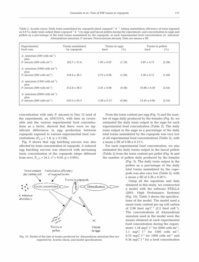
Using all the equations and dataobtained in this study, we constructeda model with the software STELLA(2001: High Perfomance Systems)(Fig. 10). Table 3 shows the specifica-tions of the model. The model used amean toxin content per ng cell carbonof 2.96 fmol ng C–1 (2.2 fmol cell–1).The concentrations of Alexandriumminutum used in the model were themeans obtained at each experimentalfood concentration during the experi-ment: 1.54 mgC l–1 for 2000 cells ml–1,1.1 mgC l–1 for 1500 cells ml–1,0.73 mgC l–1 for 1000 cells ml–1 and0.34 mgC l–1 for a food concentration
111
Table 2. Acartia clausi. Daily toxin assimilated by copepods (fmol copepod–1 d –1, taking assimilation efficiency of toxin ingestedas 3.8%), daily toxin output (fmol copepod–1 d –1) in eggs and faecal pellets during the experiment, and concentration in eggs andpellets as a percentage of the total toxins assimilated by the copepods, at each experimental food concentration (A. minutum:
Alexandrium minutum, P. micans: Prorocentrum micans). Data are means ± SE
Experimental Toxins assimilated Toxins in eggs Toxins in pelletsfood conc. by copepods fmol (%) fmol (%)
A. minutum (500 cells ml–1)plus
P. micans (500 cells ml–1) 162.7 ± 31.6 1.93 ± 0.07 (1.19) 3.87 ± 0.17 (2.38)
A. minutum (1000 cells ml–1)plus
P. micans (500 cells ml–1) 218.4 ± 26.1 2.75 ± 0.06 (1.26) 3.26 ± 0.11 (1.49)
A. minutum (1500 cells ml–1)plus
P. micans (500 cells ml–1) 412.8 ± 36.5 2.41 ± 0.06 (0.58) 10.88 ± 0.39 (2.63)
A. minutum (2000 cells ml–1)plus
P. micans (500 cells ml–1) 610.5 ± 95.3 5.38 ± 0.13 (0.88) 15.45 ± 0.96 (2.53)
Fig. 10. Model of the fate of toxins produced by Alexandrium minutum that are ingested by Acartia clausi, and model specifications

Mar Ecol Prog Ser 240: 105–115, 2002
of 500 cells ml–1 of A. minutum. The assimilation effi-ciency of toxins ingested by the copepods that bestreflects the results obtained in this study was 3.8%. Weran 2 different models. Model 1 considered all experi-mental food concentrations (Eq. 1), and Model 2 con-sidered only the 2 lowest food concentrations (500 and1000 cells ml–1) of A. minutum (Eq. 2).
Fig. 11 shows that the toxin concentration observedin the copepods was similar to the values predicted bythe model (F1,14 = 57.3, r2 = 0.81, p < 0.001, slope = 0.96and intercept = 24.6). Fig. 12 shows the evolution of thecopepod toxin concentration over time predicted byModel 1 for the highest (2000 cells ml–1) and lowest(500 cells ml–1) food concentrations of Alexandriumminutum used in this study, and by Model 2 for thefood concentration of 500 cells ml–1 of A. minutum.
DISCUSSION
This study has shown that Acartia clausi fed onAlexandrium minutum, but that feeding pressurediminished over time. Reduced feeding upon toxicphytoplankton could be due to either behaviouralrejection (Uye & Takamatsu 1990, Teegarden 1999) orphysiological incapacitation (Ives 1985, 1987, Huntleyet al. 1986). Physiological incapacitation, as a reasonfor the lowered copepod grazing rates on toxic dinofla-gellates, might be due to the interference of ingestedtoxins with feeding processes (Ives 1985). However, inour study, feeding pressure on the non-toxic dinofla-gellate Prorocentrum micans did not diminish over theexperiment, which suggests that ingestion of toxinsdid not affect the feeding capacity of the copepods. In
112
Table 3. Specifications of the model
Integration method Euler’s methodUnit time daysdt 1 dToxin content carbon cell–1 2.96 fmol ng C–1
Assimilation efficiency of toxins ingested 3.8%Alexandrium minutum abundance mg cell C l–1
Toxins assimilated (fmol copepod–1 d–1)Model 1 Toxin content carbon cell–1 × 1000 × Eq. (1) × Assimilation efficiency of toxins ingestedModel 2 Toxin content carbon cell–1 × 1000 × Eq. (2) × Assimilation efficiency of toxins ingestedToxins in copepods fmol copepod–1
Toxins in faecal pellets 2.26% of toxins assimilatedToxins in eggs 0.98% of toxins assimilatedDetoxification Eq. (3) (d–1)
Fig. 11. Acartia clausi. Relationship between observed andpredicted values obtained from the model of the toxin con-
centration of copepods
Fig. 12. Acartia clausi. Toxin concentration of copepods pre-dicted by Model 1 at food concentration of 500 cells ml–1
(dashed line) and 2000 cells ml–1 (continuous line), and forModel 2 at a food concentration of 500 cells ml–1 (dot-dashed
line) of Alexandrium minutum

Guisande et al.: Fate of PSP toxins in copepods
fact, a study carried out with the same toxic strain of A.minutum, the non-toxic dinoflagellate P. micans andthe copepod Acartia clausi showed that copepods fedmainly on Alexandrium minutum when the toxin con-tent per cell was low, but as it increased, feedingpressure increased on P. micans and decreasedon A. minutum (Guisande et al. 2002). Therefore,although it is not possible to reject the physiologicalincapacitation hypothesis, our findings support theidea that reduced grazing on toxic dinoflagellates isdue to behavioural rejection of toxic phytoplanktonspecies due to either chemosensory detection of nox-ious chemical cues or learned recognition by physi-cally handling the cells. Moreover, our results supportthe idea that behavioural rejection arises throughtrial-and-error consumption of harmful phytoplanktonthat enables grazers to distinguish those species thatshould be avoided (Uye & Takamatsu 1990), since atthe beginning of the experiment, cells of A. minutumwere ingested by the copepods.
There were differences in the toxin profiles betweenthe toxins accumulated in the copepods and the cellu-lar toxins of Alexandrium minutum (Fig. 5). The ratioGTX4+GTX1 to GTX3+GTX2 was 31.8 in A. minutumand 5.8 in the copepods. This lower GTX4+GTX1/GTX3+GTX2 ratio for toxins accumulated in copepodsthan for the toxic dinoflagellates used as food has beenobserved in other copepod species (Boyer et al. 1985),and can be due to either differences in assimilationefficiencies and/or rates of detoxification. There was nosignificant difference in the percent molar compositionof the isomers GTX4/GTX1 and GTX3/GTX2 betweencopepods on Days 8 and 12 (4 d of detoxification), indi-cating that the detoxification rate was the same forboth isomers. Therefore, the difference in toxin ratiosbetween the copepods and A. minutum seems to bedue to differences in assimilation efficiencies.
The detoxification pattern observed in this study, aninverse exponential function, fits with the usual de-toxification pattern observed in bivalves (Blanco et al.1997, Lassus et al. 2000). There is no information avail-able about detoxification rates in copepods that can becompared with our results. The detoxification rate ofPSP toxins in Acartia clausi (0.586 d–1) is higher thanthe highest detoxification rate for PSP toxins observedin the mussel Mytilus galloprovincialis (0.27 d–1, Blancoet al. 1997). The detoxification rate in the mussel M.galloprovincialis is affected by environmental vari-ables such as salinity and temperature (Blanco et al.1997). Therefore, it is possible that the detoxificationkinetic of A. clausi can also change in response to pre-vailing environmental conditions.
Despite copepod toxin detoxification, the low toxinretention efficiency of the toxins ingested (3.8%) and thefact that an increasingly lower amount of toxins were
daily ingested by the copepods during the course of theexperiment, copepods did accumulate PSP toxinsthrough dietary incorporation (Fig. 4). Such toxinaccumulation has been reported before in copepodspecies (White 1981, Boyer et al. 1985, Turrif et al. 1995,Teegarden & Cembella 1996, Frangópulos et al. 2000,Guisande et al. 2002). If we assume a mean 4.5 µg dry wtper adult Acartia clausi (Guerrero & Rodríguez 1997), thelevels of PSP toxicity in A. clausi on Day 8 of theexperiment were 12 537 and 2398 µg STXeq 100 g–1 drywt for concentrations of 2000 and 500 cells ml–1 Alexan-drium minutum, respectively. This clearly exceeds theacceptable regulatory limit of toxins in shellfish forhuman consumption (80 µg STXeq 100 g–1 soft tissue). InAcartia tonsa and Eurytemora herdmani feeding ontoxic Alexandrium spp., Teegarden & Cembella (1996)also observed low retention efficiencies of ingestedtoxins but high levels of PSP toxins accumulated in thecopepods in 12 h grazing trials of only 12 h.
Faecal pellets produced by copepods represent animportant input of material to the benthic communityand could be an alternative mechanism of toxintransfer to bottom dwellers via sedimentation. Copro-phagous organisms in the pelagic food web can alsoeat pellets and, hence, pellets produced by copepodscould make toxins available to other organisms that donot feed on toxic dinoflagellates. As far we know, ourstudy is the first report of toxins in pellets of copepods.However, the toxins in the faecal pellets representedonly a small percentage (2.26%) of the daily toxinsassimilated by the copepods, and ranged between 0.4and 2.3% of the total toxins accumulated in the cope-pod tissues. Daily detoxification in Acartia clausiaccounted for 44.3% of the toxin concentration in thecopepod tissues, and ranged between 42.9 and 237%of the daily toxins assimilated by the copepods. Thismeans that the toxins were either transformed andexcreted in the pellets as other compounds and/oreliminated as dissolved form. Hasegawa et al. (2001)showed that marine copepods release a significant partof the particle organic nitrogen ingested in the diet asdissolved nitrogen (up to 75%). As PSP toxins arenitrogenous compounds (approximately 33% of PSPtoxin weight is N), toxins could be eliminated by cope-pods in a dissolved form. In our study, a higher amountof dissolved toxins was detected in the beakers withcopepods that in those without copepods (C. Guisandepers. obs.). Gonyautoxins are intracellular compounds(Anderson & Cheng 1988), and hence toxins producedby Alexandrium minutum cannot inhibit the growth ofpotential competitors. Excretion of toxins into thewater by copepods could negatively affect non-toxicalgae and would have important effects on interspe-cific competition among species constituting the phyto-plankton community.
113

Mar Ecol Prog Ser 240: 105–115, 2002
Of the daily toxins assimilated by the copepods, asmall percentage (0.98 %) was redirected to the eggs,and hence the amount of toxins in the eggs increasedwith increasing toxin ingestion by the copepods(Fig. 7). This small amount of toxins in the eggs doesnot affect the fate of the toxins in the copepods, butdoes have an important effect on the copepods’ lifecycle, since a reduced egg hatching success was ob-served with increasing toxin content of the copepods(Fig. 9). This is probably due to the negative effect oftoxins accumulated in the eggs on egg hatching suc-cess (Frangópulos et al. 2000).
Our results explain why some studies have shownthat copepod fecundity is negatively affected by inges-tion of toxic dinoflagellates (Gill & Harris 1987, Dutz1998), whereas other studies have found no influenceof toxic dinoflagellates on egg production (Frangó-pulos et al. 2000). Copepods can ingest and accumu-late toxins up to a threshold without any negativeeffect on fecundity and, hence, egg production in-creases with increasing food concentration of the toxicalgae. However, above this threshold a larger amountof food is necessary to achieve the same egg produc-tion rate. It seems that the effect of toxins on copepodfecundity is a ‘yo-yo’ process, because each time thetoxin concentration of the copepods rises above a spe-cific threshold, the ratio egg production/food concen-tration decreases (Fig. 8). This reduced egg produc-tion with increased toxin concentration of the tissuescould be due to physiological incapacitation and/orenhanced energy expenditure of females to cope withthe ingested toxins (Dutz 1998).
Our model predicted that after an initial toxin accu-mulation in the copepod tissues, copepods eliminate allthe toxins by toxin detoxification and by reduced feed-ing pressure on toxic cells (Fig. 12). The model as-sumes that after several days the rejection efficiency is100%. However, although it is possible that copepodspreviously exposed to unpalatable food items learnto discriminate against harmful cells, such rejectionwould not be 100% efficient. Therefore (although atlow rates), copepods would continue consuming toxicdinoflagellates and hence ingesting toxins. Moreover,if chemical deterrence is concentration-dependent (Tee-garden et al. 2001), at low concentrations of toxic algaeand/or because copepods are exposed to a plethora ofdifferent food items in their habitat, then low ratesof encounter and ingestion could be insufficient to trig-ger a selective feeding response against toxic algae;hence, toxic dinoflagellates could be consumed at lowrates by the copepods. In fact, we did not find sig-nificant differences between ingestion rates over theexperiment at the 2 lower food concentrations (500 and1000 cells of Alexandrium minutum ml–1), indicatingthat there was no feeding selection against toxic
dinoflagellates at low A. minutum abundance. Thiscorroborates the hypothesis that toxic dinoflagellatesinduce grazer avoidance only when their abundanceresults in high encounter frequency with the grazers,enabling the latter to learn to distinguish toxic dinofla-gellates (Teegarden et al. 2001). Therefore, if rejectionis not 100% efficient and/or chemical deterrence isconcentration-dependent, copepods would continuefeeding on toxic algae and would not excrete all thetoxins.
Acknowledgements. This research was supported by theFATE project EVK3-CT2000-00055, a grant from the ChileanGovernment to M.F., an FPU grant to I.M., a grant from Fun-dación Provigo to I.R., and an AECI grant to A.R.V.
LITERATURE CITED
Anderson DM, Cheng PTO (1988) Intracellular localizationof saxitoxins in the dinoflagellate Gonyaulax tamarensis.J Phycol 24:12–22
Anderson DM, Kullis DM, Sullivan JJ, Hall S, Lee C (1990)Dynamics and physiology of saxitoxin production by thedinoflagellates Alexandrium spp. Mar Biol 104:511–524
Blanco J, Moroño A, Franco J, Reyero MI (1997) PSP detoxi-fication kinetics in the mussel Mytilus galloprovincialis.One- and two-compartment models and the effect of someenvironmental variables. Mar Ecol Prog Ser 158:165–175
Boyer GL, Sullivan JJ, Leblanc M, Anderson RJ (1985) Theassimilation of PSP toxins by the copepod Tigriopus cali-fornicus from dietary Protogonyaulax catenella. In: Ander-son DM, White AW, Baden DG (eds) Toxic dinoflagellates.Elsevier, New York, p 407–412
Boyer GL, Sullivan JJ, Andersen RJ, Harrison PJ, Taylor FJR(1987) Effects of nutrient limitation on toxic productionand composition in the marine dinoflagellate Proto-gonyaulax tamarensis. Mar Biol 96:123–128
Cannon JA (1996) Competition between the dinoflagellatesAlexandrium minutum and Prorocentrum micans in thePort River, South Australia. In: Yasumoto T, Oshima Y,Fukuyo Y (eds) Harmful algae. Proc 7th Int Conf ToxicPhytoplankton. Intergovernmental Oceanographic Com-mission (IOC), UNESCO, Rome, p 381–384
Chang FH, Anderson DM, Kulis DM, Till DG (1997) Toxinproduction of Alexandrium minutum (Dinophyceae) fromthe Bay of Plenty, New Zealand. Toxicon 35:393–409
Donaghay PL, Small LF (1979) Food selection capabilities ofthe estuarine copepod Acartia clausi. Mar Biol 52:137–146
Dutz J (1998) Repression of fecundity in the neritic copepodAcartia clausi exposed to the toxic dinoflagellate Alexan-drium lusitanicum: relationship between feeding and eggproduction. Mar Ecol Prog Ser 175:97–107
Franco JM, Fernández P (1993) Separation of PSP toxins byreversed phase high performance liquid chromatography,with postcolumn reaction and fluorimetric detection. Chro-matographia 35:613–620
Franco JM, Fernández P, Reguera B (1994) Toxin profiles ofnatural populations and cultures of Alexandrium minutumHalim. J Appl Phycol 6:275–279
Frangópulos M, Guisande C, Maneiro I, Riveiro I, Franco JM(2000) Short-term and long-term effects of the toxic dino-flagellate Alexandrium minutum on the copepod Acartiaclausi. Mar Ecol Prog Ser 203:161–169
114
Guisande et al.: Fate of PSP toxins in copepods
Frost BW (1972) Effects of size and concentration of food par-ticles on the feeding behaviour of the marine planktoniccopepod Calanus pacificus. Limnol Oceanogr 17:805–815
Gill CW, Harris RP (1987) Behavioural responses of the cope-pods Calanus helgolandicus and Temora longicornis todinoflagellate diets. J Mar Biol Assoc UK 67:785–801
Guerrero F, Rodríguez V (1997) Estimates of secondary pro-duction in a co-existent group of Acartia species (Cope-poda, Calanoida). Crustaceana 70:584–593
Guisande C, Frangópulos M, Maneiro I, Vergara AR, Riveiro I(2002) Ecological advantages of toxin production by thedinoflagellate Alexandrium minutum under phosphoruslimitation. Mar Ecol Prog Ser 225:169–176
Hasegawa T, Koike I, Mukai H (2001) Fate of food nitrogen inmarine copepods. Mar Ecol Prog Ser 210:167–174
Huntley ME, Sykes P, Rohan S, Marin V (1986) Chemically-mediated rejection of dinoflagellate prey by the copepodsCalanus pacificus and Paracalanus parvus: mechanism, oc-currence and significance. Mar Ecol Prog Ser 28:105–120
Ives JD (1985) The relationship between Gonyaulax tamaren-sis cell toxin levels and copepod ingestion rates. In: Ander-son DM, White AW, Baden DG (eds) Toxic dinoflagellates.Elsevier, New York, p 413–418
Ives JD (1987) Possible mechanisms underlying copepodgrazing responses to levels of toxicity in red tide dinofla-gellates. J Exp Mar Biol Ecol 112:131–145
Johansson N, Granéli E (1999) The influence of different N:Psupply ratios on cell density, chemical composition andtoxicity of Prymnesium parvum (Haptophyta) in semi-continuous cultures. J Exp Mar Biol Ecol 139:243–258
Lassus P, Bardouil M, Massselin P, Naviner M, Truquet P(2000) Comparative efficiencies of different non-toxicmicroalgal diets in detoxification of PSP-contaminatedoysters (Crassostrea gigas Thunberg). J Nat Toxins 9:1–12
Oshima Y (1995) Post-column derivatization HPLC methodsfor paralytic shellfish poisons. In: Hallegraeff GM, Ander-son DM, Cembella AD (eds) Manual on harmful marine
microalgae. IOC Manual and guide no. 33. Intergovern-mental Oceanographic Commission (IOC), UNESCO, Paris,p 81–94
Oshima Y, Sugino K, Yasumoto T (1989) Latest advances inHPLC analysis of paralytic shellfish toxins. In: Natori S,Hashimoto K, Ueno Y (eds) Mycotoxins and phycotoxins.Elsevier, Amsterdam, p 319–326
Riegman R, de Boer M, de Senerpont Domis L (1996) Growthof harmful marine algae in multispecies cultures. J Plank-ton Res 18:1851–1866
Smayda TJ (1997) Harmful algal blooms: their ecophysiologyand general relevance to phytoplankton blooms in the sea.Limnol Oceanogr 42:1137–1153
Teegarden GJ (1999) Copepod grazing selection and particlediscrimination on the basis of PSP toxic content. Mar EcolProg Ser 181:163–176
Teegarden GJ, Cembella AD (1996) Grazing of toxic dinofla-gellates, Alexandrium spp., by adult copepods of coastalMaine: implications for the fate of paralytic shellfish toxinsin marine food webs. J Exp Mar Biol Ecol 196:145–176
Teegarden GJ, Campbell RG, Durbin EG (2001) Zooplanktonfeeding behavior and particle selection in natural plank-ton assemblages containing toxic Alexandrium spp. MarEcol Prog Ser 218:213–226
Turner JT, Tester PA (1997) Toxic marine phytoplankton,zooplankton grazers, and pelagic food webs. LimnolOceanogr 42:1203–1214
Turriff N, Runge JA, Cembella AD (1995) Toxin accumulationand feeding behaviour of the planktonic copepod Calanusfinmarchicus exposed to the red-tide dinoflagellate Alex-andrium excavatum. Mar Biol 123:55–64
Uye S, Takamatsu K (1990) Feeding interactions betweenplanktonic copepods and red-tide flagellates from Japan-ese coastal waters. Mar Ecol Prog Ser 59:97–107
White AW (1981) Marine zooplankton can accumulate andretain dinoflagellate toxins and cause fish kills. LimnolOceanogr 26:103–109
115
Editorial responsibility: Otto Kinne (Editor), Oldendorf/Luhe, Germany
Submitted: January 16, 2002; Accepted: April 25, 2002Proofs received from author(s): August 22, 2002