Parallel systems of error processing in the brain
13
Parallel systems of error processing in the brain Juliana Yordanova, a,b, * Michael Falkenstein, a Joachim Hohnsbein, a and Vasil Kolev a,b a Institute of Occupational Physiology, D-44139 Dortmund, Germany b Institute of Physiology, Bulgarian Academy of Sciences, BG-1113 Sofia, Bulgaria Received 19 August 2003; revised 20 January 2004; accepted 24 January 2004 Available online 14 April 2004 Major neurophysiological principles of performance monitoring are not precisely known. It is a current debate in cognitive neuroscience if an error-detection neural system is involved in behavioral control and adaptation. Such a system should generate error-specific signals, but their existence is questioned by observations that correct and incorrect reactions may elicit similar neuroelectric potentials. A new approach based on a time – frequency decomposition of event-related brain potentials was applied to extract covert sub-components from the classical error-related negativity (Ne) and correct-response-related negativity (Nc) in humans. A unique error-specific sub-component from the delta (1.5– 3.5 Hz) frequency band was revealed only for Ne, which was associated with error detection at the level of overall performance monitoring. A sub-component from the theta frequency band (4 – 8 Hz) was associated with motor response execution, but this sub-component also differentiated error from correct reactions indicating error detection at the level of movement monitoring. It is demonstrated that error-specific signals do exist in the brain. More importantly, error detection may occur in multiple functional systems operating in parallel at different levels of behavioral control. D 2004 Elsevier Inc. All rights reserved. Keywords: Error-related negativity; Time– frequency analysis; Performance monitoring; Wavelet transform; Executive control; Parallel systems; High- resolution EEG Introduction To achieve a goal, humans have to be able to compare the consequences of their behavior with the intended outcome of this behavior. When a mismatch occurs between planned and actual results of performance, that is, when an error is made, corrective and adaptive actions are needed. Therefore, to enable error detec- tion and correction, ongoing performance requires a continuous monitoring in the brain. Recently, a specific neuroelectric signal recorded at the scalp has become an important tool for studying behavioural control and adaptation in humans (Ullsperger and Falkenstein, in press). This is a negative-going potential with midline fronto-central maximum, called error negativity (Ne, Falkenstein et al., 1990, 1991) or error- related negativity (ERN, Gehring et al., 1993). The Ne phenom- enon can be linked to the functioning of a performance monitoring system in the brain because: (1) it emerges after incorrect reactions leading to strategic behavioural re-adjustment, and (2) the sources of Ne generation are thought to be localized in the medial frontal brain regions (i.e., the anterior cingulate and the supplementary motor area, Carter et al., 1998; Dehaene et al., 1994; Ullsperger and von Cramon, 2001), known to play a major role in behaviour and motor regulation (Posner and DiGirolamo, 1998). However, current models for performance monitoring as related to Ne differ with respect to the type and specificity of underlying processes. According to the error-detection model, the Ne reflects the activity of a genuine system in the medial frontal brain which generates an error signal if a mismatch emerges between neural representations of intended and actual movements (Bernstein et al., 1995; Coles et al., 2001; Falkenstein et al., 1991, 2000; Holroyd et al., 1998). The response-conflict model posits that the Ne is not specific for errors and is generated during response competition when two or more response programs are activated simultaneously (Botvinick et al., 2001; Carter et al., 1998). The Ne has been further conceptualized as related to the subjective evaluation of action plans by the medial frontal lobe (Luu et al., 2000), to a general system estimating the motivational value of various events, among them errors and response conflict (Bush et al., 2000), or to negative reinforcement learning mediated by the dopamine neuro- transmitter system in the brain (Holroyd and Coles, 2002). Thus, knowledge about the precise functional significance of Ne is inconclusive. Yet, this knowledge is crucial with respect to major principles of performance and action control. A new perspective to understand these principles is provided by the observation that correct motor responses can also elicit a fronto-central negativity (correct-response-related negativity, CRN, Falkenstein et al., 2000; Ford, 1999; Gehring and Knight, 2000; Luu et al., 2000; Vidal et al., 2000), termed here Nc. It is at present a matter of discussion whether the Ne and Nc have the same functional origin, with errors producing a larger negativity than correct responses (Vidal et al., 2000). In this view, the frontal negativity is not specific for errors and may be associated with a process activated after both correct and incorrect responses. Alter- natively, Nc has been suggested to appear on correct trials only when subjects believe the response is an error (Scheffers and 1053-8119/$ - see front matter D 2004 Elsevier Inc. All rights reserved. doi:10.1016/j.neuroimage.2004.01.040 * Corresponding author. Institute of Physiology, Bulgarian Academy of Sciences, Acad. G. Bonchev str., bl. 23, BG-1113 Sofia, Bulgaria. Fax: +359-2-979-37-49. E-mail address: [email protected] (J. Yordanova). Available online on ScienceDirect (www.sciencedirect.com.) www.elsevier.com/locate/ynimg NeuroImage 22 (2004) 590 – 602
Transcript of Parallel systems of error processing in the brain

www.elsevier.com/locate/ynimg
NeuroImage 22 (2004) 590–602
Parallel systems of error processing in the brain
Juliana Yordanova,a,b,* Michael Falkenstein,a Joachim Hohnsbein,a and Vasil Koleva,b
a Institute of Occupational Physiology, D-44139 Dortmund, Germanyb Institute of Physiology, Bulgarian Academy of Sciences, BG-1113 Sofia, Bulgaria
Received 19 August 2003; revised 20 January 2004; accepted 24 January 2004
Available online 14 April 2004
Major neurophysiological principles of performance monitoring are
not precisely known. It is a current debate in cognitive neuroscience if
an error-detection neural system is involved in behavioral control and
adaptation. Such a system should generate error-specific signals, but
their existence is questioned by observations that correct and incorrect
reactions may elicit similar neuroelectric potentials. A new approach
based on a time– frequency decomposition of event-related brain
potentials was applied to extract covert sub-components from the
classical error-related negativity (Ne) and correct-response-related
negativity (Nc) in humans. A unique error-specific sub-component
from the delta (1.5–3.5 Hz) frequency band was revealed only for Ne,
which was associated with error detection at the level of overall
performance monitoring. A sub-component from the theta frequency
band (4–8 Hz) was associated with motor response execution, but this
sub-component also differentiated error from correct reactions
indicating error detection at the level of movement monitoring. It is
demonstrated that error-specific signals do exist in the brain. More
importantly, error detection may occur in multiple functional systems
operating in parallel at different levels of behavioral control.
D 2004 Elsevier Inc. All rights reserved.
Keywords: Error-related negativity; Time– frequency analysis; Performance
monitoring; Wavelet transform; Executive control; Parallel systems; High-
resolution EEG
Introduction
To achieve a goal, humans have to be able to compare the
consequences of their behavior with the intended outcome of this
behavior. When a mismatch occurs between planned and actual
results of performance, that is, when an error is made, corrective
and adaptive actions are needed. Therefore, to enable error detec-
tion and correction, ongoing performance requires a continuous
monitoring in the brain.
Recently, a specific neuroelectric signal recorded at the scalp
has become an important tool for studying behavioural control and
adaptation in humans (Ullsperger and Falkenstein, in press). This is
1053-8119/$ - see front matter D 2004 Elsevier Inc. All rights reserved.
doi:10.1016/j.neuroimage.2004.01.040
* Corresponding author. Institute of Physiology, Bulgarian Academy of
Sciences, Acad. G. Bonchev str., bl. 23, BG-1113 Sofia, Bulgaria. Fax:
+359-2-979-37-49.
E-mail address: [email protected] (J. Yordanova).
Available online on ScienceDirect (www.sciencedirect.com.)
a negative-going potential with midline fronto-central maximum,
called error negativity (Ne, Falkenstein et al., 1990, 1991) or error-
related negativity (ERN, Gehring et al., 1993). The Ne phenom-
enon can be linked to the functioning of a performance monitoring
system in the brain because: (1) it emerges after incorrect reactions
leading to strategic behavioural re-adjustment, and (2) the sources
of Ne generation are thought to be localized in the medial frontal
brain regions (i.e., the anterior cingulate and the supplementary
motor area, Carter et al., 1998; Dehaene et al., 1994; Ullsperger
and von Cramon, 2001), known to play a major role in behaviour
and motor regulation (Posner and DiGirolamo, 1998).
However, current models for performance monitoring as related
to Ne differ with respect to the type and specificity of underlying
processes. According to the error-detection model, the Ne reflects
the activity of a genuine system in the medial frontal brain which
generates an error signal if a mismatch emerges between neural
representations of intended and actual movements (Bernstein et al.,
1995; Coles et al., 2001; Falkenstein et al., 1991, 2000; Holroyd et
al., 1998). The response-conflict model posits that the Ne is not
specific for errors and is generated during response competition
when two or more response programs are activated simultaneously
(Botvinick et al., 2001; Carter et al., 1998). The Ne has been
further conceptualized as related to the subjective evaluation of
action plans by the medial frontal lobe (Luu et al., 2000), to a
general system estimating the motivational value of various events,
among them errors and response conflict (Bush et al., 2000), or to
negative reinforcement learning mediated by the dopamine neuro-
transmitter system in the brain (Holroyd and Coles, 2002). Thus,
knowledge about the precise functional significance of Ne is
inconclusive. Yet, this knowledge is crucial with respect to major
principles of performance and action control.
A new perspective to understand these principles is provided by
the observation that correct motor responses can also elicit a
fronto-central negativity (correct-response-related negativity,
CRN, Falkenstein et al., 2000; Ford, 1999; Gehring and Knight,
2000; Luu et al., 2000; Vidal et al., 2000), termed here Nc. It is at
present a matter of discussion whether the Ne and Nc have the
same functional origin, with errors producing a larger negativity
than correct responses (Vidal et al., 2000). In this view, the frontal
negativity is not specific for errors and may be associated with a
process activated after both correct and incorrect responses. Alter-
natively, Nc has been suggested to appear on correct trials only
when subjects believe the response is an error (Scheffers and

J. Yordanova et al. / NeuroImage 22 (2004) 590–602 591
Coles, 2000). In this context, the Nc is generated by the subjective
experience of incorrect reactions and is virtually an Ne, which
would support the error-detection model. An integrative relation-
ship between Ne and Nc is also plausible according to which the
Ne may comprise of an Nc and an additional error-specific
activation superimposed on Nc (Falkenstein et al., 2000).
Concerning functional principles of performance regulation as
suggested by different models (Carter et al., 1998; Falkenstein et
al., 2000), the central question is whether an error-specific signal
exists in the brain. Hence, the relationship between Ne and Nc is of
special theoretical importance. In previous research, this issue has
been addressed by manipulating error rate and type in terms of
detectability, motivational value, and associations with conflicting
response tendencies. Here, a new conceptual and methodological
framework is proposed to clarify the relationship between Nc and
Ne. The approach is based on the following.
An electroencephalographic (EEG) signal can be described
precisely in three dimensions: (1) amplitude, (2) time, and (3)
frequency, although phase relationships should be also quantified
for a complete signal description (e.g., Basar, 1980, 1998; Freeman
and Rogers, 2002; Regan, 1989; Ruchkin, 1988). Typically,
response- and error-related brain potentials are analyzed in the
time domain. These analyses have shown that Nc and Ne do
manifest similarities in polarity, timing, and topographical distri-
bution (Vidal et al., 2000), implying common origins of correct and
incorrect response detection and evaluation. A classical time-
domain representation of ERPs reveals the timing of underlying
neural events. However, the frequency characteristics of those
events remain uncovered and no information can be obtained
about rhythmic or oscillatory events from various frequency bands
present in the signal (Basar, 1998). The inability of the time-
domain ERP representation to extract frequency characteristics of
the signal seems to be a serious disadvantage because EEG
activities from several frequency ranges (theta, alpha, beta, and
gamma) have been associated consistently with motor performance
(e.g., Babiloni et al., 2000; Feige et al., 2000; Pfurtscheller et al.,
2000; Urbano et al., 1998; Yordanova et al., 2000). These obser-
vations point to the possibility that multiple frequency-specific
components are generated during correct and error response
production. On the other hand, analysis only in the frequency
domain does not allow to know whether and how frequency
components are time-locked to correct or incorrect response
production. To enable an adequate comparison between Ne and
Nc, one task of this study was to characterize completely the Ne
and Nc signals by analysis in the time, frequency, and time–
frequency domain. Secondly, the major question of whether error-
specific neuroelectric signals are generated in the brain was
addressed. If multiple frequency components co-exist (Basar,
1980, 1998; Kolev et al., 1997; Yordanova et al., 2000) during
correct and error response production, they remain covert in time-
domain waveforms of Nc and Ne because of their full or partial
overlapping. Importantly, such covert components, if extracted
from a complex heterogeneous structure of Ne, may reflect error-
specific processes that are not activated after correct responses.
In the present study, to elucidate the relationship between the
Nc and Ne and seek for error-specific neuroelectric signals in the
brain, response-related potentials (RRPs) to correct and incorrect
reactions were elicited and analyzed in the time, frequency, and
time–frequency domains. Frequency analyses were carried out
with fast Fourier transform (FFT), and time–frequency analyses
were performed by means of wavelet transform (WT, Mallat, 1999;
Samar et al., 1999). For quantification and reliability of topography
analysis, spatial characteristics of averaged RRPs were enhanced
by calculating current source density (CSD, Babiloni et al., 1996;
Perrin et al., 1989).
Methods
Subjects
A total of 14 subjects from 19 to 27 years of age was studied
(mean, 23.5, SD = F2.8). All of them were healthy (no medica-
tion), with normal or corrected to normal vision. None of the
subjects reported about neurologic, psychiatric, chronic somatic, or
hearing problems in the past. Written informed consent was
obtained from each participant and the study was approved by
the local ethical committee. Due to low error rate and insufficient
number of error trials, data from four subjects were excluded from
analyses.
Task
A four-choice reaction task (CRT) was employed. Four stimu-
lus types represented by the letters A, E, I, and O were delivered
randomly with equal probability of 25% in separate experimental
blocks. A total of 200 stimuli was presented in each block, with n =
50 for each stimulus type. The letters A, E, I, and O had to be
responded with the left middle, left index, right index, and right
middle fingers, respectively. They were designated as four stimu-
lus-response (SR) types (SR1, SR2, SR3, and SR4). Response
force was measured by sensometric tensors while subjects pro-
duced a flexion with each of the four fingers.
Subjects performed the CRT in two modalities—auditory and
visual. Auditory stimuli (duration 300 ms, intensity 67 dB SPL)
were delivered via headphones binaurally, with similar envelopes of
the sound pressure waves formed for all stimuli. Visual stimuli with
the same duration were shown in the middle of a monitor (visual
angles 1j horizontal/1.5j vertical, intensity 50 cd/m2) placed 1.5 m
in front of the subject’s face. Interstimulus intervals varied random-
ly between 1440 and 2160 ms (mean 1800 ms). If the response was
slower than 700 ms after stimulus onset, a feedback tone was
delivered at 700 ms after stimulus onset. This tone had to be
avoided by the subjects by responding fast enough. Sequences of
auditory and visual series were counterbalanced across subjects.
Data recording
Data were recorded on three consecutive experimental days. In
the first recording day, data from one auditory and one visual CRT
blocks were collected. In the second and third experimental days,
data from four auditory and four visual blocks were collected. A
total of nine auditory and nine visual CRT blocks was performed
by each subject. Counterbalancing of experimental blocks across
subjects was done as best as possible. Data from all nine sessions
in each modality were used for analysis to enable the collection of
a sufficient number of error trials. An error was defined as a button
press with a wrong finger. To control for effects of the number of
single trials on average amplitudes, correct responses were ran-
domly selected for each subject so that their number was equal to
the individual number of error trials. Because error rate depended
on the SR type, being significantly higher for index-finger

J. Yordanova et al. / NeuroImage 22 (2004) 590–602592
responses (SR2 and SR3) than for middle-finger responses (SR1
and SR4), only SR2 and SR3 types were further analyzed. The
mean number of trials used for averaging was 22 (SE = F2). EEG
was recorded from 64 channels with Cz as reference, with
frequency limits of 0.1–70 Hz, and a sampling rate of 250 Hz.
Data processing
EEG traces were visually inspected for gross EOG and EMG
artifacts. Contaminated trials were discarded along with records
exceeding F50 AV. Next, slight horizontal and vertical eye move-
ments preserved in the accepted trials were corrected by means of a
linear regression method for EOG correction (Gratton et al., 1983).
Error and correct EEG trials were averaged separately for each
modality.
Both the Ne and Nc were clearly visible after response-related
averaging, indicating the presence of phase-locked components in
single sweeps. Therefore, analyses were performed for the aver-
aged response-triggered potentials to extract the time–frequency
components that were most stable and locked to the response.
Response-related potential
Response-related averaging was triggered when a threshold
level of 5 N in the mechanogram was reached. In this way,
incomplete responses were disregarded. This trigger for averaging
appears about 150–200 ms later than the actual EMG displacement
indexing the actual response initiation.
From a methodological point of view, the distortion of RRPs
caused by preceding or overlapping stimulus-related components is
to be accounted for. In a recent study, it was demonstrated by
means of adjusted filtering at the level of single sweeps that such
effects are substantial for the later post-response time epochs and
that they are relatively small for time windows preceding or
coinciding with response preparation and execution (Braun et al.,
2002). On the basis of these results, RRPs were not corrected for
stimulus-related components.
Current source density
To achieve a reference-free evaluation, all data analyses were
performed after calculation of CSD of the signals (Babiloni et al.,
1996; Nunez et al., 1997; Perrin et al., 1989; Vidal et al., 2000).
The CSD transform replaces the potential at each electrode with the
CSD, thus eliminating the reference potential. The algorithm
applies the spherical Laplace operator to the potential distribution
on the surface of the head. Because the potential distribution is
only known at the electrodes used, the procedure of spherical
spline interpolation is employed to calculate the continuous poten-
tial distribution. The exact mathematical procedure is explained in
detail in Perrin et al. (1989). When applied with dense electrode
arrays (48–256 electrodes, 64 in the present study), this procedure
provides excellent estimates of the bioelectric activity of the
cortical surface (Nunez and Pilgreen, 1991).
Time-domain analysis
The time-domain analysis epoch had a length of 1600 ms,
with the moment of response production being located in the
center of this epoch. Nc and Ne were identified as the most
negative peak of RRPs at FCz within 100 ms after correct and
error responses, respectively. Nc/Ne peak amplitude and latency
were measured in the averaged RRPs against a baseline of 800–
600 ms before the button press. This baseline was chosen to
avoid the cancellation of response-related activity occurring
shortly after stimulus delivery.
Frequency-domain analysis
Frequency-domain analysis was performed by analyzing aver-
age RRPs with FFT. To focus the computations to the period of
response execution, FFT was calculated for a 1600-ms epoch
centered at the time point of button pressing. Also, a Hanning
window was applied so that activity beyond 250 ms before and
after the response was strongly reduced.
Time–frequency decomposition
To achieve a reliable analysis of slow frequency components in
the time–frequency domain and avoid possible edge effects, 8192-
ms-long epochs were used to obtain averaged RRPs. The moment
of response execution was in the center of the analysis epoch.
To represent RRPs in the time–frequency domain, RRPs were
analyzed by means of a continuous wavelet transform (CWT,
Mallat, 1999; Samar et al., 1999). Time–frequency representations
were calculated by Morlet’s wavelets as described previously (e.g.,
Jensen et al., 2002; Tallon-Baudry et al., 1997). The analytical
presentation of Morlet’s wavelet w(t, f ) is:
wðt; f Þ ¼ Aexpð�t2=2r2t Þexpð2ipftÞ;
where t is time, f is frequency, A ¼ ðrt
ffiffiffi
pp
Þ�1=2, rt is the wavelet
duration, and i = �1.
For time–frequency plots, a ratio of f0/rf = 3.8 was used, where
f0 is the central frequency and rf is the width of the Gaussian shape
in the frequency domain. The choice of the ratio f0/rf was orientedto the expected slower phase-locked components present in the
RRPs, which had an effect on the shape of the Morlet wavelet and
decreased its decay. The analysis was performed in the frequency
range 0.1–12 Hz with a central frequency at 0.1 Hz intervals. For
different f0, time and frequency resolutions can be calculated as 2rtand 2rf, respectively (Tallon-Baudry et al., 1997). rt and rf are
related by the equation rt = 1/(2prf). For example, for f0 = 1 Hz,
2rt = 1200 ms and 2rf = 0.53 Hz; for f0 = 3 Hz, 2rt = 400 ms and
2rf = 1.58 Hz; for f0 = 5 Hz, 2rt =240 ms and 2rf = 2.63 Hz.
Relevant time–frequency (TF) components were extracted in
the time domain and analyzed. To focus specifically on Nc/Ne
events, amplitudes and latencies of the negative phases of frequen-
cy-specific TF components were measured within 150 ms before
and after the response onset with the same baseline (800–600 ms
before the response), and subjected to statistical evaluation.
Phase-locking
CSD-transformed single sweeps used to obtain average RRPs
were subjected to CWT analysis and extracted in the delta (1.5–3.5
Hz) and theta (4–8 Hz) frequency ranges. They were further
analyzed to assess the phase-locking (between-sweep synchroni-
zation) of these TF components independently of their amplitude.
For a quantitative evaluation of the phase-locking, a modification
of the single-sweep wave identification (SSWI) method was
applied, with methodological details on the procedure presented
elsewhere (Kolev and Yordanova, 1997; Yordanova and Kolev,
1998a,b). In sum, the method identifies extrema (minima and
maxima) in each single sweep and replaces them by codes of �1
or + 1, respectively, placed on the corresponding latency positions.
Then, a summation of the modified sweeps is performed across
trials, with the sum value at each time point assigned to a

J. Yordanova et al. / NeuroIm
histogram bar. The histogram is thereafter normalized by dividing
the bar values by the number of sweeps included in the analysis.
For statistical assessment of phase-locking, histogram bar values
were squared and the mean of the normalized squared histogram
was measured for a period of 800–600 ms before the response
(reference) and for a period of 0–100 ms after the response
coinciding with Ne/Nc development. Measures of the reference
period were subtracted from the measures of the Ne/Nc period. To
normalize distribution, the obtained values were log10 transformed
and subjected to statistical evaluation.
Fig. 1. Grand average RRPs to correct and error responses at FCz in two modaliti
for Nc and Ne, respectively. Correct and error-related RRPs are presented separatel
the topographic maps of correct and error responses.
Statistical Analysis
Nc and Ne measures were compared at the FCz electrode where
Nc and Ne were maximal. In the time domain, the measured
parameters were Nc/Ne base-to-peak amplitude and peak latency.
In the time–frequency domain, the measured parameters were
peak amplitudes and latencies of three TF components in the
sub-delta, delta, and theta frequency bands (see Results), whose
negative phases coincided with Nc/Ne. The stability of phase
synchronization with the response was evaluated at FCz for the
delta and theta TF components.
age 22 (2004) 590–602 593
es (auditory—upper panel, and visual—lower panel). Topography maps are
y for left-hand and right-hand responses. Note that the scaling is different for

J. Yordanova et al. / NeuroImage 22 (2004) 590–602594
Nc/Ne measures were assessed statistically by means of anal-
ysis of variance with repeated measures (ANOVA). There were
three within-subjects variables: Modality (auditory vs. visual) �Accuracy (correct vs. incorrect) � Response side [SR2 (left) vs.
SR3 (right)]. Behavioural measures (RT and error number) were
subjected to the same analysis. For the sub-delta component (<1
Hz), mean amplitudes were measured for consecutive 100-ms time
windows and subjected to the same analysis with an additional
within-subjects variable time-dynamics (six levels corresponding
to the consecutive time windows).
To assess statistically the topographical distribution, an array
of nine electrodes was selected to cover midline and bilateral pre-
motor, motor, and sensorimotor regions. They were combined to
form two electrode factors Region (pre-central—FC3, FCz, FC4;
central—C3, Cz, C4; and post-central—CP3, CPz, CP4) and
Laterality (midline—FCz, Cz, CPz, contralateral /FC3, C3, CP3
for right-hand responses; or FC4, C4, CP4 for left-hand
responses/, and ipsilateral /FC4, C4, CP4 for right-hand
responses; or FC3, C3, CP3 for left-hand responses/ to the
responding hand). The factors in the overall ANOVA were:
Modality � Accuracy � Response side � Region � Laterality.
Whenever significant interactions of Accuracy with Region,
Laterality, or with Region � Laterality were obtained, data were
normalized (McCarthy and Wood, 1985). The analyses were
repeated with the normalized data to confirm the significance of
interactions. Statistical results from post hoc multiple tests were
corrected by adjusting the alpha level according to the Bonferroni
procedure. In the results, only significant main effects and
interactions will be described.
Fig. 2. Power spectra of RRPs at FCz (left panel) and C4 (right panel) of
correct and error responses produced by left and right hands in two
modalities, auditory (upper panel) and visual (lower panel).
Results
Performance
All subjects performed the task adequately. Error rate was
similar for left- and right-hand responses (mean 7.3% and 8.0%,
respectively). Reaction times (RTs) did not differ significantly
(Accuracy, F(1/9) = 1.45, P = 0.26) between error (mean 473
ms, SE = F15 ms) and correct (mean 461 ms, SE = F10.6 ms)
responses. RTs to errors were similar for the two response sides
(left: mean 475 ms, SE = F16 ms; right: mean 473 ms, SE = F15
ms), whereas RTs to correct responses were longer (F(1/9) = 13.9,
P = 0.005) for the left (mean 475 ms, SE = F12.2 ms) than right
hand (mean 453 ms, SE = F9.9 ms), as also reflected by a
marginally significant Accuracy � Response side interaction
(F(1/9) = 3.9, P = 0.08).
Correct-response and error negativities in the time-domain
Fig. 1 illustrates the CSD waveforms of time-domain RRPs at
FCz and topography maps of fronto-central negativities. RRP
morphology was similar across modalities and responding hands.
It is also shown that: (1) a negative component was observed for
both correct and error RRPs at fronto-central electrodes. (2) This
negative component was basically characterized by a phasic wave.
In many cases, it was superimposed on a slow negative wave, as
seen clearly for auditory left-hand RRPs. (3) The topography maps
illustrate that error-related RRPs had a clear maximum at FCz. A
similar midline fronto-central dominance was found for correct
responses, with an additional tendency for a greater involvement of
contralateral regions. Because of its negative polarity, scalp topog-
raphy, and peak latencies of 44.7 ms (SE = F7.4 ms) for correct
responses and 51.6 ms (SE = F5.8 ms) for incorrect responses, the
negative component in correct and error RRPs was identified as Nc
and Ne, respectively.
Base-to-peak Ne amplitude was substantially larger than Nc
amplitude (Accuracy, F(1/9) = 21.29, P < 0.001), whereas peak
latencies of Ne and Nc did not differ significantly (Accuracy,
F(1/9) = 1.29, P > 0.2). There was a significant main effect of
the response side on latency values, because for both correct
and incorrect responses, the latency of the fronto-central nega-
tivity was longer for left vs. right finger presses (F(1/9) = 7.39,
P < 0.05).
Frequency content of Nc and Ne
Fig. 2 illustrates power spectra of averaged RRPs to auditory
and visual stimuli. At FCz (left panels), correct and error-related
RRPs manifested a most prominent spectral peak at very low (<1
Hz, sub-delta) frequencies. Furthermore, RRPs were characterized
by a spectral peak between 2 and 3 Hz (delta), and a subsequent

J. Yordanova et al. / NeuroImage 22 (2004) 590–602 595
compound component between 3.5 and 8 Hz (theta). However,
delta and theta spectral components were markedly larger for error
than for correct responses.
Fig. 2 (right panels) illustrates RRPs at electrodes overlying
motor cortical areas. Because the patterns of results were similar
for C3 and C4 locations, only RRPs at C4 are depicted. It is
shown that spectral peaks at the cortex contralateral to the
responding hand (C4 for the left hand) were similar for correct
and error-related responses: Spectral peaks occurred at very low
frequency (below 1 Hz), and a prominent spectral peak was seen
in the theta frequency band (3–6 Hz). Notably, no spectral activity
in the theta frequency range was observed for ipsilateral (right-
hand) responses at C4, irrespective of their accuracy (correct or
error). Accordingly, at C3, spectral peaks in the theta frequency
range were observed only for right-hand responses (both correct
and error), but not for left-hand responses (not shown in the
figure).
Multiple time–frequency sub-components of Nc and Ne
Fig. 3 presents time–frequency decomposition plots of aver-
aged RRPs at FCz and illustrates that several distinct TF compo-
nents accompanied response production.
(1) A phase-locked component within the 0.3–1.2 Hz (sub-delta)
band was generated during both correct and error responses. In
the present experimental setup with a mean inter-stimulus
Fig. 3. Time-frequency decomposition plots of RRPs at FCz of correct (upper pane
modalities, auditory (left panel) and visual (right panel). The description of the fi
interval of 1.8 s, an analysis epoch of 8 s can cover at least four
trials. Although the response-related activity of neighboring
trials is not phase-locked in the averaged potentials, there is a
theoretical possibility that the sub-delta components in the
RRPs of interest are affected by very slow components
associated with these neighboring responses. This possibility
can be excluded, because as shown in Fig. 3, the sub-delta TF
component started as early as about 500 ms before and lasted
for at least 500 ms after the response. It was equally well
expressed for correct and incorrect responses. These observa-
tions can explain the occurrence of the slow negative shifts in
the time-domain RRPs (see Performance subsection) and of the
sub-delta spectral peaks in the frequency domain RRPs (see
Correct-response and error negativities in the time-domain
subsection).
(2) After errors, a prominent TF component was seen in the 1.5- to
8-Hz (delta and theta) frequency band. This TF component was
localized within 100 ms after error execution. Fig. 3 also
demonstrates that this component was markedly reduced after
correct responses in comparison to errors, being almost absent
for the delta (1.5–3.5 Hz) frequency range. These observations
provide evidence that the major power of the phasic Ne
component in time-domain RRPs comes from phase-locked
delta and theta EEG activity.
Fig. 4 depicts difference time–frequency plots computed by
subtracting correct from error-related time–frequency plots. (1)
l) and error (lower panel) responses produced by left and right hands in two
gure is in the text.

Fig. 4. Time-frequency decomposition plots of difference RRPs (Error RRP
minus Correct RRP) at FCz produced by left (left panel) and right (right
panel) hands in two modalities (auditory—upper panel, visual—lower
panel).
J. Yordanova et al. / NeuroImage 22 (2004) 590–602596
For each modality and response type, the most conspicuous
difference between correct and incorrect responses was focused
to frequencies between 1.5 and 3.5 Hz. This is fully in line with
observations from the frequency-domain analysis (Fig. 2) reveal-
ing that the major spectral difference between correct and error
RRPs at FCz was in the 2- to 3-Hz activity. (2) This major
correct vs. incorrect distinction was a negative component
localized within 100 ms after the response, which coincided
with Ne time localization.
Error-specific time–frequency components
To verify statistically these observations, multiresolution scales
(frequency-specific components) were extracted from the wavelet-
decomposed RRPs, evaluated quantitatively, and compared be-
tween correct and incorrect responses. Evaluations were done for
the waveforms of each scale (TF component). Three TF compo-
nents were analyzed: (1) Sub-delta, 0.3–1.2 Hz, to quantify the
slow negative shifts accompanying response production; (2) Delta
(1.5–3.5 Hz), to reflect the major frequency difference between
correct and error-related processes; and (3) Theta (4–8 Hz), to
evaluate response-related theta activity appearing after both correct
and error responses. Peak amplitude and latency were measured for
the negative phase of each of these three TF components coincid-
ing with Nc/Ne.
Fig. 5 presents the three response-related TF components and
illustrates graphically the statistical results. After adjusting the
alpha level to the number of tests performed (Bonferroni correction
for three tests), the difference between correct and error responses
could be accepted as significant for P V 0.016. There was a
significant main effect of response accuracy only for the delta TF
component magnitude (delta, F(1/9) = 15.02, P < 0.005; sub-delta,
F(1/9) = 2.04, P = 0.19; theta, F(1/9) = 6.02, P = 0.037)—Fig. 5b.
Also, negative theta phase latency depended on response accuracy
(F(1/9) = 13.28, P < 0.005), because it was significantly longer for
error (mean 50 ms, SE = F5.8 ms) than for correct responses
(mean 14 ms, SE = F6.9 ms).
No main effects of the response side were yielded for the
amplitudes of sub-delta, delta, and theta TF components. How-
ever, for the theta TF component, a significant Accuracy �Response side interaction was found (F(1/9) = 10.49, P <
0.01) resulting from a substantially larger incorrect vs. correct
theta amplitudes for left-hand responses, and a lack of incorrect
vs. correct difference for the right-hand responses (Fig. 5c).
Furthermore, negative theta phase latency significantly differen-
tiated right- from left-hand responses (F(1/9) = 11.45, P < 0.005)
by being longer for left-(mean 42 ms, SE = 4.6 ms) than for
right-hand responses (mean 22 ms, SE = 5.4 ms). Fig. 5d
demonstrates theta components at FCz and C4 and shows that
in addition to the enhancement at FCz, there was a strongly
lateralized distribution of the phase-locked theta TF component
over the hemisphere contralateral to the responding hand. In
accordance with the frequency domain finding (Fig. 2), this
lateralization was observed for both correct and error responses.
Notably, as seen in Fig. 5d, the theta oscillations over the
contralateral motor cortex were phase-shifted relative to midline
frontal–central theta oscillations.
Phase-locking
The difference between correct and error responses was accept-
ed as significant for P V 0.025 after the alpha level was adjusted to
the number of the two tests performed to assess the phase-locking.
There was a significant main effect of response accuracy only for
the delta TF component phase-locking (Accuracy, F(1/9) = 10.29,
P = 0.01) because delta oscillations were strongly synchronized to
errors, in contrast to correct responses. For the theta TF compo-
nent, errors also tended to produce better phase-locked oscillations
(Accuracy, F(1/9) = 3.88, P = 0.08), which appeared to be more
pronounced for the left hand responses (Accuracy � Response
side, F(1/9) = 3.54, P = 0.09), but these differences were not
significant.
Topography effects
To explore further the Ne/Nc distinctions associated with delta
and theta TF components, scalp topography was analyzed statis-
tically for the 1.5–3.5 and 4–8 Hz TF oscillations as carrying the
information delineating response accuracy.
Fig. 6 shows topography maps calculated for the negative
phase of the delta TF component occurring within 100 ms after
the response. It demonstrates that for each modality, the topo-
graphical distribution of this component substantially differed
between correct and incorrect responses. For correct RRPs, the
delta TF component was distributed mainly over the pre-motor
frontal cortex contralateral to the responding hand. In contrast, a

Fig. 5. Grand average time-frequency components of RRPs at FCz extracted from the (a) sub-delta (0.3–1.2 Hz), (b) delta (1.5–3.5 Hz), and (c) theta (4–8 Hz)
frequency ranges for correct and error responses produced by the left and right hands in two modalities, auditory (left panel) and visual (right panel).
Corresponding to the curves of different TF components, group means (FSE) are shown graphically to illustrate statistical results. (d) Theta (4–8 Hz)
components are shown additionally for a shorter time window at FCz and C4 to illustrate fronto-central and sensorimotor theta oscillations. Note the different
time scale for (d).
J. Yordanova et al. / NeuroImage 22 (2004) 590–602 597
clear maximum at FCz was revealed for error-related delta TF
component.
In accordance with the difference topography maps, there was a
significant Accuracy � Region � Laterality interaction (F(4/36) =
11.34, P < 0.0001) reflecting the FCz maximum for errors, and the
contralateral dominance of the negative phase for correct
responses. This interaction was confirmed after the same analysis
was carried out with normalized data sets (F(4/36) = 15.86, P <
0.0001), in which accuracy-related amplitude differences were
eliminated and electrode differences were emphasized (McCarthy
and Wood, 1985). Further testing simple accuracy effects at each of
the nine electrodes demonstrated that only at FCz was the negative
delta amplitude significantly larger for incorrect responses (Accu-
racy, F(1/9) = 15.02, P <0.004; Bonferroni corrected alpha level, P

Fig. 6. Topography maps of the delta time– frequency component of grand
average RRPs of correct and error responses produced by the left and right
hands in two modalities, auditory (upper panel) and visual (lower panel).
E�C—difference maps (Error minus Correct). Note that the scaling is
different for different topographic maps.
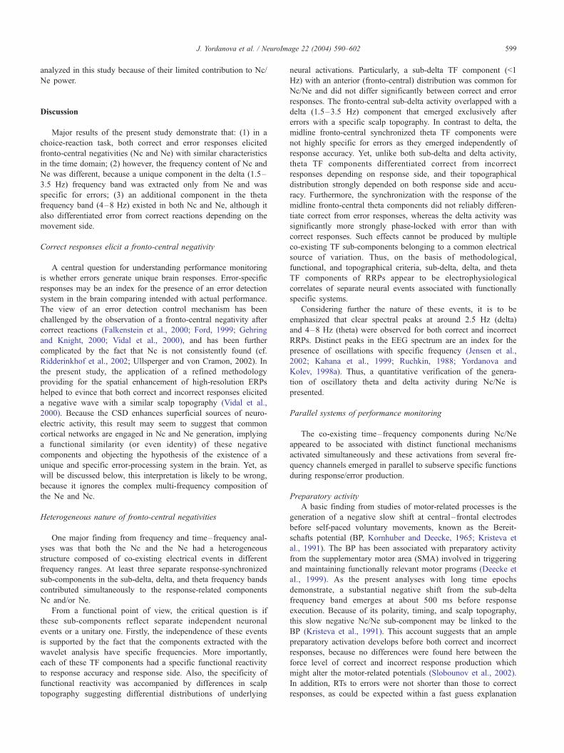
Fig. 7. Topography maps of the sub-delta and theta time– frequency
components of grand average RRPs of correct and error responses produced
by the left hand in the auditory modality. Positive and negative polarities
are marked additionally on the corresponding areas of the maps for the theta
TF component. Note that the scaling is different for different topographic
maps.
J. Yordanova et al. / NeuroImage 22 (2004) 590–602598
= 0.006). Thus, the lack of laterality effects in the difference maps
(Fig. 6) is verified by the results from testing simple effects. These
differential topography patterns indicate that distinct neuroelectric
sources contribute to Ne and Nc. Thus, a specific neural substrate
is essentially activated only after errors but not after correct
responses to produce a midline fronto-central potential.
The topography maps of sub-delta and theta TF components
coinciding with Nc/Ne are presented in Fig. 7 and show that:
(1) The topographical distribution of the sub-delta TF component
did not differ between correct and error RRPs and was
characterized by a midline fronto-central maximum, indepen-
dent of response accuracy. Because the magnitude of the sub-
delta TF component was larger than that of delta and theta TF
components in the correct RRPs (Fig. 5), the sub-delta TF
component had the largest contribution to the topographical
distribution of the Nc in the time domain (Fig. 1). This can
explain the midline fronto-central dominance of Nc typically
observed in the time domain.
(2) For correct RRPs, the negative theta phase within Nc
manifested a preponderance over the fronto-central medial
areas, with areas contralateral to response manifesting positive
values (Fig. 7). For error RRPs, the theta TF component within
Ne was strongly focused to the fronto-central midline
electrode, which modified the distribution pattern, although
theta oscillations also had positive values over the hemisphere
contralateral to the response. Because, as demonstrated in Figs.
5d and 7, theta oscillations were shifted in phase for midline
and lateral electrodes, the topography of the theta TF
component associated with the Ne/Nc was analyzed by
calculating the mean theta power within 0–100 ms after the
response. This analysis of theta power at nine electrodes
yielded a significant main effect for Accuracy (F(1/9) = 32.86,
P < 0.001), with errors eliciting larger theta oscillations than
correct responses. Yet, consistent with amplitude analysis
reported above for the FCz electrode, a significant Accuracy �Response side � Laterality interaction (F(2/18) = 5.15, P =
0.02; re-analysis with normalized data, F(2/18) = 4.55, P =
0.03) revealed that the theta oscillations were larger to error
than correct responses at midline locations only when they
were elicited by a left finger response (Accuracy � Response
side for midline electrodes, F(1/9) = 4.86, P < 0.05). At
contralateral and ipsilateral cortices, errors were associated
with larger theta oscillations, independently of the response
side (Accuracy, contralateral, F(1/9) = 12.09, P < 0.01;
ipsilateral, F(1/9) = 16.69, P < 0.005). Overall, theta
oscillations during the Ne/Nc were largest at the hemisphere
contralateral to the responding hand, and were lowest at the
ipsilateral one (Laterality, F(2/18) = 18.56, P < 0.001), but for
the fronto-central region, a midline predominance was found
(Region � Laterality, F(4/36) = 9.1, P < 0.001).
These observations indicate that the neural substrate generating
the sub-delta TF components during Nc/Ne may be similar for
correct and error responses. As opposed to the sub-delta and
similar to the delta TF component, the theta TF component of
Ne/Nc appears to stem from different neuronal networks that are
specific for correct and error responses.
Although it is possible that TF components from higher
frequency bands (alpha, beta, gamma) may also have a specific
reactivity to accuracy or response side variables, they were not
J. Yordanova et al. / NeuroImage 22 (2004) 590–602 599
analyzed in this study because of their limited contribution to Nc/
Ne power.
Discussion
Major results of the present study demonstrate that: (1) in a
choice-reaction task, both correct and error responses elicited
fronto-central negativities (Nc and Ne) with similar characteristics
in the time domain; (2) however, the frequency content of Nc and
Ne was different, because a unique component in the delta (1.5–
3.5 Hz) frequency band was extracted only from Ne and was
specific for errors; (3) an additional component in the theta
frequency band (4–8 Hz) existed in both Nc and Ne, although it
also differentiated error from correct reactions depending on the
movement side.
Correct responses elicit a fronto-central negativity
A central question for understanding performance monitoring
is whether errors generate unique brain responses. Error-specific
responses may be an index for the presence of an error detection
system in the brain comparing intended with actual performance.
The view of an error detection control mechanism has been
challenged by the observation of a fronto-central negativity after
correct reactions (Falkenstein et al., 2000; Ford, 1999; Gehring
and Knight, 2000; Vidal et al., 2000), and has been further
complicated by the fact that Nc is not consistently found (cf.
Ridderinkhof et al., 2002; Ullsperger and von Cramon, 2002). In
the present study, the application of a refined methodology
providing for the spatial enhancement of high-resolution ERPs
helped to evince that both correct and incorrect responses elicited
a negative wave with a similar scalp topography (Vidal et al.,
2000). Because the CSD enhances superficial sources of neuro-
electric activity, this result may seem to suggest that common
cortical networks are engaged in Nc and Ne generation, implying
a functional similarity (or even identity) of these negative
components and objecting the hypothesis of the existence of a
unique and specific error-processing system in the brain. Yet, as
will be discussed below, this interpretation is likely to be wrong,
because it ignores the complex multi-frequency composition of
the Ne and Nc.
Heterogeneous nature of fronto-central negativities
One major finding from frequency and time–frequency anal-
yses was that both the Nc and the Ne had a heterogeneous
structure composed of co-existing electrical events in different
frequency ranges. At least three separate response-synchronized
sub-components in the sub-delta, delta, and theta frequency bands
contributed simultaneously to the response-related components
Nc and/or Ne.
From a functional point of view, the critical question is if
these sub-components reflect separate independent neuronal
events or a unitary one. Firstly, the independence of these events
is supported by the fact that the components extracted with the
wavelet analysis have specific frequencies. More importantly,
each of these TF components had a specific functional reactivity
to response accuracy and response side. Also, the specificity of
functional reactivity was accompanied by differences in scalp
topography suggesting differential distributions of underlying
neural activations. Particularly, a sub-delta TF component (<1
Hz) with an anterior (fronto-central) distribution was common for
Nc/Ne and did not differ significantly between correct and error
responses. The fronto-central sub-delta activity overlapped with a
delta (1.5–3.5 Hz) component that emerged exclusively after
errors with a specific scalp topography. In contrast to delta, the
midline fronto-central synchronized theta TF components were
not highly specific for errors as they emerged independently of
response accuracy. Yet, unlike both sub-delta and delta activity,
theta TF components differentiated correct from incorrect
responses depending on response side, and their topographical
distribution strongly depended on both response side and accu-
racy. Furthermore, the synchronization with the response of the
midline fronto-central theta components did not reliably differen-
tiate correct from error responses, whereas the delta activity was
significantly more strongly phase-locked with error than with
correct responses. Such effects cannot be produced by multiple
co-existing TF sub-components belonging to a common electrical
source of variation. Thus, on the basis of methodological,
functional, and topographical criteria, sub-delta, delta, and theta
TF components of RRPs appear to be electrophysiological
correlates of separate neural events associated with functionally
specific systems.
Considering further the nature of these events, it is to be
emphasized that clear spectral peaks at around 2.5 Hz (delta)
and 4–8 Hz (theta) were observed for both correct and incorrect
RRPs. Distinct peaks in the EEG spectrum are an index for the
presence of oscillations with specific frequency (Jensen et al.,
2002; Kahana et al., 1999; Ruchkin, 1988; Yordanova and
Kolev, 1998a). Thus, a quantitative verification of the genera-
tion of oscillatory theta and delta activity during Nc/Ne is
presented.
Parallel systems of performance monitoring
The co-existing time–frequency components during Nc/Ne
appeared to be associated with distinct functional mechanisms
activated simultaneously and these activations from several fre-
quency channels emerged in parallel to subserve specific functions
during response/error production.
Preparatory activity
A basic finding from studies of motor-related processes is the
generation of a negative slow shift at central– frontal electrodes
before self-paced voluntary movements, known as the Bereit-
schafts potential (BP, Kornhuber and Deecke, 1965; Kristeva et
al., 1991). The BP has been associated with preparatory activity
from the supplementary motor area (SMA) involved in triggering
and maintaining functionally relevant motor programs (Deecke et
al., 1999). As the present analyses with long time epochs
demonstrate, a substantial negative shift from the sub-delta
frequency band emerges at about 500 ms before response
execution. Because of its polarity, timing, and scalp topography,
this slow negative Nc/Ne sub-component may be linked to the
BP (Kristeva et al., 1991). This account suggests that an ample
preparatory activation develops before both correct and incorrect
responses, because no differences were found here between the
force level of correct and incorrect response production which
might alter the motor-related potentials (Slobounov et al., 2002).
In addition, RTs to errors were not shorter than those to correct
responses, as could be expected within a fast guess explanation

J. Yordanova et al. / NeuroImage 22 (2004) 590–602600
for error occurrence. A possible reason is that in the four-choice
reaction task, subjects’ cognitive strategy may not rely on
stimulus prediction, and accordingly, wrong guess errors may
not be very probable. Rather, the errors can be induced by
attention fluctuations affecting the stimulus-response matching
in working memory. Hence, most errors in the four choice-
reaction task appear preceded by a central preparatory activity.
If the SMA is engaged in the elicitation of the BP as reflected by
the sub-delta TF component (Deecke et al., 1999), as well as in
the generation of Ne (Dehaene et al., 1994; Ullsperger and von
Cramon, 2001), certain interactions may occur within the SMA
between the mechanisms regulating motor preparation and action
monitoring. This issue may be an important aspect of future
research.
Error-specific signals: performance monitoring
A unique oscillatory component from the delta frequency range
(1.5–3.5 Hz) precisely focused to the medial fronto-central elec-
trode FCz, accounted for Ne expression on the scalp. This delta
sub-component was not pronounced in the Nc after correct
responses (Fig. 6). In contrast to Ne, the delta sub-component
after correct responses was distributed primarily over the contra-
lateral hemisphere, with central midline locations being also
involved. These results indicate that after errors, specific neural
sources generate a medial fronto-central electric response and these
sources are not functionally active after correct reactions. This
observation strongly argues against the assumption of the func-
tional identity of Ne and Nc. Instead, it shows that there exists a
specific medial system for performance monitoring operating in
the 2- to 3-Hz frequency channel that detects errors of overall
performance.
It is an interesting issue for further studies to establish whether
the fronto-medial error-specific delta oscillations are generated
primarily in sensorimotor choice-reaction tasks, or they may
characterize the error processing in general. In a previous study,
Ne and Nc have been described in terms of oscillations (Luu and
Tucker, 2001) after filtering out low-frequency (below 4 Hz) EEG
activity from response-locked averages. Accordingly, the contri-
bution of delta activity to Ne has not been quantitatively evaluated
by these authors. Whether and how the expression of error-specific
delta oscillations may depend on methodological (e.g., CSD vs.
monopolar signals, type of time–frequency decomposition, etc.) or
functional (type of task and error processing) factors remains to be
evaluated.
Parallel error-specific signals: Movement monitoring
In the study of Luu and Tucker (2001), the Ne/Nc signal
remaining after removing slow activity integrated theta and alpha
(4–12 Hz) frequencies. This signal was analyzed in the time
domain and was shown to be larger for errors. Although this
approach has not allowed to prove reliably that theta oscillations
are present in the RRPs or to study precisely their specific
reactivity, it has nonetheless helped to suggest that oscillatory
theta activity has an important functional role in action monitoring
(Luu and Tucker, 2001; Luu et al., 2000).
In line with these suggestions, the present study revealed
prominent theta oscillations at midline fronto-central and contra-
lateral motor cortex locations after both correct and error
responses. A similar distribution also can be observed in Figs.
2 and 3 in the study of Luu and Tucker (2001), in addition to
the sources of oscillations localized by these authors in both the
centromedial frontal cortex and bilateral sensorimotor cortices.
According to present findings, midline fronto-central theta ampli-
tudes associated with Nc and Ne did not generally differ
between correct and incorrect responses, but differentiated errors
in a response-side-specific way. Also, fronto-central theta oscil-
lations manifested sensitivity to the response hand (left or right),
because negative theta phase latency was significantly longer for
left hand responses. Moreover, topography analysis revealed that
prominent theta oscillations were enhanced over the motor
cortex contralateral to the response hand, and were substantially
suppressed over the ipsilateral motor cortex, which was true for
both correct and error responses (Figs. 2 and 5d). These
response side effects and the scalp distribution over the contra-
lateral motor areas strongly imply that synchronized theta
oscillations are functionally involved with movement production
and/or regulation.
Most importantly, error signals can be generated also in the
theta frequency channel. For midline fronto-central location, they
depend on movement side: Only for left-hand responses, did errors
enhance theta amplitude at FCz, which may be associated with
differences in the strength of neural motor representations due to
hand dominance (see RT results), or with different localizations of
motor representations of left- and right-hand responses. Over
contra- and ipsilateral fronto-central, central, and parieto-central
regions, errors produced larger theta oscillations than correct
responses. These findings indicate that theta oscillations are also
functionally associated with error monitoring, but mainly in
relation to movement execution. Thus, response-synchronized
theta oscillations may subserve a separate system of movement
monitoring, which can generate error signals at the level of central
motor regulation.
Conclusion
In a choice reaction task, two systems monitoring errors during
motor behaviour can be delineated: The first system (performance-
monitoring system) is specific for the detection of performance
errors and operates in the delta frequency range. The second
system (movement-monitoring system) is involved in the monitor-
ing of planned and executed motor programs and operates in the
theta frequency channel. Both systems can generate error signals.
The error signal of the performance-monitoring system may be
regarded as a highly specific index for overall incorrect perfor-
mance, whereas the error signal from the movement-monitoring
system may depend on the type and number of concurrently
activated motor programs. These results appear consistent with
both the error-detection model and the response-conflict model for
the generation of error negativity, implying that each of the models
has emphasized a specific aspect of the complex error signal. It is
concluded that error signaling is a function distributed at multiple
levels of information processing in the brain that are activated in
parallel.
Acknowledgments
Authors are thankful to Drs. I. Gutberlet and A. Hornecker for
helpful discussions and to J. Hoormann, L. Blanke, D. Winter and
C. Westedt for technical assistance. This work was supported by
the Deutsche Forschungsgemeinschaft (DFG), Germany (Grant

J. Yordanova et al. / NeuroImage 22 (2004) 590–602 601
No. Ho 965-5/3) by the National Science Council of the Ministry
of Education and Science, Bulgaria (Grant No. L-1316), and by the
German Federal Institute of Occupational Safety and Health
(BauA).
References
Babiloni, F., Babiloni, C., Carducci, F., Fattorini, L., Onorati, P., Urbano,
A., 1996. Spline Laplacian estimate of EEG potentials over a realistic
magnetic resonance-constructed scalp surface model. Electroencepha-
logr. Clin. Neurophysiol. 98, 363–373.
Babiloni, C., Babiloni, F., Carducci, F., Cincotti, F., Del Percio, C., De
Pino, G., Maestrini, S., Priori, A., Tisei, P., Zanetti, O., Rossini, P.M.,
2000. Movement-related electroencephalographic reactivity in Alz-
heimer disease. NeuroImage 12, 139–146.
Basar, E., 1980. EEG Brain Dynamics. Relation between EEG and Brain
Evoked Potentials. Elsevier, Amsterdam.
Basar, E., 1998. Brain Function and Oscillations. Brain Oscillations. Prin-
ciples and Approaches, vol. 1. Springer, Berlin.
Bernstein, P.S., Scheffers, M.K., Coles, M.G.H., 1995. ‘‘Where did I go
wrong?’’ A psychophysiological analysis of error detection. J. Exp.
Psychol. Hum. Percept. Perform. 21, 1312–1322.
Botvinick,M.M., Braver, T.S., Barch, D.M., Carter, C.S., Cohen, J.D., 2001.
Conflict monitoring and cognitive control. Psychol. Rev. 108, 624–652.
Braun, C.M., Villeneuve, L., Gruzelier, J.H., 2002. Topographical analysis
of stimulus-related and response-related electrical scalp activity and
interhemispheric dynamics in normal humans. Int. J. Psychophysiol.
46, 109–122.
Bush, G., Luu, P., Posner, M.I., 2000. Cognitive and emotional influences
in anterior cingulate cortex. Trends Cogn. Sci. 4, 215–222.
Carter, C.S., Braver, T.S., Barch, D.M., Botvinick, M.M., Noll, D., Cohen,
J.D., 1998. Anterior cingulate cortex, error detection, and the online
monitoring of performance. Science 280, 747–749.
Coles, M.G.H., Scheffers, M.K., Holroyd, C.B., 2001.Why is there an ERN/
Ne on correct trials? Response representations, stimulus-related compo-
nents, and the theory of error-processing. Biol. Psychol. 56, 173–189.
Deecke, L., Lang, W., Uhl, F., Beisteiner, R., Lindinger, G., Cui, R.Q.,
1999. Movement-related potentials and magnetic fields: new evidence
for SMA activation leading MI activation prior to voluntary movement.
Electroencephalogr. Clin. Neurophysiol. Suppl. 50, 386–401.
Dehaene, S., Posner, M.I., Tucker, D.M., 1994. Localization of a neural
system for error detection and compensation. Psychol. Sci. 5, 303–305.
Falkenstein, M., Hohnsbein, J., Hoormann, J., Blanke, L., 1990. Effects of
errors in choice reaction tasks on the ERP under focused and divided
attention. In: Brunia, C.H.M., Gaillard, A.W.K., Kok, A. (Eds.), Psy-
chophysiological Brain Research 1990. Tilburg Univ. Press, Tilburg,
The Netherlands, pp. 192–195.
Falkenstein, M., Hohnsbein, J., Hoormann, J., Blanke, L., 1991. Effects of
crossmodal divided attention on late ERP components: II. Error pro-
cessing in choice reaction tasks. Electroencephalogr. Clin. Neurophy-
siol. 78, 447–455.
Falkenstein, M., Hoormann, J., Christ, S., Hohnsbein, J., 2000. ERP com-
ponents on reaction errors and their functional significance: a tutorial.
Biol. Psychol. 51, 87–107.
Feige, B., Aertsen, A., Kristeva-Feige, R., 2000. Dynamic synchronization
between multiple cortical motor areas and muscle activity in phasic
voluntary movements. J. Neurophysiol. 84, 2622–2629.
Ford, J.M., 1999. Schizophrenia: the broken P300 and beyond. Psycho-
physiology 36, 667–682.
Freeman, W.J., Rogers, L.J., 2002. Fine temporal resolution of analytic
phase reveals episodic synchronization by state transitions in gamma
EEGs. J. Neurophysiol. 87, 937–945.
Gehring, W.J., Knight, R.T., 2000. Prefrontal –cingulate interactions in
action monitoring. Nat. Neurosci. 3, 421–423.
Gehring, W.J., Goss, B., Coles, M.G.H., Meyer, D.E., Donchin, E., 1993. A
neural system for error detection and compensation. Psychol. Sci. 4,
385–390.
Gratton, G., Coles, M.G.H., Donchin, E., 1983. A new method for off-line
removal of ocular artifact. Electroencephalogr. Clin. Neurophysiol. 55,
468–484.
Holroyd, C.B., Coles, M.G.H., 2002. The neural basis of human error
processing: reinforcement learning, dopamine, and the error-related
negativity. Psychol. Rev. 109, 679–709.
Holroyd, C.B., Dien, J., Coles, M.G.H., 1998. Error-related scalp potentials
elicited by hand and foot movements: evidence for an output-indepen-
dent error-processing system in humans. Neurosci. Lett. 242, 65–68.
Jensen, O., Gelfand, J., Kounios, J., Lisman, J.E., 2002. Oscillations in the
alpha band (9–12 Hz) increase with memory load during retention in a
short-term memory task. Cereb. Cortex 12, 877–882.
Kahana, M.J., Sekuler, R., Caplan, J.B., Kirschen, M., Madsen, J.R., 1999.
Human theta oscillations exhibit task dependence during virtual maze
navigation. Nature 399, 781–784.
Kolev, V., Yordanova, J., 1997. Analysis of phase-locking is informative
for studying event-related EEG activity. Biol. Cybern. 76, 229–235.
Kolev, V., Demiralp, T., Yordanova, J., Ademoglu, A., Isoglu-Alkac�, U.,1997. Time–frequency analysis reveals multiple functional components
during oddball P300. NeuroReport 8, 2061–2065.
Kornhuber, H.H., Deecke, L., 1965. Hirnpotentialanderungen bei Willkur-
bewegungen des Menschen: Bereitschaftspotential und reafferente
Potentiale. Pflugers Arch. 284, 1–17.
Kristeva, R., Cheyne, D., Deecke, L., 1991. Neuromagnetic fields accom-
panying unilateral and bilateral voluntary movements: topography and
analysis of cortical sources. Electroencephalogr. Clin. Neurophysiol. 81,
284–298.
Luu, P., Tucker, D.M., 2001. Regulating action: alternating activation of
midline frontal and motor cortical networks. Clin. Neurophysiol. 112,
1295–1306.
Luu, P., Flaisch, T., Tucker, D.M., 2000. Medial frontal cortex in action
monitoring. J. Neurosci. 20, 464–469.
Mallat, S., 1999. A Wavelet Tour of Signal Processing, 2nd ed. Academic
Press, San Diego.
McCarthy, G., Wood, C.C., 1985. Scalp distributions of event-related
potentials: an ambiguity associated with analysis of variance models.
Electroencephalogr. Clin. Neurophysiol. 62, 203–208.
Nunez, P.L., Pilgreen, K.L., 1991. The spline-Laplacian in clinical neuro-
physiology: a method to improve EEG spatial resolution. J. Clin. Neu-
rophysiol. 8, 397–413.
Nunez, P.L., Srinivasan, R., Westdorp, A.F., Wijesinghe, R.S., Tucker,
D.M., Silberstein, R.B., Cadusch, P.J., 1997. EEG coherency. I.
Statistics, reference electrode, volume conduction, Laplacians, corti-
cal imaging, and interpretation at multiple scales. Electroencepha-
logr. Clin. Neurophysiol. 103, 499–515.
Perrin, F., Pernier, J., Bertrand, O., Echallier, J.F., 1989. Spherical splines
for scalp potential and current density mapping. Electroencephalogr.
Clin. Neurophysiol. 72, 184–187.
Pfurtscheller, G., Neuper, C., Krausz, G., 2000. Functional dissociation of
lower and upper frequency mu rhythms in relation to voluntary limb
movement. Clin. Neurophysiol. 111, 1873–1879.
Posner, M.I., DiGirolamo, G.J., 1998. Executive attention: conflict, target
detection, and cognitive control. In: Parasuraman, R. (Ed.), The Atten-
tive Brain. MIT Press, Cambridge, MA, pp. 401–423.
Regan, D., 1989. Human brain electrophysiology. Evoked Potentials
and Evoked Magnetic Fields in Science and Medicine. Elsevier,
Amsterdam.
Ridderinkhof, K.R., de Vlugt, Y., Bramlage, A., Spaan, M., Elton, M., Snel,
J., Band, G.P., 2002. Alcohol consumption impairs detection of perfor-
mance errors in mediofrontal cortex. Science 298, 2209–2211.
Ruchkin, D.S., 1988. Measurement of event-related potentials: signal ex-
traction. In: Picton, T.W. (Ed.), Event-Related Potentials. EEG Hand-
book, rev. series, vol. 3. Elsevier, Amsterdam, pp. 7–43.
Samar, V.J., Bopardikar, A., Rao, R., Swartz, K., 1999. Wavelet analysis of
neuroelectric waveforms: a conceptual tutorial. Brain Lang. 66, 7–60.

J. Yordanova et al. / NeuroImage 22 (2004) 590–602602
Scheffers, M.K., Coles, M.G.H., 2000. Performance monitoring in a con-
fusing world: error-related brain activity, judgments of response accu-
racy, and types of errors. J. Exp. Psychol. Hum. Percept. Perform. 26,
141–151.
Slobounov, S., Johnston, J., Chiang, H., Ray, W., 2002. Movement-related
EEG potentials are force or end-effector dependent: evidence from a
multi-finger experiment. Clin. Neurophysiol. 113, 1125–1135.
Tallon-Baudry, C., Bertrand, O., Delpuech, C., Permier, J., 1997. Oscilla-
tory gamma-band (30–70 Hz) activity induced by a visual search task
in humans. J. Neurosci. 17, 722–734.
Ullsperger, M., von Cramon, D.Y., 2001. Subprocesses of performance
monitoring: a dissociation of error processing and response competi-
tion revealed by event-related fMRI and ERPs. NeuroImage 14,
1387–1401.
Ullsperger, M., Falkenstein, M. (Eds.), 2004. Errors, Conflicts, and
the Brain. Current Opinions on Performance Monitoring. Max
Planck Institute for Human Cognitive and Brain Sciences, Leipzig.
In press.
Urbano, A., Babiloni, C., Onorati, P., Babiloni, F., 1998. Dynamic func-
tional coupling of high resolution EEG potentials related to unilateral
internally triggered one digit movements. Electroencephalogr. Clin.
Neurophysiol. 106, 477–487.
Vidal, F., Hasbroucq, T., Grapperon, J., Bonnet, M., 2000. Is the ‘error
negativity’ specific to errors? Biol. Psychol. 51, 109–128.
Yordanova, J., Kolev, V., 1998a. A single sweep analysis of the theta
frequency band during an auditory oddball task. Psychophysiology
35, 116–126.
Yordanova, J., Kolev, V., 1998b. Phase locking of event related EEG
oscillations: analysis and application. Appl. Sig. Proc. 5, 24–33.
Yordanova, J., Devrim, M., Kolev, V., Ademoglu, A., Demiralp, T., 2000.
Multiple time frequency components account for the complex function-
al reactivity of P300. NeuroReport 11, 1097–1103.