Parallel Substrate Binding Sites in a β-Agarase Suggest a Novel Mode of Action on Double-Helical...
10
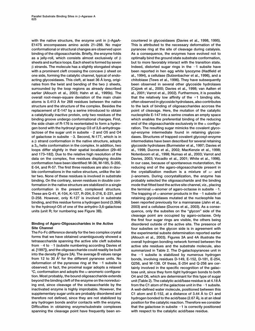
Structure, Vol. 12, 623–632, April, 2004, 2004 Elsevier Science Ltd. All rights reserved. DOI 10.1016/j.str.2004.02.020 Parallel Substrate Binding Sites in a -Agarase Suggest a Novel Mode of Action on Double-Helical Agarose (1,4) linkages as well as the occurrence of 3,6-anhydro- L-galactose bridges in the polysaccharide backbone fa- vor the formation of twisted ribbons, with alternating 4 C 1 and 1 C 4 chair conformations for the D-galactopyranose and 3,6-anhydro-L-galactose rings, respectively (Arnott Julie Allouch, 1 William Helbert, 2 Bernard Henrissat, 1 and Mirjam Czjzek 1, * 1 Architecture et Fonction des Macromole ´ cules Biologiques UMR 6098 Centre de la Recherche Scientifique and et al., 1974; Rochas et al., 1986). The earliest model of the agarose gel network involves the aggregation of Universite ´ s Aix-Marseille I and II 31 chemin Joseph Aiguier parallel double-helical structures stabilized by interchain hydrogen bonds (Arnott et al., 1974). The parallel double- F-13402 Marseille Cedex 20 2 Ve ´ ge ´ taux Marins et Biomole ´ cules helix model is reported to have a 9.5 A ˚ axial periodicity, each single chain being a left-handed 3-fold helix with UMR 7139 (CNRS/UPMC/Laboratoires Goe ¨ mar) Station Biologique de Roscoff a pitch of 19.0 A ˚ . Each strand is translated axially relative to the first one by half this distance (Arnott et al., 1974). Place Georges Teissier, BP 74 29682 Roscoff Cedex, Bretagne In agarose gels, helix propagation is interrupted by the occurrence of natural structural discontinuities or de- France fects, resulting from the occasional and random replace- ment of 3,6-anhydro-L-galactose by L-galactose-6-sul- fate. Self-association of these polysaccharides into Summary large crystalline clusters is thus prevented, and, instead, they most probably form three-dimensional networks in Agarose is a gel-forming polysaccharide with an aqueous solutions, consisting of crystalline zones of -L(1,4)-3,6-anhydro-galactose, -D(1,3)-galactose re- double helix aggregates separated by amorphous zones peat unit, from the cell walls of marine red algae. containing single-stranded helices and random coil -agarase A, from the Gram-negative bacterium (Bhattacharjee et al., 1978). Zobellia galactanivorans, is secreted to the external Certain marine bacteria can utilize agarose as a car- medium and degrades agarose with an endo-mecha- bon source. The enzymatic breakdown of agarose can nism. The structure of the inactive mutant -agarase be performed by two types of agarases, namely those A-E147S in complex with agaro-octaose has been able to cleave the -1,4-galactosidic bonds (-agarases) solved at 1.7 A ˚ resolution. Two oligosaccharide chains and those able to cleave the -1,3-anhydro-L-galactosi- are bound to the protein. The first one resides in the dic bonds (-agarases). While the former are well docu- active site channel, spanning subsites 4 to 1. A mented, -agarases are poorly known. -agarases are second oligosaccharide binding site, on the opposite found in three different families of the glycoside hy- side of the protein, was filled with eight sugar units, drolase (GH) classification (http://afmb.cnrs-mrs.fr/CAZY), parallel to the active site. The crystal structure of the namely in families GH-50, GH-86, and GH-16. Family GH16 -agarase A with agaro-octaose provides detailed in- is one of the largest families and groups together enzymes formation on agarose recognition in the catalytic site. of varying substrate specificity such as keratan-sulfate The presence of the second, parallel, binding site sug- endogalactosidases, -1,3-glucanases, mixed linkage gests that the enzyme might be able to unwind the -1,3(4)-glucanases, xyloglucan endotransferases, and double-helical structure of agarose prior to the cata- -carrageenases together with -agarases. Amino acid lytic cleavage. sequence alignment of the members of this heteroge- neous family reveals the existence of several subfamilies Introduction that coincide with the experimentally determined sub- strate specificity of the enzymes (Allouch et al., 2003). Agarose, an essentially neutral polysaccharide, is the Three-dimensional structures have been solved for major gelling component of agar. It is widely used as several of these subfamilies, namely -1,3-1,4-gluca- a texturing and gelling agent in the food and medical nase (Hahn et al., 1995a, 1995b; Juncosa et al., 1994; industries as well as for numerous chromatographic and Keitel et al., 1993), -carrageenase (Michel et al., 2001), electrophoretic techniques (De Ruiter and Rudolph, and, more recently, -agarase (Allouch et al., 2003). 1997). This hydrocolloid is found in the cell walls of Structural data for the xyloglucan endotransferase sub- various marine red algae (Rhodophyta), where it is laid family is expected to be soon available, since the crystal- out as highly ordered molecules, either associated to lization of this enzyme from Populus tremula x tremu- the cellulose microfibrils or in the microfibril-less inter- loides has been reported recently (Johansson et al., cellular matrix (Kloareg and Quatrano, 1988). Agarose 2003). Despite this apparent wealth of structural data, is an alternating copolymer of 3-linked -D-galactopyra- no structure of any family GH-16 enzyme in complex nose and 4-linked 3,6-anhydro--L-galactopyranose with its native substrate has been reported to date. This (Figure 1). Conformational analyses and X-ray diffraction information is particularly important in understanding the studies have shown that the succession of (1,3) and diversity of substrate specificity within family GH-16. Two -agarases, -agarase A and -agarase B (here- after referred to as -agaA and -agaB), have been iso- *Correspondence: [email protected]
-
Upload
independent -
Category
Documents
-
view
0 -
download
0
Transcript of Parallel Substrate Binding Sites in a β-Agarase Suggest a Novel Mode of Action on Double-Helical...
Structure, Vol. 12, 623–632, April, 2004, 2004 Elsevier Science Ltd. All rights reserved. DOI 10.1016/j .str .2004.02.020
Parallel Substrate Binding Sites in a �-AgaraseSuggest a Novel Mode of Actionon Double-Helical Agarose
�(1,4) linkages as well as the occurrence of 3,6-anhydro-L-galactose bridges in the polysaccharide backbone fa-vor the formation of twisted ribbons, with alternating 4C1
and 1C4 chair conformations for the D-galactopyranoseand 3,6-anhydro-L-galactose rings, respectively (Arnott
Julie Allouch,1 William Helbert,2
Bernard Henrissat,1 and Mirjam Czjzek1,*1Architecture et Fonction des Macromolecules
BiologiquesUMR 6098Centre de la Recherche Scientifique and et al., 1974; Rochas et al., 1986). The earliest model of
the agarose gel network involves the aggregation ofUniversites Aix-Marseille I and II31 chemin Joseph Aiguier parallel double-helical structures stabilized by interchain
hydrogen bonds (Arnott et al., 1974). The parallel double-F-13402 Marseille Cedex 202 Vegetaux Marins et Biomolecules helix model is reported to have a 9.5 A axial periodicity,
each single chain being a left-handed 3-fold helix withUMR 7139 (CNRS/UPMC/Laboratoires Goemar)Station Biologique de Roscoff a pitch of 19.0 A. Each strand is translated axially relative
to the first one by half this distance (Arnott et al., 1974).Place Georges Teissier, BP 7429682 Roscoff Cedex, Bretagne In agarose gels, helix propagation is interrupted by the
occurrence of natural structural discontinuities or de-Francefects, resulting from the occasional and random replace-ment of 3,6-anhydro-L-galactose by L-galactose-6-sul-fate. Self-association of these polysaccharides intoSummarylarge crystalline clusters is thus prevented, and, instead,they most probably form three-dimensional networks inAgarose is a gel-forming polysaccharide with anaqueous solutions, consisting of crystalline zones of�-L(1,4)-3,6-anhydro-galactose, �-D(1,3)-galactose re-double helix aggregates separated by amorphous zonespeat unit, from the cell walls of marine red algae.containing single-stranded helices and random coil�-agarase A, from the Gram-negative bacterium(Bhattacharjee et al., 1978).Zobellia galactanivorans, is secreted to the external
Certain marine bacteria can utilize agarose as a car-medium and degrades agarose with an endo-mecha-bon source. The enzymatic breakdown of agarose cannism. The structure of the inactive mutant �-agarasebe performed by two types of agarases, namely thoseA-E147S in complex with agaro-octaose has beenable to cleave the �-1,4-galactosidic bonds (�-agarases)solved at 1.7 A resolution. Two oligosaccharide chainsand those able to cleave the �-1,3-anhydro-L-galactosi-are bound to the protein. The first one resides in thedic bonds (�-agarases). While the former are well docu-active site channel, spanning subsites �4 to �1. Amented, �-agarases are poorly known. �-agarases aresecond oligosaccharide binding site, on the oppositefound in three different families of the glycoside hy-side of the protein, was filled with eight sugar units,drolase (GH) classification (http://afmb.cnrs-mrs.fr/CAZY),parallel to the active site. The crystal structure of thenamely in families GH-50, GH-86, and GH-16. Family GH16�-agarase A with agaro-octaose provides detailed in-is one of the largest families and groups together enzymesformation on agarose recognition in the catalytic site.of varying substrate specificity such as keratan-sulfateThe presence of the second, parallel, binding site sug-endogalactosidases, �-1,3-glucanases, mixed linkagegests that the enzyme might be able to unwind the�-1,3(4)-glucanases, xyloglucan endotransferases, anddouble-helical structure of agarose prior to the cata-�-carrageenases together with �-agarases. Amino acidlytic cleavage.sequence alignment of the members of this heteroge-neous family reveals the existence of several subfamilies
Introduction that coincide with the experimentally determined sub-strate specificity of the enzymes (Allouch et al., 2003).
Agarose, an essentially neutral polysaccharide, is the Three-dimensional structures have been solved formajor gelling component of agar. It is widely used as several of these subfamilies, namely �-1,3-1,4-gluca-a texturing and gelling agent in the food and medical nase (Hahn et al., 1995a, 1995b; Juncosa et al., 1994;industries as well as for numerous chromatographic and Keitel et al., 1993), �-carrageenase (Michel et al., 2001),electrophoretic techniques (De Ruiter and Rudolph, and, more recently, �-agarase (Allouch et al., 2003).1997). This hydrocolloid is found in the cell walls of Structural data for the xyloglucan endotransferase sub-various marine red algae (Rhodophyta), where it is laid family is expected to be soon available, since the crystal-out as highly ordered molecules, either associated to lization of this enzyme from Populus tremula x tremu-the cellulose microfibrils or in the microfibril-less inter- loides has been reported recently (Johansson et al.,cellular matrix (Kloareg and Quatrano, 1988). Agarose 2003). Despite this apparent wealth of structural data,is an alternating copolymer of 3-linked �-D-galactopyra- no structure of any family GH-16 enzyme in complexnose and 4-linked 3,6-anhydro-�-L-galactopyranose with its native substrate has been reported to date. This(Figure 1). Conformational analyses and X-ray diffraction information is particularly important in understanding thestudies have shown that the succession of �(1,3) and diversity of substrate specificity within family GH-16.
Two �-agarases, �-agarase A and �-agarase B (here-after referred to as �-agaA and �-agaB), have been iso-*Correspondence: [email protected]

Structure624
Table 1. Data Reduction and Refinement Statistics
�-AgaA-E147S/ �-AgaA-E147S/Native Data Statistics Octa-Agarose Dodeca-Agarose
Space group P3221 P21
Beamline ID14-eh1 ID14-eh4Wavelength (A) 0.934 0.9793Resolution (A) 37.2–1.7 41.5–1.7Total data 148,960 255,194Figure 1. Schematic Drawing of the Repeating Disaccharide UnitUnique data 34,045 60,409of AgaroseRedundancya 4.4 (4.5) 4.2 (4.2)The �(1,4) bond cleaved during hydrolysis by �-agarases is labeled.Completeness (%)a 95.3 (98.4) 98.8 (97.8)I/�Ia 12.1 (6.8) 7.2 (2.2)Rsym (%)a,b 4.2 (11.0) 7.3 (32.4)
lated from the marine bacterium Zobellia galactanivor-Refinementans Dsij and have been characterized (Jam et al.,
submitted). The catalytic domains of these enzymes Resolution range (A) 37.2–1.7 41.5–1.7share 45% sequence identity, and their experimentally N of unique reflections 32,767 60,150
Rwork (Rfree)c 15.5 (18.2) 17.2 (21.3)determined 3D structures are highly similar (Allouch etRmsd bondsd (A) 0.015 0.025al., 2003). At the biological level, a notable differenceRmsd angles (�) 1.762 2.2between the two enzymes is that �-agaA is secreted inQuality of Ramachandran plotthe extracellular medium, while �-agaB is most probably
attached to the outer cell membrane (Jam et al., submit- % of residues in most 85.5 87.1ted). In addition, �-agarase A contains a 130 residue favored regions
% of residues in additional 14.5 12.9supplementary module of unknown function at its Callowed regionsterminus, separated from the catalytic domain by a 13
% of residues in generously 0.0 0.0residue linker peptide (Jam et al., submitted). The bio-allowed regionschemical analysis of the reaction products of the two
No. of atomscatalytic domains has established that both enzymesact by an endo-mechanism. Only subtle differences Protein 2171 4340have been found between the two enzymes: the catalytic Water 355 726
Ion (Ca) 1 2module of �-AgaA preferably degrades longer oligosac-Agarose1 43 86charides and has an overall slower reaction rate thanAgarose2 74 180�-AgaB (Allouch et al., 2003). Family GH-16 �-agarasesa Values in parenthesis correspond to the highest resolution shell.perform catalysis with mechanism leading to net reten-b Rsym � �|I � Iav|/�I, where the summation is over all symmetry-tion of the anomeric configuration (Potin et al., 1993) viaequivalent reflections.a double displacement mechanism as first suggested byc R calculated on 5% of data excluded from refinement.Koshland (Koshland, 1953). The two catalytic residues d Rmsd, root-mean-square deviation.
involved in this mechanism (acid/base and nucleophile)have been identified in a related family GH-16 member(Juncosa et al., 1994) and correspond to glutamates 147 ride, spanning subsites �4 to �1 in the active site chan-and 152, respectively, in �-AgaA. nel. A second binding site on the surface of the enzyme,
Like all members of family GH-16 with a known 3D parallel to the active site cleft and occupied by an intactstructure, the crystal structures of the catalytic domains agaro-octaose, is identified and described.of native �-AgaA and �-AgaB reveal a jelly-roll fold,formed of two seven-stranded � sheets interconnected Results and Discussionby extended loops. The concave � sheet forms a longactive site cleft, surrounded by the extended loops and Overall Structureruns a full 36 A across the surface of the enzymes. Crystals of the Zobellia galactanivorans Dsij �-AgaA-This structural feature, together with the biochemical E147S in complex with the natural substrate agaro-analysis of the reaction products indicate that these two octaose, obtained by cocrystallization trials, belong to�-agarases are capable of cleaving randomly a single space group P3221, with cell dimensions a � b � 51.38 Aagarose chain. The detailed analysis of the residues in and c � 205.36 A. The structure of the complex wasthe active site channel, as well as the structural compari- determined at 1.7 A resolution by molecular replacementson with the related enzymes within family GH-16 helped with AMoRe using the native �-AgaA structure as theto pinpoint a number of residues most probably forming search model. The coordinates describing one enzymethe substrate binding subsites. However, a description molecule with two oligosaccharide chains per asymmet-of the details of agarose recognition is not possible ric unit were refined to a final R factor and Rfree of 15.5%in absence of an enzyme/substrate complex. Here we and 18.2%. A second crystal form has been obtainedreport the crystal structure of an inactive �-agaA by soaking experiments of mutant �-AgaA-E147S inobtained by site-directed replacement of the catalytic presence of agaro-dodecaose. In this case, the asym-nucleophile by a serine (referred to below as �-AgaA- metric unit contained two enzyme molecules each inE147S) in complex with long agaro-oligosaccharides. complex with two oligosaccharide chains, leading toThe unbiased averaged electron density map clearly final R and Rfree factors of 17.2% and 21.3%. Further
crystallographic statistics are reported in Table 1. Asreveals density for four units of an agaro-oligosaccha-
Parallel Substrate Binding Sites in �-Agarase A625
with the native structure, the enzyme unit in �-AgaA- countered in glycosidases (Davies et al., 1998, 1995).This is attributed to the necessary deformation of theE147S encompasses amino acids 21–288. No major
conformational or structural changes are observed upon pyranose ring at the site of cleavage during catalysis.As a consequence, the enzymes have evolved not tobinding of the oligosaccharide. Briefly, the enzyme folds
as a jelly-roll, which consists almost exclusively of � optimally bind the ground state substrate conformation,but to more favorably interact with the transition state.sheets and surface loops. Each sheet is formed by seven
� strands. The molecule has a slightly elongated shape Indeed, distorted sugar rings in the �1 subsite havebeen reported in hen egg white lysozyme (Hadfield etwith a prominent cleft crossing the concave � sheet on
one side, forming the catalytic channel, typical of endo- al., 1994), a cellulase (Sulzenbacher et al., 1996), and achitobiase (Tews et al., 1996). They have subsequentlyacting glycosidases. This cleft, at least 36 A long, origi-
nates from the twist and bending of the two � sheets, been observed in several other glycoside hydrolases(Czjzek et al., 2000; Davies et al., 1998; van Aalten etsurrounded by the loop regions as already described
earlier (Allouch et al., 2003; Hahn et al., 1995b). The al., 2001; Varrot et al., 2002). Furthermore, it is possiblethat the relatively low affinity of the 1 binding site,overall root-mean-square deviation of the main chain
atoms is 0.413 A for 268 residues between the native often observed in glycoside hydrolases, also contributesto the lack of binding of oligosaccharides accross thestructure and the structure of the complex. Besides the
replacement of E-147 by a serine introduced to obtain point of cleavage. Here, the mutation of the catalyticnucleophile E-147 into a serine creates an empty spacea catalytically inactive protein, only two residues of the
binding groove undergo conformational changes. First, which enables the preferential binding of the reducingend of the oligosaccharide exclusively in the � configu-the side chain of R-176 is reorientated to form a hydro-
gen bond with the hydroxyl group O3 of 3,6-anhydroga- ration. The resulting sugar mimicks the covalent glyco-syl-enzyme intermediate found in retaining glycosi-lactose of the sugar unit in subsite �2 and O5 and O4
of galactose in subsite �3. Second, N-177, which is in dases. Structures of trapped covalent glycosyl-enzymeintermediates have been described for several retaininga � strand conformation in the native structure, adopts
a 310 helix conformation in the complex. In addition, two glycoside hydrolases (Burmeister et al., 1997; Davies etal., 1998; Ducros et al., 2002; MacKenzie et al., 1998;loops differ slightly in their spatial localization (29–40
and 173–182). Due to the relative high resolution of the Notenboom et al., 1998; Numao et al., 2003; Varrot andDavies, 2003; Vocadlo et al., 2001; White et al., 1996).data on the complex, five residues displaying double
conformation have been identified: M-36, M-185, S-200, In our case, because of spontaneous mutarotation, thereducing end of the agaro-oligosaccharide present inE-54, and R-57. The first three residues are also in dou-
ble conformations in the native structure, unlike the lat- the crystallization medium is a mixture of �- and�-anomers. During cocrystallization, the enzyme haster two. None of these residues is involved in substrate
binding. On the contrary, seven residues in double con- probably selected the oligosaccharide and the bindingmode that fitted best the active site channel, viz., placingformation in the native structure are stabilized in a single
conformation in the present, complexed structure. the terminal �-anomer of agaro-octaose in subsite �1.The trapping of �-anomer products in the �1 subsite ofThese are Q-41, K-104, K-127, R-204, E-216, E-231, and
D-258. However, only K-127 is involved in substrate retaining glycosidases mutated at the nucleophile hasbeen reported previously for a mannanase (Jahn et al.,binding, and this residue forms a hydrogen bond (3.38A)
to the hydroxyl-O5 of one of the anhydro-3,6-galactose 2003) and a cellulase (Ducros et al., 2003). As a conse-quence, only the subsites on the “glycon” side of theunits (unit R; for numbering see Figure 3B).cleavage point are occupied by agaro-octaose. Onlythe first four sugar rings are visible, the others being
Binding of Agaro-Oligosaccharides in the Active disordered outside of the active site. The presence ofSite Channel four subsites on the glycon side is in agreement withThe Fo-Fc difference density for the two complex crystal the experimental subsite determination reported earlierforms that we have obtained unambiguously showed a (Allouch et al., 2003). Figures 3A and 4A illustrate thetetrasaccharide spanning the active site cleft subsites overall hydrogen bonding network formed between thefrom �4 to �1 (subsite numbering according Davies et active site residues and the substrate molecule, alsoal. [1997]), and the oligosaccharide could readily be built summarized in Table 2. The D-galactopyranose unit ininto the density (Figure 2A). The average B values range the �1 subsite is stabilized by numerous hydrogenfrom 12 to 30 A2 for the different pyranose units. No bonds, involving residues D-149, E-152, D-181, E-254,deformation of the pyranose ring at the �1 subsite is Q256, and W-138. Of these, E-254 and Q-256 are cer-observed; in fact, the proximal sugar adopts a relaxed tainly involved in the specific recognition of the galac-4C1 conformation and adopts the �-anomeric configura- tose unit, since they form tight hydrogen bonds to bothtion. Most probably, the bound oligosaccharide extends O4 and O6, which are determinant for this type of sugarbeyond the binding cleft of the enzyme on the nonreduc- unit (Table 2). The catalytic acid/base residue is at 4.18 Aing end, since cleavage of the octasaccharide by the from the C1 atom of the galactose unit in the �1 subsite.inactivated enzyme is highly improbable. However, the A well-defined water molecule, positioned between thissupplementary sugar units are probably disordered and C1 atom and E-152, at a distance of 3.44 A to C1 andtherefore not defined, since they are not stabilized by hydrogen bonded to the acid/base (2.67 A), is at an idealany hydrogen bonds and/or contacts with the enzyme. position for the catalytic reaction. Therefore we considerDifficulties in obtaining enzyme/substrate complexes that the galactose in subsite �1 is correctly positioned
with respect to the catalytic acid/base residue.spanning the cleavage point have frequently been en-

Structure626
Figure 2. Experimental Maps Defining the Ligand Molecules
Electronic density around the galactopyranose units of the oligosaccharide in the active site channel (A) and on the surface binding site (B).The (Fo-Fc) Fourier-difference maps before refinement are shown in the top, while omit-maps at the end of refinement, calculated using onlythe enzyme model phases without substrate, contoured at 2.5� above the mean density are shown at the bottom.
The 3,6-anhydro-galactopyranose unit in subsite �2 polar residues will form hydrogen bonds to two adjacentpyranose units and therefore form a discriminating pat-is surrounded by W-73 on one side and F-174 and R-176
on the other side. In particular, R-176 forms a hydrogen tern for the binding of the characteristic-diose subunitof oligo-agarose chains (see Table 2). The alignment ofbond to the O3 of the anhydro bridge and therefore
is crucial for binding of 3,6-anhydro-galactose in this the sequence of �-AgaA with the other family GH-16�-agarases indicates that the residues involved in thesubsite. The D-galactopyranose unit in the �3 subsite
is stabilized by stacking interactions with W-73 on one surface binding site observed here are not conserved(data not shown) in the few other agarases whose se-side and F-179 on the other side. Two further hydrogen
bonds, involving R-176 and E-144, stabilize the sugar quences are available. This site is probably a featurespecific of �-agarase A from Zobellia galactanivorans Dsij.ring in this subsite. Only two hydrogen bonds are formed
(E-144 and N-71) with the 3,6-anhydro-L-galactopyra- In the vast majority of the documented cases, poly-saccharidases have evolved supplementary substratenose unit in subsite �4, explaining the higher mobility
of this sugar ring reflected by the higher B value of this binding sites by recruitment of independent carbohy-drate binding modules (CBM) (Bourne and Henrissat,sugar (30 A2) with respect to the sugar rings in the other
subsites (12–16 A2). 2001; Gill et al., 1999; Henrissat and Davies, 2000). TheseCBMs are believed to enhance the activity of the en-zymes on insoluble substrates by increasing the effec-The Surface Binding Site
The two crystal forms that we have obtained displayed tive enzyme concentration on the polysaccharide sur-face (Gill et al., 1999). Their removal by proteolysis orwell-defined and continuous electron density for oligo-
saccharides bound to a long surface binding site (Figure genetic manipulation reduces the activity of the enzymeon insoluble polysaccharides but not on soluble sub-2B). Seven sugar rings were visible for the crystals in
space group P3221 and eight in the P21 crystal form. strates (Tomme et al., 1995). By contrast, surface bind-ing sites have been observed so far only in �-amylasesBoth complexes permit the unambiguous determination
of the orientation of the bound oligosaccharide. The mean and related enzymes (Kadziola et al., 1994; Robert etal., 2003; Skov et al., 2002). It has been proposed thatB factor values vary from 15 to 33 A2 for the individual
sugar units bound to the surface site. The hydrogen these surface sites could help the adsorption of theenzyme onto granular starch (Kadziola et al., 1998) or thebonding network and the residues forming this binding
site are depicted in Figures 3B and 4B and listed in guidance of substrate molecules for transglycosylationevents (Albenne et al., 2004). In the case of the surfaceTable 2. The base of the binding site is formed by two
tryptophan residues (W-87 and W-277), which are side binding site observed here in �-agarase A, the high num-ber of enzyme/substrate interactions of the bound oligo-by side, in an almost perpendicular orientation, enabling
stacking interactions with two successive galactose saccharide is unlikely to be an artifact of crystallizationbut certainly points to a biological role. We hypothesizeunits. In addition, a high number of hydrogen bonds
are formed with numerous asparagine and glutamine that this surface binding site is involved in the adhesionof the enzyme to its natural substrate, which is insolubleresidues that border the oligosaccharide chain on both
sides (Figures 3B and 4B). Remarkably, each of these in its naturally occurring form of a gel in algal cell walls.

Parallel Substrate Binding Sites in �-Agarase A627
Figure 3. Substrate Binding Sites of �-Aga-rase A
Detailed view of the substrate molecules,highlighting a selection of residues importantfor substrate binding (A) in the active site cleftof �-agarase A and (B) at the surface bindingsite of �-agarase A. Aromatic residues arecolored in purple, polar residues in yellow,positivly charged residues in light blue, andthe catalytic acid base as well as the mutatednucleophile are colored in red. (C) A globalview of the two agaro-oligosaccharide chainsbound to the active site channel and the sur-face site of the enzyme. These figures wereprepared with Molscript (Kraulis, 1991).
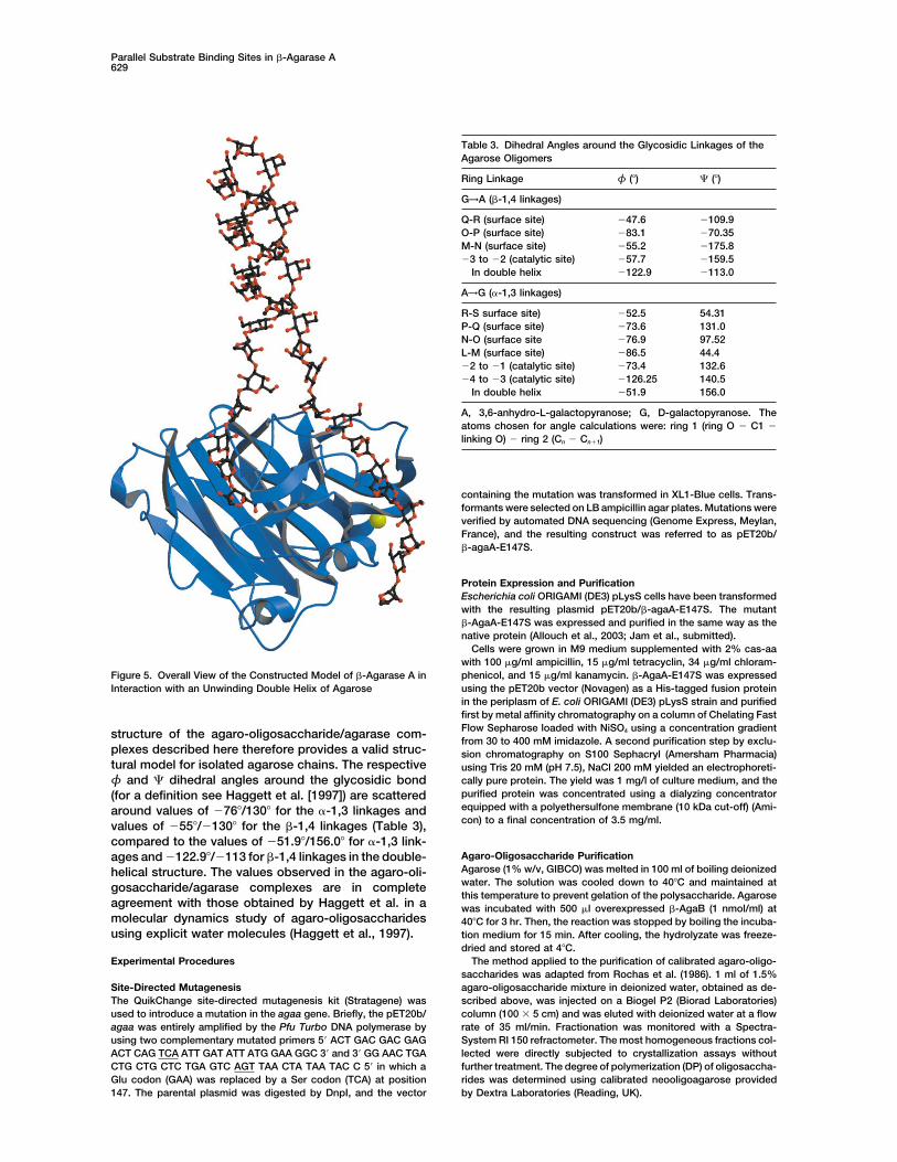
Interestingly, the oligosaccharide chains in the two bind- unwinding of the double-helical structure of agarose by�-agaA is reminiscent, but different, from the mode ofing sites are parallel to each other and their nonreducing
ends converge to only about 16 A from each other. The action of helicases that use ATP to processively unwindDNA (Delagoutte and von Hippel, 2002).curvature of the chains (Figure 3C) is such that their
continuation suggest that they could merge in the paral-lel double-helical structure of agarose (Arnott et al., Structure of Agarose
The structural model of double-helical agarose chains1974). In order to verify this (Figure 5), we have con-structed a model by extending the agarose chains be- has been obtained from low-resolution fiber diffraction
data (PDB idcode: 1aga) (Arnott et al., 1974). While af-yond the enzyme surface and ending up in the paralleldouble-helical structure (PDB code: 1aga). The chains fording a model of ordered junction zones, this approach
did not allow the description of the disordered regionscould be built readily with no steric clash, and withglycosidic bond torsional angles in the allowed regions separating the ordered double-helical regions and that
give rise to the three-dimensional network of agaroseof the Ramachandran plot of the agarose glycosidicbonds dihedral angles (Haggett et al., 1997). This gels. Moreover, because of the low resolution of the
fiber diffraction data, the very existence of double-strongly suggests that the enzyme may not only bindtwo parallel agarose chains, but that it may be able to helical regions has been challenged by some authors
(Guenet et al., 1993; Jimenez-Barbero et al., 1989). Thelocally separate the two strands of the parallel doublehelix of agarose by acting like a wedge. This is in agree- parallel binding sites that we have observed in the agaro-
oligosaccharide/agarase complexes strongly support thement with experimental data showing that �-agaA iscapable of degrading agarose gels (Jam et al., submit- parallel chain structure of Arnott (Arnott et al., 1974). The
structure of the complexes not only depicts the detailedted). A similar mode of action has already been proposedfor a starch binding domain from glucoamylase (Sori- interactions of a �-agarase with an agaro-oligosaccha-
ride, but also provides the first 1.7 A resolution structuremachi et al., 1997; Southall et al., 1999). The possible

Structure628
Figure 4. Schematic Description of the Ligand/Enzyme Interactions Highlighting the Residues Involved and Water Molecules
(A) Tetra-agaose in the catalytic active channel spanning subsites �4 to �1.(B) Octa-agarose at the surface binding site. The subsites are labeled L to R running from the reducing to the nonreducing end of theoligosaccharide. This figure was prepared with LIGPLOT (Wallace et al., 1995).
of an agaro-octaose single chain. In the single chain chain at the cleavage site, it is generally observed thatpolysaccharide binding proteins select a conformationstate, the agarose molecule is considerably more ex-
tended and less twisted than in the double-helical struc- of their ligands from a preexisting conformation occurringin solution (Imberty, 1997; Kogelberg et al., 2003). Theture. With the exception of the distortion of the substrate
Table 2. List of Direct Hydrogen Bonds between the Agarose Units and Enzyme Residues
Oligo-Agarose in the Catalytic Active Channel Oligo-Agarose at the Surface Binding Site
Residue Name Residue NameAgarose Atom and Atom Distance in A Agarose Atom and Atom Distance in A
AGL �4 O2 E-144 O2 2.77 AGL L O5 N-82 N�2 3.04O2 N-71 N�2 3.54 GAL M O4 N-82 N�2 3.20O3 N-71 O�1 3.21 O4 Q-85 O1 3.09O3 W-73 N 3.30 O4 Q-85 N2 3.70GAL �3 O2 E-144 O1 2.65 O5 Q-85 O1 3.11O2 Y-69 O� 3.8 O6 Q-98 N2 2.73O3 E-144 O1 3.57 AGL N O2 Q-98 N2 3.65O4 R-176 N�2 3.07 O3 Q-96 N2 3.59O5 R-176 N�2 2.94 O3 Q-85 O1 2.94AGL �2 O3 R-176 N�2 3.54 GAL O O2 D-271 O�2 2.75O3 R-176 N�1 2.98 O2 Q-96 O1 3.12O4 E-144 O1 3.57 AGL P O3 D-271 O�2 3.76GAL �1 O1 D-149 O�1 2.73 O4 D-271 O�2 3.56O1 D-149 O�1 3.48 GAL Q O4 N-89 N�2 3.71O1 S-147 O 3.58 O6 N-89 N�2 2.95O2 H-172 N�1 3.63 O6 Q-92 N2 3.2O2 H-172 N2 3.65 AGL R O2 K-127 N� 3.58O4 E-254 O1 2.65 O5 K-127 N� 3.40O4 E-254 O2 3.58 GAL S O3 K-127 N� 3.09O4 Q-256 O1 3.17 O4 K-127 N� 2.88
O6 R-199 O 3.39
Parallel Substrate Binding Sites in �-Agarase A629
Table 3. Dihedral Angles around the Glycosidic Linkages of theAgarose Oligomers
Ring Linkage φ (�) � (�)
G→A (�-1,4 linkages)
Q-R (surface site) �47.6 �109.9O-P (surface site) �83.1 �70.35M-N (surface site) �55.2 �175.8�3 to �2 (catalytic site) �57.7 �159.5
In double helix �122.9 �113.0
A→G (�-1,3 linkages)
R-S surface site) �52.5 54.31P-Q (surface site) �73.6 131.0N-O (surface site �76.9 97.52L-M (surface site) �86.5 44.4�2 to �1 (catalytic site) �73.4 132.6�4 to �3 (catalytic site) �126.25 140.5
In double helix �51.9 156.0
A, 3,6-anhydro-L-galactopyranose; G, D-galactopyranose. Theatoms chosen for angle calculations were: ring 1 (ring O � C1 �
linking O) � ring 2 (Cn � Cn1)
containing the mutation was transformed in XL1-Blue cells. Trans-formants were selected on LB ampicillin agar plates. Mutations wereverified by automated DNA sequencing (Genome Express, Meylan,France), and the resulting construct was referred to as pET20b/�-agaA-E147S.
Protein Expression and PurificationEscherichia coli ORIGAMI (DE3) pLysS cells have been transformedwith the resulting plasmid pET20b/�-agaA-E147S. The mutant�-AgaA-E147S was expressed and purified in the same way as thenative protein (Allouch et al., 2003; Jam et al., submitted).
Cells were grown in M9 medium supplemented with 2% cas-aawith 100 �g/ml ampicillin, 15 �g/ml tetracyclin, 34 �g/ml chloram-phenicol, and 15 �g/ml kanamycin. �-AgaA-E147S was expressedFigure 5. Overall View of the Constructed Model of �-Agarase A inusing the pET20b vector (Novagen) as a His-tagged fusion proteinInteraction with an Unwinding Double Helix of Agarosein the periplasm of E. coli ORIGAMI (DE3) pLysS strain and purifiedfirst by metal affinity chromatography on a column of Chelating FastFlow Sepharose loaded with NiSO4 using a concentration gradientstructure of the agaro-oligosaccharide/agarase com-from 30 to 400 mM imidazole. A second purification step by exclu-
plexes described here therefore provides a valid struc- sion chromatography on S100 Sephacryl (Amersham Pharmacia)tural model for isolated agarose chains. The respective using Tris 20 mM (pH 7.5), NaCl 200 mM yielded an electrophoreti-
cally pure protein. The yield was 1 mg/l of culture medium, and theφ and � dihedral angles around the glycosidic bondpurified protein was concentrated using a dialyzing concentrator(for a definition see Haggett et al. [1997]) are scatteredequipped with a polyethersulfone membrane (10 kDa cut-off) (Ami-around values of �76�/130� for the �-1,3 linkages andcon) to a final concentration of 3.5 mg/ml.values of �55�/�130� for the �-1,4 linkages (Table 3),
compared to the values of �51.9�/156.0� for �-1,3 link-Agaro-Oligosaccharide Purificationages and �122.9�/�113 for �-1,4 linkages in the double-Agarose (1% w/v, GIBCO) was melted in 100 ml of boiling deionizedhelical structure. The values observed in the agaro-oli-water. The solution was cooled down to 40�C and maintained atgosaccharide/agarase complexes are in completethis temperature to prevent gelation of the polysaccharide. Agarose
agreement with those obtained by Haggett et al. in a was incubated with 500 �l overexpressed �-AgaB (1 nmol/ml) atmolecular dynamics study of agaro-oligosaccharides 40�C for 3 hr. Then, the reaction was stopped by boiling the incuba-using explicit water molecules (Haggett et al., 1997). tion medium for 15 min. After cooling, the hydrolyzate was freeze-
dried and stored at 4�C.The method applied to the purification of calibrated agaro-oligo-Experimental Procedures
saccharides was adapted from Rochas et al. (1986). 1 ml of 1.5%agaro-oligosaccharide mixture in deionized water, obtained as de-Site-Directed Mutagenesis
The QuikChange site-directed mutagenesis kit (Stratagene) was scribed above, was injected on a Biogel P2 (Biorad Laboratories)column (100 � 5 cm) and was eluted with deionized water at a flowused to introduce a mutation in the agaa gene. Briefly, the pET20b/
agaa was entirely amplified by the Pfu Turbo DNA polymerase by rate of 35 ml/min. Fractionation was monitored with a Spectra-System RI 150 refractometer. The most homogeneous fractions col-using two complementary mutated primers 5� ACT GAC GAC GAG
ACT CAG TCA ATT GAT ATT ATG GAA GGC 3� and 3� GG AAC TGA lected were directly subjected to crystallization assays withoutfurther treatment. The degree of polymerization (DP) of oligosaccha-CTG CTG CTC TGA GTC AGT TAA CTA TAA TAC C 5� in which a
Glu codon (GAA) was replaced by a Ser codon (TCA) at position rides was determined using calibrated neooligoagarose providedby Dextra Laboratories (Reading, UK).147. The parental plasmid was digested by DnpI, and the vector

Structure630
Crystallization Cote d’Azur (France), Goemar (St. Malo, France), and the CentreNational de la Recherche Scientifique (CNRS, France).�-AgaA-E147S was concentrated to 3.5 mg/ml and stored in 50 mM
Tris buffer, pH 7.5, with 25 mM NaCl. Crystallization conditions werefirst investigated using two sparse-matrix sampling kits (Molecular Received: November 11, 2003
Revised: January 6, 2004Dimensions and Stura Footprint). Two crystal forms of the enzyme-substrate complex have been obtained. The first are issue of cocrys- Accepted: January 6, 2004
Published: April 6, 2004tallization trials of �-AgaA-E147S with agaro-octaose, purified asdescribed above. In this case, the crystallization solution contained30% PEG 4000, 200 mM ammonium acetate, and 100 mM sodium Referencesacetate, pH 4.6. Crystals were grown by mixing 2 vol of proteinsolution, equally containing about 4 mM agaro-octaose, with 1 vol Albenne, C., Skov, L.K., Mirza, O., Gajhede, M., Feller, G., D’Amico,
S., Andre, G., Potocki-Veronese, G., Van Der Veen, B.A., Monsan, P.,of precipitant solution in a hanging-drop vapor diffusion setup at20.5�C. Crystals grew within 1 week and are in space group P3221, et al . (2004). Molecular basis of the amylose-like polymer formation
catalyzed by Neisseria polysaccharea amylosucrase. J. Biol. Chem.,having unit cell parameters a � b � 51.38 A and c � 205.36 A.Thereafter, a single crystal was soaked for 30 s in successive solu- 279, 726–734.tions with increasing glycerol concentrations up to a maximum of Allouch, J., Jam, M., Helbert, W., Barbeyron, T., Kloareg, B., Henris-15%. sat, B., and Czjzek, M. (2003). The three-dimensional structures of
The second crystal form was obtained by soaking experiments. two �-agarases. J. Biol. Chem. 278, 47171–47180.First, crystals of the mutant protein �-AgaA-E147S were grown in Arnott, S., Fulmer, A., Scott, W.E., Dea, I.C., Moorhouse, R., andconditions close to those of the native enzyme (Allouch et al., 2003). Rees, D.A. (1974). The agarose double helix and its function in aga-The crystallization solution contained 24% PEG 4000, 160 mM am- rose gel structure. J. Mol. Biol. 90, 269–284.monium acetate, and 80 mM sodium acetate (pH 4.6) and led to
Bhattacharjee, S.S., Yaphe, W., and Hamer, G.K. (1978). 13C-n.m.r.crystals which belonged to space group P21 with unit cell parametersspectroscopic analysis of agar, kappa-carrageenan and iota-carra-a � 42.2 A, b � 135.0 A, c � 49.8 A, and � � 101.8 A. Thesegeenan. Carbohydr. Res. 60, C1–C3.single crystals were then transferred to a mother liquor containingBourne, Y., and Henrissat, B. (2001). Glycoside hydrolases and gly-approximately 10 mM agaro-dodecaose during one night and sub-cosyltransferases: families and functional modules. Curr. Opin.sequently soaked in 10% glycerol solution, prior to flash freezing.Struct. Biol. 11, 593–600.
Burmeister, W.P., Cottaz, S., Driguez, H., Iori, R., Palmieri, S., andData Collection and RefinementHenrissat, B. (1997). The crystal structures of Sinapis alba myrosi-Diffraction data at 1.7 A resolution were collected on beamlinesnase and a covalent glycosyl-enzyme intermediate provide insightsID14-EH1 and EH4 (ESRF, Grenoble, France) for �-AgaA-E147S/into the substrate recognition and active-site machinery of anagaro-octaose and �-AgaA-E147S/agaro-dodecaose complexes,S-glycosidase. Structure 5, 663–675.respectively.CCP4 (Collaborative Computational Project Number 4) (1994). TheThe �-AgaA-E147S/agaro-octaose cocrystals (P3221) containedCCP4 suite: programs for protein crystallography. Acta Crystallogr.one molecule in the asymmetric unit, whereas for �-AgaA-E147S/D 50, 760–763.agaro-dodecaose (P21), two molecules were present in the asym-
metric unit. In both cases, the structure was solved by molecular Czjzek, M., Cicek, M., Zamboni, V., Bevan, D.R., Henrissat, B., andreplacement, using AMoRe (CCP4, 1994; Navaza, 1994) and Esen, A. (2000). The mechanism of substrate (aglycone) specificity�-AgaA_CM as search models. The respective correlation and R in beta-glucosidases is revealed by crystal structures of mutantfactors of the molecular replacement are 64.9% and 34.2% for one maize beta-glucosidase-DIMBOA, -DIMBOAGlc, and -dhurrin com-solution for �-AgaA-E147S/agaro-octaose and 59.2% and 36.7% plexes. Proc. Natl. Acad. Sci. USA 97, 13555–13560.for two solutions for �-AgaA-E147S/agaro-dodecaose. Davies, G.J., Tolley, S.P., Henrissat, B., Hjort, C., and Schulein, M.
Both �-AgaA-E147S/agaro-octaose and �-AgaA-E147S/agaro- (1995). Structures of oligosaccharide-bound forms of the endoglu-dodecaose were built manually using TURBO (Roussel and Cambil- canase V from Humicola insolens at 1.9 A resolution. Biochemistrylau, 1991). The refinement was performed with REFMAC 5 (CCP4, 34, 16210–16220.1994). Water molecules were added using CCP4/wARP (Perrakis et
Davies, G.J., Wilson, K.S., and Henrissat, B. (1997). Nomenclatureal., 1997). The stereochemistry of the final structures was evaluatedfor sugar-binding subsites in glycosyl hydrolases. Biochem. J. 321,using PROCHECK (Laskowski et al., 1993). All refinement statistics557–559.are summarized in Table 1.Davies, G.J., Mackenzie, L., Varrot, A., Dauter, M., Brzozowski, A.M.,Schulein, M., and Withers, S.G. (1998). Snapshots along an enzy-
Construction of the Model of the �-AgaA in Complex matic reaction coordinate: analysis of a retaining beta-glycosidewith Partially Unwound Helical Agarose hydrolase. Biochemistry 37, 15280–15287.The model of �-agaA in complex with partially unwound helical
De Ruiter, G.A., and Rudolph, B. (1997). Carrageenan biotechnology.agarose was obtained by simple duplication of the agaro-octaoseTrends Food Sci. Technol. 8, 389–395.and agaro-tetraose units, as defined in the structural complex.Delagoutte, E., and von Hippel, P.H. (2002). Helicase mechanismsThese duplicated parts were then translated and rotated in a wayand the coupling of helicases within macromolecular machines. Partthat they could be connected to the nonreducing ends of the twoI: structures and properties of isolated helicases. Q. Rev. Biophys.oligosaccharides in the complex structure respectively, with respec-35, 431–478.tive φ and � dihedral angles in the allowed region. These ends
were then joined to the model of a double-helical agarose unit, the Ducros, V.M., Zechel, D.L., Murshudov, G.N., Gilbert, H.J., Szabo,L., Stoll, D., Withers, S.G., and Davies, G.J. (2002). Substrate distor-coordinates of which were accessible through the Protein Data Bank
(accession number 1aga). The model construction was performed tion by a beta-mannanase: snapshots of the Michaelis and covalent-intermediate complexes suggest a B(2,5) conformation for the tran-using the program TURBO (Roussel and Cambillau, 1991).sition state. Angew. Chem. Int. Ed. Engl. 41, 2824–2827.
Ducros, V.M., Tarling, C.A., Zechel, D.L., Brzozowski, A.M., Frand-Acknowledgmentssen, T.P., von Ossowski, I., Schulein, M., Withers, S.G., and Davies,G.J. (2003). Anatomy of glycosynthesis: structure and kinetics of theThe authors wish to thank Murielle Jam, Tristan Barbeyron, BernardHumicola insolens Cel7B E197A and E197S glycosynthase mutants.Kloareg, and Gurvan Michel (Station Biologique, Roscoff, France)Chem. Biol. 10, 619–628.for useful discussions. We would also like to thank Pedro Coutinho
for helpful advice and discussions. We are also indebted to the Gill, J., Rixon, J.E., Bolam, D.N., McQueen-Mason, S., Simpson,P.J., Williamson, M.P., Hazlewood, G.P., and Gilbert, H.J. (1999). TheESRF staff on beamlines ID14-EH1 and EH4 for help during data
collections. This work was funded by the Region Provence-Alpes- type II and X cellulose-binding domains of Pseudomonas xylanase A

Parallel Substrate Binding Sites in �-Agarase A631
potentiate catalytic activity against complex substrates by a com- phile by trapping of the covalent glycosyl-enzyme intermediate. Bio-chem. J. 335, 409–416.mon mechanism. Biochem. J. 342, 473–480.
Michel, G., Chantalat, L., Duee, E., Barbeyron, T., Henrissat, B.,Guenet, J.M., Brulet, A., and Rochas, C. (1993). Agarose chain con-Kloareg, B., and Dideberg, O. (2001). The kappa-carrageenase offormation in the sol state by neutron scattering. Int. J. Biol. Mac-P. carrageenovora features a tunnel-shaped active site: a novelromol. 15, 131–132.insight in the evolution of Clan-B glycoside hydrolases. StructureHadfield, A.T., Harvey, D.J., Archer, D.B., MacKenzie, D.A., Jeenes,9, 513–525.D.J., Radford, S.E., Lowe, G., Dobson, C.M., and Johnson, L.N.Navaza, J. (1994). AmoRe: an automated package for molecular(1994). Crystal structure of the mutant D52S hen egg white lysozymereplacement. Acta Crystallogr. A 50, 157–163.with an oligosaccharide product. J. Mol. Biol. 243, 856–872.Notenboom, V., Birsan, C., Nitz, M., Rose, D.R., Warren, R.A., andHaggett, N.M.W., Hoffmann, R.A., Howlin, B.J., and Webb, G.A.Withers, S.G. (1998). Insights into transition state stabilization of the(1997). Molecular modelling of six-ring agarose chains: effects ofbeta-1,4-glycosidase Cex by covalent intermediate accumulation inexplicit and implicit solvent. J. Mol.Model. 3, 301–310.active site mutants. Nat. Struct. Biol. 5, 812–818.
Hahn, M., Olsen, O., Politz, O., Borriss, R., and Heinemann, U.Numao, S., Kuntz, D.A., Withers, S.G., and Rose, D.R. (2003). Insights(1995a). Crystal structure and site-directed mutagenesis of Bacillusinto the mechanism of drosophila melanogaster Golgi �-manno-macerans endo-1,3–1,4-beta-glucanase. J. Biol. Chem. 270, 3081–sidase through the structural analysis of covalent reaction interme-3088.diates. J. Biol. Chem. 278, 48074–48083.
Hahn, M., Pons, J., Planas, A., Querol, E., and Heinemann, U. (1995b).Perrakis, A., Sixma, T.K., Wilson, K.S., and Lamzin, V.S. (1997).Crystal structure of Bacillus licheniformis 1,3–1,4-beta-D-glucanwARP: improvement and extension of crystallographic phases by4-glucanhydrolase at 1.8 A resolution. FEBS Lett. 374, 221–224.weightedaveraging of multiple-refined dummy atomic models. Acta
Henrissat, B., and Davies, G.J. (2000). Glycoside hydrolases and Crystallogr D 53, 448–455.glycosyltransferases. Families, modules, and implications for geno-
Potin, P., Richard, C., Rochas, C., and Kloareg, B. (1993). Purificationmics. Plant Physiol. 124, 1515–1519.
and characterization of the alpha-agarase from Alteromonas agar-Imberty, A. (1997). Oligosaccharide structures: theory versus experi- lyticus (Cataldi) comb. nov., strain GJ1B. Eur. J. Biochem. 214,ment. Curr. Opin. Struct. Biol. 7, 617–623. 599–607.Jahn, M., Stoll, D., Warren, R.A., Szabo, L., Singh, P., Gilbert, H.J., Robert, X., Haser, R., Gottschalk, T.E., Ratajczak, F., Driguez, H.,Ducros, V.M., Davies, G.J., and Withers, S.G. (2003). Expansion of Svensson, B., and Aghajari, N. (2003). The structure of barley alpha-the glycosynthase repertoire to produce defined manno-oligosac- amylase isozyme 1 reveals a novel role of domain C in substratecharides. Chem. Commun. (Camb.), 1327–1329. recognition and binding: a pair of sugar tongs. Structure 11,
973–984.Jimenez-Barbero, J., Bouffar-Roupe, C., Rochas, C., and Perez, S.(1989). Modelling studies of solvent effects on the conformational Rochas, C., Lahaye, M., Yaphe, W., and Phan Viet, M.T. (1986). 13Cstability of agarobiose and neoagarobiose and their relationship to N.M.R.-spectroscopic investigation of agarose oligomers. Carbo-agarose. Int. J. Biol. Macromol. 11, 265–272. hydr. Res. 148, 199–207.
Johansson, P., Denman, S., Brumer, H., Kallas, A.M., Henriksson, Roussel, A., and Cambillau, C. (1991). TURBO-FRODO program. InH., Bergfors, T., Teeri, T.T., and Jones, T.A. (2003). Crystallization Silicon Graphics Geometry Partners Directory 88. (Mountain View,and preliminary X-ray analysis of a xyloglucan endotransglycosylase CA: Silicon Graphics).from Populus tremula x tremuloides. Acta Crystallogr. D 59, 535–537. Skov, L.K., Mirza, O., Sprogoe, D., Dar, I., Remaud-Simeon, M.,
Albenne, C., Monsan, P., and Gajhede, M. (2002). OligosaccharideJuncosa, M., Pons, J., Dot, T., Querol, E., and Planas, A. (1994).and sucrose complexes of amylosucrase. Structural implicationsIdentification of active site carboxylic residues in Bacillus licheni-for the polymerase activity. J. Biol. Chem. 277, 47741–47747.formis 1,3–1,4-beta-D-glucan 4-glucanhydrolase by site-directed
mutagenesis. J. Biol. Chem. 269, 14530–14535. Sorimachi, K., Le Gal-Coeffet, M.F., Williamson, G., Archer, D.B.,and Williamson, M.P. (1997). Solution structure of the granular starchKadziola, A., Abe, J., Svensson, B., and Haser, R. (1994). Crystalbinding domain of Aspergillus niger glucoamylase bound to beta-and molecular structure of barley alpha-amylase. J. Mol. Biol. 239,cyclodextrin. Structure 5, 647–661.104–121.Southall, S.M., Simpson, P.J., Gilbert, H.J., Williamson, G., and Wil-Kadziola, A., Sogaard, M., Svensson, B., and Haser, R. (1998). Molec-liamson, M.P. (1999). The starch-binding domain from glucoamylaseular structure of a barley alpha-amylase-inhibitor complex: implica-disrupts the structure of starch. FEBS Lett. 447, 58–60.tions for starch binding and catalysis. J. Mol. Biol. 278, 205–217.Sulzenbacher, G., Driguez, H., Henrissat, B., Schulein, M., andKeitel, T., Simon, O., Borriss, R., and Heinemann, U. (1993). Molecu-Davies, G.J. (1996). Structure of the Fusarium oxysporum endoglu-lar and active-site structure of a Bacillus 1,3–1,4-beta-glucanase.canase I with a nonhydrolyzable substrate analogue: substrate dis-Proc. Natl. Acad. Sci. USA 90, 5287–5291.tortion gives rise to the preferred axial orientation for the leaving
Kloareg, B., and Quatrano, R.S. (1988). Structure of the cell walls group. Biochemistry 35, 15280–15287.of marine algae and ecophysiological function of the matrix polysac-
Tews, I., Perrakis, A., Oppenheim, A., Dauter, Z., Wilson, K.S., andcharides. Oceanogr. Mar. Biol. Annu. Rev. 26, 259–315.Vorgias, C.E. (1996). Bacterial chitobiase structure provides insight
Kogelberg, H., Soli, D., and Jimenez-Barbero, J. (2003). New struc- into catalytic mechanism and thes basis of Tay-Sachs disease. Nat.tural insights into carbohydrate-protein interactions from NMR Struct. Biol. 3, 638–648.spectroscopy. Curr. Opin. Struct. Biol. 13, 646–653.
Tomme, P., Driver, D.P., Amandoron, E.A., Miller, R.C., Jr., Antony,Koshland, D.E. (1953). Stereochemistry and the mechanism of enzy- R., Warren, J., and Kilburn, D.G. (1995). Comparison of a fungalmatic reactions. Biol. Rev. Camb. Philos. Soc. 28, 416–436. (family I) and bacterial (family II) cellulose-binding domain. J. Bacte-
riol. 177, 4356–4363.Kraulis, P.J. (1991). MOLSCRIPT: a program to produce both de-tailed and schematic plots of protein structures. J. Appl. Crystallogr. van Aalten, D.M.F., Komander, D., Synstad, B., Gaseidnes, S., Peter,24, 946–950. M.G., and Eijsink, V.G.H. (2001). Strutcural insights into the catalytic
mechanism of a family 18 exo-chitinase. Proc. Natl. Acad. Sci. USALaskowski, R.A., MacArthur, M.W., Moss, D.S., and Thornton, J.M.98, 8979–8984.(1993). PROCHECK: a program to check the stereochemical quality
of protein structures. J. Appl. Crystallogr. 26, 283–291. Varrot, A., and Davies, G.J. (2003). Direct experimental observationof the hydrogen-bonding network of a glycosidase along its reactionMacKenzie, L.F., Sulzenbacher, G., Divne, C., Jones, T.A., Woldike,coordinate revealed by atomic resolution analyses of endogluca-H.F., Schulein, M., Withers, S.G., and Davies, G.J. (1998). Crystalnase Cel5A. Acta Crystallogr D 59, 447–452.structure of the family 7 endoglucanase I (Cel7B) from Humicola
insolens at 2.2 A resolution and identification of the catalytic nucleo- Varrot, A., Frandsen, T., Driguez, H., and Davies, G. (2002). Structure
Structure632
of the Humicola insolens cellobiohydrolase Cel6A D416A mutantin complex with a non-hydrolysable substrate analogue, methyl-cellobiosyl-4-thio-beta-cellobioside. Acta Crystallogr D 58, 2201–2204.
Vocadlo, D.J., Davies, G.J., Laine, R., and Withers, S.G. (2001). Catal-ysis by hen egg white lysozyme proceeds via a covalent intermedi-ate. Nature 412, 835–838.
Wallace, A.C., Laskowski, R.A., and Thornton, J.M. (1995). LIGPLOT:a program to generate schematic diagrams of protein-ligand interac-tions. Protein Eng. 8, 127–134.
White, A., Tull, D., Johns, K., Withers, S.G., and Rose, D.R. (1996).Crystallographic observation of a covalent catalytic intermediate ina beta-glycosidase. Nat. Struct. Biol. 3, 149–154.
Accession Numbers
Coordinates and observed structure factor amplitudes have beendeposited in the Protein Data Bank (accession code 1URX).