p31comet Blocks Mad2 Activation through Structural Mimicry
12
p31 comet Blocks Mad2 Activation through Structural Mimicry Maojun Yang, 1 Bing Li, 1 Diana R. Tomchick, 2 Mischa Machius, 2 Josep Rizo, 1,2 Hongtao Yu, 1 and Xuelian Luo 1, * 1 Department of Pharmacology 2 Department of Biochemistry The University of Texas Southwestern Medical Center, 6001 Forest Park Road, Dallas, TX 75390, USA *Correspondence: [email protected] DOI 10.1016/j.cell.2007.08.048 SUMMARY The status of spindle checkpoint signaling depends on the balance of two opposing dy- namic processes that regulate the highly un- usual two-state behavior of Mad2. In mitosis, a Mad1-Mad2 core complex recruits cytosolic Mad2 to kinetochores through Mad2 dimeri- zation and converts Mad2 to a conformer ame- nable to Cdc20 binding, thereby facilitating checkpoint activation. p31 comet inactivates the checkpoint through binding to Mad1- or Cdc20- bound Mad2, thereby preventing Mad2 acti- vation and promoting the dissociation of the Mad2-Cdc20 complex. Here, we report the crystal structure of the Mad2-p31 comet complex. The C-terminal region of Mad2 that undergoes rearrangement in different Mad2 conformers is a major structural determinant for p31 comet bind- ing, explaining the specificity of p31 comet to- ward Mad1- or Cdc20-bound Mad2. p31 comet adopts a fold strikingly similar to that of Mad2 and binds at the dimerization interface of Mad2. Thus, p31 comet exploits the two-state be- havior of Mad2 to block its activation by acting as an ‘‘anti-Mad2.’’ INTRODUCTION A cell-cycle surveillance mechanism called the spindle checkpoint monitors the proper bipolar attachment of sister chromatids to spindle microtubules and ensures the fidelity of chromosome segregation during mitosis (Bharadwaj and Yu, 2004; Musacchio and Hardwick, 2002; Musacchio and Salmon, 2007; Yu, 2002). Check- point-dependent inhibition of a multisubunit uiquitin li- gase, the anaphase-promoting complex or cyclosome (APC/C), requires the direct binding of Mad2 to the mitotic activator of APC/C, Cdc20 (Fang et al., 1998; Hwang et al., 1998; Kim et al., 1998; Yu, 2007). Cytosolic Mad2 has an autoinhibited conformation, called N1-Mad2 or open- Mad2 (hereafter referred to as O-Mad2) that is kinetically unfavorable for Cdc20 binding (De Antoni et al., 2005; Luo et al., 2000, 2004; Yu, 2006). Upon binding to Cdc20, Mad2 undergoes a large structural change to reach the N2- or closed-Mad2 conformation (hereafter referred to as C-Mad2). Mad1—an upstream regulator of Mad2—forms a tight core complex with Mad2. In the Mad1-Mad2 complex, Mad2 also adopts the C-Mad2- conformation (Luo et al., 2002; Sironi et al., 2002). In mito- sis, the kinetochore-bound Mad1-Mad2 core complex recruits another copy of cytosolic O-Mad2 through C- Mad2–O-Mad2 dimerization (De Antoni et al., 2005; Shah et al., 2004). All available data support the following model for Mad2 activation. In this model, the Mad1-Mad2 core complex converts O-Mad2 to an intermediate Mad2 conformer (referred to as I-Mad2) that can directly bind to Cdc20 and complete the open-to-closed rearrangement (Figure 1A). Alternatively, I-Mad2 on its own can convert to an unliganded C-Mad2 that dissociates from the Mad1- Mad2 core complex, binds subsequently to Cdc20, and is more active in APC/C inhibition in vitro (Luo et al., 2004; Yu, 2006). The p31 comet protein binds to both Mad1- and Cdc20- bound C-Mad2, but not to O-Mad2 (Figure 1A; Xia et al., 2004). Through binding to Mad1-bound C-Mad2, p31 comet blocks the recruitment of O-Mad2 to the Mad1-Mad2 core complex and thereby prevents Mad1-assisted structural activation of Mad2 (Mapelli et al., 2006). Through binding to Cdc20-bound C-Mad2, p31 comet neutralizes the APC/ C-inhibitory function of Mad2 and, in collaboration with the ubiquitin-conjugating enzyme UbcH10, promotes the autoubiquitination of Cdc20 and the disassembly of Mad2-Cdc20-containing checkpoint complexes (Reddy et al., 2007; Stegmeier et al., 2007; Xia et al., 2004). Thus, Mad2 is a two-state protein with an intermediate confor- mation of finite lifetime; it is positively regulated by Mad1 and inhibited by p31 comet (Musacchio and Salmon, 2007; Yu, 2006). By opposing the Mad1-assisted structural acti- vation of Mad2 and promoting the disassembly of the Mad2-Cdc20 complex, p31 comet -dependent inhibition of Mad2 sets the threshold for checkpoint activation and enables rapid checkpoint inactivation after the proper attachment of all sister chromatids to the mitotic spindle. To investigate how p31 comet achieves its conforma- tion-specific binding to Mad2 and how it prevents the Mad1-assisted structural activation of Mad2, we have 744 Cell 131, 744–755, November 16, 2007 ª2007 Elsevier Inc.
-
Upload
utsouthwestern -
Category
Documents
-
view
2 -
download
0
Transcript of p31comet Blocks Mad2 Activation through Structural Mimicry
p31comet Blocks Mad2 Activationthrough Structural MimicryMaojun Yang,1 Bing Li,1 Diana R. Tomchick,2 Mischa Machius,2 Josep Rizo,1,2 Hongtao Yu,1 and Xuelian Luo1,*1Department of Pharmacology2Department of Biochemistry
The University of Texas Southwestern Medical Center, 6001 Forest Park Road, Dallas, TX 75390, USA
*Correspondence: [email protected] 10.1016/j.cell.2007.08.048
SUMMARY
The status of spindle checkpoint signalingdepends on the balance of two opposing dy-namic processes that regulate the highly un-usual two-state behavior of Mad2. In mitosis,a Mad1-Mad2 core complex recruits cytosolicMad2 to kinetochores through Mad2 dimeri-zation and converts Mad2 to a conformer ame-nable to Cdc20 binding, thereby facilitatingcheckpoint activation. p31comet inactivates thecheckpoint through binding to Mad1- or Cdc20-bound Mad2, thereby preventing Mad2 acti-vation and promoting the dissociation of theMad2-Cdc20 complex. Here, we report thecrystal structure of the Mad2-p31comet complex.The C-terminal region of Mad2 that undergoesrearrangement in different Mad2 conformers is amajor structural determinant for p31comet bind-ing, explaining the specificity of p31comet to-ward Mad1- or Cdc20-bound Mad2. p31comet
adopts a fold strikingly similar to that of Mad2and binds at the dimerization interface ofMad2. Thus, p31comet exploits the two-state be-havior of Mad2 to block its activation by actingas an ‘‘anti-Mad2.’’
INTRODUCTION
A cell-cycle surveillance mechanism called the spindle
checkpoint monitors the proper bipolar attachment of
sister chromatids to spindle microtubules and ensures
the fidelity of chromosome segregation during mitosis
(Bharadwaj and Yu, 2004; Musacchio and Hardwick,
2002; Musacchio and Salmon, 2007; Yu, 2002). Check-
point-dependent inhibition of a multisubunit uiquitin li-
gase, the anaphase-promoting complex or cyclosome
(APC/C), requires the direct binding of Mad2 to the mitotic
activator of APC/C, Cdc20 (Fang et al., 1998; Hwang et al.,
1998; Kim et al., 1998; Yu, 2007). Cytosolic Mad2 has an
autoinhibited conformation, called N1-Mad2 or open-
Mad2 (hereafter referred to as O-Mad2) that is kinetically
744 Cell 131, 744–755, November 16, 2007 ª2007 Elsevier Inc
unfavorable for Cdc20 binding (De Antoni et al., 2005;
Luo et al., 2000, 2004; Yu, 2006). Upon binding to
Cdc20, Mad2 undergoes a large structural change to
reach the N2- or closed-Mad2 conformation (hereafter
referred to as C-Mad2). Mad1—an upstream regulator of
Mad2—forms a tight core complex with Mad2. In the
Mad1-Mad2 complex, Mad2 also adopts the C-Mad2-
conformation (Luo et al., 2002; Sironi et al., 2002). In mito-
sis, the kinetochore-bound Mad1-Mad2 core complex
recruits another copy of cytosolic O-Mad2 through C-
Mad2–O-Mad2 dimerization (De Antoni et al., 2005;
Shah et al., 2004). All available data support the following
model for Mad2 activation. In this model, the Mad1-Mad2
core complex converts O-Mad2 to an intermediate Mad2
conformer (referred to as I-Mad2) that can directly bind to
Cdc20 and complete the open-to-closed rearrangement
(Figure 1A). Alternatively, I-Mad2 on its own can convert
to an unliganded C-Mad2 that dissociates from the Mad1-
Mad2 core complex, binds subsequently to Cdc20, and is
more active in APC/C inhibition in vitro (Luo et al., 2004;
Yu, 2006).
The p31comet protein binds to both Mad1- and Cdc20-
bound C-Mad2, but not to O-Mad2 (Figure 1A; Xia et al.,
2004). Through binding to Mad1-bound C-Mad2, p31comet
blocks the recruitment of O-Mad2 to the Mad1-Mad2 core
complex and thereby prevents Mad1-assisted structural
activation of Mad2 (Mapelli et al., 2006). Through binding
to Cdc20-bound C-Mad2, p31comet neutralizes the APC/
C-inhibitory function of Mad2 and, in collaboration with
the ubiquitin-conjugating enzyme UbcH10, promotes the
autoubiquitination of Cdc20 and the disassembly of
Mad2-Cdc20-containing checkpoint complexes (Reddy
et al., 2007; Stegmeier et al., 2007; Xia et al., 2004). Thus,
Mad2 is a two-state protein with an intermediate confor-
mation of finite lifetime; it is positively regulated by Mad1
and inhibited by p31comet (Musacchio and Salmon, 2007;
Yu, 2006). By opposing the Mad1-assisted structural acti-
vation of Mad2 and promoting the disassembly of the
Mad2-Cdc20 complex, p31comet-dependent inhibition of
Mad2 sets the threshold for checkpoint activation and
enables rapid checkpoint inactivation after the proper
attachment of all sister chromatids to the mitotic spindle.
To investigate how p31comet achieves its conforma-
tion-specific binding to Mad2 and how it prevents the
Mad1-assisted structural activation of Mad2, we have
.

Figure 1. Structure of the Mad2-p31comet Complex
(A) Schematic drawing of the proposed mechanisms of Mad2 activation by the Mad1-Mad2 core complex and the inhibition of this process by
p31comet. Upon checkpoint activation, autoinhibited O-Mad2 binds to the Mad1-Mad2 core complex through Mad2-Mad2 dimerization, which
induces a conformational change of O-Mad2 and converts it into an activated intermediate state (I-Mad2). I-Mad2 dissociates from the Mad1-
Mad2 core complex to become the active conformer, C-Mad2, with or without Cdc20. During checkpoint inactivation, p31comet binds to the
Mad1-Mad2 core complex and blocks the binding of O-Mad2, thereby preventing the generation of I-Mad2 and C-Mad2. p31comet also binds to
Cdc20-bound Mad2 and activates APC/C. The symbols used for different Mad2 conformers are shown in the shaded yellow box. The Mad2-binding
motif of Mad1 is colored red.
(B) Ribbon diagram of the Mad2-p31comet complex in two views. Mad2, p31comet, and MBP1 are colored blue, orange, and red, respectively. The N
and C termini of Mad2 and p31comet are labeled. All structural figures were generated with PyMOL (http://www.pymol.org).
determined the crystal structure of the human Mad2-
p31comet complex bound to a high-affinity Mad2-binding
peptide (MBP1) (Luo et al., 2002). Our study provides the
structural basis for the inhibition of Mad2 activation by
p31comet. Our structure reveals that p31comet adopts
a fold that is highly similar to Mad2 and binds at the
Mad2 dimerization interface. Therefore, p31comet blocks
the Mad1-assisted Mad2 activation by acting as a struc-
tural mimic of Mad2.
RESULTS AND DISCUSSION
Structure Determination and Overviewof the Mad2-p31comet ComplexThe wild-type human Mad2 protein adopts two conforma-
tions, O-Mad2 and C-Mad2, which exist in equilibrium
(Luo et al., 2004). Perhaps because of this inherent
conformational heterogeneity, we were unable to crystal-
C
lize the wild-type Mad2-p31comet complex. We circum-
vented this problem by replacing L13 in Mad2 with an
alanine. L13 is located in strand b1 of O-Mad2, whereas
this region is disordered in C-Mad2. We surmised that
the L13A mutation selectively destabilizes the open
conformer of Mad2. Indeed, Mad2 L13A exclusively adop-
ted the C-Mad2 conformation and bound to both Cdc20
and p31comet with affinities comparable to those of the
wild-type Mad2 (data not shown). In addition, the N-
terminal region of p31comet is not conserved in other spe-
cies (Figure S1 in the Supplemental Data available online)
and was dispensable for Mad2 binding (data not shown).
The N-terminal�40 residues of p31comet adopted a flexible
conformationbased on nuclear magnetic resonance (NMR)
spectroscopy (data not shown). We therefore coexpressed
Mad2 L13A and a truncation mutant of p31comet lacking its
N-terminal 35 residues (p31cometDN35; residues 36–274)
and purified the resulting complex. To further eliminate
ell 131, 744–755, November 16, 2007 ª2007 Elsevier Inc. 745
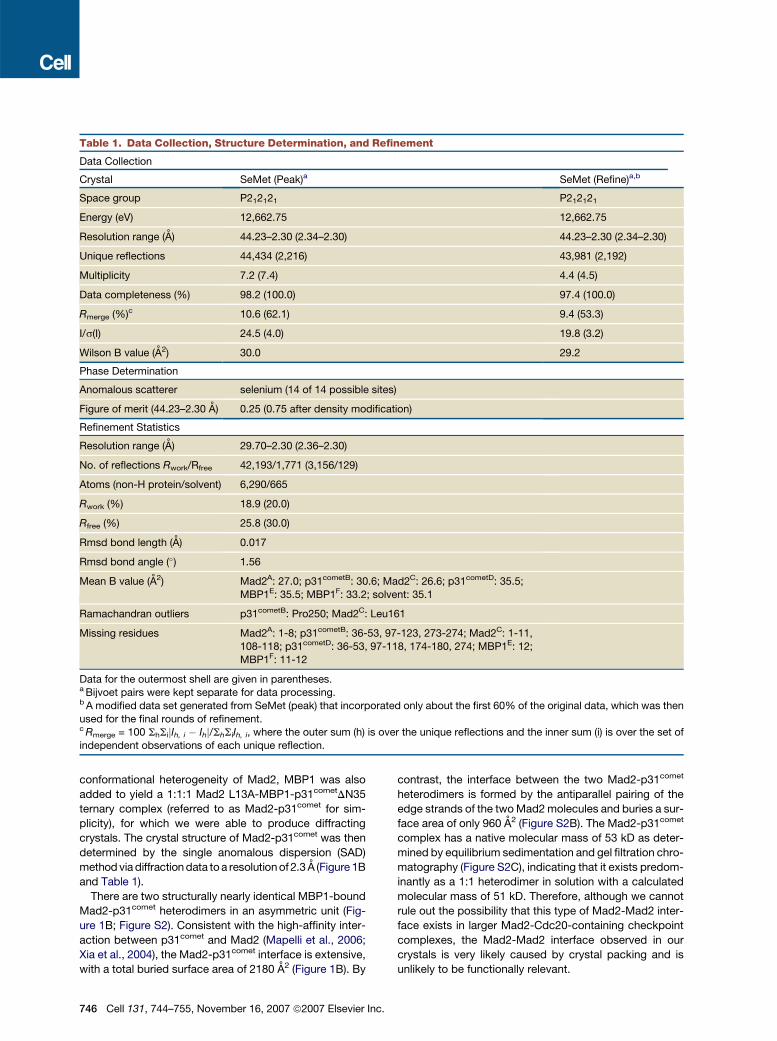
Table 1. Data Collection, Structure Determination, and Refinement
Data Collection
Crystal SeMet (Peak)a SeMet (Refine)a,b
Space group P212121 P212121
Energy (eV) 12,662.75 12,662.75
Resolution range (A) 44.23–2.30 (2.34–2.30) 44.23–2.30 (2.34–2.30)
Unique reflections 44,434 (2,216) 43,981 (2,192)
Multiplicity 7.2 (7.4) 4.4 (4.5)
Data completeness (%) 98.2 (100.0) 97.4 (100.0)
Rmerge (%)c 10.6 (62.1) 9.4 (53.3)
I/s(I) 24.5 (4.0) 19.8 (3.2)
Wilson B value (A2) 30.0 29.2
Phase Determination
Anomalous scatterer selenium (14 of 14 possible sites)
Figure of merit (44.23–2.30 A) 0.25 (0.75 after density modification)
Refinement Statistics
Resolution range (A) 29.70–2.30 (2.36–2.30)
No. of reflections Rwork/Rfree 42,193/1,771 (3,156/129)
Atoms (non-H protein/solvent) 6,290/665
Rwork (%) 18.9 (20.0)
Rfree (%) 25.8 (30.0)
Rmsd bond length (A) 0.017
Rmsd bond angle (�) 1.56
Mean B value (A2) Mad2A: 27.0; p31cometB: 30.6; Mad2C: 26.6; p31cometD: 35.5;
MBP1E: 35.5; MBP1F: 33.2; solvent: 35.1
Ramachandran outliers p31cometB: Pro250; Mad2C: Leu161
Missing residues Mad2A: 1-8; p31cometB: 36-53, 97-123, 273-274; Mad2C: 1-11,108-118; p31cometD: 36-53, 97-118, 174-180, 274; MBP1E: 12;
MBP1F: 11-12
Data for the outermost shell are given in parentheses.a Bijvoet pairs were kept separate for data processing.b A modified data set generated from SeMet (peak) that incorporated only about the first 60% of the original data, which was then
used for the final rounds of refinement.c Rmerge = 100 ShSijIh, i � Ihj/ShSiIh, i, where the outer sum (h) is over the unique reflections and the inner sum (i) is over the set of
independent observations of each unique reflection.
conformational heterogeneity of Mad2, MBP1 was also
added to yield a 1:1:1 Mad2 L13A-MBP1-p31cometDN35
ternary complex (referred to as Mad2-p31comet for sim-
plicity), for which we were able to produce diffracting
crystals. The crystal structure of Mad2-p31comet was then
determined by the single anomalous dispersion (SAD)
method via diffraction data to a resolution of 2.3 A (Figure 1B
and Table 1).
There are two structurally nearly identical MBP1-bound
Mad2-p31comet heterodimers in an asymmetric unit (Fig-
ure 1B; Figure S2). Consistent with the high-affinity inter-
action between p31comet and Mad2 (Mapelli et al., 2006;
Xia et al., 2004), the Mad2-p31comet interface is extensive,
with a total buried surface area of 2180 A2 (Figure 1B). By
746 Cell 131, 744–755, November 16, 2007 ª2007 Elsevier Inc
contrast, the interface between the two Mad2-p31comet
heterodimers is formed by the antiparallel pairing of the
edge strands of the two Mad2 molecules and buries a sur-
face area of only 960 A2 (Figure S2B). The Mad2-p31comet
complex has a native molecular mass of 53 kD as deter-
mined by equilibrium sedimentation and gel filtration chro-
matography (Figure S2C), indicating that it exists predom-
inantly as a 1:1 heterodimer in solution with a calculated
molecular mass of 51 kD. Therefore, although we cannot
rule out the possibility that this type of Mad2-Mad2 inter-
face exists in larger Mad2-Cdc20-containing checkpoint
complexes, the Mad2-Mad2 interface observed in our
crystals is very likely caused by crystal packing and is
unlikely to be functionally relevant.
.
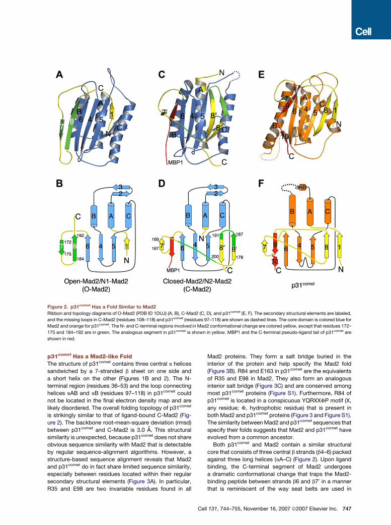
Figure 2. p31comet Has a Fold Similar to Mad2
Ribbon and topology diagrams of O-Mad2 (PDB ID 1DUJ) (A, B), C-Mad2 (C, D), and p31comet (E, F). The secondary structural elements are labeled,
and the missing loops in C-Mad2 (residues 108–118) and p31comet (residues 97–118) are shown as dashed lines. The core domain is colored blue for
Mad2 and orange for p31comet. The N- and C-terminal regions involved in Mad2 conformational change are colored yellow, except that residues 172–
175 and 184–192 are in green. The analogous segment in p31comet is shown in yellow. MBP1 and the C-terminal pseudo-ligand tail of p31comet are
shown in red.
p31comet Has a Mad2-like FoldThe structure of p31comet contains three central a helices
sandwiched by a 7-stranded b sheet on one side and
a short helix on the other (Figures 1B and 2). The N-
terminal region (residues 36–53) and the loop connecting
helices aAB and aB (residues 97–118) in p31comet could
not be located in the final electron density map and are
likely disordered. The overall folding topology of p31comet
is strikingly similar to that of ligand-bound C-Mad2 (Fig-
ure 2). The backbone root-mean-square deviation (rmsd)
between p31comet and C-Mad2 is 3.0 A. This structural
similarity is unexpected, because p31comet does not share
obvious sequence similarity with Mad2 that is detectable
by regular sequence-alignment algorithms. However, a
structure-based sequence alignment reveals that Mad2
and p31comet do in fact share limited sequence similarity,
especially between residues located within their regular
secondary structural elements (Figure 3A). In particular,
R35 and E98 are two invariable residues found in all
C
Mad2 proteins. They form a salt bridge buried in the
interior of the protein and help specify the Mad2 fold
(Figure 3B). R84 and E163 in p31comet are the equivalents
of R35 and E98 in Mad2. They also form an analogous
interior salt bridge (Figure 3C) and are conserved among
most p31comet proteins (Figure S1). Furthermore, R84 of
p31comet is located in a conspicuous YQRXXFP motif (X,
any residue; F, hydrophobic residue) that is present in
both Mad2 and p31comet proteins (Figure 3 and Figure S1).
The similarity between Mad2 and p31comet sequences that
specify their folds suggests that Mad2 and p31comet have
evolved from a common ancestor.
Both p31comet and Mad2 contain a similar structural
core that consists of three central b strands (b4–6) packed
against three long helices (aA–C) (Figure 2). Upon ligand
binding, the C-terminal segment of Mad2 undergoes
a dramatic conformational change that traps the Mad2-
binding peptide between strands b6 and b70 in a manner
that is reminiscent of the way seat belts are used in
ell 131, 744–755, November 16, 2007 ª2007 Elsevier Inc. 747
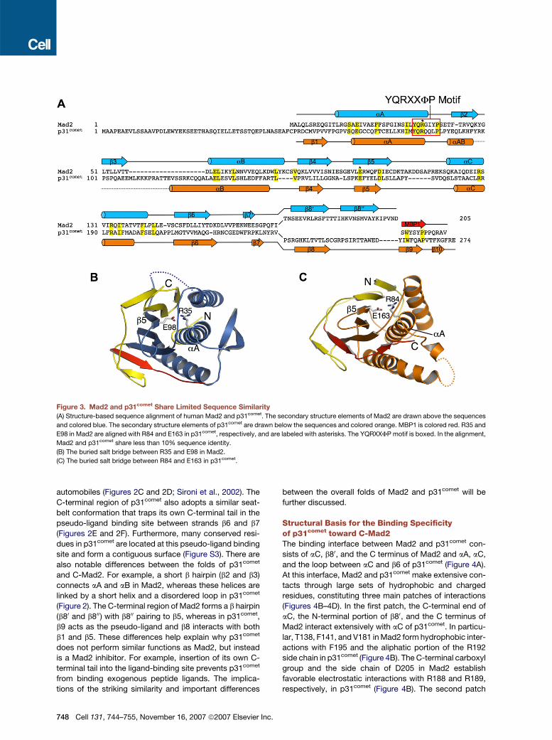
Figure 3. Mad2 and p31comet Share Limited Sequence Similarity
(A) Structure-based sequence alignment of human Mad2 and p31comet. The secondary structure elements of Mad2 are drawn above the sequences
and colored blue. The secondary structure elements of p31comet are drawn below the sequences and colored orange. MBP1 is colored red. R35 and
E98 in Mad2 are aligned with R84 and E163 in p31comet, respectively, and are labeled with asterisks. The YQRXXFP motif is boxed. In the alignment,
Mad2 and p31comet share less than 10% sequence identity.
(B) The buried salt bridge between R35 and E98 in Mad2.
(C) The buried salt bridge between R84 and E163 in p31comet.
automobiles (Figures 2C and 2D; Sironi et al., 2002). The
C-terminal region of p31comet also adopts a similar seat-
belt conformation that traps its own C-terminal tail in the
pseudo-ligand binding site between strands b6 and b7
(Figures 2E and 2F). Furthermore, many conserved resi-
dues in p31comet are located at this pseudo-ligand binding
site and form a contiguous surface (Figure S3). There are
also notable differences between the folds of p31comet
and C-Mad2. For example, a short b hairpin (b2 and b3)
connects aA and aB in Mad2, whereas these helices are
linked by a short helix and a disordered loop in p31comet
(Figure 2). The C-terminal region of Mad2 forms a b hairpin
(b80 and b800) with b800 pairing to b5, whereas in p31comet,
b9 acts as the pseudo-ligand and b8 interacts with both
b1 and b5. These differences help explain why p31comet
does not perform similar functions as Mad2, but instead
is a Mad2 inhibitor. For example, insertion of its own C-
terminal tail into the ligand-binding site prevents p31comet
from binding exogenous peptide ligands. The implica-
tions of the striking similarity and important differences
748 Cell 131, 744–755, November 16, 2007 ª2007 Elsevier Inc
between the overall folds of Mad2 and p31comet will be
further discussed.
Structural Basis for the Binding Specificityof p31comet toward C-Mad2The binding interface between Mad2 and p31comet con-
sists of aC, b80, and the C terminus of Mad2 and aA, aC,
and the loop between aC and b6 of p31comet (Figure 4A).
At this interface, Mad2 and p31comet make extensive con-
tacts through large sets of hydrophobic and charged
residues, constituting three main patches of interactions
(Figures 4B–4D). In the first patch, the C-terminal end of
aC, the N-terminal portion of b80, and the C terminus of
Mad2 interact extensively with aC of p31comet. In particu-
lar, T138, F141, and V181 in Mad2 form hydrophobic inter-
actions with F195 and the aliphatic portion of the R192
side chain in p31comet (Figure 4B). The C-terminal carboxyl
group and the side chain of D205 in Mad2 establish
favorable electrostatic interactions with R188 and R189,
respectively, in p31comet (Figure 4B). The second patch
.
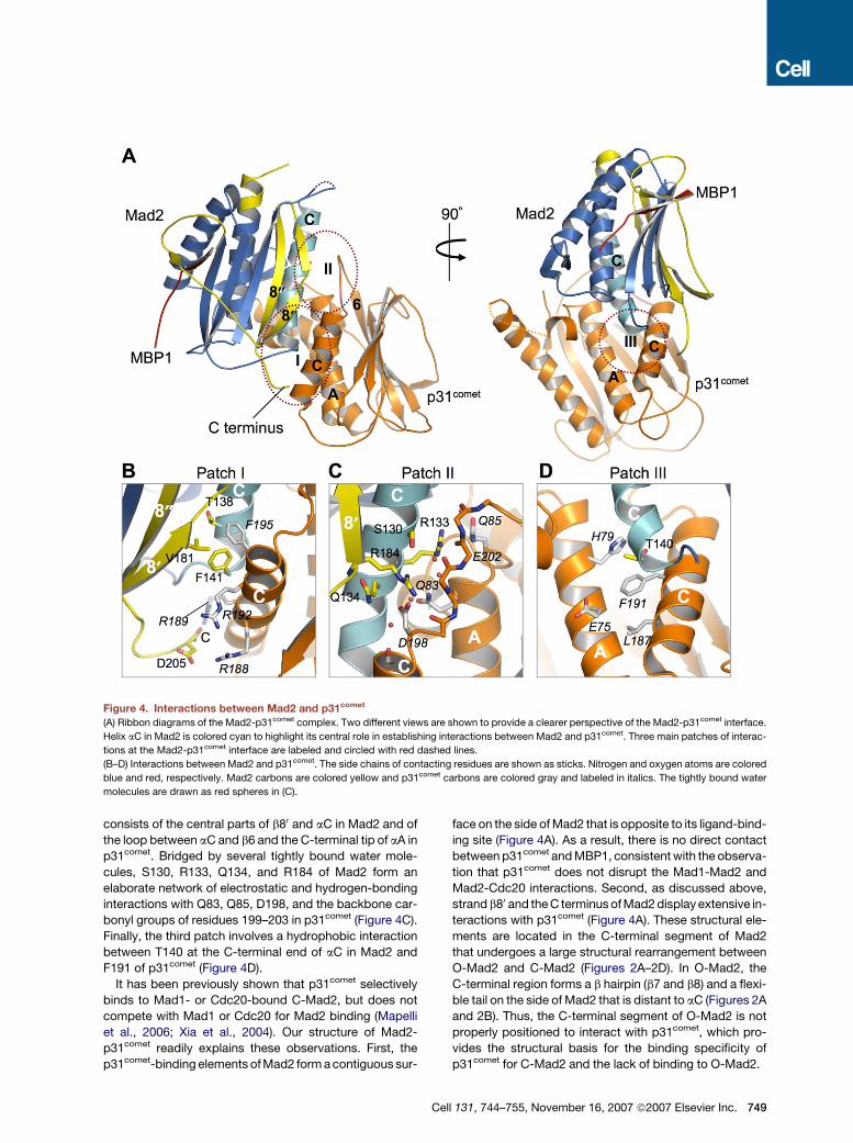
Figure 4. Interactions between Mad2 and p31comet
(A) Ribbon diagrams of the Mad2-p31comet complex. Two different views are shown to provide a clearer perspective of the Mad2-p31comet interface.
Helix aC in Mad2 is colored cyan to highlight its central role in establishing interactions between Mad2 and p31comet. Three main patches of interac-
tions at the Mad2-p31comet interface are labeled and circled with red dashed lines.
(B–D) Interactions between Mad2 and p31comet. The side chains of contacting residues are shown as sticks. Nitrogen and oxygen atoms are colored
blue and red, respectively. Mad2 carbons are colored yellow and p31comet carbons are colored gray and labeled in italics. The tightly bound water
molecules are drawn as red spheres in (C).
consists of the central parts of b80 and aC in Mad2 and of
the loop between aC and b6 and the C-terminal tip of aA in
p31comet. Bridged by several tightly bound water mole-
cules, S130, R133, Q134, and R184 of Mad2 form an
elaborate network of electrostatic and hydrogen-bonding
interactions with Q83, Q85, D198, and the backbone car-
bonyl groups of residues 199–203 in p31comet (Figure 4C).
Finally, the third patch involves a hydrophobic interaction
between T140 at the C-terminal end of aC in Mad2 and
F191 of p31comet (Figure 4D).
It has been previously shown that p31comet selectively
binds to Mad1- or Cdc20-bound C-Mad2, but does not
compete with Mad1 or Cdc20 for Mad2 binding (Mapelli
et al., 2006; Xia et al., 2004). Our structure of Mad2-
p31comet readily explains these observations. First, the
p31comet-binding elements of Mad2 form a contiguous sur-
C
face on the side of Mad2 that is opposite to its ligand-bind-
ing site (Figure 4A). As a result, there is no direct contact
between p31comet and MBP1, consistent with the observa-
tion that p31comet does not disrupt the Mad1-Mad2 and
Mad2-Cdc20 interactions. Second, as discussed above,
strand b80 and the C terminus of Mad2 display extensive in-
teractions with p31comet (Figure 4A). These structural ele-
ments are located in the C-terminal segment of Mad2
that undergoes a large structural rearrangement between
O-Mad2 and C-Mad2 (Figures 2A–2D). In O-Mad2, the
C-terminal region forms a b hairpin (b7 and b8) and a flexi-
ble tail on the side of Mad2 that is distant to aC (Figures 2A
and 2B). Thus, the C-terminal segment of O-Mad2 is not
properly positioned to interact with p31comet, which pro-
vides the structural basis for the binding specificity of
p31comet for C-Mad2 and the lack of binding to O-Mad2.
ell 131, 744–755, November 16, 2007 ª2007 Elsevier Inc. 749
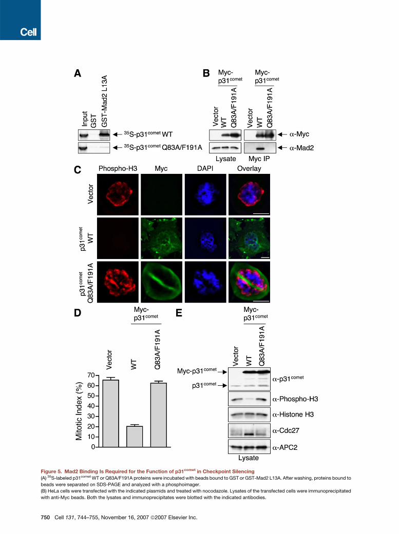
Figure 5. Mad2 Binding Is Required for the Function of p31comet in Checkpoint Silencing
(A) 35S-labeled p31comet WT or Q83A/F191A proteins were incubated with beads bound to GST or GST-Mad2 L13A. After washing, proteins bound to
beads were separated on SDS-PAGE and analyzed with a phosphoimager.
(B) HeLa cells were transfected with the indicated plasmids and treated with nocodazole. Lysates of the transfected cells were immunoprecipitated
with anti-Myc beads. Both the lysates and immunoprecipitates were blotted with the indicated antibodies.
750 Cell 131, 744–755, November 16, 2007 ª2007 Elsevier Inc.

A p31comet Mutant Deficient in Mad2 Binding Failsto Override Nocodazole-Triggered Mitotic ArrestTo validate the binding interactions between Mad2 and
p31comet observed in our crystal structure, we performed
systematic mutagenesis studies on both Mad2 and
p31comet. Among the p31comet-binding residues in Mad2,
mutations of R133, F141, and R184 significantly decreased
p31comet binding (Figure S4). Consistent with our finding, an
earlier study showed that several double mutants of Mad2,
including R133E/Q134A, R133A/F141A, and R133A/R184A,
failed to bind to p31comet (Mapelli et al., 2006). Among the
Mad2-binding residues in p31comet, only mutations of Q83
and F191 greatly diminished Mad2 binding (Figure S5).
While confirming the relevance of the Mad2-p31comet
interface in our structure, these mutagenesis results also
suggest that the interactions contributing to the binding
energy between Mad2 and p31comet are not evenly distrib-
uted across this interface. Instead, there are hot spots of
interactions that contribute the majority of the binding
energy, reminiscent of the binding of human growth factor
to its receptor (Clackson and Wells, 1995).
Previous studies have clearly demonstrated a role of
p31comet in checkpoint inactivation (Habu et al., 2002;
Reddy et al., 2007; Stegmeier et al., 2007; Xia et al.,
2004). Though it is very likely that Mad2 binding is impor-
tant for the function of p31comet, this notion has not been
formally tested. Individual mutations of Q83 and F191 in
p31comet greatly diminished, but did not abolish, Mad2
binding. We thus constructed the p31comet Q83A/F191A
double mutant, which completely lost its ability to bind
to Mad2 in vitro (Figure 5A). HeLa cells were transfected
with a control plasmid or plasmids encoding Myc-
p31comet wild-type (WT) or the Q83A/F191A double mu-
tant and treated with nocodazole, a spindle poison, to
activate the spindle checkpoint. Myc-p31comet WT, but
not Myc-p31comet Q83A/F191A, efficiently bound to the
endogenous Mad2 protein (Figure 5B). Consistent with
previous reports (Habu et al., 2002; Xia et al., 2004), over-
expression of Myc-p31comet WT significantly decreased
the percentage of cells arrested in mitosis in the presence
of nocodazole (Figures 5C and 5D) and reduced the cellu-
lar levels of phospho-histone H3 and hyperphosphory-
lated Cdc27 (Figure 5E). In contrast, upon nocodazole
treatment, expression of Myc-p31comet Q83A/F191A did
not appreciably alter the mitotic index or the levels of
phospho-histone H3 or Cdc27 hyperphosphorylation, as
compared to the control plasmid (Figures 5C–5E). Thus,
mutations of p31comet that disrupt its binding to Mad2
also disrupt the ability of p31comet to overcome spindle
checkpoint-dependent mitotic arrest. This finding further
suggests that Mad2 binding by p31comet is critically impor-
tant for its function in checkpoint silencing.
C
Structural Basis for the Blockage of Mad2Activation by p31comet
Binding of p31comet to Mad1-bound C-Mad2 blocks the
recruitment of an additional copy of O-Mad2 to the Mad1-
Mad2 core complex, thereby preventing the generation
of active Mad2 conformers, such as I-Mad2 and unli-
ganded C-Mad2, and contributing to checkpoint inactiva-
tion (Figures 1A and 1B; De Antoni et al., 2005; Luo et al.,
2004; Mapelli et al., 2006; Yu, 2006). The structure of
Mad2 bound to a fragment of Mad1 has been determined
previously (Sironi et al., 2002). The Mad2 molecules in our
Mad2-p31comet structure have virtually the same confor-
mation as Mad1-bound Mad2, with backbone rmsds of
about 1 A. By overlaying the Mad2 molecules in the Mad1-
Mad2 and Mad2-p31comet structures, we constructed
a structural model of the Mad1-Mad2-p31comet ternary
complex (Figure 6A). In this model, there are no steric
clashes between p31comet and Mad1 or between the two
p31comet molecules, suggesting that the Mad2-p31comet
binding mode in the Mad2-p31comet complex is compati-
ble with the formation of the Mad1-Mad2-p31comet ternary
complex. Mutations of Mad2 residues located on aC, in-
cluding R133, Q134, and F141, are known to disrupt the
dimerization of Mad2 and consequently its function in
the spindle checkpoint (De Antoni et al., 2005; Mapelli
et al., 2006; Sironi et al., 2001), indicating that aC is a major
dimerization determinant of Mad2. Mapelli et al. have de-
termined the crystal structure of the O-Mad2–C-Mad2
dimer (Mapelli et al., 2007, in this issue of Cell). O-Mad2 in-
deed binds to a surface on C-Mad2 that includes aC and
b80 (Figures 6B and 6C). The p31comet-binding surface of
Mad2 also contains aC and b80 (Figures 6B and 6D).
Thus, O-Mad2 and p31comet have largely overlapping
binding surfaces on C-Mad2, providing a straightforward
explanation for the ability of p31comet to block O-Mad2–
C-Mad2 dimerization and to prevent structural activation
of Mad2.
An Allosteric Model for the Activation of O-Mad2by the Mad1-Mad2 Core ComplexThe Mad1-Mad2 core complex recruits a second O-Mad2
molecule through O-Mad2–C-Mad2 dimerization and
converts O-Mad2 to I-Mad2 and unliganded C-Mad2,
thereby facilitating Mad2 binding to Cdc20 (De Antoni
et al., 2005; Yu, 2006). However, the mechanism of
Mad2 activation and the nature of I-Mad2 remain enig-
matic. Conversion of O-Mad2 to C-Mad2 involves two
main structural changes that occur at both sides of the
Mad2 molecule. On one side, b1 in O-Mad2 dissociates
from b5, traverses through the b5-aC loop, and forms an
additional turn in aA. On the other side, the C-terminal
region of O-Mad2 including b7, b8, and the flexible
(C) HeLa cells were transfected with the indicated plasmids, treated with nocodazole, and stained with DAPI (blue), anti-Myc (green), and anti-phos-
pho-H3 (red). Scale bars indicate 10 mm.
(D) The mitotic indices of the transfected cells in (C) were quantified. At least 400 cells were counted for each transfection. The averages and standard
deviations of three separate experiments are shown.
(E) Lysates of cells described in (C) and (D) were blotted with the indicated antibodies.
ell 131, 744–755, November 16, 2007 ª2007 Elsevier Inc. 751
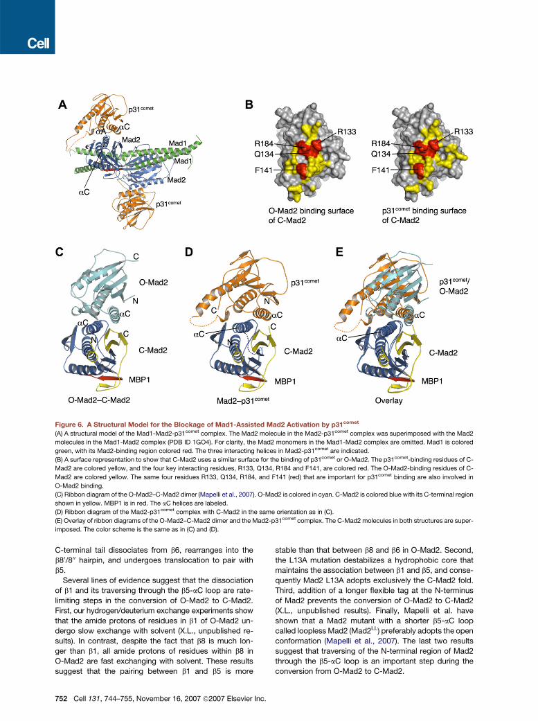
Figure 6. A Structural Model for the Blockage of Mad1-Assisted Mad2 Activation by p31comet
(A) A structural model of the Mad1-Mad2-p31comet complex. The Mad2 molecule in the Mad2-p31comet complex was superimposed with the Mad2
molecules in the Mad1-Mad2 complex (PDB ID 1GO4). For clarity, the Mad2 monomers in the Mad1-Mad2 complex are omitted. Mad1 is colored
green, with its Mad2-binding region colored red. The three interacting helices in Mad2-p31comet are indicated.
(B) A surface representation to show that C-Mad2 uses a similar surface for the binding of p31comet or O-Mad2. The p31comet-binding residues of C-
Mad2 are colored yellow, and the four key interacting residues, R133, Q134, R184 and F141, are colored red. The O-Mad2-binding residues of C-
Mad2 are colored yellow. The same four residues R133, Q134, R184, and F141 (red) that are important for p31comet binding are also involved in
O-Mad2 binding.
(C) Ribbon diagram of the O-Mad2–C-Mad2 dimer (Mapelli et al., 2007). O-Mad2 is colored in cyan. C-Mad2 is colored blue with its C-terminal region
shown in yellow. MBP1 is in red. The aC helices are labeled.
(D) Ribbon diagram of the Mad2-p31comet complex with C-Mad2 in the same orientation as in (C).
(E) Overlay of ribbon diagrams of the O-Mad2–C-Mad2 dimer and the Mad2-p31comet complex. The C-Mad2 molecules in both structures are super-
imposed. The color scheme is the same as in (C) and (D).
C-terminal tail dissociates from b6, rearranges into the
b80/800 hairpin, and undergoes translocation to pair with
b5.
Several lines of evidence suggest that the dissociation
of b1 and its traversing through the b5-aC loop are rate-
limiting steps in the conversion of O-Mad2 to C-Mad2.
First, our hydrogen/deuterium exchange experiments show
that the amide protons of residues in b1 of O-Mad2 un-
dergo slow exchange with solvent (X.L., unpublished re-
sults). In contrast, despite the fact that b8 is much lon-
ger than b1, all amide protons of residues within b8 in
O-Mad2 are fast exchanging with solvent. These results
suggest that the pairing between b1 and b5 is more
752 Cell 131, 744–755, November 16, 2007 ª2007 Elsevier Inc.
stable than that between b8 and b6 in O-Mad2. Second,
the L13A mutation destabilizes a hydrophobic core that
maintains the association between b1 and b5, and conse-
quently Mad2 L13A adopts exclusively the C-Mad2 fold.
Third, addition of a longer flexible tag at the N-terminus
of Mad2 prevents the conversion of O-Mad2 to C-Mad2
(X.L., unpublished results). Finally, Mapelli et al. have
shown that a Mad2 mutant with a shorter b5-aC loop
called loopless Mad2 (Mad2LL) preferably adopts the open
conformation (Mapelli et al., 2007). The last two results
suggest that traversing of the N-terminal region of Mad2
through the b5-aC loop is an important step during the
conversion from O-Mad2 to C-Mad2.
In an accompanying paper in this issue of Cell, Mapelli
et al. have determined the structure of Mad2LL bound to
C-Mad2 (Figure 6C; Mapelli et al., 2007). Mad2LL bound
to C-Mad2 has a fold identical to that of the free O-
Mad2 (Luo et al., 2000; Mapelli et al., 2007). Thus, the
structure of Mad2LL–C-Mad2 likely depicts the initial
docking complex between O-Mad2 and C-Mad2 in the
Mad1-Mad2 core complex (Figure 1A), not that between
I-Mad2 and C-Mad2.
On the other hand, p31comet has a fold closely related to
that of Mad2. p31comet and O-Mad2 bind to similar sur-
faces on C-Mad2. Conversely, the C-Mad2-binding site
of p31comet includes its aC, aA, and the short helix con-
necting aA and aB (Figure 6D). Likewise, the C-Mad2-
binding site of O-Mad2 is comprised of its aC, aA, and
the b2/3 hairpin that connects aA and aB (Figure 6C).
Therefore, p31comet and O-Mad2 use equivalent structural
elements for binding to C-Mad2 (Figures 2, 6C, and 6D).
However, when the C-Mad2 molecules in the structures
of O-Mad2–C-Mad2 and p31comet–C-Mad2 are superim-
posed, the equivalent structural elements of p31comet
and O-Mad2 do not overlay well (Figure 6E). In fact, if we
superimposed O-Mad2 with p31comet in our Mad2-
p31comet structure, aC and the tip of the b2/3 hairpin in
O-Mad2 would have severe steric clashes with aC of C-
Mad2 (Figure S6A). Thus, an intriguing possibility is that
the binding mode of p31comet on C-Mad2 mimics that of
I-Mad2 on C-Mad2, rather than that of O-Mad2. Binding
of C-Mad2 might induce the unfolding of the b2/3 hairpin
and a rotation of aC in O-Mad2 to alleviate the steric
clashes between O-Mad2 and C-Mad2 in our model
(Figure S6A). The rotation of aC in O-Mad2 is expected
to destabilize the hydrophobic core that involves residues
on b1, including L13 (Figure S6B). Disruption of this hydro-
phobic core could facilitate dissociation of b1 from b5,
a possible rate-limiting step in the conversion from O-
Mad2 to C-Mad2. Dissociation of b1 and its traversing
through the b5-aC loop might then allow the translocation
of the b80/800 hairpin to pair with b5, thereby generating C-
Mad2 (Figures S6). The structures of Mad2-p31comet and
O-Mad2–C-Mad2 provide the basis for designing future
experiments that can test this model for the structural ac-
tivation of Mad2. Furthermore, it is possible that the Mad2-
p31comet interaction is spatially and temporally regulated
during mitosis. The structure of Mad2-p31comet will help
us understand the regulatory mechanisms of p31comet,
once these mechanisms are discovered.
ConclusionThe crystal structure of the Mad2-p31comet complex
reveals the structural basis for how p31comet acts as an
anti-Mad2 in checkpoint inactivation and further suggests
an allosteric mechanism for the activation of the two-state
protein Mad2 by the Mad1-Mad2 core complex. Our stud-
ies provide a striking illustration of how two proteins with
similar folds can play antagonistic roles in the same
signaling pathway.
C
EXPERIMENTAL PROCEDURES
Protein Purification and Crystallization
The coding regions of human Mad2 and human p31cometDN35
(residues 36–274) were cloned into pETDuet-1 (Novagen). The L13A
mutation was introduced into pETDuet-1-Mad2-p31comet with the
QuikChange mutagenesis kit (Stratagene). Expression of pETDuet-
1-Mad2 L13A-p31comet in the bacterial strain BL21(DE3) produced N-
terminal His6-tagged Mad2 L13A and untagged p31comet proteins.
The Mad2 L13A-p31comet complex was isolated from bacterial lysate
by affinity chromatography with Ni2+-NTA agarose resin (QIAGEN)
and cleaved with TEV protease to remove the His6-tag. The complex
was further purified by anion exchange chromatography with a re-
source Q column followed by size exclusion chromatography with
a Superdex 200 column. After addition of MBP1 at a 1:1 molar ratio,
the resulting Mad2 L13A-p31comet-MBP1 complex was concentrated
to 8 mg/ml in a buffer containing 10 mM Tris (pH 8.0), 100 mM NaCl,
and 5 mM DTT. The seleno-methionine-labeled Mad2 L13A-MBP1-
p31comet complex was prepared similarly. The Mad2 L13A-MBP1-
p31comet complex was crystallized at 16�C by the vapor-diffusion
method in hanging-drop mode with a reservoir solution containing
16% (w/v) PEG 3350, 125 mM sodium phosphate (pH 5.0), and 25 mM
DTT. The crystals were cryo-protected with reservoir solution sup-
plemented with 16% (v/v) glycerol and then flash-cooled in liquid
propane. Crystals exhibited the symmetry of space group P212121
with cell dimensions of a = 69 A, b = 105 A, c = 139 A and contained
two complexes per asymmetric unit.
Data Collection and Structure Determination
All data sets were collected at beamline 19-ID (SBC-CAT) at the
Advanced Photon Source (Argonne National Laboratory, Argonne,
IL) and processed with HKL2000 (Otwinowski and Minor, 1997). Crys-
tals diffracted to a minimum Bragg spacing (dmin) of about 2.3 A.
Phases were obtained from a selenium-SAD experiment via X-rays
with an energy near the selenium K absorption edge. With data to
3.0 A, 14 of 14 possible selenium sites were located and refined with
the program SHELX (Schneider and Sheldrick, 2002; Sheldrick,
2002). Phases were subsequently extended to 2.3 A and refined with
the program MLPHARE (Otwinowski, 1991), resulting in an overall
figure of merit of 0.25. Phases were further improved by density
modification with histogram matching and 2-fold noncrystallographic
averaging in the program DM (Cowtan and Main, 1998), resulting in a fi-
nal overall figure of merit of 0.75. The resulting electron density map
was of sufficient quality to automatically construct an initial model
with the program ARP/warp (Perrakis et al., 1999). This model was
used as a starting model for the refinement with the programs CNS
(Brunger et al., 1998) and REFMAC5 (Murshudov et al., 1997) from
the CCP4 package (Consortium, 1994; Murshudov et al., 1997), inter-
spersed with manual rebuilding with the programs Coot (Emsley and
Cowtan, 2004) and O (Jones et al., 1991). Analysis of the original
data set used for phasing revealed significant radiation decay, which
was most significantly manifested through reduced electron density
for the disulfide bond formed in Mad2A between C79 and C106 (this di-
sulfide bond was not observed in Mad2C) and increased disorder in the
electron density map in this region. A modified data set was generated
that incorporated only about the first 60% of the original data, which
was then used for the final rounds of refinement. The electron density
maps calculated from this modified data set were more easily inter-
pretable and the refinement was more stable (Table 1).
Mammalian Tissue Culture, Immunoprecipitation,
and Protein Binding Assays
HeLa Tet-on (Invitrogen) cells were cultured in DMEM medium supple-
mented with 10% fetal bovine serum. The cells were transfected with
pCS2-p31comet vectors with Effectene (QIAGEN). After 24 hr, the cells
were treated with 100 ng/ml nocodazole (Sigma) for 16 hr, stained
with Hoechst 33342 (Invitrogen), and examined with an inverted
ell 131, 744–755, November 16, 2007 ª2007 Elsevier Inc. 753
fluorescence microscope (Zeiss). Cells were also fixed and stained
with DAPI and antibodies against Myc and phospho-histone H3 as de-
scribed (Tang et al., 2006). Lysates of the transfected cells were sub-
jected to immunoprecipitation and immunoblotting with the appropri-
ate antibodies. For in vitro protein binding assays, GST-Mad2 proteins
were bound to glutathione-agarose beads and incubated with 35S-la-
beled p31comet proteins obtained by in vitro translation in rabbit retic-
ulocyte lysates (Promega). After washing, proteins bound to beads
were resolved on SDS-PAGE and analyzed with a phosphoimager
(Fuji).
Supplemental Data
Six figures are available at http://www.cell.com/cgi/content/full/131/4/
744/DC1/.
ACKNOWLEDGMENTS
We thank Dr. Nick Grishin for his help with sequence analysis of
p31comet and Mad2 and Minghua Wen for her technical support. We
also thank Dr. Lin Chen and Dr. Aidong Han for their suggestions
and discussion. Results shown in this report are derived from work
performed at Argonne National Laboratory, Structural Biology Center
at the Advanced Photon Source. Argonne is operated by University
of Chicago Argonne, LLC, for the U.S. Department of Energy, Office
of Biological and Environmental Research. This work was supported
in part by grants from the National Institutes of Health (to X.L. and
H.Y.) and the Welch Foundation (to H.Y. and J.R.).
Received: April 4, 2007
Revised: June 28, 2007
Accepted: August 30, 2007
Published: November 15, 2007
REFERENCES
Bharadwaj, R., and Yu, H. (2004). The spindle checkpoint, aneuploidy,
and cancer. Oncogene 23, 2016–2027.
Brunger, A.T., Adams, P.D., Clore, G.M., DeLano, W.L., Gros, P.,
Grosse-Kunstleve, R.W., Jiang, J.S., Kuszewski, J., Nilges, M., Pannu,
N.S., et al. (1998). Crystallography & NMR system: a new software
suite for macromolecular structure determination. Acta Crystallogr. D
Biol. Crystallogr. 54, 905–921.
Clackson, T., and Wells, J.A. (1995). A hot spot of binding energy in
a hormone-receptor interface. Science 267, 383–386.
Consortium, T.C. (1994). The CCP4 suite: programs for protein crystal-
lography. Acta Crystallogr. D Biol. Crystallogr. 50, 760–763.
Cowtan, K., and Main, P. (1998). Miscellaneous algorithms for density
modification. Acta Crystallogr. D Biol. Crystallogr. 54, 487–493.
De Antoni, A., Pearson, C.G., Cimini, D., Canman, J.C., Sala, V., Nezi,
L., Mapelli, M., Sironi, L., Faretta, M., Salmon, E.D., and Musacchio, A.
(2005). The Mad1/Mad2 complex as a template for Mad2 activation in
the spindle assembly checkpoint. Curr. Biol. 15, 214–225.
Emsley, P., and Cowtan, K. (2004). Coot: model-building tools for
molecular graphics. Acta Crystallogr. D Biol. Crystallogr. 60, 2126–
2132.
Fang, G., Yu, H., and Kirschner, M.W. (1998). The checkpoint protein
MAD2 and the mitotic regulator CDC20 form a ternary complex with
the anaphase-promoting complex to control anaphase initiation.
Genes Dev. 12, 1871–1883.
Habu, T., Kim, S.H., Weinstein, J., and Matsumoto, T. (2002). Identifi-
cation of a MAD2-binding protein, CMT2, and its role in mitosis. EMBO
J. 21, 6419–6428.
Hwang, L.H., Lau, L.F., Smith, D.L., Mistrot, C.A., Hardwick, K.G.,
Hwang, E.S., Amon, A., and Murray, A.W. (1998). Budding yeast
Cdc20: a target of the spindle checkpoint. Science 279, 1041–1044.
754 Cell 131, 744–755, November 16, 2007 ª2007 Elsevier Inc.
Jones, T.A., Zou, J.Y., Cowan, S.W., and Kjeldgaard, M. (1991). Im-
proved methods for building protein models in electron density maps
and the location of errors in these models. Acta Crystallogr. A 47,
110–119.
Kim, S.H., Lin, D.P., Matsumoto, S., Kitazono, A., and Matsumoto, T.
(1998). Fission yeast Slp1: an effector of the Mad2-dependent spindle
checkpoint. Science 279, 1045–1047.
Luo, X., Fang, G., Coldiron, M., Lin, Y., Yu, H., Kirschner, M.W., and
Wagner, G. (2000). Structure of the Mad2 spindle assembly checkpoint
protein and its interaction with Cdc20. Nat. Struct. Biol. 7, 224–229.
Luo, X., Tang, Z., Rizo, J., and Yu, H. (2002). The Mad2 spindle check-
point protein undergoes similar major conformational changes upon
binding to either Mad1 or Cdc20. Mol. Cell 9, 59–71.
Luo, X., Tang, Z., Xia, G., Wassmann, K., Matsumoto, T., Rizo, J., and
Yu, H. (2004). The Mad2 spindle checkpoint protein has two distinct
natively folded states. Nat. Struct. Mol. Biol. 11, 338–345.
Mapelli, M., Filipp, F.V., Rancati, G., Massimiliano, L., Nezi, L., Stier,
G., Hagan, R.S., Confalonieri, S., Piatti, S., Sattler, M., and Musacchio,
A. (2006). Determinants of conformational dimerization of Mad2 and its
inhibition by p31comet. EMBO J. 25, 1273–1284.
Mapelli, M., Massimiliano, L., Santaguida, S., and Musacchio, A.
(2007). The Mad2 conformational dimer: structure and implications
for the spindle assembly checkpoint. Cell 131, this issue, 730–743.
Murshudov, G.N., Vagin, A.A., and Dodson, E.J. (1997). Refinement of
macromolecular structures by the maximum-likelihood method. Acta
Crystallogr. D Biol. Crystallogr. 53, 240–255.
Musacchio, A., and Hardwick, K.G. (2002). The spindle checkpoint:
structural insights into dynamic signalling. Nat. Rev. Mol. Cell Biol. 3,
731–741.
Musacchio, A., and Salmon, E.D. (2007). The spindle-assembly check-
point in space and time. Nat. Rev. Mol. Cell Biol. 8, 379–393.
Otwinowski, Z. (1991). Isomorphous replacement and anomalous
scattering. In Proceedings of the CCP4 Study Weekend: Isomorphous
Replacement and Anomalous Scattering, W. Wolf, P.R. Evans, and
A.G.W. Leslie, eds. (Warrington, UK: SERC Daresbury Laboratory),
pp. 80–86.
Otwinowski, Z., and Minor, W. (1997). Processing X-ray diffraction data
collected in oscillation mode. Methods Enzymol. 276, 307–326.
Perrakis, A., Morris, R., and Lamzin, V.S. (1999). Automated protein
model building combined with iterative structure refinement. Nat.
Struct. Biol. 6, 458–463.
Reddy, S.K., Rape, M., Margansky, W.A., and Kirschner, M.W. (2007).
Ubiquitination by the anaphase-promoting complex drives spindle
checkpoint inactivation. Nature 446, 921–925.
Schneider, T.R., and Sheldrick, G.M. (2002). Substructure solution with
SHELXD. Acta Crystallogr. D Biol. Crystallogr. 58, 1772–1779.
Shah, J.V., Botvinick, E., Bonday, Z., Furnari, F., Berns, M., and Cleve-
land, D.W. (2004). Dynamics of centromere and kinetochore proteins;
implications for checkpoint signaling and silencing. Curr. Biol. 14,
942–952.
Sheldrick, G.M. (2002). Macromolecular phasing with SHELXE. Zeits-
chrift Fur Kristallographie 217, 644–650.
Sironi, L., Melixetian, M., Faretta, M., Prosperini, E., Helin, K., and
Musacchio, A. (2001). Mad2 binding to Mad1 and Cdc20, rather than
oligomerization, is required for the spindle checkpoint. EMBO J. 20,
6371–6382.
Sironi, L., Mapelli, M., Knapp, S., De Antoni, A., Jeang, K.T., and
Musacchio, A. (2002). Crystal structure of the tetrameric Mad1-Mad2
core complex: implications of a ‘safety belt’ binding mechanism for
the spindle checkpoint. EMBO J. 21, 2496–2506.
Stegmeier, F., Rape, M., Draviam, V.M., Nalepa, G., Sowa, M.E., Ang,
X.L., McDonald, E.R., 3rd, Li, M.Z., Hannon, G.J., Sorger, P.K., et al.
(2007). Anaphase initiation is regulated by antagonistic ubiquitination
and deubiquitination activities. Nature 446, 876–881.
Tang, Z., Shu, H., Qi, W., Mahmood, N.A., Mumby, M.C., and Yu, H.
(2006). PP2A is required for centromeric localization of Sgo1 and
proper chromosome segregation. Dev. Cell 10, 575–585.
Xia, G., Luo, X., Habu, T., Rizo, J., Matsumoto, T., and Yu, H. (2004).
Conformation-specific binding of p31(comet) antagonizes the function
of Mad2 in the spindle checkpoint. EMBO J. 23, 3133–3143.
Yu, H. (2002). Regulation of APC-Cdc20 by the spindle checkpoint.
Curr. Opin. Cell Biol. 14, 706–714.
C
Yu, H. (2006). Structural activation of Mad2 in the mitotic spindle
checkpoint: the two-state Mad2 model versus the Mad2 template
model. J. Cell Biol. 173, 153–157.
Yu, H. (2007). Cdc20: a WD40 activator for a cell cycle degradation
machine. Mol. Cell 27, 3–16.
Accession Numbers
The atomic coordinates of the Mad2-MBP1-p31comet ternary complex
have been deposited in the Protein Data Bank with accession code
2QYF.
ell 131, 744–755, November 16, 2007 ª2007 Elsevier Inc. 755