A Novel Antibody against Human Factor B that Blocks Formation of the C3bB Proconvertase and Inhibits...
10
of October 31, 2014. This information is current as Activation in Disease Models Proconvertase and Inhibits Complement that Blocks Formation of the C3bB A Novel Antibody against Human Factor B Córdoba Marina Noris, B. Paul Morgan and Santiago Rodríguez de Villegas-Martínez, Mercedes Dominguez, Oscar Llorca, Fernando Ataúlfo González-Fernández, Ana Galbusera, Andrés López-Perrote, Lucia de Juana Lopez, Marta Subías, Agustín Tortajada, Sara Gastoldi, Miriam ol.1402013 http://www.jimmunol.org/content/early/2014/10/29/jimmun published online 29 October 2014 J Immunol Subscriptions http://jimmunol.org/subscriptions is online at: The Journal of Immunology Information about subscribing to Permissions http://www.aai.org/ji/copyright.html Submit copyright permission requests at: Email Alerts http://jimmunol.org/cgi/alerts/etoc Receive free email-alerts when new articles cite this article. Sign up at: Print ISSN: 0022-1767 Online ISSN: 1550-6606. Immunologists, Inc. All rights reserved. Copyright © 2014 by The American Association of 9650 Rockville Pike, Bethesda, MD 20814-3994. The American Association of Immunologists, Inc., is published twice each month by The Journal of Immunology at Red de Bibliotecas del CSIC on October 31, 2014 http://www.jimmunol.org/ Downloaded from at Red de Bibliotecas del CSIC on October 31, 2014 http://www.jimmunol.org/ Downloaded from
-
Upload
independent -
Category
Documents
-
view
0 -
download
0
Transcript of A Novel Antibody against Human Factor B that Blocks Formation of the C3bB Proconvertase and Inhibits...

of October 31, 2014.This information is current as
Activation in Disease ModelsProconvertase and Inhibits Complementthat Blocks Formation of the C3bB A Novel Antibody against Human Factor B
CórdobaMarina Noris, B. Paul Morgan and Santiago Rodríguez deVillegas-Martínez, Mercedes Dominguez, Oscar Llorca, Fernando Ataúlfo González-Fernández, AnaGalbusera, Andrés López-Perrote, Lucia de Juana Lopez, Marta Subías, Agustín Tortajada, Sara Gastoldi, Miriam
ol.1402013http://www.jimmunol.org/content/early/2014/10/29/jimmun
published online 29 October 2014J Immunol
Subscriptionshttp://jimmunol.org/subscriptions
is online at: The Journal of ImmunologyInformation about subscribing to
Permissionshttp://www.aai.org/ji/copyright.htmlSubmit copyright permission requests at:
Email Alertshttp://jimmunol.org/cgi/alerts/etocReceive free email-alerts when new articles cite this article. Sign up at:
Print ISSN: 0022-1767 Online ISSN: 1550-6606. Immunologists, Inc. All rights reserved.Copyright © 2014 by The American Association of9650 Rockville Pike, Bethesda, MD 20814-3994.The American Association of Immunologists, Inc.,
is published twice each month byThe Journal of Immunology
at Red de B
ibliotecas del CSIC
on October 31, 2014
http://ww
w.jim
munol.org/
Dow
nloaded from
at Red de B
ibliotecas del CSIC
on October 31, 2014
http://ww
w.jim
munol.org/
Dow
nloaded from

The Journal of Immunology
A Novel Antibody against Human Factor B that BlocksFormation of the C3bB Proconvertase and InhibitsComplement Activation in Disease Models
Marta Subıas,*,† Agustın Tortajada,*,† Sara Gastoldi,‡,x Miriam Galbusera,‡,x
Andres Lopez-Perrote,* Lucia de Juana Lopez,*,† Fernando Ataulfo Gonzalez-Fernandez,{
Ana Villegas-Martınez,{ Mercedes Dominguez,‖ Oscar Llorca,* Marina Noris,‡,x
B. Paul Morgan,# and Santiago Rodrıguez de Cordoba*,†
The alternative pathway (AP) is critical for the efficient activation of complement regardless of the trigger. It is also amajor player in
pathogenesis, as illustrated by the long list of diseases in which AP activation contributes to pathology. Its relevance to human
disease is further emphasized by the high prevalence of pathogenic inherited defects and acquired autoantibodies disrupting com-
ponents and regulators of the AP C3-convertase. Because pharmacological downmodulation of the AP emerges as a broad-spectrum
treatment alternative, there is a powerful interest in developing new molecules to block formation and/or activity of the AP C3-
convertase. In this paper, we describe the generation of a novel mAb targeting human factor B (FB). mAb FB48.4.2, recognizing with
high affinity an evolutionary-conserved epitope in the Ba fragment of FB, very efficiently inhibited formation of the AP C3-
proconvertase by blocking the interaction between FB and C3b. In vitro assays using rabbit and sheep erythrocytes demonstrated
that FB28.4.2 was a potent AP inhibitor that blocked complement-mediated hemolysis in several species. Using ex vivo models of
disease we demonstrated that FB28.4.2 protected paroxysmal nocturnal hemoglobinuria erythrocytes from complement-mediated
hemolysis and inhibited both C3 fragment and C5b-9 deposition on ADP-activated HMEC-1 cells, an experimental model for atyp-
ical hemolytic uremic syndrome. Moreover, i.v. injection of FB28.4.2 in rats blocked complement activation in rat serum and pre-
vented the passive induction of experimental autoimmuneMyasthenia gravis. As a whole, these data demonstrate the potential value
of FB28.4.2 for the treatment of disorders associated with AP complement dysregulation in man and animal models. The Journal
of Immunology, 2014, 193: 000–000.
Complement is an essential component of innate immunityand a major trigger of inflammatory responses. It playsa crucial role in microbial killing, apoptotic cell clear-
ance, immune complex handling and modulation of adaptive im-mune responses (1, 2). Complement is initiated by three activationpathways, the classical pathway (CP), the lectin pathway (LP),and the alternative pathway (AP). The critical step in these acti-vation pathways is the formation of labile protease complexes,termed C3-convertases (C3bBb in the AP; C4b2a in the CP/LP)that cleave C3 to generate the active fragment, C3b. When C3b isgenerated, a reactive thioester is exposed, which permits covalentbinding of C3b to the activating surface, targeting it for destructionand initiating inflammation. The incorporation of one additional C3b
molecule to the AP C3-convertase creates the C5-convertase, whichcleaves C5 thereby triggering inflammation and leukocyte recruit-ment through production of C5a. Cleavage of C5 also initiates for-mation of the membrane attack complex (MAC) (3).The efficiency of complement activation relies on the AP am-
plification loop in which the C3b generated by the C3-convertaseforms more AP C3-convertase and provides exponential amplifi-cation to the initial activation. The balance between the rate atwhich the initial trigger is amplified and the degree to which C3band the C3-convertases are inactivated determines the progressionof the complement cascade to cell damage and death. Foreignsugars on microbial pathogens (AP), Abs (CP), or mannan (LP) tipthe balance in favor of amplification, causing target opsonization
*Centro de Investigaciones Biologicas, Consejo Superior de Investigaciones Cientıf-icas, 28040 Madrid, Spain; †Centro de Investigacion Biomedica en EnfermedadesRaras, Madrid 28040, Spain; ‡IRCCS - Mario Negri Instituto for PharmacologicalResearch “Mario Negri”, Clinical Research Center for Rare Diseases “Aldo e CeleDacco”, Ranica, Bergamo 24020, Italy; x“Centro Anna Maria Astori” Parco Scien-tifico e Tecnologico Kilometro Rosso, Bergamo 24126, Italy; {Hospital Clınico SanCarlos, Universidad Complutense de Madrid, Madrid 28040, Spain; ‖Servicio deInmunologıa Microbiana, Centro Nacional de Microbiologıa, Instituto de Investiga-cion Carlos III, Madrid 28220, Spain; and #Institute of Infection and Immunity,School of Medicine, Cardiff University, Cardiff CF14 4XN, United Kingdom
ORCID: 0000-0001-6401-1874 (S.R.d.C.).
Received for publication August 6, 2014. Accepted for publication September 29,2014.
This work was supported by Spanish “Ministerio de Economıa y Competitividad” GrantsSAF2011-26583 (to S.R.d.C.) and SAF2011-22988 (to O.L.), the Fundacion Renal InigoAlvarez de Toledo, and the Seventh Framework Programme European Union ProjectEURenOmics (Grant 305608) (to S.R.d.C.). In addition, this work was supported by theAutonomous Region of Madrid (Grant S2010/BMD-2316 to S.R.d.C. and O.L.).
M.S., M.D., and S.R.d.C. conceived and planned the project; M.S., A.T., S.G., M.G.,A.L.-P., L.d.J.L., F.A.G.-F., M.D., O.L., A.V.-M., M.N., B.P.M., and S.R.d.C. de-signed and/or performed the research experiments and analyzed the data; and M.S.,O.L., M.N., B.P.M., and S.R.d.C. drafted and edited the manuscript.
Address correspondence and reprint requests to Dr. Santiago Rodrıguez de Cordoba,Centro de Investigaciones Biologicas, Consejo Superior de Investigaciones Cientıf-icas, Ramiro de Maeztu 9, 28040 Madrid, Spain. E-mail address: [email protected]
Abbreviations used in this article: AChR, acetylcholine receptor; aHUS, atypicalhemolytic uremic syndrome; AP, alternative pathway; CP, classical pathway; EAMG,experimental autoimmune MG; FB, factor B; LP, lectin pathway; MAC, membraneattack complex; MG, myasthenia gravis; NHS, normal human serum; PNH, parox-ysmal nocturnal hemoglobinuria; RU, resonance unit; SPR, surface plasmon reso-nance.
Copyright� 2014 by TheAmerican Association of Immunologists, Inc. 0022-1767/14/$16.00
www.jimmunol.org/cgi/doi/10.4049/jimmunol.1402013
Published October 29, 2014, doi:10.4049/jimmunol.1402013 at R
ed de Bibliotecas del C
SIC on O
ctober 31, 2014http://w
ww
.jimm
unol.org/D
ownloaded from

and cell lysis. In health, complement activation is strictly regulatedand limited to the activator surface (4).Pharmacological downmodulation of the AP is predicted to be
beneficial in the treatment of a long list of diseases in which com-plement activation contributes to pathology by sustaining inflamma-tion and perpetuating tissue damage (5). Indeed, inhibition of the APis emerging as the treatment of choice in diseases, like paroxysmalnocturnal hemoglobinuria (PNH), C3 glomerulopathies, atypical he-molytic uremic syndrome (aHUS), and Myasthenia gravis (MG), inwhich dysregulation of the AP C3 convertase, because of inheriteddefects or acquired autoantibodies, is the etiopathogenic factor (6–9).Previously, mAbs specific for either FB or C3b that block for-
mation of the AP C3-convertase (C3bBb) have been generated andprovided proof of concept in models that inhibition of the APprevents or ameliorates diseases like antiphospholipid Ab syn-drome, complement-mediated hemolytic anemia or ischemia/reperfusion injury (10–16). In this paper, we report the genera-tion of a novel mAb against human factor B (FB) with capacity toblock the activity of the AP. FB48.4.2, a mAb that recognizes withhigh affinity an evolutionarily conserved epitope in the Ba frag-ment of human FB, inhibits complement activation by blockingthe formation of the AP C3 proconvertase.
Materials and MethodsIsolation of complement components
C3 was prepared by an established protocol involving ammonium sulfateprecipitation, anion exchange chromatography (DEAE and Mono S; GEHealthcare), and size exclusion chromatography (Superdex 200 Increase 10/300 GL; GE Healthcare) (17); FB was affinity purified from EDTA plasmaon anti-Bb (mAb D2, in-house), followed by anion exchange chromatog-raphy (Mono Q; GE Healthcare) and size exclusion chromatography (Superdex200 Increase 10/300 GL; GE Healthcare). FD was purchased from Calbio-chem. C3b was generated by coincubating pure C3, FB, and FD as previouslydescribed (17), followed by anion exchange chromatography (Mono Q; GEHealthcare) and size exclusion chromatography (Superdex 200 Increase10/300; GE Healthcare). Concentrations of pure proteins were assessed usingabsorbance at A280nm and the following extinction coefficients: FB: 1.43cm21
(mg/ml)21, C3: 0.98cm21 (mg/ml)21, and Ig 1.4 cm21 (mg/ml)21.
Generation of mAbs
FB-deficient mice (a gift from Prof. M. Botto, Imperial College, LondonUK) were immunized with 20 mg human FB emulsified with CFA and thenboosted three times at 2-wk intervals with the same amount of FB withincomplete adjuvant. The mice were screened for the development of Absto FB by testing their sera in an ELISA using human FB–coated plates.Positive mice were given an additional boost with 20 mg of a mixture of Baand Bb fragments in PBS. Three days later, spleen cells from a mousehaving a robust immune response toward FB were fused to the x63AG8myeloma cell line. Candidate hybridomas were cloned by limiting dilution,and clones recognizing human FB by ELISA were identified and expanded.Abs were purified from tissue culture supernatants with a protein G–Sepharosecolumn (Pharmacia, Uppsala, Sweden). The purity of the mAbs was thenanalyzed by 10% SDS-PAGE, and their Ig isotype was determined with theIsoStrip Mouse mAb Isotyping Kit (Roche Applied Science). The mAbproducts of all selected hybridomas were tested in ELISA and/or Western blotfor their capacity to detect FB in human, mouse, rat, rabbit, and pig sera. Allanimal experimentation has been reviewed and approved by the ConsejoSuperior de Investigaciones Cientıficas Institutional Review Board.
FB and C3 cleavage assays
The effects of mAbs on FB and C3 cleavage were tested in an in vitro assayusing purified C3, FB and FD because all preparations of C3 containsufficient C3(H2O) to trigger formation of the AP convertase. Briefly, C3(0.55 mg), FB (0.5 mg), and FD (0.001 mg) were mixed in 25 ml AP buffer(2.9 mM barbital, 1.7 mM sodium barbital, 144 mM NaCl, 7 mM MgCl2,and 10 mM EGTA [pH 7.4]). Increasing amounts of each mAb anti FB(from 0.5 to 2 mg) were added to the reaction mix, which was then in-cubated at 37˚C for 30 min in a water bath. Cleavage of FB and C3 wasassessed after 10% SDS-PAGE under reducing conditions and Coomassieblue staining.
Hemolysis assays
The capacity of the mAbs to inhibit the AP on cellular surfaces was assessedin a hemolytic assay using rabbit erythrocytes. Titration of normal humanserum (NHS) for lysis of rabbit erythrocytes was performed before theexperiment to determine the optimal conditions for the hemolysis inhibitionassay. A final concentration of 2.5% of NHS was used in our experiments, asthis amountwas just sufficient to completely lyse the rabbit erythrocytes underthe conditions used. Experiments were done in triplicate with each reactioncontaining 100 ml rabbit erythrocytes (1 3 108/ml) in isotonic GVB buffer(AP buffer containing 0.1% gelatin), 100 ml 5% NHS in AP buffer andincreasing amounts of an anti-FB mAb or its Fab fragment, incubated for 30min at 37˚C with occasional shaking. The reactions were stopped by adding2 ml ice-cold saline. Tubes were centrifuged at 10003 g for 5 min at 4˚C, andthe supernatants read at 414 nm. Erythrocytes with NHS and 20 mM EDTAwere used as a blank and erythrocytes with NHS and water as 100% lysis.
Rabbit erythrocytes were also used to test the capacity of the mAbs toprevent C3 deposition on cell surfaces. Briefly, 100 ml rabbit erythrocytes(1 3 108/ml) in AP buffer were incubated with 5% NHS in presence ofeither eculizumab (Soliris, Alexion Pharmaceuticals) or FB28.4.2 at 37˚Cfor 30 min. C3 deposition was evaluated by flow cytometry using a rabbitpolyclonal anti human C3 Ab (in house; 1 mg/ml in PBS) or the mouseanti-human iC3b/C3dg mAb SIM320.12.2.1 (in house; 0.5 mg/ml in PBS).
The capacity of the mAb to inhibit the AP on cell surfaces was alsoassessed in a FH-dependent hemolytic assay using sheep erythrocytes anda well-characterized serum from an aHUS patient carrying the FH-W1183Lmutation (18). In brief, 1 3 108/ml sheep erythrocytes were incubatedwith 20% patient serum in AP buffer with increasing concentrations ofFB28.4.2 or control mAb for 30 min at 37˚C. Percent lysis was calcu-lated as described above.
The capacity of the mAb FB28.4.2 to inhibit acidified lysis of PNHerythrocytes (the Ham test) (19) was assessed by incubating (37˚C for 1 h)PNH erythrocytes (4% v/v final) with 25% NHS in AP buffer acidified byaddition of 9% HCl 0.2 M in the presence of increasing amounts ofFB28.4.2 (from 9 mg/ml [0.06 mM] to 75 mg/ml [0.5mM]). Hemolysis insupernatants was read at 540 nm, and percent lysis was calculated using0 and 100% lysis controls.
The capacity of FB28.4.2.to inhibit hemolysis in sera from other specieswas tested in hemolytic assays essentially as described above. For rat andpig sera, rabbit erythrocytes in AP buffer were used and the serum dosesselected (just sufficient to cause 100% lysis) were 7.5% (rat) and 10% (pig).For mouse serum, Ab-sensitized rabbit erythrocytes in CP buffer were usedas described (20) with a selected serum dose of 4%. Dose of mAb wasvaried as described.
Surface plasmon resonance studies
All analyses were carried out on a Biacore x100 (GE Healthcare). For thekinetic characterization of human FB binding to mAb FB28.4.2, we useda single-cycle-kinetics method. This approach consisted of five consecutiveinjections of FB (analyte) at increasing concentrations over a surface inwhich the FB28.4.2 Ab was captured using an anti-mouse Ab. Each in-jection lasted for 150 s separated by a dissociation period of 180 s, duringwhich FB-free buffer was injected. The cycle was completed with an ex-tended dissociation period of 1300 s and a regeneration step to release boththe FB28.4.2 and FB. This protocol is essentially based on the sameprinciples as a traditional multicycle analysis (in which after concentrationinjection and dissociation period, there is a regeneration step) and can bedescribed with the same equations but using a modified model supported bythe Biacore software. The model is based on a one-to-one binding inter-action, each analyte injection is individually fitted, and the total responsewas calculated. The numerical model considers the amount of analyte boundto Ab in the previous injection and its dissociation and also contains termsfor mass transport, drift, and bulk refractive index mismatches. Importantly,this, multicycle approach provides kinetic rate constants that are verysimilar to those obtained using the single-cycle approach (21, 22).
For the functional analysis of the mAb FB28.4.2, C3b (1000 resonanceunits [RU]) was amine-coupled to a CM5 (carboxymethylated dextran)sensor chip as instructed by the manufacturer (NHS/EDC coupling kit; GEHealthcare). To characterize the binding of FB to C3b in the absence orpresence of FB28.4.2, FB was flowed across the chip for 120 s alone orpremixed with up to a 4-fold molar excess of mAb; binding was allowed todecay for 300 s. The chip surfacewas regenerated by flowing 20 mMEDTA.To characterize the binding kinetics of FB to the mAbs, 300 RU mAb wascoupled to a CM5 sensor chip using the Mouse Ab capture Kit (BR-1008-38; GE Healthcare); increasing concentrations of FB were flowed in thepresence of 20 mM HEPES, 150 mMNaCl, and 5 mMMgCl2 for 150 s andallowed to decay for 1300 s. The chip surface was regenerated with 50 mM
2 NOVEL mAb THAT INHIBITS COMPLEMENT TARGETING FB
at Red de B
ibliotecas del CSIC
on October 31, 2014
http://ww
w.jim
munol.org/
Dow
nloaded from

sodium acetate (pH 4.2) and 50 mM NaOH. All data were double-re-ferenced (data from control cell and blank injection subtracted). Data wereevaluated using Biaevaluation software (version 4.1; GE Healthcare).
Flow cytometry assays
EDTA blood was collected from eculizumab-treated PNH patients orhealthy individuals after informed consent. Erythrocytes were harvestedby centrifugation, washed with PBS several times until the supernatantremained clear, and stored for up to 1 wk in ACD-A buffer at 4˚C. C3fragments deposition on the PNH erythrocytes membrane was measured byflow cytometry. Briefly, a 0.4% suspension of erythrocytes was incubatedwith a rabbit polyclonal anti human C3 Ab (in-house; 1 mg/ml) or themouse anti-human iC3b/C3dg mAb clone SIM320.12.2.1 (in-house; 0.5mg/ml in PBS) for 30 min at room temperature and then with an anti-rabbitor an anti-mouse IgG Ab labeled with Alexa 488 (Life Technologies) 0.5mg/ml in PBS.
C3 fragment and C5b-9 deposition on HMEC-1 cells
The human microvascular endothelial cell line of dermal origin HMEC-1was cultured as described (23). HMEC-1 were plated on glass slides andused when confluent. Cells were activated with 10 mM ADP (Sigma-Aldrich) for 10 min, thereafter cells were incubated for 4 h with serumfrom an aHUS patient in the presence or absence of the FB28.4.2 mAbor in the presence or the absence of sCR1 (150 mg/ml; Celldex). At theend of the incubation step HMEC-1 were fixed in 3% paraformaldehydeand stained with FITC-conjugated rabbit anti-human C3c-complement(DakoCytomation) or with rabbit anti-human C5b-9 (Calbiochem) fol-lowed by FITC-conjugated secondary Ab (Jackson ImmunoResearchLaboratories). A confocal inverted laser microscope (LSM 510 Meta;Zeiss) was used for acquisition of the fluorescent staining on endothelialcell surface. Fifteen fields, systematically digitized along the surface, wereacquired using a computer-based image analysis system. The area occu-pied by the fluorescent staining was evaluated by automatic edge detectionusing built-in specific functions of the software ImageJ (National Institutesof Health, Bethesda, MD), and expressed as pixel2 per field analyzed. Foreach sample the mean of 15 fields (excluding the lowest and the highestvalues) was calculated. Results are expressed as mean 6 SE. Data wereanalyzed by ANOVA. A p value , 0.05 was considered to be statisticallysignificant.
Testing FB28.4.2 in experimental autoimmune MG
Experimental autoimmune MG (EAMG) was passively induced in youngadult female Lewis rats essentially as described (20, 24). In brief, rats wereweighed and prebled, then injected i.p. with the antiacetylcholine receptor(AChR) mAb35 (1 mg/kg in PBS). At the same time as disease induction,rats were injected i.v. with FB28.4.2. mAb (8 mg in 1 ml PBS; six rats) orPBS as control (1 ml PBS; six rats). Rats were monitored for signs ofclinical disease and weighed at 4, 24, and 48 h postdisease induction andbled at 4 h and at the time of sacrifice. Rats were sacrificed when diseasescore exceeded 3 (total hind limb paralysis) and/or weight loss exceeded20% of starting weight; all remaining rats were sacrificed at 48 h. Soleusmuscles were harvested from all rats at sacrifice and flash frozen in OCTmedium for histological analyses. Serum obtained preinduction, at 4 h, andat sacrifice was tested for hemolytic activity as described above. Musclewas sectioned (10 mm) and stained with rhodamine-labeled bungarotoxinto identify AChR-positive muscle endplates. Intact endplates were countedin five randomly selected fields at 320 magnification from each musclesection.
ResultsGeneration and characterization of a mouse mAb targetinghuman FB that inhibits activation of the complementalternative pathway
Mouse mAbs to human FB were generated as described inMaterials and Methods. Reasoning that mAbs directed to evolu-tionary conserved epitopes were likely to be targeting functionaldomains in FB, we immunized FB-deficient mice and mAbs fromresulting hybridomas were tested for their capacity to recognizeboth human and mouse FB in ELISA and Western blot. Intact FBand both Ba and Bb fragments were included in these analyses. Inaddition, all mAb were tested for their capacity to block FBcleavage by FD in the presence of C3b. FB28.4.2 (IgG2b) was
selected for further characterization because it recognized anepitope in the Ba fragment of FB (Fig. 1A) with very high affinity(KD 3.55 nM; Fig. 1B) and cross-reacted with FB in primate,mouse (weakly), rat, rabbit, goat, sheep, and pig serum, indicatingthat it detected an evolutionarily conserved epitope (Fig. 1C);furthermore, this mAb blocked the FD-mediated cleavage of FB,preventing the generation of the C3bBb convertase (Fig. 1D). Theability of mAb FB28.4.2 to inhibit the AP activity in human, rat,and pig serum was confirmed in hemolysis assays using sheep andrabbit erythrocytes. FB28.4.2 and its Fab fragment inhibited thelysis of rabbit erythrocytes in normal human, rat, and pig serum(Fig. 2A, 2C). Furthermore, FB28.4.2 prevents lysis of sheeperythrocytes in 20% EGTA serum from an aHUS patient carryingthe factor H C-terminal mutation W1183L (Fig. 2B). In humanserum, 50% inhibition of lysis in these assays was achieved at ap-proximately equimolar concentrations of mAb FB28.4.2 (6 mg/ml,0.04 mM) and FB (4.5 mg/ml, 0.05 mM) (Fig. 2). Efficiency ofinhibition in rat and pig sera was of a similar magnitude, but noinhibition of lysis by mouse serum was seen in the concentrationrange tested (Fig. 2C).
FB28.4.2 inhibits AP activation by preventing assembly of theC3bB proconvertase
The assays described above indicated that the FB28.4.2 efficientlyprevented complement activation by blocking the formation ofthe AP C3 convertase. To analyze the mechanism by which theFB28.4.2 achieves this inhibition, we performed surface plasmonresonance (SPR) experiments using a C3b-coated amine-coupledCM5 sensor chip. Flowing FB over the sensor chip resulted inbinding of FB to C3b and formation of the C3bB proconvertase,which, as expected, was sensitive to accelerated decay by EDTA.Importantly, preincubation of FB with Fab fragment of FB28.4.2,which alone showed no binding to the C3b-coated SPR chip,completely blocked the interaction between C3b and FB (Fig. 3).Previous functional data and structural models have demonstratedthat the interaction between C3b and FB involves two interactions:1) between the von Willebrand type A domain of FB and theC345C domain of C3b and 2) between the Ba domain of FB andthe MG2, MG6, MG7, ANA, and CUB domains of C3b (25–29).These data have also shown that the interaction between the Badomain and C3b is critical for stabilizing an open conformation ofFB in the C3bB complex essential for cleavage and activation ofFB by FD. In fact, small variations in the KD values for the in-teraction between the Ba domain and C3b, for example, thosecaused by the R32Q polymorphism in the Ba domain of FB, haveimportant consequences for the formation rate of the C3bB pro-convertase and activity of the AP (17). In light of these functionaland structural data, our SPR results indicate that FB28.4.2 or itsFab fragment binds the Ba domain of FB, blocking formation ofthe C3bB proconvertase and activation of FB.
FB28.4.2 prevents lysis of PNH erythrocytes in acidifiedhuman serum
PNH is a rare complement-mediated hemolytic anemia that arisesfrom an acquired somatic mutation in the PIGA gene resulting inthe nonmalignant proliferation of hematopoietic stem cell clonesand the generation of erythrocytes lacking all GPI-anchored pro-teins, including the complement regulators CD55 and CD59,which are consequently susceptible to complement-mediate lysis(14). A classical assay to reveal the susceptibility of PNH eryth-rocytes to complement-mediated lysis, the Ham test (19), involvesexposing the PNH erythrocytes to NHS acidified to pH 6.4 toinitiate activation of the AP. We have performed the Ham test witherythrocytes obtained from six PNH patients under treatment with
The Journal of Immunology 3
at Red de B
ibliotecas del CSIC
on October 31, 2014
http://ww
w.jim
munol.org/
Dow
nloaded from

eculizumab (Soliris; Alexion Pharmaceutics), a humanized anti-C5 mAb that inhibits MAC formation by blocking C5 activation.Because these patients are treated with eculizumab, PNH eryth-rocytes are not lysed and circulate in the plasma of these patientscoated with C3 activated fragments; their relative abundancedepends on the clone size in each particular patient. C3 depositson PNH erythrocytes lacking CD59 and CD55 can be demon-strated either by a Coombs direct test or by flow cytometry (Fig. 4,inset). Different percentages of erythrocytes from eculizumab-treated patients, which roughly correlate with the proportion ofC3-coated erythrocytes, are lysed when exposed to acidified NHS.Addition of FB28.4.2 inhibited very efficiently the lysis of PNHerythrocytes in all patient samples in a concentration-dependentfashion (Fig. 4). Importantly, 50% inhibition of lysis was achievedin all cases at a concentration of Ab that was essentially equimolarwith FB in the serum.Treatment with eculizumab ameliorates the symptoms associ-
ated with chronic intravascular hemolysis and significantlyimproves the quality of life of PNH patients (30, 31). However,because eculizumab does not block the activity of the AP C3convertase, C3 opsonization of PNH erythrocytes persists and mayresult in significant extracellular hemolysis, explaining the per-sistent hemolytic anemia observed in about half of the PNHpatients treated with eculizumab (32, 33). Blocking formation ofthe AP C3 convertase would improve treatment of PNH by pre-venting both extravascular and intravascular hemolysis throughthe inhibition of C3 deposition and MAC formation, respectively.To investigate the efficacy of FB28.4.2 in preventing opson-
ization of erythrocytes, we incubated rabbit erythrocytes withNHS in the presence of either eculizumab or FB28.4.2. Flow
cytometry analysis revealed that 100% of rabbit erythrocytesincubated with NHS in the presence of eculizumab were heavilyopsonized with C3 fragments; in contrast, no deposition of C3fragments was detected on erythrocytes exposed to NHS in thepresence of inhibitory concentrations of FB28.4.2 (Fig. 5). Thesedata illustrate the potential usefulness of a humanized version ofFB28.4.2 in the treatment of PNH patients to prevent both opso-nization of the erythrocytes and their complement-mediated lysis.
FB28.4.2 blocks C3 and C5b-9 deposition on activated HMEC-1 cells exposed to serum from aHUS patients
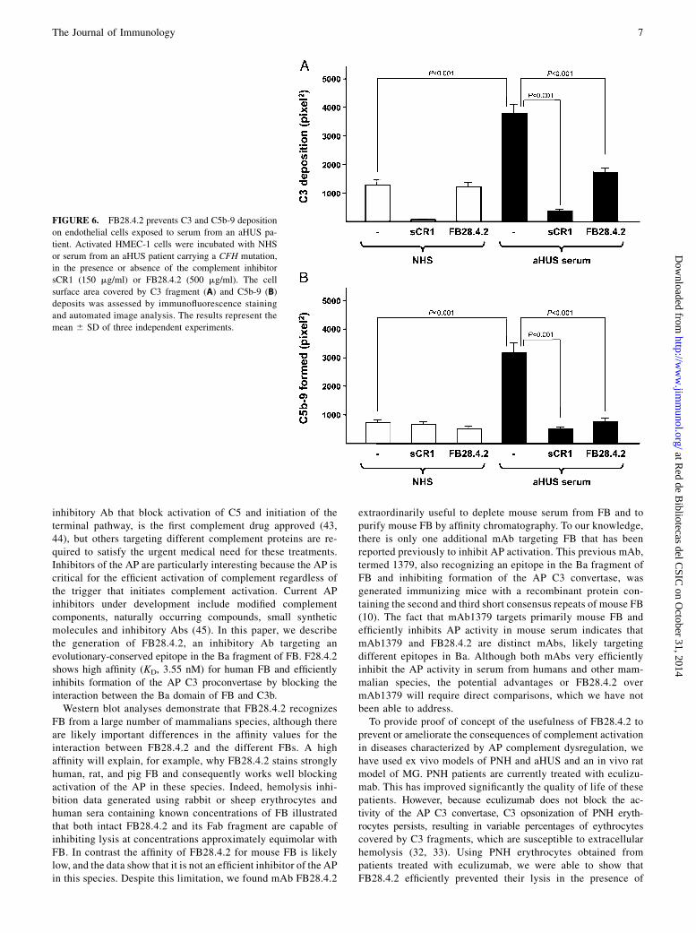
aHUS is a rare, life-threatening disease characterized by throm-bocytopenia, hemolytic anemia, and acute renal failure. aHUS istriggered by vascular endothelial damage, which in most cases isassociated with mutations and polymorphisms in complementgenes. Overwhelming experimental evidence shows that thesepathogenic genetic variations confer defective protection of cel-lular surfaces from complement activation (34). ADP-activatedHMEC-1 cells are a good ex vivo aHUS model; they over-express P-selectin, which works as a receptor for C3b capable ofinitiating complement activation (35). When these activated cellsare exposed to serum from aHUS patients carrying mutations insoluble complement components that impair complement regula-tion, the surface of these cells becomes covered by C3 fragmentsand C5b-9 deposits; NHS has no such effect (36, 37). We haveused this experimental setting to evaluate the capacity of theFB28.4.2 Ab to prevent complement-mediated endothelial celldamage. When activated HMEC-1 cells were incubated with se-rum from an aHUS patient carrying a pathogenic CFH mutation inthe presence of the mAb, both C3 fragment and C5b-9 deposits
FIGURE 1. FB28.4.2 binds an epitope in the Ba domain of FB and prevents activation of both FB and C3. (A) ELISA and Western blot analyses using
intact FB and Bb and Ba fragments. Proteins were mixed together prior to SDS PAGE and Western blot. R, reduced conditions. NR, non-reduced con-
ditions. A goat anti-hFB polyclonal Ab was used as a positive control recognizing both Bb and Ba fragments. (B) Sensogram showing the kinetic char-
acterization of human FB binding to mAb FB28.4.2 captured with an anti-mouse IgG Ab surface. (C) Coomassie-stained gel showing that addition of mAb
FB28.4.2 to a mixture of C3 (0.55 mg), FB (0.5 mg), and FD (0.001 mg) in AP buffer prevents cleavage of both FB and C3. (D) Western blot analyses of
EDTA–plasma samples (0.1 ml) from different mammals to illustrate recognition by FB28.4.2. Purified hFB (0.08 mg) and mFB (0.08 mg) were included for
comparison.
4 NOVEL mAb THAT INHIBITS COMPLEMENT TARGETING FB
at Red de B
ibliotecas del CSIC
on October 31, 2014
http://ww
w.jim
munol.org/
Dow
nloaded from

were significantly decreased (Fig. 6). For comparison, we in-cluded in these experiments a recombinant truncated form of the
membrane-associated complement receptor 1 (sCR1) capable of
inhibiting AP activation and decreasing complement deposition
on the cell surface. As illustrated in Fig. 6, the results obtained
with FB28.4.2 were comparable to those obtained with sCR1.
FB28.4.2 prevents the development of EAMG in rats
MG is an autoimmune disease caused by Abs against the nicotinicAChR. These Abs activate complement at the neuromuscular
endplate causing microlysis and reducing the number of AChR,
thus hampering the correct transmission of impulses between motor
neurons and muscle leading to weakness and fatigability (38–40).
FIGURE 2. FB28.4.2 blocks AP activation and prevents complement-mediate lysis of rabbit and sheep erythrocytes. (A) Rabbit erythrocytes activate the
AP and are lysed in the presence of human serum. Both FB28.4.2 (left panel) and its Fab fragment (right panel) inhibited lysis of rabbit erythrocytes in
2.5% of normal human serum. The results represent the mean 6 SD of three independent experiments. (B) In contrast to rabbit erythrocytes, lysis of sheep
erythrocytes by human serum is factor H dependent. Figure shows that FB28.4.2 prevents lysis of sheep erythrocytes in 20% EGTA serum from an aHUS
patient carrying the factor H C-terminal mutation W1183L. The percentage of aHUS required to obtain .80% lysis was determined by exposing sheep
erythrocytes to increasing amounts of aHUS serum (inset). The results represent the mean 6 SD of three independent experiments. (C) Rat (7.5% for 100%
lysis) and pig (10%) sera efficiently lysed rabbit erythrocytes under AP conditions; mouse serum (4%) required classical pathway triggering using Ab-
sensitized rabbit erythrocytes. FB28.4.2 efficiently inhibited lysis by rat and pig sera but not mouse serum under these conditions. The results represent the
mean 6 SD of three independent experiments.
FIGURE 3. FB28.4.2 prevents binding of FB
to C3b. Sensogram showing binding of FB
(flowed at 0.5 mM) to a CM5 Biacore chip
coated with 1000 RU C3b. Preincubation of FB
with 4-fold excess of purified FB28.4.2 Fab
fragment completely block binding of FB to the
chip. Bar indicates the duration of FB injection
and the arrow the point of EDTA injection. This
experiment was performed twice with identical
results.
The Journal of Immunology 5
at Red de B
ibliotecas del CSIC
on October 31, 2014
http://ww
w.jim
munol.org/
Dow
nloaded from

EAMG can be induced actively by AChR immunization or pas-sively by transfer of anti-AChR Abs in mice and rats (20, 24).Strong experimental evidence supports the critical role of com-plement in the development of both the human disease and therodent models (7, 41).We used the passive EAMG model in rats to test whether the
FB28.4.2 Ab, inhibitory for rat complement in vitro (Fig. 2C), isable to block the AP in vivo in rats and prevent the development ofthe disease. EAMG was induced by passive administration of theanti-AChR mAb35. By 24 h after disease induction, all controlanimals (n = 6) receiving the triggering mAb presented substantialweight loss (average, ∼10% body mass) (Fig. 7A) and varyingdegrees of paralysis (Fig. 7B). At this time point, two animals hadreached a clinical score of 3 (total hind limb paralysis) and weresacrificed; the remaining four rats progressed and all had reachedor exceeded a score of 3 at 48 h and were sacrificed. In contrast,five of six animals treated with a single i.v. dose (8 mg) of FB-28.4.2 at the time of disease induction had no detectable disease at24 h (Fig. 7B) (no weakness or tail tone loss) and one had sometail tone loss (clinical score of 1); the treated group showedminimal weight loss (average 1.7% body mass). After 48 h, ratstreated with mAb FB28.4.2 showed marginally increased signs ofdisease (three of six with clinical score of 1; Fig. 7B) and weightloss (Fig. 7A). All rats were sacrificed at 48 h to collect tissues andserum.
Complement hemolytic activity was measured in serum harvestedfrom the rats prior to disease induction, then at 4, 24, and 48 hpostinduction, using a specific AP assay (as described in Materialsand Methods). Four hours after FB28.4.2 administration, plasma APhemolytic activity was drastically reduced by 90% compared withpredose and PBS controls. At 24 and 48 h, the hemolytic activitywas still markedly reduced—by 70 and 50%, respectively (Fig. 7C).To confirm the impact of the mAb on pathology at the end-plate
in EAMG, intact Bungarotoxin-stained endplates were counted inSoleus muscle sections from control and treated rats. In controlEAMG rats, intact endplates were rare, averaging 1.6 per field at320 magnification; in contrast, endplates in FB28.4.2-treatedanimals were significantly more frequent, averaging 10.6 perfield at 320 magnification (Fig. 7D).
These data illustrate that FB28.4.2 efficiently blocks comple-ment activation in vivo and prevents complement-mediated end-plate damage and the development of paralysis in EAMG, furthersupporting the large body of evidence indicating that EAMG isa complement-mediated disease and suggesting that AP inhibitionis a potential therapy for the human disease.
DiscussionDuring the last 15 y, a number of linkage studies and genome-wide association studies have revealed a strong association be-tween mutations and polymorphisms in complement genes anddifferent diseases such as aHUS, C3 glomerulopathies, and AMD.Subsequent functional analyses of various disease-associatedgenetic variants have demonstrated that complement dysregu-lation is a major contributor to pathogenesis in these disorders(42). These findings, in addition to the long-time realization thatunwanted complement activation sustains the “vicious cycle” ofinflammation and perpetuates tissue damage in many patholog-ical conditions, have put complement inhibition in focus fordevelopment of new therapeutic agents (16). Eculizumab, an
FIGURE 4. FB28.4.2 prevents lysis of PNH erythrocytes in acidified
serum. Erythrocytes (8%) from six PNH patients with different sizes of PNH
clones and densities of C3 deposits were incubated in 50% acidified NHS
alone or in the presence of increasing concentrations of FB28.4.2 mAb.
Despite the differences in the percentage of original lysis, which roughly
correlates with the size of the PNH clone evident from C3 deposition
(demonstrated either by a Coombs direct test (46) or by flow cytometry),
FB28.4.2 efficiently prevented the hemolysis in all cases. Data points rep-
resent mean 6 SD of three different experiments.
FIGURE 5. FB28.4.2 prevents C3 deposition on erythrocytes. Rabbit
erythrocytes were incubated with NHS in the presence of eculizumab
(20 mg) or FB28.4.2 (9 mg) at 37˚C in AP buffer and analyzed by flow
cytometry for the presence of C3 activated fragments using a rabbit
polyclonal anti-C3 Ab or an anti-iC3b/C3dg mouse mAb (SIM320.12.2.1).
As a negative control, rabbit erythrocytes were incubated with NHS in the
presence of EDTA. This experiment was performed twice with identical
results.
6 NOVEL mAb THAT INHIBITS COMPLEMENT TARGETING FB
at Red de B
ibliotecas del CSIC
on October 31, 2014
http://ww
w.jim
munol.org/
Dow
nloaded from
inhibitory Ab that block activation of C5 and initiation of theterminal pathway, is the first complement drug approved (43,44), but others targeting different complement proteins are re-quired to satisfy the urgent medical need for these treatments.Inhibitors of the AP are particularly interesting because the AP iscritical for the efficient activation of complement regardless ofthe trigger that initiates complement activation. Current APinhibitors under development include modified complementcomponents, naturally occurring compounds, small syntheticmolecules and inhibitory Abs (45). In this paper, we describethe generation of FB28.4.2, an inhibitory Ab targeting anevolutionary-conserved epitope in the Ba fragment of FB. F28.4.2shows high affinity (KD, 3.55 nM) for human FB and efficientlyinhibits formation of the AP C3 proconvertase by blocking theinteraction between the Ba domain of FB and C3b.Western blot analyses demonstrate that FB28.4.2 recognizes
FB from a large number of mammalians species, although thereare likely important differences in the affinity values for theinteraction between FB28.4.2 and the different FBs. A highaffinity will explain, for example, why FB28.4.2 stains stronglyhuman, rat, and pig FB and consequently works well blockingactivation of the AP in these species. Indeed, hemolysis inhi-bition data generated using rabbit or sheep erythrocytes andhuman sera containing known concentrations of FB illustratedthat both intact FB28.4.2 and its Fab fragment are capable ofinhibiting lysis at concentrations approximately equimolar withFB. In contrast the affinity of FB28.4.2 for mouse FB is likelylow, and the data show that it is not an efficient inhibitor of the APin this species. Despite this limitation, we found mAb FB28.4.2
extraordinarily useful to deplete mouse serum from FB and topurify mouse FB by affinity chromatography. To our knowledge,there is only one additional mAb targeting FB that has beenreported previously to inhibit AP activation. This previous mAb,termed 1379, also recognizing an epitope in the Ba fragment ofFB and inhibiting formation of the AP C3 convertase, wasgenerated immunizing mice with a recombinant protein con-taining the second and third short consensus repeats of mouse FB(10). The fact that mAb1379 targets primarily mouse FB andefficiently inhibits AP activity in mouse serum indicates thatmAb1379 and FB28.4.2 are distinct mAbs, likely targetingdifferent epitopes in Ba. Although both mAbs very efficientlyinhibit the AP activity in serum from humans and other mam-malian species, the potential advantages or FB28.4.2 overmAb1379 will require direct comparisons, which we have notbeen able to address.To provide proof of concept of the usefulness of FB28.4.2 to
prevent or ameliorate the consequences of complement activationin diseases characterized by AP complement dysregulation, wehave used ex vivo models of PNH and aHUS and an in vivo ratmodel of MG. PNH patients are currently treated with eculizu-mab. This has improved significantly the quality of life of thesepatients. However, because eculizumab does not block the ac-tivity of the AP C3 convertase, C3 opsonization of PNH eryth-rocytes persists, resulting in variable percentages of eythrocytescovered by C3 fragments, which are susceptible to extracellularhemolysis (32, 33). Using PNH erythrocytes obtained frompatients treated with eculizumab, we were able to show thatFB28.4.2 efficiently prevented their lysis in the presence of
FIGURE 6. FB28.4.2 prevents C3 and C5b-9 deposition
on endothelial cells exposed to serum from an aHUS pa-
tient. Activated HMEC-1 cells were incubated with NHS
or serum from an aHUS patient carrying a CFH mutation,
in the presence or absence of the complement inhibitor
sCR1 (150 mg/ml) or FB28.4.2 (500 mg/ml). The cell
surface area covered by C3 fragment (A) and C5b-9 (B)
deposits was assessed by immunofluorescence staining
and automated image analysis. The results represent the
mean 6 SD of three independent experiments.
The Journal of Immunology 7
at Red de B
ibliotecas del CSIC
on October 31, 2014
http://ww
w.jim
munol.org/
Dow
nloaded from

acidified serum. As expected, these PNH erythrocytes were cov-ered by C3 fragments, which impeded our ability to demonstrate
that FB28.4.2 would also be useful in preventing C3 opsonization
of PNH erythrocytes. However, experiments performed with rab-
bit erythrocytes to compare the capacity of eculizumab and
FB28.4.2 to inhibit complement-mediated lysis and C3 deposition
strongly support the suggestion that FB28.4.2 would improve
treatment of PNH by preventing both extravascular and intravas-
cular hemolysis through the inhibition of C3 deposition and MAC
formation, respectively.aHUS is another disease in which the mAb FB28.4.2 could be
use to restore the AP complement dysregulation associated with
mutations in complement genes and to prevent the vascular en-
dothelial damage caused by excessive terminal pathway activation.
We have tested this possibility in a recently developed aHUS ex vivo
model based on the capacity of ADP-activated HMEC-1 cells to
activate the AP, which results in strong C3 fragment and C5b-9
deposition when these activated cells are exposed to serum from
aHUS patients. In agreement with the conclusions from our ex-
periments in the PNH model, when ADP-activated HMEC-1 cells
were incubated with aHUS serum in the presence of FB28.4.2, C3
fragment and C5b-9 deposition was drastically reduced.The experiments performed with PNH erythrocytes and HMEC-
1 cells are in vitro experiments. To test whether the FB28.4.2 Ab is
able to block the AP in vivo, we used the passive EAMG model in
rats. We were able to show that a single i.v. dose of 8 mg FB28.4.2blocked significantly the AP in the rat serum for at least 48 h,
which in this acute animal model was sufficient to prevent the
complement-mediated endplate damage and the development of
hind-limb paralysis in the animals treated with the anti-AChR
mAb35. As a whole, the data presented in this paper demon-
strate the potential value of FB28.4.2 for the treatment of diseasescaused by complement dysregulation in humans and animal models.
AcknowledgmentsWe thank Prof. Claire L. Harris for comments and advice during the SPR
experiments and Dr. Martın Alcorlo and Laura Blanco Sobero for help at
the initial stages of this work. We also thank Dr. Edwin Ades and Fransisco
J. Candal of the U.S. Centers for Disease Control and Prevention, Dr. Thomas
Lawley of Emory University (Atlanta, GA) who developed HMEC-1 used in
this study, and CellDex (Needham, MA) for providing sCR1.
DisclosuresS.R.d.C., M.D., M.S., and O.L. are named on a patent held by the Agencia
Estatal Consejo Superior de Investigaciones Cientificas and the Instituto de
la Salud Carlos III that protects the use of the mAb FB28.4.2 as a comple-
ment inhibitor. This situation has not influenced the results and interpreta-
tions in this article. The other authors have no financial conflicts of interest.
References1. Walport, M. J. 2001. Complement: first of two parts.N. Engl. J. Med. 344: 1058–1066.2. Walport, M. J. 2001. Complement: second of two parts. N. Engl. J. Med. 344:
1140–1144.3. Morgan, B. P. 1999. Regulation of the complement membrane attack pathway.
Crit. Rev. Immunol. 19: 173–198.4. Lachmann, P. J. 2009. The amplification loop of the complement pathways. Adv.
Immunol. 104: 115–149.5. Holers, V. M. 2008. The spectrum of complement alternative pathway-mediated
diseases. Immunol. Rev. 223: 300–316.6. Parker, C. J. 2007. The pathophysiology of paroxysmal nocturnal hemoglobin-
uria. Exp. Hematol. 35: 523–533.7. T€uz€un, E., and P. Christadoss. 2013. Complement associated pathogenic
mechanisms in myasthenia gravis. Autoimmun. Rev. 12: 904–911.8. Pickering, M. C., V. D. D’Agati, C. M. Nester, R. J. Smith, M. Haas,
G. B. Appel, C. E. Alpers, I. M. Bajema, C. Bedrosian, M. Braun, et al. 2013. C3glomerulopathy: consensus report. Kidney Int. 84: 1079–1089.
FIGURE 7. FB28.4.2 prevents development of EAMG in rats. After anti-AChR administration (time 0), change in weight (A), clinical score (B), and
hemolytic activity (C) were monitored in both groups, control animals were injected i.v. with vehicle alone (n = 6), and animals were treated with FB28.4.2
(n = 6). The impact of the mAb on pathology was confirmed by counting the number of intact neuromuscular junctions (endplates) in bungarotoxin–
rhodamine-stained Soleus muscle sections (average of 5 fields of view at320 magnification; counted blind) from control and FB28.4.2-treated rats (D). The
results represent the mean 6 SD of three independent experiments. Clinical score as follows: 0, can grip and lift lid of cage; 1, can grip but cannot lift; 2,
cannot grip; 3, cannot grip + hind limbs paralyzed; and 4, moribund. All differences obtained from the comparison between using FB28.4.2 mAb and
vehicle alone were statistically significant.
8 NOVEL mAb THAT INHIBITS COMPLEMENT TARGETING FB
at Red de B
ibliotecas del CSIC
on October 31, 2014
http://ww
w.jim
munol.org/
Dow
nloaded from

9. Skerka, C., M. Jozsi, P. F. Zipfel, M. A. Dragon-Durey, and V. Fremeaux-Bacchi.2009. Autoantibodies in haemolytic uraemic syndrome (HUS). Thromb. Hae-most. 101: 227–232.
10. Thurman, J. M., D. M. Kraus, G. Girardi, D. Hourcade, H. J. Kang, P. A. Royer,L. M. Mitchell, P. C. Giclas, J. Salmon, G. Gilkeson, and V. M. Holers. 2005. Anovel inhibitor of the alternative complement pathway prevents antiphospholipidantibody-induced pregnancy loss in mice. Mol. Immunol. 42: 87–97.
11. Leinhase, I., M. Rozanski, D. Harhausen, J. M. Thurman, O. I. Schmidt,A. M. Hossini, M. E. Taha, D. Rittirsch, P. A. Ward, V. M. Holers, et al. 2007.Inhibition of the alternative complement activation pathway in traumatic braininjury by a monoclonal anti-factor B antibody: a randomized placebo-controlledstudy in mice. J. Neuroinflammation 4: 13.
12. DiLillo, D. J., A. W. Pawluczkowycz, W. Peng, A. D. Kennedy, P. V. Beum,M. A. Lindorfer, and R. P. Taylor. 2006. Selective and efficient inhibition of thealternative pathway of complement by a mAb that recognizes C3b/iC3b. Mol.Immunol. 43: 1010–1019.
13. Lindorfer, M. A., A. W. Pawluczkowycz, E. M. Peek, K. Hickman, R. P. Taylor,and C. J. Parker. 2010. A novel approach to preventing the hemolysis of par-oxysmal nocturnal hemoglobinuria: both complement-mediated cytolysis and C3deposition are blocked by a monoclonal antibody specific for the alternativepathway of complement. Blood 115: 2283–2291.
14. Risitano, A. M. 2012. Paroxysmal nocturnal hemoglobinuria and othercomplement-mediated hematological disorders. Immunobiology 217: 1080–1087.
15. Pawluczkowycz, A. W., M. A. Lindorfer, J. N. Waitumbi, and R. P. Taylor. 2007.Hematin promotes complement alternative pathway-mediated deposition of C3activation fragments on human erythrocytes: potential implications for thepathogenesis of anemia in malaria. J. Immunol. 179: 5543‑5552.
16. Holers, V. M., and J. M. Thurman. 2004. The alternative pathway of complementin disease: opportunities for therapeutic targeting. Mol. Immunol. 41: 147–152.
17. Montes, T., A. Tortajada, B. P. Morgan, S. Rodrıguez de Cordoba, andC. L. Harris. 2009. Functional basis of protection against age-related maculardegeneration conferred by a common polymorphism in complement factor B.Proc. Natl. Acad. Sci. USA 106: 4366–4371.
18. Sanchez-Corral, P., C. Gonzalez-Rubio, S. Rodrıguez de Cordoba, andM. Lopez-Trascasa. 2004. Functional analysis in serum from atypical HemolyticUremic Syndrome patients reveals impaired protection of host cells associatedwith mutations in factor H. Mol. Immunol. 41: 81–84.
19. Ham, T. H., and J. H. Dingle. 1939. Studies on destruction of red blood cells.II.Chronic hemolytic anemia with paroxysmal nocturnal hemoglobinuria:certainimmunological aspects of the hemolytic mechanism with special reference toserum complement. J. Clin. Invest. 18: 657–672.
20. Hepburn, N. J., A. S. Williams, M. A. Nunn, J. C. Chamberlain-Banoub,J. Hamer, B. P. Morgan, and C. L. Harris. 2007. In vivo characterization andtherapeutic efficacy of a C5-specific inhibitor from the soft tick Ornithodorosmoubata. J. Biol. Chem. 282: 8292–8299.
21. Karlsson, R., P. S. Katsamba, H. Nordin, E. Pol, and D. G. Myszka. 2006.Analyzing a kinetic titration series using affinity biosensors. Anal. Biochem. 349:136–147.
22. Trutnau, H. H. 2006. New multi-step kinetics using common affinity biosensorssaves time and sample at full access to kinetics and concentration. J. Biotechnol.124: 191–195.
23. Ruiz-Torres, M. P., F. Casiraghi, M. Galbusera, D. Macconi, S. Gastoldi,M. Todeschini, F. Porrati, D. Belotti, E. M. Pogliani, M. Noris, and G. Remuzzi.2005. Complement activation: the missing link between ADAMTS-13 deficiencyand microvascular thrombosis of thrombotic microangiopathies. Thromb. Hae-most. 93: 443–452.
24. Hepburn, N. J., J. L. Chamberlain-Banoub, A. S. Williams, B. P. Morgan, andC. L. Harris. 2008. Prevention of experimental autoimmune myasthenia gravisby rat Crry-Ig: A model agent for long-term complement inhibition in vivo.Mol.Immunol. 45: 395–405.
25. Milder, F. J., L. Gomes, A. Schouten, B. J. Janssen, E. G. Huizinga,R. A. Romijn, W. Hemrika, A. Roos, M. R. Daha, and P. Gros. 2007. Factor Bstructure provides insights into activation of the central protease of the com-plement system. Nat. Struct. Mol. Biol. 14: 224–228.
26. Rooijakkers, S. H., J. Wu, M. Ruyken, R. van Domselaar, K. L. Planken,A. Tzekou, D. Ricklin, J. D. Lambris, B. J. Janssen, J. A. van Strijp, and P. Gros.2009. Structural and functional implications of the alternative complementpathway C3 convertase stabilized by a staphylococcal inhibitor. Nat. Immunol.10: 721–727.
27. Torreira, E., A. Tortajada, T. Montes, S. Rodriguez de Cordoba, and O. Llorca.2009. Coexistence of closed and open conformations of complement factor B inthe alternative pathway C3bB(Mg2+) proconvertase. J. Immunol. 183:7347‑7351.
28. Torreira, E., A. Tortajada, T. Montes, S. Rodrıguez de Cordoba, and O. Llorca.2009. 3D structure of the C3bB complex provides insights into the activation andregulation of the complement alternative pathway convertase. Proc. Natl. Acad.Sci. USA 106: 882–887.
29. Janssen, B. J., L. Gomes, R. I. Koning, D. I. Svergun, A. J. Koster,D. C. Fritzinger, C. W. Vogel, and P. Gros. 2009. Insights into complementconvertase formation based on the structure of the factor B-cobra venom factorcomplex. EMBO J. 28: 2469–2478.
30. Hillmen, P., C. Hall, J. C. Marsh, M. Elebute, M. P. Bombara, B. E. Petro,M. J. Cullen, S. J. Richards, S. A. Rollins, C. F. Mojcik, and R. P. Rother. 2004.Effect of eculizumab on hemolysis and transfusion requirements in patients withparoxysmal nocturnal hemoglobinuria. N. Engl. J. Med. 350: 552–559.
31. Hillmen, P., N. S. Young, J. Schubert, R. A. Brodsky, G. Socie, P. Muus, A. Roth,J. Szer, M. O. Elebute, R. Nakamura, et al. 2006. The complement inhibitoreculizumab in paroxysmal nocturnal hemoglobinuria. N. Engl. J. Med. 355:1233–1243.
32. Risitano, A. M., R. Notaro, L. Marando, B. Serio, D. Ranaldi, E. Seneca,P. Ricci, F. Alfinito, A. Camera, G. Gianfaldoni, et al. 2009. Complementfraction 3 binding on erythrocytes as additional mechanism of disease in par-oxysmal nocturnal hemoglobinuria patients treated by eculizumab. Blood 113:4094–4100.
33. Hill, A., R. P. Rother, L. Arnold, R. Kelly, M. J. Cullen, S. J. Richards, andP. Hillmen. 2010. Eculizumab prevents intravascular hemolysis in patients withparoxysmal nocturnal hemoglobinuria and unmasks low-level extravascular he-molysis occurring through C3 opsonization. Haematologica 95: 567–573.
34. Rodrıguez de Cordoba, S., M. S. Hidalgo, S. Pinto, and A. Tortajada. 2014.Genetics of atypical hemolytic uremic syndrome (aHUS). Semin. Thromb.Hemost. 40: 422–430.
35. Morigi, M., M. Galbusera, S. Gastoldi, M. Locatelli, S. Buelli, A. Pezzotta,C. Pagani, M. Noris, M. Gobbi, M. Stravalaci, et al. 2011. Alternative pathwayactivation of complement by Shiga toxin promotes exuberant C3a formation thattriggers microvascular thrombosis. J. Immunol. 187: 172–180.
36. Valoti, E., M. Alberti, A. Tortajada, J. Garcia-Fernandez, S. Gastoldi, L. Besso,E. Bresin, G. Remuzzi, S. Rodriguez de Cordoba, and M. Noris. 2014. A novelatypical hemolytic uremic syndrome-associated hybrid CFHR1/CFH geneencoding a fusion protein that antagonizes factor H-dependent complementregulation. J. Am. Soc. Nephrol. DOI: 10.1681/ASN.2013121339.
37. Noris, M., M. Galbusera, S. Gastoldi, P. Macor, F. Banterla, E. Bresin,C. Tripodo, S. Bettoni, R. Donadelli, E. Valoti, et al. 2014. Dynamics of com-plement activation in atypical HUS and how to monitor eculizumab therapy.Blood DOI: 10.1182/blood-2014-02-558296.
38. Hughes, B. W., M. L. Moro De Casillas, and H. J. Kaminski. 2004. Patho-physiology of myasthenia gravis. Semin. Neurol. 24: 21–30.
39. De Baets, M., and M. H. Stassen. 2002. The role of antibodies in myastheniagravis. J. Neurol. Sci. 202: 5–11.
40. Engel, A. G. 1979. The immunopathological basis of acetylcholine receptordeficiency in myasthenia gravis. Prog. Brain Res. 49: 423–434.
41. Sahashi, K., A. G. Engel, E. H. Lambert, and F. M. Howard, Jr. 1980. Ultra-structural localization of the terminal and lytic ninth complement component(C9) at the motor end-plate in myasthenia gravis. J. Neuropathol. Exp. Neurol.39: 160–172.
42. de Cordoba, S. R., A. Tortajada, C. L. Harris, and B. P. Morgan. 2012. Com-plement dysregulation and disease: from genes and proteins to diagnostics anddrugs. Immunobiology 217: 1034–1046.
43. Rother, R. P., S. A. Rollins, C. F. Mojcik, R. A. Brodsky, and L. Bell. 2007.Discovery and development of the complement inhibitor eculizumab for thetreatment of paroxysmal nocturnal hemoglobinuria. Nat. Biotechnol. 25: 1256–1264.
44. Legendre, C. M., C. Licht, P. Muus, L. A. Greenbaum, S. Babu, C. Bedrosian,C. Bingham, D. J. Cohen, Y. Delmas, K. Douglas, et al. 2013. Terminal com-plement inhibitor eculizumab in atypical hemolytic-uremic syndrome. N. Engl.J. Med. 368: 2169–2181.
45. Ricklin, D., and J. D. Lambris. 2013. Complement in immune and inflammatorydisorders: therapeutic interventions. J. Immunol. 190: 3839‑3847.
46. Coombs, R. R. A., A. E. Mourant, and R. R. Race. 1945. A new test for thedetection of weak and incomplete Rh agglutinins. Br. J. Exp. Pathol. 26: 255–266.
The Journal of Immunology 9
at Red de B
ibliotecas del CSIC
on October 31, 2014
http://ww
w.jim
munol.org/
Dow
nloaded from