Oxidation of Low Density Lipoprotein Upon Sequential Exposure to Copper Ions
14
PLEASE SCROLL DOWN FOR ARTICLE This article was downloaded by: [Tel Aviv University] On: 21 September 2009 Access details: Access Details: [subscription number 906383925] Publisher Informa Healthcare Informa Ltd Registered in England and Wales Registered Number: 1072954 Registered office: Mortimer House, 37-41 Mortimer Street, London W1T 3JH, UK Free Radical Research Publication details, including instructions for authors and subscription information: http://www.informaworld.com/smpp/title~content=t713642632 Oxidation of Low Density Lipoprotein Upon Sequential Exposure to Copper Ions Edit Schnitzer a ; Menahem Fainaru b ; Dov Lichtenberg a a Dept. of Physiology and Pharmacology Sackler Faculty of Medicine Tel Aviv University, Ramat Aviv, Tel Aviv, Israel b Dept. of Internal Medicine A, Beilinson Medical Center Sackler Faculty of Medicine Tel Aviv University, Ramat Aviv, Tel Aviv, Israel Online Publication Date: 01 August 1995 To cite this Article Schnitzer, Edit, Fainaru, Menahem and Lichtenberg, Dov(1995)'Oxidation of Low Density Lipoprotein Upon Sequential Exposure to Copper Ions',Free Radical Research,23:2,137 — 149 To link to this Article: DOI: 10.3109/10715769509064028 URL: http://dx.doi.org/10.3109/10715769509064028 Full terms and conditions of use: http://www.informaworld.com/terms-and-conditions-of-access.pdf This article may be used for research, teaching and private study purposes. Any substantial or systematic reproduction, re-distribution, re-selling, loan or sub-licensing, systematic supply or distribution in any form to anyone is expressly forbidden. The publisher does not give any warranty express or implied or make any representation that the contents will be complete or accurate or up to date. The accuracy of any instructions, formulae and drug doses should be independently verified with primary sources. The publisher shall not be liable for any loss, actions, claims, proceedings, demand or costs or damages whatsoever or howsoever caused arising directly or indirectly in connection with or arising out of the use of this material.
-
Upload
independent -
Category
Documents
-
view
1 -
download
0
Transcript of Oxidation of Low Density Lipoprotein Upon Sequential Exposure to Copper Ions
PLEASE SCROLL DOWN FOR ARTICLE
This article was downloaded by: [Tel Aviv University]On: 21 September 2009Access details: Access Details: [subscription number 906383925]Publisher Informa HealthcareInforma Ltd Registered in England and Wales Registered Number: 1072954 Registered office: Mortimer House,37-41 Mortimer Street, London W1T 3JH, UK
Free Radical ResearchPublication details, including instructions for authors and subscription information:http://www.informaworld.com/smpp/title~content=t713642632
Oxidation of Low Density Lipoprotein Upon Sequential Exposure to Copper IonsEdit Schnitzer a; Menahem Fainaru b; Dov Lichtenberg a
a Dept. of Physiology and Pharmacology Sackler Faculty of Medicine Tel Aviv University, Ramat Aviv, TelAviv, Israel b Dept. of Internal Medicine A, Beilinson Medical Center Sackler Faculty of Medicine Tel AvivUniversity, Ramat Aviv, Tel Aviv, Israel
Online Publication Date: 01 August 1995
To cite this Article Schnitzer, Edit, Fainaru, Menahem and Lichtenberg, Dov(1995)'Oxidation of Low Density Lipoprotein UponSequential Exposure to Copper Ions',Free Radical Research,23:2,137 — 149
To link to this Article: DOI: 10.3109/10715769509064028
URL: http://dx.doi.org/10.3109/10715769509064028
Full terms and conditions of use: http://www.informaworld.com/terms-and-conditions-of-access.pdf
This article may be used for research, teaching and private study purposes. Any substantial orsystematic reproduction, re-distribution, re-selling, loan or sub-licensing, systematic supply ordistribution in any form to anyone is expressly forbidden.
The publisher does not give any warranty express or implied or make any representation that the contentswill be complete or accurate or up to date. The accuracy of any instructions, formulae and drug dosesshould be independently verified with primary sources. The publisher shall not be liable for any loss,actions, claims, proceedings, demand or costs or damages whatsoever or howsoever caused arising directlyor indirectly in connection with or arising out of the use of this material.
Free Rud Res., Vol. 23, No. 2, pp. 137-149 Reprints available directly from the publisher Photocopying permitted by license only
0 1995 Harwood Academic Publishers GmbH Printed in Malaysia
OXIDATION OF LOW DENSITY LIPOPROTEIN UPON SEQUENTIAL EXPOSURE TO COPPER IONS
EDIT SCHNITZER", MEN AHEM FAINARU2 and
Dept. of Physiology and Pharmacology' and Dept. of Internal Medicine A, Beilinson Medical Center2 Sackler Faculty of Medicine Tel Aviv University Ramat Aviv, Tel
Aviv 69978 Israel
DOV LICHTENBERG'"
(Received June 20th, 1994; in revised form, October 28th, 1994)
Copper-induced LDL oxidation is characterized by an 'induction phase' (lag phase) during which the endogenous antioxidants are consumed, followed by a 'propagation phase' in which the LDL-associated polyunsaturated fatty acids are oxidized. Oxidation products may play an important role in the propagation of the oxidative process in the arterial intima as they increase the permeability of the damaged endothelium to various plasma components, including LDL. We therefore found it of interest to investigate the kinetics of LDL oxidation in vitro under conditions where LDL is sequentially exposed to Cu2'-induced oxidation.
The results of our studies demonstrate that when native LDL is exposed to copper oxidation in a medium containing oxidized LDL, oxidation of the added LDL may be almost instantaneous. Furthermore, even when native LDL is added to 'oxidizing LDL' towards the end of the lag phase or during the propagation phase it becomes oxidized after a very short lag. This oxidation process, occurring in spite of the possible protective effect of the antioxidants present in the newly added LDL, indicates that although antioxidants prolong the latency period by preventing the formation of active free radicals, when such radicals are present in the system, oxidation propagates. These results lend strong support to the generally accepted paradigm regarding the mechanism of propagation of Iipid oxidation.
In viewoftheeffect of oxidation products on the permeability of theendothelium, theobserved shortening of the lag period may result in a vicious cycle, independent of the LDL-associated antioxidants, leading to continuing oxidation and foam cell formation.
KEY WORDS: LDL, oxidation, lipoproteins, atherosclerosis.
INTRODUCTION
Atherosclerosis is a complex and multifactorial disease caused by excessive lipid uptake by macrophages and smooth muscle cells and consequent 'foam cell' formation and cell death.'-3 Previous pathological, microscopic, histochemical and biochemical studies indicate that a major cause of plaque-formation in the intima of major arteries is the accessive entry of modified low density lipoproteins (LDL) into the cells. LDL modific- ation, which occurs in or near the artery, interferes with its internalization through the tightly controlled mechanism mediated by the LDL receptor. Instead, internalization occurs via a 'scavenger pathway' which has no such control me~hanisrn.~
Recent studies led to the hypothesis that the most common modification of LDL responsible for plaque formation is LDL-oxidation by mechanisms involving free radicals andlor lipoxygena~e.~,~ Such mechanisms are believed to result in peroxidation of polyunsaturated fatty acids [PUFA]. The breakdown products of the resultant lipid
'This work constitutes a part of Mrs. Schnitzer's Ph.D. thesis, to be submitted. *Correspondence
137
Downloaded By: [Tel Aviv University] At: 12:18 21 September 2009
138 E. SCHNITZER, M. FAINARU AND D. LICHTENBERG
peroxides (mostly aldehydes) react with apolipoprotein B (apo B), thus modifying it to the extent that it is no longer recognized by the LDL re~ep to r .~ Furthermore, accumu- lation of oxidized LDL in the arterial wall results in the release of ‘diffusible toxins’.8 These, in turn, among other deleterious effects, increase the infiltration of LDL into the intima, and thereby contribute to the propagation of the atherogenic process.*-”
Appropriate methods for determination of the susceptibility of LDL to oxidation in vivo are not available. Much effort has therefore been devoted to studyin the in vitro oxidation of isolated LDL by Cu2+ and other ‘inducers of oxidation’!” In these reactions, the oxidation was monitored either by measuring the consumption of PUFAs or oxygen’* or the production of lipid hydro peroxide^,'^ conjugated dienesi4 or thiobarbituric acid reactive substances (TBARS), mostly malondialdehyde (MDA).” The most popular method for monitoring the initial stages of oxidation is based on continuous recording of the increase of the conjugated dienes, which absorb UV light at 234nm.I4 Such studies revealed that LDL oxidation is preceded by a latent (lag) phase which is commonly used as a criterion for ‘LDL resistance’ to oxidation. During this phase, all the naturally occuring lipid-soluble antioxidants (mostly vitamin E) are consumed and only minimal oxidation of fatty acids occurs.
If LDL oxidation occurs in the intima of an artery and a significant fraction of the antioxidants have been consumed, further influx of LDL into the intima (along with the LDL-associated antioxidants) may result in prolongation of the lag. By contrast, the oxidation of that LDL which was depleted of its antioxidants may continue, independent of the further addition of LDL (and Vitamin E). Moreover, the initially formed peroxy radicals may shorten the lag period prior to diene formation from the added LDL. Thus, LDL addition to partially oxidized LDL may theoretically be ‘inhibitory’, ‘independent’ or ‘stimulatory ’.
The elucidation of this issue carries pathologic relevance. Yet, it has not been explicitly addressed thus far. In the present communication we describe experiments which clearly demonstrate both the lack of inhibitory effect of added ‘native LDL’ on the propagation of LDL oxidation and the ‘stimulatory’ nature of LDL oxidation towards added native LDL.
MATERIALS AND METHODS
LDL was isolated from human plasma by density gradient ultracentrifufugation according to our modification’6 of the method of Redgrave et al. Briefly, the density of 2.0 ml of human plasma, obtained after 12 hours of fasting, was raised to 1.21 g/ml with solid KBr and overlayered with KBr ‘density solutions’ containing 1mM EDTA (3.0 ml of a solution of a density d = 1.063 g/ml, 3.0 ml of a density d = 1.019 g/ml and 2.5 ml of d = 1.006 g/ml). The sample was then centrifuged in an SW 41 rotor at 40,000 rpm in a L-80 Beckman ultracentrifuge for 24 hours at 4°C. Fractions of 0.5ml were collected from the bottom of the tube after tube-piercing. Each fraction was analysed for cholesterol and density. The fractions corresponding to LDL (densities of d = 1.02-1.05 g/ml), were pooled and dialysed against PBS buffer (3.3mM NaH2P04, 3.3mM Na2HP04, 146mM NaCl and O.lmM EDTA at pH 7.4). The whole procedure was carried out in the dark, at 4”C, and was completed within 48 hours of blood drawing. LDL fractions isolated by a similar procedure were shown by Dobrian et al. to have a similar oxidizability to LDL fractions from which residual albumin has been removed either by affinity chromatography or by gel filtration.”
Prior to studying the Cu2’-induced LDL oxidation, the LDL fraction was dialysed
Downloaded By: [Tel Aviv University] At: 12:18 21 September 2009
LDL OXIDATION 139
again against five changes of a 1 liter solution of the same composition only that EDTA was present in the dialysing medium, according to our modification of the method of Kleinveld et al.:I9 The concentration of EDTA was chosen such that its final concen- tration, after dilution of the LDL to a concentration of 50pglm1, was 1pM. LDL samples were stored at 4°C and used within three days of preparation.
Protein concentration was determined according to Lowry.” Total cholesterol was determined using the commercially available enzymatic kit (Boehiringer-Mannheim). The LDL protein/cholesterol weight ratio was about I :2, indicating that the protein present in the samples was predominantly, if not exclussively, apo B (i.e. that contam- ination by albumin was minimal).
Oxidation was monitored at room temperature (22 k 2°C) or at 37°C by continuous recording of absorbance at 234nm, following the addition of CuC12I9, using a Kontron (Uvikon 930) double beam spectrophotometer, equipped with a I2 position automated sample changer. Measurements were carried out in 1.5 ml quartz cuvettes containing the LDL solutions, following the addition of a freshly prepared CuC12 solution (750pM, 20p1) to a final Cu2+ concentration of 10pM. Time courses of the absorbence at 234 nm are presented as measured against a reference containing LDL at the same concentra- tion, (to ‘subtract’ the reference). Each of the time courses presented in this report is representative of 2-1 2 similar experiments (as mentioned in the corresponding figure legends).
The oxidative modification of LDL was also determined by measuring spectropho- tometrically the amount of malondialdehyde (MDA) equivalents, using triobarbituric acid (TBA) according to the method of Buege and Aust.2’ Specifically, 10pM CuClz were added to a beaker containing lOOml of LDL solution (50pglml). From this beaker, three 1.5ml portions were transfered into quartz cuvettes for monitoring of diene formation (see results). Ten 4 ml aliquotes were taken from the remaining volume at 0,45, 54, 66 and 90 minutes (two aliquotes at each time). In addition, a 20 ml sample of this solution was transfered into another beaker 45 min after CuCll-addition and more native LDL (50p1ml) was added to this beaker. Six2 ml aliquotes were withdrawn from this LDL-rich (100pglml) solution for TBARS measurements at 54,66 and 90 minutes (two aliquotes at each time point). The same procedure was also carried out with another 20ml aliquote of the original solution 54 minutes after CuC12 addition which was then assayed for TBARS at 66 and 90 minutes. In each sample taken for TBARS assay, the oxidation was immediately terminated by the addition of 24pM EDTA and 20pM BHT (butylated hydroxytoluene) followed by freezing (at -70°C). All the samples were lyophilized and the residues were subsequently dissolved in distilled water (0.5ml). TBA reagent (Iml) was then added and the solutions heated for 20 min at 100°C. Following cooling to room temperature, the solutions were centrifuged at 8000 rpm for 10 min and the absorbance of the supernatant was determined at 532 nm. The amount of TBARS was determined by comparison to a calibration graph made by applying the same procedure to malondialdehyde solutions of varying concentrations. TBARS were also assayed in the three solutions (original + the two solutions made by LDL addition) 48h after exposure to CuC12. For these assays, concentrating the solutions (by lyophilization and redilution in smaller volume) was not required. Except of lyophilization, the same procedure was carried out for these samples.
Simulations of kinetic profiles in ‘sequential exposure’ experiments. To be able to reach conclusions regarding the correlation between the oxidation of ‘oxidizing LDL‘ and added ‘native LDL’ the experimental time-courses of diene
Downloaded By: [Tel Aviv University] At: 12:18 21 September 2009
140 E. SCHNITZER, M. FAINARU AND D. LICHTENBERG
accumulation had to be compared with simulated time-courses (see discussion). Sim- ulations were based on the experimental time courses of oxidation of the first batch of LDL as observed in each experiment after the addition of CuCh and were carried out on a PC 486 (Packard Bell) using Lotus for computations and Sigmaplot for plotting the simulated time courses.
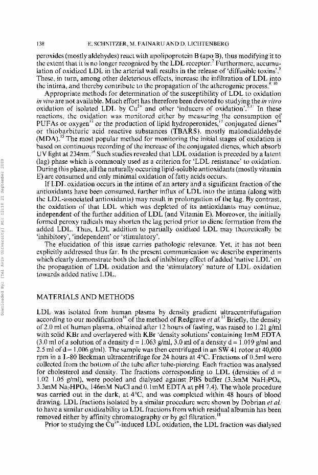
RESULTS
Two questions were addressed in this study regarding sequential exposure of LDL to Cu2+. First we asked how the presence of LDL ‘oxidation products’ affects the kinetics of the oxidation of added native LDL (and specifically the lag prior to oxidation). This question is partially answered by the data given in Fig. 1. Curve a in this figure depicts the time-dependence of conjugated diene production observed upon addition of CuClz (1OtM) to LDL (50pg LDL protein/ml). Curve b describes the time-dependence of Cu ‘-induced oxidation as in curve a only that at the arrow more native LDL (same
O.D.
234nm
1.2
1 .0
0.8
0.6
0.4
0.2
0.0 0 100 300 300
Time( min)
FIGURE 1 Time dependence of diene accumulation during copper-induced oxidation. Diene level was monitored by the absorbance at 234nm after mixing (at time zero) 0. IpM LDL (SOpg proteidml) with 1OpM CuC12 (Curve a). Curve b is the time dependence obtained upon addition of native LDL (50pg/ml) at the time of maximal absorbance (arrow) to the oxidized LDL (as in a). The presented time courses are representative for four experiments carried out on two different LDL preparations.
Downloaded By: [Tel Aviv University] At: 12:18 21 September 2009

LDL OXIDATION 141
O.D. 334nm
1 .o
0.8
0.6
0.4
0.2
0.0 0 50 100 150 200 250 300
Time( min)
FIGURE 2 The effect of oxidized LDL on the oxidation of added native LDL. In all three experiments, CuC12 (10pM) was added to LDL (1Opg protein/ml) and during its oxidation additional LDL (50pg/ml) was introduced at the times indicated by the arrows. The effect of the oxidized LDL on the lagprecedingoxidation of the added LDL was determined in comparison to the lag preceding oxidization of the first batch of LDL (lOpg/rnl) (Early stages of curve a), The presented time courses are representative for three experiments carried out on the same LDL preparation.
amount) was added. The oxidation of the added LDL was not instantaneous, i.e. the presence of oxidation products was not sufficient for complete elimination of the lag phase. However, the lag observed following the second addition was much shorter than that observed after the first exposure to Cu2+, indicating that the products of oxidation stimulate oxidation of the newly added native LDL.
Furthermore, even when the oxidation of native LDL (50pg LDL proteidml) was carried out in the presence of much less oxidized LDL (produced by preincubation of merely 10pg LDL protein/ml), a very significant shortening of the lag phase of the added LDL was observed (Fig. 2a). Moreover, in comparison to the long lag preceding oxidation of native LDL (early stage of curve 2a), the lag observed after further addition of LDL (50pg LDL proteidml) was shortened even when it was added at the onset of the ‘propagation phase’ (Fig. 2b) but not earlier (Fig. 2c).
This effect was observed only when sufficient LDL became oxidized prior to further addition of native LDL. As an example, when LDL (50pg proteidml) was added
Downloaded By: [Tel Aviv University] At: 12:18 21 September 2009
I42 E. SCHNITZER. M. I’AINARU AND D. LICHTENBERG
1.4
1.2
1 .o O . D .
234nm 0.8
0.6
0.4
0.2
0.0 0 50 100 150 200 250 300
Time( min)
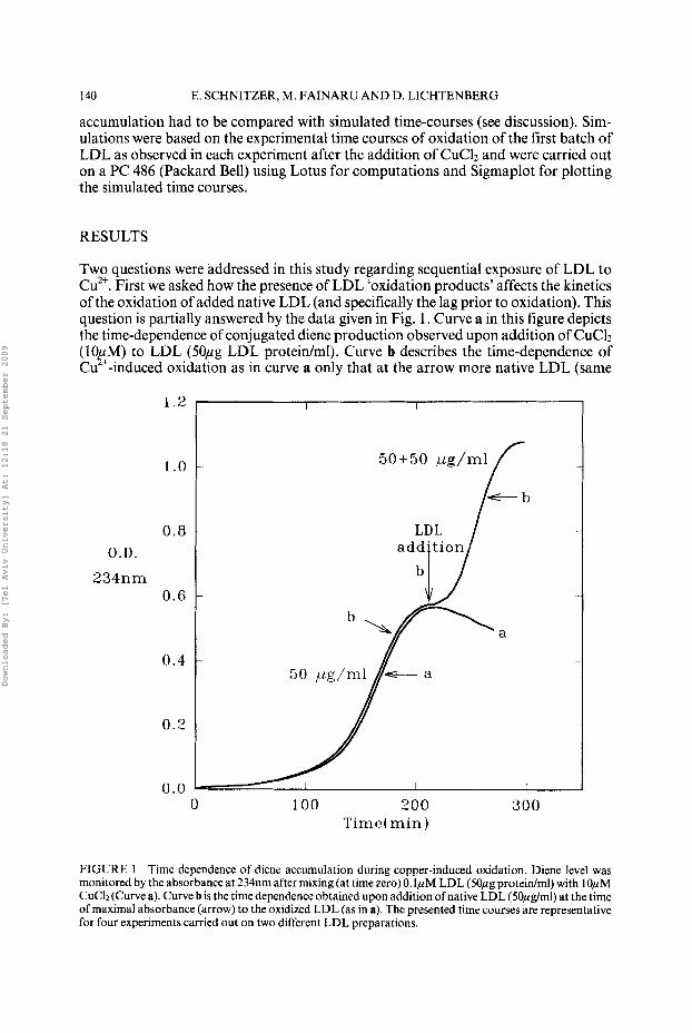
FIGURE 3 The effect of the concentration of oxidized LDL on the oxidation ofadded LDL. Varying LDL concentrations (&20pg/ml, as indicated in the figure) were mixed with CuC12 (10pM) at time zero. At the time indicated by the arrow, 50pg LDL protein/ml were added and diem accumulation was continuously monitored. The presented time courses are representative for three experiments carried out on the same LDL preparation.
towards the end of the lag phase in the oxidation of 1-2Opg LDL/ml, the lag preceding oxidation of the added LDL was significantly shortened only if the initial LDL concentration was higher than 5pglml (Fig. 3). It thus appears that LDL oxidation is markedly accelerated if oxidation products are present in the solution at a sufficiently high concentration.
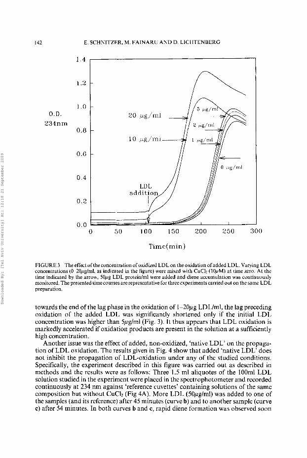
Another issue was the effect of added, non-oxidized, ‘native LDL’ on the propaga- tion of LDL oxidation. The results given in Fig. 4 show that added ‘native LDL’ does not inhibit the propagation of LDL-oxidation under any of the studied conditions. Specifically, the experiment described in this figure was carried out as described in methods and the results were as follows: Three 1.5 ml aliquotes of the lOOml LDL solution studied in the experiment were placed in the spectrophotometer and recorded continuously at 234 nm against ‘reference cuvettes’ containing solutions of the same composition but without CuClz (Fig 4A). More LDL (50pg/ml) was added to one of the samples (and its reference) after 45 minutes (curve b) and to another sample (curve c) after 54 minutes. In both curves b and c, rapid diene formation was observed soon
Downloaded By: [Tel Aviv University] At: 12:18 21 September 2009
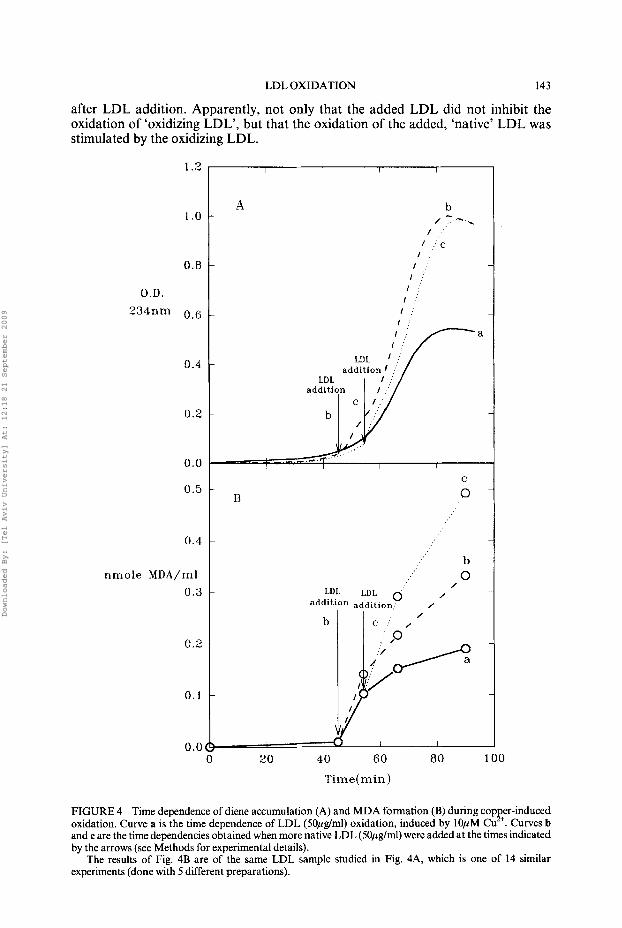
LDL OXIDATION 143
after LDL addition. Apparently, not only that the added LDL did not inhibit the oxidation of ‘oxidizing LDL’, but that the oxidation of the added, ‘native’ LDL was stimulated by the oxidizing LDL.
1.2
1 .o
0.8
O.D. 234nm 0.6
0.4
0.2
0.0
0.5
0.4
nmole MDA/ml 0 .3
0.2
0.1
0.0
A b - / ,..--.
/
I : I : c
I : 1 .
l j I :
1 ; 1 .
A b - / ,..--.
/
I : I : c
I : 1 .
l j I :
1 ;
addition
I I
B
C
0
b 0
/ LDL LDL 0 1
add’t1on addition
20 40 60 80 100
Time(min)
FIGURE 4 Time dependence of diene accumulation (A) and MDA formation (B) during copp+er-induced oxidation. Curve a is the time dependence of LDL (SOpg/ml) oxidation, induced by 10pM Cu . Curves b and c are the time dependencies obtained when more native LDL (5Opg/ml) were added at the times indicated by the arrows (see Methods for experimental details).
The results of Fig. 4B are of the same LDL sample studied in Fig. 4A, which is one of 14 similar experiments (done with 5 different preparations).
Downloaded By: [Tel Aviv University] At: 12:18 21 September 2009
144 E. SCHNITZER, M. FAINARU AND D. LICHTENBERG
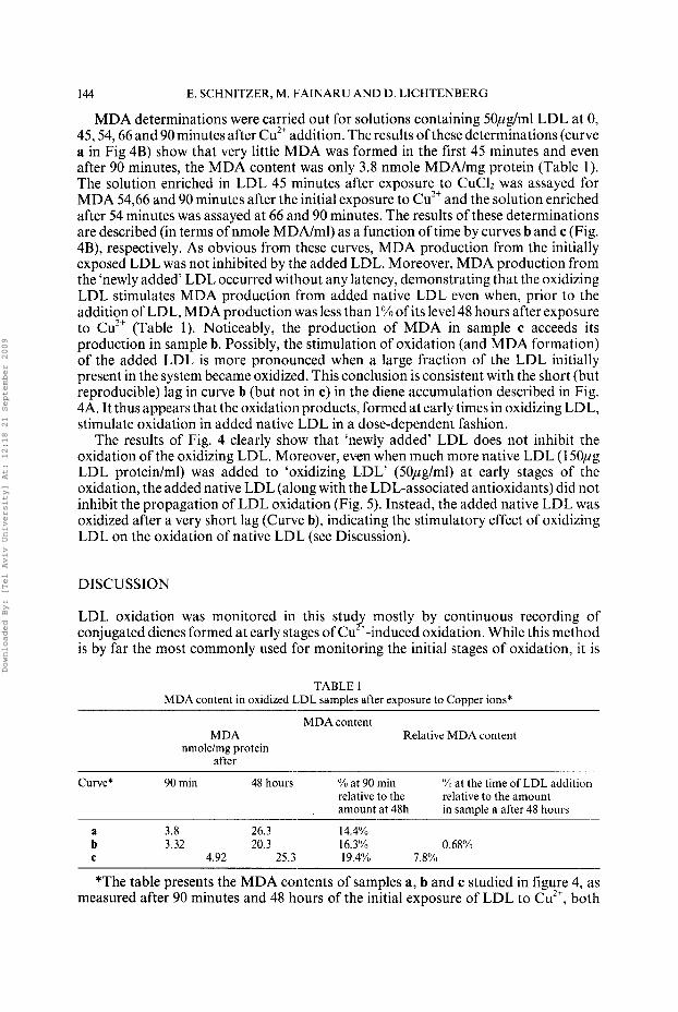
MDA determinations were carried out for solutions containing 50pglml LDL at 0, 45,54,66 and 90 minutes after Cu2+ addition. The results of these determinations (curve a in Fig 4B) show that very little MDA was formed in the first 45 minutes and even after 90 minutes, the MDA content was only 3.8 nmole MDA/mg protein (Table 1). The solution enriched in LDL 45 minutes after exposure to CuCh was assayed for MDA 54,66 and 90 minutes after the initial exposure to Cu2+ and the solution enriched after 54 minutes was assayed at 66 and 90 minutes. The results of these determinations are described (in terms of nmole MDA/ml) as a function of time by curves band c (Fig. 4B), respectively. As obvious from these curves, MDA production from the initially exposed LDL was not inhibited by the added LDL. Moreover, MDA production from the ‘newly added’ LDL occurred without any latency, demonstrating that the oxidizing LDL stimulates MDA production from added native LDL even when, prior to the addition of LDL, MDA production was less than 1% of its level 48 hours after exposure to Cu2+ (Table 1). Noticeably, the production of MDA in sample c acceeds its production in sample b. Possibly, the stimulation of oxidation (and MDA formation) of the added LDL is more pronounced when a large fraction of the LDL initially present in the system became oxidized. This conclusion is consistent with the short (but reproducible) lag in curve b (but not in c) in the diene accumulation described in Fig. 4A. It thus appears that the oxidation products, formed at early times in oxidizing LDL, stimulate oxidation in added native LDL in a dose-dependent fashion.
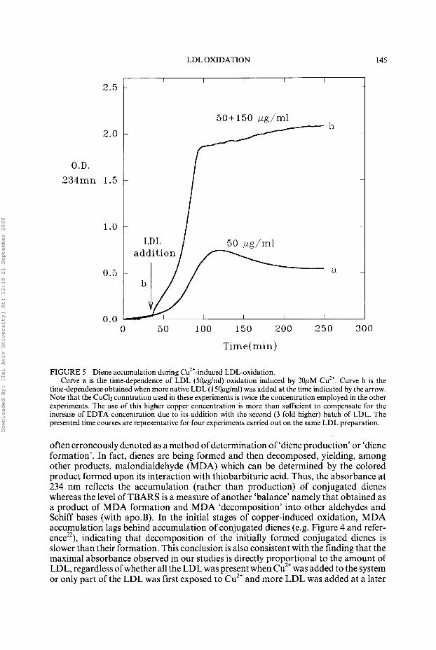
The results of Fig. 4 clearly show that ‘newly added’ LDL does not inhibit the oxidation of the oxidizing LDL. Moreover, even when much more native LDL (1 50pg LDL protein/ml) was added to ‘oxidizing LDL’ (50pglml) at early stages of the oxidation, the added native LDL (along with the LDL-associated antioxidants) did not inhibit the propagation of LDL oxidation (Fig. 5). Instead, the added native LDL was oxidized after a very short lag (Curve b), indicating the stimulatory effect of oxidizing LDL on the oxidation of native LDL (see Discussion).
DISCUSSION
LDL oxidation was monitored in this study mostly by continuous recording of conjugated dienes formed at early stages of Cu2’-induced oxidation. While this method is by far the most commonly used for monitoring the initial stages of oxidation, it is
TABLE 1 MDA content in oxidized LDL samples after exposure to Copper ions*
MDA content MDA Relative MDA content
nmolelmg protein after
Curve* 90 min 48 hours O/U at 90 min ‘Yn at the time of LDL addition relative to the amount at 48h
relative to the amount in sample a after 48 hours
a 3.8 26.3 14.4% ~
b 3.32 20.3 16.3% 0.68% C 4.92 25.3 19.4% 7.8%
*The table presents the MDA contents of samples a, b and c studied in figure 4, as measured after 90 minutes and 48 hours of the initial exposure of LDL to Cu2+, both
Downloaded By: [Tel Aviv University] At: 12:18 21 September 2009
LDL OXIDATION 145
O.D. 234mn
2.0
1.5
1 .o
0.5
0.0 0 50 100 150 200 250 300
Time( min)
FIGURE 5 Diene accumulation during Cu2+-induced LDL-oxidation. Curve a is the time-dependence of LDL (50pglml) oxidation induced by 20pM Cu”. Curve b is the
time-dependence obtained when more native LDL (150,uglml) was added at the time indicated by the arrow. Note that the CuCh conntration used in these experiments is twice the concentration employed in the other experiments. The use of this higher copper concentration is more than sufficient to compensate for the increase of EDTA concentration due to its addition with the second (3 fold higher) batch of LDL. The presented time courses are representative for four experiments carried out on the same LDL preparation.
often erroneously denoted as a method of determination of ‘diene production’ or ‘diene formation’. In fact, dienes are being formed and then decomposed, yielding, among other products, malondialdehyde (MDA) which can be determined by the colored product formed upon its interaction with thiobarbituric acid. Thus, the absorbance at 234 nm reflects the accumulation (rather than production) of conjugated dienes whereas the level of TBARS is a measure of another ‘balance’ namely that obtained as a product of MDA formation and MDA ‘decomposition’ into other aldehydes and Schiff bases (with ap0.B). In the initial stages of copper-induced oxidation, MDA accumulation lags behind accumulation of conjugated dienes (e.g. Figure 4 and refer- ence”), indicating that decomposition of the initially formed conjugated dienes is slower than their formation. This conclusion is also consistent with the finding that the maximal absorbance observed in our studies is directly proportional to the amount of LDL, regardless ofwhether all the LDL was present when Cu2+ was added to the system or only part of the LDL was first exposed to Cu2+ and more LDL was added at a later
Downloaded By: [Tel Aviv University] At: 12:18 21 September 2009
146 E. SCHNITZER, M. FAINARU AND D. LICHTENBERG
stage. It therefore appears that the time course of diene accumulation prior to the observation of a maximal rate of increase of absorbance is only slightly affected by conversion of conjugated dienes to other products and is therefore a measure of diene formation. Two major conclusions can be drawn from our experiments:
1. Upon addition of native LDL to partially oxidized LDL, conjugated diene formation from the ‘newly added’ LDL occurs after a much shorter ‘lag phase’ than that obtained in the absence of ‘oxidizing’ or oxidized LDL.
2. Addition of native LDL to the oxidizing LDL does not inhibit the continuing accumulation of conjugated dienes from the oxidizing LDL.
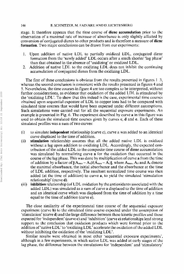
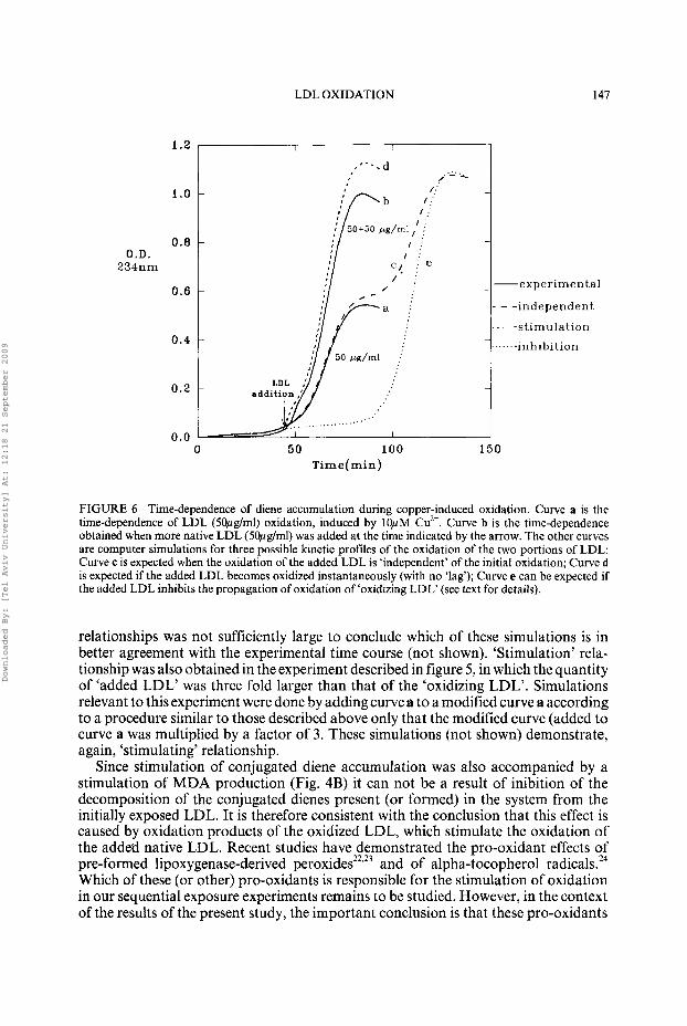
The first of these conclusions is obvious from the results presented in figures 1-3, whereas the second conclusion is consistent with the results presented in figures 4 and 5 . Nevertheless, the time courses in figure 4 are too complex to be interpreted, without further considerations, as evidence that oxidation of the added LDL is stimulated by the ‘oxidizing LDL’. To show that this indeed is the case, experimental time courses obtained upon sequential exposure of LDL to copper ions had to be compared with simulated time courses that would have been expected under different assumptions. Such simulations were carried out for ’all the sequential exposure experiments. An example is presented in Fig. 6 . The experiment described by curve a in this figure was used to obtain the simulated time courses given by curves c, d and e. Each of these simulated profiles was a sum of two curves:
6)
(ii)
(iii)
to simulate independent relationship (curve c), curve a was added to an identical curve displaced to the time of addition. stimulation relationship assumes that all the added native LDL is oxidized without a lag upon addition to oxidizing LDL. Accordingly, the expected con- tribution of the added LDL to the composite time course of diene accumulation was simulated by normalizing curve a for the oxidation that occurred in the course of the lag phase. This was done by multiplication of curve a from the time of addition by a factor of [(A,,, - Ao)/(Amax - At)], where Amax, AO and At denote the maximal absorbance, the initial absorbance and the absorbance at the time of LDL addition, respectively. The resultant normalized time course was then added (at the time of addition) to curve a, to yield the simulated ‘stimulation relationship’ (curve d). inhibition relationship (of LDL oxidation by the antioxidants associated with the added LDL) was simulated as a sum of curve a displaced to the time of addition and an identical curve which was displaced from the time of addition by a time equal to the time of addition (curve e).
The close similarity of the experimental time course of the sequential exposure experiment (curve b) to the simulated time course expected under the assumption of ‘stimulation’ (curve d) and the large difference between these kinetic profiles and those expected for ‘independent’ (curve c ) and ‘inhibition’ (curve e) relationships lend strong support to the conclusion that oxidation products which were formed prior to the addition of ‘native LDL’ to ‘oxidizing LDL’ accelerate the oxidation of the added LDL without inhibiting the oxidation of the ‘oxidizing LDL’.
Similar results were obtained in most other ‘sequential exposure experiments’, although in a few experiments, in which native LDL was added at early stages of the lag phase, the difference between the simulations for ‘independent’ and ‘stimulatory’
Downloaded By: [Tel Aviv University] At: 12:18 21 September 2009
LDL OXIDATION 147
1.2
1.0
0 . 8 O.D.
234nm
0.6
0.4
I I
_... ,‘--. d ,. - .
-experimental
- - independent
- - - - s t imu la t ion inhibi t ion
...’ 0.0 I
0 5 0 100 150 Time(min)
FIGURE 6 Time-dependence of diene accumulation during copper-induced oxidation. Curve a is the time-dependence of LDL (5Opglml) oxidation, induced by 10pM Cu2+. Curve b is the time-dependence obtained when more native LDL (5Opg/ml) was added at the time indicated by the arrow. The other curves are computer simulations for three possible kinetic profiles of the oxidation of the two portions of LDL: Curve c is expected when the oxidation of the added LDL is ‘independent’ of the initial oxidation; Curve d is expected if the added LDL becomes oxidized instantaneously (with no ‘lag’); Curve e can be expected if the added LDL inhibits the propagation of oxidation of ‘oxidizing LDL‘ (see text for details).
relationships was not sufficiently large to conclude which of these simulations is in better agreement with the experimental time course (not shown). ‘Stimulation’ rela- tionship was also obtained in the experiment described in figure 5, in which the quantity of ‘added LDL’ was three fold larger than that of the ‘oxidizing LDL’. Simulations relevant to this experiment were done by adding curve a to a modified curve a according to a procedure similar to those described above only that the modified curve (added to curve a was multiplied by a factor of 3. These simulations (not shown) demonstrate, again, ‘stimulating’ relationship.
Since stimulation of conjugated diene accumulation was also accompanied by a stimulation of MDA production (Fig. 4B) it can not be a result of inibition of the decomposition of the conjugated dienes present (or formed) in the system from the initially exposed LDL. It is therefore consistent with the conclusion that this effect is caused by oxidation products of the oxidized LDL, which stimulate the oxidation of the added native LDL. Recent studies have demonstrated the pro-oxidant effects of pre-formed lipoxygenase-derived peroxide^^^.'^ and of alpha-tocopherol radicals.24 Which of these (or other) pro-oxidants is responsible for the stimulation of oxidation in our sequential exposure experiments remains to be studied. However, in the context of the results of the present study, the important conclusion is that these pro-oxidants
Downloaded By: [Tel Aviv University] At: 12:18 21 September 2009
148 E. SCHNITZER, M. FAINARU AND D. LICHTENBERG
can transfer quickly to newly added LDL and rapidly overcome (‘swamp’) the antiox- idant capacity of the newly added LDL, thus induce an almost instantaneous oxidation of the added LDL.
The lack of inhibitory effect of added LDL on the propagation of oxidation of ‘oxidizing LDL’ is not trivial. In general, the complex nature of the kinetics of copper-induced oxidation in vitro is explained by the higher tendency of the naturally- occurring lipid-associated antioxidants to become oxidized, as compared to the poly- unsaturated, LDL-associated, fatty acids (PUFA). More specifically, the ‘lag phase’ is believed to be a sum of a Vitamin E-independent component and a Vitamin E-depen- dent component, attributed to Vitamin E oxidation prior to the oxidation of PUFA.’ Addition of native LDL, along with the accompanying naturally occuring antioxi- dants, to ‘oxidizing LDL’ in the course of oxidation could have conceivably inhibited the propagation of LDL oxidation. Such an effect was never observed, indicating that complete consumption of Vitamin E within a given LDL particle is followed by a ‘propagation stage’ within this particle, which can not be inhibited by the addition of LDL-associated Vitamin E. This added Vitamin E is probably confined to the LDL particles such that the rate of its transport to other (partially oxidized) LDL particles is very slow on the time scale of oxidation. This in contrast to the rapid transfer of ‘oxidation-promoters’ from the partially oxidized LDL to the added LDL, as discussed above.
This finding is consistent with previous observationsZ5 which demonstrated the problematic nature of in vitro incorporation of vitamin E into LDL. The incorporation was ‘reasonably successful’ only when the added vitamin E was dissolved in dimethyl- sulfoxide (DMSO) and incubated with plasma for at least 3 hours prior to isolation of LDL. By that procedure up to 10% of added vitamin E became incorporates into the LDL, thus increasing the vitamin E content of the LDL by up to four fold of the basal value. Incorporation of Vitamin E into LDL particles after its isolation or after incubation with less externally added vitamin E had only slight effect on the lag preceeding oxidation, in full agreement with our results.
In conclusion, this study shows that LDL oxidation enhances the rate of ollidation of subsequently added native LDL without reducing the rate of propagation of the oxidizing LDL. These results are compatible with what is generally understood about the mechanism of propagation of lipid oxidation, thus strengthening the prevailing concepts. In view of the effect of oxidation products on the permeation of LDL through the arterial wall,*-” the self-accelerating nature of LDL oxidation is likely to result in a vicious cycle of events leading to foam cell production and to the development of an atherosclerotic plague.’-’
Acknowledgments The authors thank Mrs. Zahava Schafer for her expert technical assistence. Special thanks are due to Prof. Herman Esterbauer for the stimulating discussions that led to this study.
References 1. R. Ross and J.A. Glomset, (1976). The pathogenesis of atherosclerosis (part 1). New England Journol
o j Medicine, 295,369-371. 2. R. Ross, (1986). The pathogenesis of atherosclerosis ~ an update. New England Journal OJ Medicine,
314,488-500. 3. J.C. Geer, H.C. McGill and J.P. Strong, (1961). The fine structure of human atherosclerotic lesions.
American Journal of Pathology, 38,263-281.
Downloaded By: [Tel Aviv University] At: 12:18 21 September 2009
LDL OXIDATION 149
4. U. Beisiegel (1992). Apolipoprotein as ligands for lipoprotein receptor. In ‘Structure and function of Apolipoproteins’ (Rossuneu M, ed., CRC in Press, Inc., Boca Raton, An Arbor, London, Tokyo), pp. 269-294.
5. H. Esterbauer, J. Gebicki, H. Phul and G. Jurgens (1992). The Role of Lipid Peroxidation and antioxidants in oxidative modification of LDL. Free Radical Biology and Medicine, 13,341-390.
6. D. Steinberg and workshop participants (1992). Antioxidants in the prevention of human atheroscle- rosis. Summary of the proceedings of a National Heart, Lung, and Blood Institute Workshop: September 5-6, 1991, Bethesda, Maryland. Circulation, 85,2338-2344.
7. H. Esterbauer and G. Jurgens (1993). Mechanistic and Genetic aspects of susceptibility of LDL to oxidation. Current Opinion in Lipidology, 4,114124.
8. H. Esterbauer (1992). Lipid peroxidation and its role in atherosclerosis. Nutrition Metabolism and Cardiovascular Diseases, 2,55-57.
9. H. Esterbauer (1993). Cytotoxicity and genotoxicity of lipid-oxidation products. American Journal of Clinical Nutrition, 57, (su ppl), 7795-786s.
10. D. Steinberg, S. Parthasarathy, T.E. Carew, J.C. Khoo and J.L. Witztum (1989). Beyond cholesterol. Modifications of low-density lipoprotein that increase its atherogenicity. New England Journal of Medicine, 320,915-924.
11. B. Halliwell and S. Chirico (1993). Lipid peroxidation, its mechanism, measurement, and significance. American Journal of Clinical Nutrition, 57, (Suppl), 7 15S--725S.
12. M. Mino, M. Mikim, M. Miyake and T. Ogihava (1989). Nutritional assessment of vitamin E in oxidative stress. Annuls New York Academy of Science, 570,296-320.
13. M. El-Saadani, H. Esterbaur, M. El-Sayed, M. Goher, A.Y. Nassar, and G. Juergens (1989). A spectrophotometric assay for lipid peroxides in serum lipoproteins using commercially available reagent. Journal of’lipid Research, 30,627-630.
14. H. Esterbaur, G. Striegel, H. Puhl, and M. Rotheneder (1989). Continuous monitoring of in vitro oxidation of human low Density lipoprotein. Free radicals Research communications, 6,67-75.
15. J. Schuh, G.F. Fairclough, Jr. and R.H. Haschemeyer (1978). Oxygen-mediated hetrogeneity of apo-low-density lipoprotein. Proceedings of the National of Academy of Sciences of the U.S,A, 75,
16. M. Fainaru, Z. Schafer, D. Gavish, A. Hare], and M. Schwartz (1988). Interactions between human and carp (cyprimus carpio) low density lipoprotein (LDL) and LDL receptors. Comparative Biochem- istry and Physiology, 91B, 331-338.
17. T.G. Redgrave, D.C.K. Roberts and C.E. West (1975). Separation of plasma lipoproteins by density gradient ultracentrifugation. Analytical Biochemistry, 65,4249.
18. A. Dobrian, R. Mova, M. Simionescu and N. Simionescu (1993). In vitro formation of oxidatively- modified and reassembled human low-density lipoproteins: antioxidant effect of albumin. Biochimica Biophysica acta1169, 12-24.
19. H.A. Kleinveld, H.L.M. Hak-Lemmers, A.F.H. Stalenhoff, and P.N.M. Demacker (1992). Improved measurement of low-density lipoprotein susceptibility to Copper-Induced oxidation: Application of a short procedure for isolating low-density lipoprotein. Clinical Chemistry, 38,2066-2072.
20. O.H. Lowry, N.J. Rosenbrough, A.L. Farr, and R.J. Randall, (1951). Journal of Biological chemistry,
21. J.A. Buege, and S.D. Aust (1978). Microsomal Lipid peroxidation. In ‘Methods in Enzymology’ Vol. 52 (Biomembranes part C , Microsomal, cytochrom p-450 and other hemoprotein systems; Colowich, S.P. and Kaplan N.O. eds., Academic press Inc. New York, London), pp. 302-310.
22. V.J. Oleary, V.M. Darley-Usmar, L.J. Russell and D. Stone (1992). Pro-oxidant effects of lipoxygenase- derived peroxides on copper-initiated oxidation of low-density lipoprotein. Biochemical Journal, 282, 63 1-634.
23. B. Frei and J.M. Gazianu (1993). Content of antioxidants, preformed lipid hydroperoxides, and cholesterol as predictors of the susceptibility of human LDL to metal ion-dependent and independent oxidation. Journal of Lipid Research, 34,2135-2 145.
24. V.W. Bowry and R. Stoker (1993). Tocopherol-mediated peroxidation. The proxidant effect of Vitamin E on the radical-initiated oxidation of human low-density lipoprotien. Journal of American Chemical Society, 115,6029-6044.
25. H. Esterbaur,M. Dieber-Rotheneder, G. Striegl, and G. Waeg (1991). Role of Vitamin E in preventing the oxidation of low-density lipoprotein. Ameriran Journal of Clinical Nutrition, 53,314s-3215.
3173-3177.
193,265-270.
Accepted by Professor H. Esterbaur
Downloaded By: [Tel Aviv University] At: 12:18 21 September 2009