Family Dermochelyidae (Superfamily Chelonioidea) from the Upper Cretaceous of North Japan
Osteology of Huabeisaurus allocotus (Sauropoda:Titanosauriformes) from the Upper Cretaceous of ChinaMichael D. D’Emic1*, Philip D. Mannion2, Paul Upchurch3, Roger B. J. Benson4, Qiqing Pang5,
Cheng Zhengwu6
1 Anatomical Sciences Department, Stony Brook University, Health Sciences Center, Stony Brook, New York, United States of America, 2 Department of Earth Science and
Engineering, Imperial College London, South Kensington Campus, London, United Kingdom, 3 Department of Earth Sciences, University College London, London, United
Kingdom, 4 Department of Earth Sciences, University of Oxford, Oxford, United Kingdom, 5 Shijiazhuang University of Economics, Shijiazhuang, People’s Republic of
China, 6 Institute of Geology, Chinese Academy of Geological Sciences, Beijing, People’s Republic of China
Abstract
Background: The Late Cretaceous titanosauriform sauropod Huabeisaurus allocotus Pang and Cheng is known from teethand much of the postcranial skeleton. Its completeness makes it an important taxon for integrating and interpretinganatomical observations from more fragmentary Cretaceous East Asian sauropods and for understanding titanosauriformevolution in general.
Methodology/Principal Findings: We present a detailed redescription of Huabeisaurus allocotus and a suite of anatomicalcomparisons with other titanosauriforms that demonstrate its validity via autapomorphies (e.g., division of some presacralvertebral laminae, reduced development of caudal ribs, the development of fossae relative to one another in caudalvertebral neural arches, high tibia-to-femur ratio). Huabeisaurus shares many features with other Cretaceous East Asiansauropods (e.g., pendant cervical ribs, anterior-middle caudal vertebrae with a nearly flat anterior centrum face and aconcave posterior centrum face) that are absent in sauropods from other landmasses and strata, suggesting a closerelationship among many of these forms within the clade Somphospondyli.
Conclusions/Significance: Restudy of Huabeisaurus provides further evidence for the existence of a clade ofsomphospondylans – Euhelopodidae – mainly found in the Cretaceous of East Asia. Euhelopodidae represents a fourthexample of the evolution of narrow crowns within Sauropoda, along with diplodocoids, brachiosaurids, and advancedtitanosaurs (lithostrotians). Despite being known from fewer species than Diplodocoidea, Brachiosauridae, or Lithostrotia,euhelopodids possessed a broader range of tooth shapes than any of these clades, suggesting that euhelopodidsexemplified a comparably broad range of feeding strategies and perhaps diets.
Citation: D’Emic MD, Mannion PD, Upchurch P, Benson RBJ, Pang Q, et al. (2013) Osteology of Huabeisaurus allocotus (Sauropoda: Titanosauriformes) from theUpper Cretaceous of China. PLoS ONE 8(8): e69375. doi:10.1371/journal.pone.0069375
Editor: Andrew A. Farke, Raymond M. Alf Museum of Paleontology, United States of America
Received March 7, 2013; Accepted June 6, 2013; Published August 2, 2013
Copyright: � 2013 D’Emic et al. This is an open-access article distributed under the terms of the Creative Commons Attribution License, which permitsunrestricted use, distribution, and reproduction in any medium, provided the original author and source are credited.
Funding: Funding was provided by the Geological Society of America to MDD, an Alexander von Humboldt Research Fellowship (Museum fur Naturkunde,Berlin) and a Junior Research Fellowship (Imperial College London) to PDM, and Leverhulme Trust Research Grant RPG-129 to PU. The funders had no role in studydesign, data collection and analysis, decision to publish, or preparation of the manuscript.
Competing Interests: The authors have declared that no competing interests exist.
* E-mail: [email protected]
Introduction
The sauropod Huabeisaurus allocotus was excavated from Upper
Cretaceous sediments of northeast China in the 1990 s. The
holotype of Huabeisaurus is a partially articulated individual
composed of teeth, representative elements from all regions of
the axial column (cervical, dorsal, sacral, and caudal vertebrae),
ribs, complete pectoral and pelvic girdles, and nearly complete
limbs. Due to its relative completeness, Huabeisaurus represents an
important taxon for understanding sauropod evolution in Asia.
The original description of the species noted strong similarities
between the osteology of Huabeisaurus and other Cretaceous East
Asian sauropods, and in general, previous studies have pointed to
some East Asian Cretaceous sauropod (e.g., Nemegtosaurus,
Phuwiangosaurus) as the sister taxon of Huabeisaurus. In the 13 years
since the original description of Huabeisaurus, 17 new sauropod
species have been erected from the Cretaceous of East Asia (see
lists in [1,2]). Many authors have noted similarities among
Cretaceous East Asian sauropods, often suggesting that several
of these taxa belong to a clade grounded on a genus with well-
known anatomy (e.g., Nemegtosauridae, [3–5]; Opisthocoelicau-
dinae, [6]; Euhelopodidae, [2,7]; see [8] for further discussion).
Cladistic support was recently presented for a Euhelopodidae that
consisted of exclusively Cretaceous-aged members [2], in contrast
with traditional studies and early cladistic analyses that posited the
existence of a Euhelopodidae with Jurassic forms (see [5,7] for
detailed discussion). Both earlier and later analyses suggest some
degree of endemism in East Asia, though its temporal duration
remains uncertain. On a broader scale, an interesting evolutionary
pattern has been recognized wherein all pre-Jurassic Cretaceous
Asian sauropods lie outside of Neosauropoda, whereas all
Cretaceous Asian sauropods are titanosauriforms [5]. Refining
and explaining these paleobiogeographic patterns through time
rests on detailed comparisons and comprehensive phylogenetic
PLOS ONE | www.plosone.org 1 August 2013 | Volume 8 | Issue 8 | e69375

studies including East Asian sauropods, which are currently
lacking.
With the aim of presenting comparative osteological data for
Huabeisaurus, we examined the hypodigm firsthand at the
Shijiazhuang University Museum. Below, we redescribe the
anatomy of Huabeisaurus, highlighting similarities and differences
with other Cretaceous East Asian sauropods. Based on synapo-
morphies recovered by existing cladistic datasets, we place
Huabeisaurus in the wider context of sauropod evolution by
exploring its lower level affinities. Finally, we compare the
disparity of tooth shapes among derived sauropod clades, noting
the exceptional range of shapes found within Euhelopodidae.
Results
Systematic paleontologyDinosauria Owen 1842 [9]
Sauropoda Marsh 1878 [10]
Titanosauriformes Salgado, Coria, and Calvo 1997 [11]
Somphospondyli Wilson and Sereno 1998 [12]
? Euhelopodidae Romer 1956 [13] sensu D’Emic 2012 [2]
Huabeisaurus Pang and Cheng 2000 [14]
Huabeisaurus allocotus Pang and Cheng 2000 [14]
HolotypeHBV-20001, a single, partially articulated individual comprising
two teeth, four cervical vertebrae, six partial dorsal vertebrae, a
sacrum composed of six vertebrae, 30 caudal vertebrae, four
dorsal ribs, 13 chevrons, left and right scapulae, left and right
coracoids, left radius, right ilium, left and right pubes, left and
right ischia, left and right femora, left and right tibiae, left and
right fibulae, held in the collections of Shijiazhuang University
Museum, Shijiazhuang, People’s Republic of China.
Emended diagnosisAutapomorphic features of H. allocotus (newly recognized herein
except for the last feature) include: (1) posterior cervical vertebrae
with divided prezygodiapophyseal lamina (i.e., PRDL-F present),
(2) anterior dorsal vertebrae with divided anterior spinodiapophy-
seal lamina (i.e., anterior SPDL-F present), (3) postzygapophyseal
spinodiapophyseal fossa (POSDF) larger than postzygapophyseal
centrodiapophyseal fossa (POCDF) on anterior-middle caudal
vertebrae, (4) caudal vertebrae with small caudal ribs (‘transverse
processes’) that disappear around caudal vertebra eight, (5) ventral
one-third of anterior-middle caudal vertebral centra expanded
posteriorly, (6) two longitudinal ridges on the lateral faces of mid-
caudal vertebral centra (ventral to the ridge at the neurocentral
junction), (7) coracoid with tubercle near anterodorsal edge of
lateral face, (8) distal end of radius about twice as broad
transversely as midshaft (convergently acquired in derived
titanosaurs), (9) tubercle on ischial plate projects from posterior
margin, (10) ratio of tibia to femur length high (0.75; [14]).
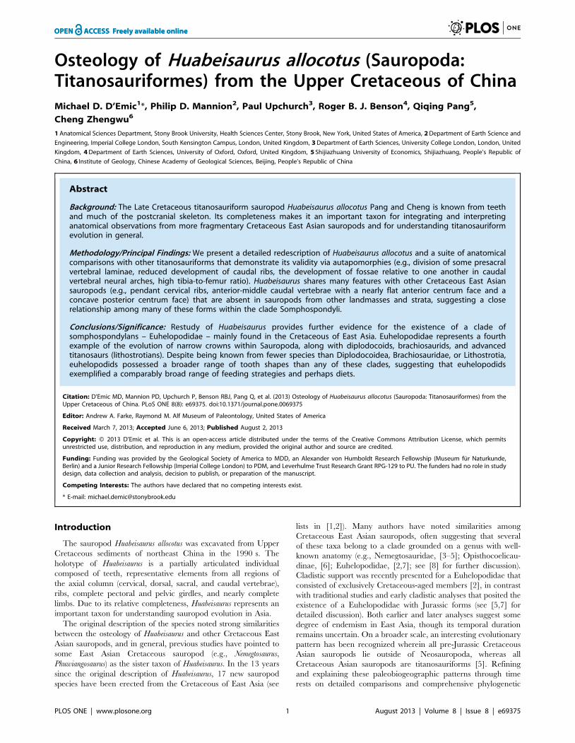
Type locality, horizon, and ageHuabeisaurus comes from Kangdailiang and Houyu, Zhaojiagou
Town, Tianzhen County, Shanxi province, China (Fig. 1). The
holotype was found in the unnamed upper member of the
Huiquanpu Formation, which is Late Cretaceous (?Cenomanian–
?Campanian) in age based on ostracods, charophytes, and fission-
track dating [17,18].
CommentsThe holotype of H. allocotus comprises more bones than
originally listed [14] (e.g., four dorsal ribs versus one). The
augmented list presented herein is based on personal observation
of the skeleton in March 2012 and examination of the quarry map
of the holotype located in the Museum of Shijiazhuang University.
Because many of the bones of HBV-20001 are currently restored
with plaster, the proposed autapomorphies above and the
description that follows are based on firsthand observation
confirmed by reference to pre-restoration photographs of H.
allocotus. Our diagnosis differs from that listed in the original
description [14], which used a combination of characters, nearly
all of which have a broader phylogenetic distribution, to diagnose
H. allocotus.
The isolated humerus designated the paratype [14] comes from
a locality over 200 meters away from the type locality of H.
allocotus, in a fluvially deposited sandy conglomeratic layer in the
lower member of the Huiquanpu Formation (see fig. 4 in [15]).
This layer is roughly 90 m lower stratigraphically than the type
horizon of H. allocotus, which comes from the upper member of the
Huiquanpu Formation. The humerus thus comes from a stratum
representing a different and likely older depositional environment
than that of H. allocotus, and does not overlap anatomically with
the holotypic skeleton, and so cannot currently be referred to that
taxon. The horizon that yielded the humerus also contains
hadrosaurid specimens referred to cf. Shantungosaurus sp. and
indeterminate ankylosaurid material [15].
The absence of sutures between the neural arches and centra of
cervical, dorsal, and caudal vertebrae suggests that the specimen
was nearing somatic maturity, but the open sutures between the
scapula and coracoid and the ilium and some sacral ribs suggest
that it had not reached full skeletal maturity.
Paleoenvironment and taphonomyThe geology of the type locality area was described in a series of
reports [14,15,18–22]. The specimen was found near the base of
the upper member of the Huiquanpu Formation, in a fluvially
Figure 1. Holotype locality of H. allocotus (HBV-20001) from theUpper Cretaceous of Shanxi, China, marked by ‘x’.doi:10.1371/journal.pone.0069375.g001
Osteology of Huabeisaurus allocotus
PLOS ONE | www.plosone.org 2 August 2013 | Volume 8 | Issue 8 | e69375
deposited silty mudstone (Fig. 2). This locality has also produced
the ankylosaur Tianzhenosaurus youngi, theropod material referred to
cf. Szechuanosaurus campi (now regarded as a nomen dubium
[23]), and indeterminate hadrosaurid material [14,15].
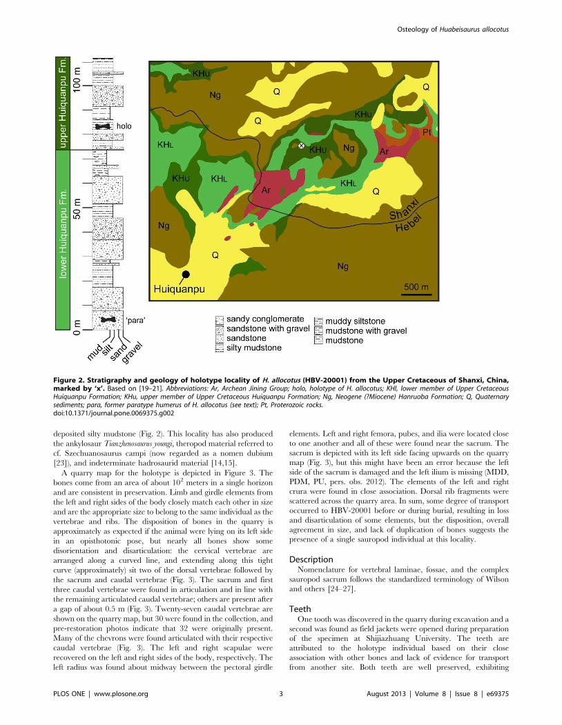
A quarry map for the holotype is depicted in Figure 3. The
bones come from an area of about 102 meters in a single horizon
and are consistent in preservation. Limb and girdle elements from
the left and right sides of the body closely match each other in size
and are the appropriate size to belong to the same individual as the
vertebrae and ribs. The disposition of bones in the quarry is
approximately as expected if the animal were lying on its left side
in an opisthotonic pose, but nearly all bones show some
disorientation and disarticulation: the cervical vertebrae are
arranged along a curved line, and extending along this tight
curve (approximately) sit two of the dorsal vertebrae followed by
the sacrum and caudal vertebrae (Fig. 3). The sacrum and first
three caudal vertebrae were found in articulation and in line with
the remaining articulated caudal vertebrae; others are present after
a gap of about 0.5 m (Fig. 3). Twenty-seven caudal vertebrae are
shown on the quarry map, but 30 were found in the collection, and
pre-restoration photos indicate that 32 were originally present.
Many of the chevrons were found articulated with their respective
caudal vertebrae (Fig. 3). The left and right scapulae were
recovered on the left and right sides of the body, respectively. The
left radius was found about midway between the pectoral girdle
elements. Left and right femora, pubes, and ilia were located close
to one another and all of these were found near the sacrum. The
sacrum is depicted with its left side facing upwards on the quarry
map (Fig. 3), but this might have been an error because the left
side of the sacrum is damaged and the left ilium is missing (MDD,
PDM, PU, pers. obs. 2012). The elements of the left and right
crura were found in close association. Dorsal rib fragments were
scattered across the quarry area. In sum, some degree of transport
occurred to HBV-20001 before or during burial, resulting in loss
and disarticulation of some elements, but the disposition, overall
agreement in size, and lack of duplication of bones suggests the
presence of a single sauropod individual at this locality.
DescriptionNomenclature for vertebral laminae, fossae, and the complex
sauropod sacrum follows the standardized terminology of Wilson
and others [24–27].
TeethOne tooth was discovered in the quarry during excavation and a
second was found as field jackets were opened during preparation
of the specimen at Shijiazhuang University. The teeth are
attributed to the holotype individual based on their close
association with other bones and lack of evidence for transport
from another site. Both teeth are well preserved, exhibiting
Figure 2. Stratigraphy and geology of holotype locality of H. allocotus (HBV-20001) from the Upper Cretaceous of Shanxi, China,marked by ‘x’. Based on [19–21]. Abbreviations: Ar, Archean Jining Group; holo, holotype of H. allocotus; KHl, lower member of Upper CretaceousHuiquanpu Formation; KHu, upper member of Upper Cretaceous Huiquanpu Formation; Ng, Neogene (?Miocene) Hanruoba Formation; Q, Quaternarysediments; para, former paratype humerus of H. allocotus (see text); Pt, Proterozoic rocks.doi:10.1371/journal.pone.0069375.g002
Osteology of Huabeisaurus allocotus
PLOS ONE | www.plosone.org 3 August 2013 | Volume 8 | Issue 8 | e69375
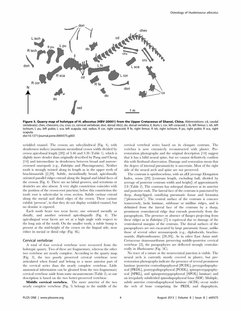
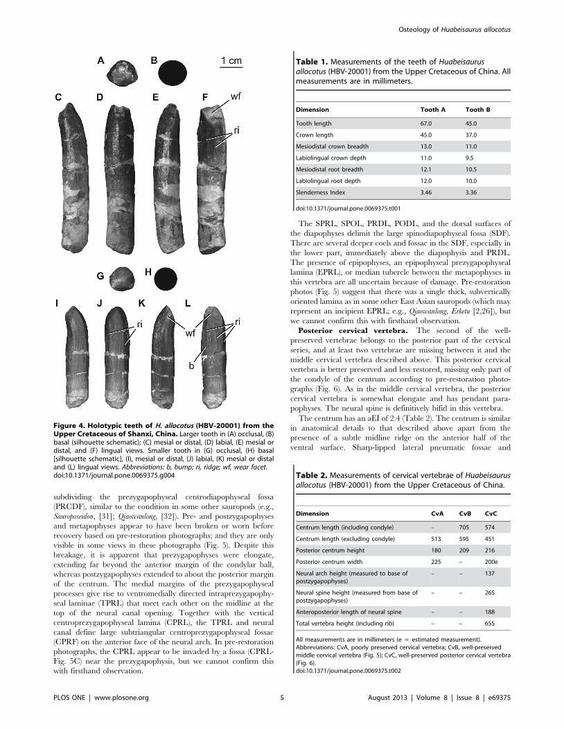
wrinkled enamel. The crowns are subcylindrical (Fig. 4), with
slenderness indices (maximum mesiodistal crown width divided by
crown apicobasal length [28]) of 3.46 and 3.36 (Table 1), which is
slightly more slender than originally described by Pang and Cheng
[14] and intermediate in slenderness between broad and narrow-
crowned sauropods (e.g., Euhelopus and Phuwiangosaurus). Neither
tooth is strongly twisted along its length as in the upper teeth of
brachiosaurids [2,29]. Subtle, mesiodistally broad, apicobasally
oriented parallel ridges extend along the lingual and labial faces of
the crowns (Fig. 4). There are no labial grooves, and serrations or
denticles are also absent. A very slight constriction coincides with
the position of the crown-root junction; below this constriction the
tooth root is subcircular in cross section. Subtle carinae extend
along the mesial and distal edges of the crown. These carinae
exhibit ‘prewear’, in that they do not display wrinkled enamel, but
no dentine is exposed.
Each tooth bears two wear facets: one oriented mesially or
distally, and another oriented apicolingually (Fig. 4). The
apicolingual wear facets are set at a high angle with respect to
the long axis of the tooth. On the smaller tooth, a subtle bump is
present at the mid-height of the crown on the lingual side, near
either its mesial or distal edge (Fig. 4L).
Cervical vertebraeA total of four cervical vertebrae were recovered from the
holotypic quarry. Two of these are fragmentary, whereas the other
two vertebrae are nearly complete. According to the quarry map
(Fig. 3), the two poorly preserved cervical vertebrae were
articulated when found and belong to a more anterior part of
the cervical series than the nearly complete vertebrae. Little
anatomical information can be gleaned from the two fragmentary
cervical vertebrae aside from some measurements (Table 2), so our
description is based on the two better-preserved vertebrae.
Middle cervical vertebra. The more anterior of the two
nearly complete vertebrae (Fig. 5) belongs to the middle of the
cervical vertebral series based on its elongate centrum. The
vertebra is now extensively reconstructed with plaster. Pre-
restoration photographs and the original description [14] suggest
that it has a bifid neural spine, but we cannot definitively confirm
this with firsthand observation. Damage and restoration mean that
the degree of internal pneumaticity is uncertain. Most of the right
side of the neural arch and spine are not preserved.
The centrum is opisthocoelous, with an aEI (average Elongation
Index, sensu [29] [centrum length, excluding ball, divided by
average of posterior centrum width and height]) of approximately
2.8 (Table 2). The centrum has subequal diameters at its anterior
and posterior ends. The lateral face of the centrum is punctured by
deep, sharp-lipped, ramifying pneumatic fossae and foramina
(‘‘pleurocoels’’). The ventral surface of the centrum is concave
transversely, lacks laminae, subfossae or midline ridges, and is
delimited from the lateral face of the centrum by a sharp,
prominent ventrolateral ridge that extends posteriorly from the
parapophysis. The presence or absence of flanges projecting from
these ridges as in Euhelopus [7] is equivocal due to damage of the
ventrolateral margins of the centrum. The dorsal surfaces of the
parapophyses are not excavated by large pneumatic fossae, unlike
those of several other neosauropods (e.g., diplodocids, brachio-
saurids, Haplocanthosaurus; [28,30]). As in other East Asian mid-
Cretaceous titanosauriforms preserving middle-posterior cervical
vertebrae [2], the parapophyses are deflected strongly ventrolat-
erally in Huabeisaurus (Fig. 5C).
No trace of a suture at the neurocentral junction is visible. The
neural arch is currently mostly covered in plaster, but pre-
restoration photographs indicate the presence of several prominent
laminae (posterior centrodiapophyseal [PCDL], prezygodiapophy-
seal [PRDL], postzygodiapophyseal [PODL], spinoprezygapophy-
seal [SPRL], and spinopostzygapophyseal [SPOL] laminae) and
an irregularly subdivided spinodiapophyseal fossa (SDF). Multiple,
subtle anterior centrodiapophyseal laminae (ACDL) occur under
the web of bone comprising the PRDL and diapophysis,
Figure 3. Quarry map of holotype of H. allocotus (HBV-20001) from the Upper Cretaceous of Shanxi, China. Abbreviations: cd, caudalvertebra(e); chev, chevrons; cru, crus; cv, cervical vertebrae; dori, dorsal rib(s); do, dorsal vertebra; il, ilium; L cor, left coracoid; L fe, left femur; L ish, leftischium; L pu, left pubis; L sca, left scapula; rad, radius; R cor, right coracoid; R fe, right femur; R ish, right ischium; R pu, right pubis; R sca, rightscapula.doi:10.1371/journal.pone.0069375.g003
Osteology of Huabeisaurus allocotus
PLOS ONE | www.plosone.org 4 August 2013 | Volume 8 | Issue 8 | e69375
subdividing the prezygapophyseal centrodiapophyseal fossa
(PRCDF), similar to the condition in some other sauropods (e.g.,
Sauroposeidon, [31]; Qiaowanlong, [32]). Pre- and postzygapophyses
and metapophyses appear to have been broken or worn before
recovery based on pre-restoration photographs; and they are only
visible in some views in these photographs (Fig. 5). Despite this
breakage, it is apparent that prezygapophyses were elongate,
extending far beyond the anterior margin of the condylar ball,
whereas postzygapophyses extended to about the posterior margin
of the centrum. The medial margins of the prezygapophyseal
processes give rise to ventromedially directed intraprezygapophy-
seal laminae (TPRL) that meet each other on the midline at the
top of the neural canal opening. Together with the vertical
centroprezygapophyseal lamina (CPRL), the TPRL and neural
canal define large subtriangular centroprezygapophyseal fossae
(CPRF) on the anterior face of the neural arch. In pre-restoration
photographs, the CPRL appear to be invaded by a fossa (CPRL-
Fig. 5C) near the prezygapophysis, but we cannot confirm this
with firsthand observation.
The SPRL, SPOL, PRDL, PODL, and the dorsal surfaces of
the diapophyses delimit the large spinodiapophyseal fossa (SDF).
There are several deeper coels and fossae in the SDF, especially in
the lower part, immediately above the diapophysis and PRDL.
The presence of epipophyses, an epipophyseal prezygapophyseal
lamina (EPRL), or median tubercle between the metapophyses in
this vertebra are all uncertain because of damage. Pre-restoration
photos (Fig. 5) suggest that there was a single thick, subvertically
oriented lamina as in some other East Asian sauropods (which may
represent an incipient EPRL; e.g., Qiaowanlong, Erketu [2,26]), but
we cannot confirm this with firsthand observation.
Posterior cervical vertebra. The second of the well-
preserved vertebrae belongs to the posterior part of the cervical
series, and at least two vertebrae are missing between it and the
middle cervical vertebra described above. This posterior cervical
vertebra is better preserved and less restored, missing only part of
the condyle of the centrum according to pre-restoration photo-
graphs (Fig. 6). As in the middle cervical vertebra, the posterior
cervical vertebra is somewhat elongate and has pendant para-
pophyses. The neural spine is definitively bifid in this vertebra.
The centrum has an aEI of 2.4 (Table 2). The centrum is similar
in anatomical details to that described above apart from the
presence of a subtle midline ridge on the anterior half of the
ventral surface. Sharp-lipped lateral pneumatic fossae and
Figure 4. Holotypic teeth of H. allocotus (HBV-20001) from theUpper Cretaceous of Shanxi, China. Larger tooth in (A) occlusal, (B)basal (silhouette schematic); (C) mesial or distal, (D) labial, (E) mesial ordistal, and (F) lingual views. Smaller tooth in (G) occlusal, (H) basal[silhouette schematic], (I), mesial or distal, (J) labial, (K) mesial or distaland (L) lingual views. Abbreviations: b, bump; ri, ridge; wf, wear facet.doi:10.1371/journal.pone.0069375.g004
Table 1. Measurements of the teeth of Huabeisaurusallocotus (HBV-20001) from the Upper Cretaceous of China. Allmeasurements are in millimeters.
Dimension Tooth A Tooth B
Tooth length 67.0 45.0
Crown length 45.0 37.0
Mesiodistal crown breadth 13.0 11.0
Labiolingual crown depth 11.0 9.5
Mesiodistal root breadth 12.1 10.5
Labiolingual root depth 12.0 10.0
Slenderness Index 3.46 3.36
doi:10.1371/journal.pone.0069375.t001
Table 2. Measurements of cervical vertebrae of Huabeisaurusallocotus (HBV-20001) from the Upper Cretaceous of China.
Dimension CvA CvB CvC
Centrum length (including condyle) – 705 574
Centrum length (excluding condyle) 513 595 451
Posterior centrum height 180 209 216
Posterior centrum width 225 – 200e
Neural arch height (measured to base ofpostzygapophyses)
– – 137
Neural spine height (measured from base ofpostzygapophyses)
– – 265
Anteroposterior length of neural spine – – 188
Total vertebra height (including rib) – – 655
All measurements are in millimeters (e = estimated measurement).Abbreviations: CvA, poorly preserved cervical vertebra; CvB, well-preservedmiddle cervical vertebra (Fig. 5); CvC, well-preserved posterior cervical vertebra(Fig. 6).doi:10.1371/journal.pone.0069375.t002
Osteology of Huabeisaurus allocotus
PLOS ONE | www.plosone.org 5 August 2013 | Volume 8 | Issue 8 | e69375
foramina (‘‘pleurocoels’’) are shallower than in the middle cervical
vertebra and are restricted to the anterior half of the centrum. No
trace of a neurocentral junction suture is visible.
The neural arch and spine are proportionally taller than in the
middle cervical vertebra, representing about twice the centrum
height. The prezygapophyses have flat articular faces that face
dorsomedially and only extend a short distance beyond the
anterior condylar ball. The CPRL descends to the centrum and is
single throughout its length. The lateral part of the underside of
the prezygapophysis gives rise to a well-developed, bifurcated
PRDL (Fig. 6), which is an autapomorphy of Huabeisaurus (though
a similar structure occurs in the anterior dorsal vertebrae of the
basal eusauropod Mamenchisaurus [33]). A pre-epipophysis (follow-
ing the nomenclature of Wilson and Upchurch [7]) lies at the
junction of the CPRL and divided PRDL. Although Ksepka and
Norell [34] followed Wilson and Upchurch [7] in describing the
pre-epipophysis as originating from different laminae in Euhelopus
and Erketu, the condition is the same in those taxa as in
Huabeisaurus. The bifurcated PRDL creates a deep fossa (PRDL-
F) on the anterior face of the pendant diapophysis. The
diapophysis is also connected to the centrum and neural arch by
ACDL, PCDL and PODL. The short, posterodorsally oriented
ACDL is single, unlike the divided ACDL of the middle cervical
vertebra. The region where the diapophysis meets the top of the
tuberculum is marked by a small, rounded process on the exposed
left side of the vertebra. Below the anterodorsally oriented, sheet-
like PCDL, a sharp ridge extends from the posterior end of the
PCDL to the ventral end of the ACDL near the boundary between
the centrum and neural arch. The postzygapophyses have
shallowly concave articular surfaces that face mainly ventrally.
They are connected by an intraprezygapophyseal lamina (TPOL)
that extends ventromedially to the dorsolateral margins of the
posterior neural canal opening (Fig. 6). A tall, robust epipophysis
caps the SPOL near each postzygapophysis (Fig. 6), similar to the
morphology seen in Erketu and Mongolosaurus ([35,36]).
The neural spine is deeply divided, with the floor of the notch
lying level with the zygapophyses. The presence of a median
tubercle is equivocal due to damage. The metapophyses have a
straight, inclined dorsal profile in lateral view. Their cross sections
are apostrophe-shaped in dorsal view, thickening posteriorly
around the SPOL. In lateral view, the stout SPRL is strongly
concave, whereas the SPOL is more broadly curved. No distinct
epipophyseal prezygapophyseal lamina (EPRL) is present, but the
lateral face of each metapophysis bears an irregularly subdivided
spinodiapophyseal fossa (SDF). In particular, there are at least
three small subfossae within the upper part of this fossa.
Cervical ribsOnly the capitula, tubercula, and part of the anterior and
posterior processes of the cervical ribs are preserved. The ribs are
fused to their respective vertebrae (Figs 5–6). The cervical ribs are
pendant, extending ventrally for a distance subequal to the height
of the centrum, as in several other East Asian Cretaceous
sauropods [2]. In both cervical vertebrae, the tuberculum is
notably slender anteroposteriorly, especially in comparison with
the capitulum. The cervical ribs are currently broken, but the
original description notes that at least some originally exceeded
centrum length [14].
Dorsal vertebraeParts of six dorsal vertebrae are preserved: one partial anterior
dorsal vertebral neural arch, one partial dorsal vertebral centrum,
three posterior dorsal vertebrae that are nearly complete but
currently heavily reconstructed with plaster, and one that has been
plastered into the sacrum. None of the dorsal vertebrae have
observable neurocentral sutures.
The anterior dorsal neural arch is missing its neural spine,
postzygapophyses, and pedicles, and has been deformed antero-
laterally (Fig. 7). The total width across the diapophyses is
483 mm. The absence of a parapophysis on the neural arch
indicates that this represents one of the first three dorsal vertebrae.
This identification is also supported by the size and shape of the
prezygapophyses, which have articular surfaces that are wider
than long, widely spaced from each other, and placed near the
front of the neural spine’s base rather than on distinct peduncles.
Broken surfaces reveal coarse camellate pneumaticity internally,
and external laminae and fossae are well developed. Prezygapo-
physes are connected to the diapophyses by a thick, single PRDL.
A small, shallow, elliptical CPRF sits below the prezygapophyses,
Figure 5. Holotypic middle cervical vertebra of H. allocotus(HBV-20001) from the Upper Cretaceous of Shanxi, China.Taken during original preparation in (A) left lateral, (B) ventral, (C)anterior, and (D) posterior views. Abbreviations: ?cprl-f, possiblecentroprezygapophyseal lamina fossa; cvr, cervical rib; ?eprl, possibleepipophyseal-prezygapophyseal lamina; pn fo, pneumatic fossa; pocdf,postzygapophyseal centrodiapophyseal fossa; pep, pre-epipophysis; sdf,spinodiapophyseal fossa. Dashed lines indicate broken bone margins.doi:10.1371/journal.pone.0069375.g005
Osteology of Huabeisaurus allocotus
PLOS ONE | www.plosone.org 6 August 2013 | Volume 8 | Issue 8 | e69375
bounded by wide, low-relief laminae (Fig. 7A). The lateral side of
the vertebra, below the zygodiapophyseal table, bears a very small,
circular centrodiapophyseal fossa (CDF) and broad, shallow,
postzygapophyseal centrodiapophyseal fossa (POCDF), as in the
vertebrae of the pectoral region in some titanosaurs (e.g.,
Rapetosaurus [37]). The anterior surface of the neural arch above
the zygodiapophyseal table is irregularly subdivided: it lacks a
prespinal lamina (at least in its preserved part) and only possesses
an SPRL on the right side (Fig. 7A). The SPDL is divided into
parallel anterior and posterior portions; on the right side of the
vertebra these laminae are distinct from one another throughout
their length, but on the left side of the vertebra these laminae are
poorly developed and the posterior SPDL is united with the
PODL. Between the branches of the SPDL, the SPDL-F is deep
on the right side and shallow on the left side. The posterior SPDL
is badly damaged on the right side. Near the midline, the anterior
SPDL divides to form a small pocket-like anterior SPDL-F, which
we interpret as an autapomorphy of Huabeisaurus (Fig. 7B). The
diapophyses of this vertebra are dorsoventrally tall and ‘flat-
topped’ (Fig. 7A–B), as in most somphospondylans ([2,38]), and
project mainly laterally. The distal end of the diapophysis expands
to form a low, rounded, anteriorly projecting process. Below the
rounded upper region the distal end forms a subcircular, shallow
concavity that faces outwards.
The fragmentary dorsal vertebral centrum is opisthocoelous and
1.5 times wider than tall (Fig. 8). Its anterior, dorsal, and lateral
faces are broken, revealing coarse camellate internal pneumaticity
consisting of branching multi-centimeter scale chambers that
nearly fill the centrum (Fig. 8B). It is too incomplete to determine
its position in the dorsal vertebral sequence.
The three posterior dorsal vertebrae are more complete but are
largely reconstructed with plaster. Pre-restoration photos only exist
for two of these vertebrae (Figs. 9–10), so our description focuses
on these. Both of these vertebrae (Figs. 9–10) are missing parts of
their neural spines, transverse processes, zygapophyses, and the
anterior faces of their centra. Both centra were likely opisthocoe-
lous based on their deeply concave posterior cotyles. The more
anterior of the two vertebrae (Fig. 9A–C) is 1.4 times wider than
tall with a centrum length nearly equal to its width, whereas the
more posterior vertebra has centrum diameters that are subequal
(Table 3). The ventral surfaces of the centra are broad and gently
convex transversely, without developing a ventral keel. The centra
have camellate pneumaticity; the state of pneumaticity in the
neural arches is unknown. The lateral pneumatic foramen
(pleurocoel) is undivided, slightly acuminate anteriorly and
posteriorly, and not set in a fossa in the centrum of the more
anterior dorsal vertebra (Fig. 9C). In contrast, the foramen in the
posterior dorsal vertebra is divided by a complex set of laminae
internally, oval (i.e., not posteriorly acuminate), and set in a fossa
(Fig. 10C).
Parapophyses and the laminae that connect them to other
landmarks are poorly preserved, covered with plaster, and/or
difficult to recognize in pre-restoration photographs in both
vertebrae, precluding definitive identification of laminae and
fossae delimited by them. On the lateral faces of the vertebrae, the
PCDL do not bifurcate towards their ventral ends, unlike the
condition in many somphospondylans [2,11].
The large prezygapophyses of both vertebrae have flat articular
surfaces that face dorsally and slightly medially at approximately
25u to the horizontal. Each prezygapophysis curves ventrally near
the midline, indicating the presence of a hypantrum. The region
surrounding the hyposphene is damaged or covered in all three
vertebrae (Figs. 9–10), but it was probably present, as is the case in
most sauropods excluding derived titanosaurs and many rebba-
chisaurids [11,33,39,40]. On both sides of each vertebra, a stout
CPRL extends dorsally and curls medially to support the medial
part of the prezygapophysis from below. Between these CPRL
there is a slot-like excavation (left and right CPRF) above the
anterior neural canal opening. The stout centropostzygapophyseal
laminae (CPOL) bound the CPOF medially; these fossae are
separated on the midline by a well-developed ventral strut of the
TPOL.
Laminae and fossae of the neural spine are only readily
identifiable on the more posterior dorsal vertebra (Fig. 10).
Anteriorly, a single prespinal lamina (PRSL) extends for almost the
entire length of the neural spine, except near its base, where it
bifurcates slightly. Laterally the SPRF are bounded by anterior
SPDL that terminate in the space between the prezygapophyses
and diapophysis, suggesting that this is perhaps a stranded lamina
in transition according to the pattern of ‘‘lamina capture’’ [25].
The anterior SPDL extends dorsally to an anterior triangular
lateral process (Fig. 10B, D); the posterior triangular lateral process
is broken. Some other sauropods appear to have separate anterior
and posterior triangular lateral processes (e.g., Phuwiangosaurus,
Wilson [25]: fig. 11E). Medial to these triangular lateral processes
at the apex of the neural spine is a longitudinal fossa. Posterior to
the anterior SPDL is a broad fossa (SPDL-F), bounded posteriorly
by the posterior SPDL as in several other titanosauriforms [25].
Figure 6. Holotypic posterior cervical vertebra of H. allocotus (HBV-20001) from the Upper Cretaceous of Shanxi, China. In (A)anterior, (B), anterolateral, (C) left lateral, (D), posterior, and (E) ventrolateral views. (A), (D), and (E) were taken during original preparation; (B) and (C)photographed in 2012. Abbreviations: acdl, anterior centrodiapophyseal lamina; ep, epipophyses; fo, subfossae within spinodiapophyseal fossa; met,metapophysis; pep, pre-epipophysis; prdl-f, prezygodiapophyseal lamina fossa; sdf, spinodiapophyseal fossa. Striped pattern indicates broken surface;dashed lines indicate broken bone margins.doi:10.1371/journal.pone.0069375.g006
Osteology of Huabeisaurus allocotus
PLOS ONE | www.plosone.org 7 August 2013 | Volume 8 | Issue 8 | e69375

The posterior face of the neural spine is damaged, but appears to
have a broad POSL.
Dorsal ribsSix dorsal ribs are indicated on the quarry map (Fig. 3), but only
one nearly complete element and three fragments could be located
in the collections of Shijiazhuang University (Fig. 11). The shafts of
all dorsal ribs are ‘plank-like’ (i.e. three to four times wider than
broad in cross section), as in other titanosauriforms [33]. Near the
articulation facets for the vertebra, the most complete dorsal rib (a
left element) has a subtriangular cross section. This rib has a large,
sharp-lipped pneumatic foramen proximally, as in other titano-
sauriforms [12,33]. This pneumatic foramen is currently covered
by a supporting jacket, but is visible through the support and in
pre-restoration photographs (Fig. 11C). Opposite the capitulum,
on the proximal end, the bone expands into a prominent flange as
in some titanosaurs (e.g., Pitekunsaurus, MAU-Pv-AG-446-33;
Paleontologıa de Vertebrados, Museo Municipal ‘‘Argentino
Urquiza’’, Rincon de los Sauces, Argentina MDD pers. obs.
2010). This ridge also appears to be present in some ribs of
Opisthocoelicaudia (Borsuk-Bialynicka, 1977: plate 10, figure 4B), but
we have not yet confirmed this with firsthand observation. The
posterior edge of the plank-like region is very thin, whereas
anteriorly the rib thickens and has a transversely rounded margin.
Towards its proximal end, the shaft develops a broad, rounded
ridge on its lateral face that is continuous with the anterior margin
of the shaft. The best-preserved rib of Huabeisaurus has a curved
length of 1450 mm and a straight length (i.e., a line from one
extreme to the other) of 1420 mm.
Another dorsal rib (Fig. 11D–F) probably pertains to one of the
last dorsal vertebrae based on its small size and the short distance
between the tuberculum and capitulum. Its shaft is also plank-like
in cross section. The other two fragmentary ribs are broken
portions of shafts.
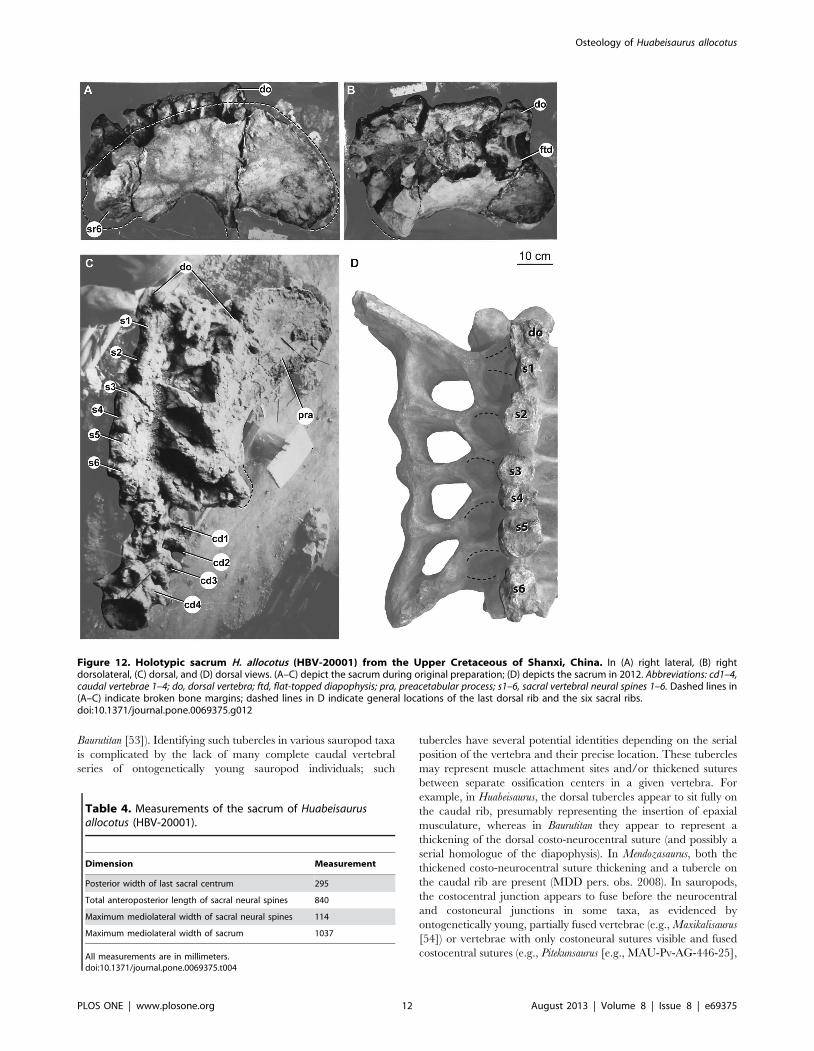
SacrumA nearly complete sacrum consisting of six vertebrae was
recovered from the quarry, originally only lacking some ribs
(Fig. 12). The original description of Huabeisaurus suggested that
Figure 7. Holotypic anterior dorsal neural arch of H. allocotus (HBV-20001) from the Upper Cretaceous of Shanxi, China. In (A)anterior, (B) dorsal, and (C) left lateral views with interpretive line drawings. Note that the post. spdl is broken dorsally in (B). Abbreviations: ant. spdl,anterior spinodiapophyseal lamina; cam, camellate internal pneumaticity exposed on broken surface; cdf, centrodiapophyseal fossa; cprl-f,centroprezygapophyseal lamina fossa; cpol, centropostzygapophyseal lamina; ftd, flat-topped diapophysis; lprz, left prezygapophysis; pocdf,postzygapophyseal centrodiapophyseal fossa; podl, postzygodiapophyseal lamina; post. spdl, posterior spinodiapophyseal lamina; rdi, right diapophysis;rprz, right prezygapophysis; spdl-f, spinodiapophyseal lamina fossa; sprl, spinoprezygapophyseal lamina. Dashed lines indicate broken bone margins.doi:10.1371/journal.pone.0069375.g007
Figure 8. Holotypic anterior dorsal centrum of H. allocotus(HBV-20001) from the Upper Cretaceous of Shanxi, China. In (A)posterior and (B) dorsal views. Abbreviations: cmra, camerate internalpneumaticity exposed on broken surface. Dashed lines indicate brokenbone margins.doi:10.1371/journal.pone.0069375.g008
Osteology of Huabeisaurus allocotus
PLOS ONE | www.plosone.org 8 August 2013 | Volume 8 | Issue 8 | e69375
only five sacral vertebrae were present based on the number of
sacral ribs and intercostal foramina. The sacrum is currently
heavily restored with plaster, but pre-restoration photographs
show the sacrum in two oblique dorsal views and right lateral view
(Fig. 12A–C). These photographs reveal that the last dorsal
vertebra was taphonomically shifted posteriorly and to the right,
crushing the right first sacral rib (Fig. 12A–C). Pre-restoration
photographs and the number of broken sacral neural spines
currently visible (Fig. 12D) indicate that the sacrum was composed
of at least six vertebrae. The first vertebra crushed into the sacrum
could represent a seventh sacral vertebra; because the ribs of this
dorsal vertebra are not observable firsthand or in photographs, we
cannot verify whether or not these ribs contacted the ilium. We
interpret this vertebra as the last dorsal vertebra because: (1) its
neural spine does not appear to be fused to the neural spine
posterior to it, and (2) the usual sacral vertebral count for all but
the most basal somphospondylans is six (with seven vertebrae in
Neuquensaurus as the only derived exception [41,42]).
The sacrum is only slightly longer along its centra than wide
across its ribs, as in most sauropods (Table 4). The pre-restoration
photographs suggest that at least the alar arms (sensu [27]) of the
second through fourth sacral ribs were fused to the right ilium, and
that the neural spines of these vertebrae were fused into a broad
plate as in some titanosaurs (e.g., Epachthosaurus [43]; Malawisaurus
[44]). The broken tops of the neural spines of the sacrum reveal
that neural spines 3–5 fused in contact with one another at this
level, whereas pre- and postspinal laminae separate the other
sacral neural spines. Pre-restoration photographs indicate that the
postacetabular process of the ilium is missing, exposing the iliac
articular faces of the fifth and sixth sacral ribs (Fig. 12A–C). The
corrugated texture apparent in lateral view on the fifth and sixth
sacral ribs suggests a cartilaginous attachment, implying that they
were not fused to the ilium at the time of death. However, we
cannot confirm this based on firsthand observation.
The first sacral vertebra bears a prominent convex anterior ball,
whereas the last sacral vertebral centrum is flat to slightly concave
posteriorly. These articular surfaces are approximately subcircular
in outline. The centra of sacral vertebrae 1–6 are fused together
and their boundaries are marked by low, rounded ridges. There
are no obvious pneumatic fossae or foramina, but these might be
filled and covered with matrix or plaster. The centra of the middle
sacral vertebrae do not narrow considerably compared to the first
and last sacral vertebrae, unlike the situation in some titanosaurs
(e.g., Neuquensaurus [2,42]). As far as can be ascertained from the
restored sacrum, the ventral surfaces of sacral centra are convex
transversely.
There is no evidence that a postspinal lamina (POSL) was
present on the posterior midline of the neural spine of sacral
vertebra 6. This spine also seems to lack the lateral laminae
observed on spines 1–5.
On pre-restoration photographs, flat-topped alar processes are
visible on the sacral vertebral diapophyses, as in the sacra (and
dorsal vertebrae) of most somphospondylans [2,38]. All five ribs
have acetabular arms that are fused laterally to form a well-
developed sacricostal yoke. Intervertebral foramina, transverse
foramina, and intracostal foramina (sensu [27]), if once present,
are now filled by plaster. The size and shape of the dorsal and
ventral intercostal foramina have also been modified by plaster
restoration. Vertebral laminae above the zygodiapophyseal table
(i.e., SPRL, SPDL, SPOL) are also mostly covered by plaster.
Figure 9. Holotypic dorsal vertebra of H. allocotus (HBV-20001) from the Upper Cretaceous of Shanxi, China. Photographs and linedrawings of dorsal vertebra in (A) anterior, (B) posterior, and (C) right lateral views. (A–C) were taken during original preparation. Abbreviations: ?acpl,?anterior centroparapophyseal lamina; cam, camerate internal pneumaticity exposed on broken bone surface; cpol, centropostzygapophyseal lamina; cprl,centroprezygapophyseal lamina; hypa, accessory ventral articulation of hypantrum; pcdl, posterior centrodiapophyseal lamina; pl, pneumatic foramen(pleurocoel) in centrum; ?sprl, ?spinoprezygapophyseal lamina; tpol, ventral strut of intrapostzygapophyseal lamina. Dashed lines indicate broken bonemargins.doi:10.1371/journal.pone.0069375.g009
Osteology of Huabeisaurus allocotus
PLOS ONE | www.plosone.org 9 August 2013 | Volume 8 | Issue 8 | e69375

Caudal vertebraeAlthough only 21 caudal vertebrae were listed in the holotype of
H. allocotus [14], at least 27 are shown on the quarry map (Fig. 3)
and 30 are present in the collections of the Museum of
Shijiazhuang University. The caudal series is preserved as
sequences of consecutive vertebrae with gaps: the first four are
present, then at least two are missing, then eighteen vertebrae,
then a gap of at least two, then one vertebra, then a gap of at least
one, then seven vertebrae (Figs 13, 14, 15). Thus, representative
elements of at least the first thirty-five caudal vertebrae are
preserved in H. allocotus. The last preserved caudal vertebra still
has a well-developed neural arch, suggesting that at least fifteen
vertebrae are missing from the end of the tail based on
comparisons with Gobititan (IVPP 12579; Institute of Vertebrate
Paleontology and Paleoanthropology, Beijing, China [MDD,
PDM and PU pers. obs. 2012]). Thus, the true number of caudal
vertebrae in H. allocotus was probably close to 50, as in most
sauropods (e.g. Camarasaurus, which has 53 [45], and Shunosaurus,
which has 44 [46]), with the exception of the lengthened tails of
diplodocids and shortened tails of some derived titanosaurs
[28,47–49]. Here, we refer to the 30 caudal vertebrae as ‘Cd1–
Cd30’, with these numbers reflecting their sequence and position
as preserved. Below, we divide the available specimens into
‘anterior’ (caudal vertebrae 1–8) and ‘middle’ (caudal vertebrae 9–
30) sections based on the presence and absence of caudal ribs,
respectively. We abbreviate caudal vertebra 1, 2, etc., as Cd1,
Cd2, etc.
Anterior caudal vertebrae. The first eight caudal vertebrae
are generally well preserved, although there are missing parts and
some features are hidden by plaster. In particular, part of the
centrum of Cd1 is heavily restored, the spine of Cd4 is damaged,
the arch and spine of Cd5 are heavily restored with plaster, and
the centrum of Cd8 is strongly crushed transversely.
The original description described all caudal vertebral centrum
articulations as amphicoelous, with the posterior concavity slightly
deeper [14]. This is true for the anterior region of the tail –
anterior articular surfaces of the anterior-middle caudal vertebral
centra (Cd 1–12) are shallowly concave, whereas the larger,
posterior surfaces are more strongly concave (Fig. 13), a feature
that has been interpreted as incipient opisthocoely ([6], but see
Discussion below). Centra are taller than wide and in lateral view
are short anteroposteriorly compared to their height (Table 5). For
Figure 10. Holotypic dorsal vertebra of H. allocotus (HBV-20001) from the Upper Cretaceous of Shanxi, China. Photographs and linedrawings of middle-posterior dorsal vertebra in (A), anterior, (B) posterior, (C), right lateral, and (D), dorsal views. Abbreviations: ant. spdl, anteriorspinodiapophyseal lamina; atlp, anterior transverse lateral process; cam, camerate internal pneumaticity exposed on broken bone surface; cpof,centropostzygapophyseal fossa; cprl, centroprezygapophyseal lamina; fo, fossa; ?pcpl, ?posterior centroparapophyseal lamina; post. spdl, posteriorspinodiapophyseal lamina; sprl, spinoprezygapophyseal lamina; pl, pneumatic foramen (pleurocoel) in centrum; prsl, prespinal lamina. Dashed linesindicate broken bone margins.doi:10.1371/journal.pone.0069375.g010
Table 3. Measurements of dorsal vertebrae of Huabeisaurusallocotus (HBV-20001) from the Upper Cretaceous of China.
Dimension DvA DvB
Centrum length (excluding condyle) 215 –
Posterior centrum height 203 229
Posterior centrum width 286 260
Mediolateral width of left prezygapophysis 123 83
Mediolateral width across prezygapophyses 276 178
All measurements are in millimeters. Abbreviations: DvA, dorsal vertebra inFigure 9; DvB, dorsal vertebra in Figure 10.doi:10.1371/journal.pone.0069375.t003
Osteology of Huabeisaurus allocotus
PLOS ONE | www.plosone.org 10 August 2013 | Volume 8 | Issue 8 | e69375

example, the length to posterior height ratio for Cd2 is about 0.5.
Chevron facets are poorly preserved in the first few vertebrae,
supporting the view that these are from the anteriormost region of
the tail, since these are lacking in articulated sauropod specimens.
In Cd1–5, the ventral surface of the centrum is broad and flat
transversely and mildly concave anteroposteriorly. In Cd5 a
shallow midline groove is present between the posterior chevron
facets. All caudal vertebral centra are solid, lacking pneumatic
fossae or foramina below or into the caudal ribs.
The neural arch occupies most of the length of the dorsal
surface of the centrum in the anteriormost caudal vertebrae, but
from Cd5 onwards it is displaced to cover the anterior part of the
centrum, as in other titanosauriforms [11]. In lateral view, the
prezygapophyses project steeply anterodorsally. The articular
facets of the zygapophyses are flat; in the most anterior caudal
vertebrae (Cd1–4) the facets form a 45-degree angle with the
horizontal. Each prezygapophysis is supported ventrally by a
TPRL and a CPRL. The lateral face of the prezygapophysis bears
a tubercle (Fig. 13) that likely represents the serial homologue of
the diapophysis in the dorsal vertebrae (see below; [40]). There is
no dorsal process or tubercle on the prezygapophyseal portion of
the SPRL (‘SPRL process’), whereas such a feature is found in
some somphospondylans (e.g., Phuwiangosaurus [e.g., SM K11;
Sirindhorn Museum Kalasin Collection, Changwat Kalasin,
Thailand], Tangvayosaurus [TV2; Musee des Dinosaures, Savanna-
khet, Laos], MDD pers. obs. 2008). On the lateral face of the
vertebra, a nearly horizontal lamina (PODL) separates two deep
fossae: the POSDF above, and the POCDF below (Fig. 13). In
most titanosauriforms, the POCDF is deeper and better developed
than the POSDF (e.g., Mendozasaurus [50]), but in H. allocotus the
reverse is true, representing an autapomorphy. The postzygapo-
physeal articular surfaces are large, flat, lack hyposphenes, and
face ventrolaterally.
The neural spines are relatively low and subequal to centrum
height (Table 5). The spine of Cd1 projects posterodorsally,
whereas those of the remaining anterior caudal vertebrae are
directed dorsally. In the most anterior caudal vertebrae (Cd1–6),
the spine is wider transversely than anteroposteriorly throughout
its length, and expands transversely towards its summit. This
results in weakly developed posterolateral lobes in the first several
caudal vertebrae. The SPRL create a moderately deep SPRF,
which is partially divided by a PRSL. The PRSL expands
transversely as it approaches the summit (the same is true for the
POSL described below). The SPOL are single, thin plates that
project prominently, extending to the posterolateral lobes atop the
neural spine. These create a deep SPOF that occupies most of the
posterior surface of the spine. There is a well-developed POSL on
the midline of the SPOF, extending from a point level with the
dorsal margins of the postzygapophyses to the spine summit. The
spine summit was probably convex transversely to weakly trifid in
the anterior caudal vertebrae, as in several titanosaurs (e.g.
Futalognkosaurus [51] and Xianshanosaurus [52]). From Cd5 onwards,
the summits of the spines are generally flat. In Cd7 and all
subsequent caudal vertebrae, the spines are laterally compressed
plates and lack PRSL and POSL, though SPRL and SPOL are
present.
Caudal ribs occur only on Cd1–8, which is unusual for
sauropods, whose caudal ribs typically disappear around Cd14–
16 [30]. We regard this feature as a local autapomorphy of
Huabeisaurus (caudal ribs also disappear by Cd10 in the derived
titanosaurs Opisthocoelicaudia and Alamosaurus [33]). In Huabeisaurus
the caudal ribs are tall at their bases but taper quickly to a blunt,
subtriangular end more rapidly than in other sauropods, giving
them a reduced appearance. The bases of the ribs typically occupy
the dorsal part of the centrum and lower part of the neural arch as
in other sauropods [33]. The precise orientation of some of the
caudal ribs is difficult to determine because of distortion;
nevertheless, in most specimens the ribs appear to have projected
posterolaterally so that their distal tips lie level with or slightly
posterior to the posterior margin of the centrum. Such backswept
caudal ribs are considered to be a titanosauriform synapomorphy
[1,2]. The rib is supported ventrally by two low rounded ridges.
One of these ridges extends ventrally and slightly posteriorly from
the ventral margin of the rib to virtually the posterior margin of
the centrum, and might represent a serial homologue of the
posterior centroparapophyseal lamina (PCPL) of the dorsal
vertebrae. The other ridge extends ventrally from the anterior
face of the rib to the anterior margin of the centrum and is less
steeply inclined, and possibly represents a serial homologue of the
anterior centroparapophyseal lamina (ACPL) of the dorsal
vertebrae. The fossa between these laminae is shallow and does
not further invade the body of the centrum.
All caudal ribs appear to be fully fused to their respective neural
arches and centra. The first several caudal vertebral ribs of
Huabeisaurus have an anteroposteriorly elongate tubercle on their
dorsal surface, which is best preserved in the second and fourth
caudal vertebrae (Fig. 13). Similar elongate tubercles sit on the
dorsal surfaces of the caudal ribs of several titanosauriforms (e.g.,
Mendozasaurus [IANIGLA-PV-065-5, 26; Instituto Argentino de
Nivologıa, Glaciologıa y Ciencias Ambientales, Colleccion Paleo-
vertebrados, Mendoza, Argentina], MDD pers. obs. 2008;
Figure 11. Holotypic dorsal ribs of H. allocotus (HBV-20001)from the Upper Cretaceous of Shanxi, China. Anterior dorsal rib in(A) lateral, (B) posterior; and (C) medial views. Posterior dorsal rib in (D)lateral, (E) posterior, and (F) medial views. (C) was taken during originalpreparation, all others were taken in 2012. Abbreviations: ca, capitulum;fl, flange; pn, pneumatic opening; tu, tuberculum. Dashed lines indicatebroken bone margins.doi:10.1371/journal.pone.0069375.g011
Osteology of Huabeisaurus allocotus
PLOS ONE | www.plosone.org 11 August 2013 | Volume 8 | Issue 8 | e69375
Baurutitan [53]). Identifying such tubercles in various sauropod taxa
is complicated by the lack of many complete caudal vertebral
series of ontogenetically young sauropod individuals; such
tubercles have several potential identities depending on the serial
position of the vertebra and their precise location. These tubercles
may represent muscle attachment sites and/or thickened sutures
between separate ossification centers in a given vertebra. For
example, in Huabeisaurus, the dorsal tubercles appear to sit fully on
the caudal rib, presumably representing the insertion of epaxial
musculature, whereas in Baurutitan they appear to represent a
thickening of the dorsal costo-neurocentral suture (and possibly a
serial homologue of the diapophysis). In Mendozasaurus, both the
thickened costo-neurocentral suture thickening and a tubercle on
the caudal rib are present (MDD pers. obs. 2008). In sauropods,
the costocentral junction appears to fuse before the neurocentral
and costoneural junctions in some taxa, as evidenced by
ontogenetically young, partially fused vertebrae (e.g., Maxikalisaurus
[54]) or vertebrae with only costoneural sutures visible and fused
costocentral sutures (e.g., Pitekunsaurus [e.g., MAU-Pv-AG-446-25],
Figure 12. Holotypic sacrum H. allocotus (HBV-20001) from the Upper Cretaceous of Shanxi, China. In (A) right lateral, (B) rightdorsolateral, (C) dorsal, and (D) dorsal views. (A–C) depict the sacrum during original preparation; (D) depicts the sacrum in 2012. Abbreviations: cd1–4,caudal vertebrae 1–4; do, dorsal vertebra; ftd, flat-topped diapophysis; pra, preacetabular process; s1–6, sacral vertebral neural spines 1–6. Dashed lines in(A–C) indicate broken bone margins; dashed lines in D indicate general locations of the last dorsal rib and the six sacral ribs.doi:10.1371/journal.pone.0069375.g012
Table 4. Measurements of the sacrum of Huabeisaurusallocotus (HBV-20001).
Dimension Measurement
Posterior width of last sacral centrum 295
Total anteroposterior length of sacral neural spines 840
Maximum mediolateral width of sacral neural spines 114
Maximum mediolateral width of sacrum 1037
All measurements are in millimeters.doi:10.1371/journal.pone.0069375.t004
Osteology of Huabeisaurus allocotus
PLOS ONE | www.plosone.org 12 August 2013 | Volume 8 | Issue 8 | e69375

MDD pers. obs. 2010). In other taxa, the rib fuses after the neural
arch and centrum (e.g., Bonitasaura, MDD pers. obs. 2010).
Generally in sauropods, passing distally along the caudal
vertebral sequence as the caudal ribs disappear, the costoneural
junction merges with the position of the costocentral junction at
the location of the neurocentral junction, leaving a roughened,
elongate tubercle that sometimes contains a furrow (e.g.,
Camarasaurus [55]; Tastavinsaurus [56]; Pitekunsaurus, [e.g., MAU-
Pv-AG-446-10, 211, –24], MDD pers. obs. 2010; Huabeisaurus, see
below). Salgado et al. [11] considered this elongate tubercle to be
a characteristic of some derived titanosaurs, but it has a wider
phylogenetic distribution.
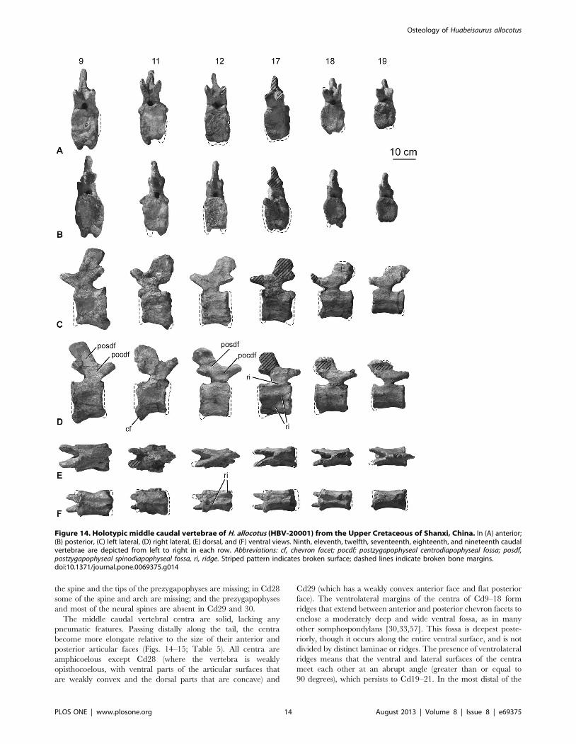
Middle and posterior caudal vertebrae. Preserved caudal
vertebrae 9–30 are only slightly damaged or distorted. For
example, the neural spine is heavily restored on Cd10; parts of
the articular faces of the centrum of Cd11 are missing; the
centrum of Cd13 has been transversely compressed by crushing;
the spine summits of Cd18 and 21 are damaged; in Cd25 parts of
Figure 13. Holotypic anterior caudal vertebrae of H. allocotus (HBV-20001) from the Upper Cretaceous of Shanxi, China. In (A)anterior; (B) posterior, and (C) left lateral views. First, second and sixth caudal vertebrae are depicted from left to right in each row. Lateral view ofsecond caudal vertebra reversed. Abbreviations: di, diapophysis; fo, fossa; lo, lobe; tu, tubercle; pocdf; postzygapophyseal centrodiapophyseal fossa; podl,postzygodiapophyseal lamina; posdf, postzygapophyseal spinodiapophyseal fossa; posl, postspinal lamina; prcdf, prezygapophyseal centrodiapophysealfossa; prsl, prespinal lamina; spol, spinopostzygapophyseal lamina; sprl, spinoprezygapophyseal lamina; tu, tubercle. Striped pattern indicates brokensurface; dashed lines indicate broken bone margins.doi:10.1371/journal.pone.0069375.g013
Osteology of Huabeisaurus allocotus
PLOS ONE | www.plosone.org 13 August 2013 | Volume 8 | Issue 8 | e69375
the spine and the tips of the prezygapophyses are missing; in Cd28
some of the spine and arch are missing; and the prezygapophyses
and most of the neural spines are absent in Cd29 and 30.
The middle caudal vertebral centra are solid, lacking any
pneumatic features. Passing distally along the tail, the centra
become more elongate relative to the size of their anterior and
posterior articular faces (Figs. 14–15; Table 5). All centra are
amphicoelous except Cd28 (where the vertebra is weakly
opisthocoelous, with ventral parts of the articular surfaces that
are weakly convex and the dorsal parts that are concave) and
Cd29 (which has a weakly convex anterior face and flat posterior
face). The ventrolateral margins of the centra of Cd9–18 form
ridges that extend between anterior and posterior chevron facets to
enclose a moderately deep and wide ventral fossa, as in many
other somphospondylans [30,33,57]. This fossa is deepest poste-
riorly, though it occurs along the entire ventral surface, and is not
divided by distinct laminae or ridges. The presence of ventrolateral
ridges means that the ventral and lateral surfaces of the centra
meet each other at an abrupt angle (greater than or equal to
90 degrees), which persists to Cd19–21. In the most distal of the
Figure 14. Holotypic middle caudal vertebrae of H. allocotus (HBV-20001) from the Upper Cretaceous of Shanxi, China. In (A) anterior;(B) posterior, (C) left lateral, (D) right lateral, (E) dorsal, and (F) ventral views. Ninth, eleventh, twelfth, seventeenth, eighteenth, and nineteenth caudalvertebrae are depicted from left to right in each row. Abbreviations: cf, chevron facet; pocdf; postzygapophyseal centrodiapophyseal fossa; posdf,postzygapophyseal spinodiapophyseal fossa, ri, ridge. Striped pattern indicates broken surface; dashed lines indicate broken bone margins.doi:10.1371/journal.pone.0069375.g014
Osteology of Huabeisaurus allocotus
PLOS ONE | www.plosone.org 14 August 2013 | Volume 8 | Issue 8 | e69375

preserved caudal vertebrae (i.e. Cd22 and more posteriorly), the
centra become subcircular in transverse cross section (Fig. 15).
Cd30 is unusual in possessing a sharp ridge that extends along the
midline of its ventral surface, perhaps the result of crushing.
In anterior-middle caudal vertebrae, the posterior chevron
facets are relatively large whereas the anterior chevron facets are
less distinguished from one another. Chevron facets can be traced
along the tail until at least Cd29. In some of the anterior-middle
caudal vertebrae (e.g., Cd11), the ventral third of the centrum is
subtly expanded posteriorly, giving the posterior margin a
backswept appearance in lateral view (Fig. 14). This feature is
regarded as an autapomorphy of Huabeisaurus. Chevron facets of
the anterior-middle caudal vertebrae (especially Cd6–11) are
unusually large for a sauropod, with a dorsoventral dimension
subequal to that of the neural canal (Figs. 13–14). Hypertrophied
chevron facets occur in some other East Asian Cretaceous
sauropods, including Phuwiangosaurus (SM K11), Tangvayosaurus
(TV2, MDD pers. obs. 2008), an unnamed specimen from
Mongolia (IGM 100/3005; Mongolian Institute of Geology,
Ulanbaatar, Mongolias [34]), and ‘Huanghetitan’ ruyangensis
(41HIII-0001 [Henan Geological Museum, Zhengzhou, People’s
Republic of China]; MDD, PDM, PU, pers. obs. 2012). These
large posterior chevron facets of Huabeisaurus have a small fossa
within them. From Cd24 onwards the anterior facets are slightly
more prominent than the posterior ones.
In anterior-middle caudal vertebrae, the lateral surface of the
centrum is divided by a variable number of anteroposteriorly
oriented ridges. A single ridge occurs at about two-thirds of the
height of the centrum in the anterior vertebrae of this series (e.g.,
Cd9, 11), whereas a second, ventral ridge is present in more
posterior vertebrae in the series (e.g., Cd13–21). Furthermore, a
third ridge represents the thickened neurocentral suture on
approximately Cd12–25. The presence of a ridge situated
approximately two-thirds of the way up the lateral surface can
be observed in a wide array of sauropods (e.g. the basal
eusauropod Cetiosaurus [58], the diplodocid Apatosaurus [59], and
the basal titanosaur Andesaurus [1]), but the possession of two or
more ridges is less common [58] and is regarded as a local
autapomorphy of Huabeisaurus within Macronaria.
The neural arch is located on the anterior half of the centrum in
most of the middle and distal caudal vertebrae, as in the anterior
part of the series (Figs. 13–15), a feature that characterizes
Titanosauriformes [11,28]. Prezygapophyses are large and project
anteriorly well beyond the anterior margin of the centrum. Their
Figure 15. Holotypic posterior caudal vertebrae of H. allocotus (HBV-20001) from the Upper Cretaceous of Shanxi, China. In (A)anterior; (B) posterior, (C) left lateral, (D) right lateral, (E) dorsal, and (F) ventral views. Twentieth, twenty-first, twenty-second, twenty-fourth, twenty-eighth, twenty-ninth, and thirtieth caudal vertebrae are depicted from left to right in each row. Abbreviations: cf, chevron facet; ncj, neurocentraljunction; ri, ridge. Striped pattern indicates broken surface; dashed lines indicate broken bone margins.doi:10.1371/journal.pone.0069375.g015
Osteology of Huabeisaurus allocotus
PLOS ONE | www.plosone.org 15 August 2013 | Volume 8 | Issue 8 | e69375
articular faces are flat and face mainly medially in Cd11 and 12.
The postzygapophyses are flat facets located at the posteroventral
corner of the spine. These facets typically face lateroventrally and
taper ventrally towards the midline at the top of the posterior
neural canal opening. Because of the erect orientation of the
neural spines and the anterior location of the neural arch,
postzygapophyses generally lie anterior to the posterior margin of
the centrum, with few exceptions (e.g., Cd12).
In the more anterior of the middle caudal vertebrae (Cd9–18)
there are well-defined SPRL that fade out at approximately mid-
height on the spine. These SPRL create a small but deep prespinal
fossa at the base of the spine that can be observed as far distally as
Cd23 (Fig. 14–15). The prezygapophyses are linked to the
postzygapophyses by a low rounded ridge that forms a ‘shoulder’
where the top of the neural arch meets the base of the neural
spine. This subtle feature is absent from approximately Cd23
onwards. There is a well-developed postspinal fossa defined by
SPOL that again fades out at approximately mid-length of the
spine. Passing distally along the tail, this postspinal fossa and
associated SPOL decrease in prominence until they disappear
altogether around Cd17 and more distal caudal vertebrae. The
neural spine itself is a laterally compressed, vertically oriented
plate of bone in Cd9–18. The posterodorsal corner of the neural
spine generally terminates posterior to or level with the posterior
margin of the centrum in these vertebrae. More posteriorly in the
series, the neural spine decreases in height and increases in length
anteroposteriorly, developing a posterodorsal process that projects
beyond the posterior margin of the centrum.
ChevronsEleven chevrons were listed in the holotype of H. allocotus by
Pang and Cheng [14], but thirteen are visible in pre-reconstruc-
tion photographs (Fig. 16), and twelve are currently present in the
Museum of Shijiazhuang University. These elements correspond
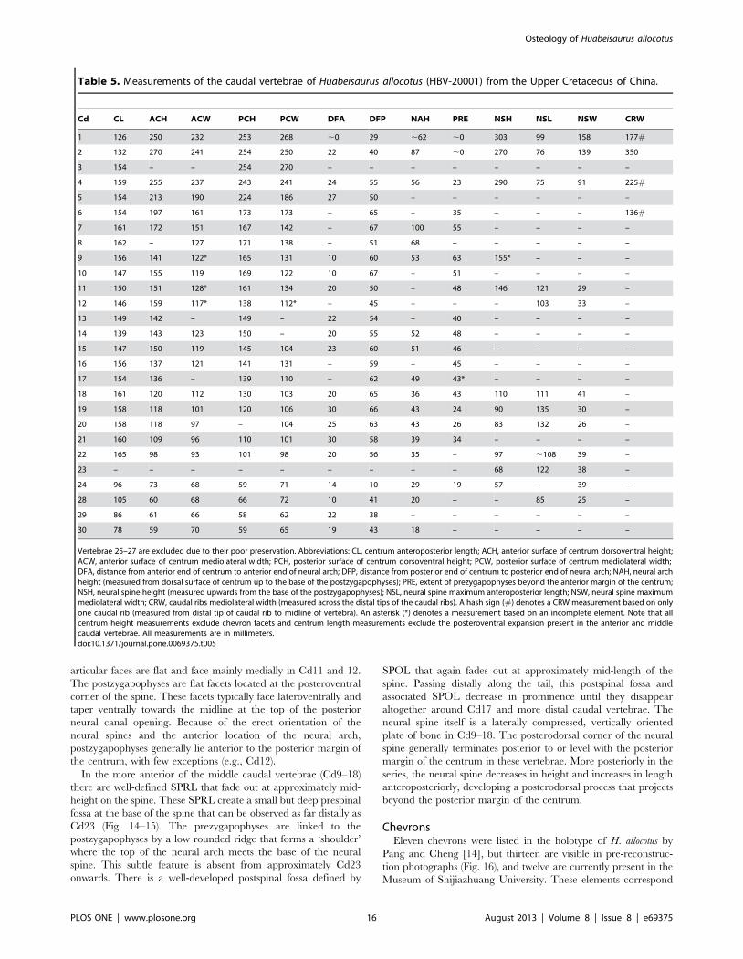
Table 5. Measurements of the caudal vertebrae of Huabeisaurus allocotus (HBV-20001) from the Upper Cretaceous of China.
Cd CL ACH ACW PCH PCW DFA DFP NAH PRE NSH NSL NSW CRW
1 126 250 232 253 268 ,0 29 ,62 ,0 303 99 158 177#
2 132 270 241 254 250 22 40 87 ,0 270 76 139 350
3 154 – – 254 270 – – – – – – – –
4 159 255 237 243 241 24 55 56 23 290 75 91 225#
5 154 213 190 224 186 27 50 – – – – – –
6 154 197 161 173 173 – 65 – 35 – – – 136#
7 161 172 151 167 142 – 67 100 55 – – – –
8 162 – 127 171 138 – 51 68 – – – – –
9 156 141 122* 165 131 10 60 53 63 155* – – –
10 147 155 119 169 122 10 67 – 51 – – – –
11 150 151 128* 161 134 20 50 – 48 146 121 29 –
12 146 159 117* 138 112* – 45 – – – 103 33 –
13 149 142 – 149 – 22 54 – 40 – – – –
14 139 143 123 150 – 20 55 52 48 – – – –
15 147 150 119 145 104 23 60 51 46 – – – –
16 156 137 121 141 131 – 59 – 45 – – – –
17 154 136 – 139 110 – 62 49 43* – – – –
18 161 120 112 130 103 20 65 36 43 110 111 41 –
19 158 118 101 120 106 30 66 43 24 90 135 30 –
20 158 118 97 – 104 25 63 43 26 83 132 26 –
21 160 109 96 110 101 30 58 39 34 – – – –
22 165 98 93 101 98 20 56 35 – 97 ,108 39 –
23 – – – – – – – – – 68 122 38 –
24 96 73 68 59 71 14 10 29 19 57 – 39 –
28 105 60 68 66 72 10 41 20 – – 85 25 –
29 86 61 66 58 62 22 38 – – – – – –
30 78 59 70 59 65 19 43 18 – – – – –
Vertebrae 25–27 are excluded due to their poor preservation. Abbreviations: CL, centrum anteroposterior length; ACH, anterior surface of centrum dorsoventral height;ACW, anterior surface of centrum mediolateral width; PCH, posterior surface of centrum dorsoventral height; PCW, posterior surface of centrum mediolateral width;DFA, distance from anterior end of centrum to anterior end of neural arch; DFP, distance from posterior end of centrum to posterior end of neural arch; NAH, neural archheight (measured from dorsal surface of centrum up to the base of the postzygapophyses); PRE, extent of prezygapophyses beyond the anterior margin of the centrum;NSH, neural spine height (measured upwards from the base of the postzygapophyses); NSL, neural spine maximum anteroposterior length; NSW, neural spine maximummediolateral width; CRW, caudal ribs mediolateral width (measured across the distal tips of the caudal ribs). A hash sign (#) denotes a CRW measurement based on onlyone caudal rib (measured from distal tip of caudal rib to midline of vertebra). An asterisk (*) denotes a measurement based on an incomplete element. Note that allcentrum height measurements exclude chevron facets and centrum length measurements exclude the posteroventral expansion present in the anterior and middlecaudal vertebrae. All measurements are in millimeters.doi:10.1371/journal.pone.0069375.t005
Osteology of Huabeisaurus allocotus
PLOS ONE | www.plosone.org 16 August 2013 | Volume 8 | Issue 8 | e69375

to positions covering nearly the entire length of the preserved
caudal vertebral series. The chevrons are generally well preserved,
but there is some distortion and damage (Fig. 16). The
correspondence of chevrons to particular caudal vertebrae is
uncertain, so we only assign them relative numbers (Fig. 16), e.g.,
Ch1, Ch2 for the first and second chevrons, respectively.
The chevrons are Y-shaped in anterior or posterior view with a
haemal canal that is unbridged proximally [14], although the
haemal canal of the smallest preserved chevron is crushed
transversely, giving it an artificially closed appearance (Fig. 16).
In lateral view, the chevrons are straight or curve slightly
posteriorly towards their distal ends, except in the posteriormost-
preserved element, which terminates distally in a subtriangular
plate formed from small anterior and posterior projections. The
proximal articular surfaces are transversely compressed and
anteroposteriorly ‘kinked’ in lateral view at their proximal ends,
giving them contiguous but distinct anterior and posterior facets
best developed in the more anterior elements (e.g., Ch5, Fig. 16).
The haemal canal is proximodistally short relative to chevron
length in the more anterior elements (36–44% in Chs1–4), but in
the more distal elements (Chs5–12) the canal represents more than
50% of proximodistal chevron length (Table 6). The haemal canal
typically represents about 30% of chevron length in most
sauropods, but titanosauriforms usually have a canal length to
chevron length ratio of approximately 50% [2,33,60]. Ventrally,
the haemal canal merges into shallow grooves on the anterior and
posterior faces of the blade of the chevron. These grooves grow
fainter and are replaced by ridges distally on the blade. The blade
of the chevron becomes transversely compressed towards its
termination, but is relatively unexpanded anteroposteriorly except
in the last two preserved elements in the series. A subtle but
distinct projection on the posterior midline occurs along the
proximal portion of the chevron blade, forming a vertically
elongate ridge. As a result of this ridge, the region immediately
below the haemal canal abruptly widens anteroposteriorly relative
to the proximal rami. This subtle feature characterizes some other
sauropod taxa (e.g., Alamosaurus [61]; Phuwiangosaurus [SM K11]
and Tangvayosaurus [TV2; MDD pers. obs. 2008]). None of the
chevrons have lateral ridges along the distal blades. There is no
indication that chevrons of the middle-distal portion of the series
developed the ‘forked’ or ‘skid’-like structure seen in basal
eusauropods and diplodocoids [28,30,33]. The most distal of the
preserved chevrons (Ch13) does not possess the ventral midline slit
seen in ‘forked’ chevrons [28].
ScapulaAlthough the exact orientation of the pectoral girdle in vivo is
uncertain, the scapulae and coracoids are generally thought to
have been oblique to the major planes of orientation of the rest of
the body, rendering orientational descriptors somewhat difficult to
select. Here, the scapulae and coracoids will be described as if the
long-axis of the scapular blade is horizontally oriented. Aside from
the glenoid region, which is damaged or missing in both
specimens, the preserved parts of the left and right scapulae
complement one another to give a full picture of the morphology
of the element. The scapulae consist of a broad proximal plate
comprising an acromion and acromial fossa and a blade that forms
more than half the length of the bone (Fig. 17; Table 7). The
lateral surface of the acromial plate is excavated anterior to the
acromial ridge and dorsal to the glenoid region. The acromial
ridge is slightly posteriorly deflected, such that it is oriented at an
acute angle to the long axis of the scapular blade. Immediately
posterior to the glenoid articular surface, the ventral margin of the
scapula is broad and convex transversely, but rapidly narrows as it
merges into the base of the blade (Fig. 17). No prominent
subtriangular process seems to occur along the posteroventral edge
of the proximal scapula, though its absence could be due to
damage. A broad ridge extends longitudinally along the proximal
third of the lateral face of the blade. The dorsal margin of the
blade is straight, whereas the ventral margin expands distally such
that the ratio of the maximum to minimum blade dorsoventral
height is 1.7 (Fig. 17, Table 7), less than the originally described
Figure 16. Holotypic chevrons of H. allocotus (HBV-20001) from the Upper Cretaceous of Shanxi, China. Photographed during originalpreparation in (A) lateral, (B) posterior; and (C) anterior views. Dashed lines indicate broken bone margins.doi:10.1371/journal.pone.0069375.g016
Osteology of Huabeisaurus allocotus
PLOS ONE | www.plosone.org 17 August 2013 | Volume 8 | Issue 8 | e69375

value of ca. 2 [14]. The development of the acromion and distal
expansion of the blade are similar to those of other somphos-
pondylans and not as marked as in rebbachisaurids [62,63]. The
medial side of the blade is not currently observable because of the
fragile nature of the scapula in the exhibit, but pre-restoration
photographs (Fig. 17) and the original description [14] indicate
that it was gently concave dorsoventrally. Thus, the scapular blade
has a flat cross section as in most somphospondylans ([33]; Fig. 16).
CoracoidBoth coracoids (Fig. 18) are well preserved aside from damage
around their margins (Fig. 18C, E) which has been restored with
plaster (Fig. 18B, D). In articulation with the scapula, the dorsal
margin of the coracoid is situated below the level of the scapular
acromion plate, and the two are separated by a V-shaped notch, as
is the case in all sauropods except some derived titanosaurs
[28,64]. The coracoids are dorsoventrally tall, curved plates with
thick humeral and scapular articulations and well developed
infraglenoid lips. Each is about 1.3 times taller than long (Table 7),
as is the case for most sauropods aside from some derived
titanosaurs [33]. The coracoids were not fused to their respective
scapulae and each bears an essentially fully enclosed coracoid
foramen. The latter is situated close to the scapular articulation,
just above coracoid mid-height. It has an elliptical outline, with the
long axis oriented anteroposteriorly [14]. The lateral surface of the
coracoid is convex in both directions, although it becomes slightly
concave anteroposteriorly towards the glenoid region. In lateral
view, the anterior and dorsal margins meet at a rounded corner.
This profile represents the plesiomorphic state that occurs in most
sauropods, and contrasts with the derived subrectangular cora-
coids observed in taxa such as Apatosaurus and advanced
titanosaurs [28,33]. The coracoid is mediolaterally thickest at
the glenoid, which has a subrectangular or nearly D-shaped
outline (with the straight edge of the ‘D’ forming the medial
margin) and faces posteroventrally. As in most other sauropods,
the lateral part of the glenoid surface curls upwards so that it is
visible in lateral view. This part of the glenoid is strongly expanded
outwards so that it projects laterally well beyond the lateral surface
Table 6. Measurements of the chevrons of Huabeisaurus allocotus (HBV-20001) from the Upper Cretaceous of China.
Dimension C1 C2 C4 C5 C6 C7 C9 C10 C12
Total dorsoventral height 368 346 317 300 253 220 246 225* 106
Maximum mediolateral width of proximal end 119 152 88 83 110 – – 69 57
Mediolateral width of proximal ramus 41 38 33 29 25 29 22 23 29
Anteroposterior length of proximal ramus 62 62 46 43 40 43 30 37 36
Haemal canal depth 155 124 140 150 125 103 169 152 57
Haemal canal depth/total chevron height 0.42 0.36 0.44 0.5 0.49 0.47 0.69 — 0.54
Maximum anteroposterior length of distal blade 56 69 50 40 39 46 36 35 47
Numbers denote sequence as preserved, not actual sequence in vertebral series. An asterisk denotes a measurement based on an incomplete element. Chevrons 3, 8and 11 are too incomplete to provide useful measurements. All measurements are in millimeters.doi:10.1371/journal.pone.0069375.t006
Figure 17. Holotypic scapulae of H. allocotus (HBV-20001) from the Upper Cretaceous of Shanxi, China. Left scapula in (A) lateral view;right scapula in (B) lateral and (C) medial views. Silhouette next to (B) shows schematic cross section of base of scapular blade. (A) and (B)photographed in 2012; (C) photographed during original preparation. Abbreviations: af, acromial fossa; con, concavity, gl, glenoid. Striped patternindicates broken surface; dashed lines indicate broken bone margins.doi:10.1371/journal.pone.0069375.g017
Osteology of Huabeisaurus allocotus
PLOS ONE | www.plosone.org 18 August 2013 | Volume 8 | Issue 8 | e69375
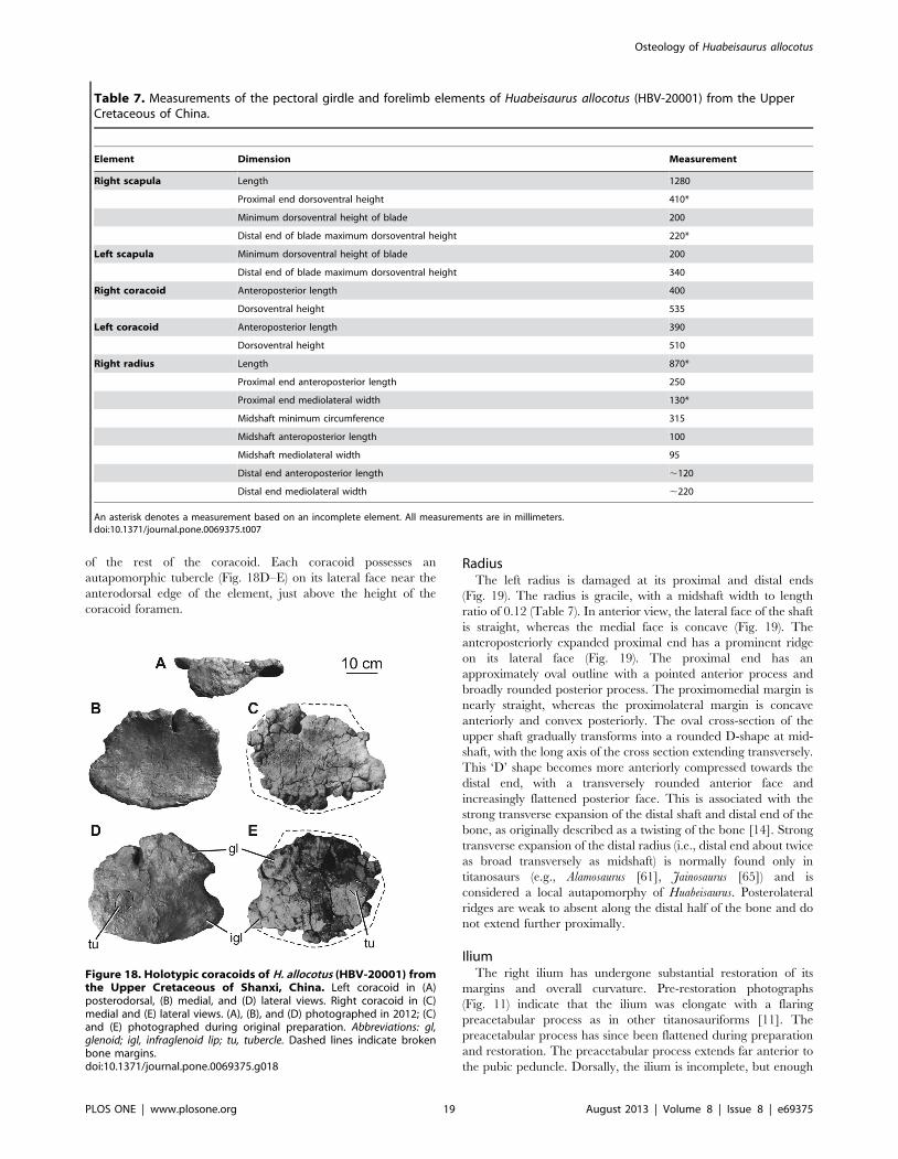
of the rest of the coracoid. Each coracoid possesses an
autapomorphic tubercle (Fig. 18D–E) on its lateral face near the
anterodorsal edge of the element, just above the height of the
coracoid foramen.
RadiusThe left radius is damaged at its proximal and distal ends
(Fig. 19). The radius is gracile, with a midshaft width to length
ratio of 0.12 (Table 7). In anterior view, the lateral face of the shaft
is straight, whereas the medial face is concave (Fig. 19). The
anteroposteriorly expanded proximal end has a prominent ridge
on its lateral face (Fig. 19). The proximal end has an
approximately oval outline with a pointed anterior process and
broadly rounded posterior process. The proximomedial margin is
nearly straight, whereas the proximolateral margin is concave
anteriorly and convex posteriorly. The oval cross-section of the
upper shaft gradually transforms into a rounded D-shape at mid-
shaft, with the long axis of the cross section extending transversely.
This ‘D’ shape becomes more anteriorly compressed towards the
distal end, with a transversely rounded anterior face and
increasingly flattened posterior face. This is associated with the
strong transverse expansion of the distal shaft and distal end of the
bone, as originally described as a twisting of the bone [14]. Strong
transverse expansion of the distal radius (i.e., distal end about twice
as broad transversely as midshaft) is normally found only in
titanosaurs (e.g., Alamosaurus [61], Jainosaurus [65]) and is
considered a local autapomorphy of Huabeisaurus. Posterolateral
ridges are weak to absent along the distal half of the bone and do
not extend further proximally.
IliumThe right ilium has undergone substantial restoration of its
margins and overall curvature. Pre-restoration photographs
(Fig. 11) indicate that the ilium was elongate with a flaring
preacetabular process as in other titanosauriforms [11]. The
preacetabular process has since been flattened during preparation
and restoration. The preacetabular process extends far anterior to
the pubic peduncle. Dorsally, the ilium is incomplete, but enough
Table 7. Measurements of the pectoral girdle and forelimb elements of Huabeisaurus allocotus (HBV-20001) from the UpperCretaceous of China.
Element Dimension Measurement
Right scapula Length 1280
Proximal end dorsoventral height 410*
Minimum dorsoventral height of blade 200
Distal end of blade maximum dorsoventral height 220*
Left scapula Minimum dorsoventral height of blade 200
Distal end of blade maximum dorsoventral height 340
Right coracoid Anteroposterior length 400
Dorsoventral height 535
Left coracoid Anteroposterior length 390
Dorsoventral height 510
Right radius Length 870*
Proximal end anteroposterior length 250
Proximal end mediolateral width 130*
Midshaft minimum circumference 315
Midshaft anteroposterior length 100
Midshaft mediolateral width 95
Distal end anteroposterior length ,120
Distal end mediolateral width ,220
An asterisk denotes a measurement based on an incomplete element. All measurements are in millimeters.doi:10.1371/journal.pone.0069375.t007
Figure 18. Holotypic coracoids of H. allocotus (HBV-20001) fromthe Upper Cretaceous of Shanxi, China. Left coracoid in (A)posterodorsal, (B) medial, and (D) lateral views. Right coracoid in (C)medial and (E) lateral views. (A), (B), and (D) photographed in 2012; (C)and (E) photographed during original preparation. Abbreviations: gl,glenoid; igl, infraglenoid lip; tu, tubercle. Dashed lines indicate brokenbone margins.doi:10.1371/journal.pone.0069375.g018
Osteology of Huabeisaurus allocotus
PLOS ONE | www.plosone.org 19 August 2013 | Volume 8 | Issue 8 | e69375

of its margin is preserved to show that it possesses a preacetabular
process with a low profile. The pubic peduncle is incomplete, but it
is apparent that the ilium contributed about half of the margin of
the acetabulum. The postacetabular process is missing (Fig. 11A–
C). No evidence of pneumaticity is apparent in the ilium, but
much of the surface is obscured by plaster.
PubisBoth the left and right pubes were recovered from the quarry.
Parts of the pubes are currently restored with plaster, but pre-
restoration photographs show that the majority of these bones are
preserved (Fig. 20). Both pubes are currently displayed in field
jackets; as such, their lateral surfaces cannot be observed.
The pubis is composed of an anterior plank-like body with a
posterior thin, curved flange of bone that forms the floor of the
pelvis. The iliac peduncle of the pubis is compressed, with its long
axis about 1.7 times greater than the axis perpendicular to this (see
Table 8). The iliac articular surface appears to narrow mediolat-
erally towards its posterior end, but it is not clear if this is a
preservational artefact. Although partly reconstructed, the acetab-
ular region slopes such that it faces dorsally and laterally, with the
lateral margin forming a gently upturned rim. The proximal and
anterior margins of the pubis meet at an angle of ,60u. Despite
poor preservation along this region, it is clear that no well-
developed ambiens process is present. The obturator foramen
enters the bone obliquely and has an elliptical outline, with the
long axis oriented in the same plane as that of the pubis shaft. On
the medial surface, the foramen is situated within a well-defined
fossa. The articular surface for the ischium is relatively short (0.35
of pubis length; Table 8). In lateral view, the symphysis meets the
ischial articulation at a sharp angle.
The medial surface of the pubis is anteroposteriorly convex
along its proximal portion, but becomes anteroposteriorly concave
along the middle third as a result of the medial deflection of the
pubic symphysis. Distally, this medial surface appears to flatten,
but neither pubis is well enough preserved in this region for this
morphology to be confirmed with certainty. In medial view, the
anterior margin of the pubis is concave, and a prominent anterior
expansion of the distal end forms an anterior boot (Fig. 20). The
left pubis preserves a flattened subtriangular area on the
posteromedial surface of the distal end for articulation with the
equivalent corresponding area on the left pubis.
IschiumBoth the left and right ischia were recovered from the quarry
and are essentially complete. The ischia retain their three-
dimensional geometry and their morphology is unobstructed by
plaster, although the left is better preserved than the right (Fig. 20).
The ischium is considerably shorter than the pubis (ischium: pubis
length ratio is 0.73, see Table 8, [14]). Whereas the pubes and
ischia are subequal in length in most sauropods [28,64], this
shortening of the ischium occurs in most somphospondylans
[2,11,28,33,66]. Aside from the robust, semicircular iliac pedun-
cle, the ischium is formed from a thin plate of bone throughout its
length. The iliac peduncle comprises about one-fifth of the
proximodistal length of the element, giving the ischium a large
(about one-fourth) contribution to the margin of the acetabulum.
Posteriorly, the iliac peduncle narrows mediolaterally to form a
triangular projection in lateral view. The iliac articular surface
slopes to face dorsolaterally; it is also mildly convex anteroposte-
riorly (Fig. 20). The acetabular surface is wide transversely in the
region of the iliac peduncle, narrows in its central region, and
widens again (though not as strongly) towards the pubic
articulation. This morphology has also been observed in most
rebbachisaurids (e.g. Demandasaurus, see [67] and ‘‘Ornithopsis
eucamerotus’’ from the Lower Cretaceous of the Isle of Wight, UK
[68]). As in the latter taxon and the Late Cretaceous Chinese
somphospondylan Sonidosaurus (LHV 0010; Long Hao Geologic
and Paleontological Research Center, Hohhot, Nei Mongol,
People’s Republic of China, PDM pers. obs. 2007), the ischium
of Huabeisaurus possesses a prominent ridge that extends medially
from the margin of the acetabulum on the section formed by the
iliac peduncle.
The bone lining the posterior pelvic floor is thin and forms a
deep bowl; anteriorly, this region forms the articular surface for
the pubis. This articulation has a narrow subtriangular outline in
anterior view, with concave dorsomedial and convex ventrolateral
margins and a tapered ventromedial tip. In lateral view, the pubic
articulation and the symphysis are demarcated by a change of
direction: the proximal part of the symphysis extends posteroven-
trally towards a small transversely expanded area, which turns
more steeply ventrally.
The distal shafts of the ischia are twisted so that they are nearly
coplanar when in articulation (Fig. 20C). The shafts themselves
approach the condition of having a straight ischial articulation, as
in titanosaurs (i.e., ‘‘plate-like’’ ischia that lack an emargination
anteroventrally (e.g., Andesaurus [1]; Alamosaurus [61]). The
posterolateral face of the ischium bears a ridge-like tubercle at
about mid-length as in most neosauropods [2,28]. This tubercle
differs from that of other neosauropods in that it projects out from
the margin of the ischium, rather than sitting on the plate of the
bone, making it visible as a kink in the profile of the dorsal margin
of the distal shaft in lateral view (Fig. 20). We interpret this
Figure 19. Holotypic left radius of H. allocotus (HBV-20001)from the Upper Cretaceous of Shanxi, China. In (A) proximal, (B)anterior, (C) lateral, and (D) posterior views. Silhouette between (C) and(D) shows schematic cross section of the diaphysis. (A) photographed in2012; (B–D) photographed during original preparation. Abbreviations: gl,glenoid; igl, infraglenoid lip; tu, tubercle. Dashed lines indicate brokenbone margins.doi:10.1371/journal.pone.0069375.g019
Osteology of Huabeisaurus allocotus
PLOS ONE | www.plosone.org 20 August 2013 | Volume 8 | Issue 8 | e69375

posterior placement and projection of this tubercle as an
autapomorphy of Huabeisaurus. Although the distal tip of the
ischium is incomplete posteriorly, the distal blade is clearly
expanded transversely relative to the minimum width of the blade.
The distal end is at least five times as wide transversely as thick
dorsoventrally.
FemurA nearly complete left femur and the distal fourth of a right
femur were collected from the quarry. The hindlimb was originally
described as robust [14], but the femur is somewhat gracile
(midshaft width divided by length = 0.16; Table 9) and has a
mediolaterally expanded, eccentric midshaft cross section (medio-
lateral width divided by anteroposterior breadth = 1.7; Table 9),
like most titanosauriforms [33]. The femoral head appears to
project slightly dorsally, but the exact proximal outline is uncertain
because of damage (Fig. 21). The proximal end bears a prominent
lateral bulge, with the lateral margin above medially deflected, as
in most titanosauriforms [33]. This proximolateral margin is
anteroposteriorly thinner than the rest of the femur, forming a
trochanteric flange towards the anterior face of the shaft. The
posterior surface of the proximal end has two dorsoventrally short,
nearly vertical ridges, one situated near the midline and the other
near the femoral head. These define the medial edges of shallow,
vertically oriented troughs, and the lateral ridge bounds the
trochanteric flange. It is difficult to determine whether this
posterior surface morphology is entirely genuine or the result of
deformation. The fourth trochanter is situated close to the
posteromedial margin of the shaft, just above the mid-length of
the femur (Table 9). This trochanter is reduced to a low, rounded
ridge that is not visible in anterior view. The medial surface of the
fourth trochanter is gently rugose, but there is no concavity medial
to this process. The anterior surface of the shaft is gently convex
mediolaterally, whereas the posterior surface is flat, becoming
mediolaterally convex towards its medial and lateral margins
(Fig. 21).
The distal ends of both femora are poorly preserved but nearly
complete. The fibular and tibial condyles appear to be subequal in
size. There is a deep concavity on the posterior surface between
the two distal condyles. The fibular condyle is poorly preserved,
but clearly divided (Fig. 21). The medial face of the tibial condyle
is nearly flat and faces medially and slightly proximally, as is
typical for sauropods. The distal condyles are only weakly divided
distally and are oriented perpendicularly with respect to the long
axis of the bone, unlike the medially or laterally beveled distal
femora of some titanosauriforms (see [7] for discussion). The distal
Figure 20. Holotypic pubes and ischia of H. allocotus (HBV-20001) from the Upper Cretaceous of Shanxi, China. Left pubis in (A) medialview. Right pubis and ischium in (B) lateral view (only loosely articulated to depict their size relative to one another). Articulated ischia in (C)posterodorsal view. Outlines below ischia indicate articulated view of distal ends. (A and the ischium in B) photographed in 2012; (C and the pubis inB) photographed during original preparation. Abbreviations: acet, acetabulum; tu, tuberosity. Dashed lines indicate broken bone margins.doi:10.1371/journal.pone.0069375.g020
Table 8. Measurements of the pelvic girdle elements ofHuabeisaurus allocotus (HBV-20001) from the UpperCretaceous of China.
Element Dimension Measurement
Right ilium Dorsoventral height anterior to pubicpeduncle
570*
Right pubis Length 810*
Proximal end anteroposterior length 445
Iliac peduncle anteroposterior length 270
Iliac peduncle mediolateral width 160
Dorsoventral height of ischiadic articulation 350
Distal end anteroposterior length 340
Distal end mediolateral width 130
Left pubis Length 990
Proximal end anteroposterior length 325*
Right ischium Length 725
Proximal end anteroposterior length 395
Iliac peduncle anteroposterior length 205
Iliac peduncle mediolateral width 100
Dorsoventral height of pubic articulation 285
Distal end anteroposterior length 208
Distal end mediolateral width ,48
An asterisk denotes a measurement based on an incomplete element. Allmeasurements are in millimeters.doi:10.1371/journal.pone.0069375.t008
Osteology of Huabeisaurus allocotus
PLOS ONE | www.plosone.org 21 August 2013 | Volume 8 | Issue 8 | e69375

articular surface is mediolaterally concave and anteroposteriorly
convex, although this convexity is only strongly developed on the
tibial condyle. The distal condyles do not extend onto the anterior
surface of the femur. The anterior half of the distal surface,
between the two distal condyles, is deflected such that it faces
ventrally, but also slightly anteriorly. The anteroposterior depth of
the midshaft of the femur equals slightly more than one-third of
the depth of the distal condyles, as in most sauropods except
derived diplodocoids [40].
TibiaBoth tibiae were recovered adjacent to their respective fibulae.
The majority of their cnemial crests and the distal end of the left
element are not preserved. Both tibiae have been reconstructed
with an unnatural twist between their proximal and distal ends, as
evident from comparing pre-restoration photographs with the
tibiae on display in the exhibit (Fig. 22). The ratio of the length of
the tibia to that of the femur is approximately 0.75, as noted in the
original description [14]. This value is higher than in other
sauropods, which typically have tibia: femur length ratios between
0.56–0.68 (see The autapomorphic tibia: femur ratio of Huabeisaurus in
the Discussion). The tibiae are gracile elements, with a midshaft
width to length ratio of 0.11 (Table 9). In dorsal view, the proximal
end has a subrectangular outline, with a concave posterior margin.
The proximal articular surface is weakly convex in both directions,
although it forms a very gentle concavity at the anteromedial
corner. The medial edge of the proximal end forms an
anteroposteriorly reduced projection that overhangs the medial
margin of the remainder of the tibia; this is more prominently
developed in the right tibia. Based on pre-restoration photographs,
the cnemial crest appears to have projected mainly laterally
(Fig. 22). There is no small process in the cnemial fossa (i.e.,
‘second cnemial crest’ [69]), on the lateral surface of the proximal
end, posterior to the true cnemial crest).
The posterior surface of the proximal part of the tibia,
approximately down to the level of the distal tip of the cnemial
crest, maintains the mediolateral concavity that occurs on the
posterior margin of the proximal articular end. The shaft has a
flattened ‘D’-shape in horizontal cross-section, with a flat to
mediolaterally concave posterolateral surface. Distally, the anterior
surface of the tibia expands transversely to form the subtriangular
area seen in most other sauropods. This region is mediolaterally
concave, although this is almost certainly the result of crushing.
Although incomplete medially, the distal end is clearly not strongly
expanded transversely relative to the midshaft (Fig. 22), and is
anteroposteriorly compressed, as in other titanosauriforms [11].
FibulaBoth fibulae were recovered from the quarry and are well
preserved, aside from missing portions of their proximal ends (in
Figure 21. Holotypic left femur of H. allocotus (HBV-20001) fromthe Upper Cretaceous of Shanxi, China. Photographed duringoriginal preparation in (A) posterior, (B) medial, (C) anterior, and (D)distal views. Silhouette between (B) and (C) shows schematic crosssection of the diaphysis. Abbreviations: fic, fibular condyle; ftr, fourthtrochanter; tic, tibial condyle. Striped pattern indicates broken surface,dashed lines indicate broken bone margins.doi:10.1371/journal.pone.0069375.g021
Table 9. Measurements of the hindlimb elements ofHuabeisaurus allocotus (HBV-20001) from the UpperCretaceous of China.
Element Dimension Measurement
Right femur Midshaft anteroposterior length ,115
Midshaft mediolateral width ,265
Distal end anteroposterior length 270
Distal end mediolateral width 535
Left femur Length 1560
Proximal end anteroposterior length 160
Proximal end mediolateral width 430
Midshaft minimum circumference 645
Midshaft anteroposterior length 145
Midshaft mediolateral width 245
Distal end anteroposterior length 380
Distal end mediolateral width 465
Right tibia Length 1170
Proximal end anteroposterior length 208
Proximal end mediolateral width 287
Midshaft minimum circumference 430
Distal end anteroposterior length 160
Distal end mediolateral width 278
Right fibula Length 950*
Proximal end anteroposterior length 305*
Proximal end mediolateral width 90
Midshaft minimum circumference 315
Midshaft anteroposterior length 115
Midshaft mediolateral width 80
Left fibula Length 1150
Proximal end anteroposterior length 305*
Proximal end mediolateral width 95
Midshaft minimum circumference 315
Midshaft anteroposterior length 120
Midshaft mediolateral width 75
An asterisk denotes a measurement based on an incomplete element. Allmeasurements are in millimeters.doi:10.1371/journal.pone.0069375.t009
Osteology of Huabeisaurus allocotus
PLOS ONE | www.plosone.org 22 August 2013 | Volume 8 | Issue 8 | e69375
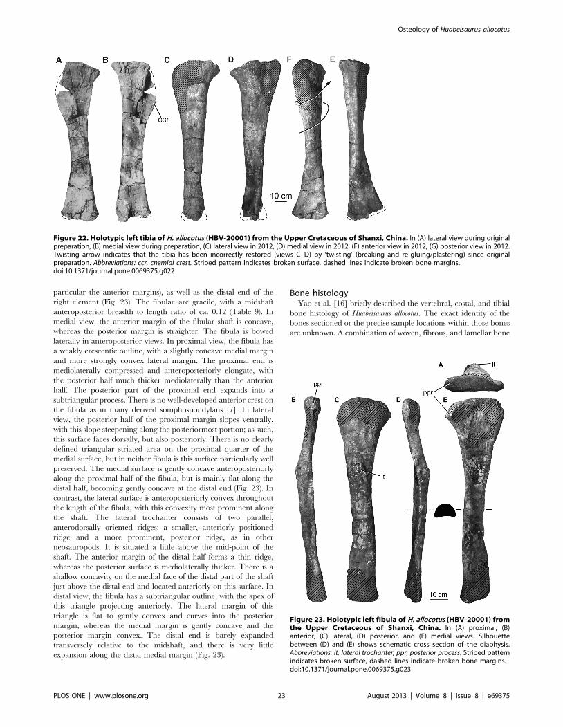
particular the anterior margins), as well as the distal end of the
right element (Fig. 23). The fibulae are gracile, with a midshaft
anteroposterior breadth to length ratio of ca. 0.12 (Table 9). In
medial view, the anterior margin of the fibular shaft is concave,
whereas the posterior margin is straighter. The fibula is bowed
laterally in anteroposterior views. In proximal view, the fibula has
a weakly crescentic outline, with a slightly concave medial margin
and more strongly convex lateral margin. The proximal end is
mediolaterally compressed and anteroposteriorly elongate, with
the posterior half much thicker mediolaterally than the anterior
half. The posterior part of the proximal end expands into a
subtriangular process. There is no well-developed anterior crest on
the fibula as in many derived somphospondylans [7]. In lateral
view, the posterior half of the proximal margin slopes ventrally,
with this slope steepening along the posteriormost portion; as such,
this surface faces dorsally, but also posteriorly. There is no clearly
defined triangular striated area on the proximal quarter of the
medial surface, but in neither fibula is this surface particularly well
preserved. The medial surface is gently concave anteroposteriorly
along the proximal half of the fibula, but is mainly flat along the
distal half, becoming gently concave at the distal end (Fig. 23). In
contrast, the lateral surface is anteroposteriorly convex throughout
the length of the fibula, with this convexity most prominent along
the shaft. The lateral trochanter consists of two parallel,
anterodorsally oriented ridges: a smaller, anteriorly positioned
ridge and a more prominent, posterior ridge, as in other
neosauropods. It is situated a little above the mid-point of the
shaft. The anterior margin of the distal half forms a thin ridge,
whereas the posterior surface is mediolaterally thicker. There is a
shallow concavity on the medial face of the distal part of the shaft
just above the distal end and located anteriorly on this surface. In
distal view, the fibula has a subtriangular outline, with the apex of
this triangle projecting anteriorly. The lateral margin of this
triangle is flat to gently convex and curves into the posterior
margin, whereas the medial margin is gently concave and the
posterior margin convex. The distal end is barely expanded
transversely relative to the midshaft, and there is very little
expansion along the distal medial margin (Fig. 23).
Bone histologyYao et al. [16] briefly described the vertebral, costal, and tibial
bone histology of Huabeisaurus allocotus. The exact identity of the
bones sectioned or the precise sample locations within those bones
are unknown. A combination of woven, fibrous, and lamellar bone
Figure 22. Holotypic left tibia of H. allocotus (HBV-20001) from the Upper Cretaceous of Shanxi, China. In (A) lateral view during originalpreparation, (B) medial view during preparation, (C) lateral view in 2012, (D) medial view in 2012, (F) anterior view in 2012, (G) posterior view in 2012.Twisting arrow indicates that the tibia has been incorrectly restored (views C–D) by ‘twisting’ (breaking and re-gluing/plastering) since originalpreparation. Abbreviations: ccr, cnemial crest. Striped pattern indicates broken surface, dashed lines indicate broken bone margins.doi:10.1371/journal.pone.0069375.g022
Figure 23. Holotypic left fibula of H. allocotus (HBV-20001) fromthe Upper Cretaceous of Shanxi, China. In (A) proximal, (B)anterior, (C) lateral, (D) posterior, and (E) medial views. Silhouettebetween (D) and (E) shows schematic cross section of the diaphysis.Abbreviations: lt, lateral trochanter; ppr, posterior process. Striped patternindicates broken surface, dashed lines indicate broken bone margins.doi:10.1371/journal.pone.0069375.g023
Osteology of Huabeisaurus allocotus
PLOS ONE | www.plosone.org 23 August 2013 | Volume 8 | Issue 8 | e69375

textures with occasional ‘rest lines’ and some Haversian remod-
eling were presented [16]. These thin sections are now lost.
Isolated humerusAlthough the humerus listed as the paratype of H. allocotus by
Pang and Cheng [14] cannot be referred to the species because of
a lack of overlapping elements, we provide a brief description here
because of its significance (see Discussion below). The humerus is
nearly complete, missing only its deltopectoral crest and parts of its
proximal and distal ends (Fig. 24). It is gracile (midshaft width
divided by length = 0.16; Table 10), with subequally expanded
proximal and distal ends. There is a broad deltopectoral fossa
along the proximal half of the anterior surface and a sharp
longitudinal ridge posteriorly (Fig. 24). The posterior surface of the
distal end forms a deep supracondylar fossa, bounded by sharp
ridges, as is the case in many other titanosauriforms [1,30]. Many
phylogenetically informative characters (e.g., squared proximolat-
eral corner; posterolateral bulge on proximal end of humerus)
cannot be ascertained in this humerus because of damage.
Discussion
Affinities of Huabeisaurus allocotusHuabeisaurus is a relatively complete and well-preserved sauro-
pod, with parts of all major regions of the body preserved. The
new data concerning Huabeisaurus presented above and the wealth
of new Cretaceous Asian sauropod discoveries over the past
decade prompt a reevaluation of its affinities. A number of named
Cretaceous East Asian titanosauriforms await detailed description
and comparisons; since we have yet to make first-hand observa-
tions of many of these species, our understanding of the
phylogenetic relationships of them is limited. We refrain from
incorporating Huabeisaurus into a cladistic analysis until more
detailed data from other taxa are available. However, comments
can be made as to its affinities based on synapomorphies recovered
in previous cladistic analyses. Below, we evaluate previous
hypotheses of affinity between Huabeisaurus and other Cretaceous
East Asian sauropods.
Huabeisaurus allocotus has never been incorporated into a cladistic
analysis, but several authors have commented on its affinities on
comparative grounds. Buffetaut et al. [4] reviewed the sauropods
of Thailand and described Late Jurassic and Early Cretaceous
teeth similar to those of euhelopodids (in the traditional sense, i.e.,
Euhelopus, Mamenchisaurus and Omeisaurus) and others more remi-
niscent of Nemegtosaurus and Phuwiangosaurus (which they termed
nemegtosaurids). Following these comparisons, Huabeisaurus was
regarded as a nemegtosaurid by Buffetaut et al. [4]. Upchurch
et al. [30] considered Huabeisaurus to represent a titanosauriform
or titanosaur outside of Lithostrotia on the basis of a variety of
features (see below). You et al. [6] suggested a close relationship
Figure 24. Isolated right humerus (HBV-20002) from the UpperCretaceous of Shanxi, China. Photographed during originalpreparation in (A) anterior, (B) medial, (C) posterior, and (D) distalviews. This bone was formerly regarded as the paratype of H. allocotus.Silhouette between (A) and (B) shows schematic cross section of thediaphysis. Abbreviations: rac, radial condyle; ri, ridge. Striped patternindicates broken surface, dashed lines indicate broken bone margins.doi:10.1371/journal.pone.0069375.g024
Table 10. Measurements of the left humerus (HBV-20002)from the Upper Cretaceous of China previously referred to asthe paratype of Huabeisaurus allocotus.
Dimension Measurement
Length 1220
Proximal end mediolateral width 410
Midshaft minimum circumference 560
Midshaft anteroposterior length 140
Midshaft mediolateral width 200
Distal end anteroposterior width 190
Distal end mediolateral width 390
All measurements are in millimeters.doi:10.1371/journal.pone.0069375.t010
Figure 25. Tibia to femur ratio in sauropods. Linear regression(solid black line) and 95% confidence intervals (dotted lines) are shown,as well as pGLS line (red dashed line). Note that Huabeisaurus has anunusually high tibia to femur ratio, only approached by some distantlyrelated diplodocines.doi:10.1371/journal.pone.0069375.g025
Osteology of Huabeisaurus allocotus
PLOS ONE | www.plosone.org 24 August 2013 | Volume 8 | Issue 8 | e69375

between Huabeisaurus, Borealosaurus, and Opisthocoelicaudia. Mo et al.
[70] highlighted similarities among Huabeisaurus, Fusuisaurus, and
Sonidosaurus. Tanimoto et al. [71] considered Huabeisaurus to be
closely related to Nemegtosaurus, Borealosaurus and the ‘‘Toba
sauropod’’ of Japan (as members of Nemegtosauridae). Finally, it
has recently been suggested [2] that Huabeisaurus is a member of
Euhelopodidae. Below we reevaluate the evidence for nemegto-
saurid, opisthocoelicaudine, and euhelopodid affinities for Huabei-
saurus.
Huabeisaurus as a nemegtosauridBuffetaut et al. [4] suggested that Huabeisaurus is a member of
Nemegtosauridae on the basis of similarities shared with
Phuwiangosaurus, which they also considered to be a nemegto-
saurid. No specific features were mentioned to support nemegto-
saurid affinities for Huabeisaurus by Buffetaut et al. ([4]:97), but
five were listed to link Phuwiangosaurus and Nemegtosaurus: (1)
‘‘narrow premaxillae’’, (2) ‘‘a broad quadrate fossa’’, (3) ‘‘a very
small supratemporal fenestra’’, (4) ‘‘a quadrangular frontal’’, (5)
‘‘and a postorbital with a very long anteroventral ramus’’.
However, these features are all more widely distributed among
sauropods. Because all sauropods have four premaxillary teeth, the
breadth of the premaxilla (feature 1) is related to tooth breadth.
Nemegtosaurus and Phuwiangosaurus have narrow teeth and
therefore narrow premaxillae, but these traits were apparently
acquired independently in these taxa (see below) as in other
sauropod lineages (e.g., Diplodocoidea versus Titanosauria [12]).
Most sauropods have what could be construed as a broad quadrate
fossa (feature 2), and its breadth does not particularly link
Phuwiangosaurus and Nemegtosaurus [5,30]. A small supratem-
poral fenestra (feature 3) characterizes many titanosauriforms [72].
Sauropod frontals are generally quadrangular (feature 4), with four
nearly straight sides bordering the orbit and postorbital laterally,
prefrontal and nasal anteriorly, parietal posteriorly, and comple-
mentary frontal medially, although these borders have varying
lengths among sauropods [30]. The length of the anteroventral
(jugal) ramus of the postorbital (feature 5) is indeed long in
Phuwiangosaurus and Nemegtosaurus (total postorbital length
divided by anteroposterior length of proximal body ca. 2.3), but
similar values occur in some other sauropods (e.g., Tapuiasaurus
[73]).
The dental features cited by Tanimoto et al. [71] in support of
nemegtosaurid affinities for Huabeisaurus were refuted by Saegusa
and Tomida [74], who pointed out that these features have a
wider distribution among sauropods.
Furthermore, nemegtosaurid affinities for Phuwiangosaurus are
refuted by several recent cladistic analyses that indicate that
Phuwiangosaurus lies outside of Titanosauria [2,75,76], with
Nemegtosaurus nested within lithostrotian titanosaurs [33,57,73].
Therefore, the several similarities shared between Huabeisaurus
and Phuwiangosaurus (see ‘‘Description’’ above) are not indicative
of nemegtosaurid affinities.
Huabeisaurus as an opisthocoelicaudineYou et al. [6] suggested that Huabeisaurus, Opisthocoelicaudia, and
Borealosaurus are closely related within Opisthocoelicaudinae on the
basis of opisthocoelous caudal vertebrae. However, this is
problematic because the middle caudal vertebrae (ca. position
17–23) of Opisthocoelicaudia are not strongly opisthocoelous as in the
holotypic middle caudal vertebra of Borealosaurus; instead they are
amphiplatyan ([77]: pl. 6). Huabeisaurus likewise has amphiplatyan
caudal vertebrae in this part of the tail and only displays an
anterior convexity in two middle-distal caudal centra (Cd28–29;
see above). The most common condition for anterior-middle
sauropod caudal vertebral centra is one in which the anterior face
of the centrum is slightly more concave than the posterior face
[78]. Although Opisthocoelicaudia, Huabeisaurus, and Borealosaurus are
all unusual with respect to the primitive titanosauriform condition
of caudal vertebral articulation, they are unusual in different (i.e.,
non-homologous) ways; opisthocoely occurs in different parts of
the tail and to different degrees in these genera. Several other
Cretaceous East Asian genera have caudal vertebral central
articular surfaces similar to those of Huabeisaurus (i.e., the posterior
concavity is deeper than the anterior concavity), including
Tangvayosaurus (TV2, MDD pers. obs. 2008) and Phuwiangosaurus
(PW1, MDD pers. obs. 2008; [75]), providing evidence for a close
relationship among these taxa.
Another feature recognized herein links elements referred to
Huabeisaurus and Borealosaurus, albeit in a complex manner. The
isolated humerus (HBV-2002), formerly the paratype of Huabei-
saurus (see above), has a longitudinal ridge on the posterior face of
the humeral shaft. A comparable ridge also occurs on a humerus
referred to Borealosaurus wimani ([6]; LPM 0170, MDD, PDM, PU
pers. obs. 2012). Unfortunately, like the referral of the humerus
HBV-20002 to Huabeisaurus, the referral of the humerus LPM
0170 to Borealosaurus cannot be substantiated, as the association of
the hypodigm of Borealosaurus is unknown [6]. Indeed, the humerus
referred to Borealosaurus seems to be much too small (610 mm long)
to belong to the same individual as the holotypic mid-caudal
vertebra (112 mm long) based on comparisons with complete
sauropods of similar size (e.g., Rapetosaurus [37]). Nonetheless, the
uniquely shared presence of a posterior longitudinal ridge on these
two isolated humeri (HBV-20002, Fig. 24 and LPM 0170) suggests
further evidence of close relatedness among Cretaceous East Asian
sauropods. Alternately, these two isolated humeri may pertain to
the same taxon.
A new hypothesis: Huabeisaurus is a euhelopodidHuabeisaurus appears to be a titanosauriform based on the
presence of numerous synapomorphies of the group, including
elongate middle cervical vertebrae, anterior and middle caudal
Figure 26. Crown slenderness index versus number of generaknown from teeth in Sauropodomorpha and derived clades.The mean (white circle), standard deviation (tick mark) and range(horizontal line) of tooth breadth for each clade is indicated (log10
Sauropodomorpha slenderness index range extends towards zero asindicated by the arrow). Sketches of tooth crowns (from left to right,Euhelopus, Huabeisaurus, Phuwiangosaurus) and red field depict thewide range of crown shapes in Euhelopodidae compared to otherderived sauropod clades.doi:10.1371/journal.pone.0069375.g026
Osteology of Huabeisaurus allocotus
PLOS ONE | www.plosone.org 25 August 2013 | Volume 8 | Issue 8 | e69375

vertebral neural arches set on the anterior half of the centrum,
middle caudal vertebrae with posteriorly projecting transverse
processes, pneumatic and plank-like dorsal ribs, a laterally flared
iliac preacetabular process, a pubis at least three times as long as
the puboischial contact, and an ischium with a raised tubercle on
its posterolateral face [2,11,30,33].
Huabeisaurus is a member of Somphospondyli based on the
presence of somphospondylous vertebral pneumaticity, anterior
dorsal vertebrae with spinodiapophyseal laminae that are low in
relief on the front of the neural spine, a scapular glenoid that is
beveled medially, and a scapular blade that is straight on the
acromial side [2,12,30,33].
A number of anatomical features suggest that Huabeisaurus is a
member of Euhelopodidae (sensu [2]), a clade that also has been
suggested to include Euhelopus, Erketu, Daxiatitan, Dongyangosaurus,
Baotianmansaurus, ‘Huanghetitan’ ruyangensis, Qiaowanlong, Tangvayo-
saurus and Phuwiangosaurus [2]. These features include: bifid cervical
neural spines, pendant cervical ribs, enlarged chevron facets on
anterior-middle caudal vertebrae, posterior centrum articulation
concave with anterior centrum articulation nearly flat in the
anterior–middle caudal vertebrae, and an ilium with a low,
pointed preacetabular process [2]. Although we provisionally
conclude that Huabeisaurus is a member of Euhelopodidae, there
are some potential caveats that accompany this interpretation.
First, placement of Huabeisaurus within Euhelopodidae has not yet
been supported by inclusion in a formal cladistic analysis.
Mannion et al. [79] found support for a monophyletic Euhelopo-
didae in some of their phylogenetic analyses, however, this clade
breaks up to form a paraphyletic grade at the base of Titanosauria
when some characters are treated as continuous data. Moreover,
Huabeisaurus shares some similarities with more derived titanosauri-
forms (e.g., the distal expansion of the radius). It is therefore
conceivable that future phylogenetic analyses will result in
separation of the large Euhelopodidae found by D’Emic [2] into
several smaller clades distributed among different regions of the
somphospondylan evolutionary tree. One of these groups could be
Huabeisauridae, with Huabeisaurus closely related to forms such as
Tangvayosaurus, similar to the taxonomic scheme proposed by Pang
and Cheng [14]. As noted above, these issues can only be resolved
through more extensive first-hand examination of Cretaceous
Asian sauropod specimens and augmented phylogenetic analyses.
Referral of other specimens to HuabeisaurusPang and Cheng [14] tentatively suggested that ‘Titanosaurus’
falloti be referred to Huabeisaurus. However,‘T.’ falloti is only
represented by femoral remains, and its femur bears no uniquely
shared features with Huabeisaurus. Furthermore, the femora of
Huabeisaurus and ‘T.’ falloti differ in the bevel of the distal end
versus the long axis of the bone (compare Fig. 21 with fig. 17 in
[80]), so the two cannot represent the same genus.
The autapomorphic tibia: femur ratio of HuabeisaurusThe ratio of the length of the tibia to that of the femur is
approximately 0.75 in Huabeisaurus. We tested whether this was
autapomorphically high compared to other sauropods given
possible allometric scaling relationships, initially by performing
ordinary least squares regression of log10 (tibia length) against log10
(femur length) for a sample of 34 other sauropod individuals
(Fig. 25; see File S1 for data) using R version 2.15.1 [81] (see
File S2 for R code). The regression equation is:
log10 tibia lengthð Þ~1:04 � log10 femur lengthð Þ{0:332
Confidence intervals (95%) about the coefficient (slope) of log10
(femur length) are 0.94 to 1.14, so isometry of sauropod hindlimb
proportions cannot be rejected. Confidence intervals about the y-
intercept are 20.644 to 20.019. Using the above equation, the
femoral length of Huabeisaurus predicted a log10 (tibia length) of
2.99 log10(mm), with 95% confidence intervals of 2.93–3.06
(Fig. 25) that do not contain the actual value of 3.07 log10(mm).
We performed a second ordinary least squares analysis in which
individuals of Apatosaurus (N = 2), Camarasaurus (N = 6), Omeisaurus
(N = 3), and diplodocines (N = 6) were collapsed to mean values.
The regression equation for this reduced sample size (N = 21) is:
log10 tibia lengthð Þ~1:04 � log10 femur lengthð Þ{0:165
Confidence intervals are 0.855 to 1.11 for the coefficient and
20.568 to 0.238 for the y-intercept. Huabeisaurus has a predicted
log10 (tibia length) of 2.98 log10(mm), with 95% confidence
intervals of 2.92–3.03, also excluding the actual value of 3.07
log10(mm).
Some diplodocines from the Late Jurassic Morrison Formation
approach the tibia: femur ratio observed in Huabeisaurus (Fig. 25).
However, because diplodocines are only distantly related to
Huabeisaurus, we interpret the ratio in Huabeisaurus as an
autapomorphy.
Finally, to model the expected covariances among specimens as
part of our regression analysis, and thus circumvent problems of
phylogenetic relatedness of the data, we performed phylogenetic
generalized least squares regression (pGLS; [82]) based on our
specimen-level data (N = 34) using a composite phylogeny based
on previous cladistic studies ([2,33]; see Files S3 and S4 for
phylogenies at the generic and specific levels, respectively). Branch
lengths were calibrated using geologic time (with a minimum
length of 0.01 Ma among specimens of Apatosaurus, Camarasaurus,
Omeisaurus, and Diplodocus and Barosaurus). Lambda, a measure of
phylogenetic signal [83], was estimated as part of the model fitting
process using the R packages nlme version 3.1–104 [84] and ape
version 3.0–5 [85]. The maximum likelihood estimate of lambda
(0.68) indicates an intermediate level of phylogenetic signal in the
relationship between log102 femoral length and tibia length (when
lambda = 1.0 branch lengths are left unaltered; when lambda = 0
scales the tree to a star phylogeny, representing zero phylogenetic
signal). Estimates of the coefficient and y-intercept of the pGLS
regression line were within their 95% confidence intervals derived
from ordinary least squares regression (coefficient = 1.01, y-
intercept = 20.230). Importantly for our purposes, the phyloge-
netically informed regression line passes further from Huabeisaurus,
confirming that its hindlimb proportions are indeed autapo-
morphic (Fig. 25).
Tooth shape in EuhelopodidaeEuhelopodids (sensu [2]; e.g., Euhelopus, Phuwiangosaurus, Tang-
vayosaurus, Erketu) are united by axial and appendicular synapo-
morphies and have a notable diversity of tooth shapes. Figure 26
plots the slenderness index (SI; crown length divided by maximum
crown breadth) for four derived sauropod clades (Diplodocoidea,
Brachiosauridae, Euhelopodidae, and Lithostrotia; data modified
from [29]; see File S5 for data) versus the number of genera that
have teeth included in the hypodigm. Euhelopodidae (inclusive of
Osteology of Huabeisaurus allocotus
PLOS ONE | www.plosone.org 26 August 2013 | Volume 8 | Issue 8 | e69375
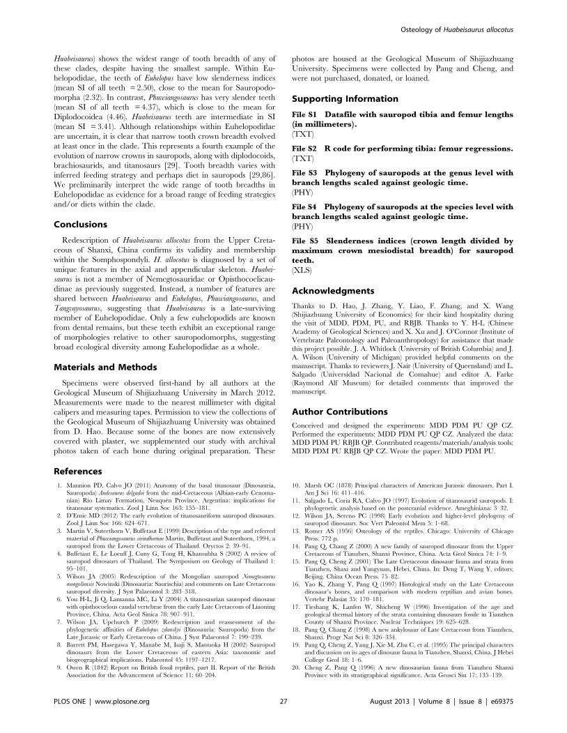
Huabeisaurus) shows the widest range of tooth breadth of any of
these clades, despite having the smallest sample. Within Eu-
helopodidae, the teeth of Euhelopus have low slenderness indices
(mean SI of all teeth = 2.50), close to the mean for Sauropodo-
morpha (2.32). In contrast, Phuwiangosaurus has very slender teeth
(mean SI of all teeth = 4.37), which is close to the mean for
Diplodocoidea (4.46). Huabeisaurus teeth are intermediate in SI
(mean SI = 3.41). Although relationships within Euhelopodidae
are uncertain, it is clear that narrow tooth crown breadth evolved
at least once in the clade. This represents a fourth example of the
evolution of narrow crowns in sauropods, along with diplodocoids,
brachiosaurids, and titanosaurs [29]. Tooth breadth varies with
inferred feeding strategy and perhaps diet in sauropods [29,86].
We preliminarily interpret the wide range of tooth breadths in
Euhelopodidae as evidence for a broad range of feeding strategies
and/or diets within the clade.
Conclusions
Redescription of Huabeisaurus allocotus from the Upper Creta-
ceous of Shanxi, China confirms its validity and membership
within the Somphospondyli. H. allocotus is diagnosed by a set of
unique features in the axial and appendicular skeleton. Huabei-
saurus is not a member of Nemegtosauridae or Opisthocoelicau-
dinae as previously suggested. Instead, a number of features are
shared between Huabeisaurus and Euhelopus, Phuwiangosaurus, and
Tangvayosaurus, suggesting that Huabeisaurus is a late-surviving
member of Euhelopodidae. Only a few euhelopodids are known
from dental remains, but these teeth exhibit an exceptional range
of morphologies relative to other sauropodomorphs, suggesting
broad ecological diversity among Euhelopodidae as a whole.
Materials and Methods
Specimens were observed first-hand by all authors at the
Geological Museum of Shijiazhuang University in March 2012.
Measurements were made to the nearest millimeter with digital
calipers and measuring tapes. Permission to view the collections of
the Geological Museum of Shijiazhuang University was obtained
from D. Hao. Because some of the bones are now extensively
covered with plaster, we supplemented our study with archival
photos taken of each bone during original preparation. These
photos are housed at the Geological Museum of Shijiazhuang
University. Specimens were collected by Pang and Cheng, and
were not purchased, donated, or loaned.
Supporting Information
File S1 Datafile with sauropod tibia and femur lengths(in millimeters).
(TXT)
File S2 R code for performing tibia: femur regressions.
(TXT)
File S3 Phylogeny of sauropods at the genus level withbranch lengths scaled against geologic time.(PHY)
File S4 Phylogeny of sauropods at the species level withbranch lengths scaled against geologic time.
(PHY)
File S5 Slenderness indices (crown length divided bymaximum crown mesiodistal breadth) for sauropodteeth.
(XLS)
Acknowledgments
Thanks to D. Hao, J. Zhang, Y. Liao, F. Zhang, and X. Wang
(Shijiazhuang University of Economics) for their kind hospitality during
the visit of MDD, PDM, PU, and RBJB. Thanks to Y. H-L (Chinese
Academy of Geological Sciences) and X. Xu and J. O’Connor (Institute of
Vertebrate Paleontology and Paleoanthropology) for assistance that made
this project possible. J. A. Whitlock (University of British Columbia) and J.
A. Wilson (University of Michigan) provided helpful comments on the
manuscript. Thanks to reviewers J. Nair (University of Queensland) and L.
Salgado (Universidad Nacional de Comahue) and editor A. Farke
(Raymond Alf Museum) for detailed comments that improved the
manuscript.
Author Contributions
Conceived and designed the experiments: MDD PDM PU QP CZ.
Performed the experiments: MDD PDM PU QP CZ. Analyzed the data:
MDD PDM PU RBJB QP. Contributed reagents/materials/analysis tools:
MDD PDM PU RBJB QP CZ. Wrote the paper: MDD PDM PU.
References
1. Mannion PD, Calvo JO (2011) Anatomy of the basal titanosaur (Dinosauria,
Sauropoda) Andesaurus delgadoi from the mid-Cretaceous (Albian-early Cenoma-
nian) Rıo Limay Formation, Neuquen Province, Argentina: implications for
titanosaur systematics. Zool J Linn Soc 163: 155–181.
2. D’Emic MD (2012) The early evolution of titanosauriform sauropod dinosaurs.
Zool J Linn Soc 166: 624–671.
3. Martin V, Suteethorn V, Buffetaut E (1999) Description of the type and referred
material of Phuwiangosaurus sirindhornae Martin, Buffetaut and Suteethorn, 1994, a
sauropod from the Lower Cretaceous of Thailand. Oryctos 2: 39–91.
4. Buffetaut E, Le Loeuff J, Cuny G, Tong H, Khansubha S (2002) A review of
sauropod dinosaurs of Thailand. The Symposium on Geology of Thailand 1:
95–101.
5. Wilson JA (2005) Redescription of the Mongolian sauropod Nemegtosaurus
mongoliensis Nowinski (Dinosauria: Saurischia) and comments on Late Cretaceous
sauropod diversity. J Syst Palaeontol 3: 283–318.
6. You H-L, Ji Q, Lamanna MC, Li Y (2004) A titanosaurian sauropod dinosaur
with opisthocoelous caudal vertebrae from the early Late Cretaceous of Liaoning
Province, China. Acta Geol Sinica 78: 907–911.
7. Wilson JA, Upchurch P (2009) Redescription and reassessment of the
phylogenetic affinities of Euhelopus zdanskyi (Dinosauria: Sauropoda) from the
Late Jurassic or Early Cretaceous of China. J Syst Palaeontol 7: 199–239.
8. Barrett PM, Hasegawa Y, Manabe M, Isaji S, Matsuoka H (2002) Sauropod
dinosaurs from the Lower Cretaceous of eastern Asia: taxonomic and
biogeographical implications. Palaeontol 45: 1197–1217.
9. Owen R (1842) Report on British fossil reptiles, part II. Report of the British
Association for the Advancement of Science 11: 60–204.
10. Marsh OC (1878) Principal characters of American Jurassic dinosaurs. Part I.
Am J Sci 16: 411–416.
11. Salgado L, Coria RA, Calvo JO (1997) Evolution of titanosaurid sauropods. I:
phylogenetic analysis based on the postcranial evidence. Ameghiniana: 3–32.
12. Wilson JA, Sereno PC (1998) Early evolution and higher-level phylogeny of
sauropod dinosaurs. Soc Vert Paleontol Mem 5: 1–68.
13. Romer AS (1956) Osteology of the reptiles. Chicago: University of Chicago
Press. 772 p.
14. Pang Q, Chang Z (2000) A new family of sauropod dinosaur from the Upper
Cretaceous of Tianzhen, Shanxi Province, China. Acta Geol Sinica 74: 1–9.
15. Pang Q, Cheng Z (2001) The Late Cretaceous dinosaur fauna and strata from
Tianzhen, Shaxi and Yangyuan, Hebei, China. In: Deng T, Wang Y, editors;
Beijing. China Ocean Press. 75–82.
16. Yao K, Zhang Y, Pang Q (1997) Histological study on the Late Cretaceous
dinosaur’s bones, and comparison with modern reptilian and avian bones.
Vertebr Palasiat 35: 170–181.
17. Tieshang K, Lanfen W, Shicheng W (1996) Investigation of the age and
geological thermal history of the strata containing dinosaurs fossile in Tianzhen
County of Shanxi Province. Nuclear Techniques 19: 625–628.
18. Pang Q, Chang Z (1998) A new ankylosaur of Late Cretaceous from Tianzhen,
Shanxi. Progr Nat Sci 8: 326–334.
19. Pang Q, Cheng Z, Yang J, Xie M, Zhu C, et al. (1995) The principal characters
and discussion on its ages of dinosaur fauna in Tianzhen, Shanxi, China. J Hebei
College Geol 18: 1–6.
20. Cheng Z, Pang Q (1996) A new dinosaurian fauna from Tianzhen Shanxi
Province with its stratigraphical significance. Acta Geosci Sin 17: 135–139.
Osteology of Huabeisaurus allocotus
PLOS ONE | www.plosone.org 27 August 2013 | Volume 8 | Issue 8 | e69375

21. Pang Q, Cheng Z, Yang J, Xie M, Zhu C, et al. (1996) The preliminary report
on Late Cretaceous dinosaur fauna expeditions in Tianzhen, Shanxi. J HebeiCollege of Geology 19: 227–235.
22. Pang Q, Cheng Z (2001) A new dinosaur fauna of the Late Cretaceous from
Tianzhen, Shanxi and Yangyuan, Hebei, China. In: Hao D, Niu S, Liu Y,
editors; Shijiazhuang. 19–21.
23. Carrano MT, Benson RBJ, Sampson SD (2012) The phylogeny of Tetanurae
(Dinosauria: Theropoda). J Sys Palaeontol 10: 211–300.
24. Wilson JA (1999) A nomenclature for vertebral laminae in sauropods and other
saurischian dinosaurs. J Vert Paleontol 19: 639–653.
25. Wilson JA (2012) New vertebral laminae and patterns of serial variation in
vertebral laminae of sauropod dinosaurs. Contrib Mus Paleontol, U Michigan
32: 91–110.
26. Wilson JA, D’Emic MD, Ikejiri T, Moacdieh EM, Whitlock JA (2011) Anomenclature for vertebral fossae in sauropods and other saurischian dinosaurs.
PLoS ONE: e17114.
27. Wilson JA (2011) Anatomical terminology for the sacrum of sauropod dinosaurs.
Contrib Mus Paleontol, U Michigan 32: 59–69.
28. Upchurch P (1998) The phylogenetic relationships of sauropod dinosaurs.
Zool J Linn Soc 124: 43–103.
29. Chure D, Britt BB, Whitlock JA, Wilson JA (2010) First complete sauropod
dinosaur skull from the Cretaceous of the Americas and the evolution of
sauropod dentition. Naturwissenschaften 97: 379–391.
30. Upchurch P, Barrett PM, Dodson P (2004) Sauropoda. In: Weishampel DB,
Dodson P, Osmolska H, editors. Berkeley: University of California Press. 259–
324.
31. Rose PJ (2007) A new titanosauriform sauropod (Dinosauria: Saurischia) fromthe Early Cretaceous of central Texas and its phylogenetic relationships.
Palaeontol Electronica 10: 1–65.
32. You H-L, Li D-Q (2009) The first well-preserved Early Cretaceous
brachiosaurid dinosaur in Asia. Proc R Soc B: Biol Sci 276: 4077–4082.
33. Wilson JA (2002) Sauropod dinosaur phylogeny: critique and cladistic analysis.
Zool J Linn Soc 136: 217–276.
34. Ksepka DT, Norell MA (2010) The illusory evidence for Asian Brachiosauridae:
New material of Erketu ellisoni and a phylogenetic reappraisal of basalTitanosauriformes. Am Mus Novitates 3700.
35. Mannion PD (2011) A reassessment of Mongolosaurus haplodon Gilmore, 1933, a
titanosaurian sauropod dinosaur from the Early Cretaceous of Inner Mongolia,
People’s Republic of China. J Sys Palaeontol 9: 355–378.
36. Ksepka DT, Norell MA (2006) Erketu ellisoni, a long-necked sauropod from Bor
Guve (Dornogov Aimag, Mongolia). Am Mus Novitates 3508: 1–16.
37. Curry Rogers KA (2009) The postcranial osteology of Rapetosaurus krausei
(Sauropoda: Titanosauria) from the Late Cretaceous of Madagascar. J Vert
Paleontol 29: 1046–1086.
38. Sanz JL, Powell JE, Le Loueff J, Martınez R, Pereda Suberbiola X (1999)
Sauropod remains from the Upper Cretaceous of Lano (northcentral Spain).
Titanosaur phylogenetic relationships. Estudios del Museo de Ciencias Naturales
de Alava 14: 235–255.
39. Bonaparte JF, Gonzalez Riga BJ, Apesteguıa S (2006) Ligabuesaurus leanzai gen. et
sp. nov. (Dinosauria, Sauropoda), a new titanosaur from the Lohan Cura
Formation (Aptian, Lower Cretaceous) of Neuquen, Patagonia, Argentina.
Cretaceous Res 27: 364–376.
40. Whitlock JA (2011) A phylogenetic analysis of Diplodocoidea (Saurischia:
Sauropoda). Zool J Linn Soc 161: 872–915.
41. D’Emic MD, Wilson JA (2011) New remains attributable to the holotype of
Neuquensaurus australis (Dinosauria: Sauropoda): implications for saltasaurine
systematics. Acta Palaeontol Pol 56: 61–73.
42. Salgado L, Apesteguıa S, Heredia SE (2005) A new specimen of Neuquensaurus
australis, a Late Cretaceous saltasaurine titanosaur from north Patagonia. J Vert
Paleontol 25: 623–634.
43. Martınez RD, Gimenez O, Rodrıguez J, Luna M, Lamanna MC (2004) Anarticulated specimen of the basal titanosaurian (Dinosauria: Sauropoda)
Epachthosaurus sciuttoi from the early Late Cretaceous Bajo Barreal Formation
of Chubut province, Argentina. J Vert Paleontol 24: 107–120.
44. Gomani E (2005) Sauropod dinosaurs from the Early Cretaceous of Malawi,
Africa. Paleontol Electronica 8: 1–37.
45. Osborn HF, Mook CC (1921) Camarasaurus, Amphicoelias, and other sauropods of
Cope. Mem Am Mus Nat Hist New Series 3: 247–387.
46. Zhang Y-H (1988) The Middle Jurassic dinosaur fauna from Dashanpu, Zigong,
Sichuan. Sauropod dinosaurs. Shunosaurus. Chengdu: Sichuan Publishing Houseof Science and Technology. 106 p.
47. Berman DS, McIntosh JS (1978) Skull and relationships of the Upper Jurassic
sauropod Apatosaurus (Reptilia: Saurischia). Bull Carnegie Mus Nat Hist 8: 1–35.
48. McIntosh JS (1990) Sauropoda. In: Weishampel DB, Dodson P, Osmolska H,
editors. The Dinosauria. Berkeley, CA: University California Press. 345–401.
49. Wilson JA, Martınez RA, Alcober OA (1999) Distal tail of a titanosaurian
sauropod from the Upper Cretaceous of Mendoza, Argentina. J Vert Paleontol
19: 592–595.
50. Whitlock JA, D’Emic MD, Wilson JA (2011) Cretaceous diplodocids in Asia?
Re-evaluating the phylogenetic affinities of a fragmentary specimen. Palaeontol
54: 351–364.
51. Calvo JO, Porfiri JD, Gonzalez Riga BJ, Kellner AWA (2008) Anatomy ofFutalognkosaurus dukei Calvo, Porfiri, Gonzalez Riga & Kellner, 2007 (Dinosauria,
Titanosauridae) from the Neuquen Group (Late Cretaceous), Patagonia,
Argentina. Arquivos do Museu Nacional, Rio de Janeiro 65: 511–526.
52. Lu J, Xu L, Jiang X, Jia S, Li M, et al. (2009) A preliminary report on the new
dinosaurian fauna from the Cretaceous of the Ruyang Basin, Henan Province of
Central China. J Paleontol Soc Korea 25: 43–56.
53. Campos DA, Kellner AWA, Bertini RJ, Santucci RM (2005) On a titanosaurid
(Dinosauria, Sauropoda) vertebral column from the Bauru Group, Late
Cretaceous of Brazil. Arquivos do Museu Nacional, Rio de Janeiro 63: 656–593.
54. Kellner AWA, Campos DA, Azevedo SAK, Trotta MNF, Henriques DDR, etal. (2006) On a new titanosaur sauropod from the Bauru Group, Late
Cretaceous of Brazil. Boletim do Museu Nacional (Geologia) 74: 1–31.
55. Ostrom JH, McIntosh JS (1966) Marsh’s Dinosaurs. New Haven: Yale
University Press. 388 pp.
56. Canudo JI, Royo-Torres R, Cuenca-Bescos G (2008) A new sauropod:
Tastavinsaurus sanzi gen. et sp. nov. from the Early Cretaceous (Aptian) of Spain.
J Vert Paleontol 28: 712–731.
57. Curry Rogers KA (2005) Titanosauria: a phylogenetic overview. In: Curry
Rogers KA, Wilson JA, editors. The Sauropods: Evolution and Paleobiology.
Berkeley: University of California Press. 50–103.
58. Upchurch P, Martin J (2003) The anatomy and taxonomy of Cetiosaurus
(Saurischia: Sauropoda) from the Middle Jurassic of England. J Vert Paleontol
23: 208–231.
59. Gilmore CW (1936) Osteology of Apatosaurus with special reference to specimens
in the Carnegie Museum. Mem Carnegie Mus 11: 175–300.
60. Curry Rogers KA, Forster CA (2001) The last of the dinosaur titans: a new
sauropod from Madagascar. Nature 412: 530–534.
61. Gilmore CW (1946) Reptilian fauna of the North Horn Formation of central
Utah. United States Department of the Interior Geological Survey Professional
Paper 210-C: 29–53.
62. Mannion PD (2009) A rebbachisaurid sauropod from the Lower Cretaceous of
the Isle of Wight, England. Cretaceous Res 30: 521–526.
63. Sereno PC, Beck AL, Dutheil DB, Larsson HCE, Lyon GH, et al. (1999)
Cretaceous sauropods from the Sahara and the uneven rate of skeletal evolution
among dinosaurs. Science 286: 1342–1347.
64. Upchurch P (1995) The evolutionary history of sauropod dinosaurs. Phil
Trans R Soc London B: Biol Sci 349: 365–390.
65. Wilson JA, Carrano MT, Barrett PM (2011) An associated partial skeleton of
Jainosaurus cf. septentrionalis (Dinosauria: Sauropoda) from the Late Cretaceous of
Chhota Simla, central India. Palaeontol 54: 981–998.
66. McIntosh JS (1990) Sauropoda. In: Weishampel DB, Dodson P, Osmolska H,editors. The Dinosauria. Berkeley: University of California Press. 345–401.
67. Mannion PD, Upchurch P, Mateus O, Barnes R, Jones MEH (2012) New
information on the anatomy and systematic position of Dinheirosaurus lourinhanensis
(Sauropoda: Diplodocoidea) from the Late Jurassic of Portugal, with a review of
European diplodocoids. J Syst Palaeontol 10: 521–551.
68. Upchurch P, Mannion PD, Barrett PM (2011) Sauropod dinosaurs. In: Batten
DJ, editor. Field Guide to English Wealden Fossils. London: Palaeontol
Association. 476–525.
69. Bonaparte JF, Heinrich W-D, Wild R (2000) Review of Janenschia Wild, with the
description of a new sauropod from the Tendaguru beds of Tanzania and a
discussion on the systematic value of procoelous caudal vertebrae in the
Sauropoda. Palaeontogr, Abteilung A 256: 25–76.
70. Mo J-Y, Wang W, Huang Z, Huang X, Xu X (2006) A basal titanosauriform
from the Early Cretaceous of Guangxi, China. Acta Geol Sinica 80: 486–489.
71. Tanimoto M, Suzuki C, Takahashi T, Nabana S (2006) Nemegtosaurid fossils
from Japan. Chigakukenkyu 54: 223–227.
72. Curry Rogers KA, Forster CA (2004) The skull of Rapetosaurus krausei
(Sauropoda: Titanosauria) from the Late Cretaceous of Madagascar. J Vert
Paleontol 24: 121–144.
73. Zaher H, Pol D, Carvalho AB, Nascimento PM, Riccomini C, et al. (2011) A
complete skull of an Early Cretaceous sauropod and the evolution of advanced
titanosaurians. PLoS ONE 6: e16663.
74. Saegusa H, Tomida Y (2011) Titanosauriform teeth from the Cretaceous of
Japan. An Acad Bras Cienc 83: 247–265.
75. Suteethorn S, Le Loeuff J, Buffetaut E, Suteethorn V (2010) Description of
topotypes of Phuwiangosaurus sirindhornae, a sauropod from the Sao Khua
Formation (Early Cretaceous) of Thailand, and their phylogenetic implications.
Neues Jahrb Geol Palaontol Abh 256: 109–121.
76. Carballido JL, Pol D, Cerda I, Salgado L (2011) The osteology of Chubutisaurus
insignis del Corro, 1975 (Dinosauria: Neosauropoda) from the ‘middle’
Cretaceous of central Patagonia, Argentina. J Vert Paleontol 31: 93–110.
77. Borsuk-Bialynicka M (1977) A new camarasaurid sauropod Opisthocoelicaudia
skarzynskii gen. n., sp. n. from the Upper Cretaceous of Mongolia. Palaeontol
Polonica: 5–63.
78. D’Emic MD (2013) Revision of the sauropod dinosaurs of the Lower Cretaceous
Trinity Group, southern USA, with the description of a new genus. J Syst
Palaeontol. doi: 10.1080/14772019.2012.667446.
79. Mannion PD, Upchurch P, Barnes N, Mateus O (2013) Osteology of the Late
Jurassic Portuguese sauropod dinosaur Lusotitan atalaiensis (Macronaria) and the
evolutionary history of basal titanosauriforms. Zool J Linn Soc 168: 98–206.
80. Wilson JA, Upchurch P (2003) A revision of Titanosaurus Lydekker (Dinosauria-
Sauropoda), the first dinosaur genus with a ‘Gondwanan’ distribution. J Syst
Palaeontol 1: 125–160.
Osteology of Huabeisaurus allocotus
PLOS ONE | www.plosone.org 28 August 2013 | Volume 8 | Issue 8 | e69375
81. R Core Team website: R: A language and environment for statistical computing.
R Foundation for Statistical Computing, Vienna, Austria. ISBN 3–900051–07–0, Available: http://www.R-project.org/. Accessed 1 May 2013.
82. Grafen A (1989) The phylogenetic regression. Phil Trans R Soc London B: Biol
Sci 326: 119–157.83. Pagel M (1998) Inferring evolutionary processes from phylogenies. Zoolog Scr
26: 331–348.
84. Pinheiro J, Bates D, DebRoy S, Sarkar D, R Core Team (2012) nlme: linear and
nonlinear mixed effects models. In. 3.1-104 ed.
85. Paradis E, Claude J, Strimmer K (2004) APE: analyses of phylogenetics and
evolution in R language. Bioinf 20: 289–290.
86. Whitlock J (2011) Inferences of diplodocoid (Sauropoda: Dinosauria) feeding
behavior from snout shape and microwear analyses. PLoS One 6: e18304.
Osteology of Huabeisaurus allocotus
PLOS ONE | www.plosone.org 29 August 2013 | Volume 8 | Issue 8 | e69375
Copyright © 2022 FDOKUMEN