Preclassic Maya Population: Human Remains, Osteology, and Health
67
CHAPTER 12 Preclassic Maya Population: Human Remains, Osteology, and Health Rhan-Ju Song As an organic substance, bone is highly subject to its depositional environment and its preservation can vary tremendously depending upon its burial context. In the Maya lowlands, owing to external factors, preservation is generally poor to fair for human skeletal remains, which has limited osteological research. Soil conditions, water leaching, insect and animal activity, rootlet acidity and bioturbation from tree roots have all acted to deteriorate bone and tooth enamel, and have sometimes produced misleading pseudo-pathologies (e.g., Saul and Saul 1989). Some of the older, Preclassic period human remains, especially, have not withstood the passage of time very well. Due to the often poor preservation of human remains, many early osteological investigations of Maya remains were, historically, descriptive in nature (see Comas 1970). Like the discipline of biological anthropology in general, early research was often engrossed with anthropometric analyses and skeletal evidence of cranial and dental modification (e.g., Blom 1933; Cave et al. 1939; Cifuentes 1963; Elliott and Bleibtreu 1965; Genoves 1970; Hambly 1937; Longyear 1940; Romero 1970; Stewart 1941, 1974). A general assessment of ancient health, if it was even examined (e.g., Gann 1918), was often interpreted independently of the cultural context. Fortunately, following Saul’s (1972: 8) example, most current physical anthropologists utilize an “osteobiographic” approach to investigate ancient human remains, with the acknowledgment that “life history (can be) recorded in bone”. This approach reflects the conscious attempt of biological anthropologists and archaeologists alike to integrate the two subfields of anthropology (e.g., Blakely 1977; Buikstra 1977; Martin and Bumsted 1982; among
Transcript of Preclassic Maya Population: Human Remains, Osteology, and Health

CHAPTER 12
Preclassic Maya Population:
Human Remains, Osteology, and Health
Rhan-Ju Song
As an organic substance, bone is highly subject to its depositional environment and its
preservation can vary tremendously depending upon its burial context. In the Maya lowlands,
owing to external factors, preservation is generally poor to fair for human skeletal remains, which
has limited osteological research. Soil conditions, water leaching, insect and animal activity,
rootlet acidity and bioturbation from tree roots have all acted to deteriorate bone and tooth
enamel, and have sometimes produced misleading pseudo-pathologies (e.g., Saul and Saul 1989).
Some of the older, Preclassic period human remains, especially, have not withstood the passage
of time very well.
Due to the often poor preservation of human remains, many early osteological investigations
of Maya remains were, historically, descriptive in nature (see Comas 1970). Like the discipline
of biological anthropology in general, early research was often engrossed with anthropometric
analyses and skeletal evidence of cranial and dental modification (e.g., Blom 1933; Cave et al.
1939; Cifuentes 1963; Elliott and Bleibtreu 1965; Genoves 1970; Hambly 1937; Longyear 1940;
Romero 1970; Stewart 1941, 1974). A general assessment of ancient health, if it was even
examined (e.g., Gann 1918), was often interpreted independently of the cultural context.
Fortunately, following Saul’s (1972: 8) example, most current physical anthropologists
utilize an “osteobiographic” approach to investigate ancient human remains, with the
acknowledgment that “life history (can be) recorded in bone”. This approach reflects the
conscious attempt of biological anthropologists and archaeologists alike to integrate the two
subfields of anthropology (e.g., Blakely 1977; Buikstra 1977; Martin and Bumsted 1982; among
others). Importantly, such bioarchaeological studies examine human remains holistically, within
the context of archaeological evidence, paleodietary, environmental and ecological models, and
ethnohistoric data.
One area in which skeletal data has been scrutinized to explain archaeological findings
concerns the “Classic Maya collapse”. Citing results from Saul's (1972) study of the Maya from
Altar de Sacrificios, which suggested increased anemia, infection, and ill health in the Late
Classic period, ecological and biological models of the collapse have been proposed (Culbert
1988; Santley 1990; Santley et al. 1986; Willey and Shimkin 1973). However, in this case, more
recent skeletal data has refuted the theory that increased population pressure, reduced food
resources, concomitant malnutrition and disease were the direct catalysts for the collapse of
Classic period Maya socio-politico-economic systems (see review in Wright and White 1996).
Due to a previous preoccupation with Classic period monumental archaeology (especially of
the elite class), poor preservation factors in the tropical lowlands, and practical difficulties
accessing early burials, the quality and quantity of Preclassic period human remains is limited.
To date, very few significant assemblages of Formative period skeletal material have been
recovered from lowland Maya sites.
Notable Maya sites with Preclassic remains that have been analyzed and reported include
Altar de Sacrificios (Saul 1972) and Cuello (Saul and Saul 1991). At Altar de Sacrificios, 21
individuals were from Preclassic contexts (ca. 1000 B.C.-A.D. 300) (Saul 1972: Table 1).
Significantly, at Cuello, 131 Preclassic period individuals were recovered, with 29 of these from
the Early-Middle Formative (Swasey and Bladen phases: 1200-650 B.C.), and 102 from the Late
Formative Chicanel phase (Hammond et al. 1995; Saul and Saul 1991). This is, by the far, the
most significant assemblage of Preclassic Maya remains.

Aside from these, skeletal remains from several other sites have enhanced the
osteobiographic record of Preclassic Maya. From the burial data, more than 10% of the
interments found at both Altun Ha and Copan are Formative in date. This amounts to 58
individuals from 33 Middle and Late Preclassic burials at Altun Ha (Pendergast 1979, 1982,
1990; see Welsh 1988: Table IX), and 60 or more individuals at Copan, also dating to the Middle
and Late Preclassic (Storey 1992). In addition, skeletal remains excavated at Uaxactun consist of
five Middle Preclassic individuals and 15 Late Preclassic period individuals (Ruz 1965; see
Welsh 1988: Table VII).
Remains exist from several other sites in the Maya Lowlands, but poor preservation hinders
their utilization in comparative osteological analyses. At Lamanai, for example, 11 Preclassic
individuals are very poorly preserved and not amenable to complete analysis (White 1986), while
a similar situation characterizes the seven Late Preclassic period burials from the site of Blue
Creek in northern Belize (H. Haines, personal communication, 1997). Lastly, at Colha, a small
sample of Middle Preclassic remains has been excavated, with osteological analyses of four
individuals reported, so far, by Young (1994).
Immediately outside the Maya sphere, Preclassic remains from a much different burial
context have been recorded by J. Brady (1997). In this case, reconnaissance of caves
immediately southeast of the Maya area has revealed Preclassic Mesoamerican ceramics as well
as human remains. At Talgua Cave, in northeastern Honduras, the remains derive from large
surface ossuaries below and above the water line, amounting to between 100 and 200 interments
(Brady 1997: 24). Unfortunately, due to the cave environment, the skeletal remains could not be
analyzed due to poor preservation or calcite “crystallization”.
Interestingly, numerous Preclassic human interments or offerings have been recorded in
caves in lower Central America (Brady 1995; Gordon 1898; Healy 1974; Rue et al. 1989). While

not unequivocally Maya, the ceramics directly associated with the human remains appear clearly
influenced by Mesoamerican cultures such as the Maya (Brady 1997).
However, while a pattern of early cave interment exists for lower Central America, the
evidence northward is scant due to lack of research interest and logistical difficulties (Brady
1997). An exception is the recent cave investigations in Belize by the Western Belize Regional
Cave Project (see Awe 1998; Awe and Lee 1999), but while Preclassic Maya ceramics have been
recorded at cave entrances (J. Awe, personal communication, 1998), no human material has been
associated with such early pottery.
In sum, because of the scarcity of such early human remains compared to the relative
abundance of Classic period skeletons, any discovery of Preclassic period human skeletal
remains is noteworthy. Comparisons of Preclassic period skeletal health will be made herein
only with remains from Altar de Sacrificios (Saul 1972) and Cuello (Saul and Saul 1991), and
with some reference to the dental data from Altun Ha (Song 1997). Skeletal samples from other
sites are either too small for significant comparisons, or their osteological analyses are pending or
incompletely published.
The Burial Record at Cahal Pech and Pacbitun
Identifications were made by the author using standard osteological techniques (see
Brothwell 1981; Buikstra and Ubelaker 1994; Ubelaker 1989). Poor preservation of some
remains from Cahal Pech and Pacbitun has resulted in a classification of “indeterminate”, or
uncertain (?), category for sex. Most individuals have been assigned an age, or age category.
At Cahal Pech, at least 75 burials or human deposits, encompassing at least 88 individuals,
and the fingers of 20+ adults, were encountered during excavations between 1988 and 1997,
spanning the Middle Preclassic to Late Classic periods. The majority of burials date to the
Classic period, but with increased focus on Preclassic period settlement at the site, the
assemblage of early human remains has grown steadily (see Awe 1992; Healy and Awe 1995,
1996). The individuals (n=23) who compose the Preclassic sample at Cahal Pech (17 burials,
and two caches with human remains) represent approximately 26% of the total skeletal
assemblage from the site (Table 1).
At Pacbitun, 21 burials representing at least 27 individuals have been identified from
excavations between 1984-1997, spanning the Middle Preclassic to Terminal Classic periods. As
at Cahal Pech, the majority of these individuals date to the Classic period. Only two burials,
each containing one individual, date to the Preclassic, representing 7% of the total skeletal
assemblage at Pacbitun (Table 1).
Regarding the sample, the 25 Preclassic individuals from the two sites, including caches with
human remains, consist of: nine subadults, four adults of indeterminate sex, two adult females,
three adult males, two possible adult females, four possible adult males, and one possible adult of
indeterminate sex.
The Preclassic remains can be divided into the following temporal groups (noting local phase
names for Cahal Pech and Pacbitun, respectively):
Middle Preclassic (Kanluk phase, Mai phase) 3 individuals
Middle-Late Preclassic transition (late Kanluk-early Xakal phase, 5 individuals
late Mai-early Puc phase)
Late Preclassic (early Xakal phase, Puc phase) 9 individuals
Terminal Preclassic (late Xakal phase, Ku phase) 8 individuals
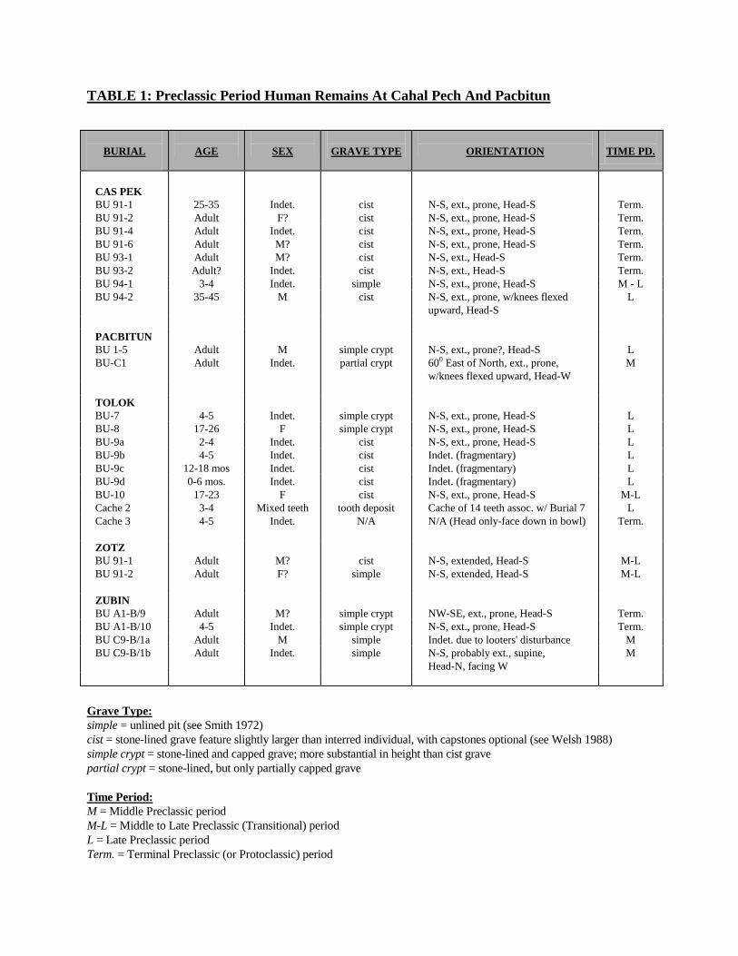
TABLE 1: Preclassic Period Human Remains At Cahal Pech And Pacbitun
BURIAL
AGE
SEX
GRAVE TYPE
ORIENTATION
TIME PD.
CAS PEK
BU 91-1
25-35
Indet.
cist
N-S, ext., prone, Head-S
Term.
BU 91-2 Adult F? cist N-S, ext., prone, Head-S Term.
BU 91-4 Adult Indet. cist N-S, ext., prone, Head-S Term.
BU 91-6
BU 93-1
BU 93-2
Adult
Adult
Adult?
M?
M?
Indet.
cist
cist
cist
N-S, ext., prone, Head-S
N-S, ext., Head-S
N-S, ext., Head-S
Term.
Term.
Term.
BU 94-1 3-4 Indet. simple N-S, ext., prone, Head-S M - L
BU 94-2 35-45 M cist N-S, ext., prone, w/knees flexed
upward, Head-S
L
PACBITUN
BU 1-5
BU-C1
Adult
Adult
M
Indet.
simple crypt
partial crypt
N-S, ext., prone?, Head-S
600 East of North, ext., prone,
w/knees flexed upward, Head-W
L
M
TOLOK
BU-7
4-5
Indet.
simple crypt
N-S, ext., prone, Head-S
L
BU-8 17-26 F simple crypt N-S, ext., prone, Head-S L
BU-9a 2-4 Indet. cist N-S, ext., prone, Head-S L
BU-9b 4-5 Indet. cist Indet. (fragmentary) L
BU-9c 12-18 mos Indet. cist Indet. (fragmentary) L
BU-9d 0-6 mos. Indet. cist Indet. (fragmentary) L
BU-10
Cache 2
17-23
3-4
F
Mixed teeth
cist
tooth deposit
N-S, ext., prone, Head-S
Cache of 14 teeth assoc. w/ Burial 7
M-L
L
Cache 3
4-5 Indet. N/A N/A (Head only-face down in bowl) Term.
ZOTZ
BU 91-1
BU 91-2
Adult
Adult
M?
F?
cist
simple
N-S, extended, Head-S
N-S, extended, Head-S
M-L
M-L
ZUBIN
BU A1-B/9
Adult
M?
simple crypt
NW-SE, ext., prone, Head-S
Term.
BU A1-B/10 4-5 Indet. simple crypt N-S, ext., prone, Head-S Term.
BU C9-B/1a Adult M simple Indet. due to looters' disturbance M
BU C9-B/1b Adult Indet. simple N-S, probably ext., supine,
Head-N, facing W
M
Grave Type:
simple = unlined pit (see Smith 1972)
cist = stone-lined grave feature slightly larger than interred individual, with capstones optional (see Welsh 1988)
simple crypt = stone-lined and capped grave; more substantial in height than cist grave
partial crypt = stone-lined, but only partially capped grave
Time Period:
M = Middle Preclassic period
M-L = Middle to Late Preclassic (Transitional) period
L = Late Preclassic period
Term. = Terminal Preclassic (or Protoclassic) period
No discrete burials have been retrieved from the terminal Early Preclassic (Cunil phase), the
earliest occupation in the Valley, but several burnt long bone fragments have been found at Cahal
Pech from this time. These could reflect ritual cannibalism or, alternatively, early cremation
practices (Awe 1992: 335).
The Cahal Pech and Pacbitun Preclassic burial sample is very small compared to that of the
site of Cuello, but is comparable to that of Altar de Sacrificios (13 adults, 8 subadults) and Seibal
(6 adults). Reflecting the diversity of excavation strategies at Cahal Pech and Pacbitun,
interments have derived from contexts as variable as (elite) tombs and “elaborate crypts” to (non-
elite) simple pits, cists, and “partial” or “simple” crypts. However, only simple pit, cist and
partial or simple crypt graves have been found dating to the Preclassic period at the two sites
examined here.
Seventeen of the 75 Cahal Pech burials, in addition to one solitary head cache and one tooth
cache, date to the Preclassic period. All originate from the peripheral settlements of Cahal Pech,
namely Cas Pek, Tolok, Zotz and Zubin (see Table 1). Including the subadult tooth cache, these
interments represent the remains of at least 23 individuals, with some graves having multiple
individuals (see Healy et al. 1998).
Of the 17 Preclassic period interments/human deposits at Cahal Pech, six burials were
uncovered in 1991 from the Zotz Group (Zotz 91-1 and 91-2) and Cas Pek (Cas Pek 91-1, 91-2,
91-4 and 91-6) (Aimers 1992; Awe 1992; Awe et al. 1992a,b; Vinuales 1992). Three of these
interments date to the transition between the late Middle Preclassic and early Late Preclassic,
while the other three are Late Preclassic. Due to problematic post-excavation/storage
identification, Cas Pek Burials 91-2 and 91-6 are not discussed here.

In 1993, two Late Preclassic burials were excavated at Tolok (Burials 7 and 8), as well as a
Late Preclassic tooth cache (Cache 2) found associated with Burial 7 (Powis 1994; Song et al.
1994). Likewise, two Terminal Preclassic interments (Burials A1-B/9 and A1-B/10) were
discovered at Zubin, a minor center located 2 km south of the site core of Cahal Pech (Iannone
1994), and two Terminal Preclassic (Protoclassic) burials at Cas Pek (Cas Pek 93-1, 93-2)
(Sunahara and Awe 1994).
Excavations in 1994 uncovered a Terminal Preclassic head cache (Tolok Cache 3), a Middle-
Late Preclassic burial (Tolok Burial 10), and a Late Preclassic primary interment also at Tolok
(Burial 9) (Powis and Hohmann 1995). At Cas Pek, two interments were discovered in the large
platform structure, Nohoch Na, one dating to the late Middle/early Late Preclassic transition
period (Cas Pek 94-1), the other to the Late Preclassic (Cas Pek 94-2) (Lee and Awe 1995).
Elsewhere, two more Middle Preclassic period individuals were recovered from a single
interment at Zubin (Burial C9-B/1).
During the 1995 field season, no burials dating to the Formative period were found at Cahal
Pech, but archaeological investigations at Pacbitun led to the discovery of a Middle Preclassic
partial crypt grave beneath Plaza C (Arendt et al. 1996). This single adult interment represents
the second Formative period burial discovered at Pacbitun, where excavations in 1987 uncovered
a Late Preclassic crypt burial deep within Structure 1 (Healy 1990: 255).
This report will present detailed skeletal information for eight Preclassic period burials and
two Preclassic caches excavated at Cahal Pech and its vicinity (e.g., Cas Pek and Tolok), and the
two Preclassic period burials from Pacbitun. Shorter commentaries are provided on five
additional Preclassic burials from other Cahal Pech settlement groups (Zotz and Zubin). Taken
together, these burials and caches contain the remains of at least 21 individuals.1 The remaining
Preclassic burials noted above (but not discussed in detail here) have been previously described,
and will not be dealt with further. These Preclassic remains are included in reports by Awe
(1992), Awe, Aimers and Blanchard (1992), Schwake (1996), Sunahara and Awe (1994) and
Vinuales (1992).
Preclassic Period Skeletal Remains at Cahal Pech
Cas Pek: Burial 91-1
This Terminal Preclassic (A.D. 0-250) cist burial contained the fragmentary remains of a
single adult individual of indeterminate sex. Human remains include most of the cranium, which
is very fragmentary, and two teeth, a right M2 and an unidentifiable single root tooth. Dental
attrition is slight in these teeth, as are deposits of calculus.
From the fragmentary cranial bones, it appears that most sutures are open or partially fused.
The partially fused portion of the sagittal suture just anterior of the lambda point indicates an
adult at least mid-twenties in age. Thus, an age range of 25-35 years can be attributed to this
individual. Upon reconstruction, some left parietal fragments appear to bulge slightly (laterally),
but the state of the bones prevents a clear assessment of the presence of cranial modification.
With respect to health, no pathology is noted on any of the cranial remains. However, in the
single root tooth, which could possibly be a mandibular premolar, extensive carious activity
resulted in almost complete destruction of the crown through to the root. The maxillary molar
also has a small interfissure pit on the occlusal surface that may be a carious lesion.
Finally, a portion of the buccal surface of the possible premolar has a linear enamel
hypoplasia of narrow width and medium depth at the midcrown level, which suggests a brief
period of ill health (most likely undernutrition and/or infectious disease) sometime between the
ages of 4 and 5 years.
Cas Pek: Burial 91-4
Due to modern-day construction activities, this cist burial became evident in cross-sections of
a bulldozed Preclassic period platform (see Vinuales 1992). Dated to the Terminal Preclassic,
the extended, prone, individual was characterized in the field as having mediocre to good
preservation. Although fragmentary, most of the skeleton is present.
The remains of an adult individual of indeterminate sex are represented. Identified skeletal
remains include several diaphyseal fragments of long bones (arms, legs), several vertebral and rib
fragments, scapular portions, carpal remains, and many miscellaneous bone fragments.
Unfortunately, full osteological analysis took place two years after the excavation and storage of
this individual, and dental remains could not be re-located for examination.
Significant observations include the extreme porosity and pitting of vertebral fragments,
some of which have extensive bony outgrowths on the exterior surfaces of the body. One
fragment, a probable thoracic or (small) lumbar vertebra is notable for a discernible fracture
down the height of the body, with evidence of healing. Repair of the fracture resulted in a slight
protrusion of bone on either the superior or inferior surface of the vertebral body.
The nature of these remains, which are characteristic of osteoporosis, may be taken to infer
an adult individual of older age, i.e. over 40 years, but the lack of other skeletal age indicators
prevents a firm statement of this fact. Finally, several unidentifiable flat bone fragments,
possibly from the leg bones, also appear diseased, in this case, with periostitic lesions.
Cas Pek: Burial 94-1
This primary interment was an unlined pit containing a fully flexed subadult individual
placed in an extended prone position (Figure 1). The individual’s cranium was placed face down
within a complete ceramic vessel and dates to the transition between the late Middle Preclassic
and early Late Preclassic.
A notable feature of this individual is the state of skeletal preservation. Relative to other
remains at Cahal Pech, and considering the young skeletal age, this child's bones are very well
preserved, though fragmented. For instance, the amount of delicate cranial fragments suggests
that most of the cranium is preserved, though fragmented. Amazingly, two ear bones (incus and
malleus) were even recovered from the skull area. Post-cranial remains are similarly well
preserved, but owing to the young skeletal age, long bones consist of diaphyseal fragments only.
Exemplifying the very good preservation of this individual, all deciduous teeth of this
individual were recovered, as were most of the secondary tooth crowns (see below).
Primary Dentition:
Upper x x x x x x x x x x
Right m2 m1 c i2 i1 i1 i2 c m1 m2 Left
Lower x x x x x x x x x x
Secondary Dentition:
Upper X X X X X X X X
Right M3 M2 M1 P2 P1 C I2 I1 I1 I2 C P1 P2 M1 M2 M3 Left
Lower X X X X X X X X
Post-Cranial Remains:
Scapulae: few pieces, very fragmentary
Ribs: most preserved, though fragmented
Vertebrae: approximately half preserved, though fragmented

Arm and Leg Long Bones: most of diaphyses preserved, but epiphyses absent
Carpals/Tarsals/Metacarpals/Metatarsals/Phalanges: many fragments
Other bones: most of this skeleton is preserved, but the fragmentary state of many bones
precludes clear identification at this time
Age, Sex And Skeletal Health
Following Ubelaker (1989: Fig. 71), the height of secondary tooth crowns and root formation
suggest that this child was between the ages of 3-4 years. Typical of subadults, sex of this child
is indeterminate.
In terms of skeletal health, no indication of pathology is evident on the cranial or post-cranial
remains. Specifically, cranial lesions of cribra orbitalia and porotic hyperostosis were searched
for, but both are noticeably absent. Likewise, inspection of post-cranial remains turned up no
clear evidence of infection, disease or trauma.
The only pathology observed in this child is apparent in the dental remains. Specifically, a
single, shallow hypoplastic line was detected on the secondary upper left I1 crown. The relative
position of this band indicates a short period of ill health or malnutrition at around 2.8 years of
age (following Song 1997: Table 6.2.3).
Additionally, several deciduous teeth exhibit surface pitting. These include the left and right
i2’s, left and right upper c’s, left and right m
1’s, left m
2, left m1 and m2. These pits predominantly
occur on the labial surfaces and along occlusal interfissures. Some pits are also associated with
hypocalcification discolorations (orange, brown and cream-beige), which occur in the central
midcrown regions of several deciduous teeth. While some of the pits are likely true carious
lesions, others are probably the result of preservation factors in the burial environment.
Lastly, attesting to the early death and cessation of development in this child, all secondary
crowns are characterized by hypocalcification staining. Enamel hypocalcification reflects a
disruption in the maturation phase of enamel development and it can appear as either localized or
general regions of orange, brown, opaque white or beige colored enamel.
Cas Pek: Burial 94-2
This Late Preclassic intrusive primary burial was a stone-lined cist grave containing the
remains of one adult individual. Skeletal position was, again, face and stomach down, with head
to the south, but the individual’s legs were flexed in a ninety-degree angle at the knee, so that the
lower legs were directed upward. Once again, the individual was interred with its cranium
placed face down within a ceramic vessel.
Like Burial 94-1 at Cas Pek, skeletal preservation of this individual is good to very good,
although fragmentation occurred during excavation and removal. The following inventory
outlines the extent of skeletal completeness for this individual.
Cranium: mostly complete, but fragmented; frontal portions consist of large supraorbitals;
temporal portions consist of large mastoids; several maxillary fragments preserved;
Sutures: most of sagittal suture is obliterated except near the frontal suture which is
closed but visible; superior portion of lambdoidal suture is closed and obliterated,
but other areas of this suture are only partially fused; portions of ectocranial
coronal suture (halfway between sagittal intersection and most lateral point of
coronal suture) appear partially closed; according to Montagu (1960: 609), fusion
in this region occurs between 24-38 years. Also: one portion of right occipital
along lambdoidal suture appears partially fused, but not obliterated; according to
Montagu (1960: 609), fusion in this region occurs between the ages of 26 - 42/47
years; mandible: mostly complete: square knobby chin, flared gonial angles,
pronounced muscle markings, but some thinning/shortening of body height; right
ramus portion not preserved
The following Secondary Dentition is present for Cas Pek Burial 94-2:
Upper X X X X? X
Right M3 M2 M1 P2 P1 C I2 I1 I1 I2 C P1 P2 M1 M2 M3 Left
Lower X X X X X X X X X

Post-Cranial Remains:
Clavicles: very fragmentary; most of left diaphysis preserved, but missing both epiphyses;
robust muscle markings; no pathology evident
Scapulae: both very fragmentary; right scapula has bony lipping along acromion edge (near
articular surface with humerus); right acromion articular surface is porous and rough
Ribs and Vertebrae: most present, though fragmented; thoracic and lumbar vertebrae appear
very porous (cancellous) and edges of bodies have prominent ridges; some
vertebral bodies have medium to heavy lipping, with spicule formation on the edges
Innominates: well preserved but very fragmented; auricular surface very pitted and porous - two
fragments suggest stage 30-34, or more likely, 35-39 years, following Ubelaker
(1989); narrow sciatic notch evident; fragmentary iliac crest but visibly fully fused;
acetabulum: surface smooth, but portions are very porous and there is some
lipping of acetabular rim; ischium: fragmentary, but fully fused to body; sacroiliac
facet: slight porosity with parts of surface rough; two fragments of pubic
symphysis: prominent outer rim (see Ubelaker 1989:75), furrows/ridges evident
but not deep, one fragment with fully formed ventral ridge similar to stage 4
(Ubelaker1989: Fig. 88); symphyseal surface not smooth, but with slight ridges,
and bone porosity; pubic surface is between Ubelaker’s (1989: Fig. 86) stages b
(age 29) and c (age 56 years), although closer to stage c
Sacrum: fragmentary remains
Long Bones: most well preserved though fragmented; Notable Observations: femurs-both
mostly complete, with heads appearing very pitted and porous; cancellous bone
extends down all of neck; right femur: estimated total length of 42.5 cm; left femur:
maximum head diameter of 4.8 cm; right tibia: some lipping along edges of
proximal articular surface; right humerus: estimated total length of 31 cm,
maximum head diameter of 4.76 cm; right ulna: robust, most preserved, estimated
total length of approximately 26 cm; right radius: total length of 24.8 cm
Hands/feet: most remains preserved; slight to medium bony extensions/lipping on inferior
surfaces of some metatarsals, especially along lateral side of first left metatarsal
(medium-heavy lipping); bony extensions (slight, medium and heavy) evident on
tips of distal foot phalanges (heaviest along edges of first distal phalange) and
proximal first foot phalange; slight to medium lipping of head and base of both
first distal hand phalanges
Patellae: both recovered, in very good condition; generally robust, although left patella
seems slightly smaller; right patella has medium lipping on edges of medial and
lateral articular facets, and surface of medial facet appears bumpy and porous (to
a lesser extent also visible on medial facet of left patella); both patellae have
regions of rough bony plaque on the anterior surface

Age, Sex And Skeletal Health
Upon discovery in the field, the robustness of this skeleton was immediately apparent in the
cranial remains. Further examination of the pelvis and long bones indicate that the individual is
most likely male.
Attesting to the completeness of this individual, an age range can be gauged from numerous
features, such as the cranial remains (suture closure); dentition (i.e., attrition); extent of
cancellous bone in the femoral head and neck; vertebral wear; auricular surface appearance; two
fragments of pubic symphyses; and overall bone porosity. From this evidence, an age range of
35/40-50 years is suggested for the individual from Burial 94-2.
In situ measurement of this individual was 117 cm from the superior cranial region to the
knee region, and approximately 34 cm from the knee to the tarsals region, which was flexed
upward. The lower leg measurement is probably shorter than the actual living height since the
distal regions of the lower legs are poorly preserved, and the feet bones were disturbed in the
grave. Upon reconstruction, the total length of the right femur indicates an estimated height
range of 157.6 cm to 164.5 cm.
Skeletal evidence of pathology is noted in the cranial remains. Lesions of porotic
hyperostosis are evident as porosity on the posterior parietal regions, along the sagittal axis, and
leading to the occipital, which is also pitted. Concomitant thickening of the diploe layer in these
parietal regions is also observed.
Upon reconstruction of the cranial vault, artificial modification was apparent in the flattening
of the occipital bone and bulging of both parietals, with an accompanying slight cleavage of the
sagittal axis.

Other than osteoarthritic lipping and bone porosity (see above), no skeletal pathology appears
on any post-cranial (long) bones, but one should note the very fragmentary nature of the tibiae,
fibulae and left arm elements.
In terms of dental health, the state of this individual’s teeth accurately reflects his relatively
long life. Other than mandibular bone resorption, where teeth had been lost ante-mortem, and
alveolar degeneration and porosity surrounding other tooth sockets, present teeth are afflicted
with extensive dental attrition, slight to medium calculus, and extensive carious destruction in
some teeth.
From the mandibular fragments, alveolar resorption and bone overgrowth is evident in
sockets of the left and right I1, left M1 and right M2. The amount of bone growth in these sockets
suggests that these teeth were lost several years antemortem. Likewise, some alveolar resorption
seems to have occurred in right maxillary fragments, but bone growth in these tooth sockets is
not as extensive as mandibular fragments. Thus, it appears that, minimally, the maxillary right
canine, P3 and P
4 were lost some time prior to death.
Generally, alveolar regions of the maxilla extending toward the nasal cavity appear resorbed
and very porous, indicative of periodontitis. This condition might also be the cause of alveolar
resorption and porosity surrounding the right mandibular M1. Periodontal infection in this region
may have resulted from irritation of the gums by calculus deposits. The cervical deposit of
calculus along this mandibular M1 is judged to be medium in severity, while other teeth also
exhibit slight to medium deposits (see Brothwell 1981: Fig. 6.14).
Carious destruction of tooth surfaces is evident in the left mandibular M2, right mandibular
M3, and the left maxillary P3. In the left M2, a large carious pit is present on the mesial crown
surface, along the cemento-enamel junction, measuring 3.5 mm across at the widest. The right

mandibular M3 is affected by extensive carious destruction, with almost complete destruction of
the tooth crown and half of the roots. Only approximately 1/4 of the crown enamel remains. The
upper left P3
has a large pit on its mesial crown and root surface measuring approximately 5 mm
across. This lesion extends from the cemento-enamel junction region to the occlusal surface of
the tooth.
Lastly, with respect to attrition, dental wear in this individual reflects his older adult age. In
this individual, severe attrition is observed on several teeth. In particular, dental wear resulted in
complete erosion of the mandibular right M1 crown, almost to the cemento-enamel junction.
Similarly, the crown of the right maxillary I2
is worn away to the root level.
Tolok: Burial 7
This Late Preclassic grave contained one fully extended subadult individual lying face down,
with skull to the south in a simple crypt. Preservation of this subadult is poor to mediocre, and it
consists of fragmented skull bones and teeth, right clavicle, rib fragments, some vertebral
remains (minus bodies), highly fragmented long bones (minus epiphyses), few cancellous bone
fragments of the pelvis and small portions of the right iliac blade, and two metatarsal fragments.
No mandible, scapulae, sternum, patellae, hand or foot bones were clearly identified at the time
of excavation, but fragmentation and poor preservation of this individual can account for this.
Cranium: most remains are preserved, but very fragmentary; several fragments of the orbital
roof are absent of cribra orbitalia
Primary Dentition:
Upper x
Right m2 m1 c i2 i1 i1 i2 c m1 m2 Left
Lower
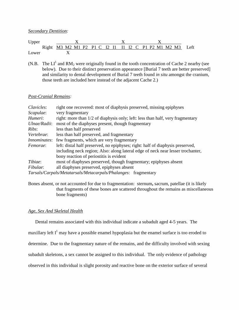
Secondary Dentition:
Upper X X X
Right M3 M2 M1 P2 P1 C I2 I1 I1 I2 C P1 P2 M1 M2 M3 Left
Lower X
(N.B. The LI1 and RM2 were originally found in the tooth concentration of Cache 2 nearby (see
below). Due to their distinct preservation appearance [Burial 7 teeth are better preserved]
and similarity to dental development of Burial 7 teeth found in situ amongst the cranium,
those teeth are included here instead of the adjacent Cache 2.)
Post-Cranial Remains:
Clavicles: right one recovered: most of diaphysis preserved, missing epiphyses
Scapulae: very fragmentary
Humeri: right: more than 1/2 of diaphysis only; left: less than half, very fragmentary
Ulnae/Radii: most of the diaphyses present, though fragmentary
Ribs: less than half preserved
Vertebrae: less than half preserved, and fragmentary
Innominates: few fragments, which are very fragmentary
Femorae: left: distal half preserved, no epiphyses; right: half of diaphysis preserved,
including neck region; Also: along lateral edge of neck near lesser trochanter,
bony reaction of periostitis is evident
Tibiae: most of diaphyses preserved, though fragmentary; epiphyses absent
Fibulae: all diaphyses preserved, epiphyses absent
Tarsals/Carpals/Metatarsals/Metacarpals/Phalanges: fragmentary
Bones absent, or not accounted for due to fragmentation: sternum, sacrum, patellae (it is likely
that fragments of these bones are scattered throughout the remains as miscellaneous
bone fragments)
Age, Sex And Skeletal Health
Dental remains associated with this individual indicate a subadult aged 4-5 years. The
maxillary left I1 may have a possible enamel hypoplasia but the enamel surface is too eroded to
determine. Due to the fragmentary nature of the remains, and the difficulty involved with sexing
subadult skeletons, a sex cannot be assigned to this individual. The only evidence of pathology
observed in this individual is slight porosity and reactive bone on the exterior surface of several
long bones (i.e., along neck of right femur near lesser trochanter region), which indicates a
general periostitic infection.
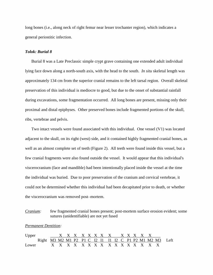
Tolok: Burial 8
Burial 8 was a Late Preclassic simple crypt grave containing one extended adult individual
lying face down along a north-south axis, with the head to the south. In situ skeletal length was
approximately 134 cm from the superior cranial remains to the left tarsal region. Overall skeletal
preservation of this individual is mediocre to good, but due to the onset of substantial rainfall
during excavations, some fragmentation occurred. All long bones are present, missing only their
proximal and distal epiphyses. Other preserved bones include fragmented portions of the skull,
ribs, vertebrae and pelvis.
Two intact vessels were found associated with this individual. One vessel (V1) was located
adjacent to the skull, on its right (west) side, and it contained highly fragmented cranial bones, as
well as an almost complete set of teeth (Figure 2). All teeth were found inside this vessel, but a
few cranial fragments were also found outside the vessel. It would appear that this individual's
viscerocranium (face and mandible) had been intentionally placed inside the vessel at the time
the individual was buried. Due to poor preservation of the cranium and cervical vertebrae, it
could not be determined whether this individual had been decapitated prior to death, or whether
the viscerocranium was removed post–mortem.
Cranium: few fragmented cranial bones present; post-mortem surface erosion evident; some
sutures (unidentifiable) are not yet fused
Permanent Dentition:
Upper X X X X X X X X X X X X X
Right M3 M2 M1 P2 P1 C I2 I1 I1 I2 C P1 P2 M1 M2 M3 Left
Lower X X X X X X X X X X X X X X X X

Post–Cranial Remains:
Scapulae: highly fragmented
Humeri: fragmentary, with post-mortem surface erosion evident; all epiphyses missing
Ulnae and Radii: all epiphyses missing
Carpals: some fragments present
Metacarpals and Hand Phalanges: some fragments present
Ribs: few extremely fragmented ribs
Vertebrae: less than half present; atlas preserved; others very fragmentary
Innominates: very fragmentary and poorly preserved; portions of ischium and body of ilium
present; no sciatic notch, iliac crest or pubic symphyses
Sacrum: very fragmentary
Femorae: both femoral heads present; condyles missing; approximate total length of right
femur (minus distal epiphyses) is 38cm; gracile muscle markings
Tibiae: both missing proximal and distal epiphyses; post-mortem surface erosion evident
Fibulae: fragmentary; most of diaphyses present but missing epiphyses; possible total
length of left fibula is approximately 30cm
Tarsals: some fragments present; left navicular and calcaneous identified, both observed
with degenerated surfaces (appearing porous) - probably due to preservation
Metatarsals and Foot Phalanges: some fragments present
Bones absent, or not accounted for due to fragmentation: clavicle, sternum, patellae
Age, Sex And Skeletal Health
Examination of the skeletal remains from Tolok Burial 8 indicates a young adult female of
approximately 17 to 26 years. This is based on complete eruption of the permanent dentition; the
open state of endocranial sutures; and the lack of age–related dental attrition or skeletal
degeneration. No evidence of skeletal pathology, trauma, or infection is observed, but dental
remains have slight to medium calculus deposits, enamel hypoplasia, hypocalcification staining
and carious lesions.
In this individual, most teeth exhibit a single prominent hypoplastic groove of acute severity,
but there is extensive hypoplasia on the right mandibular M3 and broad shallow bands
characterize a maxillary left canine and mandibular left P2. The positions of these defects
indicate that a period of ill health occurred sometime between 3 and 3.5 years of age.

Furthermore, the wide band on the maxillary left canine suggests that this same period was an
extended period of ill health lasting approximately four months (following Song 1997: Table
6.2.3).
Additionally, the right M1 and M2 have areas of beige/cream coloured hypocalcification
staining, which is a dental pathology resulting from insufficient enamel maturation. The right
M2, like the left M2, also has several very small interfissure carious pits on the occlusal surface.
The posterior surfaces of both femurs and fibulae appear to have localized patches of
porosity, possibly indicating mild periostitis, but it is also likely that post-mortem diagenesis may
be the culprit of this “pathology”. No pathological lesions are identifiable on any of the humeri,
ulnae, radii, vertebrae, innominates and tibiae, but one must note their fragmentary state.
Tolok: Burial 9
The remains of four subadult individuals were interred in this single Late Preclassic stone-
lined cist grave. Typical of burials at Tolok, as well as other localities throughout Cahal Pech,
the Primary Individual was fully extended, prone, and arranged along a north-south axis, with the
head to the south (Figure 3). Partial remains of the Second Individual were found concentrated
around the upper torso and shoulder areas, while the remains of a Third Individual were
concentrated in the cranial region, and those of a Fourth Individual were discovered around the
right shoulder of the Primary Individual.
Interestingly, the Primary Individual lacked both legs and feet, and it appeared severed at the
waist (see Figure 3). Measurement of this individual in the field, from the superior cranial region
to the distal pelvic extent, gave a length of approximately 47 cm. The absence of this child's
lower body, which was nonetheless capped with stones where the legs should have been,
suggests that some sort of skeletal interference or “mutilation” occurred. However, it cannot be

confirmed whether the individual’s legs were defleshed prior to burial (when the body was still
“fresh”), or plundered and dismembered afterward, when skeletalization had already occurred.
Overall, preservation of most skeletal elements for the Primary Individual is good to very
good, although fragmentary. Field observations indicate that most of the cranium is represented.
Mainly because of its young age, long bones that are preserved consist of the diaphyses only.
Recovered long bones include both humeri (the right being very fragmented), and both ulnae and
radii. Partial remains of both clavicles are also present. Fortunately, most ribs and cervical
vertebrae are preserved, which makes them (ribs) available for chemical analysis (see White et
al., this volume). The innominates are represented by poorly preserved fragments of the iliac
blades only, as the skeleton was “mutilated” at this junction.
Cranium: other than fragmentary remains of the calvaria (frontal, parietals, occipital),
remains of the cranium also include fragments of alveolar maxillae and some
mandibular fragments, all with associated teeth, as well as numerous loose teeth;
orbital fragments do not exhibit any evidence of cribra orbitalia
In situ appearance of the skull of the Primary Individual indicated that it was slightly
modified (Figure 4). The pseudocircular cranial modification involved vertical occipital
deformation, as well as frontal flattening or sloping. Evidence suggests that the skull
deformation was antemortem, as opposed to disfigurement due to postmortem interment
pressure. Such cranial modification could be formed by binding padded boards or cloth
bandages around the skull during early childhood growth (Stewart 1974).
The following teeth belong to the Primary Individual (as indicated by maxillary and
mandibular fragments):

Primary Dentition:
Upper x x x x x x x x x x
Right m2 m1 c i2 i1 i1 i2 c m1 m2 Left
Lower x x x x x x x x x
Secondary Dentition:
Upper X X X X X X X X X X X
Right M3 M2 M1 P2 P1 C I2 I1 I1 I2 C P1 P2 M1 M2 M3 Left
Lower X X X X X X X X X X X X X
Dental development of these teeth suggests an age range of 2-5 years [3-4 (+ 12 mths)]
(Ubelaker 1989: Fig. 71). However, in examining the fragmented arm bones, estimated total
lengths suggest a younger age, i.e., 2 years (see Ubelaker 1989: Table 14). After considering the
fact that teeth often develop at an advanced rate and, in particular, develop earlier among North
American indigenous groups (Ubelaker 1989), the age range for this child is placed at between 2-
4 years.
The following teeth, which were primarily found in a clump on the east (left) shoulder of the
primary child (with some associated cranial remains), are present for the Second Individual:
Primary Dentition:
Upper x
Right m2 m1 c i2 i1 i1 i2 c m1 m2 Left
Lower
Secondary Dentition:
Upper X X X
Right M3 M2 M1 P2 P1 C I2 I1 I1 I2 C P1 P2 M1 M2 M3 Left
Lower
(A deciduous lower left canine was also found outside of the south wall associated with this
burial, and may belong to this individual.)

For this Second Individual, levels of crown and root development of the permanent dentition
suggest an age range of 4-5 years. Estimated total long bone lengths also concur with an age of
approximately 4 years.
Outside of dental remains, this child is represented by both humeri, one ulna, both femurs,
and a left tibia. The relative sizes of the leg bones indicate that they could not belong to the
younger Primary Individual (already described). The long bones of the Second Individual were
placed along the corresponding sides of the Primary Individual’s upper torso and arm region
(i.e., left arms and legs of the Second Individual were placed alongside the left arm of the
Primary Individual). From the in situ appearance, it can be tentatively suggested that the arms
and legs of the second child were disarticulated from its body prior to deposition on the main
child. At this time, no other identified bones support a complete skeletal state for this Second
Individual.
Next, a Third Individual is represented by the following dentition, which was recovered
scattered among, and south of (“above”), the cranium of the Primary Individual:
Primary Dentition:
Upper x x x x x x
Right m2 m1 c i2 i1 i1 i2 c m1 m2 Left
Lower x x x x
The age of this infant is approximately one year, based on dental development, and since
most teeth were only unerupted crowns, it is suggested that this child was originally represented
by at least a cranium. Low preservation potential of infant remains explains the lack of cranial
bones.
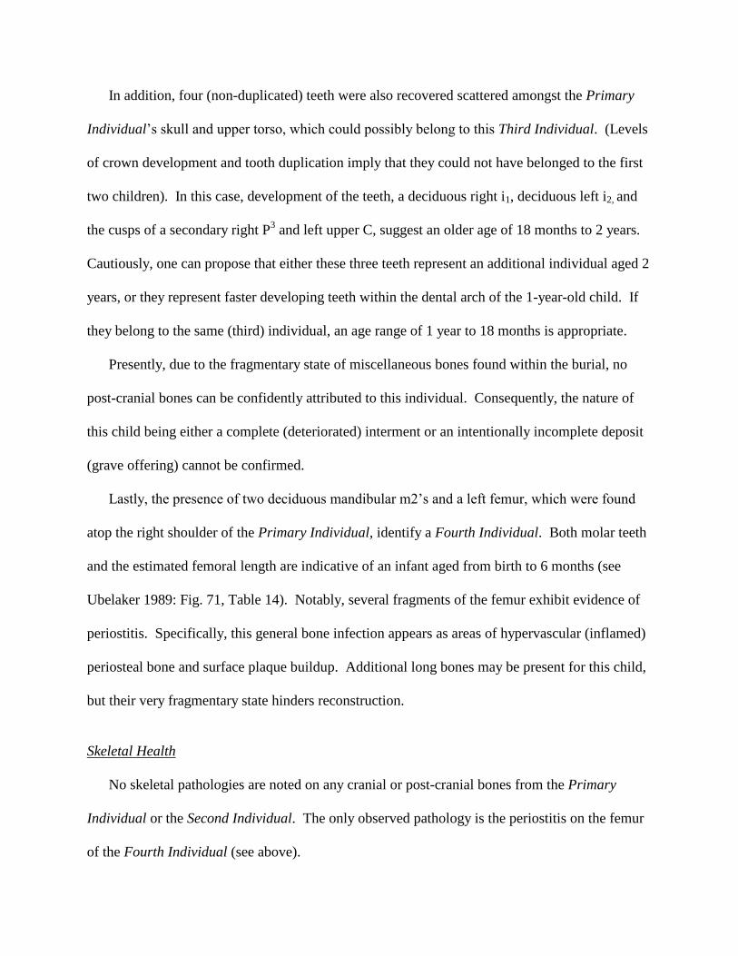
In addition, four (non-duplicated) teeth were also recovered scattered amongst the Primary
Individual’s skull and upper torso, which could possibly belong to this Third Individual. (Levels
of crown development and tooth duplication imply that they could not have belonged to the first
two children). In this case, development of the teeth, a deciduous right i1, deciduous left i2, and
the cusps of a secondary right P3 and left upper C, suggest an older age of 18 months to 2 years.
Cautiously, one can propose that either these three teeth represent an additional individual aged 2
years, or they represent faster developing teeth within the dental arch of the 1-year-old child. If
they belong to the same (third) individual, an age range of 1 year to 18 months is appropriate.
Presently, due to the fragmentary state of miscellaneous bones found within the burial, no
post-cranial bones can be confidently attributed to this individual. Consequently, the nature of
this child being either a complete (deteriorated) interment or an intentionally incomplete deposit
(grave offering) cannot be confirmed.
Lastly, the presence of two deciduous mandibular m2’s and a left femur, which were found
atop the right shoulder of the Primary Individual, identify a Fourth Individual. Both molar teeth
and the estimated femoral length are indicative of an infant aged from birth to 6 months (see
Ubelaker 1989: Fig. 71, Table 14). Notably, several fragments of the femur exhibit evidence of
periostitis. Specifically, this general bone infection appears as areas of hypervascular (inflamed)
periosteal bone and surface plaque buildup. Additional long bones may be present for this child,
but their very fragmentary state hinders reconstruction.
Skeletal Health
No skeletal pathologies are noted on any cranial or post-cranial bones from the Primary
Individual or the Second Individual. The only observed pathology is the periostitis on the femur
of the Fourth Individual (see above).

With respect to dental health, all teeth are relatively free of caries and calculus, but several
teeth belonging to the Primary Individual and the Second Individual exhibit evidence of linear
enamel hypoplasia. In the primary child, hypoplastic lesions on permanent incisors and canines
indicate that minor stresses occurred at approximately 2.8 years, 3.5 years and sometime between
4 and 4.2 years of age. Since crowns are incompletely developed, the ages of defect formation
are extrapolated from mean unworn crown heights of Maya from Altun Ha and regression
equations determined for such teeth (see Song 1997: Table 6.2.3; Goodman and Song 1999:
Table 9.5).
For the Second Individual, aged 4-5 years, lesions on the permanent upper left I1 and I
2
indicate that two periods of ill health or dietary stress occurred, namely at 3.7 and 4 years of age.
Tolok: Burial 10
For this single adult interment, dating to the Middle to Late Preclassic transition, skeletal
preservation is good, with most bones present. Specifically, most cranial bones are preserved, as
are all long bones, most of the innominates, sacrum, hands, and feet. Ribs and vertebrae are
fragmentary and very incomplete. In situ measurement of this individual in the field was
approximately 153 cm, from the superior cranial region to the most distal point on the left tibia
(since remains of the feet were disturbed).
Cranium: remains are very fragmentary; most sutures appear unfused or partially fused (i.e.,
some parts of the coronal and sagittal sutures), but the coronal suture of the left
temple region (above sphenoid) is closed and obliterated; no evidence of pathology
The following Secondary Dentition was recovered from Burial 10:
Upper X X X X X X X X X X X X X X X X
Right M3 M2 M1 P2 P1 C I2 I1 I1 I2 C P1 P2 M1 M2 M3 Left
Lower X X X X X X X X X X X X X X

Accompanying the teeth, most of the maxillary alveolar region is well preserved, as is the
intact mandible. From the appearance of alveolar bone in the mandibular right M1 and left M2
regions, it seems that bone resorption, and possibly infection around the right M1, took place
some time antemortem when the teeth were lost. In particular, the reactive bone growth that
partially filled the socket of the right mandibular M1 appears lumpy and porous (Figure 5). This
mass of bone spicules fills 1/2 to 2/3 of the socket depth.
On the other hand, the socket of the absent left M2 appears relatively free of bone resorption,
except for minimal bone spicule formation and some porosity. The comparative socket bone
growth suggests that the right M1 was lost earlier in life than the left M2. Outside of these
alveolar portions, alveolar resorption (and periodontitis?) is also apparent along the rest of the
labial border of the mandible. This is evident as eroded areas of porous bone surrounding the sockets.
Judging from the mandibular remains, early antemortem tooth loss was not unheard of for
ancient Maya. As also demonstrated by other remains at Tolok (Song et al. 1994), tooth loss
often afflicted relatively young individuals, and it can be considered characteristic of the
population. As such, resorbed mandibles suggesting advanced age often conflict with non-dental
indicators of younger age (see Song et al. 1994: 153). In the Maya case, the nature of maize
processing and consequent ingestion of grit can be cited for such antemortem loss.
Examination of dental calculus reveals slight deposits on most teeth (molars, premolars,
canines, some incisors), while the maxillary right I1
and I2
have moderate, or medium, deposits
(see Brothwell 1981: Fig. 6.14).
Significantly, linear enamel hypoplasias are evident on most teeth and they appear as a single
(narrow) groove per tooth. Specifically, the enamel defects are observed on mandibular left I1, I2,
C, M1; mandibular right I1, I2, C; maxillary left I1, I
2, C, M
1; and maxillary right I
1, I
2, M
1. Like

data from other hypoplasia studies (Goodman and Armelagos 1985; Skinner and Goodman 1992;
Song 1997), prominent grooves are apparent on the canines, particularly the mandibular canines.
Using Song's (1997) regression formulae for Altun Ha Maya, a single significant period of stress
is determined to have occurred around the age of 2.3 to 2.7 years (also see Goodman and Song
1999: Table 9.5).
Finally, carious lesions afflict several teeth. In the RM2, they include a large carious pit (0.5
cm long) along the buccal groove (which might also be a form of developmental defect known as
foramen caecum molare (FCM) [see Capasso and Di Tota 1992]), which penetrates into the
centre of the tooth, as well as one small interfissure/occlusal pit. Similarly, the LM1 has a small
FCM-like buccal pit which may be a carie, in addition to a small interfissure/occlusal pit, and the
LM2 has a midline pit along the lingual groove which penetrates dentine and measures 2.5-3mm
wide. Lastly, an interfissure pit is evident on the occlusal surface of the RM3, but this may be
due to preservation factors.
Post-Cranial Remains:
Scapula: left one fragmented
Humeri: left one mostly present (estimated total length of 28-29cm) and has large septal
aperture (foramen): 0.8cm wide; right one lacks both epiphyses
Ulnae/Radii: fragmentary, poorly preserved epiphyses (left ulna: estimated total length 19.5cm)
Carpals/Metacarpals/Hand Phalanges: most preserved
Ribs: most preserved, though fragmented
Vertebrae: most preserved, though fragmented
Innominates: both elements in good shape, but lacking pubic symphyses
Sacrum: mostly present, though fragmented
Femorae: both mostly preserved, though fragmented; right: total length of 38-39cm
suggests a living stature range of 144.44 to 152.07 cm
Patella: 1/4 of right one recovered
Tibiae: both mostly preserved, but lacking distal epiphyses
Fibulae: both mostly preserved, though lacking well-preserved epiphyses
Tarsals/Metatarsals/Foot Phalanges: 12 or more fragments
Bones absent, or not accounted for due to fragmentation: both clavicles, right scapula, left patella

Age, Sex And Skeletal Health
Several skeletal features were examined for this individual. Firstly, the dentition indicates
that the individual was a young adult. This is based on the eruption and crown/root development
of the third molar, which is present for all four quadrants of the dental arch. Importantly, it
should be noted that the third molar is an extremely variable tooth when it comes to eruption age.
According to Ubelaker (1989: Fig. 71), the period of M3 development and eruption is usually
between 15-21 years. Following Ubelaker, this individual’s age is placed closer to 21 years.
Also, with respect to dental indicators, attrition was assessed to determine relative age.
Although attrition is known to be variable between and within populations, it can be examined
comparatively with other skeletal age markers to suggest youth or old age. In this case, the
minimal wear on the mandibular molars, which exhibit attrition of the cuspal tips only, suggest
the third stage of wear for M1, according to Brothwell (1981: Fig. 3.9). This stage of wear
characterizes an age range of 17-25 years. Keeping in mind that the Maya maize diet readily
erodes enamel surfaces, the minimal wear of this individual’s teeth corresponds well with a
young adult age.
Similarly, in examining the iliac crest of the left innominate, the stage of epiphyseal fusion
suggests an age in the 16-23 year range of fusion times (Brothwell 1981: Fig. 3.4). Thus, with
the limited evidence, an age range of approximately 17-23 years is suggested for the individual
from Tolok Burial 10, with 21 years being a reasonable estimate.
With respect to sex, the appearance of the cranial remains and innominates suggest that the
individual in Burial 10 was female. In particular, the gracility of the skull, as well as other (long)
bones, and the obtuse angle of the sciatic notch of the innominates point to a female sex.

Other than the dental hypoplasia and possible periodontitis noted above, no evidence of
pathology is observed in the skeletal remains, except among some vertebrae. In this case, the
inferior and superior surfaces of some lumbar vertebral bodies appear pitted and eroded, with
raised regions of bumpy bone. While these lesions do not resemble “normal” osteoarthritic
lipping of the vertebral bodies, it is likely that they represent an arthritic reaction.
Tolok: Cache 2
In addition to the multiple burials excavated during the 1993 field season, a tooth cache
similar to that excavated at Yakalche in northern Belize (Pendergast et al. 1968) was found
associated with Burial 7 at Tolok. The cache consists of one small jadeite bead and fourteen
teeth of mixed dentition, which were all concentrated in a small isolated scatter approximately 60
cm south of the grave (see Powis 1994: Fig. 8).
Based on comparable dental development and the proximity of this cache to Tolok Burial 7
(noted above), two teeth, a lower M2 and an extra LI1, are attributed to the child from this burial
and are not accounted for here. Minimally, Cache 2 teeth represent the dentition of one child.
Since there is no evidence to indicate distinct individuals, it is assumed that these teeth belong to
the same individual. Compared to the teeth from Burial 7, the teeth from this cache are poorly
preserved and appear highly eroded. The deciduous teeth have moderate wear. No pathologies
can be identified.
The remaining 12 teeth from Cache 2 include:
Primary Dentition:
Upper x x x
Right m2 m1 c i2 i1 i1 i2 c m1 m2 Left
Lower x x x

Secondary Dentition:
Upper X X X X
Right M3 M2 M1 P2 P1 C I2 I1 I1 I2 C P1 P2 M1 M2 M3 Left
Lower X X
Crown and root development of these teeth indicate an age of approximately 3-4 years.
Dental development of these teeth suggests that most of the teeth in Cache 2 cannot belong to the
child from Burial 7, which is about one year older. If there is some tooth overlap, only the
deciduous teeth could possibly belong to Burial 7. Here, the secondary teeth, which lack roots
and dental attrition, indicate that the teeth were removed from the mandible and maxilla after
skeletalization of the body had occurred.
This cache, associated with Burial 7, which has been dated to the Late Preclassic (Powis
1994), represents one of the earliest tooth caches recorded thus far for the entire Maya region.
The tooth cache at Yakalche is dated to the late Post–Classic period (thirteenth to fifteenth
centuries A.D.) and consisted of 379 teeth and a “small, irregular subglobular jadeite bead”
(Pendergast et al. 1968: 638). To the best of the author’s knowledge, the only other reported
Maya tooth cache derives from Lubaantun and dates to the Classic period (Hammond 1975). A
total of 56 teeth were found in a house mound at this site and they were “within an area so small
as to suggest that they were buried in a container of some perishable material” (Hammond,
personal communication to F.P. Saul, in Saul 1975: 389). This may possibly be the case for the
Preclassic Tolok tooth cache as well.
To explain the significance of this tooth cache, one can look to interpretations of the tooth
cache at Yakalche (Pendergast et al. 1968) for possible relevance to the Tolok situation. J.E.S.
Thompson, in a personal communication to Pendergast and colleagues (1968: 642), associated
the tooth offering at Yakalche with evidence of child sacrifice to Itzamna, or God D. This god,

like the other two Old Gods (Gods L and N), is often represented as toothless except for a single
molar at each corner of the mouth.
The Old Gods are thought to have presided over Xibalba in Maya cosmology (see Schele and
Miller 1986), and they were well–observed religious figures in the Maya area. Pendergast and
colleagues (1968) have suggested that children's teeth were removed in order to give them a
greater resemblance to Itzamna, prior to their sacrifice in his name. The teeth may have served as
a secondary offering to the deity, or as an offering to any one of the Old Gods, considering their
similar toothless depictions.
Tolok: Cache 3
Dating to the Terminal Preclassic period (Powis and Hohmann 1995), this cache consists of
the cranial remains of a subadult individual placed within a ceramic vessel (Figure 6). In situ
observations indicate that the skull was placed in the vessel as an intact whole. The evidence
suggests that decapitation of the individual may have taken place while flesh was still present
(although whether it was the cause of death, or it occurred shortly thereafter, cannot be
determined), but it is curious that no remains of a mandible were found. However, the remains
of mandibular dentition do indicate its original inclusion with the skull.
Cranium: after cleaning, it appears that the crown (superior surface) of the skull rested on the
bottom of the vessel. Generally, cranial remains are fragmentary. While the frontal
and parietals are well preserved, occipital and temporal remains are very
fragmented. All observable sutures are fully open. No pathological features are
detected on any cranial remains, but one must note the fragmentary nature of the
bones and their differential preservation
The following teeth were recovered from the vessel, which all belong to the same individual:

Primary Dentition:
Upper x x x x x x x x x
Right m2 m1 c i2 i1 i1 i2 c m1 m2 Left
Lower x x x x x x x x
Secondary Dentition:
Upper X X X X X X X X X X X X X
Right M3 M2 M1 P2 P1 C I2 I1 I1 I2 C P1 P2 M1 M2 M3 Left
Lower X X X X X X X
Age, Sex And Skeletal Health
Based on the secondary tooth crowns and some partial root development, the age of this
individual can be placed at between 4-5 years. Once again, due to the subadult age of this
individual, sex is indeterminate. As noted above, there is no pathology in the cranial remains.
From the dental remains, other than a small carious pit found on the mesial crown surface of
the upper left deciduous m1, the only apparent irregularities are the hypoplastic bands present on
six secondary tooth crowns. Measurements of these bands relative to the occlusal edge (since
their cemento-enamel junctions are not yet developed) indicate that moderate stresses affected
this child at 3 years of age, and again at around 4 years (following Song 1997: Table 6.2.3;
Goodman and Song 1999: Table 9.5).
Zotz: Burial 91-1
This late Middle to early Late Preclassic interment from within the round structure (Str. 2) at
Zotz was a cist burial containing the fragmentary remains of an extended individual aligned
north-south with its head to the south. Although poorly preserved, it is thought that this adult
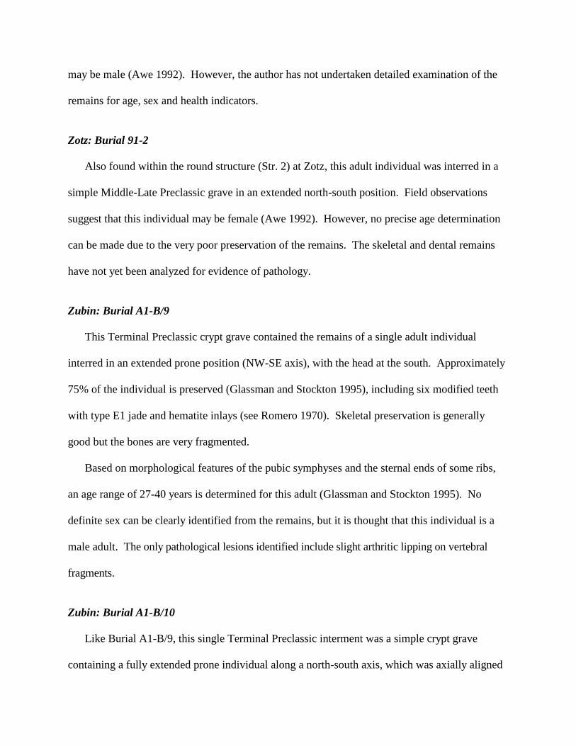
may be male (Awe 1992). However, the author has not undertaken detailed examination of the
remains for age, sex and health indicators.
Zotz: Burial 91-2
Also found within the round structure (Str. 2) at Zotz, this adult individual was interred in a
simple Middle-Late Preclassic grave in an extended north-south position. Field observations
suggest that this individual may be female (Awe 1992). However, no precise age determination
can be made due to the very poor preservation of the remains. The skeletal and dental remains
have not yet been analyzed for evidence of pathology.
Zubin: Burial A1-B/9
This Terminal Preclassic crypt grave contained the remains of a single adult individual
interred in an extended prone position (NW-SE axis), with the head at the south. Approximately
75% of the individual is preserved (Glassman and Stockton 1995), including six modified teeth
with type E1 jade and hematite inlays (see Romero 1970). Skeletal preservation is generally
good but the bones are very fragmented.
Based on morphological features of the pubic symphyses and the sternal ends of some ribs,
an age range of 27-40 years is determined for this adult (Glassman and Stockton 1995). No
definite sex can be clearly identified from the remains, but it is thought that this individual is a
male adult. The only pathological lesions identified include slight arthritic lipping on vertebral
fragments.
Zubin: Burial A1-B/10
Like Burial A1-B/9, this single Terminal Preclassic interment was a simple crypt grave
containing a fully extended prone individual along a north-south axis, which was axially aligned

with Structure A1. The head was also placed at the south end, like all other Cahal Pech
Formative period burials.
Less than 10% of the skeleton is preserved (Glassman and Stockton 1995). Dental remains
(22 teeth) indicate a young child between 4 and 5 years of age. No pathologies are observed, but
one must note the poor preservation of the remains. Sex is indeterminate.
Zubin: Burial C9-B/1a
Due to looters' disturbance of this Middle Preclassic multiple interment prior to formal
excavation, the orientation of the primary individual could not be determined. In general,
preservation is fair to good. Examination by S. Schwake (personal communication, 1997)
deemed this individual to be an adult of male sex (Iannone 1995: 46).
Specifically, this individual can be aged to a young adult, i.e., early 20's, from the state of the
cranial sutures. In particular, the sagittal suture is clearly open and unfused in the lambda region,
while all of the lambdoidal and squamosal sutures are also unfused. According to Montagu
(1960), these regions start to fuse together in the mid-to-late twenties. The fusion lines still
evident in vertebral bodies also reflect a young adult age.
From the author's personal observations, there seems to be slight cranial deformation in this
individual, in the form of flattened occipital bones and some bulging of the right parietal towards
the occipital. However, the fragmentary state of the remains precludes a firm statement at this time.
Regarding pathology, the most serious evidence is in the cranial remains. Pitting across most
of the posterior and sagittal regions of the right parietal, as well as a thinned outer layer, suggest
possible porotic hyperostosis. In addition, circular lesions characteristic of osteomyelitis are
observed on parietal fragments, with ante-mortem remodeling also evident (S. Schwake, personal
communication, 1997).

The following Secondary Dentition belongs to Individual C9-B/1a:
Upper X X X X X X X X X X X X X X X X
Right M3 M2 M1 P2 P1 C I2 I1 I1 I2 C P1 P2 M1 M2 M3 Left
Lower X X X X X X
In general, the teeth exhibit medium to very heavy calculus deposits, with moderate dental
attrition. Additionally, moderate to severe enamel hypoplasia (and pitting) is evident on
maxillary central incisors, canines and first molars, and mandibular first molars (Figures 7a,b).
The positions of these lesions indicate an extended period of ill health between 1.2 and 2.3 years
of age. Dental attrition is moderate and there is slight to very heavy deposits of calculus.
Zubin: Burial C9-B/1b
The second individual in this “simple” Middle Preclassic interment was positioned in a north-
south alignment, probably extended, with its head at the north end facing west. Unlike most
burials at Cahal Pech, this individual was buried in a supine position, lying on its back. Skeletal
preservation is good, but very fragmentary, and most of the lower body was destroyed by looter
activity. An adult age range is discerned from the remains, but sex is indeterminate.
Notable skeletal features include gracile long bones (i.e., left radius); preservation of distinct
fusion lines of the iliac crest (innominates), suggesting young adult age; and slight arthritic
lipping of the head of a distal phalanx.
Recovered teeth include a maxillary RP3 and a left (?) I1. The mandibular I1 has slight to
medium calculus deposits, medium to heavy dental attrition and possible hypocalcification
staining of the labial enamel.
Finally, traumatic injury, with subsequent healing, is observed in one unidentified long bone
fragment, and it appears as a thick underlying cortex of bony regrowth over the fracture (S.
Schwake, personal communication, 1997). In addition, one vertebral fragment has marked
curvature, possibly due to a crushing force related to workload stress (S. Schwake, personal
communication, 1997).
Preclassic Period Skeletal Remains at Pacbitun
Pacbitun: Burial 1-5
This individual was recovered from a Late Preclassic simple, slab-lined crypt, on the central
axis, deep within Structure 1 at Pacbitun. The burial was likely extended, aligned north-south,
with head to the south. It was not possible to determine if the body lay prone or supine, nor the
precise orientation of the limbs. Preservation was poor.
Cranium: large number of cranial fragments which, unfortunately, are very small; includes
15 cranial vault fragments, petrous portion of temporal bone, and inion region of
occipital; one fragment of the mandible includes mental protuberance
Permanent Dentition:
Upper X X X X
Right M3 M2 M1 P2 P1 C I2 I1 I1 I2 C P1 P2 M1 M2 M3 Left
Lower
N.B. The left upper canine originally had a circular inlay measuring 3 mm in diameter (lost).
Post-Cranial Remains:
Ulnae and Radii: proximal end of right radius preserved
Metacarpals/Hand phalanges: distal and medial hand phalanges identified
Femorae: portions of femoral heads and shafts preserved
Age, Sex and Skeletal Health
Analysis by Helmuth (1988) identified heavy dental calculus deposition, caries, and
indications of osteoarthritis. The size of the femoral head (47mm diameter) and femoral wall

thickness point strongly to a male sex. Considering the dental calculus, arthritis, loss of
mandibular teeth and high caries frequency, this male individual must have been over 40 years
old at death. It is likely, but cannot be proven, that he was around 50, or even 60, years of age.
Finally, one distal hand phalanx bore indications of arthritis, probably caused by a previous
fracture, while the medial hand phalanx showed evidence of a healed fracture (Helmuth 1988).
Pacbitun: Burial C-1
This single Middle Preclassic period burial at Pacbitun consisted of an adult individual buried
in a stone-lined cist or partial crypt grave beneath the floor of Plaza C in the site core (Arendt et
al. 1996). The burial was aligned 600 east of north, with the upper body extended and prone.
The knees were tightly flexed upward against the thighs, which is similar to the burial position of
Cas Pek Burial 94-2 (Figure 8).
Overall, preservation was fair to poor. Post-mortem root disturbance especially wrecked
havoc with the chest and pelvic areas, which, together with the absent cranial remains, prevent an
assessment of sex. Most of the cranium could not be located due to disturbance and damage
from much later (Classic period) placement of a large, uncarved stela adjacent to Pacbitun Burial
C-1 (see Figure 8).
Remains of the mandible appear resorbed, with the body reduced in height. Antemortem
tooth loss of at least four molar teeth is noted. This resulted in almost complete bone resorption
of the left M1 socket; complete bone overgrowth of the left M2; and partial (approximately 1/3)
bony spicule formation in the sockets of both third molars. As a result of the loss of the left M1
and M2, more alveolar resorption and reduced body height is apparent in left mandibular
fragments, when compared to the right.

However, while the mandible appears relatively gracile, the chin is noticeably pronounced,
squarish and knobby at the processes.
The following Secondary Dentition was present for Pacbitun Burial 95-1:
Upper X X
Right M3 M2 M1 P2 P1 C I2 I1 I1 I2 C P1 P2 M1 M2 M3 Left
Lower X X X X X X X X X X X
Calculus deposits are slight in amount and dental attrition is slight to moderate.
Post-Cranial Remains:
Scapulae, Ribs and Vertebrae: very fragmentary
Arm and Leg Long Bones: good condition, but most epiphyses not preserved
Left Patella: only proximal half preserved
Hand and Foot Bones: most bones well preserved
Bones not preserved or accounted for due to fragmentation: innominates, sacrum, clavicles,
sternum.
Age, Sex And Skeletal Health
Due to the very poor preservation of pelvic remains, and inaccessibility of the cranium,
positioned near the base of the stela, no reliable skeletal markers are available for age and sex
determination. However, an adult individual aged between 30-40 years can be inferred from
dental remains and their degree of attrition. Admittedly, this latter trait is not as reliable as pelvic
and cranial markers for age estimation, but it is the only criterion available.
For sex determination, a female sex is implied by the relative gracility of all long bones, but
the nature of mandibular features suggest a male sex (of slight body dimensions). At this time,
Pacbitun Burial 95-1 can only be described as an adult individual of indeterminate sex.
Regarding skeletal pathology, arthritis appears to afflict one middle toe phalange, with bony
growths evident on the distal end (head) of the phalange.

From the dental remains, linear enamel hypoplasias are observed on mandibular canines and
premolars. A brief period of minor ill health is determined to have occurred sometime between 4
and 5 years of age. Antemortem tooth loss of at least four molars, and possibly six or more teeth,
is also noted.
Carious lesions are evident on several molars. In maxillary teeth, the right M2 (possible M
3)
has an interfissure carie on the occlusal surface that penetrates the dentine and is 1.74 mm in
diameter. Similarly, the left M2 (possible M
3) has a large occlusal carie through the dentine
measuring 4.15 mm at its widest point. In the mandibular right M1, an eroded region measuring
4.45 mm across is observed on the mesial crown surface near the cemento-enamel junction, and
another small carious pit afflicts the buccal surface along the midline (buccal groove). Finally, in
the mandibular right M2, a large interfissure occlusal carie is evident on the lateral half of the
crown, extending through the dentine into the root, and measuring 3.70 mm wide. A localized
carious pit is also apparent on the buccal groove, which measures 2.92 mm wide and penetrates
the dentine.
Skeletal Indicators of Preclassic Maya Health
While the state of skeletal preservation and fragmentation hinders clear assessments of
skeletal health for many of the Preclassic individuals identified at Cahal Pech and Pacbitun,
remains for some are sufficient to observe the presence of various skeletal pathologies (Table 2).
In particular, porotic hyperostosis, a cranial inflammation due to an anemic state, is observed
in Cas Pek Burial 94-2. This condition, which could either reflect dietary anemia or the anemic
response of infectious disease (Macadam and Kent 1992), is evident as areas of pitted, porous
bone on the outer cranial surface of the parietals, in addition to a widening of the inner diploe

layer. It is perhaps noteworthy that there is not more evidence for spongy or porotic hyperostosis
at either Cahal Pech or Pacbitun. Saul and Saul (1991:156) note a similar scarcity at Cuello
during the Preclassic. This would seem to suggest a better level of health and nutrition among
the Maya in the Preclassic period than during the Classic period, when this condition is much
more common.
Cranial pathology is also evident in remains from Zubin Burial C9-B/1b, whose parietal
bones display circular osteomyelitic lesions and evidence of ante-mortem bone remodeling (S.
Schwake, personal communication, 1997).
With regards to bone infection, mandibular fragments from Tolok Burial 10 and Cas Pek
Burial 94-2 indicate possible periodontitis infection. This condition appears as regions of
alveolar bone resorption and porous bone surrounding the tooth sockets, in association with
antemortem loss of mandibular molars. General bone infection is also observed as periostitis in
the long bones of Cas Pek Burial 91-4 (over 40 years), Tolok Burial 9d (aged 0-6 months), and
Tolok Burial 7 (aged 4-5 years).
While arthritis (or osteoarthritis) is known from many Classic and Postclassic period Maya
skeletal specimens, it has been less well documented for the Preclassic (Saul and Saul 1991:152).
At Cahal Pech and Pacbitun, several adult vertebrae and feet show signs of “normal”
osteoarthritic lesions that most likely simply represent work-related and age-related changes in
these mobile areas (e.g., Zubin Burial A1-B/9, Pacbitun Burial 1-5 and C-1).
On the other hand, evidence of significant vertebral pathology is detected in the remains from
Cas Pek Burial 91-4. In this case, it is likely that the individual suffered from osteoporosis, an
age-related degenerative condition of bone demineralization.

Finally, traumatic injury is observed in remains from Cas Pek Burial 91-4, Zubin Burial C9-
B/1b, and Pacbitun Burial 1-5. In all cases, the individuals survived the damaging incident for
some time after the occurrence, with evidence of bony re-growth over the fractures.
Table 2 Skeletal Pathologies Observed in the Preclassic Sample
BURIAL PATHOLOGY
Cas Pek 91-4 periostitis on several unidentifiable bone fragments
(probably leg bones), fracture of one vertebral body,
osteoporosis of vertebrae
Cas Pek 94-2 porotic hyperostosis, vertebral osteoarthritis (lipping),
possible periodontitis
Tolok Bu-7 periostitis of right femur
Tolok Bu-9d periostitis on left femur - with areas of hypervascular
(inflamed) periosteal bone, and surface plaque buildup
Tolok Bu-10 possible periodontitis infection in mandible
Zubin C9-B/1a cranial osteomyelitis, porotic hyperostosis
Zubin C9-B/1b crushed vertebrae (probably due to workload stress),
healed fracture on one long bone fragment
Zubin A1-B/9 osteoarthritic lipping of vertebrae
Pacbitun Bu 1-5 osteoarthritic lesions on hand phalanx; healed hand fracture
Pacbitun Bu C-1 osteoarthritic lesions on toe phalange
Dental Pathology
Due to the state of preservation of the skeletal material, the most common indicators of stress
are apparent in the dental remains. While teeth may not always accurately reflect disease patterns
in the skeleton, they are considered to be good indicators of general health. In the Maya case,
where bone preservation can be very poor, they are often the only hard tissue remains preserved
well enough for such analysis. At Cahal Pech and Pacbitun, preservation is generally poor to fair
for human remains, with the exception of some well-preserved remains from the Cas Pek and
Tolok settlement groups.
In particular, regarding dental health, the incidence of linear enamel hypoplasia (LEH) is
examined since such defects reflect episodes of chronic, often low grade, stress (Wright 1990).
Health and/or nutritional stress that occur during the period of enamel matrix formation can
result in developmental defects of enamel, namely LEH. Hypoplasias can be manifest as shallow
or deep, wide or narrow, horizontal grooves or bands; small pits arranged generally, or
horizontally, across the enamel surface of tooth crowns; or, as regions of entirely missing enamel
over small or large areas of dentine (FDI 1982: 160). However, the predominant defect types are
lines or grooves.
Enamel defects can result from a multitude of factors, such as malnutrition, fever, vitamin
deficiencies, viral infections, prematurity, hereditary conditions, and localized trauma (Cuttress
and Suckling 1982; Pindborg 1982; Goodman and Rose 1990). In archaeological samples, the
primary cause of enamel hypoplasia will have been systemic metabolic factors, such as
malnutrition and infectious disease (gastrointestinal and respiratory infections).
Among ancient and living Maya, dental enamel hypoplasias are a relatively common
affliction, i.e., more than 50% incidence (Danforth 1986; Infante and Gillespie 1974; Saul and
Saul 1991; Song 1997; Sweeney and Guzman 1966; White 1986; Wright 1994). At Cahal Pech,
a similar situation exists with the dental remains (Maar and Varney 1993; Song 1993, 1995,
1996; Song et al. 1994). Moreover, like other studies, the Cahal Pech remains reflect the
vulnerability of teeth, such as upper central incisors and mandibular canines, which display the
highest frequencies of enamel defects (Goodman and Armelagos 1985; Skinner and Goodman
1992).

Advantageously, hypoplastic enamel defects can be measured in relation to the cemento-
enamel junction (CEJ) of the tooth to determine the age at which stress(es) occurred during the
individual's childhood. Many studies, of both Maya and non-Maya (i.e., Corruccini et al. 1985;
Goodman et al. 1984; Saul 1972; Saul and Saul 1989, 1991; Schulz and McHenry 1975; Song
1995, 1996, 1997; White 1986; among others), find a consistent patterning of defects between
the ages of 2-4 years, which is also the case for the Formative Maya of the Belize Valley.
For many prehistoric populations, this age coincides with the time of weaning. Other than
dietary changes, this period is a time of increased health stress, when children are becoming more
exposed to pathogens and health hazards due to increased mobility and natural curiosity, as well
as decreased immunity from reduced breastmilk consumption. Coupled with nutritionally
inadequate weanling diets (maize gruel), children at this time (2-4 years in the Maya case) often
face health and dietary stresses that disrupt normal enamel formation. In particular, weanling
diarrhea and anemia are debilitating conditions that often result from the disease and dietary
stresses that accompany weaning, in addition to factors such as environmental pathogen load and
settlement density (see Scrimshaw 1964).
In the Maya case, the association of enamel defects with weaning is often based on
ethnographic studies that indicate a late weaning age for Maya children (see Tozzer 1941:125).
Specifically, children are weaned on maize gruel, which is notably deficient in amino acids and
insufficient for proper nutrition.
However, this association may not be as simple and direct as previously believed (see
Goodman and Song 1999). Recent investigations of enamel development and microstructure
suggest that the period most often represented by dental defects simply reflects the peak period of
enamel development in teeth (Skinner and Goodman 1992). During this peak period of enamel

growth, there is a higher susceptibility to enamel defects due to the unique nature of enamel
microstructural prisms. Such enamel prisms can be recognized in the middle third of the tooth
crown, where defects are the most common (Goodman and Armelagos 1985; Goodman and Rose
1990). With more enamel growing during the period of 2-4 years than any other time, it could be
expected that defects would more likely arise at this time, partly due to increased sensitivity to
metabolic insults (Goodman and Armelagos 1985).
Undoubtedly, one cannot dismiss the coincidental health and dietary factors that compound
the susceptibility of enamel to defective development during this period of childhood, but current
analyses of enamel defects must consider alternative causes than just the role of maize and
weanling diets in the evolution of poor health among ancient Maya.
The nature of hypoplastic defects from the Preclassic skeletal remains suggests that health
and dietary stresses were rarely severe in this early period. Of the 15 individuals with permanent
dentitions, 9 have teeth with enamel defects (see Table 4). Only Zubin C9-B/1a has teeth with
severe LEH. These defects appear as both moderately deep and wide horizontal bands (see FDI
1982), and deep narrow lines on several teeth, which are also accompanied by hypocalcification
staining. “Aging” of these enamel defects are based on regression formulae determined
previously by the author for Altun Ha dental remains (Song 1997: Table 6.2.3). Utilization of
this standard accounts for higher crown heights of Maya teeth, buried cuspal enamel and better
correlation of developmental timing between different tooth types (see Goodman and Song
1999).
Notably, among nine Cahal Pech subadults, the absence of defects in deciduous teeth, which
develop in utero, reflect minimal stress during the prenatal and neonatal period, and this can be
interpreted to indicate generally good maternal and infant health.

While the findings are not statistically significant, since sample size is small, the tendency for
minor lesions in secondary teeth parallels the situation found, thus far, among the ancient Maya
of the Pasion region of Guatemala (Wright 1994) and at Altun Ha, in northern Belize (Song
1997).
Table 3 Enamel Hypoplasia in the Cahal Pech and Pacbitun Preclassic Samples
BURIAL AGE OF STRESS EPISODE(S)
Cas Pek 91-1 between 4-5 yrs (short-term, single pd.)
Cas Pek 94-1 2.8 yrs (short-term, single pd.)
Pacbitun BU-C1 between 4-5 yrs (short-term, single pd.)
Tolok B8 3 to 3.5 yrs (prolonged, single pd.)
Tolok B9a 2.8, 3.5, between 4 and 4.2 yrs. (three pds.)
Tolok B9b 3.7, 4 yrs (two short periods)
Tolok B10 between 2.3-2.7 yrs (short-term, single pd.)
Tolok Cache 3 3 and 4 yrs (two short periods)
Zubin C9-B/1a approx. 1.2 to 2.3 yrs (long-term, single pd.)
Miscellaneous Dental Features
With respect to other dental conditions, the Preclassic Cahal Pech and Pacbitun teeth reflect
patterns of caries incidence, calculus, attrition and antemortem tooth loss typical of Maya
remains. Caries usually afflicted adults and older adults, with a moderate rate of frequency at the
individual level, but most lesions are minor or moderate in severity.
As a result of the starchy maize diet, calculus deposits are generally moderate to severe in
adult Maya individuals, as is the case for early Cahal Pech and Pacbitun. In particular, the
increased salivary environment of the lower dentition contributes to heavier deposits along the
lingual surfaces of mandibular teeth.
Like other Maya samples, teeth from Cahal Pech and Pacbitun also exhibit marked dental
attrition in adult individuals. Principally due to the gritty nature of maize diets resulting from
stone processing, dental attrition often results in complete destruction of tooth crowns among
adults. Consequently, it is often a misleading indicator of relative age, as individuals who are
otherwise skeletally young (i.e., estimated as 30-40 years) often exhibit extensive attrition and
early tooth loss (see Song et al. 1994). Such antemortem tooth loss and subsequent alveolar
resorption can be attributed to a combination of heavy calculus deposits, carious destruction of
teeth, a gritty diet and poor hygiene. These factors would have also been important causes of
periodontitis in surrounding bone.
Cranial / Dental Modification
In the Maya region, modification of the normal shape of the skull was fairly common. While
some head shaping may have been unintentional, several different types of cranial modification
have been observed (see Romero 1970; Saul 1972; Stewart 1974), predominantly consisting of
occipital and/or frontal flattening, and vertical elongation. At Cahal Pech, only Tolok Burial 9a
and Cas Pek Burial 94-2 appear to have altered skulls dating to the Preclassic period. (Cranial
modification can also be attributed to Cas Pek Burial 91-1 and Zubin Burial C9-B/1a, but their
statuses are inconclusive due to the fragmentary nature of the bones.)
Like the “typical” Maya pattern, these skulls exhibit vertical occipital deformation and slight
frontal sloping, which results in bulging parietal regions. Additionally, in the case of Cas Pek
94-2, cleaving of the sagittal axis adjacent to the parietals results. These cranial modifications
could have been produced by binding padded boards to the skull during early childhood growth

(Stewart 1974), but additional unintentional flattening of the occipital region due to sleeping
posture is also possible.
Elsewhere at Cahal Pech, cranial modification is apparent in individuals from Burial 5 at
Tolok (Song 1993); Burials 2-B/2 and 2-B/3 at Zotz (Maar and Varney 1993); and Str. B-4
Burial 1 in the site core (J. Awe, personal communication, 1995), all previously described.
According to Romero (1970) and Stewart (1974), such cultural practices were common in
ancient Maya society and crosscut age, sex and social status.
More variable in Mesoamerica was the practice of dental modification, which included
simple and intricate patterns of dental filings and inlays (see Romero 1970). At Cahal Pech,
Preclassic period dental modification is only noted, thus far, in Zubin Burial A1-B/9 (a possible
male), which is dated to the Terminal Preclassic, and has six teeth with globular pyrite and
jadeite inlays. At Pacbitun, evidence for an inlay (lost) was present in the dentition of the older
male individual in Burial 1-5 (Late Preclassic). No filings are identified in the examined
dentitions, but its presence in the teeth of a young/middle adult female at Cuello during the
Middle Formative (Romero's (1970) A1 type) suggests that it was practiced as early as 900-800
B.C. in the Maya lowlands (E. Krejci, personal communication 1993, to Hammond et al.
1995:122; also see Saul and Saul 1991: 155).
Table 4 Cranial / Dental Modification
BURIAL TYPE OF MODIFICATION
Cas Pek 94-2 occipital flattening (w/parietal bulging)
Tolok Bu-9a fronto-occipital
Zubin A1-B/9 globular pyrite and jadeite dental inlays
Pacbitun Bu 1-5 unidentified circular inlay (lost)

Stature Estimates
The precise determination of stature or height from skeletal remains is an uncertain process.
At Cahal Pech, only three individuals were sufficiently well preserved to estimate stature (Table
5). From the long bones of these adult individuals, male and female height estimates are found
to be similar to ranges for Maya remains from Altar de Sacrificios, Tikal, Seibal, and Barton
Ramie during the Preclassic period (Danforth 1994; Saul 1972). The lone male from Cas Pek,
for whom we have good measurements, had a height range of 157.64-164.48cm (5 ft 3” - 5 ft 6”)
(mean of 161cm), while two females, both from Tolok, have heights between 134 cm+ and 152
cm (4 ft 6”-5 ft 1”).
Osteological evidence from sites such as Altar de Sacrificios, Cuello and Tikal suggest that
adult stature significantly decreased from the Preclassic to Late Classic periods, which has been
attributed to heightened environmental and dietary stress through time (associated with increased
population density) (Haviland 1967; Saul 1972; Saul and Saul 1991).
While there is insufficient evidence from the few remains of the Belize Valley Maya to draw
conclusions about stature reductions, the Formative period heights cited here do correspond well
with the skeletons of the same time period from other sites.
However, Danforth (1994) questions the belief that stature significantly declined from the
Preclassic period to the Late Classic. Upon scrutinizing the data, she notes that there is: 1) an
inconsistency of skeletal sample size through time, which is often rather limited; 2)
methodological problems associated with stature estimates and sex identification; and 3)
problems with the appropriateness of comparing sites in which stature differences may simply
reflect varying adaptations to different population densities and dietary resources.
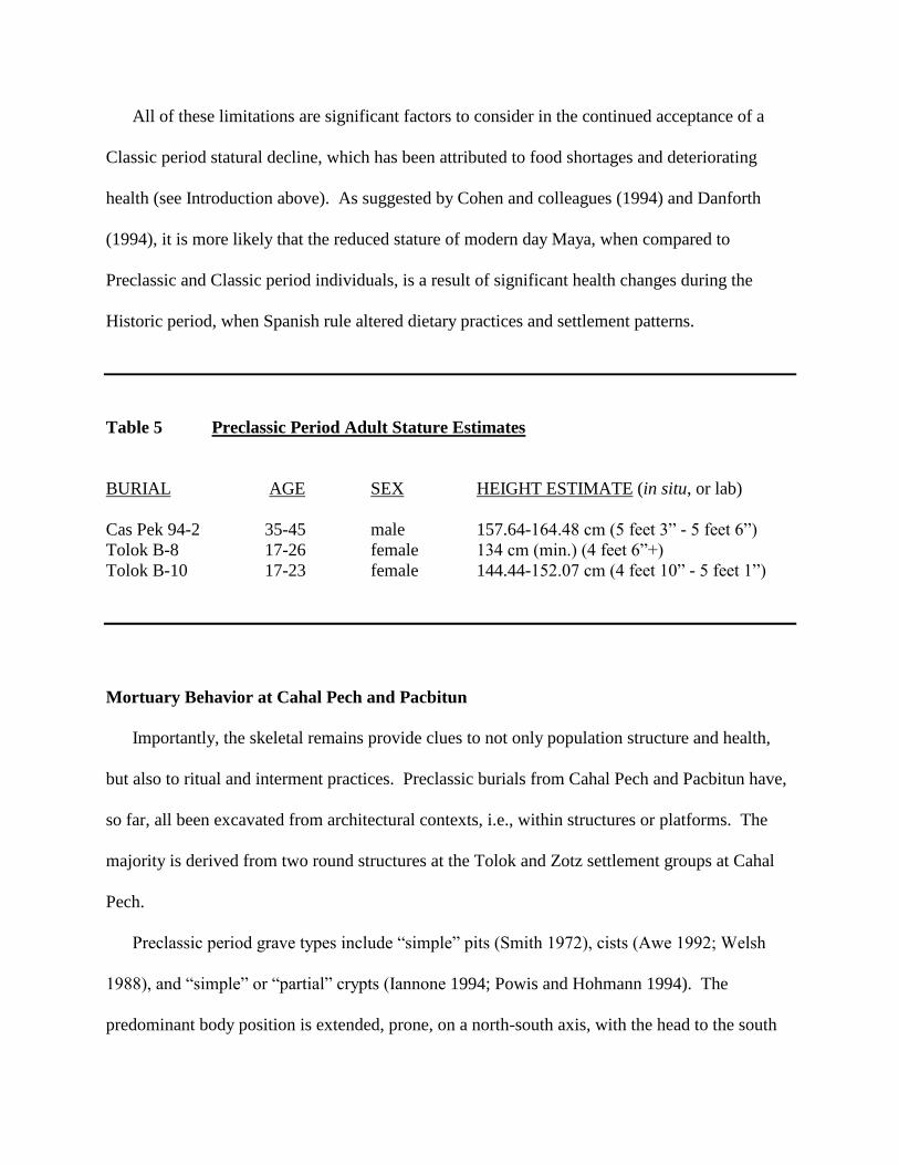
All of these limitations are significant factors to consider in the continued acceptance of a
Classic period statural decline, which has been attributed to food shortages and deteriorating
health (see Introduction above). As suggested by Cohen and colleagues (1994) and Danforth
(1994), it is more likely that the reduced stature of modern day Maya, when compared to
Preclassic and Classic period individuals, is a result of significant health changes during the
Historic period, when Spanish rule altered dietary practices and settlement patterns.
Table 5 Preclassic Period Adult Stature Estimates
BURIAL AGE SEX HEIGHT ESTIMATE (in situ, or lab)
Cas Pek 94-2 35-45 male 157.64-164.48 cm (5 feet 3” - 5 feet 6”)
Tolok B-8 17-26 female 134 cm (min.) (4 feet 6”+)
Tolok B-10 17-23 female 144.44-152.07 cm (4 feet 10” - 5 feet 1”)
Mortuary Behavior at Cahal Pech and Pacbitun
Importantly, the skeletal remains provide clues to not only population structure and health,
but also to ritual and interment practices. Preclassic burials from Cahal Pech and Pacbitun have,
so far, all been excavated from architectural contexts, i.e., within structures or platforms. The
majority is derived from two round structures at the Tolok and Zotz settlement groups at Cahal
Pech.
Preclassic period grave types include “simple” pits (Smith 1972), cists (Awe 1992; Welsh
1988), and “simple” or “partial” crypts (Iannone 1994; Powis and Hohmann 1994). The
predominant body position is extended, prone, on a north-south axis, with the head to the south

(see Table 1). Of the 17 primary burials for which the position is discernible, this pattern is
evident for 14 individuals, one of whom also had his knees flexed back and pointing upward
(Cas Pek Burial 94-2).
In comparison, Pacbitun Burial C-1 and Zubin Burial C9-B/1b deviate most from this
mortuary pattern. The Pacbitun individual was interred in an extended, prone position, but the
body was aligned 600 east of north, with the head at the west end, and its knees tightly flexed
upward. This arrangement even deviates from the common pattern of burial during the Classic
period for other Pacbitun graves: of 20 burials for which the position was discernible, 16
individuals were found in the extended position, but they were primarily supine and with the legs
crossed (Healy 1990). Similar to the Cahal Pech burials, however, these extended interments
were north-south in alignment, with the heads to the south.
In further contrast, the Zubin burial had a north-south orientation, but its head was at the
north end, facing west, and the entire extended body was supine in position. Interestingly, this
individual is also distinct from the other Preclassic remains in its dentition, which exhibit the
most severe enamel hypoplasias.
At Cuello, grave orientation similarly follows a consistent north-south placement (Hammond
et al. 1995). The earliest Middle Preclassic interments were flexed, as well as extended, and both
prone and supine in position. Tightly flexed individuals were found in both Middle and Late
Preclassic burials, and they were positioned lying on their backs, with their knees drawn up
(Hammond et al. 1995). This is a practice that has not yet been observed at Cahal Pech or
Pacbitun.
Grave goods vary between interments but, notably, several Preclassic individuals at Cahal
Pech were interred with their crania placed face down within whole vessels. Such instances were
found in Cas Pek Burials 94-1 and 94-2, and Tolok Burials 8 and 10. This custom continued in
later times at Cahal pech. For example, during the Classic period at Zopilote, the second
individual from Tomb 1 (ca. A.D. 600) had its cranium placed face down within a vessel, but its
skull was also covered with another inverted vessel (Cheetham et al. 1993). This placement of
an inverted vessel over the skull was also evident in a Late Classic skull interment from the Zotz
group (Burial 92-2-B/7) (Aimers 1992).
Outside of Cahal Pech, the pot-over-skull association is a predominant feature of Middle and
Late Preclassic period burials from Cuello, where it has been considered a form of skull
protection (Robin 1989). According to Welsh (1988), most Maya citizens, regardless of status,
probably interred individuals with vessels below or above the skull and it continued as a common
burial practice into the Classic period. Other than practical reasons for placing a vessel over a
skull, i.e., protection, such a custom has also been interpreted as representing special veneration
of the dead (Welsh 1988). Alternatively, covering of the skull with a vessel might symbolize the
individual’s descent into the darkened underworld (Xibalba).
Cognizant of this interpretation, one should then note the deposition of Cache 3 in the round
structure (Str. 14) at Tolok. This Terminal Preclassic “skull-in-bowl” offering displays a related
practice, with the caching of a 4-5 year old child's skull within a complete Aguacate Orange
tetrapod bowl. Regarding cultural significance, the purposeful interment of only the skull is
highly suggestive of some sort of ritual associated with sacrifice or veneration. Following Robin
(1989), the young age of the skull argues against its deposition as a form of ancestor worship. In
the larger scheme of events, the deposition of the cache in the round structure (Tolok Str. 14)
might then represent a dedicatory offering to the structure itself (see Welsh 1988: 170). Noting
the presence of other burials, and the nature of some deposits (i.e., Tolok Cache 2, the tooth
offering) associated with this structure, which suggest a public/ritual function for Structure 14
(Powis 1994), the possibility of the cache serving as a votive offering seems likely.
Tolok Burial 9 is noteworthy for the multiple interment of four subadults. Firstly, the
Primary Individual, aged 2-4 years old, should be distinguished because of its interment with the
partial remains of others. Skeletal evidence suggests that the Second Individual (4-5 years) is
represented by most long bones (humeri, ulna, femurs, tibia), but it appears that the arms and legs
were disarticulated prior to deposition. These long bones were placed along the corresponding
sides of the main child's upper torso and arm region. On the other hand, the Third Individual (1-
1.5 years) seems to be only represented by dental remains and, possibly, a cranium. At the very
least, maxillary and mandibular portions of the skull containing the unerupted secondary teeth
constitute this child’s remains. Lastly, the Fourth Individual, a child aged birth to 6 months, is
only identified by two teeth and a femur.
The nature of the three secondary subadult deposits atop the main child’s shoulders, upper
torso and near the cranium suggests that they were “grave goods” for the main child (also see
Powis and Hohmann 1995). Human sacrifice can be suggested to account for the secondary
individuals but, ultimately, the nature of death of all four children is perplexing.
Interestingly, the Primary Individual of Tolok Burial 9 lacks both legs and feet, appearing
severed at the waist. The absence of this individual’s lower body, which was nonetheless capped
with stones where the legs should have been, suggests that some sort of skeletal interference
occurred. With this in mind, it is very interesting that the legs of another child (Second
Individual) would be placed with the interment. Following Welsh (1988), one can only speculate
that the main child’s legs were removed, and similarly offered as grave goods to honor another
individual. Circumstances involving missing skeletal elements might indicate that the legs were

defleshed prior to burial (i.e., Burials 385-1 and 385-2 at Dzibilchaltun) and, thus, may represent
instances of ritual cannibalism or sacrifice (see Welsh 1988: 81).
Regarding the circumstances of the full length (head to feet) grave, which extended beyond
the severed skeleton of the Primary Individual (and thus, seems impractical), it can be argued
that the construction of the full cist grave was meant to allow burial of a “complete” body, that
might have had representations of legs constructed from some sort of perishable material.
Overall, the circumstances of ritual behavior surrounding Tolok Burial 9 conforms well with
other acts of ritual, skeletal “mutilation” and sacrifice evident at the site and particularly with
Structure 14 (see Powis 1996).
Elsewhere at Cahal Pech, similar (but Late Classic) evidence of possible skeletal interference,
or “mutilation”, is apparent at Zotz, where one individual (Burial 2-B/2) had its lower right leg
missing, while another (Burial 2-B/3) had an extra femur deposited with the lower left leg
(Aimers 1992: 5-6).
At Cuello, similar evidence exists from three double burials dating to early Middle Preclassic
and Late Preclassic contexts (see Robin 1989). In these cases, each primary individual was
interred with additional disarticulated leg bones, which were interpreted to be sacrificial remains
placed in honor of the principal individuals (Robin 1989).
Conclusions
Healthwise, the skeletal remains reflect general good health for the Preclassic period Maya at
both Cahal Pech and Pacbitun. The most common pathologies are enamel defects of teeth, but
they are minor in severity and reflect short-term periods of ill health (infectious disease) and/or
dietary stress (weaning). Skeletal pathologies such as periostitis, porotic hyperostosis and
osteoarthritis are also not severe in nature and, even considering the sample size, are not very

common. The most serious skeletal pathologies include: traumatic fractures to individuals in Cas
Pek Burial 91-4, Zubin Burial C9-B/1b, and Pacbitun Burial 1-5, one of which (Cas Pek) is likely
due to osteoporosis; periostitis of long bones of the individual in Cas Pek Burial 91-4, Tolok
Burial 7, and Tolok Burial 9d; porotic hyperostosis of the individual in Cas Pek Burial 94-2, and
possibly in Zubin Burial C9-B/1; and periodontitis and alveolar absorption among the individuals
in Cas Pek Burial 94-2 and Tolok Burial 10.
Comparatively, the Cahal Pech and Pacbitun data accord well with other Formative Maya
human skeletal remains. For instance, osteological analyses of material from Altar de Sacrificios
(Saul 1972) and Cuello (Saul and Saul 1991), sites with the largest Preclassic samples, indicate
that the health status of individuals from the Preclassic period was relatively better than that of
individuals from the Classic period. Although the Belize Valley sample size is smaller,
particularly the remains that have been amenable to detailed analysis, the Formative sample from
Cahal Pech and Pacbitun also indicate a relatively healthy population.
Notably, the general health trend of ancient Maya is accompanied by some archaeological
and bone chemical evidence suggesting increased maize reliance over time, leading to
speculation of dietary factors as the cause of deteriorating health (White et al. 1993).
Nonetheless, in the Belize Valley, evidence for abundant floral, faunal, riverine, and even
exchanged coastal resources, suggests that despite increased reliance on maize, diets probably
remained varied and adequate over time (Powis et al. 1999; see also Stanchly, this volume). This
regional distinction characterizes the variability evident across the Maya region, which leads
White (2000: xxii) to conclude that “…although there exists a pan-Maya ideological grounding
for dietary regimes, there is little to offer at present in terms of universal dietary patterning within
the Maya sphere.”
It is suggested that any changes in health that may have occurred in the Classic period are
probably due more to increased population and settlement density, and environmental alteration
associated with increased agricultural intensification, than directly to an increased reliance on
maize alone. More importantly, the political-economic climate of the Classic period is a greater
determinant of the nature of Maya health during this time, particularly when it is compared to the
less socially stratified Preclassic period.
Considering that the Preclassic period was the era in which Classic period cultural
complexity germinated, the analysis of human remains from this period can enhance the
understanding of early complex Maya society. Specifically, such analyses can shed light on
population health and mortuary behavior prior to major Classic period changes in demography,
settlement patterns, socio-economic organization, and subsistence. The Preclassic skeletal
sample at Cahal Pech and Pacbitun is small, thus far, but this does not lessen its significance in
the larger picture of the prehistory of the Maya.
Footnotes
1. The remaining Preclassic individuals (Cas Pek 91-2, 91-6, 93-1, 93-2; Zotz 91-1, 91-2) have
not been analyzed in detail by the author. Some isolated human bone and dental remains have
also been found in construction fill dated to Preclassic contexts at both sites, but these results are
also not presented here. Except for Zotz Burials 91-1 and 91-2, the Zubin remains, and Pacbitun
Burial 1-5, the author was responsible for primary analyses of all individuals discussed in this
chapter. Detailed descriptions of all skeletal data from Zubin have been previously reported by
Glassman and Stockton (1995) and Schwake (1996), but are also reported here based on
subsequent examinations by the author. The Late Preclassic grave (Burial 1-5) from Pacbitun
was analyzed by Helmuth (1988), with the information presented here with his permission.

REFERENCES CITED
Aimers, J.J.
1992 A Third Season of Excavations at the Zotz Group, Cahal Pech.Ms. in possession of the
author.
Arendt, C., R.J. Song and P.F. Healy
1996 The 1995 excavations in Plaza C, Pacbitun, Belize: A Middle Preclassic burial and a Late
Classic stela. In Belize Valley Preclassic Maya Project: Report on the 1995 Field Season,
ed. by P.F. Healy and J.J. Awe, 128-138. Occasional Papers in Anthropology No. 12, Trent
University, Peterborough, ON.
Awe, J.J.
1992 Dawn in the Land Between the Rivers: Formative Occupation at Cahal Pech, Belize and its
Implications for Preclassic Occupation in the Maya Lowlands. Ph.D. Dissertation, Institute of
Archaeology, University College London, U.K.
Awe, J.J. (editor)
1998 The Western Belize Regional Cave Project: A Report of the 1997 Field Season.
Anthropology Occasional Paper No.1, Dept. of Anthropology, Univ. of New Hampshire,
Durham, NH.
Awe, J.J., J.J. Aimers and C. Blanchard
1992 A Preclassic round structure from the Zotz Group at Cahal Pech. In Progress Report of
the Fourth Season (1991) of Investigations at Cahal Pech, Belize, ed. by J.J. Awe and
M.D. Campbell, 119-140. Trent University, Peterborough, ON.
Awe, J.J., C. Bill and M.D. Campbell
1990 The Cahal Pech, Belize Project: A Progress Report of the Second (1989) Season of
Investigations. Ms., Dept. of Anthropology, Trent University, Peterborough, ON.
Awe, J.J. and M.D. Campbell
1988 Site Core Investigations at Cahal Pech, Cayo District, Belize: Preliminary Report of the
1988 Season. Ms., Dept. of Anthropology, Trent University, Peterborough, ON.
Awe, J.J. and D.F. Lee (editors)
1999 The Western Belize Regional Cave Project: A Report of the 1998 Field Season.
Anthropology Occasional Paper No.2, Dept. of Anthropology, Univ. of New Hampshire,
Durham, NH.
Awe, J.J., J. Vinuales, M. Velasco and R. Novela
1992 Investigations at the Cas Pek Group in the western periphery of Cahal Pech, Belize. In
Progress Report of the Fourth Season (1991) of Investigations at Cahal Pech, Belize, ed. by
J.J. Awe and M.D. Campbell, 35-50. Trent University, Peterborough, ON.

Blakely, R.L. (ed.)
1977 Biocultural Adaptation in Prehistoric America. Southern Anthropological Society
Proceedings No. 11. University of Georgia Press, Athens, GA.
Blom, F.
1933 A Maya skull from Uloa Valley, Republic of Honduras. Middle American Research Institute
Publication 5: 6-24. Tulane University, New Orleans, LA.
Brady, J.E.
1995 A reassessment of the chronology and function of Gordon’s Cave #3, Copan, Honduras.
Ancient Mesoamerica 6: 29-38.
1997 Palaces of the dead: Glimpses of the afterlife from a Middle Preclassic cave ossuary. In
Proceedings of the 1995 and 1996 Latin American Symposia “Death, Burial, and the
Afterlife: Landscapes and Mindscapes of the Ancient Maya”, ed. by A. Cordy-Collins and G.
Johnson, 21-32. San Diego Museum Papers No. 34, San Diego, CA.
Brothwell, D.R.
1981 Digging Up Bones. Cornell University Press, Ithaca, NY.
Buikstra, J.E.
1977 Biocultural dimensions of archeological study: A regional perspective. In Biocultural
Adaptation in Prehistoric America, ed. by R.L. Blakely, 67-84. Univ. of Georgia Press,
Athens, GA.
Buikstra, J.E. and D.H. Ubelaker (eds.)
1994 Standards for data collection from human skeletal remains. Arkansas Archeological Survey
Research Series No. 44. Arkansas Archeological Survey, Fayetteville AK.
Capasso, L.L. and G. Di Tota
1992 Foramen caecum molare as a developmental defect of the enamel. In Recent Contributions to
the Study of Enamel Developmental Defects, ed. by A.H. Goodman and L.L. Capasso, 91-
106. Journal of Paleopathology Monographic Publications-2. Edigrafital: Teramo, Italy.
Cave, A.J.E., S.S. Grosjean and H. Cummins
1939 Report on two skulls from British Honduras. Smithsonian Institute Bureau of American
Ethnology Bulletin 123, 59-60. Washington, D.C.
Cheetham, D.T., J.J. Awe and D.M. Glassman
1994 Sacrifice at the end of the road: Preliminary report of the osteological analysis of human
remains from the Zopilote Group, Cahal Pech, Belize. In Belize Valley Archaeological
Reconnaissance Project: Progress Report of the 1993 Field Season, ed. by J.J., 179-184.
Trent University, Peterborough, ON.
Cifuentes, A.O.
1963 Odontologia y Mutilaciones Dentarias Mayas. Editorial Universitaria, Guatemala.

Cohen, M.N., K.A. O’Connor, M.E. Danforth, K.P. Jacobi and C.W. Armstrong
1994 Health and death at Tipu. In In the Wake of Contact, ed. by C.S. Larsen and G.R. Milner,
121-133. Wiley-Liss, New York.
Comas, J.
1970 History of physical anthropology in Middle America. In Handbook of Middle American
Indians, Volume 9: Physical Anthropology, ed. by T.D. Stewart (R. Wauchope, general
editor), 3-21. Austin: Univ of Texas Press
Conlon, J.M. and J.J. Awe
1991 The Cahal Tzinic Group at Cahal Pech, Belize: Preliminary comments of the 1990 season
of investigations. In Belize Valley Archaeological Reconnaissance Project: Report of the
Third Season (1990) of Investigations at Cahal Pech, Belize, ed. by J.J. Awe and M.D.
Campbell, 4-15. Trent University, Peterborough, ON.
Corruccini, R.S., J.S. Handler and K.P. Jacobi
1985 Chronological distribution of enamel hypoplasias and weaning in a Caribbean slave
population. Human Biology 57: 699-711.
Culbert, T.P.
1988 The collapse of Classic Maya civilization. In The Collapse of Ancient States and
Civilizations, ed. by N. Yoffee and G.L. Cowgill, 69-101. University of Arizona Press,
Tucson.
Cutress, T.W. and G.W. Suckling
1982 The assessment of non-carious defects of enamel. International Dentistry Journal 2: 117-
22.
Danforth, M.E.
1986 Health Patterns in the Classic Maya: Dental Evidence. Paper presented at the Annual
Meeting of the American Anthropological Association, Philadelphia, PA.
1994 Stature change in the prehistoric Maya of the southern lowlands. Latin American
Antiquity 5: 206-211.
Elliot, O. and H. Bleibtreu
1965 The human skeletal remains. In Prehistoric Maya Settlements in the Belize Valley, ed. by
G.R. Willey, W.R. Bullard Jr, J.B. Glass & J.C. Gifford, 535-539. Papers of the Peabody
Museum of Archaeology and Ethnology, Harvard Univ. Vol LIV. Cambridge, Mass.
Federation Dentaire Internationale (FDI)
1982 An epidemiologic index of developmental defects of dental enamel (DDE Index).
International Dentistry Journal 32: 159-167.

Gann, T.W.F.
1918 The Maya Indians of Southern Yucatan and Northern British Honduras. Smithsonian
Institution Bureau of American Ethnology Bull. 64. Washington, DC.
Genoves, S.
1970 Anthropometry of late prehistoric human remains. In Handbook of Middle American
Indians, Volume 9: Physical Anthropology, ed. by T.D. Stewart (R.Wauchope, general
editor), 35-49. Austin: Univ of Texas Press.
Glassman, D.M. and T.C. Stockton
1995 Zubin- Human Skeletal Summary. Ms. on file with the author, Dept. of Anthropology,
Southwest Texas State University, San Marcos, TX.
Goodman, A.H. and G.J. Armelagos
1985 The chronological distribution of enamel hypoplasia in human permanent incisor and
canine teeth. Archives of Oral Biology 30: 503-507.
Goodman, A.H., G.J. Armelagos and J.C. Rose
1984 The chronological distribution of enamel hypoplasias from prehistoric Dickson Mounds
populations. Am. J. Phys. Anthropol. 65: 259-266.
Goodman, A.H. and J.C. Rose
1990 Dental enamel hypoplasias as indicators of nutritional status. In Advances in Dental
Anthropology, ed. by M.A. Kelley and C.S. Larsen, 279-293. Wiley-Liss, New York.
Goodman, A.H. and R.J. Song
1999 Sources of variation in estimated ages at formation of linear enamel hypoplasias. In
Human Growth in the Past: Studies from Bones and Teeth, ed. by R. Hoppa and C.
FitzGerald, 210-239. Cambridge University Press, England.
Gordon, G.B.
1898 Caverns of Copan, Honduras. Peabody Museum of Archaeology and Ethnology Memoirs
1: 137-148.
Hambly, W.D.
1937 Skeletal material from San Jose ruin, British Honduras. Field Mus. Natural Hist., Anthr.
Mem., 25: 1-19
Hammond, N.
1975 Lubaantun, A Classic Maya Realm. Peabody Museum of Archaeology and Ethnology
Monograph No. 2. Harvard University, Cambridge, MA.
Hammond, N., A. Clarke and S. Donaghey
1995 The long goodbye: Middle Preclassic Maya archaeology at Cuello, Belize. Latin American
Antiquity 6(2): 120-128.

Haviland, W.A.
1967 Stature at Tikal, Guatemala: Implications for ancient Maya demography and social
organization. American Antiquity 32: 316-25.
Healy, P.F.
1974 The Cuyamel Caves: Preclassic sites in northeast Honduras. American Antiquity 39: 435-447.
1990 Excavations at Pacbitun, Belize: Preliminary report on the 1986 and 1987 investigations. J. of
Field Archaeology 17(3): 247-262.
Healy, P.F. and J.J. Awe (eds)
1995 Belize Valley Preclassic Maya Project: Report on the 1994 Field Season. Occasional Papers
in Anthropology No. 10, Trent University, Peterborough, ON.
1996 Belize Valley Preclassic Maya Project: Report on the 1995 Field Season. Occasional Papers in
Anthropology No. 12, Trent University, Peterborough, ON.
Healy, P.F., J.J. Awe and H. Helmuth
1998 An ancient Maya multiple burial at Caledonia, Cayo District, Belize. Journal of Field
Archaeology 25:261-274.
Helmuth, H.
1988 Report on the human remains from Pacbitun. Ms. on file, Dept. of Anthropology, Trent
University, Peterborough, ON.
Hohmann, B. and A. Hartnett
1995 Living on the edge: Late Classic peripheral activities at the Figueroa Group. In Belize Valley
Archaeological Reconnaissance Project: Progress Report of the 1994 Field Season, Vol. 2,
ed. by J. Conlon and J. Awe, 11-32. Ms., Institute of Archaeology, University College
London, England.
Iannone, G.
1993 Time among the thorns: Results of the 1992 field season at Zubin, Cayo District, Belize. In
Belize Valley Archaeological Reconnaissance Project: Progress Report of the 1992 Field
Season, ed. by J.J. Awe, 10-44. Trent University, Peterborough, ON.
1994 More time among the thorns: Results of the 1993 field season at Zubin, Cayo District, Belize.
In Belize Valley Archaeological Reconnaissance Project: Progress Report of the 1993 Field
Season, ed. by J.J. Awe, 32-108. Trent University, Peterborough, ON.
1995 One last time among the thorns: Results of the 1994 field season at Zubin, Cayo District,
Belize. In Belize Valley Archaeological Reconnaissance Project: Progress Report of the
1994 Field Season, Vol. 1, ed. by G. Iannone and J.M. Conlon, 11-122. Ms., Institute of
Archaeology, University College London, England.
Infante, P.F. and G.M. Gillespie
1974 An epidemiological study of linear enamel hypoplasia of deciduous anterior teeth in
Guatemalan children. Archives of Oral Biology 19: 1055-1061.
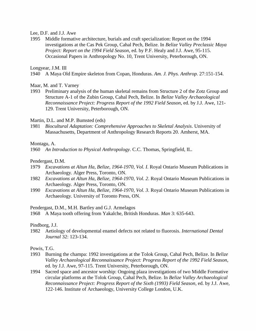
Lee, D.F. and J.J. Awe
1995 Middle formative architecture, burials and craft specialization: Report on the 1994
investigations at the Cas Pek Group, Cahal Pech, Belize. In Belize Valley Preclassic Maya
Project: Report on the 1994 Field Season, ed. by P.F. Healy and J.J. Awe, 95-115.
Occasional Papers in Anthropology No. 10, Trent University, Peterborough, ON.
Longyear, J.M. III
1940 A Maya Old Empire skeleton from Copan, Honduras. Am. J. Phys. Anthrop. 27:151-154.
Maar, M. and T. Varney
1993 Preliminary analysis of the human skeletal remains from Structure 2 of the Zotz Group and
Structure A-1 of the Zubin Group, Cahal Pech, Belize. In Belize Valley Archaeological
Reconnaissance Project: Progress Report of the 1992 Field Season, ed. by J.J. Awe, 121-
129. Trent University, Peterborough, ON.
Martin, D.L. and M.P. Bumsted (eds)
1981 Biocultural Adaptation: Comprehensive Approaches to Skeletal Analysis. University of
Massachusetts, Department of Anthropology Research Reports 20. Amherst, MA.
Montagu, A.
1960 An Introduction to Physical Anthropology. C.C. Thomas, Springfield, IL.
Pendergast, D.M.
1979 Excavations at Altun Ha, Belize, 1964-1970, Vol. I. Royal Ontario Museum Publications in
Archaeology. Alger Press, Toronto, ON.
1982 Excavations at Altun Ha, Belize, 1964-1970, Vol. 2. Royal Ontario Museum Publications in
Archaeology. Alger Press, Toronto, ON.
1990 Excavations at Altun Ha, Belize, 1964-1970, Vol. 3. Royal Ontario Museum Publications in
Archaeology. University of Toronto Press, ON.
Pendergast, D.M., M.H. Bartley and G.J. Armelagos
1968 A Maya tooth offering from Yakalche, British Honduras. Man 3: 635-643.
Pindborg, J.J.
1982 Aetiology of developmental enamel defects not related to fluorosis. International Dental
Journal 32: 123-134.
Powis, T.G.
1993 Burning the champa: 1992 investigations at the Tolok Group, Cahal Pech, Belize. In Belize
Valley Archaeological Reconnaissance Project: Progress Report of the 1992 Field Season,
ed. by J.J. Awe, 97-115. Trent University, Peterborough, ON.
1994 Sacred space and ancestor worship: Ongoing plaza investigations of two Middle Formative
circular platforms at the Tolok Group, Cahal Pech, Belize. In Belize Valley Archaeological
Reconnaissance Project: Progress Report of the Sixth (1993) Field Season, ed. by J.J. Awe,
122-146. Institute of Archaeology, University College London, U.K.
1996 Excavations of Middle Formative Period Round Structures at the Tolok Group, Cahal Pech,
Belize. M.A. Thesis, Dept. of Anthropology, Trent University, Peterborough, ON.
Powis, T.G. and B.M. Hohmann
1994 Middle Formative Architecture in the Periphery of Cahal Pech, Belize: The Case of Two
Circular Platforms at the Tolok Group. Paper presented at the 59th
Annual Meeting of the
Society for American Archaeology, Anaheim, CA.
1995 From private household to public ceremony: Middle Formative occupation at the Tolok
Group, Cahal Pech, Belize. In Belize Valley Preclassic Maya Project: Report on the 1994
Field Season, ed. by P.F. Healy and J.J. Awe, 45-94. Occasional Papers in Anthropology
No. 10, Trent University, Peterborough, ON.
Robin, C.
1989 Preclassic Maya Burials at Cuello, Belize. British Archaeological Reports (BAR),
International Series 480. Oxford, England.
Romero, J.
1970 Dental mutilation, trephination and cranial deformation. In Handbook of Middle
American Indians Vol. 9, ed. by R. Wauchope, 50-67. University of Texas Press, Austin.
Rue, D.J., A.C. Freter and D.A. Ballinger
1989 The caverns of Copan revisited: Preclassic sites in the Sesesmil River Valley, Copan,
Honduras. J. of Field Archaeology 16: 395-404.
Ruz L., A.
1965 Tombs and funerary practices in the Maya Lowlands. In Handbook of Middle American
Indians, Vol. 2, Archaeology, Part 1, ed. by R. Wauchope, 441-468. University of Texas
Press, Austin.
Santley, R.S.
1990 Demographic archaeology in the Maya Lowlands. In Precolumbian Population History in the
Maya Lowlands, ed. byT.P. Culbert and D.S. Rice, 325-343. University of New Mexico
Press, Albuquerque, NM.
Santley, R.S., T.W. Killion and M.T. Lycett
1986 On the Maya collapse. J. of Anthropological Research 42: 123-159.
Saul, F.P.
1972 The Human Skeletal Remains of Altar de Sacrificios: An Osteobiographic Analysis. Harvard
Univ., Peabody Museum of Archaeology and Ethnology, Paper 63(2). Cambridge, MA.
1975 Human remains from Lubaantun. In Lubaantun, A Classic Maya Realm, ed. by N. Hammond,
389-410. Peabody Museum of Archaeology and Ethnology Monograph No. 2. Harvard
University, Cambridge, MA.

Saul, F.P. and J.M. Saul
1989 Osteobiography: A Maya example. In Reconstruction of Life from the Skeleton, ed. by M.Y.
Iscan and K.A.R. Kennedy, 287-302. Alan R. Liss, New York.
1991 The Preclassic population of Cuello. In Cuello, An Early Maya Community in Belize, ed. by
N. Hammond, 134-158. Cambridge University Press, Cambridge.
Schele, L. and M.E. Miller
1986 The Blood of Kings. George Braziller, New York.
Schulz, P.D. and H. McHenry
1975 Age distribution of enamel hypoplasias in prehistoric California Indians. J. of Dental
Research 54: 913.
Schwake, S.
1996 Ancestors among the thorns: The burials of Zubin, Cayo District, Belize. In The Social
Archaeology Research Program: Progress Report of the Second (1996) Season, ed. by G.
Iannone, 84-105. Department of Anthropology, Trent University, Peterborough.
Scrimshaw, N.S.
1964 Ecological factors in nutritional disease. Am. J. Clinical Nutrition 14: 112-122.
Skinner, M. and A.H. Goodman
1992 Anthropological uses of developmental defects of enamel. In Skeletal Biology of Past
Peoples: Research Methods, ed. by S.R. Saunders and M.A. Katzenberg, 153-174. Wiley-
Liss, New York.
Smith, A.L.
1972 Excavations at Altar de Sacrificios: Architecture, Settlement, Burials and Caches. Peabody
Museum of Archaeology and Ethnology Paper 62(2). Harvard University, Cambridge, MA.
Song, R.J.
1993 Human skeletal remains from the Tolok Group, Cahal Pech. In Belize Valley Archaeological
Reconnaissance Project: Progress Report of the 1992 Field Season, ed. by J.J. Awe, 116-
120. Ms., Trent University, Peterborough, ON.
1995 Bones and bowls of the Formative Maya: A preliminary report of the human skeletal
remains from Cahal Pech and the implications for mortuary behavior. In Belize Valley
Preclassic Maya Project: Report on the 1994 Field Season, ed. by P.F. Healy and J.J. Awe,
173-197. Occasional Papers in Anthropology No. 10, Trent University, Peterborough, ON.
1996 The Formative Period Skeletal Assemblage at Cahal Pech, Belize. Paper presented at the 61st
Annual Meetings of the Society for American Archaeology, New Orleans, LA.
1997 Developmental Defects of Enamel in the Maya of Altun Ha, Belize: Implications for Ancient
Maya Childhood Health. M.A. Thesis, Trent University, Peterborough, ON. University
Microfilms International, Ann Arbor, MI.

Song, R.J., B. Hohmann, D. Mardiros and D. Glassman
1994 All in the family circle: A second interim report of the human skeletal remains from Tolok,
Cahal Pech, Belize 1993. In Belize Valley Archaeological Reconnaissance Project: Progress
Report of the 1993 Field Season, ed. by J.J. Awe, 147-163. Ms., Institute of Archaeology,
University College London, U.K.
Stewart, T.D.
1941 New examples of tooth mutilation from Middle America. Am. J. Phys. Anthrop. 28: 117-24.
1974 Human skeletal remains from Dzibilchaltun, Yucatan, Mexico, with a review of cranial
deformity types in the Maya Region. Middle American Research Institute Publication #31.
Tulane University, New Orleans, LA.
Storey, R.
1992 The children of Copan: Issues in paleopathology and paleodemography. Ancient
Mesoamerica 3: 161-167.
Sunahara, K.S. and J.J. Awe
1994 A slice through time: 1993 investigations at the Cas Pek Group, Cahal Pech. In Belize
Valley Archaeological Reconnaissance Project: Progress Report of the Sixth (1993) Field
Season, ed. by J.J. Awe, 193-210. Institute of Archaeology, University College London,
U.K.
Sweeney, E.A. and M. Guzman
1966 Oral conditions in children from three Highland villages in Guatemala. Archives of Oral
Biology 11: 97-98.
Tozzer, A.M.
1941 Landa’s Relacion de las Cosas de Yucatan. Peabody Museum of Archaeology and
Ethnology Paper 18. Harvard University, Cambridge, MA.
Ubelaker, D.
1989 Human Skeletal Remains: Excavation, Analysis, Interpretation, 2nd
Edition. Manuals on
Archeology 2. Taraxacum, Washington, D.C.
Vinuales, J.
1992 Preliminary Study of the Cas Pek Group, Cahal Pech, Belize. B.A. Thesis, Institute of
Archaeology, University College London, U.K.
Welsh, W.B.M.
1988 An Analysis of Classic Lowland Maya Burials. British Archaeological Reports (BAR)
International Series 409. Oxford, England.
White, C.D.
1986 Paleodiet and Nutrition of the Ancient Maya at Lamanai, Belize. M.A. Thesis, Dept. of
Anthropology, Trent University, Peterborough, ON.

2000 Introduction: Ancient Maya diet. In Reconstructing Ancient Maya Diet, ed. by C.D.
White, ix-xxvii. University of Utah Press, Salt Lake City.
White, C.D., P.F. Healy and H.P. Schwarcz
1993 Intensive agriculture, social status and Maya diet at Pacbitun, Belize. Journal of
Anthropological Research 49: 347-375.
Willey, G.R. and D.B. Shimkin
1973 The Maya collapse: A summary view. In The Classic Maya Collapse, ed. by T.P. Culbert,
457-502. School of American Research, Advanced Seminar Series, Santa Fe, NM. Univ.
of New Mexico Press, Albuquerque.
Wright, L.E.
1990 Stresses of conquest: A study of Wilson bands and enamel hypoplasias in the Maya of
Lamanai, Belize. Am. J. of Human Biology 2: 25-35.
1994 The Sacrifice of the Earth? Diet, Health and Inequality in the Pasion Maya Lowlands.
PhD Dissertation, University of Chicago. University Microfilms, Ann Arbor, MI.
Wright, L.E. and C.D. White
1996 Human biology in the Classic Maya collapse: Evidence from paleopathology and
paleodiet. J. of World Prehistory 10(2): 147-198.
Young, D.
1994 Analysis of human skeletal remains from Operation 2031, Colha, Belize. In Continuing
Archaeology at Colha, Belize, ed. by T.R. Hester, H.J. Shafer and J.D. Eaton, 59-63. Studies
in Archeology 16, Texas Archeological Research Laboratory. The University of Texas at
Austin.

LIST OF FIGURES
Figure 1: Cas Pek Burial 94-1, with cranium inside Preclassic vessel.
Figure 2: Preclassic vessel containing viscerocranium of Tolok Burial 8.
Figure 3: Tolok Burial 9, with legs of primary individual missing.
Figure 4: In situ photograph of pseudocircular cranial modification in Tolok Burial 9-Indiv 1.
Figure 5: Mandible of Tolok Burial 10, with reactive bone evident in socket of right M1.
Figure 6: Tolok Cache 3 vessel containing subadult cranium.
Figure 7a: Linear enamel hypoplasia of Zubin Burial C9-B/1 anterior teeth, with calculus
evident.
Figure 7b: Linear enamel hypoplasia of Zubin Burial C9-B/1 molar teeth, with calculus evident.
Figure 8: Pacbitun Burial C-1, with base of stela at upper left (west).