Vitellogenin is not an appropriate biomarker of feminisation in a Crustacean
Orcokinin Peptides in Developing andAdult Crustacean Stomatogastric
Nervous Systems and Pericardial Organs
LINGJUN LI,1,2 STEFAN R. PULVER,2 WAYNE P. KELLEY,1
VATSALA THIRUMALAI,2 JONATHAN V. SWEEDLER,1AND EVE MARDER2*
1Department of Chemistry and Beckman Institute, University of Illinois,Urbana, Illinois 61801
2Volen Center and Biology Department, Brandeis University,Waltham, Massachusetts 02454-9110
ABSTRACTThe orcokinins are a family of neuropeptides recently isolated from several crustacean
species. We found orcokinin-like immunoreactivity in the stomatogastric nervous systemsand pericardial organs of three decapod crustacean species, Homarus americanus, Cancerborealis, and Panulirus interruptus. The neuropil of the stomatogastric ganglion was stainedin adults of all three species as well as in embryonic and larval H. americanus. In H.americanus, the somata giving rise to this projection were found in the inferior ventricularnerve. Matrix-assisted laser desorption/ionization mass spectrometry mass profiling andsequencing with postsource decay led to the identification of six different orcokinin familypeptides, including those previously described in other decapods and two novel shorterpeptides. Application of exogenous [Ala13]orcokinin to the stomatogastric ganglion of H. ameri-canus resulted in changes in the pyloric rhythm. Specifically, the number of lateral pyloric (LP)neuron spikes/burst decreased, and the phase of firing of the pyloric neurons was altered.Together, these data indicate that the orcokinins are likely to function as modulators of thecrustacean stomatogastric ganglion. J. Comp. Neurol. 444:227–244, 2002. © 2002 Wiley-Liss, Inc.
Indexing terms: MALDI-TOF mass spectrometry; neuromodulators; Homarus americanus; Cancer
borealis; Panulirus interruptus; confocal microscopy
The crustacean stomatogastric nervous system containsthe central pattern-generating networks that controlrhythmic movements of the foregut (Harris-Warrick et al.,1992). One of the most remarkable features of this ner-vous system is the richness and diversity of its neuro-modulatory control systems. Specifically, although the sto-matogastric ganglion (STG) contains only 26–30 neurons,18–20 different neuromodulators are found in neuronsthat project into the STG (Marder et al., 1995; Marder andCalabrese, 1996). Moreover, these and additional sub-stances are also present in the hemolymph, as a conse-quence of their release by the pericardial organs and otherneurosecretory structures (Christie et al., 1995b).
The orcokinins are a novel family of neuropeptides thatwere originally purified from the nervous system of thecrayfish Orconectes limosus (Stangier et al., 1992) andmeasured by ELISA in various neural structures, includ-ing the STG, in several crustacean species (Bungart et al.,1994). Subsequently, different orcokinin analogues wereisolated, and it is now evident that the orcokinins are a
family of closely related peptides (Bungart et al., 1995a,b;Dircksen et al., 2000; Yasuda-Kamatani and Yasuda,2000).
Immunocytochemical localizations of the orcokinin fam-ily reveal cells and fibers that stain in the sixth abdominalganglion of O. limosus and fibers throughout the gut. We(this paper) and Skiebe et al. (2002) have combined im-munocytochemistry and matrix-assisted laser desorption/ionization (MALDI) time-of-flight mass spectrometry(TOF MS) to describe the presence and structure of theorcokinin peptides in the stomatogastric nervous systemsand pericardial organs of several crustacean species.
*Correspondence to: Dr. Eve Marder, Volen Center, MS 013, BrandeisUniversity, 415 South St., Waltham, MA 02454.E-mail: [email protected]
Received 17 July 2001; Revised 12 October 2001; Accepted 16 November2001
Published online the week of January 28, 2002
THE JOURNAL OF COMPARATIVE NEUROLOGY 444:227–244 (2002)
© 2002 WILEY-LISS, INC.DOI 10.1002/cne.10139

Because the orcokinin antisera used for immunocytochem-istry (Dircksen et al., 2000) do not distinguish amongseveral structurally distinct peptide forms, biochemicalcharacterization of the peptides in different tissues andspecies allows us to determine unambiguously which ofthe peptides are found in the stomatogastric nervous sys-tem of each crustacean studied.
Despite the extensive biochemical characterization ofthe orcokinins, relatively little is known of their physio-logical roles. To date, the bioactivity of the orcokinins hasbeen demonstrated in the hindgut of the crayfish (Stangieret al., 1992; Bungart et al., 1995b; Dircksen et al., 2000),but no electrophysiological demonstrations of the effects ofthe orcokinins on the nervous system have been reported.The distribution of orcokinin-like immunoreactivity in thestomatogastric nervous system suggested that orcokininfamily peptides could function as modulators of the sto-matogastric nervous system motor patterns, motivatingus to study the modulation of the pyloric rhythm of thestomatogastric nervous system in Homarus americanusby orcokinin.
Recent work has shown that the full complement ofneuromodulators in the adult stomatogastric nervous sys-tem does not appear until the end of larval life. Instead,individual sensory and descending neurons that projectinto the STG acquire their full complement of neurotrans-mitters sequentially (Fenelon et al., 1998, 1999; Kilman etal., 1999; Le Feuvre et al., 2001). The timing of appear-ance of the orcokinins in development has not been previ-ously studied. Therefore, one of the goals of this work wasto determine the developmental timing of orcokinin ap-pearance in the stomatogastric nervous system and peri-cardial organs of H. americanus and to compare thesewith the developmental appearance of the other majorSTG neuromodulators.
Previous work on the modulatory projections to the STGon related crustacean species has demonstrated signifi-cant differences in the distribution of modulators in thestomatogastric nervous system (Beltz et al., 1984; Mortinand Marder, 1991; Turrigiano and Selverston, 1991;Skiebe, 1999), including species differences in the cotrans-mitter complement of identified projection neurons (Mey-rand et al., 2000). For this reason, here we also compared
the distribution of orcokinin family peptides in the adultnervous system of three species of crustaceans, H. ameri-canus, Cancer borealis, and Panulirus interruptus. Thesewill be further compared with the results for Cherax de-structor obtained by Skiebe et al. (2002). Some of this workhas been presented in abstract form (Li et al., 2001).
MATERIALS AND METHODS
Animals and dissection
Experiments were performed on embryonic, larval, ju-venile [thorax length (TL) 1–2 cm], and adult H. america-nus. Embryos, larvae, and juveniles were obtained fromthe lobster-rearing facility at the New England Aquarium(Boston, MA); adults were obtained from Commercial Lob-ster (Boston, MA). Experiments were also performed onadult C. borealis and P. interruptus (TL 6–8 cm). C. bo-realis and P. interruptus were obtained from CommercialLobster and Tomlinson Commercial Fishery (San Diego,CA), respectively. Animals were kept in large tanks ofcirculating 10–14°C seawater. H. americanus embryosand larvae were staged according to Helluy and Beltz(1991).
In adult, older larval, and juvenile animals, the STGand nerves of the stomatogastric nervous system weredissected free of the stomach and pinned flat onto aSylgard-lined petri dish, as is customary for STG prepa-rations (Harris-Warrick et al., 1992). The pericardial or-gans (POs) in adults of all three species were dissected byremoving the carapace covering the heart. The longitudi-nal body muscles, hyperdermis, and heart were then re-moved and pinned ventral side up in a Sylgard-lined petridish. The PO nerves surrounding the heart were identifiedand removed. The dissection procedures for embryos andsmaller larvae for the stomatogastric nervous system inH. americanus were carried out as previously described(Kilman et al., 1999). Briefly, the foregut was removed,split open along the midline, and pinned flat onto a smallSylgard cube.
The saline composition for H. americanus and P. inter-ruptus was (in mM) NaCl, 479; KCl, 12.74; MgSO4, 20;Na2SO4, 3.91; CaCl2, 13.67; HEPES, 5; pH 7.45. For C.borealis the saline composition was (in mM) NaCl, 440;KCl, 11; MgCl2, 26; CaCl2, 13; Trizma base, 11; maleicacid, 5; pH 7.4–7.5.
Whole-mount immunocytochemistry
Tissues were processed for whole-mount immunocyto-chemistry following standard protocols for this system(Beltz and Kravitz, 1983). Tissues were fixed overnight at4°C with one of two solutions. Embryonic and larval H.americanus preparations were fixed with 2% paraformal-dehyde and 15% picric acid in 0.2 M sodium phosphatebuffer, pH 7.3 (Stefanini et al., 1967). Juvenile H. ameri-canus, C. borealis, and P. interruptus were fixed with 4%paraformaldehyde in 0.1 M sodium phosphate buffer, pH7.4. In all species, the same structures were labeled witheither Stefanini fixative or 4% paraformaldehyde (S.R.P.,unpublished observations).
After fixation, preparations were washed five or sixtimes for 1 hour per wash in 0.1 M sodium phosphatebuffer, pH 7.4, containing 0.3% Triton X-100 and 0.1%sodium azide (PTA). The tissues were then incubated forapproximately 12–24 hours in a polyclonal primary anti-serum raised in rabbits against a thyroglobulin conjugate
Abbreviations
CabTRPIa Cancer borealis tachykinin-related peptide IaCo commissuresCoG commissural gangliondvn dorsal ventricular nervedpon dorsal posterior esophageal nerveion inferior esophageal nerveivn inferior ventricular nerveIV neuron inferior ventricular neuronLP neuron lateral pyloric neuronlvn lateral ventricular nervenp neuropilOG esophageal ganglionon esophageal nervePD neuron pyloric dilator neuronpdn pyloric dilator nervePY neuron pyloric neuronpyn pyloric nervePS pyloric suppressorsom somatason superior esophageal nerveSTG stomatogastric ganglionstn stomatogastric nerve
228 L. LI ET AL.
of [Asn13]orcokinin that cross reacts with others of thefamily of orcokinin peptides (Bungart et al., 1994; Dirck-sen et al., 2000). This antibody was a gift of HeinrichDircksen (Bonn, Germany) and was used at a final dilu-tion of 1:5,000. The primary antiserum was diluted in PTAplus 10% goat normal serum (GNS) to reduce nonspecificstaining. Preparations were washed in PTA as describedabove and then incubated overnight in PTA plus 10% GNSalong with anti-rabbit Alexafluor 488 (Molecular Probes,Eugene, OR) diluted 1:300–1:400. Finally, preparationswere washed five or six times for 1 hour per wash in 0.1 Msodium phosphate buffer, pH 7.4. The tissues weremounted in 80% glycerol in 0.02 M sodium phosphatebuffer, pH 7.4, on glass slides. All processing was carriedout at 4°C.
Imaging
Preparations were viewed as whole mounts with a Bio-Rad MRC 600 laser scanning confocal microscope with akrypton/argon mixed gas laser or a Leica TCS spectralconfocal microscope fitted with argon (458/488 nm) andkrypton (568 nm) lasers. For imaging antibodies coupledto Alexafluor 488 on the Bio-Rad MRC 600, we used astandard BHS (exciter filter, 488 nm DF 10; dichroic re-flector, 510 nm LP; emission filter, 515 nm LP) filter block.The z-axis spacing was 1–2 �m in embryos and larval H.americanus. In adult animals, the z-axis spacing was 3 �mexcept in the commissural ganglia (CoGs), which weresometimes sectioned at 4 or 5 �m intervals because of thelarge size of these ganglia. The resulting stacked imageswere compiled into maximum projections, then processedwith Confocal Assistant (Bio-Rad) and Adobe Photoshop5.5 (Adobe Systems, Mountain View, CA). Images wereprinted on a Codonics NP-1600 Printer (Codonics Inc.,Middleburg Heights, OH).
Cellular sample preparation forMALDI-MS analysis
Small pieces of tissue samples or single neurons weredissected and prepared for MALDI-MS analysis. Physio-logical saline was replaced with an aqueous MALDI ma-trix solution, 10 mg/ml of 2,5-dihydroxybenzoic acid (DHB;ICN Pharmaceuticals, Costa Mesa, CA) to remove theextracellular salts associated with the tissue sample (Gar-den et al., 1996). Tungsten needles and fine forceps wereused to dissect and transfer small pieces of tissue or singleneurons onto a MALDI sample plate containing 0.5 �l ofeither regular aqueous DHB (10 mg/ml) matrix solution orconcentrated DHB (50 mg/ml) in acetone:water (4:1)mixed solvent. The tissue samples on the plate weresmashed before drying at ambient temperature, followedby MALDI-MS analysis.
Microbore RP-HPLC of homogenates
Peptides were extracted from four brains from H. ameri-canus, nine brains from C. borealis and a single brain fromP. interruptus. In addition, 12 POs from H. americanusand 13 POs from P. interruptus were individually ex-tracted and pooled. The extraction of tissues was per-formed using acidified acetone (1:40:6 HCl:acetone:H2O)as described previously (Floyd et al., 1999). Briefly, sam-ples were homogenized in a microhomogenizer (JenconsScientific Ltd.), and the supernatant was drawn off andcentrifuged (Baxter Biofuge 15, Mcgraw Park, IL). This
process was repeated several times, after which an addi-tion of H2O was made to the resulting extract, which wasthen concentrated under a stream of nitrogen to approxi-mately 300 �l. Separations were performed utilizing amicrobore HPLC instrument (Magic 2002; MichromBioResources, Auburn, CA). An aliquot of the extract wasinjected onto a reverse-phase C-18 column (Reliasil). Both1.0 � 150 mm and 0.5 � 150 mm columns were utilized,each with 5 �m particle size and 30 nm pore size. Thecolumn was equilibrated with solvent A at a programmedtemperature of 35°C. An aliquot of the aqueous extractwas injected onto the column at a constant flow rate of 50�l/minute and a gradient developed from 5% to 98% ofsolvent B in 34 minutes. Solvent A consisted of 2% aceto-nitrile (ACN), 98% H2O, and 0.1% trifluoroacetic acid(TFA; v/v). Solvent B consisted of 95% ACN, 5% H2O, and0.1% TFA (v/v). Sample peaks were detected via absor-bance at 214 and 280 nm wavelengths, and the eluent wascollected by a small-volume fraction collection system (Gil-son FC 203B, Middletown, WI). To identify peptides ofinterest, each fraction was screened using MALDI-MS;approximately 0.25 �l of each fraction was deposited on aMALDI-MS sample plate, followed by the same volume ofan �-cyano-4-hydroxycinnamic acid matrix (10 mg/ml in6:3:1 ACN:H2O:3% TFA; Aldrich, Milwaukee, WI). Thusmore than 95% of each fraction was available for furtherassays.
MALDI-MS
MALDI is a soft ionization method that vaporizes andgenerates ions of analyte molecules without fragmentingthem by incorporating the analyte into an excess of amatrix. The matrix is typically a small organic compoundwith absorbance at a wavelength of the laser employed.MALDI mass spectra were obtained using a Voyager DESTR (PE Biosystems, Framingham, MA) time-of-flightmass spectrometer equipped with delayed ion extraction.A pulsed nitrogen laser (337 nm) was used as thedesorption/ionization source, and positive-ion mass spec-tra were acquired using both linear and reflectron modes.Each representative mass spectrum shown is thesmoothed average of 128–256 laser pulses. Mass calibra-tion was performed externally using a mixture of syn-thetic peptide standards (PE Biosystems). Mass accuracywas typically better than 0.01%. Because the signal inten-sity of MALDI spectra depends on the incorporation of thepeptides into matrix crystals, it is difficult to relate peakheight with the quantity of analyte present. Thus, MALDIis used primarily as a qualitative technique, and repre-sentative spectra of multiple replicates (n � 3) are shownin the current study.
Postsource decay (PSD) analysis
Semipurified microbore LC fractions containing thepeptide of interest or 2 pmol of synthetic peptide standardwere subjected to PSD analysis. The peptide standard[Ala13]orcokinin was synthesized by the Protein SciencesFacility (Biotechnology Center, University of Illinois). Thetotal acceleration voltage was 20 kV, and a delay time of75 nsec was used. By the use of timed ion selector, differ-ent precursor ions were selected from a mixture of pep-tides and subjected to fragmentation. Under these exper-imental conditions, the mass accuracy of the precursor ionwas within 30 ppm, and the average error on the massassignment of the PSD ions was better than 0.3 Da. Spec-
229CRUSTACEAN STOMATOGASTRIC NERVOUS SYSTEM ORCOKININS
tra were obtained by accumulating data from 100–256laser shots. To obtain complete PSD spectra, a series ofreflectron TOF spectral segments was acquired, each op-timized to focus fragment ions within different m/z ranges(Spengler, 1997). Each segment was stitched together us-ing the Biospectrometry Workstation software to generatea composite PSD spectrum.
Electrophysiology
The entire stomatogastric nervous system was dis-sected. The STG was desheathed and Vaseline wells weremade on the motor nerves for extracellular recordings.The preparations were continuously superfused withchilled saline (9–12°C). Intracellular recordings weremade with glass microelectrodes filled with 0.6 M K2SO4and 20 mM KCl, with typical resistances of 20–30 M�.Lateral pyloric (LP), pyloric (PY), and pyloric dilator (PD)neurons were identified on the basis of the motor nervesinto which their axons project. The STG was left attachedto the CoGs and the esophageal ganglion (OG) via thesomatogastric nerve (stn). Physiological saline containing2 � 10–6M [Ala13]orcokinin was superfused over the en-tire preparation. Data were acquired using an Axoclamp2A amplifier and a Digidata 1200 data acquisition board(Axon Instruments, Foster City, CA). Bursts and spikeswere extracted using in-house software from the intracel-lular traces of the LP, PY, and PD neurons. From thesedata, the burst frequency, the number of spikes per burst,and the burst durations were calculated for each neurontype. Data obtained in normal saline were compared withthose in orcokinin using paired t-tests in Microsoft Excel.
RESULTS
The stomatogastric nervous system
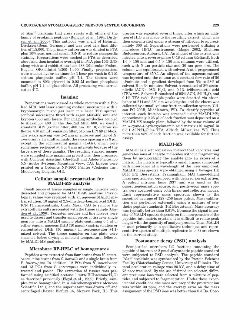
The stomatogastric nervous system consists of the sin-gle STG with about 30 neurons, the paired CoGs, thesingle OG, and their connecting nerves (Fig. 1). This gen-eral organization is the same in the three speciesstudied—H. americanus, C. borealis, and P. interruptus—with the exception that the stn is shortened in the crabpreparations relative to what is seen in the lobsters.
Previous work has demonstrated in H. gammarus thatthe complete stomatogastric nervous system is presentbefore 50% of embryonic development (E50) and that asearly as E50 neurogenesis in the STG is complete (Fenelonet al., 1998). More recently, LeFeuvre et al. (2001) sug-gested that most of the fibers projecting to the STG fromthe CoGs and the OG are also present by midembryoniclife. Nonetheless, the gross anatomical features of thestomatogastric nervous system change during embryonicand larval life, as the nerves elongate and ganglia changeshape. Despite the fact that the gross anatomical featuresof the stomatogastric nervous system are fully formedearly in embryonic life, many of the neuromodulatorsfound in projections to the STG from more anterior struc-tures do not appear until later in development (Fenelon etal., 1998, 1999; Kilman et al., 1999; Le Feuvre et al.,2001), so we have studied orcokinin immunoreactivity atdifferent developmental stages.
Orcokinin labeling in the STG in embryonic,larval, and adult H. americanus
Figure 2 shows whole mounts of STGs at different de-velopmental stages labeled with the orcokinin antibody.
Figure 2A shows that the neuropil of the STG of an E57embryo was already densely immunoreactive for orcoki-nin. The single input nerve to the STG, the stn, showed atleast one stained fiber, but none of the somata was la-beled, and there were no labeled fibers in the posteriormotor nerves. Similar staining patterns were seen in sixembryos from E57 to E90. The same essential features ofthe orcokinin labeling were seen at later developmentaltimes, except at later stages two stained fibers were visi-ble in the stn. In a small number of preparations (1 of 6embryos, 2 of 14 larvae, 0 of 4 juveniles, and 1 of 5 adults),one or two lightly labeled fibers were seen in the dorsalventricular nerve (dvn). Figure 2B shows the STG from an
Fig. 1. Schematic diagram of the stomatogastric nervous system.The single STG receives modulatory descending inputs from neuronsin the OG and the paired CoGs via the single stn. The ivn contains thesomata of the PS neurons and connects the stomatogastric nervoussystem to the brain. The ion, son, dvn, pdn, pyn, and dpon are alsoindicated. Ganglion size and nerve width are not to scale.
230 L. LI ET AL.
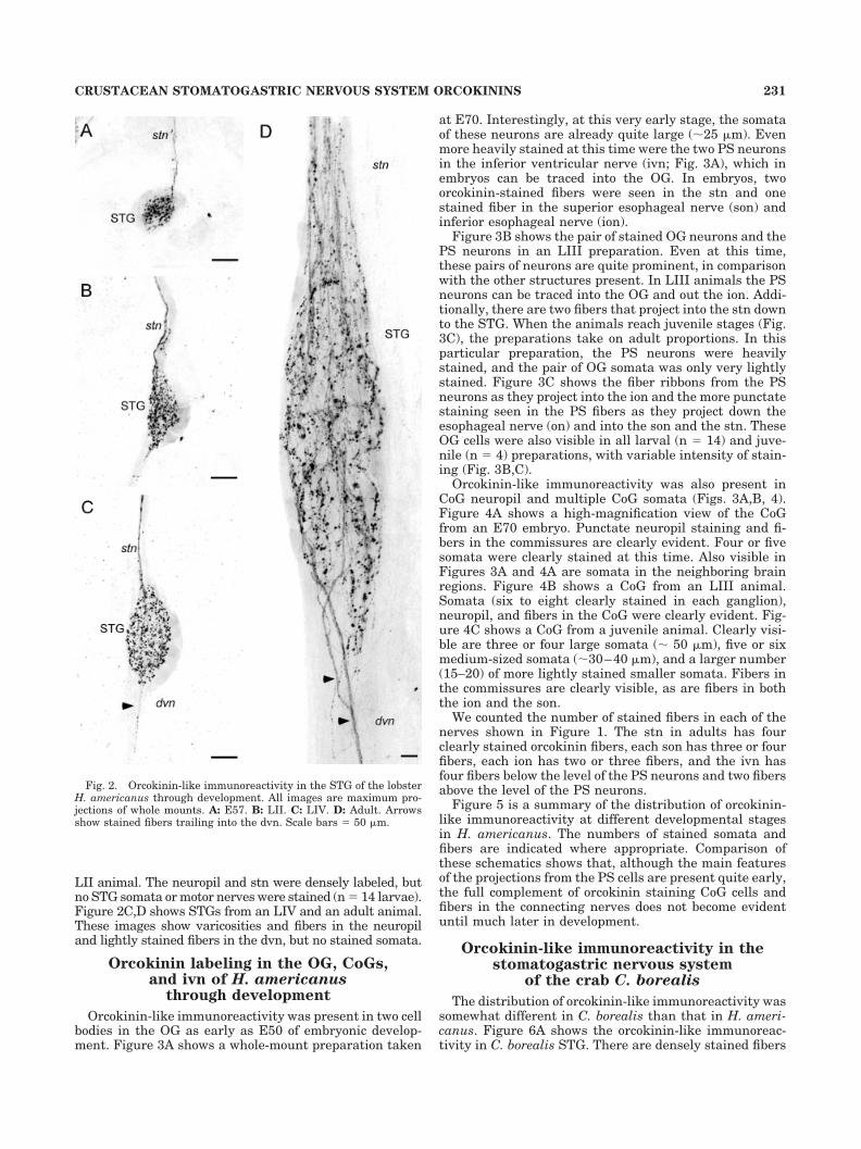
LII animal. The neuropil and stn were densely labeled, butno STG somata or motor nerves were stained (n � 14 larvae).Figure 2C,D shows STGs from an LIV and an adult animal.These images show varicosities and fibers in the neuropiland lightly stained fibers in the dvn, but no stained somata.
Orcokinin labeling in the OG, CoGs,and ivn of H. americanus
through development
Orcokinin-like immunoreactivity was present in two cellbodies in the OG as early as E50 of embryonic develop-ment. Figure 3A shows a whole-mount preparation taken
at E70. Interestingly, at this very early stage, the somataof these neurons are already quite large (�25 �m). Evenmore heavily stained at this time were the two PS neuronsin the inferior ventricular nerve (ivn; Fig. 3A), which inembryos can be traced into the OG. In embryos, twoorcokinin-stained fibers were seen in the stn and onestained fiber in the superior esophageal nerve (son) andinferior esophageal nerve (ion).
Figure 3B shows the pair of stained OG neurons and thePS neurons in an LIII preparation. Even at this time,these pairs of neurons are quite prominent, in comparisonwith the other structures present. In LIII animals the PSneurons can be traced into the OG and out the ion. Addi-tionally, there are two fibers that project into the stn downto the STG. When the animals reach juvenile stages (Fig.3C), the preparations take on adult proportions. In thisparticular preparation, the PS neurons were heavilystained, and the pair of OG somata was only very lightlystained. Figure 3C shows the fiber ribbons from the PSneurons as they project into the ion and the more punctatestaining seen in the PS fibers as they project down theesophageal nerve (on) and into the son and the stn. TheseOG cells were also visible in all larval (n � 14) and juve-nile (n � 4) preparations, with variable intensity of stain-ing (Fig. 3B,C).
Orcokinin-like immunoreactivity was also present inCoG neuropil and multiple CoG somata (Figs. 3A,B, 4).Figure 4A shows a high-magnification view of the CoGfrom an E70 embryo. Punctate neuropil staining and fi-bers in the commissures are clearly evident. Four or fivesomata were clearly stained at this time. Also visible inFigures 3A and 4A are somata in the neighboring brainregions. Figure 4B shows a CoG from an LIII animal.Somata (six to eight clearly stained in each ganglion),neuropil, and fibers in the CoG were clearly evident. Fig-ure 4C shows a CoG from a juvenile animal. Clearly visi-ble are three or four large somata (� 50 �m), five or sixmedium-sized somata (�30–40 �m), and a larger number(15–20) of more lightly stained smaller somata. Fibers inthe commissures are clearly visible, as are fibers in boththe ion and the son.
We counted the number of stained fibers in each of thenerves shown in Figure 1. The stn in adults has fourclearly stained orcokinin fibers, each son has three or fourfibers, each ion has two or three fibers, and the ivn hasfour fibers below the level of the PS neurons and two fibersabove the level of the PS neurons.
Figure 5 is a summary of the distribution of orcokinin-like immunoreactivity at different developmental stagesin H. americanus. The numbers of stained somata andfibers are indicated where appropriate. Comparison ofthese schematics shows that, although the main featuresof the projections from the PS cells are present quite early,the full complement of orcokinin staining CoG cells andfibers in the connecting nerves does not become evidentuntil much later in development.
Orcokinin-like immunoreactivity in thestomatogastric nervous system
of the crab C. borealis
The distribution of orcokinin-like immunoreactivity wassomewhat different in C. borealis than that in H. ameri-canus. Figure 6A shows the orcokinin-like immunoreac-tivity in C. borealis STG. There are densely stained fibers
Fig. 2. Orcokinin-like immunoreactivity in the STG of the lobsterH. americanus through development. All images are maximum pro-jections of whole mounts. A: E57. B: LII. C: LIV. D: Adult. Arrowsshow stained fibers trailing into the dvn. Scale bars � 50 �m.
231CRUSTACEAN STOMATOGASTRIC NERVOUS SYSTEM ORCOKININS

Fig. 3. Orcokinin-like immunoreactivity in the OGs of H. americanus during development. A: E70.B: LIII. C: Juvenile (thorax length 1.8 cm). Arrows indicate the stained OG somata. Scale bars � 50 �m.
232 L. LI ET AL.
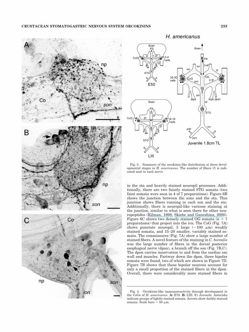
in the stn and heavily stained neuropil processes. Addi-tionally, there are two faintly stained STG somata (twofaint somata were seen in 4 of 7 preparations). Figure 6Bshows the junction between the sons and the stn. Thisjunction shows fibers running in each son and the stn.Additionally, there is neuropil-like varicose staining atthe junction, similar to what is seen there for other neu-ropeptides (Kilman, 1998; Skiebe and Ganeshina, 2000).Figure 6C shows two densely stained OG somata (n � 7preparations) that project into the ivn. The CoG (Fig. 7A)shows punctate neuropil, 3 large (�100 �m) weaklystained somata, and 15–20 smaller, variably stained so-mata. The commissures (Fig. 7A) show a large number ofstained fibers. A novel feature of the staining in C. borealiswas the large number of fibers in the dorsal posterioresophageal nerve (dpon), a branch off the son (Fig. 7B,C).The dpon carries innervation to and from the cardiac sacwall and muscles. Partway down the dpon, three bipolarsomata were found, two of which are shown in Figure 7D.Figure 7B shows that these bipolar neurons account foronly a small proportion of the stained fibers in the dpon.Overall, there were considerably more stained fibers in
Fig. 4. Orcokinin-like immunoreactivity through development inthe CoGs of H. americanus. A: E70. B: LIII. C: Juvenile. Asterisksindicate groups of lightly stained somata. Arrows show darkly stainedsomata. Scale bars � 50 �m.
Fig. 5. Summary of the orcokinin-like distribution at three devel-opmental stages in H. americanus. The number of fibers (f) is indi-cated next to each nerve.
233CRUSTACEAN STOMATOGASTRIC NERVOUS SYSTEM ORCOKININS

C. borealis than in H. americanus. In C. borealis therewere six to ten stained fibers in the stn, five or six stainedfibers in the son, three or four stained fibers in the ion, andthree or four stained fibers in the ivn.
Distribution of orcokinin-likeimmunoreactivity in the stomatogastric
nervous system of P. interruptus
The distribution of orcokinin-like immunoreactivity inP. interruptus was less extensive than in the other species(n � 5, three adults and two juveniles). Figure 8A showsorcokinin staining in the STG of a juvenile animal. The stndescending projection contains a tightly packed bundle offour to six small-diameter fibers that enter the STG andramify through the neuropil. No somata or posterior motornerves were stained. A novel feature of the P. interruptuspattern was the presence of two small (�20 �m) somata atthe junction of the sons and stn. Orcokinin-staining neu-ropil was also seen in the stn close to the junction with thesons (Fig. 8B). Figure 8C shows the presence of two so-mata in the OG that project axons up the ivn, toward thebrain (n � 5). The distribution of immunoreactivity inthe CoGs of P. interruptus is less extensive than seen inthe other animals and is shown in the summary diagramin Figure 9, along with the summary of the distribution ofimmunoreactivity in C. borealis.
Orcokinin staining in the pericardial organs
The crustacean POs are one of the major neurosecretorystructures in the animal, releasing numerous hormonesthat regulate a variety of physiological processes (Cooke
and Sullivan, 1982; Keller, 1992; Dircksen, 1997). Figure10 shows that dense orcokinin-like immunoreactivity ispresent in fibers and varicosities in the POs of all threespecies studied here (n � 5 H. americanus, n � 3 C.borealis, n � 5 P. interruptus).
MALDI analysis of orcokinin family peptides
For the preceding sections of this paper, we used anantibody to map the distribution of orcokinin-like neu-ropeptides. For this section we use biochemical techniquesto determine the identity of the peptides that give rise tothe orcokinin-like immunoreactivity. MALDI-MS is a pow-erful technique for the simultaneous detection of multiplepeptides present at significant levels in biological tissuesor even single cells with minimal sample preparation(Jimenez et al., 1994; Garden et al., 1996, 1998; Martin etal., 1999). Since the original orcokinin was isolated fromthe crayfish Orconectes limosus (Stangier et al., 1992),analogues of orcokinin have been identified in tissues fromthe shore crab Carcinus maenas (Bungart et al., 1995a).The sequence of the original orcokinin is Asn-Phe-Asp-Glu-Ile-Asp-Arg-Ser-Gly-Phe-Gly-Phe-Asn ([Asn13]), andthe three analogues of orcokinin differ in only one aminoacid residue and so are named [Ser9]-, [Ala13]-, and[Val13]orcokinin (Bungart et al., 1995a).
We identified a total of six orcokinin-related peptideswith MALDI-MS in C. borealis, H. americanus, and P.interruptus based on molecular weight measurements andthe known peptide sequences from O. limosus and C.maenas. Some of these peptides are unique to one species,and others are common to all three species (Table 1).
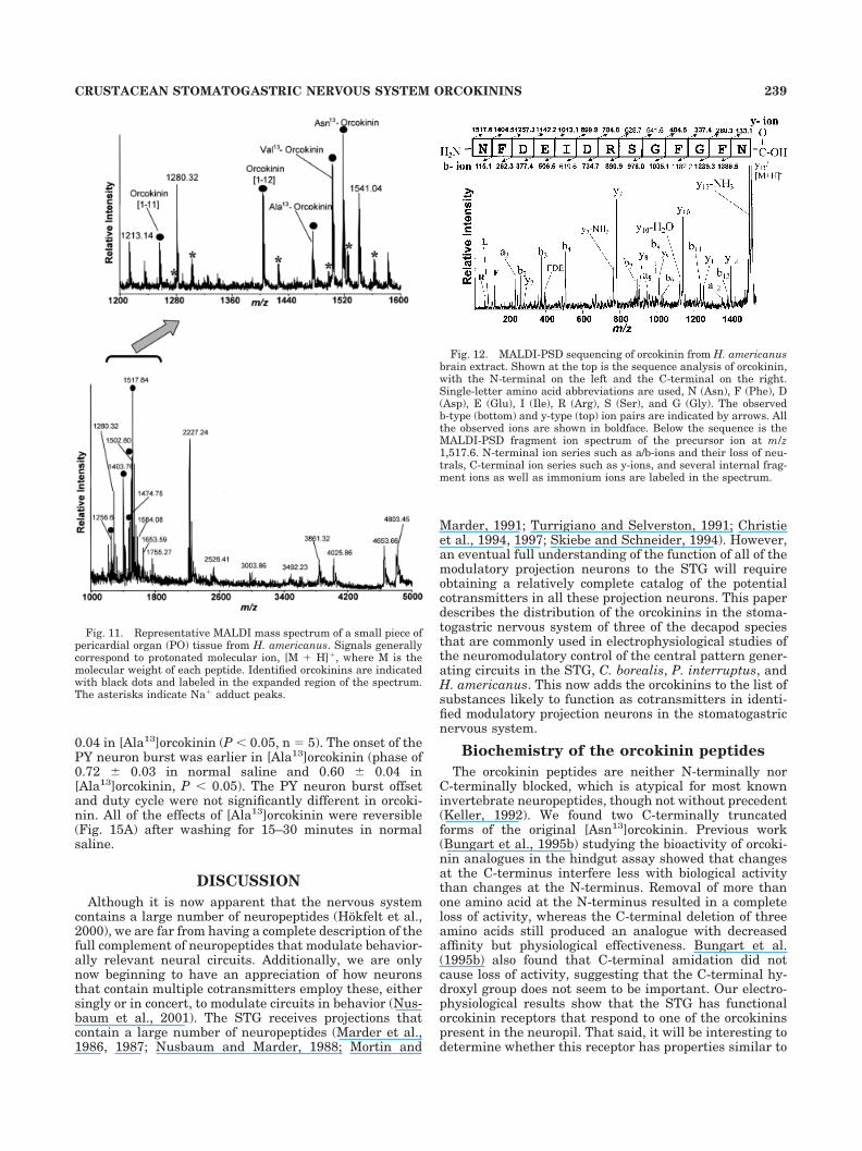
Because the immunocytochemistry studies showed in-tense orcokinin staining in POs, to verify the peptideidentity and determine the actual form(s) of the orcokininspresent in this neurosecretory tissue that contribute tothe orcokinin-like immunoreactivity seen in the POs,MALDI-MS peptide profiling of PO tissue from the threespecies was performed. Figure 11 shows a representativeMALDI mass spectrum of PO tissue from H. americanus.Five forms of orcokinins were present, including[Asn13]orcokinin, its two C-terminally truncated forms,orcokinin[1–11] and orcokinin[1–12], and [Ala13]- and[Val13]orcokinins. MALDI-MS analysis of PO tissue ex-tract from the crab C. borealis revealed the presence of[Ala13]-, [Val13]-, and [Ser9]orcokinins. PO tissue extractfrom P. interruptus contained the [Asn13]- and [Val13]-orcokinins. Although molecular weight information is cru-cial for peptide identification, unambiguous identificationis often achieved via mass spectrometric sequencing tech-niques. Therefore, we performed PSD (Spengler, 1997; Liet al., 1999) analysis of several orcokinin-containing LCfractions obtained from brain extracts to confirm theseassignments. Figure 12 shows a representative PSD frag-mentation ion spectrum of the precursor ion at m/z 1,517.6and the peptide sequence analysis from H. americanusbrain extract LC fraction. To use the established nomen-clature, cleavage of an amide bond produces b-type ions(charge is retained at the N-terminus) and y-type ions(charge is retained at the C-terminus; Siuzdak, 1996). Thesequence-specific fragment ions can thus be assigned asshown in Figure 12. By using the MS-Product databasedeveloped by the UCSF Mass Spectrometry Facility(http://prospector.ucsf.edu), with the entered orcokininpeptide sequence, fragment ions resulting from PSD pro-cesses were calculated by the software. The fragment ions
Fig. 6. Orcokinin-like immunoreactivity in STG and OG of adultC. borealis. A: STG and stn. Note the two faintly stained STG somata(arrows). B: Montage (of three micrographs) of the junction of the sonsand stn shows varicose ramifications (arrow). C: OG with two darklystained somata (arrows) projecting up into the ivn. Scale bars �50 �m.
234 L. LI ET AL.

detected in the obtained PSD spectra match well with thepredicted fragment pattern, which further confirms theidentity of [Asn13]orcokinin.
Putative [Ala13]orcokinin (m/z 1,474.7) was present inbrain extracts from C. borealis, H. americanus, and P.interruptus and was often the most intense peak in C.borealis tissue samples. Figure 13 shows the peptide se-quence of [Ala13]orcokinin (Fig. 13A) and PSD spectraobtained from the putative [Ala13]orcokinin containing LCfraction from C. borealis brain extract (Fig. 13B, upper
trace) and the synthetic [Ala13]orcokinin standard (Fig.13B, lower trace). The almost identical fragmentation pat-tern observed between the synthetic and the putative[Ala13]orcokinin PSD spectra confirms the identity of[Ala13]orcokinin in the brain extract. In fact, the extensivefragmentation obtained from the precursor ion at m/z1,474.6 in the brain extract allowed complete de novosequencing of the peptide. The immonium ions observed inthe low-mass region of the spectrum (as highlighted inFig. 13, inset) indicate the presence of amino acids, includ-
Fig. 7. Orcokinin staining in the CoGs and nerves of adult C. borealis. A: CoG. Arrows indicate twolarge, weakly stained somata. B: Branch of the dpon off of the son. C: Stained fibers splitting intodifferent branches of the dpon. D: Two biopolar somata (arrows) in the medial branch of the dpon. Scalebars � 50 �m.
235CRUSTACEAN STOMATOGASTRIC NERVOUS SYSTEM ORCOKININS

ing Ala (44 Da), Arg (129, 70, 87, 112 Da), Ile (86 Da), Asn(87 Da), Asp (88 Da), Glu (102 Da), and Phe (120 Da) andthe absence of His (110 Da). Mass signal pairs (mb � my �mprecursor � 1) at m/z 115.1/1,361.5, 262.3/1,214.3, 377.4/1,099.2, 506.5/970.0, 619.6/856.9, 734.7/741.8, 890.9/585.6, 978.0/498.5, 1035.1/441.5, 1182.2/294.3, 1239.3/237.3, and 1386.5/90.1 were assigned as paired b-type andy-type ions (see Fig. 13A). The b-type ions were deter-mined based on the detection of their corresponding a-typeions (loss of CO, yielding a 28 Da lower mass ion). Theamino acid sequence of the putative [Ala13]orcokinin peakwas deduced by using the mass difference between con-secutive b- or y-type ions. The sequence was determined tobe Asn-Phe-Asp-Glu-Ile/Leu-Asp-Arg-Ser-Gly-Phe-Gly-Phe-Ala, where the Ile and Leu could not be distinguishedbecause of the identical mass of both residues. However,the 100% identity to the known sequence strongly sug-gests an Ile instead of Leu. Three other orcokinin peptideswere confirmed similarly (data not shown). Thus, by usinga MALDI-based sequencing approach, all of the newlyidentified orcokinin peptides have been confirmed.
After confirming all of the identified orcokinin peptides,a MALDI-MS survey of the distribution of various forms oforcokinins in different tissues from three crustacean spe-cies was conducted (Table 1). As noted, some of the orco-kinin forms are unique to one species, and others are
Fig. 8. Orcokinin-like immunoreactivity in the stomatogastric ner-vous system of P. interruptus. A: STG. B: Junction of the stn and sons.Arrows indicate two small stained somata. C: OG. Arrowheads indi-cate the path of the two stained fibers in the ivn that project from thestained OG somata. The ivn is folded over the top of the left ion. Scalebars � 50 �m.
Fig. 9. Summary diagrams of the distribution of orcokinin-likeimmunoreactivity in the stomatogastric nervous system of C. borealisand juvenile (6 cm TL) P. interruptus.
236 L. LI ET AL.
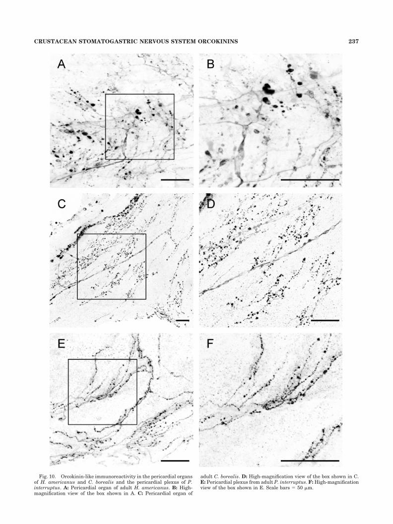
Fig. 10. Orcokinin-like immunoreactivity in the pericardial organsof H. americanus and C. borealis and the pericardial plexus of P.interruptus. A: Pericardial organ of adult H. americanus. B: High-magnification view of the box shown in A. C: Pericardial organ of
adult C. borealis. D: High-magnification view of the box shown in C.E: Pericardial plexus from adult P. interruptus. F: High-magnificationview of the box shown in E. Scale bars � 50 �m.
237CRUSTACEAN STOMATOGASTRIC NERVOUS SYSTEM ORCOKININS
present in all three species. For example, [Ser9]orcokininwas present only in C. borealis, whereas [Ala13]- and[Val13]orcokinins were detected in all three species. Fig-ure 14 shows representative MALDI mass spectra ob-tained from pieces of the STG from H. americanus and C.borealis. [Ser9]orcokinin was seen only in C. borealis, and[Asn13]orcokinin was detected only in H. americanus. How-ever, [Ala13]- and [Val13]orcokinins and two C-terminallytruncated orcokinins were detected in both species. Fur-thermore, MALDI mass profiling of CoG, STG, PO, andbrain tissue in C. borealis also revealed the presence of amass peak at m/z 1,554.7, which may correspond to an-other variant of orcokinin, [Thr8-His13]orcokinin, origi-nally isolated from the crayfish P. clarkii (Yasuda-Kamatani and Yasuda, 2000). Because of the relativelylow intensity of the signal, PSD fragmentation analysiswas not attempted. Thus, the assignment of this peak as[Thr8-His13]orcokinin remains tentative.
Single-cell MALDI was attempted from several identi-fied neurons from H. americanus. As shown in Table 1,direct MALDI assay of single PS neurons (n � 2, threereplicates for each cell) from H. americanus revealed thepresence of three forms of orcokinins, including [Ala13]-,[Val13]-, and [Asn13]orcokinins, corresponding to the in-tense orcokinin-like staining observed in this pair of neu-rons in the animal by immunocytochemistry.
Orcokinin-like immunoreactivity was present in theSTG and other regions of the nervous system in H. ameri-canus quite early in development. We were curiouswhether there was a developmental regulation of the formof orcokinin expressed. Therefore, we analyzed tissuesfrom the embryo and larval developmental stages (Table1). STGs from the embryo and early larvae showed allthree orcokinins found in the adult STG, [Ala13]-, [Asn13]-,and [Val13]orcokinin.
Electrophysiology
In previous bioassays, several orcokinin forms were bi-ologically active (Bungart et al., 1995b). As a first step indetermining the possible physiological roles of the orcoki-nins as potential modulators of the STG, we synthesized[Ala13]orcokinin for use in electrophysiological studies.We chose [Ala13]orcokinin because it is present in all threespecies studied and is often a prominent peak in theMALDI spectrum. Therefore, this peptide appeared to be agood candidate for a physiologically active orcokinin.
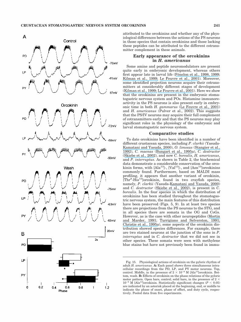
The pyloric rhythm of the STG consists of repeatingsequences of activity in the LP, PY, and PD neurons (Fig.15A). When [Ala13]orcokinin was applied in the bath, thispattern of activity changed. Specifically, the number of LPneuron spikes/burst decreased from 10 1.7 to 5.5 1.8(n � 5, P 0.05). This was accompanied by a decrease inthe duration of the LP burst from 470 5 to 238 6 msec(n � 5, P 0.05). The PY neurons depolarized slightly;consequently, the amplitude of the PY membrane poten-tial oscillations decreased in the presence of [Ala13]-orcokinin. Additionally, in all five preparations, there wasa modest increase in burst frequency with the applicationof [Ala13]orcokinin.
Figure 15B shows the effect of [Ala13]orcokinin on thephase relationships of the pyloric network neurons. Thesephase plots show the relative timing of the neurons’ dis-charge patterns when burst frequency is normalized. ThePD neurons’ duty cycles (fraction of the burst period forwhich the neuron was active) were unaffected by[Ala13]orcokinin. The LP neuron burst was terminatedearlier in orcokinin. Specifically, the LP off phase was0.8 0.02 in normal saline and 0.63 0.04 in orcokinin(P 0.01; n � 5, paired t-test). The LP neuron duty cyclewas shortened from 0.28 0.03 in control saline to 0.16
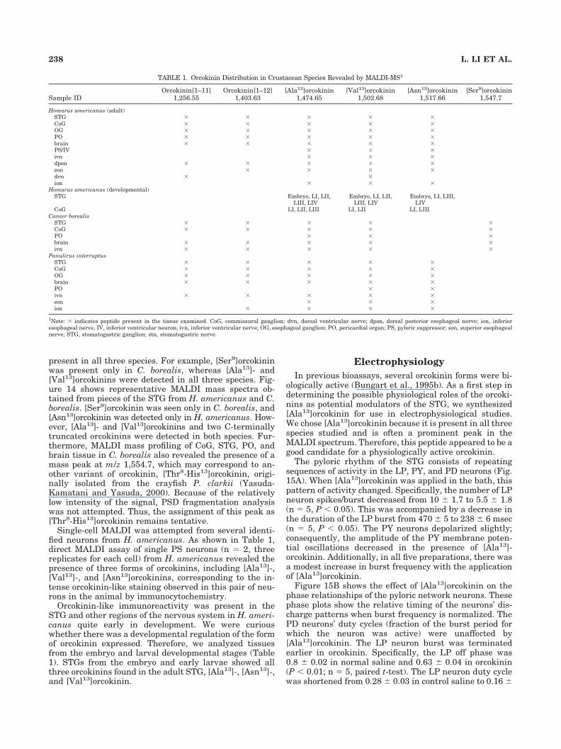
TABLE 1. Orcokinin Distribution in Crustacean Species Revealed by MALDI-MS1
Sample IDOrcokinin[1–11]
1,256.55Orcokinin[1–12]
1,403.63[Ala13]orcokinin
1,474.65[Val13]orcokinin
1,502.68[Asn13]orcokinin
1,517.66[Ser9]orcokinin
1,547.7
Homarus americanus (adult)STG � � � � �CoG � � � � �OG � � � � �PO � � � � �brain � � � � �PS/IV � � �ivn � � �dpon � � � � �son � � � �dvn � �ion � � �
Homarus americanus (developmental)STG Embryo, LI, LII,
LIII, LIVEmbryo, LI, LII,
LIII, LIVEmbryo, LI, LIII,
LIVCoG LI, LII, LIII LI, LII LI, LIII
Cancer borealisSTG � � � � �CoG � � � � �PO � � �brain � � � � �ivn � � � � �
Panulirus interruptusSTG � � � � �CoG � � � � �OG � � � � �brain � � � � �PO � �ivn � � � � �son � � �ion � � � �
1Note: � indicates peptide present in the tissue examined. CoG, commissural ganglion; dvn, dorsal ventricular nerve; dpon, dorsal posterior esophageal nerve; ion, inferioresophageal nerve, IV, inferior ventricular neuron; ivn, inferior ventricular nerve; OG, esophageal ganglion; PO, pericardial organ; PS, pyloric suppressor; son, superior esophagealnerve; STG, stomatogastric ganglion; stn, stomatogastric nerve.
238 L. LI ET AL.
0.04 in [Ala13]orcokinin (P 0.05, n � 5). The onset of thePY neuron burst was earlier in [Ala13]orcokinin (phase of0.72 0.03 in normal saline and 0.60 0.04 in[Ala13]orcokinin, P 0.05). The PY neuron burst offsetand duty cycle were not significantly different in orcoki-nin. All of the effects of [Ala13]orcokinin were reversible(Fig. 15A) after washing for 15–30 minutes in normalsaline.
DISCUSSION
Although it is now apparent that the nervous systemcontains a large number of neuropeptides (Hokfelt et al.,2000), we are far from having a complete description of thefull complement of neuropeptides that modulate behavior-ally relevant neural circuits. Additionally, we are onlynow beginning to have an appreciation of how neuronsthat contain multiple cotransmitters employ these, eithersingly or in concert, to modulate circuits in behavior (Nus-baum et al., 2001). The STG receives projections thatcontain a large number of neuropeptides (Marder et al.,1986, 1987; Nusbaum and Marder, 1988; Mortin and
Marder, 1991; Turrigiano and Selverston, 1991; Christieet al., 1994, 1997; Skiebe and Schneider, 1994). However,an eventual full understanding of the function of all of themodulatory projection neurons to the STG will requireobtaining a relatively complete catalog of the potentialcotransmitters in all these projection neurons. This paperdescribes the distribution of the orcokinins in the stoma-togastric nervous system of three of the decapod speciesthat are commonly used in electrophysiological studies ofthe neuromodulatory control of the central pattern gener-ating circuits in the STG, C. borealis, P. interruptus, andH. americanus. This now adds the orcokinins to the list ofsubstances likely to function as cotransmitters in identi-fied modulatory projection neurons in the stomatogastricnervous system.
Biochemistry of the orcokinin peptides
The orcokinin peptides are neither N-terminally norC-terminally blocked, which is atypical for most knowninvertebrate neuropeptides, though not without precedent(Keller, 1992). We found two C-terminally truncatedforms of the original [Asn13]orcokinin. Previous work(Bungart et al., 1995b) studying the bioactivity of orcoki-nin analogues in the hindgut assay showed that changesat the C-terminus interfere less with biological activitythan changes at the N-terminus. Removal of more thanone amino acid at the N-terminus resulted in a completeloss of activity, whereas the C-terminal deletion of threeamino acids still produced an analogue with decreasedaffinity but physiological effectiveness. Bungart et al.(1995b) also found that C-terminal amidation did notcause loss of activity, suggesting that the C-terminal hy-droxyl group does not seem to be important. Our electro-physiological results show that the STG has functionalorcokinin receptors that respond to one of the orcokininspresent in the neuropil. That said, it will be interesting todetermine whether this receptor has properties similar to
Fig. 11. Representative MALDI mass spectrum of a small piece ofpericardial organ (PO) tissue from H. americanus. Signals generallycorrespond to protonated molecular ion, [M � H]�, where M is themolecular weight of each peptide. Identified orcokinins are indicatedwith black dots and labeled in the expanded region of the spectrum.The asterisks indicate Na� adduct peaks.
Fig. 12. MALDI-PSD sequencing of orcokinin from H. americanusbrain extract. Shown at the top is the sequence analysis of orcokinin,with the N-terminal on the left and the C-terminal on the right.Single-letter amino acid abbreviations are used, N (Asn), F (Phe), D(Asp), E (Glu), I (Ile), R (Arg), S (Ser), and G (Gly). The observedb-type (bottom) and y-type (top) ion pairs are indicated by arrows. Allthe observed ions are shown in boldface. Below the sequence is theMALDI-PSD fragment ion spectrum of the precursor ion at m/z1,517.6. N-terminal ion series such as a/b-ions and their loss of neu-trals, C-terminal ion series such as y-ions, and several internal frag-ment ions as well as immonium ions are labeled in the spectrum.
239CRUSTACEAN STOMATOGASTRIC NERVOUS SYSTEM ORCOKININS

those described for crayfish hindgut. Moreover, it is cer-tainly possible that there are multiple orcokinin receptors,expressed either by different target tissues in the animalor even within the same tissue.
Although we found a number of different orcokininforms within each species, we saw no evidence that differ-ent analogues of this peptide family are found in differenttissues. Thus far, the biochemical data (Table 1) show thatmultiple forms of the orcokinins are found in the sametissue, and there is a simple correspondence to the distri-bution seen immunocytochemically and that seen by di-rect biochemical investigation. Although the antibody weused was raised against [Asn13]orcokinin, the very closestructural similarity of all of these forms makes it highlylikely that the antibody will label all of the orcokininforms that are detected biochemically. Therefore, MALDIdata from different regions of the stomatogastric nervoussystem and various connecting nerves are important todetermine the expression and distribution of various
forms of orcokinins. In fact, MALDI analyses of singleidentified orcokinin staining neurons, the PS cells, wereperformed to investigate whether individual neurons ex-press multiple forms of the orcokinins (see discussion be-low). In Procambarus clarkii, there are two orcokiningenes, both of which encode the sequences for [Ala13]-,[Val13]-, [Asn13]-, and [Thr8-His13]orcokinins (Yasuda-Kamatani and Yasuda, 2000). This suggests that multipleforms of the orcokinins might be commonly expressed inindividual neurons.
Orcokinins as cotransmitters inthe PS/IV neurons
Dando and Selverston (1972) first characterized thephysiological actions of a pair of neurons found in P.interruptus at the base of the brain (Claiborne and Sel-verston, 1984b) that project down the stn and significantlyalter the motor patterns of the STG. In P. interruptus,these neurons contain histamine (Claiborne and Selver-ston, 1984a) and FLRFamide-like peptides (Kilman,1998). In C. borealis the IV neuron somata are sometimesfound in the ivn and also contain histamine andFLRFamide-like peptides (Christie, 1995; Christie et al.,2002). In H. gammarus, the IV neurons are called thepyloric suppressor (PS) neurons, have been extensivelystudied electrophysiologically (Cazalets et al., 1990a,b;Meyrand et al., 1991, 1994), and also contain histamineand FLRFamide peptides (Le Feuvre et al., 2001). In H.americanus, the PS/IV neurons contain histamine (Mul-loney and Hall, 1991; Pulver et al., 2002). We show herewith immunocytochemistry that in H. americanus theseneurons also contain one or more of the orcokinins. Byusing MALDI-MS, peptide profiles from single isolated PSneurons from H. americanus were obtained. As shown inTable 1, three forms of orcokinins were detected, in addi-tion to the detection of two extended FLRFamide peptides,SDRNFLRFamide and TNRNFLRFamide (data notshown). Thus the PS neurons of H. americanus containhistamine, extended FLRFamide peptides, and orcoki-nins. Orcokinin is also seen in the PS neurons in thefreshwater crustacean C. destructor (Skiebe et al., 2002).Taken together, these data provide additional evidence forthe orcokinins as cotransmitters in the PS/IV neurons. Itwill be interesting to determine in the future whether anyof the physiological actions of the PS/IV neurons can be
Fig. 13. MALDI-PSD analysis of the putative [Ala13]orcokininpeptide. A: Sequence of orcokinin, with the N-terminal on the left andthe C-terminal on the right. Single-letter amino acid abbreviationsare used, N (Asn), F (Phe), D (Asp), E (Glu), I (Ile), R (Arg), S (Ser), G(Gly), and A (Ala). The observed b-type (bottom) and y-type (top) ionpairs are indicated by arrows. B: MALDI-PSD fragment ion spectra ofboth the ion at m/z 1,474.6 from the LC fraction of brain extract fromC. borealis (upper trace) and the synthetic orcokinin standard (lowertrace). N-terminal ion series such as a/b-ions and their loss of neu-trals, C-terminal ion series such as y-ions, and several internal frag-ment ions as well as immonium ions are labeled in the upper tracespectrum. The inset highlights the immonium ion region, with theamino acid residues labeled. The masses of the fragment ion signalslabeled with asterisks in the lower trace are identical to fragment ionsdetected in the upper trace.
Fig. 14. MALDI-MS of small pieces of STG tissues from C. borealis(A) and H. americanus (B). Peaks labeled with numbers representorcokinin peptides: 1, orcokinin[1–11]; 2, orcokinin[1–12]; 3,[Ala13]orcokinin; 4, [Val13]orcokinin; 5, [Ser9]orcokinin; 6,[Asn13]orcokinin.
240 L. LI ET AL.
attributed to the orcokinins and whether any of the phys-iological differences between the actions of the PS neuronsin those species that contain orcokinins and those lackingthese peptides can be attributed to the different cotrans-mitter complement in these animals.
Early appearance of the orcokininsin H. americanus
Some amine and peptide neuromodulators are presentquite early in embryonic development, whereas othersfirst appear late in larval life (Fenelon et al., 1998, 1999;Kilman et al., 1999; Le Feuvre et al., 2001). Moreover,some identified projection neurons acquire their cotrans-mitters at considerably different stages of development(Kilman et al., 1999; Le Feuvre et al., 2001). Here we showthat the orcokinins are present in the embryonic stoma-togastric nervous system and POs. Histamine immunore-activity in the PS neurons is also present early in embry-onic time in both H. gammarus (Le Feuvre et al., 2001)and H. americanus (Pulver et al., 2002). This suggeststhat the PS/IV neurons may acquire their full complementof cotransmitters early and that the PS neurons may playsignificant roles in the physiology of the embryonic andlarval stomatogastric nervous system.
Comparative studies
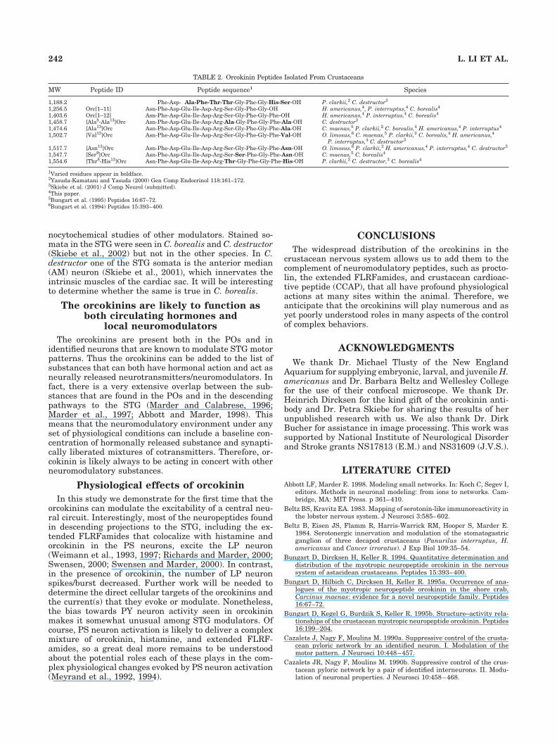
To date orcokinins have been identified in a number ofdifferent crustacean species, including P. clarkii (Yasuda-Kamatani and Yasuda, 2000), O. limosus (Stangier et al.,1992), C. maenas (Bungart et al., 1995a), C. destructor(Skiebe et al., 2002), and now C. borealis, H. americanus,and P. interruptus. As shown in Table 2, the biochemicaldata demonstrate a considerable conservation of the orco-kinin forms, with [Ala13]-, [Val13]-, and [Asn13]orcokininscommonly found. Furthermore, based on MALDI massprofiling, it appears that another variant of orcokinin,[Thr8-His13]orcokinin, found in two crayfish species,namely, P. clarkii (Yasuda-Kamatani and Yasuda, 2000)and C. destructor (Skiebe et al., 2002), is present in C.borealis. In the four species in which the distribution oforcokinins has been studied throughout the stomatogas-tric nervous system, the main features of this distributionhave been preserved (Figs. 5, 9). In at least two speciesthere are projections from the PS neurons to the STG, andin all species there are somata in the OG and CoGs.However, as is the case with other neuropeptides (Mortinand Marder, 1991; Turrigiano and Selverston, 1991;Christie et al., 1995a), some aspects of the orcokinin dis-tribution showed species differences. For example, thereare two stained neurons at the junction of the sons in P.interruptus and in C. destructor that we did not see inother species. These somata were seen with methyleneblue stains but have not previously been found in immu-
Fig. 15. Physiological actions of orcokinin on the pyloric rhythm ofadult H. americanus. A: Each panel shows three simultaneous intra-cellular recordings from the PD, LP, and PY motor neurons. Top,control. Middle, in the presence of 2 � 10–6 M [Ala13]orcokinin. Bot-tom, wash. B: Effects of orcokinin on the phase relations of the pyloricmotor pattern. Open bars, control; solid bars, in the presence of 2 �10–6 M [Ala13]orcokinin. Statistically significant changes (P 0.05)are indicated by an asterisk placed at the beginning, end, or middle toindicate the phase of onset, phase of offset, and duty cycle, respec-tively. Pooled data from five experiments.
241CRUSTACEAN STOMATOGASTRIC NERVOUS SYSTEM ORCOKININS
nocytochemical studies of other modulators. Stained so-mata in the STG were seen in C. borealis and C. destructor(Skiebe et al., 2002) but not in the other species. In C.destructor one of the STG somata is the anterior median(AM) neuron (Skiebe et al., 2001), which innervates theintrinsic muscles of the cardiac sac. It will be interestingto determine whether the same is true in C. borealis.
The orcokinins are likely to function asboth circulating hormones and
local neuromodulators
The orcokinins are present both in the POs and inidentified neurons that are known to modulate STG motorpatterns. Thus the orcokinins can be added to the list ofsubstances that can both have hormonal action and act asneurally released neurotransmitters/neuromodulators. Infact, there is a very extensive overlap between the sub-stances that are found in the POs and in the descendingpathways to the STG (Marder and Calabrese, 1996;Marder et al., 1997; Abbott and Marder, 1998). Thismeans that the neuromodulatory environment under anyset of physiological conditions can include a baseline con-centration of hormonally released substance and synapti-cally liberated mixtures of cotransmitters. Therefore, or-cokinin is likely always to be acting in concert with otherneuromodulatory substances.
Physiological effects of orcokinin
In this study we demonstrate for the first time that theorcokinins can modulate the excitability of a central neu-ral circuit. Interestingly, most of the neuropeptides foundin descending projections to the STG, including the ex-tended FLRFamides that colocalize with histamine andorcokinin in the PS neurons, excite the LP neuron(Weimann et al., 1993, 1997; Richards and Marder, 2000;Swensen, 2000; Swensen and Marder, 2000). In contrast,in the presence of orcokinin, the number of LP neuronspikes/burst decreased. Further work will be needed todetermine the direct cellular targets of the orcokinins andthe current(s) that they evoke or modulate. Nonetheless,the bias towards PY neuron activity seen in orcokininmakes it somewhat unusual among STG modulators. Ofcourse, PS neuron activation is likely to deliver a complexmixture of orcokinin, histamine, and extended FLRF-amides, so a great deal more remains to be understoodabout the potential roles each of these plays in the com-plex physiological changes evoked by PS neuron activation(Meyrand et al., 1992, 1994).
CONCLUSIONS
The widespread distribution of the orcokinins in thecrustacean nervous system allows us to add them to thecomplement of neuromodulatory peptides, such as procto-lin, the extended FLRFamides, and crustacean cardioac-tive peptide (CCAP), that all have profound physiologicalactions at many sites within the animal. Therefore, weanticipate that the orcokinins will play numerous and asyet poorly understood roles in many aspects of the controlof complex behaviors.
ACKNOWLEDGMENTS
We thank Dr. Michael Tlusty of the New EnglandAquarium for supplying embryonic, larval, and juvenile H.americanus and Dr. Barbara Beltz and Wellesley Collegefor the use of their confocal microscope. We thank Dr.Heinrich Dircksen for the kind gift of the orcokinin anti-body and Dr. Petra Skiebe for sharing the results of herunpublished research with us. We also thank Dr. DirkBucher for assistance in image processing. This work wassupported by National Institute of Neurological Disorderand Stroke grants NS17813 (E.M.) and NS31609 (J.V.S.).
LITERATURE CITED
Abbott LF, Marder E. 1998. Modeling small networks. In: Koch C, Segev I,editors. Methods in neuronal modeling: from ions to networks. Cam-bridge, MA: MIT Press. p 361–410.
Beltz BS, Kravitz EA. 1983. Mapping of serotonin-like immunoreactivity inthe lobster nervous system. J Neurosci 3:585–602.
Beltz B, Eisen JS, Flamm R, Harris-Warrick RM, Hooper S, Marder E.1984. Serotonergic innervation and modulation of the stomatogastricganglion of three decapod crustaceans (Panurilus interruptus, H.americanus and Cancer irroratus). J Exp Biol 109:35–54.
Bungart D, Dircksen H, Keller R. 1994. Quantitative determination anddistribution of the myotropic neuropeptide orcokinin in the nervoussystem of astacidean crustaceans. Peptides 15:393–400.
Bungart D, Hilbich C, Dircksen H, Keller R. 1995a. Occurrence of ana-logues of the myotropic neuropeptide orcokinin in the shore crab,Carcinus maenas: evidence for a novel neuropeptide family. Peptides16:67–72.
Bungart D, Kegel G, Burdzik S, Keller R. 1995b. Structure–activity rela-tionships of the crustacean myotropic neuropeptide orcokinin. Peptides16:199–204.
Cazalets J, Nagy F, Moulins M. 1990a. Suppressive control of the crusta-cean pyloric network by an identified neuron. I. Modulation of themotor pattern. J Neurosci 10:448–457.
Cazalets JR, Nagy F, Moulins M. 1990b. Suppressive control of the crus-tacean pyloric network by a pair of identified interneurons. II. Modu-lation of neuronal properties. J Neurosci 10:458–468.
TABLE 2. Orcokinin Peptides Isolated From Crustaceans
MW Peptide ID Peptide sequence1 Species
1,188.2 Phe-Asp- Ala-Phe-Thr-Thr-Gly-Phe-Gly-His-Ser-OH P. clarkii,2 C. destructor3
1,256.5 Orc[1–11] Asn-Phe-Asp-Glu-Ile-Asp-Arg-Ser-Gly-Phe-Gly-OH H. americanus,4, P. interruptus,4 C. borealis4
1,403.6 Orc[1–12] Asn-Phe-Asp-Glu-Ile-Asp-Arg-Ser-Gly-Phe-Gly-Phe-OH H. americanus,4 P. interruptus,4 C. borealis4
1,458.7 [Ala8-Ala13]Orc Asn-Phe-Asp-Glu-Ile-Asp-Arg-Ala-Gly-Phe-Gly-Phe-Ala-OH C. destructor3
1,474.6 [Ala13]Orc Asn-Phe-Asp-Glu-Ile-Asp-Arg-Ser-Gly-Phe-Gly-Phe-Ala-OH C. maenas,5 P. clarkii,2 C. borealis,4 H. americanus,4 P. interruptus4
1,502.7 [Val13]Orc Asn-Phe-Asp-Glu-Ile-Asp-Arg-Ser-Gly-Phe-Gly-Phe-Val-OH O. limosus,6 C. maenas,5 P. clarkii,2 C. borealis,4 H. americanus,4
P. interruptus,4 C. destructor3
1,517.7 [Asn13]Orc Asn-Phe-Asp-Glu-Ile-Asp-Arg-Ser-Gly-Phe-Gly-Phe-Asn-OH O. limosus,6 P. clarkii,2 H. americanus,4 P. interruptus,4 C. destructor3
1,547.7 [Ser9]Orc Asn-Phe-Asp-Glu-Ile-Asp-Arg-Ser-Ser-Phe-Gly-Phe-Asn-OH C. maenas,5 C. borealis4
1,554.6 [Thr8-His13]Orc Asn-Phe-Asp-Glu-Ile-Asp-Arg-Thr-Gly-Phe-Gly-Phe-His-OH P. clarkii,2 C. destructor,3 C. borealis4
1Varied residues appear in boldface.2Yasuda-Kamatani and Yasuda (2000) Gen Comp Endocrinol 118:161–172.3Skiebe et al. (2001) J Comp Neurol (submitted).4This paper.5Bungart et al. (1995) Peptides 16:67–72.6Bungart et al. (1994) Peptides 15:393–400.
242 L. LI ET AL.
Christie A. 1995. Chemical neuroanatomy of the crab stomatogastric gan-glion: a study using immunocytochemistry and laser scanning confocalmicroscopy. PhD thesis, Department of Biology, Brandeis University. p156.
Christie AE, Hall C, Oshinsky M, Marder E. 1994. Buccalin-like andmyomodulin-like peptides in the stomatogastric ganglion of the crabCancer borealis. J Exp Biol 193:337–343.
Christie AE, Baldwin D, Turrigiano G, Graubard K, Marder E. 1995a.Immunocytochemical localization of multiple cholecystokinin-like pep-tides in the stomatogastric nervous system of the crab, Cancer borealis.J Exp Biol 198:263–271.
Christie AE, Skiebe P, Marder E. 1995b. Matrix of neuromodulators inneurosecretory structures of the crab, Cancer borealis. J Exp Biol198:2431–2439.
Christie AE, Lundquist T, Nassel DR, Nusbaum MP. 1997. Two noveltachykinin-related peptides from the nervous system of the crab Can-cer borealis. J Exp Biol 200:2279–2294.
Christie AE, Stein W, Quinlan JE, Beenhakker MP, Marder E, NusbaumMP. 2002. Actions of a histaminergic/peptidergic projection neuron onrhythmic motor patterns in the stomatogastric nervous system of thecrab Cancer borealis. In preparation.
Claiborne B, Selverston A. 1984a. Histamine as a neurotransmitter in thestomatogastric nervous system of the spiny lobster. J Neurosci 4:708–721.
Claiborne B, Selverston A. 1984b. Localization of stomatogastric IV neuroncell bodies in the lobster brain. J Comp Physiol 154:27–32.
Cooke IM, Sullivan RE. 1982. Hormones and neurosecretion. In: AtwoodHL, Sandeman DC, editors. The biology of Crustacea: neurobiology.New York: Academic Press. p 205–290.
Dando MR, Selverston AI. 1972. Command fibres from the supraoesopha-geal ganglion to the stomatogastric ganglion in Panulirus argus.J Comp Physiol 78:138–175.
Dircksen H. 1997. Conserved crustacean cardioactive peptide: neural net-works and function in arthropod evolution. In: Coast GM, Webster SG,editors. Arthropod endocrinology—perspectives and recent advances.Cambridge: Cambridge University Press.
Dircksen H, Burdzik S, Sauter A, Keller R. 2000. Two orcokinins and thenovel octapeptide orcomyotropin in the hindgut of the crayfish Or-conectes limosus: identified myostimulatory neuropeptides originatingtogether in neurones of the terminal abdominal ganglion. J Exp Biol203:2807–2818.
Fenelon V, Casasnovas B, Faumont S, Meyrand P. 1998. Ontogeneticalteration in peptidergic expression within a stable neuronal popula-tion in lobster stomatogastric nervous system. J Comp Neurol 399:289–305.
Fenelon VS, Kilman V, Meyrand P, Marder E. 1999. Sequential develop-mental acquisition of neuromodulatory inputs to a central pattern-generating network. J Comp Neurol 408:335–351.
Floyd PD, Li L, Moroz TP, Sweedler JV. 1999. Characterization of peptidesfrom Aplysia using microbore liquid chromatography with matrix-assisted laser desorption/ionization time-of-flight mass spectrometryguided purification. J Chromalog A 830:105–113.
Garden RW, Moroz LL, Moroz TP, Shippy SA, Sweedler JV. 1996. Excesssalt removal with matrix rinsing: direct peptide profiling of neuronsfrom marine invertebrates using matrix-assisted laser desorption/ionization time-of-flight mass spectrometry. J Mass Spectrom 31:1126–1130.
Garden RW, Shippy SA, Li L, Moroz TP, Sweedler JV. 1998. Proteolyticprocessing of the Aplysia egg-laying hormone prohormone. Proc NatlAcad Sci USA 95:3972–3977.
Harris-Warrick RM, Marder E, Selverston AI, Moulins M. 1992. Dynamicbiological networks. The stomatogastric nervous system. Cambridge,MA: MIT Press.
Helluy SM, Beltz BS. 1991. Embryonic development of the Americanlobster: quantitative staging and characerization of an embryonic moltcycle. Biol Bull 180:355–371.
Hokfelt T, Broberger C, Xu ZQ, Sergeyev V, Ubink R, Diez M. 2000.Neuropeptides—an overview. Neuropharmacology 39:1337–1356.
Jimenez CR, van Veelen PA, Li KW, Wildering WC, Geraerts WPM, TjadenUR, van der Greef J. 1994. Neuropeptide expression and processing asrevealed by direct matrix-assisted laser desorption ionization massspectrometry of single neurons. J Neurochem 62:404–409.
Keller R. 1992. Crustacean neuropeptides: structures, functions and com-parative aspects. Experientia 48:439–448.
Kilman VL. 1998. Multiple roles of neuromodulators throughout life: an
anatomical study of the crustacean stomatogastric nervous system.PhD thesis, Neuroscience Program, Brandeis University. p 173.
Kilman VL, Fenelon V, Richards KS, Thirumalai V, Meyrand P, Marder E.1999. Sequential developmental acquisition of cotransmitters in iden-tified sensory neurons of the stomatogastric nervous system of thelobsters, Homarus americanus and Homarus gammarus. J Comp Neu-rol 408:318–334.
Le Feuvre Y, Fenelon VS, Meyrand P. 2001. Ontogeny of modulatoryinputs to motor networks: early established projection and progressiveneurotransmitter acquisition. J Neurosci 21:1313–1326.
Li L, Garden RW, Romanova EV, Sweedler JV. 1999. In situ sequencing ofpeptides from biological tissues and single cells using MALDI-PSD/CIDanalysis. Anal Chem 71:5451–5458.
Li L, Pulver S, Kelley WP, Sweedler JV, Marder E. 2001. Comparativestudies of the orcokinin peptides in crustacean species by immuno-chemistry and mass spectrometry. Soc Neurosci Abstr 27:909.1.
Marder E, Calabrese RL. 1996. Principles of rhythmic motor pattern gen-eration. Physiol Rev 76:687–717.
Marder E, Hooper SL, Siwicki KK. 1986. Modulatory action and distribu-tion of the neuropeptide proctolin in the crustacean stomatogastricnervous system. J Comp Neurol 243:454–467.
Marder E, Calabrese RL, Nusbaum MP, Trimmer B. 1987. Distributionand partial characterization of FMRFamide-like peptides in the stoma-togastric nervous systems of the rock crab, Cancer borealis, and thespiny lobster, Panulirus interruptus. J Comp Neurol 259:150–163.
Marder E, Christie AE, Kilman VL. 1995. Functional organization ofcotransmission systems: lessons from small nervous systems. InvertNeurosci 1:105–112.
Marder E, Jorge-Rivera JC, Kilman V, Weimann JM. 1997. Peptidergicmodulation of synaptic transmission in a rhythmic motor system. In:Festoff BW, Hantaı D, Citron BA, editors. Advances in organ biology.Greenwich, CT: JAI Press Inc. p 213–233.
Martin G, Sorokine O, Moniatte M, Juchault P, Van Dorsselaer A. 1999.Profiling the peptides in two neurohemal organs, the sinus gland andlateral nervous plexus of terrestrial isopods (Crustacea), by mass spec-trometry. Can J Zool 77:1300–1308.
Meyrand P, Simmers J, Moulins M. 1991. Construction of a pattern-generating circuit with neurons of different networks. Nature 351:60–63.
Meyrand P, Weimann JM, Marder E. 1992. Multiple axonal spike initia-tion zones in a motor neuron: serotonin activation. J Neurosci 12:2803–2812.
Meyrand P, Simmers J, Moulins M. 1994. Dynamic construction of a neuralnetwork from multiple pattern generators in the lobster stomatogastricnervous system. J Neurosci 14:630–644.
Meyrand P, Faumont S, Simmers J, Christie AE, Nusbaum MP. 2000.Species-specific modulation of pattern-generating circuits. Eur J Neu-rosci 12:2585–2596.
Mortin LI, Marder E. 1991. Differential distribution of �-pigment dispers-ing hormone (�-PDH)-like immunoreactivity in the stomatogastric ner-vous system of five species of decapod crustaceans. Cell Tissue Res265:19–33.
Mulloney B, Hall WM. 1991. Neurons with histaminelike immunoreactiv-ity in the segmental and stomatogastric nervous systems of the crayfishPacifastacus leniusculus and the lobster Homarus americanus. CellTissue Res 266:197–207.
Nusbaum MP, Marder E. 1988. A neuronal role for a crustacean redpigment concentrating hormone-like peptide: neuromodulation of thepyloric rhythm in the crab, Cancer borealis. J Exp Biol 135:165–181.
Nusbaum MP, Blitz DM, Swensen AM, Wood D, Marder E. 2001. The rolesof co-transmission in neural network modulation. Trends Neurosci24:146–154.
Pulver SR, Thirumalai V, Richards KS, Marder E. 2002. The distributionand actions of histamine in the adult and developing stomatogastricnervous system of the lobster, Homarus americanus. In preparation.
Richards KS, Marder E. 2000. The actions of crustacean cardioactivepeptide on adult and developing stomatogastric ganglion motor pat-terns. J Neurobiol 44:31–44.
Siuzdak G. 1996. Sequencing of peptides and proteins. In: Siuzdak G,editor. Mass spectrometry for biotechnology. San Diego: AcademicPress. p 93–99.
Skiebe P. 1999. Allatostatin-like immunoreactivity within the stomatogas-tric nervous system and the pericardial organs of the crab Cancerpagurus, the lobster Homarus americanus, and the crayfish Cheraxdestructor and Procambarus clarkii. J Comp Neurol 403:85–105.
243CRUSTACEAN STOMATOGASTRIC NERVOUS SYSTEM ORCOKININS
Skiebe P, Ganeshina O. 2000. Synaptic neuropil in nerves of the crustaceanstomatogastric nervous system: an immunocytochemical and electronmicroscopical study. J Comp Neurol 420:373–397.
Skiebe P, Schneider H. 1994. Allatostatin peptides in the crab stomatogas-tric nervous system: inhibition of the pyloric motor pattern and distri-bution of allatostatin-like immunoreactivity. J Exp Biol 194:195–208.
Skiebe P, Dreger M, Meseke M, Evers JF, Hucho F. 2002. Identification oforcokinins in single neurons in the stomatgastric nervous system of thecrayfish, Cherax destructor. J Comp Neurol 444:245–259.
Spengler B. 1997. Post-source decay analysis in matrix-assisted laserdesorption/ionization mass spectrometry of biomolecules. J Mass Spec-trom. 32:1019–1036.
Stangier J, Hilbich C, Burdzik S, Keller R. 1992. Orcokinin: a novelmyotropic peptide from the nervous system of the crayfish, Orconecteslimosus. Peptides 13:859–864.
Stefanini M, De Martino C, Zamboni L. 1967. Fixation of ejaculated sper-matozoa for electron microscopy. Nature 216:173–174.
Swensen AM. 2000. Network consequences of convergent modulation in
the stomatogastric nervous system of the crab, Cancer borealis. Thesis,Neuroscience Progam, Brandeis University. p 143.
Swensen AM, Marder E. 2000. Multiple peptides converge to activate thesame voltage-dependent current in a central pattern-generating cir-cuit. J Neurosci 20:6752–6759.
Turrigiano GG, Selverston AI. 1991. Distribution of cholecystokinin-likeimmunoreactivity within the stomatogastric nervous systems of fourspecies of decapod crustacea. J Comp Neurol 305:164–176.
Weimann JM, Marder E, Evans B, Calabrese RL. 1993. The effects of SDRN-FLRFamide and TNRNFLRFamide on the motor patterns of the stoma-togastric ganglion of the crab Cancer borealis. J Exp Biol 181:1–26.
Weimann JM, Skiebe P, Heinzel H-G, Soto C, Kopell N, Jorge-Rivera JC,Marder E. 1997. Modulation of oscillator interactions in the crab sto-matogastric ganglion by crustacean cardioactive peptide. J Neurosci17:1748–1760.
Yasuda-Kamatani Y, Yasuda A. 2000. Identification of orcokinin gene-relatedpeptides in the brain of the crayfish Procambarus clarkii by the combina-tion of MALDI-TOF and on-line capillary HPLC/Q-Tof mass spectrome-tries and molecular cloning. Gen Comp Endocrinol 118:161–172.
244 L. LI ET AL.
Copyright © 2022 FDOKUMEN


![[Clinical and laboratory features of patients with pericardial effusion]](https://static.fdokumen.com/doc/165x107/6325f6805547884b71068814/clinical-and-laboratory-features-of-patients-with-pericardial-effusion.jpg)