Oral Methylthioadenosine Administration Attenuates Fibrosis and Chronic Liver Disease Progression in...
15
Oral Methylthioadenosine Administration Attenuates Fibrosis and Chronic Liver Disease Progression in Mdr22/2 Mice M. Ujue Latasa 1. , Carmen Gil-Puig 1,2. , Maite G. Ferna ´ ndez-Barrena 1,3 , Carlos M. Rodrı´guez-Ortigosa 1,3 , Jesu ´ s M. Banales 1,3 , Raquel Urtasun 1 , Saioa Gon ˜i 1 , Miriam Me ´ ndez 1 , Sara Arcelus 3 , Nerea Juanarena 1 , Juan A. Recio 4 , Sophie Lotersztajn 5,6 , Jesu ´ s Prieto 1,3 , Carmen Berasain 1 , Fernando J. Corrales 1 , Jon Lecanda 1,2" , Matı´as A. A ´ vila 1 * " 1 Division of Hepatology and Gene Therapy, CIMA, University of Navarra, Pamplona, Spain, 2 Digna Biotech, Madrid, Spain, 3 CIBERehd, University Clinic, University of Navarra, Pamplona, Spain, 4 Vall d’Hebron Research Institute, Institute of Oncology and Hospital, Barcelona, Spain, 5 Inserm, U955, Cre ´ teil, France, 6 Universite ´ Paris-Est, Faculte ´ de Me ´ decine, UMR-S955, Cre ´ teil, France Abstract Background: Inflammation and fibrogenesis are directly related to chronic liver disease progression, including hepatocellular carcinoma (HCC) development. Currently there are few therapeutic options available to inhibit liver fibrosis. We have evaluated the hepatoprotective and anti-fibrotic potential of orally-administered 59-methylthioadenosine (MTA) in Mdr2 2/2 mice, a clinically relevant model of sclerosing cholangitis and spontaneous biliary fibrosis, followed at later stages by HCC development. Methodology: MTA was administered daily by gavage to wild type and Mdr2 2/2 mice for three weeks. MTA anti- inflammatory and anti-fibrotic effects and potential mechanisms of action were examined in the liver of Mdr2 2/2 mice with ongoing fibrogenesis and in cultured liver fibrogenic cells (myofibroblasts). Principal Findings: MTA treatment reduced hepatomegaly and liver injury. a-Smooth muscle actin immunoreactivity and collagen deposition were also significantly decreased. Inflammatory infiltrate, the expression of the cytokines IL6 and Mcp-1, pro-fibrogenic factors like TGFb2 and tenascin-C, as well as pro-fibrogenic intracellular signalling pathways were reduced by MTA in vivo. MTA inhibited the activation and proliferation of isolated myofibroblasts and down-regulated cyclin D1 gene expression at the transcriptional level. The expression of JunD, a key transcription factor in liver fibrogenesis, was also reduced by MTA in activated myofibroblasts. Conclusions/Significance: Oral MTA administration was well tolerated and proved its efficacy in reducing liver inflammation and fibrosis. MTA may have multiple molecular and cellular targets. These include the inhibition of inflammatory and pro- fibrogenic cytokines, as well as the attenuation of myofibroblast activation and proliferation. Downregulation of JunD and cyclin D1 expression in myofibroblasts may be important regarding the mechanism of action of MTA. This compound could be a good candidate to be tested for the treatment of (biliary) liver fibrosis. Citation: Latasa MU, Gil-Puig C, Ferna ´ndez-Barrena MG, Rodrı ´guez-Ortigosa CM, Banales JM, et al. (2010) Oral Methylthioadenosine Administration Attenuates Fibrosis and Chronic Liver Disease Progression in Mdr22/2 Mice. PLoS ONE 5(12): e15690. doi:10.1371/journal.pone.0015690 Editor: Massimo Federici, University of Tor Vergata, Italy Received August 17, 2010; Accepted November 21, 2010; Published December 29, 2010 Copyright: ß 2010 Latasa et al. This is an open-access article distributed under the terms of the Creative Commons Attribution License, which permits unrestricted use, distribution, and reproduction in any medium, provided the original author and source are credited. Funding: This work was supported by the agreement between FIMA and the "UTE project CIMA"; RTICC-RD06 00200061 (CB, FJC, MAA), CiberEhd (MGF-B, JB, CRO, JP), FIS-PI070392 and PI070402 (CB, MAA); SAF2008-01540 (FJC); Instituto de Salud Carlos III Fellowship (SG); Ramo ´ n y Cajal Program (MUL) and Torres Quevedo Program (CG-P, RU); INSERM, Universite ´ Paris-Est, the Agence Nationale de la Recherche and the Fondation pour la Recherche Medicale (SL). The funders had no role in study design, data collection and analysis, decision to publish, or preparation of the manuscript. Competing Interests: The authors have declared that no competing interests exist. * E-mail: [email protected] . These authors contributed equally to this work. " These authors also contributed equally to this work. Introduction Liver diseases are currently the fifth cause of mortality in the Western world, and as opposed to other major causes of mortality their incidence is increasing [1,2]. The principal causes of the most common liver ailments are well known, and include chronic alcohol consumption, infection by hepatitis viruses, genetic conditions such as hemochromatosis or a1-antitrypsin deficiency, autoimmune hepatitis and metabolic disorders [1,2]. Regardless of the etiology, chronic tissue injury and inflammation are increas- ingly recognized as driver mechanisms in liver disease progression from fibrosis, characterized by the excessive accumulation of extracellular matrix (ECM), to hepatocellular carcinoma (HCC) [1–3]. The molecular links in the so-called inflammation-fibrosis- PLoS ONE | www.plosone.org 1 December 2010 | Volume 5 | Issue 12 | e15690
-
Upload
independent -
Category
Documents
-
view
2 -
download
0
Transcript of Oral Methylthioadenosine Administration Attenuates Fibrosis and Chronic Liver Disease Progression in...
Oral Methylthioadenosine Administration AttenuatesFibrosis and Chronic Liver Disease Progression inMdr22/2 MiceM. Ujue Latasa1., Carmen Gil-Puig1,2., Maite G. Fernandez-Barrena1,3, Carlos M. Rodrıguez-Ortigosa1,3,
Jesus M. Banales1,3, Raquel Urtasun1, Saioa Goni1, Miriam Mendez1, Sara Arcelus3, Nerea Juanarena1,
Juan A. Recio4, Sophie Lotersztajn5,6, Jesus Prieto1,3, Carmen Berasain1, Fernando J. Corrales1, Jon
Lecanda1,2", Matıas A. Avila1*"
1 Division of Hepatology and Gene Therapy, CIMA, University of Navarra, Pamplona, Spain, 2 Digna Biotech, Madrid, Spain, 3 CIBERehd, University Clinic, University of
Navarra, Pamplona, Spain, 4 Vall d’Hebron Research Institute, Institute of Oncology and Hospital, Barcelona, Spain, 5 Inserm, U955, Creteil, France, 6 Universite Paris-Est,
Faculte de Medecine, UMR-S955, Creteil, France
Abstract
Background: Inflammation and fibrogenesis are directly related to chronic liver disease progression, includinghepatocellular carcinoma (HCC) development. Currently there are few therapeutic options available to inhibit liver fibrosis.We have evaluated the hepatoprotective and anti-fibrotic potential of orally-administered 59-methylthioadenosine (MTA) inMdr22/2 mice, a clinically relevant model of sclerosing cholangitis and spontaneous biliary fibrosis, followed at later stagesby HCC development.
Methodology: MTA was administered daily by gavage to wild type and Mdr22/2 mice for three weeks. MTA anti-inflammatory and anti-fibrotic effects and potential mechanisms of action were examined in the liver of Mdr22/2 mice withongoing fibrogenesis and in cultured liver fibrogenic cells (myofibroblasts).
Principal Findings: MTA treatment reduced hepatomegaly and liver injury. a-Smooth muscle actin immunoreactivity andcollagen deposition were also significantly decreased. Inflammatory infiltrate, the expression of the cytokines IL6 and Mcp-1,pro-fibrogenic factors like TGFb2 and tenascin-C, as well as pro-fibrogenic intracellular signalling pathways were reduced byMTA in vivo. MTA inhibited the activation and proliferation of isolated myofibroblasts and down-regulated cyclin D1 geneexpression at the transcriptional level. The expression of JunD, a key transcription factor in liver fibrogenesis, was alsoreduced by MTA in activated myofibroblasts.
Conclusions/Significance: Oral MTA administration was well tolerated and proved its efficacy in reducing liver inflammationand fibrosis. MTA may have multiple molecular and cellular targets. These include the inhibition of inflammatory and pro-fibrogenic cytokines, as well as the attenuation of myofibroblast activation and proliferation. Downregulation of JunD andcyclin D1 expression in myofibroblasts may be important regarding the mechanism of action of MTA. This compound couldbe a good candidate to be tested for the treatment of (biliary) liver fibrosis.
Citation: Latasa MU, Gil-Puig C, Fernandez-Barrena MG, Rodrıguez-Ortigosa CM, Banales JM, et al. (2010) Oral Methylthioadenosine Administration AttenuatesFibrosis and Chronic Liver Disease Progression in Mdr22/2 Mice. PLoS ONE 5(12): e15690. doi:10.1371/journal.pone.0015690
Editor: Massimo Federici, University of Tor Vergata, Italy
Received August 17, 2010; Accepted November 21, 2010; Published December 29, 2010
Copyright: � 2010 Latasa et al. This is an open-access article distributed under the terms of the Creative Commons Attribution License, which permitsunrestricted use, distribution, and reproduction in any medium, provided the original author and source are credited.
Funding: This work was supported by the agreement between FIMA and the "UTE project CIMA"; RTICC-RD06 00200061 (CB, FJC, MAA), CiberEhd (MGF-B, JB,CRO, JP), FIS-PI070392 and PI070402 (CB, MAA); SAF2008-01540 (FJC); Instituto de Salud Carlos III Fellowship (SG); Ramon y Cajal Program (MUL) and TorresQuevedo Program (CG-P, RU); INSERM, Universite Paris-Est, the Agence Nationale de la Recherche and the Fondation pour la Recherche Medicale (SL). The fundershad no role in study design, data collection and analysis, decision to publish, or preparation of the manuscript.
Competing Interests: The authors have declared that no competing interests exist.
* E-mail: [email protected]
. These authors contributed equally to this work.
" These authors also contributed equally to this work.
Introduction
Liver diseases are currently the fifth cause of mortality in the
Western world, and as opposed to other major causes of mortality
their incidence is increasing [1,2]. The principal causes of the most
common liver ailments are well known, and include chronic
alcohol consumption, infection by hepatitis viruses, genetic
conditions such as hemochromatosis or a1-antitrypsin deficiency,
autoimmune hepatitis and metabolic disorders [1,2]. Regardless of
the etiology, chronic tissue injury and inflammation are increas-
ingly recognized as driver mechanisms in liver disease progression
from fibrosis, characterized by the excessive accumulation of
extracellular matrix (ECM), to hepatocellular carcinoma (HCC)
[1–3]. The molecular links in the so-called inflammation-fibrosis-
PLoS ONE | www.plosone.org 1 December 2010 | Volume 5 | Issue 12 | e15690
cancer axis in the liver are currently being elucidated in experimental
models of acute and chronic injury. These links include a variety of
intracellular pathways triggered by extracellular mediators like the
cytokines interleukin 1 (IL1), IL6 and tumour necrosis factor alpha
(TNFa), platelet-derived growth factors and transforming growth
factor beta (TGFb), among others [3–5]. These growth factors and
cytokines stimulate ECM synthesis by the activation of hepatic
stellate cells and myofibroblasts in the hepatic parenchyma [3–5].
Targeted interference with these and other mediators has clearly
shown a beneficial effect on the course of the experimental disease,
lending support to their pathological role [6,7]. However, in spite of
these remarkable advances the availability of safe and efficacious
therapies to halt the progression of fibrosis and liver disease in
humans is still limited [8,9]. The use of experimental models of
chronic liver injury resembling the human pathology is important to
evaluate candidate drugs with chances of succeeding in the clinical
setting. One such experimental model are the Mdr2/Abcb4-deficient
mice (Mdr22/2), which lack the canalicular phosphatidylcholine
flippase [10]. The absence of phosphatidylcholine from bile
occurring in these mice leads to bile regurgitation into the portal
tracts, causing periportal inflammation and injury early in life (2–3
weeks), periportal fibrosis (4 weeks), and the appearance of
preneoplastic lesions (at 4–6 months) [10–12]. These pathogenic
characteristics resemble what occurs in human primary sclerosing
cholangitis and biliary fibrosis, making these mice an excellent model
to study disease mechanisms, and a test ground for hepatoprotective
and antifibrotic therapies [10–13]. In analogy to the situation found
in humans, protracted injury and inflammation lead to HCC
development in virtually 100% of Mdr22/2 mice by 16 months [14].
We and others have previously demonstrated the in vivo anti-
inflammatory effects of 59-methylthioadenosine (MTA), a sulphur-
containing adenine nucleoside produced from S-adenosylmethio-
nine (SAMe) during polyamine biosynthesis [15]. Parenteral MTA
administration reduces the production of inflammatory mediators
triggered by bacterial lipopolysaccharide or pro-inflammatory
peptides in mice [16–18], and protects from liver injury in rats
treated with CCl4 or with the carcinogen diethylnitrosamine
[19,20]. MTA is therefore a compound with an attractive
pharmacological profile for the treatment of chronic liver disease.
Using the Mdr22/2 mice as a model, here we show for the first
time that orally administered MTA attenuates liver injury and
inflammation, and inhibits the progression of hepatic fibrosis. The
mechanism of action of MTA is likely to be multifaceted. MTA
reduced the production of inflammatory and pro-fibrogenic
mediators and inhibited the proliferation and activation of
fibrogenic cells. These observations gathered in the Mdr22/2
mouse model indicate that MTA could be useful for the treatment
of biliary fibrosis and conditions such as primary sclerosing
cholangitis, a severe disease frequently associated with hepatobil-
iary cancer and with limited therapeutic options.
Methods
Ethics statementAnimals received humane care and study protocols complied with
our institution’s guidelines and the recommendations of the
European Accreditation of Laboratory Animal Care. The protocol
was approved by the Committee on the Ethics of Animal
Experiments of the University of Navarra (protocol number: 025-10).
Animal studiesMale Mdr22/2 and Mdr2+/+ mice (The Jackson Laboratory, Bar
Harbor, ME) were fed standard laboratory diet. Two groups of
mice per genotype were established (2 months old, n = 8 per
group). One group received a daily oral dose of 30 mg/Kg body
weight of MTA dissolved in 0.2% dimethylsulfoxide by gavage for
three weeks, the control group received the same volume of vehicle
(0.2% dimethylsulfoxide). MTA was from Enantia S.L. (Barcelona,
Spain). This dose of MTA has been used previously, but was
administered intraperitoneally [16,17]. Liver enzymes and bilirru-
bin levels were analyzed in serum (CobasH analyzer, Roche).
Livers were excised, weighted, and were either snap frozen or fixed
in formalin and paraffin-embedded.
Metabolite measurement in liver tissuesGlutathione (GSH) was measured using the Gluthatione Assay
Kit (Sigma, St. Louis, USA). Tissue samples were processed as
recommended by the manufacturer. SAMe was measured as
previously described [21].
Tissue staining and immunohistochemistryTissue sections were stained with Picro-Sirius Red to visualize
collagen deposition as reported [22]. For morphometric analysis
images were captured at 100X magnification (Nikon Eclipse
1000), and percentage of collagen cover per field was estimated
with an Arkon software (Arkon Resources Inc. Arcadia, CA) as
described [22]. Values are the means of five fields taken from
different tissue sections per mouse. Immunodetection of a-smooth
muscle actin (aSMA) on liver sections was carried out as described
using a monoclonal anti-aSMA antibody (#A2547) from Sigma
(St. Louis, MO) [22]. Tenascin-C and Ki-67 immunostainings
were performed using rabbit polyclonal antibodies from Millipore
(#AB1951) (Millipore Iberica, Madrid, Spain) and Abcam
(#ab833) (Cambridge, UK) respectively. Immunostaining for
CD45 was performed with a rat-anti-mouse CD45 antibody
(clone 30-F11, #103101) from BioLegend (San Diego, CA).
Cell culture and treatmentsLiver myofibroblasts were isolated from wild type and Mdr22/2
mice by collagenase perfusion and density-gradient purification in
Nycodenz, and cultured in Dulbecco’s Modified Eagle’s medium
(DMEM) containing 10% fetal calf serum (FCS) as described [23].
For [3H]thymidine incorporation myofibroblasts were incubated
under the specified conditions for 24 h with 2 mCi/ml [3H]thymi-
dine. Cells were lysed and radioactivity measured as described [22].
Human recombinant platelet derived growth factor BB (PDGF) was
from Calbiochem (#521225) (Merck KGaA, Darmstadt, Ger-
many). MTA showed no cytotoxic effects, as determined by
measuring lactate-dehydrogenase in culture media using the
Cytotox assay (Promega, Madison, WI). Apoptosis was measured
in cultured myofibroblasts using the Cell Death Detection Assay
from Roche (Barcelona, Spain) as previously described [22].
The murine monocyte/macrophage RAW 264.7 cells (American
Type Culture Collection, ATCC) were cultured in DMEM
supplemented with 10% FCS. Where indicated cells were treated
with Salmonella typhymurium lipopolysaccharide (LPS) (50 mg/mL)
from Sigma (#L6511, Lot#3944110). The adenosine receptor
agonist 59-(N-Ethylcarboxamido) adenosine (NECA) and the
adenosine A2B receptor antagonist MRS 1754 were from Sigma,
the adenosine A2A receptor antagonist ZM 241385 was purchased
from Tocris Bioscience (Bristol, UK). TNFa concentration in
conditioned media was measured using an enzyme-linked immu-
nosorbent assay (ELISA) kit from BD Biosciences (San Diego, CA).
RNA isolation and quantitative real-time RT-PCRTotal RNA was extracted using the TRI Reagent (Sigma). Real
time PCR was performed using an iCycler (BioRad, Hercules, CA)
Therapeutic Effects of MTA in Biliary Fibrosis
PLoS ONE | www.plosone.org 2 December 2010 | Volume 5 | Issue 12 | e15690

Figure 1. Oral administration of MTA to Mdr22/2 mice reduces hepatomegaly and serum parameters of liver injury. Mdr2+/+ andMdr22/2 mice were treated with MTA during three weeks. Increased liver-to-body weight ratio (%) in Mdr22/2 mice was reduced by MTA (A). Serumtransaminases (B), alkaline phosphatase (C) and bilirubin (D), levels that are increased in Mdr22/2 mice were attenuated by MTA. *P,0.05 vsuntreated Mdr2+/+ mice, #P,0.05 vs untreated Mdr22/2 mice.doi:10.1371/journal.pone.0015690.g001
Figure 2. MTA administration reduces the spontaneous fibrosis that develops in Mdr22/2 mice. Representative Sirius Red-stained liversections from Mdr2+/+, control Mdr22/2 and MTA-treated Mdr22/2 mice (Mdr22/2 + MTA) (A). Fibrosis was quantified as function of mean percentageof stained area (B). *P,0.05 vs Mdr2+/+ mice, #P,0.05 vs untreated Mdr22/2 mice.doi:10.1371/journal.pone.0015690.g002
Therapeutic Effects of MTA in Biliary Fibrosis
PLoS ONE | www.plosone.org 3 December 2010 | Volume 5 | Issue 12 | e15690

and the iQ SYBR Green Supermix (BioRad) as reported [22]. Gene
expression was determined using the DDCT calculation as described
[21,22]. We designed all primers to distinguish between genomic
and cDNA amplification and sequenced all PCR products to
confirm the specificity. Primers for a1(I)-procollagen, matrix
metalloprotease-13 (MMP13) and TIMP1 determination were
described previously [22]. aSMA, TGFb1 and TGFb2 primers
were as reported [11], TNFa, IL6 and inducible NO synthase
(iNOS) primers were as described [17], tenascin-C primers were as
reported [24]. Monocyte chemotactic protein-1 (Mcp-1) primers
were: sense 59-CCACTCACCTGCTGCTACTC-39, antisense 59-
TTCACATTCAAAGGTGCTGAAG-39. JunD primers were:
sense 59-CGCCCATCGACATGGACAC-39, antisense 59-GT-
TGACGTGGCTGAGGACTT-39. Adenosine A2B receptor prim-
ers were: sense 59-TGGCGCTGGAGCTGGTTA-39, antisense 59-
GCAAAGGGGATGGCGAAG-39.
Western blot analysisTissues and cells were homogenized and analyzed by Western
blotting as described [22]. The antibodies used were: anti-cyclin-
D1 (#sc-450), anti-c-Jun N-terminal kinase (JNK) (#sc-571), anti-
phospho-extracellular signal-regulated kinase (Tyr204) (pErk1/2)
(#sc7383), anti-Smad1/5/8 (#sc-6031-R), anti-JunD (#sc-74)
from Santa Cruz Biotechnology (Santa Cruz, CA); anti-phospho-
JNK(Thr183/Tyr185) (#9251), anti-c-Jun (#9165), anti-phospho-c-
Jun (Ser63) (#9261), and anti-phospho-S6 ribosomal protein
(Ser235/Ser236)(#4857) were from Cell Signaling Technology
(Danvers, MA); anti-phospho-Smad2 (Ser465/467) (#AB3849),
anti-Smad2/3 (#05-914), anti-phospho-Smad1/5/8(Ser463/465)
(#AB3848), anti-Erk1/2 (#06-182) were from Millipore (Billerica,
MA). GAPDH antibodies (#MCA4739) were from AbD-Serotec
(Dusseldorf, Germany).
Chromatin immunoprecipitation (ChIP)ChIP assay was performed in mouse myofibroblasts essentially
as described [18], using anti-JunD (#sc-74x) (Santa Cruz
Biotechnology) or anti-tri-methyl-H3K4 (#ab8580) (Abcam)
antibodies, or a control rabbit IgG (Santa Cruz Biotechnology).
The promoter region 2905 to 2725 of the murine cyclin D1
gene, which includes a growth factor-responsive binding site for
activator protein 1 (AP1) factors [25], was analyzed by RT-PCR
with the following primers: 59-AACGAAGCCAATCAAGAAGC-
39, 59-CAGTATCCCCCTCCTCCACT-39. Values were nor-
malized to average values of inputs.
Statistical analysisData are means 6SEM. Analyses were performed using
GraphPad Prism version 5.00 (GraphPad Software, San Diego,
USA). Data were compared among groups using the Student t test.
A P value of ,0.05 was considered significant.
Results
MTA treatment reduces liver injury in Mdr22/2 miceBy three months of age Mdr22/2 mice typically display
hepatomegaly and elevated serum levels of hepatic enzymes
Figure 3. MTA administration reduces aSMA expression in Mdr22/2 mice. Immunohistochemical staining of aSMA in representative liversections from wild type, control Mdr22/2 mice, and MTA-treated Mdr22/2 mice (Mdr22/2 + MTA). The increased number of positive periductalmyofibroblasts was reduced upon MTA administration (A). aSMA mRNA levels in Mdr2+/+, control Mdr22/2, and MTA-treated Mdr22/2 mice (B). AU:arbitrary units. *P,0.05 vs untreated Mdr22/2 mice.doi:10.1371/journal.pone.0015690.g003
Therapeutic Effects of MTA in Biliary Fibrosis
PLoS ONE | www.plosone.org 4 December 2010 | Volume 5 | Issue 12 | e15690
[10,11,14]. MTA administration was well tolerated, and treated
animals gained similar weight as controls. MTA reduced the
liver to body weight ratio (Fig. 1A), and improved aspartate
aminotransferase (AST) and alanino aminotransferase (ALT)
serum levels (Fig. 1B). Circulating levels of alkaline phosph-
atase (AP) and bilirubin were also lowered by MTA (Fig. 1C
and 1D).
MTA treatment attenuates liver fibrosis in Mdr22/2 miceMTA significantly reduced the periductal fibrosis that sponta-
neously develops in these mice [11], as determined by Sirius Red
staining of crosslinked collagen in liver sections and morphometric
quantitation of stained areas (Fig. 2A and B). MTA administration
to Mdr2+/+ mice had no effect on collagen levels (not shown). As
previously described [11], we found an increase in periductal
aSMA-positive cells and aSMA mRNA levels, which were also
reduced by MTA treatment (Fig. 3A and B). In accordance with
the histology, the expression of a1(I)procollagen mRNA was
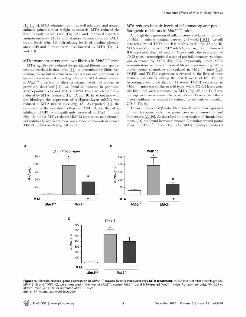
reduced in MTA treated mice (Fig. 4A). As reported [11], the
expression of the interstitial collagenase MMP13, and that of its
inhibitor TIMP1, was significantly increased in Mdr22/2 mice
(Fig. 4B and C). MTA reduced MMP13 expression, and although
not statistically significant there was a tendency towards decreased
TIMP1 mRNA levels (Fig. 4B and C).
MTA reduces hepatic levels of inflammatory and pro-fibrogenic mediators in Mdr22/2 mice
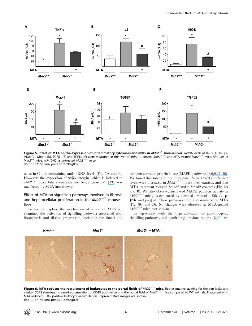
Although the expression of inflammatory cytokines in the liver
of Mdr22/2 mice is maximal between 2–8 weeks [10,11], we still
detected elevated TNFa and IL6 mRNA levels (Fig. 5A and B).
MTA tended to reduce TNFa mRNA, and significantly lowered
IL6 expression (Fig. 5A and B). Consistently, the expression of
iNOS gene, a transcriptional target of pro-inflammatory cytokines,
was decreased by MTA (Fig. 5C). Importantly, upon MTA
administration we observed reduced Mcp-1 expression (Fig. 5D), a
pro-fibrogenic chemokine up-regulated in Mdr22/2 mice [12].
TGFb1 and TGFb2 expression is elevated in the liver of these
animals, particularly during the first 8 weeks of life [10–12].
Accordingly we found that by 11 weeks TGFb1 expression in
Mdr22/2 mice was similar to wild types, while TGFb2 levels were
still high, and were attenuated by MTA (Fig. 5E and F). These
findings were accompanied by a significant decrease in inflam-
matory infiltrate, as assessed by staining for the leukocyte marker
CD45 (Fig. 6).
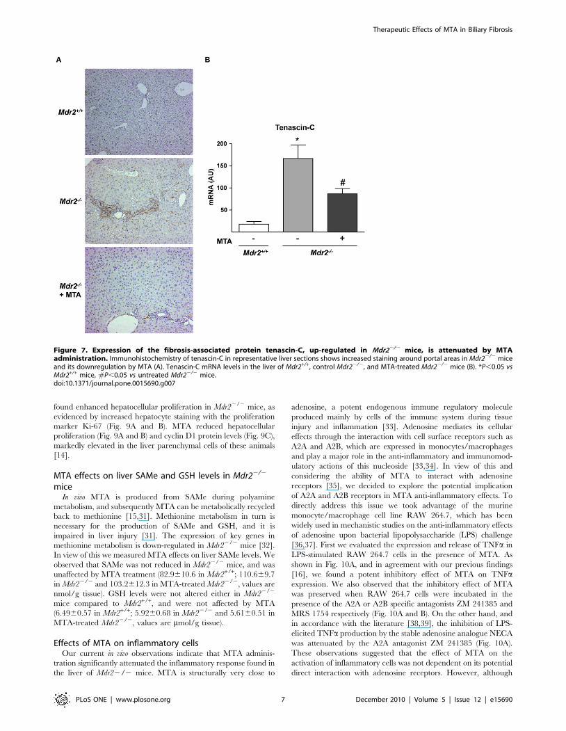
Tenascin-C is a TGFb-inducible extracellular protein expressed
in liver fibrogenic cells that participates in inflammation and
fibrogenesis [24,26]. As described in other models of chronic liver
injury [24], we found increased tenascin-C staining around portal
areas in Mdr22/2 mice (Fig. 7A). MTA treatment reduced
Figure 4. Fibrosis-related gene expression in Mdr22/2 mouse liver is attenuated by MTA treatment. mRNA levels of a1(I) procollagen (A),MMP13 (B) and TIMP1 (C), were measured in the liver of Mdr2+/+, control Mdr22/2, and MTA-treated Mdr22/2 mice. AU: arbitrary units. *P,0.05 vsMdr2+/+ mice, #P,0.05 vs untreated Mdr22/2 mice.doi:10.1371/journal.pone.0015690.g004
Therapeutic Effects of MTA in Biliary Fibrosis
PLoS ONE | www.plosone.org 5 December 2010 | Volume 5 | Issue 12 | e15690
tenascin-C immunostaining and mRNA levels (Fig. 7A and B).
However, the expression of avb6 integrin, which is induced in
Mdr22/2 mice biliary epithelia and binds tenascin-C [13], was
unaffected by MTA (not shown).
Effect of MTA on signalling pathways involved in fibrosisand hepatocellular proliferation in the Mdr22/2 mouseliver
To further explore the mechanism of action of MTA we
examined the activation of signalling pathways associated with
fibrogenesis and disease progression, including the Smad and
mitogen-activated protein kinase (MAPK) pathways [3,6,9,27–30].
We found that total and phosphorylated Smad1/5/8 and Smad2
levels were increased in Mdr22/2 mouse liver extracts, and that
MTA treatment reduced Smad2 and p-Smad2 contents (Fig. 8A
and B). We also observed increased MAPK pathway activity in
Mdr22/2 mice, as evidenced by elevated levels of p-Erk1/2, p-
JNK and p-c-Jun. These pathways were also inhibited by MTA
(Fig. 8C and D). No changes were observed in MTA-treated
Mdr2+/+ mice (not shown).
In agreement with the hyperactivation of pro-mitogenic
signalling pathways, and confirming previous reports [6,30], we
Figure 5. Effect of MTA on the expression of inflammatory cytokines and iNOS in Mdr22/2 mouse liver. mRNA levels of TNFa (A), IL6 (B),iNOS (C), Mcp-1 (D), TGFb1 (E) and TGFb2 (F) were measured in the liver of Mdr2+/+, control Mdr22/2, and MTA-treated Mdr22/2 mice. *P,0.05 vsMdr2+/+ mice, #P,0.05 vs untreated Mdr22/2 mice.doi:10.1371/journal.pone.0015690.g005
Figure 6. MTA reduces the recruitment of leukocytes to the portal fields of Mdr22/2 mice. Representative staining for the pan-leukocytemarker CD45 showing increased accumulation of CD45 positive cells in the portal field of Mdr22/2 mice compared to WT animals. Treatment withMTA reduced CD45 positive leukocyte accumulation. Representative images are shown.doi:10.1371/journal.pone.0015690.g006
Therapeutic Effects of MTA in Biliary Fibrosis
PLoS ONE | www.plosone.org 6 December 2010 | Volume 5 | Issue 12 | e15690
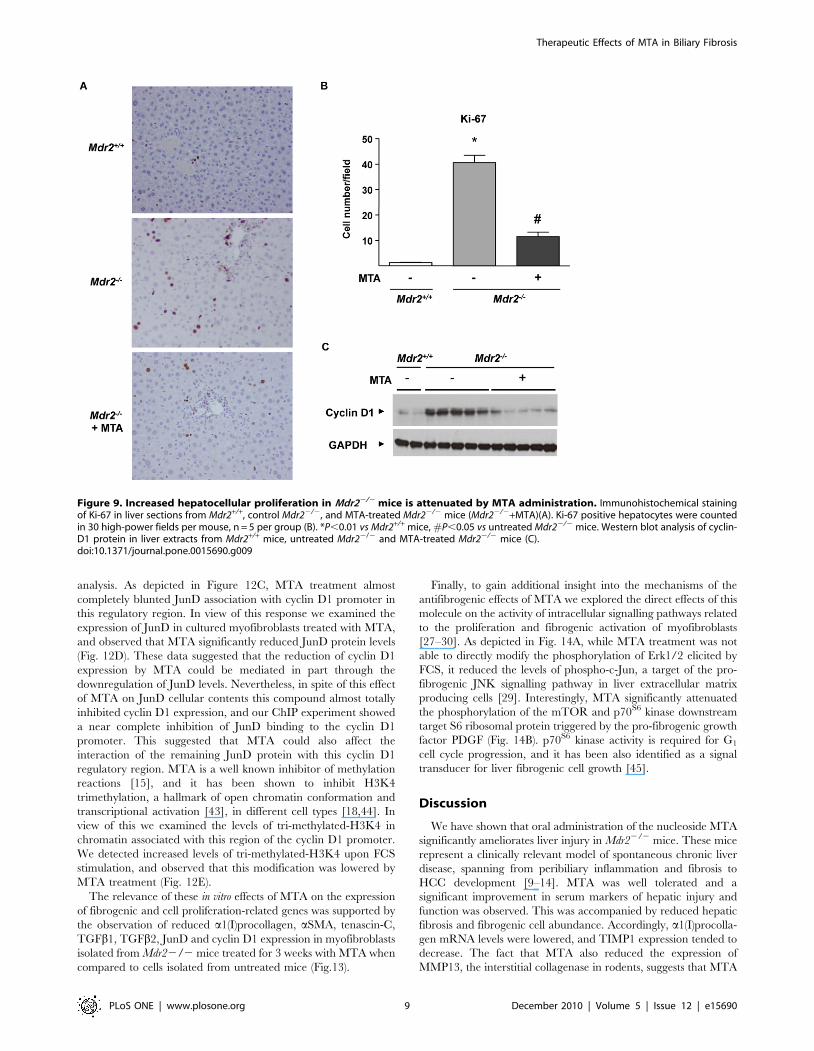
found enhanced hepatocellular proliferation in Mdr22/2 mice, as
evidenced by increased hepatocyte staining with the proliferation
marker Ki-67 (Fig. 9A and B). MTA reduced hepatocellular
proliferation (Fig. 9A and B) and cyclin D1 protein levels (Fig. 9C),
markedly elevated in the liver parenchymal cells of these animals
[14].
MTA effects on liver SAMe and GSH levels in Mdr22/2
miceIn vivo MTA is produced from SAMe during polyamine
metabolism, and subsequently MTA can be metabolically recycled
back to methionine [15,31]. Methionine metabolism in turn is
necessary for the production of SAMe and GSH, and it is
impaired in liver injury [31]. The expression of key genes in
methionine metabolism is down-regulated in Mdr22/2 mice [32].
In view of this we measured MTA effects on liver SAMe levels. We
observed that SAMe was not reduced in Mdr22/2 mice, and was
unaffected by MTA treatment (82.9610.6 in Mdr2+/+; 110.669.7
in Mdr22/2 and 103.2612.3 in MTA-treated Mdr22/2, values are
nmol/g tissue). GSH levels were not altered either in Mdr22/2
mice compared to Mdr2+/+, and were not affected by MTA
(6.4960.57 in Mdr2+/+; 5.9260.68 in Mdr22/2 and 5.6160.51 in
MTA-treated Mdr22/2, values are mmol/g tissue).
Effects of MTA on inflammatory cellsOur current in vivo observations indicate that MTA adminis-
tration significantly attenuated the inflammatory response found in
the liver of Mdr22/2 mice. MTA is structurally very close to
adenosine, a potent endogenous immune regulatory molecule
produced mainly by cells of the immune system during tissue
injury and inflammation [33]. Adenosine mediates its cellular
effects through the interaction with cell surface receptors such as
A2A and A2B, which are expressed in monocytes/macrophages
and play a major role in the anti-inflammatory and immunomod-
ulatory actions of this nucleoside [33,34]. In view of this and
considering the ability of MTA to interact with adenosine
receptors [35], we decided to explore the potential implication
of A2A and A2B receptors in MTA anti-inflammatory effects. To
directly address this issue we took advantage of the murine
monocyte/macrophage cell line RAW 264.7, which has been
widely used in mechanistic studies on the anti-inflammatory effects
of adenosine upon bacterial lipopolysaccharide (LPS) challenge
[36,37]. First we evaluated the expression and release of TNFa in
LPS-stimulated RAW 264.7 cells in the presence of MTA. As
shown in Fig. 10A, and in agreement with our previous findings
[16], we found a potent inhibitory effect of MTA on TNFaexpression. We also observed that the inhibitory effect of MTA
was preserved when RAW 264.7 cells were incubated in the
presence of the A2A or A2B specific antagonists ZM 241385 and
MRS 1754 respectively (Fig. 10A and B). On the other hand, and
in accordance with the literature [38,39], the inhibition of LPS-
elicited TNFa production by the stable adenosine analogue NECA
was attenuated by the A2A antagonist ZM 241385 (Fig. 10A).
These observations suggested that the effect of MTA on the
activation of inflammatory cells was not dependent on its potential
direct interaction with adenosine receptors. However, although
Figure 7. Expression of the fibrosis-associated protein tenascin-C, up-regulated in Mdr22/2 mice, is attenuated by MTAadministration. Immunohistochemistry of tenascin-C in representative liver sections shows increased staining around portal areas in Mdr22/2 miceand its downregulation by MTA (A). Tenascin-C mRNA levels in the liver of Mdr2+/+, control Mdr22/2, and MTA-treated Mdr22/2 mice (B). *P,0.05 vsMdr2+/+ mice, #P,0.05 vs untreated Mdr22/2 mice.doi:10.1371/journal.pone.0015690.g007
Therapeutic Effects of MTA in Biliary Fibrosis
PLoS ONE | www.plosone.org 7 December 2010 | Volume 5 | Issue 12 | e15690

apparently MTA was not acting as an A2B receptor agonist we
found that this molecule dramatically synergized with LPS in the
up-regulation of A2B mRNA levels (Fig. 10C). Increased A2B
expression has been linked to the activation of anti-inflammatory
responses [37,39,40]. Interestingly this effect of MTA was not
reproduced by the adenosine analogue NECA (Fig. 10C).
Effect of MTA on cultured liver myofibroblastsNext we tested whether MTA had a direct effect on liver
myofibroblasts, which participate in biliary fibrosis and represent a
relevant source of extracellular matrix in Mdr22/2 mice [9,30,41].
Given that isolated myofibroblasts display high proliferation and
activity upon in vitro culture, we pre-incubated the cells in serum
free medium for 12 h to attenuate activation and proliferation,
and subsequently cultures were stimulated with 10% FCS to
promote myofibroblast activation in the presence or absence of
MTA. We observed that the expression of the fibrogenic markers
a1(I)procollagen and aSMA elicited by FCS treatment was
inhibited by MTA (Fig. 11A and B). Similar observations were
made when myofibroblasts were challenged with TGFb (not
shown). Additionally, TGFb1 and TGFb2 expression triggered by
FCS was also attenuated by MTA (Fig. 11C and D). In agreement
with previous reports [30] we found that FCS treatment increased
Mcp-1 expression, and this response was also reduced by MTA
(Fig. 11E), while IL6 expression was unaltered (Fig. 11F).
We also examined whether MTA could affect myofibroblast
proliferation. Figure 12A shows that MTA reduced FCS-
triggered DNA synthesis, without causing any toxicity (deter-
mined by lactate dehydrogenase release, not shown). Additionally
we observed that MTA did not induce apoptosis in cultured
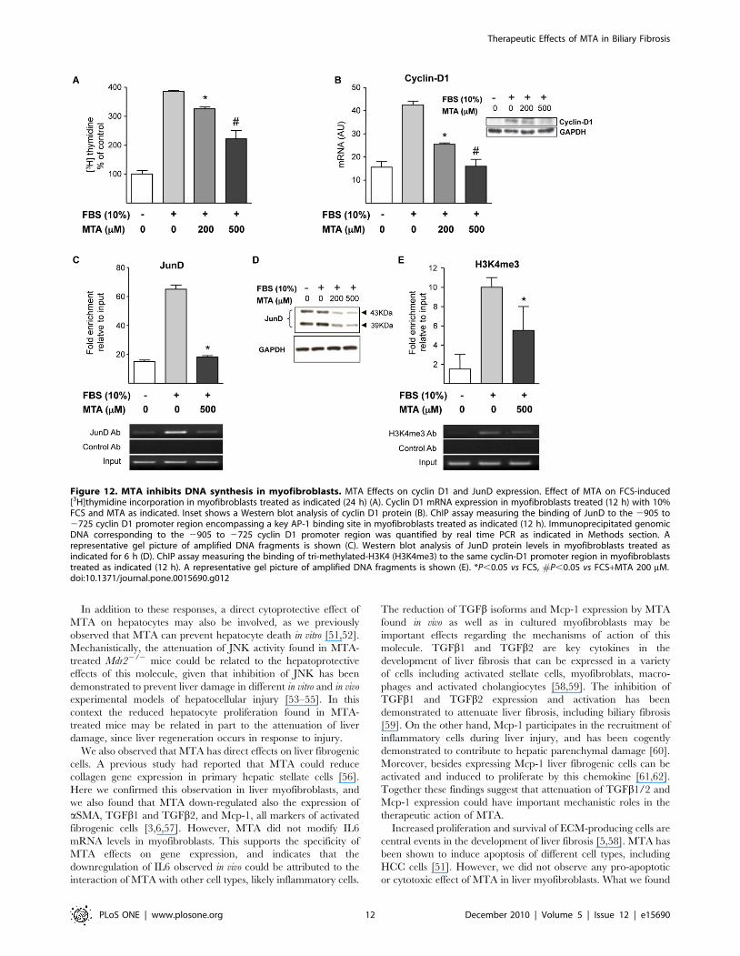
myofibroblasts (data not shown). Our [3H]thymidine incorpora-
tion experiments suggested that MTA inhibited cell cycle
progression through the S phase (DNA synthesis), therefore we
examined the expression of cyclin D1, a key cyclin in the
transition from G0/G1 to S phase. We found that cyclin D1
expression was reduced by MTA treatment both at the mRNA
and protein levels (Figure 12B). The expression of cyclin D1 can
be regulated by AP-1 transcription factors, and among the
components of the AP-1 complex JunD has been identified as a
critical transcription factor in liver fibrogenesis [27,42]. There-
fore we examined the binding of JunD to a region of the cyclin
D1 promoter that contains a key AP-1 binding site [25] by ChIP
Figure 8. MTA modulates intracellular signalling pathways activated in Mdr22/2 mouse liver. Phosphorylation levels of Smad1/5/8 (A),Smad2 (B), JNK and c-Jun (C) and Erk1/2 (D) were analyzed by western blotting in liver extracts from Mdr2+/+, control Mdr22/2, and MTA-treatedMdr22/2 mice. *P,0.05 vs Mdr2+/+ mice, #P,0.05 vs untreated Mdr22/2 mice.doi:10.1371/journal.pone.0015690.g008
Therapeutic Effects of MTA in Biliary Fibrosis
PLoS ONE | www.plosone.org 8 December 2010 | Volume 5 | Issue 12 | e15690
analysis. As depicted in Figure 12C, MTA treatment almost
completely blunted JunD association with cyclin D1 promoter in
this regulatory region. In view of this response we examined the
expression of JunD in cultured myofibroblasts treated with MTA,
and observed that MTA significantly reduced JunD protein levels
(Fig. 12D). These data suggested that the reduction of cyclin D1
expression by MTA could be mediated in part through the
downregulation of JunD levels. Nevertheless, in spite of this effect
of MTA on JunD cellular contents this compound almost totally
inhibited cyclin D1 expression, and our ChIP experiment showed
a near complete inhibition of JunD binding to the cyclin D1
promoter. This suggested that MTA could also affect the
interaction of the remaining JunD protein with this cyclin D1
regulatory region. MTA is a well known inhibitor of methylation
reactions [15], and it has been shown to inhibit H3K4
trimethylation, a hallmark of open chromatin conformation and
transcriptional activation [43], in different cell types [18,44]. In
view of this we examined the levels of tri-methylated-H3K4 in
chromatin associated with this region of the cyclin D1 promoter.
We detected increased levels of tri-methylated-H3K4 upon FCS
stimulation, and observed that this modification was lowered by
MTA treatment (Fig. 12E).
The relevance of these in vitro effects of MTA on the expression
of fibrogenic and cell proliferation-related genes was supported by
the observation of reduced a1(I)procollagen, aSMA, tenascin-C,
TGFb1, TGFb2, JunD and cyclin D1 expression in myofibroblasts
isolated from Mdr22/2 mice treated for 3 weeks with MTA when
compared to cells isolated from untreated mice (Fig.13).
Finally, to gain additional insight into the mechanisms of the
antifibrogenic effects of MTA we explored the direct effects of this
molecule on the activity of intracellular signalling pathways related
to the proliferation and fibrogenic activation of myofibroblasts
[27–30]. As depicted in Fig. 14A, while MTA treatment was not
able to directly modify the phosphorylation of Erk1/2 elicited by
FCS, it reduced the levels of phospho-c-Jun, a target of the pro-
fibrogenic JNK signalling pathway in liver extracellular matrix
producing cells [29]. Interestingly, MTA significantly attenuated
the phosphorylation of the mTOR and p70S6 kinase downstream
target S6 ribosomal protein triggered by the pro-fibrogenic growth
factor PDGF (Fig. 14B). p70S6 kinase activity is required for G1
cell cycle progression, and it has been also identified as a signal
transducer for liver fibrogenic cell growth [45].
Discussion
We have shown that oral administration of the nucleoside MTA
significantly ameliorates liver injury in Mdr22/2 mice. These mice
represent a clinically relevant model of spontaneous chronic liver
disease, spanning from peribiliary inflammation and fibrosis to
HCC development [9–14]. MTA was well tolerated and a
significant improvement in serum markers of hepatic injury and
function was observed. This was accompanied by reduced hepatic
fibrosis and fibrogenic cell abundance. Accordingly, a1(I)procolla-
gen mRNA levels were lowered, and TIMP1 expression tended to
decrease. The fact that MTA also reduced the expression of
MMP13, the interstitial collagenase in rodents, suggests that MTA
Figure 9. Increased hepatocellular proliferation in Mdr22/2 mice is attenuated by MTA administration. Immunohistochemical stainingof Ki-67 in liver sections from Mdr2+/+, control Mdr22/2, and MTA-treated Mdr22/2 mice (Mdr22/2+MTA)(A). Ki-67 positive hepatocytes were countedin 30 high-power fields per mouse, n = 5 per group (B). *P,0.01 vs Mdr2+/+ mice, #P,0.05 vs untreated Mdr22/2 mice. Western blot analysis of cyclin-D1 protein in liver extracts from Mdr2+/+ mice, untreated Mdr22/2 and MTA-treated Mdr22/2 mice (C).doi:10.1371/journal.pone.0015690.g009
Therapeutic Effects of MTA in Biliary Fibrosis
PLoS ONE | www.plosone.org 9 December 2010 | Volume 5 | Issue 12 | e15690

down-regulates overall tissue remodelling activity, inhibiting
fibrosis progression rather than stimulating fibrosis resolution
[3].
Previous studies have shown that MTA exerts remarkable
biological effects, its pharmacologic administration modulates
different pathways and cellular responses, including gene expres-
sion, cell cycle progression and inflammation [15–20]. Therefore,
the mechanisms behind MTA therapeutic action on a complex
model such as Mdr22/2 mice are likely multifaceted. Our current
findings agree with those reported by Simile et al., who showed
that parenteral MTA administration to rats undergoing chronic
CCl4-induced liver injury reduced fibrosis [19]. This effect was
mechanistically related to MTA antioxidant properties, that
counteracted the oxidative stress and subsequent injury generated
by CCl4 metabolism [19]. However, oxidative stress is not a
prominent feature in Mdr22/2 mice [14]. Accordingly, we found
that GSH levels were unaltered in Mdr22/2 mouse liver, and were
not affected by MTA, suggesting that MTA effects were not
attributable to its antioxidant potential. In vivo MTA is metabolized
to methionine in the so called methionine salvage pathway, and in
the liver methionine is efficiently metabolized into SAMe [15].
SAMe is a key methyl donor with significant hepatoprotective
properties, including antifibrotic activity in experimental cholesta-
sis [31,46]. In view of the metabolic links between MTA and
SAMe, we measured SAMe levels in control and MTA-treated
Mdr22/2 mouse liver but did not observe major differences,
indicating that MTA effects were not likely mediated through its
potential contribution to the SAMe pool. Moreover, this may
suggest that the antifibrotic effects of SAMe in experimental
cholestasis [46] could be mediated in part by the metabolic or non-
enzymatic conversion of SAMe into MTA [15].
Interestingly, MTA reduced the expression of the extracellular
matrix protein tenascin-C in the liver of Mdr22/2 animals. This
may be directly related to MTA antifibrotic effects, since tenascin-
C is required for the development of experimental liver fibrosis
[24]. Furthermore, tenascin-C is up-regulated in chronic hepatitis
C, and its expression in peritumoral activated stellate cells in HCC
associates with poor clinical prognosis [26,38,47]. In view of this,
tenascin-C may be a previously unrecognized player in fibrosis
and HCC development in Mdr22/2 mice, and its attenuation by
MTA a key event in the anti-fibrotic and anti-neoplastic properties
of this molecule [20]. Moreover, tenascin-C binds and activates
avb6 integrin, a recently identified pharmacological target in
biliary fibrosis [13].
Figure 10. Effect of MTA on the activation of inflammatory cells: interaction with adenosine receptors. As indicated in the figure serum-starved murine monocyte/macrophages RAW 264.7 cells were pre-treated for 30 min with the A2A receptor antagonist ZM 241385 (10 mM) or theA2B receptor antagonist MRS 1754 (1 mM), then MTA or the adenosine receptor agonist NECA (20 mM) where added to the cultures for another30 min. Subsequently and where indicated cells were treated with LPS (50 ng/ml) for up to 5 h. TNFa mRNA levels in cell lysates (A) and TNFa proteincontents in conditioned media (B) were measured. *P,0.01 vs control, **P,0.01 vs LPS, #P,0.05 vs LPS. The expression of the adenosine A2Breceptor was measured in RAW 264.7 cells pre-treated with MTA or the adenosine agonist NECA (20 mM) for 30 min and then where indicated withLPS (50 ng/ml) for up to 5 h (C). *P,0.01 vs control, **P,0.01 vs LPS.doi:10.1371/journal.pone.0015690.g010
Therapeutic Effects of MTA in Biliary Fibrosis
PLoS ONE | www.plosone.org 10 December 2010 | Volume 5 | Issue 12 | e15690

As occurs in human chronic liver disease, progression of liver
injury in Mdr22/2 mice is associated with persistent inflammation.
The observation that anti-inflammatory compounds like ibuprofen
or curcumin ameliorate injury and fibrosis in Mdr22/2 mice
[6,30], and that TNFa blockade prevents hepatocarcinogenesis in
these animals [6], underscore the role played by inflammation in
this model. We and others have previously shown that MTA has a
significant immunomodulatory potential. MTA inhibits NF-kB
mediated signalling, reduces inflammatory cytokine production by
macrophages and lymphocytes, and attenuates the cellular effects
of these cytokines [16–18]. We now observed that in Mdr22/2
mice MTA reduced the expression of IL6, Mcp-1, the NO-
generating enzyme iNOS, and the profibrogenic factor TGFb2.
Accordingly MTA attenuated the activation of Erk1/2, JNK and
Smad2, intracellular pathways triggered by these cytokines and
which are directly implicated in fibrogenesis [3,5,27–30]. There-
fore, interference with inflammatory cytokine and NO generation
likely contributes to MTA hepatoprotective and anti-fibrogenic
effects in this model [10,48].
From a mechanistic perspective, and in view of the remarkable
anti-inflammatory effects of MTA, we cannot overlook the fact
that this molecule is structurally very close to adenosine, a purine
nucleoside released form cells with notorious anti-inflammatory
properties [15,33]. Adenosine interacts with different G-protein-
coupled cell surface receptors including the A1, A2A, A2B and
A3 receptors. Until recently adenosine anti-inflammatory effects
were mainly attributed to its interaction with the high affinity
A2A receptor subtype [49], however an important role for the
low affinity A2B receptor in the anti-inflammatory response to
adenosine is currently emerging [34,38,40]. Using specific
pharmacological antagonists of the A2A and A2B receptors we
evaluated their potential implication in the immunomodulatory
effects of MTA on mouse RAW 264.7 monocytes/macrophages
challenged with bacterial LPS, a prototypic model of the
inflammatory response. We found that the inhibition of LPS-
elicited TNFa production by MTA was not impaired in the
presence of these drugs, suggesting that MTA effects were not
mediated through these receptors. Nevertheless, the involvement
of the adenosine signalling system in the biological effects of
MTA cannot be completely disregarded. Interestingly we
observed that MTA treatment resulted in a remarkable
potentiation of the expression of A2B receptors triggered by
LPS, and this effect was not shared by the adenosine receptor
agonist NECA. As previously mentioned A2B receptors display
low affinity for adenosine, but their expression is up-regulated
during tissue injury and inflammation, conditions in which
interstitial adenosine concentrations raise. The expression of A2B
receptors seems to be very important for the attenuation of the
systemic inflammatory response, as illustrated by the fact that
A2B knockout mice show enhanced cytokine production and
overall mortality during sepsis [39,50]. Therefore the stimulatory
effect of MTA on A2B receptor expression could also contribute
to dampen inflammation in the chronically injured Mdr22/2
mouse liver.
Figure 11. Effects of MTA on liver myofibroblast activation and cytokine expression. Primary myofibroblasts were serum starved (12 h)and then treated (12 h) with 10% FCS and MTA as indicated. mRNA levels of a1(I)procollagen (A), aSMA (B), TGFb1 (C), TGFb2 (D), Mcp-1 (E) and IL6 (F)were measured at the end of treatments. *P,0.05 vs FCS, #P,0.05 vs FCS+MTA 200 mM.doi:10.1371/journal.pone.0015690.g011
Therapeutic Effects of MTA in Biliary Fibrosis
PLoS ONE | www.plosone.org 11 December 2010 | Volume 5 | Issue 12 | e15690
In addition to these responses, a direct cytoprotective effect of
MTA on hepatocytes may also be involved, as we previously
observed that MTA can prevent hepatocyte death in vitro [51,52].
Mechanistically, the attenuation of JNK activity found in MTA-
treated Mdr22/2 mice could be related to the hepatoprotective
effects of this molecule, given that inhibition of JNK has been
demonstrated to prevent liver damage in different in vitro and in vivo
experimental models of hepatocellular injury [53–55]. In this
context the reduced hepatocyte proliferation found in MTA-
treated mice may be related in part to the attenuation of liver
damage, since liver regeneration occurs in response to injury.
We also observed that MTA has direct effects on liver fibrogenic
cells. A previous study had reported that MTA could reduce
collagen gene expression in primary hepatic stellate cells [56].
Here we confirmed this observation in liver myofibroblasts, and
we also found that MTA down-regulated also the expression of
aSMA, TGFb1 and TGFb2, and Mcp-1, all markers of activated
fibrogenic cells [3,6,57]. However, MTA did not modify IL6
mRNA levels in myofibroblasts. This supports the specificity of
MTA effects on gene expression, and indicates that the
downregulation of IL6 observed in vivo could be attributed to the
interaction of MTA with other cell types, likely inflammatory cells.
The reduction of TGFb isoforms and Mcp-1 expression by MTA
found in vivo as well as in cultured myofibroblasts may be
important effects regarding the mechanisms of action of this
molecule. TGFb1 and TGFb2 are key cytokines in the
development of liver fibrosis that can be expressed in a variety
of cells including activated stellate cells, myofibroblats, macro-
phages and activated cholangiocytes [58,59]. The inhibition of
TGFb1 and TGFb2 expression and activation has been
demonstrated to attenuate liver fibrosis, including biliary fibrosis
[59]. On the other hand, Mcp-1 participates in the recruitment of
inflammatory cells during liver injury, and has been cogently
demonstrated to contribute to hepatic parenchymal damage [60].
Moreover, besides expressing Mcp-1 liver fibrogenic cells can be
activated and induced to proliferate by this chemokine [61,62].
Together these findings suggest that attenuation of TGFb1/2 and
Mcp-1 expression could have important mechanistic roles in the
therapeutic action of MTA.
Increased proliferation and survival of ECM-producing cells are
central events in the development of liver fibrosis [5,58]. MTA has
been shown to induce apoptosis of different cell types, including
HCC cells [51]. However, we did not observe any pro-apoptotic
or cytotoxic effect of MTA in liver myofibroblasts. What we found
Figure 12. MTA inhibits DNA synthesis in myofibroblasts. MTA Effects on cyclin D1 and JunD expression. Effect of MTA on FCS-induced[3H]thymidine incorporation in myofibroblasts treated as indicated (24 h) (A). Cyclin D1 mRNA expression in myofibroblasts treated (12 h) with 10%FCS and MTA as indicated. Inset shows a Western blot analysis of cyclin D1 protein (B). ChIP assay measuring the binding of JunD to the 2905 to2725 cyclin D1 promoter region encompassing a key AP-1 binding site in myofibroblasts treated as indicated (12 h). Immunoprecipitated genomicDNA corresponding to the 2905 to 2725 cyclin D1 promoter region was quantified by real time PCR as indicated in Methods section. Arepresentative gel picture of amplified DNA fragments is shown (C). Western blot analysis of JunD protein levels in myofibroblasts treated asindicated for 6 h (D). ChIP assay measuring the binding of tri-methylated-H3K4 (H3K4me3) to the same cyclin-D1 promoter region in myofibroblaststreated as indicated (12 h). A representative gel picture of amplified DNA fragments is shown (E). *P,0.05 vs FCS, #P,0.05 vs FCS+MTA 200 mM.doi:10.1371/journal.pone.0015690.g012
Therapeutic Effects of MTA in Biliary Fibrosis
PLoS ONE | www.plosone.org 12 December 2010 | Volume 5 | Issue 12 | e15690

was a significant inhibition by MTA of FCS-stimulated DNA
synthesis. In line with this response we observed that MTA
reduced the expression of cyclin D1, a key G1 cyclin associated
with liver fibrogenic cell proliferation [63]. AP-1 transcription
factors can stimulate the expression of genes involved in the
activation and proliferation of liver fibrogenic cells, and among
them JunD is known to play an important role in liver fibrogenesis
[27,42]. To gain further insight into the mechanism of action of
MTA, we examined the binding of JunD to cyclin D1 promoter
and found that it was markedly reduced in myofibroblasts treated
with this compound. This can be due in part to the observed
downregulation of JunD levels in MTA-treated cells, and therefore
reduction of JunD expression may be a relevant mechanism in the
antifibrogenic action of MTA.
In order to better understand the mechanisms behind the
antiproliferative effects of MTA, we examined the activity of three
signalling pathways that are involved in fibrogenic cell prolifera-
tion. We found that while MTA did not affect FCS-triggered
Erk1/2 phosphorylation, it reduced phopho-c-Jun levels and
significantly attenuated PDGF elicited ribosomal S6 protein
phosphorylation. Both, the JNK and the mTOR and p70S6 kinase
pathways have been demonstrated as key regulators of hepatic
fibrogenic cell proliferation [29,45], and therefore their inhibition
by MTA could be relevant in the mechanism of action of this
compound. The selective interaction of MTA with protein kinase-
mediated intracellular signalling pathways as described here is
likely to have profound effects on cellular behaviour and thus
deserves further examination.
We also noticed that MTA could attenuate the increase in
H3K4 trimethylation elicited by FCS stimulation in the cyclin D1
promoter. H3K4 trimethylation is a marker of transcriptional
active chromatin, and this modification contributes to gene
activation [64]. MTA is an inhibitor of methylation reactions
[15], including histone methylation [18], our observations suggest
Figure 13. Expression of fibrogenic activation and cellular proliferation-related genes in myofibroblasts isolated from the liver ofcontrol and MTA-treated Mdr22/2 mice. Mdr22/2 mice were treated for three weeks with MTA or vehicle as described in the Methods section.At the end of the treatments hepatic myofibroblasts were isolated and the mRNA levels of the indicated genes were measured. Data are means6SEM of three independent cell preparations per condition. *P,0.05 vs vehicle-treated Mdr22/2 mice.doi:10.1371/journal.pone.0015690.g013
Figure 14. Effect of MTA on liver myofibroblast intracellularsignalling pathways. Liver myofibroblasts were pre-treated with MTAfor 30 min in the absence of serum and then stimulated with 10% FCSfor 30 min or PDGF (20 ng/ml) for 60 min. Phosphorylation levels ofErk1/2 and c-Jun upon FCS stimulation (A), and S6 ribosomal proteinafter PDGF treatment (B) were analyzed by western blotting.Representative blots are shown.doi:10.1371/journal.pone.0015690.g014
Therapeutic Effects of MTA in Biliary Fibrosis
PLoS ONE | www.plosone.org 13 December 2010 | Volume 5 | Issue 12 | e15690
that interference with this epigenetic mechanism could account in
part for the effects of MTA on cyclin D1 gene expression.
Moreover, this finding makes it tempting to speculate that MTA
could exert its antifibrogenic effects also through an overall action
on the epigenetic programming. In this regard 3 deazaneplanocin,
another methyltransferase inhibitor, has been recently reported to
prevent the activation of liver fibrogenic cells, however its efficacy
and tolerability in chronic in vivo models of liver fibrosis have not
been tested yet [65]. Together these findings suggest that the
antifibrogenic effects of MTA observed in vivo may be mediated in
part through the direct inhibition of myofibroblast activation and
proliferation. Nevertheless, a direct effect of MTA on bile duct
epithelial cell damage and cholangiocyte activation, important
events in the pathogenesis of liver injury in Mdr22/2 mice [30],
cannot be discarded and merit future evaluation. Figure 15
summarizes the proposed mechanisms of action of MTA in
Mdr22/2 mice mainly based on the observations reported in this
study.
In summary, here we have demonstrated that oral MTA
administration is an effective therapeutic strategy to attenuate
disease progression in a relevant model of liver injury and fibrosis.
In addition to our current study, MTA has been previously tested
in in vivo experimental models never showing untoward effects
[16,17,66,67], and an ID50 of 2.960.4 g/kg was estimated when
administered intramuscularly to rats [19]. In humans MTA
administration is also well tolerated [68,69]. These considerations,
together with the efficacy described herein, suggest that MTA
could be a good candidate to be clinically tested in the context of
liver injury and fibrosis.
Author Contributions
Conceived and designed the experiments: MAA. Performed the experi-
ments: MUL CG-P MGF-B CMR-O MM JMB SG SA NJ RU JL FJC.
Analyzed the data: FJC MUL RU JP JL CB MAA. Contributed reagents/
materials/analysis tools: JMB SL JAR. Wrote the paper: JL MAA.
References
1. Williams R (2006) Global challenges in liver disease. Hepatology 44: 521–6.
2. Siegel AB, Zhu AX (2009) Metabolic syndrome and hepatocellular carcinoma.
Cancer 115: 5651–5661.
3. Bataller R, Brenner DA (2005) Liver fibrosis. J Clin Invest 115: 209–218.
4. Elsharkawy AM, Mann DA (2007) Nuclear factor-kB and the hepatic
inflammation-fibrosis-cancer axis. Hepatology 46: 590–597.
5. Lotersztajn S, Julien B, Teixeira-Clerc F, Grenard P, Mallat A (2005) Hepatic
fibrosis: molecular mechanisms and drug targets. Annu Rev Pharmacol Toxicol
45: 605–628.
6. Pikarski E, Porat RM, Stein I, Abramovitch R, Amit S, et al. (2004) NF-kB
functions as a tumour promoter in inflammation-associated cancer. Nature 431:
461–466.
7. Berasain C, Castillo J, Perugorria MJ, Latasa MU, Prieto J, et al. (2009) Inflammation
and liver cancer: new molecular links. Ann NY Acad Sci; 1155: 206–221.
8. Pinzani M, Rombouts K, Colagrande S (2005) Fibrosis in chronic liver diseases:
diagnosis and management. J Hepatol 42(Suppl): S22–S36.
9. Popov Y, Schuppan D (2009) Targeting liver fibrosis: strategies for development
and validation of antifibrotic therapies. Hepatology 50: 1294–1306.
10. Fickert P, Fuchsbichler A, Wagner M, Zollner G, Kaser A, et al. (2004)
Regurgitation of bile acids from leaky bile ducts causes sclerosing cholangitis in
Mdr2 (Abcb4) knockout mice. Gastroenterology 127: 261–274.
11. Popov Y, Patsenker E, Fickert P, Trauner M, Schuppan D (2005) Mdr2
(Abcb4)-/- mice spontaneously develop severe biliary fibrosis via massive
dysregulation of pro- and antifibrogenic genes. J Hepatol 43: 1045–1054.
12. Nakken KE, Nygard S, Haaland T, Berge KE, Arnkværn K, et al. (2007)
Multiple inflammatory-, tissue remodelling- and fibrosis genes are differentially
transcribed in the livers of Abcb4 (-/-) mice harbouring chronic cholangitis.
Scand J Gastroenterol 42: 1245–1255.
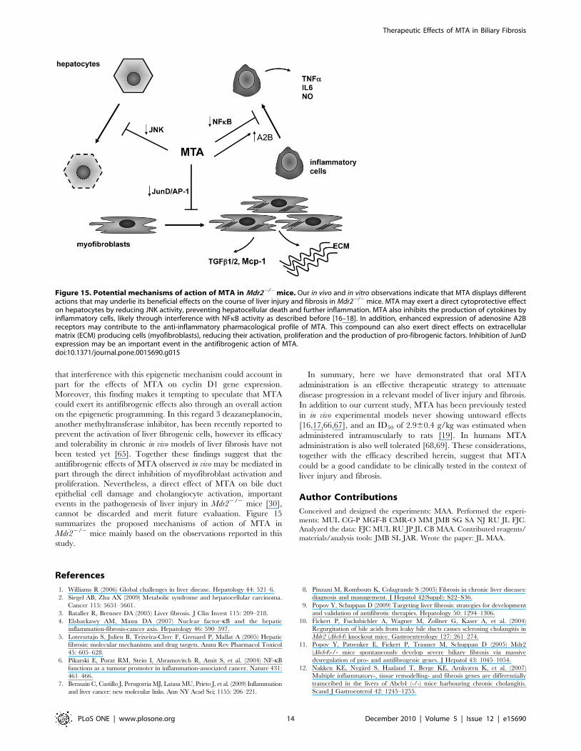
Figure 15. Potential mechanisms of action of MTA in Mdr22/2 mice. Our in vivo and in vitro observations indicate that MTA displays differentactions that may underlie its beneficial effects on the course of liver injury and fibrosis in Mdr22/2 mice. MTA may exert a direct cytoprotective effecton hepatocytes by reducing JNK activity, preventing hepatocellular death and further inflammation. MTA also inhibits the production of cytokines byinflammatory cells, likely through interference with NFkB activity as described before [16–18]. In addition, enhanced expression of adenosine A2Breceptors may contribute to the anti-inflammatory pharmacological profile of MTA. This compound can also exert direct effects on extracellularmatrix (ECM) producing cells (myofibroblasts), reducing their activation, proliferation and the production of pro-fibrogenic factors. Inhibition of JunDexpression may be an important event in the antifibrogenic action of MTA.doi:10.1371/journal.pone.0015690.g015
Therapeutic Effects of MTA in Biliary Fibrosis
PLoS ONE | www.plosone.org 14 December 2010 | Volume 5 | Issue 12 | e15690

13. Patsenker E, Popov Y, Stickel F, Jonczyk A, Goodman SL, et al. (2008)
Inhibition of integrin avb6 on cholangiocytes blocks transforming growth factor-b activation and retards biliary fibrosis progression. Gastroenterology 135:
660–670.
14. Katzenellenbogen M, Pappo O, Barash H, Klopstock N, Mizrahi L, et al. (2006)Multiple adaptive mechanisms to chronic liver disease revealed at early stages of
liver carcinogenesis in the Mdr2-knockout mice. Cancer Res 66: 4001–4010.15. Avila MA, Garcia-Trevijano ER, Lu SC, Corrales FJ, Mato JM (2004)
Methylthioadenosine. Int J Biochem Cell Biol 36: 2125–2130.
16. Hevia H, Varela-Rey M, Corrales FJ, Berasain C, Martınez-Chantar ML, et al.(2004) 59-Methythioadenosine modulates the inflammatory response to endo-
toxin in mice and in rat hepatocytes. Hepatology 39: 1088–1098.17. Moreno B, Hevia H, Santamaria M, Sepulcre J, Munoz J, et al. (2006)
Methylthioadenosine reverses brain autoimmune disease. Ann Neurol 60:323–334.
18. Iglesias-Ara A, Xia M, Ramani K, Mato JM, Lu SC (2008) S-Adenosylmethi-
onine inhibits lipopolysaccharide-induced gene expression via modulation ofhistone methylation. Hepatology 47: 1655–1666.
19. Simile MM, Banni S, Angioni E, Carta G, De Miglio MR, et al. (2001) 59-Methylthioadenosine administration prevents lipid peroxidation and fibrogenesis
induced in rat liver by carbon-tetrachloride intoxication. J Hepatol 34: 386–394.
20. Pascale RM, Simile MM, De Miglio MR, Feo F (2002) Chemoprevention ofhepatocarcinogenesis: S-adenosyl-L-methionine. Alcohol 27: 193–198.
21. Berasain C, Hevia H, Fernandez-Irigoyen J, Larrea E, Caballerıa J, et al. (2004)Methylthioadenosine phosphorylase gene expression is impaired in human liver
cirrhosis and hepatocarcinoma. Biochim Biophys Acta 1690: 276–284.22. Perugorria MJ, Latasa MU, Nicou A, Cartagena-Lirola H, Castillo J, et al.
(2008) The epidermal growth factor receptor ligand amphiregulin participates in
the development of mouse liver fibrosis. Hepatology 48: 1251–1261.23. Li LY, Grenard P, Van Nhieu JT, Julien B, Mallat A, et al. (2003) Heme
oxygenase-1 is an antifibrogenic protein in human hepatic myofibroblasts.Gastroenterology 125: 460–469.
24. El-Karef A, Yoshida T, Gabazza EC, Nishioka T, Inada H, et al. (2007)
Deficiency of tenascin-C attenuates liver fibrosis in immune-mediated chronichepatitis in mice. J Pathol 211: 86–94.
25. Eto I. Molecular cloning and sequence analysis of the promoter region ofmouse cyclin D1 gene. Cell Prolif 2000; 33: 167–187.
26. El-Karef A, Kaito M, Tanaka H, Ikeda K, Nishioka T, et al. (2007) Expressionof large tenascin-C splice variants by hepatic stellate cells/myofibroblasts in
chronic hepatitis C. J Hepatol 46: 664–673.
27. Mann J, Mann DA (2009) Transcriptional regulation of hepatic stellate cells.Adv Drug Delivery Rev 61: 497–512.
28. Tao J, Mallat A, Gallois C, Belmadani S, Mery PF, et al. (1999) Biological effectsof C-type natriuretic peptide in human myofibroblastic hepatic stellate cells.
J Biol Chem 274: 23761–23769.
29. Kluwe J, Pradere JP, Gwak GY, Mencin A, De Minicis S, et al. (2010)Modulation of hepatic fibrosis by c-Jun-N-terminal kinase inhibition. Gastro-
enterology 138: 347–359.30. Baghdasaryan A, Claudel T, Kosters A, Gumhold J, Silbert D, et al. (2010)
Curcumin improves sclerosing cholangitis in Mdr2-/- mice by inhibition ofcholangiocyte inflammatory response and portal myofibroblast proliferation.
Gut 59: 521–530.
31. Mato JM, Martınez-Chantar ML, Lu SC (2008) Methionine metabolism andliver disease. Annu Rev Nutr 28: 273–293.
32. Katzenellenbogen M, Mizrahi L, Pappo O, Klopstock N, Olam D, et al. (2007)Molecular mechanisms of liver carcinogenesis in the Mdr2-knockout mice. Mol
Cancer Res 5: 1159–1170.
33. Jacobson KA, Gao Z (2006) Adenosine receptors as therapeutic targets. Nat RevDrug Discovery 5: 247–264.
34. Hasko G, Csoka B, Nemeth ZH, Vizi ES, pacher P (2009) A2B adenosinereceptors in immunity and inflammation. Trends Immunol. 30: 263–270.
35. Munshi R, Clanachan AS, Baer HP (1988) 59-deoxy-59-methylthioadenosine: a
nucleoside which differentiates between adenosine receptor types. BiochemPharmacol 37: 2085–2089.
36. Nemeth ZH, Leibovich SJ, Deitch EA, Vizi S, Szabo C, et al. (2003) cDNAMicroarray analysis reveals a nuclear factor-kB-independent regulation of
macrophage function by adenosine. J Pharmacol Exp Ther 306: 1042–1049.37. Nemeth ZH, Lutz CS, Csoka B, Deitch EA, Leibovich SJ, et al. (2005)
Adenosine augments IL-10 production by macrophages through an A2B recptor-
mediated posttranscriptional mechanism. J Immunol 175: 8260–8270.38. Kreckler LM, Wan TC, Ge Z, Auchampach JA (2006) Adenosine inhibits tumor
necrosis factor-a release from mouse peritoneal macrophages via A2A and A2B
but not the A3 adenosine receptor. J Pharmacol Exp Ther 317: 172–180.
39. Ryzhov S, Zaynagetdinov R, Goldstein AE, Novitskiy SV, Blackburn MR, et al.
(2008) Effetc of A2B adenosine receptor gene ablation on adenosine-dependentregulation of proinflammatory cytokines. J Pharm Exp Ther 324: 694–700.
40. Aherne CM, Kewley EM, Eltzschig HK (2010) The resurgence of A2Badenosine receptor signalling. Biochim Biophys Acta May 27. [Epub ahead of
print].41. Dranoff JA, Wells RG (2010) Portal fibroblasts: underappreciated mediators of
biliary fibrosis. Hepatology 51: 1438–1444.
42. Smart DE, Green K, Oakley F, Weitzman JB, Yaniv M, et al. (2006) JunD is aprofibrogenic transcription factor regulated by Jun N-terminal kinase-indepen-
dent phosphorylation. Hepatology 44: 1432–1440.
43. Guenther MG, Levine SS, Boyer LA, Jaenisch R, Young RA (2007) A
chromatin landmark and transcription initiation at most promoters in human
cells. Cell 130: 77–88.
44. Song M-R, Ghosh A (2004) FGF2-induced chromatin remodelling regulates
CNTF-mediated gene expression and astrocyte differentiation. Nat Neurosci 7:
229–235.
45. Gabele E, Reif S, Tsukada S, Bataller R, Yata Y, et al. (2005) The role of p70S6K
in hepatic stellate cell collagen gene expression and cell proliferation. J Biol
Chem 280: 13374–13382.
46. Yang H, Ramani K, Xia M, Ko KS, Li TWH, et al. (2009) Dysregulation of
glutathione synthesis during cholestasis in mice: molecular mechanisms and
therapeutic implications. Hepatology 49: 1982–1991.
47. Ju M, Qiu S, Fan J, Xiao Y, Gao Q, et al. (2009) Peritumoral activated hepatic
stellate cells predict poor clinical outcome in hepatocellular carcinoma. Am J Clin
Pathol 131: 498–510.
48. Spirli C, Fabris L, Duner E, Fiorotto R, Roskams T, et al. (2003) Cytokine-
stimulated nitric oxide production inhibits adenylyl cyclase and cAMP-
dependent secretion in cholangiocytes. Gastroenterology 124: 737–753.
49. Sitkovsky MV (2003) Use of the A2A adenosine receptor as a physiological
immunosuppressor and to engineer inflammation in vivo. Biochem Pharmacol
65: 493–501.
50. Csoka B, Nemeth ZH, Rosenberger P, Eltzschig HK, Spolarics Z, et al. (2010)
A2B adenosine receptors protect against sepsis-induced mortality by dampening
excessive inflammation. J Immunol 185: 542–550.
51. Ansorena E, Garcia-Trevijano ER, Martinez-Chantar ML, Huang Z, Chen L,
et al. (2002) S-Adenosylmethionine and methylthioadenosine are antiapoptotic
in cultured rat hepatocytes but proapoptotic in human hepatoma cells.
Hepatology 35: 274–280.
52. Ansorena E, Berasain C, Lopez Zabalza MJ, Avila MA, Garcıa-Trevijano ER,
et al. (2006) Differential regulation of the JNK/AP-1 pathway by S-
adenosylmethionine and methylthioadenosine in primary rat hepatocytes versus
HuH7 hepatoma cells. Am J Physiol Gastrointest Liver Physiol 290:
G1186–G1193.
53. Marderstein EL, Bucher B, Guo Z, Feng X, Reid K, et al. (2003) Protection of
rat hepatocytes from apoptosis by inhibition of c-Jun N-terminal kinase. Surgery
134: 280–284.
54. Uehara T, Bennett B, Sakata ST, Satoh Y, Bilter G, et al. (2005) JNK mediates
hepatic ischemia reperfusion injury. J Hepatol 42: 850–859.
55. Gunawan BK, Liu ZX, Hanawa N, Gaarde WA, Kaplowitz N (2006) c-Jun N-
terminal kinase plays a major role in murine acetaminophen hepatotocicity.
Gastroenterology 131: 165–178.
56. Nieto N, Cederbaum AI, S-Adenosylmethionine blocks collagen I production by
preventing transforming growth factor-b induction of the COL1A2 promoter
(2005) J Biol Chem 280: 30963–30974.
57. Marra F, DeFranco R, Grappone C, Milani S, Pastacaldi S, et al. (1998) Increased
expression of monocyte chemotactic protein-1 during active hepatic fibrogenesis:
correlation with monocyte infiltration. Am J Pathol 152: 423–430.
58. Friedman SL (2008) Mechanisms of hepatic fibrogenesis. Gastroenterology 134:
1655–1669.
59. Schuppan D, Popov Y (2009) Rationale and targets for antifibrotic therapies.
Gastroenterol Clin Biol 33: 949–957.
60. Zamara E, Galastri S, Aleffi S, Petrai I, Aragno M, et al. (2007) Prevention of
severe toxic liver injury and oxidative stress in MCP-1-deficient mice. J Hepatol
46: 230–238.
61. Marra F, Valente AJ, Pinzani M, Abboud HE (1993) Cultured human liver fat-
storing cells produce monocyte chemotactic protein-1. Regulation by proin-
flammatory cytokines. J Clin Invest 92: 1674–1680.
62. Kruglov EA, Nathanson RA, Nguyen T, Dranoff JA (2006) Secretion of MCP-
1/CCL2 by bile duct epithelia induces myofibroblastic trasndifferentiation of
portal fibroblasts. Am J Physiol Gastrointest Liver Physiol 290: G765–
G771.
63. Kawada N, Ikeda K, Seki S, Kuroki T (1999) Expression of cyclins D1, D2 and E
correlates with proliferation of rat stellate cells in culture. J Hepatol 30: 1057–1064.
64. Wysoka J, Swigut T, Xiao H, Milne TA, Kwon SY, et al. (2006) A PHD finger of
NURF couples histone H3 lysine 4 trimethylation with chromatin remodelling.
Nature 442: 86–90.
65. Mann J, Chu DCK, Maxwell A, Oakley F, Zhu N, Tsukamoto H, Mann DA.
MeCP2 controls an epigenetic pathway that promotes myofibroblast transdiffer-
entiation and fibrosis. Gastroenterology 2010; 138: 705–714.
66. Chen H, Xia M, Lin M, Yang H, Kuhlenkamp J, et al. (2007) Role of
methionine adenosyltransferase 2A and S-adenosylmethionine in mitogen-
induced growth of human colon cancer cells. Gastroenterology 133: 207–219.
67. Andreu-Perez P, Hernandez-Losa J, Moline T, Gil R, Grueso J, et al. (2010)
Methylthioadenosine (MTA) inhibits melanoma cell proliferation and in vivo
tumor growth. BMC Cancer 10: 265.
68. Stramentinoli G, Gennari F. Adenosine derivatives of anti- inflammatory and
analgesic activity, and therapeutic compositions which contain them as their
active principle. Stramentinoli patent 4,454,122. Filed: Aug 6, 1982; Issued: Jun
12, 1984.
69. Moratti E. Pharmaceutical compositions containing 5-deoxy-5-methylthioade-
nosine s-adenosylmethionine and their salts for reducing seborrhoea. Moratti
patent 5,753,213. Filed: Mar 13, 1990; Issued: May 19, 1998.
Therapeutic Effects of MTA in Biliary Fibrosis
PLoS ONE | www.plosone.org 15 December 2010 | Volume 5 | Issue 12 | e15690