optimizing the production of bacteriocins by lactic acid bacteria
94
OPTIMIZING THE PRODUCTION OF BACTERIOCINS BY LACTIC ACID BACTERIA ISOLATED FROM FOODS USING IMPROVED DEFERRED ANTAGONISM ASSAY A THESIS SUBMITTED TO THE GRADUATE DIVISION OF THE UNIVERSITY OF HAWAI’I IN PARTIAL FULFILLMENT OF THE REQUIREMENTS FOR THE DEGREE OF MASTER OF SCIENCE IN FOOD SCIENCE DECEMBER 2017 By Zhijun Zhan Thesis Committee Dr. Yong Li, Chairperson Dr. Soojin Jun Dr. Chin Nyean Lee
-
Upload
khangminh22 -
Category
Documents
-
view
0 -
download
0
Transcript of optimizing the production of bacteriocins by lactic acid bacteria
OPTIMIZING THE PRODUCTION OF BACTERIOCINS BY LACTIC ACID BACTERIA
ISOLATED FROM FOODS USING IMPROVED DEFERRED ANTAGONISM ASSAY
A THESIS SUBMITTED TO THE GRADUATE DIVISION OF THE UNIVERSITY OF
HAWAI’I IN PARTIAL FULFILLMENT OF THE REQUIREMENTS FOR THE DEGREE
OF
MASTER OF SCIENCE
IN
FOOD SCIENCE
DECEMBER 2017
By
Zhijun Zhan
Thesis Committee
Dr. Yong Li, Chairperson
Dr. Soojin Jun
Dr. Chin Nyean Lee


iii
ACKNOWLEDGMENTS
I would acknowledge and express my deep gratitude to my thesis advisor, Dr. Yong Li,
for his guidance, support and help with completing this research. The door to Dr. Li’s office
was always open whenever I ran into a trouble or had a question about my research or
writing. Dr. Li has extraordinary patience, motivation, enthusiasm, and immense knowledge.
With his guidance, I learned a lot, not only in the scientific area but also on a personal level.
Guidance from Dr. Jin Dong was also critical to the completion of my research. She taught
me about all the experimental skills during this study.
Besides my advisor, I would like to thank Dr. Soojin Jun and Dr. Chin Nyean Lee for
their guidance in the creation of this thesis and for serving on my thesis committee. Thanks to
Dr. Jun for his guidance in the courses that helped me obtain abundant knowledge about food
processing and food engineering. His encouragement and support helped me stay active in
food science research. Thanks to Dr. Lee who shared plentiful information about bacteriocins
and probiotics with me. His help and guidance encouraged me to be confident in my research.
My sincere thanks go to my family for providing me with unfailing support and
continuous encouragement throughout my two years of study and through the process of
conducting research and writing this thesis. This accomplishment would not have been
possible without them.
Finally, I would like to thank all my friends at University of Hawaii at Manoa for their
help and support.

iv
ABSTRACT
Bacteriocins are antimicrobial peptides or proteins produced by certain bacteria. Lactic
acid bacteria (LAB) are common bacteriocin-producers and often used in the production of
fermented food. These bacteria and bacteriocins they produce can inhibit certain bacteria
causing food spoilage and foodborne disease. It is time consuming and labor intensive to
isolate bacteriocin producing LAB from food. This study aimed to (1) improve the
bacteriocin-producer isolation method, deferred antagonism assay, by optimizing media
composition; (2) isolate and identify bacteriocin producing LAB from fermented foods; and
(3) determine the influence of different growth conditions on the production of bacteriocins
by those LAB isolates.
To identify more appropriate media to isolate bacteriocin-producers, three types of media
(de Man, Rogosa and Sharpe [MRS] agar, M17 agar, and Elliker agar) with two types of
buffering salts (disodium-β-glycerophosphate and the combination of Na2HPO4 and
NaH2PO4) at different initial media pH (5.5-6.9) were tested with known six LAB strains via
deferred antagonism assay. Tween 80 and ethanol were added at 1% to the isolation media to
assess their effect on bacteriocin production. Both bacteriocin producing and bacteriocin non-
producing LAB formed inhibition zones on MRS agar with two types of buffering salts.
There was no inhibition zone formed by bacteriocin non-producers in the other two types of
media (M17 agar and Elliker agar). The bacteriocin-producers generated significantly larger
inhibition zones in Elliker agar than in M17 agar. The buffering salts did not significantly
affect the size of inhibition zones. But disodium-β-glycerophosphate was known to inhibit the
growth of Lactobacillus bulgaricus in previous study. The size of inhibition zones, formed by
v
the tested bacteriocin producing LAB, enlarged with the application of higher initial media
pH and the supplement 1% Tween 80. Therefore, the Elliker agar with buffering salts
(Na2HPO4 and NaH2PO4) and 1% Tween 80 at pH 6.9 was the most appropriate bottom
media in deferred antagonism assay.
The improved deferred antagonism assay was employed to isolate bacteriocin producing
LAB from kimchee, sauerkraut, yogurt and kefir. Bacteriocin-encoding genes in the isolates
were amplified and sequenced. In addition, the inhibition spectrum of the isolates’
bacteriocins was also determined by inoculated with four common pathogenic bacteria. A
total of 10 bacteriocin producing LAB were isolated from those fermented food samples,
which included 8 Lactococcus lactis and 2 Lactobacillus plantarum. Based on the result of
random amplification of polymorphic DNA-polymerase chain reaction, the Lactococcus
lactis isolates and Lactobacillus plantarum isolates could be divided into 4 groups and 1
group, respectively. Isolated Lactococcus lactis carried nisin Z and lactococcin 972 genes,
and isolated Lactobacillus plantarum carried plantaricin S gene. Three types of the
Lactococcus lactis isolates not only inhibited the growth of Listeria monocytogenes but also
showed antimicrobial activity against Staphylococcus aureus.
To optimize the production of bacteriocins by those LAB isolates, the influence of
different liquid culture media (MRS, M17 and Elliker broth) at different initial pH (5.5-6.9)
with 1% Tween 80 and/or 1% ethanol on their antimicrobial activity was determined. The
Lactococcus lactis isolates produced more bacteriocins in MRS broth than in M17 broth and
Elliker broth. In comparison, the type of culture medium did not significantly affect
bacteriocin production by the Lactobacillus plantarum isolates. Higher initial medium pH

vi
increased the bacteriocin production. The bacteriocin units of all isolates at pH 6.9 were more
than three-fold higher than at pH 5.5. As for the supplements, 1% Tween 80 effectively
increased bacteriocin production by the Lactococcus lactis isolates, and 1% ethanol showed
remarkable enhancement in bacteriocin production by the Lactobacillus plantarum isolates.
In summary, the optimized bottom media improves the deferred antagonism assay and
provides a more effective approach to isolating bacteriocin-producers. The bacteriocin
producing LAB isolates identified in this study can potentially be used as starter cultures in
fermented foods to improve their quality and safety. Further work is needed to purify their
bacteriocins and test them as natural antimicrobials in food preservation.
vii
TABLE OF CONTENTS
ACKNOWLEDGMENTS ....................................................................................................................................... iii
ABSTRACT ................................................................................................................................................................ iv
TABLE OF CONTENTS ........................................................................................................................................ vii
LIST OF TABLES .................................................................................................................................................... ix
LIST OF FIGURES .................................................................................................................................................... x
Chapter 1 Introduction ............................................................................................................................................... 1
Chapter 2 Literature Review .................................................................................................................................... 3
2.1 Lactic acid bacteria ......................................................................................................................................... 3
2.1.1 Introduction .............................................................................................................................................. 3
2.1.2 Characteristics of major lactic acid bacteria...................................................................................... 4
2.1.3 Metabolism of lactic acid bacteria ....................................................................................................... 7
2.1.4 Inhibition of LAB against spoilage bacteria and pathogens .......................................................... 9
2.2 Bacteriocins ....................................................................................................................................................12
2.2.1 Introduction ............................................................................................................................................12
2.2.2 Classification ..........................................................................................................................................13
2.2.3 Difference between bacteriocins and antibiotics ............................................................................15
2.2.4 Factors affecting the production of bacteriocin ..............................................................................16
2.2.5 Application of Bacteriocin ..................................................................................................................18
2.2.6 Isolation of bacteriocin-producers .....................................................................................................21
Chapter 3 Improved deferred antagonism assay for isolating bacteriocin-producing lactic acid bacteria
.......................................................................................................................................................................................24
3.1 Introduction ....................................................................................................................................................24
3.2 Materials and Methods .................................................................................................................................27
3.2.1 Bacterial strains and growth conditions ...........................................................................................27
3.2.2 Bacteriocin deferred antagonism assay ............................................................................................27
3.2.3 Test different media in deferred antagonism assay .......................................................................29
3.2.4 Test different initial pH of the bottom media .................................................................................30
3.2.5 Test the effect of Tween 80 and ethanol in bottom media ...........................................................30

viii
3.2.6 Statistical analysis .................................................................................................................................30
3.3 Results ..............................................................................................................................................................31
3.3.1 Effect of bottom media agar type ......................................................................................................31
3.3.2 Effect of initial pH of bottom media .................................................................................................34
3.3.3 Effect of Tween 80 and ethanol added to the bottom media .......................................................37
3.4 Discussions .....................................................................................................................................................40
Chapter 4 Isolation, identification and characterization of bacteriocin producing lactic acid bacteria
from foods ..................................................................................................................................................................43
4.1 Introduction ....................................................................................................................................................43
4.2 Materials and Methods .................................................................................................................................45
4.2.1 Sample collection ..................................................................................................................................45
4.2.2 Isolation of bacteriocin producing bacteria from foods ................................................................45
4.2.3 Extraction of DNA from the isolates ................................................................................................46
4.2.4 Identification of the isolates by 16S rDNA amplification and sequencing ..............................47
4.2.5 Differentiation of the isolates via RAPD-PCR ...............................................................................48
4.2.6 Detection of bacteriocin genes of the isolates .................................................................................49
4.2.7 Determination of the inhibitory spectrum of bacteriocins produced by the isolates ..............49
4.2.8 Optimal conditions for bacteriocin production by representative isolates ...............................51
4.3 Results ..............................................................................................................................................................52
4.3.1 Isolation of bacteriocin-producers from foods................................................................................52
4.3.2 Identification of isolated LAB ............................................................................................................53
4.3.3 Differentiation of the bacteriocin producing LAB isolates ..........................................................54
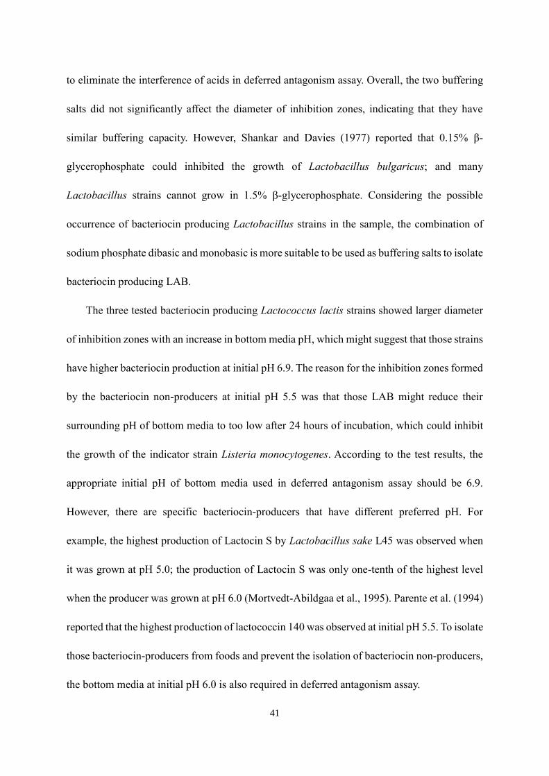
4.3.4 Identification of bacteriocin genes of representative isolates ......................................................56
4.3.5 The inhibition spectrum of representative isolates ........................................................................59
4.3.6 The effect of growth conditions on bacteriocin production of representative isolates ..........59
4.4 Discussions .....................................................................................................................................................64
Chapter 5 Conclusions .............................................................................................................................................68
Appendix A Diameter of inhibition zones formed by the testing strains in Chapter 3 .............................71
Appendix B Bacteriocin units of different isolates in Chapter 4 ...................................................................73
References ..................................................................................................................................................................75

ix
LIST OF TABLES
Table Page
1 The components of tested bottom media…………………………………………..29
2 List of primers used for the bacteriocin-specific PCR targeting LAB……………..50
3 Sequencing information for bacteriocin producing LAB isolates………………… 53
4 Identification of bacteriocin genes of representative LAB isolates amplified by primer
sets Nis Z, Bac 147 and Pln S…………………………...……………………..…..58
A1 The effects of bottom media composition on the diameter of inhibition zones formed by
bacteriocin-producing strains (Ki, L7-3 and B) and bacteriocin non-producing strains
(R7-4, LGG and LBP) .……………………………………………………………….71
A2 The effects of pH of bottom media on the diameter of inhibition zones formed by
bacteriocin-producing strains (Ki, L7-3 and B) and bacteriocin non-producing strains
(R7-4, LGG and LBP)…….....………………………………………………………72
A3 The effects of Tween 80 and/or ethanol on the diameter of inhibition zones formed by
bacteriocin-producing strains (Ki, L7-3 and B) and bacteriocin non-producing strains
(R7-4, LGG and LBP)………………………………………………………………..72
B1 Influences of medium on bacteriocin production (BU/ml) by representative isolates..73
B2 Influences of initial medium pH on bacteriocin production (BU/ml) by representative
isolates………………………………………………………………………………....73
B3 Influences of supplements on bacteriocin production (BU/ml) by representative
isolates………………………………………………………..……………………….74
x
LIST OF FIGURES
Figure Page
1 Simplified catabolic pathways of fermentative LAB………………………….…….….9
2 Bacteriocin deferred antagonism assay…………………………………………………28
3 The effects of bottom media composition on the diameter of inhibition zones……….32
4 The inhibition zones formed by bacteriocin producers and bacteriocin non-producers in
different media………………………………………………………………………….33
5 The effects of pH of the bottom media on the diameter of inhibition zones…………...35
6 The inhibition zones formed by bacteriocin producers and bacteriocin non-producers at
different initial medium pH…………………………………………………………….36
7 The effects of Tween 80 and/or ethanol on the diameter of inhibition zones………..…..38
8 The inhibition zones formed by bacteriocin producers in the media with different
supplements…………………………………………………………. …………………39
9 Inhibition zones caused by lactic acid bacteria isolated from the fermented foods…….52
10 RAPD-PCR gel profiles for bacteriocin producing LAB isolates from the fermented
foods…………………………………………………………………………………….55
11 Agarose gel electrophoresis of PCR amplicons from bacteriocin genes………………..57
12 Influences of medium on bacteriocin production by isolates…………...………………61
13 Influences of initial medium pH on bacteriocin production by isolates..…………...….62
14 Influences of supplements on bacteriocin production by Lactococcus lactis isolates.…63

1
Chapter 1
Introduction
Microbes are abundant and ubiquitous in our living surroundings. From about 10,000
years ago, when human beings were still hunting foods, there were already problems caused
by microbes including food spoilage and the diseases caused by pathogens. With the
development of agriculture, food preservation has become more and more important.
Although humans did not realize the existence of microorganisms, they had already preserved
their foods via fermentation around 4,000 B.C. In 1676, Antonie van Leeuwenhoek utilized a
crude microscope to observe some small living things, which were microbes. About 200
years later, Lazzaro Spallanzani showed that meat would not spoil when it was boiled in a
sealed container. In 1862, Louis Pasteur designed the “swan-necked flask” experiment to
prove the existence of microorganisms in nature and microorganisms being a cause of food
spoilage. These discoveries eventually led to the invention of canning of foods.
Nowadays, it is well known that human lives are closely intertwined with
microorganisms, which not only cause problems but also benefit humans. Diseases caused by
foodborne pathogens are a major public health concern. According to the Centers for Disease
Control and Prevention (CDC), the annual estimate of foodborne illness in the United States
reaches 47.8 million, and 3,037 people die due to ingestion of contaminated food. To
eliminate pathogens potentially present in food, a commonly used approach is pasteurization.
However, consumers’ concern on the loss of nutrients during thermal processes has provoked
increased demand for fresh and minimally processed foods. While these foods are usually

2
stored under refrigeration conditions, certain pathogenic bacteria, such as Listeria
monocytogenes, have developed resistance to low temperature preservation and can grow in
refrigerated foods.
Bacteriocins are antimicrobial proteins and peptides produced by bacteria. Lactic acid
bacteria (LAB) are common bacteriocin-producers and widely found in fermented foods. The
application of bacteriocin producing LAB as starter cultures in fermented foods can not only
promote the fermentation by producing organic acids but also preserve the foods by inhibiting
the growth of spoilage bacteria and pathogens. Bacteriocins produced by LAB have great
potential as natural food preservatives. To isolate bacteriocin-producers from fermented foods,
conventional methods are flip spot assay and flip streak method. These methods are very
tedious and time consuming. Henning et al. (2015) designed a deferred antagonism assay which
can simultaneously screen multiple LAB isolates for bacteriocin producing capability on agar
directly. However, certain bacteriocin non-producing LAB can produce excessive organic acids
or other antimicrobial compounds, which may interfere with the isolation of bacteriocin-
producers in deferred antagonism assay.
The immediate objectives of this study were to improve deferred antagonism assay to
reduce the chance of isolating bacteriocin non-producing LAB, and apply the improved method
to isolate and identify bacteriocin producing LAB from certain fermented foods. Besides, the
factors which can affect the production of bacteriocins by the isolates were investigated. The
data from this study will hopefully facilitate the isolation of bacteriocin-producers and the
discovery of novel bacteriocins which can be utilized in food preservation.

3
Chapter 2
Literature Review
2.1 Lactic acid bacteria
2.1.1 Introduction
Lactic acid bacteria (LAB) are characterized as Gram-positive, low-GC content, acid
tolerant, usually non-motile and non-sporeforming bacteria, which produce lactic acid as a
sole or major product of fermentative metabolism from glucose. The first pure culture of
LAB was isolated by J. Lister in 1873, which was about ten years after Louis Pasteur’s study
on lactic acid fermentation (Shareck et al. 2004). The similarity between milk souring
bacteria and other lactic acid-producing bacteria was recognized in the early 1900s. In 1890,
cheese and sour milk were introduced; nowadays, LAB are commonly used as starter cultures
in a variety of fermented foods, such as yogurt, kefir, butter milk, and kimchee.
According to cell morphology, members of LAB can be divided into rod and cocci.
Lactobacilli and Carnobacterium are typical rod-shaped LAB. The other LAB genera are
cocci. Besides, according to their glucose fermentation modes, LAB can be divided into
homofermentative and heterofermentative bacteria. Homofermentative LAB convert sugars
to lactic acid as their sole end product, which include Lactococcus, Pediococcus, and
Streptococcus and so on. The role of homofermentative LAB in food fermentation processing
is mainly related to decreasing the pH of food by producing enough lactic acids. In contrast to
homofermentative LAB, heterofermentative LAB, like Leuconostoc and some Lactobacillus
species, utilize the pentose phosphate pathway to replace the phosphoketolase pathway or
phosphogluconate pathway, which can produce not only lactic acid but also acetic acid,

4
ethanol and/or carbon dioxide (König and Fröhlich, 2017). Therefore, heterofermentative
LAB contribute to the texture and flavor of fermented foods.
2.1.2 Characteristics of major lactic acid bacteria
Due to ubiquitous occurrence of LAB in food and their significant contributions to human
gut health, plenty of research has been focused on the characterization of LAB in hope of
gaining better understanding of their physiology and exploring the potential of their
metabolites as natural preservatives. The major genera of LAB are Lactobacillus,
Leuconostoc, Pediococcus, Lactococcus, and Streptococcus. The other LAB, including
Aerococcus, Carnobacterium, Enterococcus, Oenococcus, Sporolactobacillus,
Tetragenococcus, Vagococcus, and Weissella, belong to the order Lactobacillales.
2.1.2.1 Characteristics of Lactobacillus
Lactobacillus are gram-positive, facultatively anaerobic or microaerophilic, rod-shaped,
non-sporeforming bacteria. The genomes of Lactobacillus are highly variable, which have
1,100 to 3,200 protein-coding genes (Mendes-Soares et al., 2014). The wealth of genome
compositions attributes to high diversity among species in the genus Lactobacillus. Julius et
al. (2008) isolated 23 representative Lactobacillus strains from fermented milk to test their
functional characteristics. They reported that Lactobacillus spp. showed a high tolerance to
acidic conditions of pH 2.5. Lb. fermentum strains had the highest resistance to acidic
conditions, which could maintain 100% survival after exposure to pH 2.0. In addition,
Lactobacillus spp. also showed bile toxicity resistance, antibiotic susceptibility,
hydrophobicity, mucin degradation, DNase activity and antigenotoxic characteristics
5
(Guarner and Schaafsma, 1998).
2.1.2.2 Characteristics of Leuconostoc
Leuconostoc are gram-positive, facultatively anaerobic, non-sporeforming, ovoid cocci.
Some strains of Leuconostoc show thermoduric capacity and can resist pasteurization
(Martley and Crow,1993). Leuconostoc cells are able to survive for a long time in
unfavorable surroundings. Hostile environmental conditions promote the formation of slime
or glycocalyx on Leuconostoc cells, resulting in biofilms which protect the cells against
detrimental agents (Kim et al., 2000). The slime formed by Leuconostoc spp. can also cause
food products to spoil (Ennahar et al., 2003). In dairy technology, Leuconostoc spp. play an
important role as non-starter lactic acid bacteria (NSLAB). As heterofermentative bacteria,
Leuconostoc spp. contribute to the formation of aroma and texture of specific dairy products.
2.1.2.3 Characteristics of Pediococcus
Pediococcus are gram-positive, facultatively anaerobic, non-sporeforming, cocci.
Pediococci are the major component of microbial flora in various types of crops (Cai et al.,
1999). They commonly grow with other plant-associated LAB during fermentation.
Pediococcus can utilize many carbohydrates as their carbon sources, which can ferment
maltose, sucrose, and methyl glucoside to produce lactic acid as the sole end product (Bergan
et al., 1984). In addition, Pediococcus can stand harsh environments. For example, P.
pentosaceus shows tolerance to 1,0% NaCl. P. pentosaceus and P. acidilactici can survive at
50℃.
6
2.1.2.4 Characteristics of Lactococcus
Lactococcus are gram-positive, facultatively anaerobic or microaerophilic, non-
sporeforming, non-motile, cocci. Lactococci are mesophilic and alkaline susceptive, which
can grow at 10℃ but cannot be cultured at 45℃ or pH 9.6 (Cogan et al., 1997). Some
Lactococcus lactis strains are tolerant to stressful conditions. Mannu et al. (2000) reported six
strains of L. lactis were able to grow in medium containing 6.5% NaCl. Lactococcus,
particularly L. lactis, are commonly used in the dairy industry to manufacture fermented
foodssuch as cheeses. L. lactis subsp. lactis and L. lactis subsp. cremoris are important starter
cultures in dairy fermentation (Hayes et al., 2006). Those strains can rapidly acidify the milk
by fermenting lactose and glucose to produce lactic acid, which in turn inhibits the growth of
spoilage bacteria. Additionally, those strains also contribute to the production of aroma
compounds in fermented dairy products (Ayad et al. 1999). The flavor compounds produced
by lactococci are due to their citrate fermentation, which can generate diacetyl, carbon
dioxide, acetoin, pyruvate, 2,3-butanediol, and acetaldehyde (Bandell et al. 1998).
2.1.2.5 Characteristics of Streptococcus
Streptococcus are gram-positive, facultatively anaerobic, non-motile, non-sporeforming
cocci. Streptococcus genus is common flora of the mouth, nose, and throat (Skinner and
Quesnel 1978). Species of Streptococcus are classified based on their hemolytic properties
(Patterson, 1996). Alpha-hemolytic species can oxidize the iron in hemoglobin molecules in
red blood cells. Beta-hemolytic species can cause complete rupture of red blood cells.
Gamma-hemolytic species cause no hemolysis. Some streptococci can cause human disease.

7
Strep. pyogenes, Strep. agalactiae, and Strep. pneumonia are notable as the pathogens of
serious acute infections in human beings (Hardie and Whiley, 1995). Some Strep. mitis strains
are aggressively pathogenic in immunologically compromised individuals, giving rise to
septicemia or the adult respiratory distress syndrome (Hardie and Whiley, 1994). Strep.
anginosus has been associated with abscesses in various parts of the body, such as the mouth,
brain, liver and other organs (Whiley et al., 1992). Although some Streptococci can cause
human diseases, many Streptococcus species are not pathogenic and can be applied in the
food industry. Streptococci are critical ingredients in producing yogurts and Swiss cheeses by
working as starter cultures (Beresford et al., 2001).
2.1.3 Metabolism of lactic acid bacteria
Kniel et al. (2012) mentioned in their book that carbohydrates are used as carbon and
energy sources by homofermentative and heterofermentative LAB through different catabolic
pathways, which are related to the formation of adenosine-tri-phosphate (ATP). There are
three main catabolic pathways utilized by fermentive LAB, including Embden-Meyerhof-
Parnas pathway, Enter-Doudoroff pathway, and Heterofermentative pathway. Figure 1
illustrates the simplified catabolic pathways of LAB.
The Embden-Meyerhof-Parnas (EMP) pathway is the most crucial catabolic pathway. In
this pathway, LAB can convert one molecule of glucose to two molecules of pyruvic acids.
However, at that point, the oxidation-reduction reactions are not balanced, which still need to
oxidize the pyruvate to lactic acid. Meanwhile, two molecules of ATP generate. As the only
end product of EMP pathway is lactic acid, EMP pathway is considered the most effective

8
pathway. The organisms using this pathway are called “homolactic”.
The Enter-Doudoroff pathway is more important in dairy fermentations than other two
pathways. The reason is that milk contains abundant lactose which, as a disaccharide, can be
broken down into glucose and galactose during fermentation. The other pathways cannot
metabolize the galactose portion of lactose. Therefore, those LAB using EMP pathway or
heterofermentative pathway will waste half of this energy source from lactose. However,
those LAB which use Enter-Doudoroff pathway can metabolize galactose and produce one
ATP molecule.
The LAB that utilize the heterofermentative pathway can metabolize five carbon sugars,
such as pentoses. Hence, this type of LAB is more abundant in the environments that have
adequate pentose and less hexoses.

9
Figure 1. Simplified catabolic pathways of Fermentive LAB
2.1.4 Inhibition of LAB against spoilage bacteria and pathogens
The capacity of LAB to produce antimicrobial substances has been realized and used to
preserve food since long time ago. Several investigations have proved that various species of
LAB can exert antagonistic actions against intestinal and foodborne pathogens (Gibson et al.,
1997). The antimicrobial activities of LAB may be due to a) decreasing pH by the production
of organic acids; b) producing hydrogen peroxide; and c) producing bacteriocins (Sanders,
1993).
2.1.4.1 The effect of organic acids
LAB can produce and accumulate organic acids during food fermentation by metabolizing

10
carbohydrate source. The accumulation of lactic acid and short chain fatty acids (SCFA), such
as acetic acid and propionic acid, results in a reduction in pH, which can inhibit many Gram-
positive and Gram-negative bacteria. Lactic acid and acetic acid are known to inhibit
Staphylococcus aureus in the early stage of its growth (Haines and Harmon, 1973). Goepfert
and Hicks (1969) reported that Salmonella cells are inhibited when they grow at pH lower than
4.4. Adams and Hall (1988) showed that the combination of lactic acid and acetic acid can
inhibit the growth of E. coli and Salmonella. The reason is that lactic acid can increase the mole
ratio of inhibitory undissociated acetic acid. In addition, lactic acid and acetic acid can inhibit
the growth of Helicobacter pylori that might cause ulcers and even stomach cancer (Midolo et
al., 1995).
2.1.4.2 The effect of hydrogen peroxide
In aerobic conditions, LAB could produce hydrogen peroxide (H2O2). H2O2 can form
destructive hydroxyl radical to peroxidate membrane lipids (Morris, 1979; Kong and Zotolla
Zotolla Zotolla 1999on, 1980) and increase membrane permeability (Kong and Davison, 1980).
Moreover, H2O2 can destruct nucleic acids and proteins in cell (Piard, and Desmazeaud, 1992).
There are several reports on the effect of H2O2 produced by LAB on other microorganisms.
LAB can utilize the NADH oxidase, pyruvate oxidase, and NADH peroxidase to produce
H2O2 (Murphy and Condon, 1984). H2O2-producing LAB are commonly present in the vagina
of normal women, but they are absent from women with bacterial vaginosis. The production of
H2O2 by LAB represents an antimicrobial defense mechanism of the normal vaginal ecosystem
and protects against genital colonization by pathogens (Naidu et al., 1999). Hillier et al. (1992)

11
showed that women colonized by H2O2-positive LAB had lower chance to suffer bacterial
vaginosis, symptomatic candidiasis, and vaginal colonization by Gardnerella vaginalis,
Bacteroides, Peptostreptococcus, Mycoplasma hominis, Ureaplasma urealyticum, and Viridans
streptococci. Hawes et al. (1996) found that the women colonized by H2O2-producing LAB
showed a decrease in the acquisition of vaginal infections.
2.1.4.3 The effect of bacteriocins
LAB can produce a wide range of bactericidal proteins which are deemed as bacteriocins.
The production of bacteriocin is considered to be a strategy that certain bacteria use to compete
with other bacteria. In recent years, there are an increasing number of studies on bacteriocins
produced by LAB. For example, Nisin, as the most well-known bacteriocin, is produced by
Lactococcus lactis, and can inhibit pathogenic Listeria monocytogens. Gibson et al. (1997)
reported that L. reuteri produced the bacteriocin reuterin which inhibited Salmonella and
Listeria. Jacobsen et al. (2003) utilized the living culture of Leuconostoc carnosum 4010 and
its bacteriocin to effectively inhibit the growth of Listeria monocytogenes.More and more
studies have focused on bacteriocins in that bacteriocins are considered natural and safe
products to be applied in food systems, which are readily accepted by the consumer.
12
2.2 Bacteriocins
2.2.1 Introduction
Food spoilage and foodborne disease are common problems provoking concerns of food
quality and public health all over the world. Bacterial pathogens, including Salmonella
enterica, Escherichia coli O157:H7, Listeria monocytogenes, Staphylococcus aureus,
Clostridium botulinum, and so on, have posed serious hazards to the public for many years.
The infection by these pathogens can cause diarrhea, typhoidal fever, hemorrhage colitis, and
even death. Many antibacterial substances produced by animals, plants, insects, and bacteria,
such as hydrogen peroxide, fatty acids, organic acids, ethanol, antibiotics, and bacteriocins,
have already been used by the food industry to improve the quality and safety of food
products.
Bacteria can produce bacteriocins via their ribosomes as secondary metabolites.
Bacteriocins are antibacterial peptides or proteins which are bactericidal or bacteriostatic. In
comparison with chemical agents, bacteriocins are nearly harmless to humans because they
lead to less modification of nutritional and organoleptic properties of foods and have less
toxicity to human beings. Unlike antibiotics, bacteriocins cause less bacterial resistance,
which indicates target bacteria might have further evolved. Bacteriocins are produced by
bacteria to kill other related (narrow spectrum) or unrelated (wide spectrum) microbiota as
their inherent defense weapons. They are a strategy for maintaining the population of
bacteriocin-producers and reducing the number of competitors in hope of obtaining more
nutrients and living space. LAB like Lactococcus, Streptococcus, Pediococcus, and
Lactobacillus are major bacteriocin-producers and are commonly used in the production of

13
fermented foods, which reflects the safety of bacteriocins (Kniel et al., 2012). Therefore, it is
necessary to understand the antimicrobial property of bacteriocins and isolate bacteriocin
producing bacteria to find safer and more effective approaches to combating pathogens.
2.2.2 Classification
Both Gram-positive and Gram-negative bacteria can produce bacteriocins. However, the
bacteriocin produced by Gram-positive species have boarder applications because their
producers have already been used as starter cultures in fermented foods, which ensures the
harmlessness of those bacteriocins. Those bacteriocins can be classified into four classes
based on their molecular structure, molecular mass, thermostability and so on.
2.2.2.1 Class I-bacteriocin
Class I-bacteriocins represent those heat stable modified peptides containing unusual
amino acids, such as lanthionine or methyllanthionine residues, which are called lantibiotics
(Nissen and Nes, 1997). Based on the structure, this class can be subdivided into subclass Ia
which includes relatively elongated, flexible, positively charged peptides, and subclass Ib
which has globular, rigid and either negatively charged or uncharged peptides (Klaenhammer,
1993). LAB commonly produce this type of bacteriocin to attack other Gram-positive
bacteria. Class I bacteriocins can bind to target molecules to prevent cell wall synthesis, and
even get inserted into cell membranes and lead to the formation of pores on cell membranes
causing cells to die.

14
2.2.2.2 Class-II bacteriocin
Class-II bacteriocins are those heat-stable, small and unmodified peptides, which do not
contain any unusual amino acids, and are called non-lantibiotics. Class-II bacteriocins can be
further divided into three subclasses (Ennahar et al., 2000). Subclass-IIa bacteriocins are
mainly those pediocin-like antilisterial bacteriocins. Subclass-IIb bacteriocins have two
peptides and are called two-component bacteriocins. Subclass-IIc bacteriocins are thiol-
activated bacteriocins, such as circular bacteriocins. In general, Class II bacteriocin peptides
can get into the membrane of target cells causing depolarization and death by applying their
amphiphilic helical structures (Drider et al., 2006). The amphiphilic Class II bacteriocin
peptides can combine with the N-terminal or C-terminal of target protein molecules and cause
them to lose their activities.
2.2.2.3 Class-III bacteriocin
Class-III bacteriocins have apparent differences from the first two classes, which are heat-
labile and larger (>10 kDa) protein molecules (Savadogo et al., 2006). Most of bacteriocins in
this class are bacteriolysins which can lyse the target cell by cell wall hydrolytic activity
(Johnsen et al., 2004). Lysostaphin produced by Gram-positive Staphylococcus species is a
typical bacteriolysin, which can kill other Gram-positive bacterial cells through damaged cell
walls (Cotter et al., 2005).
2.2.2.4 Class-IV bacteriocin
Those heat stable, complex bacteriocins, containing lipid or carbohydrate moieties,

15
belong to the Class-IV (Heng et al., 2007). This type of bacteriocins was found after the
observation that bacteriocin activities had not been abolished until being treated with protease
and glycolytic or lipolytic enzyme (Garneau et al., 2002). The mode action of this type of
bacteriocins needs further study.
2.2.3 Difference between bacteriocins and antibiotics
Bacteriocins are antimicrobial peptides which have bactericidal or bacteriostatic mode of
action against their producers’ closely related species. Although the functions of bacteriocins
and traditional antibiotics are similar, there are still remarkable differences between them.
Bacteriocins have the potential to solve problems caused by the use of antibiotics and may
take the place of antibiotics in foods and pharmaceuticals. Unlike antibiotics, bacteriocins are
naturally produced by certain bacteria including the gut probiotic bacteria to against intestinal
infections, which would not lead to collateral damage to the human commensal microbiota
(Blaser, 2011). Those human commensal microbiotas play key roles in human health and are
susceptive to antibiotics due to their broad-spectrum antimicrobial activities (Cotter et al.,
2012). Moreover, bacteriocins are less toxic and have resulted in less incidence of atopic and
autoimmune disease than antibiotics (Blaser, 2011). As to the development of cell tolerance to
bacteriocin, any antimicrobial compounds including bacteriocin have the potential to cause
resistance in microbiota. But some possible strategies can be used to minimize the emergence
of bacteriocin resistance. For example, application of bacteriocins with distinct mechanisms
of action in combination can keep their effects on the mutation of bacteria.

16
2.2.4 Factors affecting the production of bacteriocin
Bacteriocin biosynthesis occurs at the end of bacterial exponential growth phase (Piard
and Desmazeaud, 1992). Bacteria produced bacteriocin to compete with other bacteria for
getting more resources to grow. The production of bacteriocins by LAB can be affected by
several factors, such as culture medium, culture pH, and medium supplement.
2.2.4.1 Effect of culture medium
Bacteriocin production is affected by the type of medium used to cultivate the
bacteriocin-producers. The culture media which contain more essential nutrients required by
bacteriocin-producers can promote their production of bacteriocins. Geis et al. (1983)
compared the bacteriocin production of lactic streptococci grown in Elliker broth, M17 broth,
and milk. The maximum bacteriocin production was found in Elliker broth followed by M17
broth. Piard et al. (1990) also observed that Elliker medium buffered with sodium β-
glycerophosphate yielded more lacticin 481 than M17 medium. Muriana and Klaenhammer
(1987) reported that the maximum lactacin F production was observed in MRS medium.
Vignolo et al. (1995) reported that the production of lactocin 705 was much higher in MRS
broth than in Elliker broth, M17 broth or BHI broth.
2.2.4.2 Effect of culture pH
Some LAB strains may prefer cultures with different pH and this can subsequently affect
their production of bacteriocins. Cabo et al. (2001) found that the production of nisin by
Lactococcus lactis was noticeably higher at pH 6.0 than at pH 5.5. Turgis et al. (2016)

17
showed that the production of nisin was significantly higher at pH 7 than pH 6.0 or pH 5.5.
Vignolo et al. (1995) found that the maximum output of lactocin 705 by Lactobacillus casei
CRL 705 was achieved in MRS broth at pH 6.5-7.5. Lactobacillus sakei subsp. sakei 2a
secreted abundant bacteriocin at pH 5.5-7.0 (Malheiros et al., 2015). The production of
lactococcin 140 was obtained at pH 5.5 (Parente et al., 1994). Mortvedt-Abildgaa (1995)
reported that lactocin S would lose its bactericidal activity when the producer Lactobacillus
sake L45 was cultured at pH higher than 6.0.
2.2.4.3 Effect of medium supplement
The addition of certain chemical reagents, such as tween and ethanol, into the culture
medium may improve the production of bacteriocins by LAB. Tween (polysorbate) works as a
surfactant which can increase the permeability of cell membranes and enhance the production
of bacteriocins; it can also accelerate the diffusion of bacteriocins and improve their
antimicrobial effect (Vignolo et al., 1995). Malheiros et al. (2015) reported that the addition of
Tween 20 and Tween 80 increased the bacteriocin production by L. sakei 2a. Tween 20 can also
enhance the activity of bacteriocins produced by L. sakei and L. curvatus ACU-1 (Castro MP.
et al., 2011). Martinez et al. (2015) reported that Tween 80 can work as a stimulating additive
to promote the production of bacteriocin-like inhibitory substances by Bifidobacterium lactis
in skim milk. Radha and Padmavathi (2017) reported that 0.24% Tween 20 could increase the
bacteriocin production by Lactobacillus delbrueckii subsp. bulgaricus. Ravi et al. (2017)
observed that both Tween 20 and Tween 80 could increase the bacteriocin production by LAB
isolated from mango pulp.
18
Additionally, low concentration of ethanol can cause slightly unfavorable growth
condition which might increase the production of bacteriocin (De Vuyst et al., 1996).
Mortvedt-Abildgaa et al. (1995) reported that low concentration of ethanol might prevent the
bacteriocin aggregation and stabilize the bacteriocin. Callewaert et al. (1999) showed that
ethanol not only stimulated the production of amylovorin L471 but also prevented the
absorption of the bacteriocin to the producer cells during prolonged fermentation.
2.2.5 Application of Bacteriocin
Bacteriocins have many applications in food preservation, via directly adding into foods
or in combination with other preservation approaches, such as pasteurization, high-pressure
processing or irradiation (Abriouel et al., 2010; Keymanesh et al., 2009). The addition of pure
or mixed bacteriocins or bacteriocin producing LAB to foods can increase their shelf life and
safety by inhibiting common spoilage bacteria and foodborne pathogens. It can also control
adventitious bacteria which may cause foreign odors. Bacteriocin producing LAB have been
found in various fermented foods.
2.2.5.1 Application in dairy products
Many bacteria, particularly LAB, are used as starter cultures in fermented dairy products
like yogurt, kefir, and cheese. Their antimicrobial products, bacteriocins, can even be utilized
directly in those fermented dairy products. O’Sullivan et al. (2002) reported that bacteriocins
were incorporated into cheese or yogurt as dried concentrate powders or used as the products
of starter cultures. Many Swiss style cheeses rely on bacteriocin producing cultures to retard
the gas blowing caused by clostridia. Nisin-producing Lactococci were found to effectively
19
act against clostridia spoilage (Hirsch et al., 1951). A combination of lactose fermenting,
nisin-producing and proteinase-positive L. lactis strains showed antimicrobial effect on
Listeria monocytogenes, Clostridium sporogenes and Staphylococcus aureus in cheese
spreaders and pasteurized processed cheese (Zotolla 1994). Besides nisin, propionicin PLG-1
from P. thoenii P127 is also commonly used in Swiss-type cheese. This bacteriocin can kill
Listeria monocytogenes, Pseudomonas fluorescens and Yersinia enterocolitica (Lyon et al.,
1993).
As for yogurt, applying a combination of nisin-producing strains and traditional starter
cultures can prevent the growth of spoilage bacteria and extend its shelf life (Yamauchi et al.,
1996). Streptococcus thermophilus is one of the starter cultures used in yogurt. It contributes
to the development of yogurt semisolid texture by producing lactic acid. It also produces
bacteriocins that can inhibit the growth of Listeria monocytogenes and to some extent, S.
aureus (Yang et al., 2012). Bifidobacterium is common prebiotics used in yogurt, which can
produce acidocin B. Brink et al. (1994) reported that acidocin B could act effectively against
Clostridium sp. in fermented foods.
2.2.5.2 Application in meat products
For meat products, the contamination by Listeria monocytogenes is a serious concern. L.
monocytogenes is widely distributed in nature and can get into ready-to-eat meat products
(Nesbakken et al., 1996). Many bacteriocins produced by LAB showed their capability of
reducing or inhibiting the growth of L. monocytogenes in meat products. Jacobsen et al.
(2003) utilized the living culture of Leuconostoc carnosum 4010 and its bacteriocin in sliced

20
meat products. The treatment effectively inhibited the growth of L.monocytogenes. The
bacteriocin sakacin P and pediocin AcH, isolated from Lactobacillus and Pediococcus
acidilactici cultures, respectively, showed remarkable antilisterial activity in a Listeria-
seeded raw pork meat matrix and suppressed Listeria for six weeks (Kouakou et al., 2010).
Sabia et al. (2003) also reported that bacteriocin enterocin 416 K1 produced by Enterococcus
casseliflavus showed strong antilisterial activity in Italian sausages.
In addition to those antilisterial bacteriocins, some other bacteriocins have also been
isolated from fermented meat products, which can effectively treat and prevent microbial
infections. For example, Todorov et al. (2010) found the bacteriocin producing strain
Enterococcus foecium ST5Ha from smoked salmon can produce a pediocin-like bacteriocin.
This bacteriocin inhibit not only Listeria spp. but also HSV-1 virus, which is a type of human
virus.
2.2.5.3 Application in fruits and vegetables
Unlike meat products and dairy products, fruits and vegetables are consumed raw to keep
their freshness. Unfortunately, some pathogenic microbes may contaminate those foods. A
combination of bacteriocins and chemical preservatives can decrease the risk of microbial
contamination. Molinos et al. (2008) reported that washing treatments with enterocin AS-48
could efficiently reduce the counts of L. monocytogenes in sliced melon, watermelon, pear,
and kiwi in 24 hours. Moreover, a combination of enterocin AS-48 and 12 mM carvacrol, as
well as with 100 mM n-propyl p-hydroxybenzoate showed increased antilisterial activity and
suppressed the regrowth of Listeria. In addition to Listeria, enterocin AS-48 also had

21
inhibitor effect against Staphylococcus aureus in vegetable sauces. Although its antimicrobial
activity was limited when it was used alone, a combination of AS-48 and 20 mM
hydrocinnamic acid or 126 mM carvacrol could lower the viable count of S. aureus below the
detection limit (Grande et al., 2007). Enterocin AS-48 also showed outstanding effect on the
reduction of spoilage Lactobacillus strains, such as Lb. collinoides and Lb. diolivorans, when
it was used along with high-intensity pulsed electric field treatment in apple juice (Martinez-
Viedma et al., 2008).
There were other bacteriocins that can also be used to treat fresh fruits and vegetables.
For example, Carvalho et al. (2008) reported remarkable effects of bovicin HC5, a
bacteriocin produced by S. bovis HC5, against vegetative cells of Alicyclobacillus
acidoterrestris which is a common spoilage bacterium in pasteurized acidic drinks. An
interesting finding by Carvalho et al. was that spores of A. acidoterrestris were more
sensitive to bovicin HC5 than vegetative cells.
2.2.6 Isolation of bacteriocin-producers
Approaches to isolating bacteriocin-producers are based on their antimicrobial capability.
The first antimicrobial susceptibility test that utilized diffusion of the antibiotic substance
through agar medium was done by Fleming in 1924 with penicillin against Staphylococcus
aureus (Hoover and Steenson., 2014).
2.2.6.1 Flip streak method
This method requires the use of an aseptic loop to inoculate fresh LAB culture onto the
surface of a culture medium agar by streaking. After incubation for 24 hours, the agar is

22
flipped onto the cover of the petri dish by using an aseptic spatula. Then the indicator strain is
streaked on the inverted agar. After incubation for 24 hours, the bacteriocin producing strain
can form inhibition line on the inverted plate (Spelhaug and Harlander, 1989). In this method,
each plate usually just tests one type of LAB. Therefore, this method is complex and
inefficient.
2.2.6.2 Flip spot method
Test organisms need to be cultured and diluted 10-fold in peptone water. Diluted cultures
are spotted onto the surface of an agar plate. Each spot should keep approximately 3 cm
distance. Plates are incubated for 24 hours, and the agar is reversed as in the flip streak
method. The indicator strain is inoculated into temporarily melt soft agar and poured onto the
surface of the inverted agar. This two-layers plate is incubated for 24 hours, and the inhibition
capability of LAB is detected by observing inhibition zones (Spelhaug and Harlander, 1989).
This method can identify multiple LAB strains at the same time. The disadvantage is that
pure LAB cultures must be obtained before the inhibition test, which can be tedious and
laborious.
2.2.6.3 Deferred antagonism assay
The sample containing mixed LAB strains is diluted and spread plated on the agar
medium. A temporarily melt softer agar is poured onto the base agar immediately, and the
two-layers plate was incubated for 24 hours. After the LAB colonies show up, a temporarily
melt indicator layer with indicator organisms is overlaid on the two-layer plate. The three-
layer plate is incubated for 24 hours until the indicator lawn grows to completion. The

23
bacteriocin producing LAB show their antimicrobial capability by forming clear inhibition
zones (Henning et al., 2015). This method does not require the use of pure LAB cultures and
can screen more LAB strains at one time than flip spot method. However, certain bacteriocin
non-producers can produce excessive organic acids and also form inhibition zones in this
assay.

24
Chapter 3
Improved deferred antagonism assay for isolating bacteriocin producing
lactic acid bacteria
3.1 Introduction
Microbes play an important role in food safety. Pathogenic organisms can cause illness
through contaminated food. According to Centers for Disease Control and Prevention (CDC),
Listeria monocytogenes is one of the most dangerous foodborne pathogens, and it causes
approximately 1,600 people to become sick and 260 people to die annually in the United
States. Listeria monocytogenes is non-sporeforming, gram-positive, facultative anaerobic, rod
shape bacterium. Although there is no evidence on the infectious dose of Listeria
monocytogenes in humans, Golnazarian et al. (1989) reported that mice infected orally
showed variable responses, with 50% infectious doses ranging from 1.7 x 10³ to 9.9 x 10⁶
CFU. Listeria monocytogenes infections can cause diarrhea, fever, septicemia, meningitis,
encephalitis or abortion. Moreover, this pathogen has high resistance to undesirable
environments, which can grow at 14% NaCl, pH 4.4, or - 0.1℃ conditions (Walker et al.
1990). Therefore, those heat sensitive foods, such as fresh produce and fermented foods, are
likely to harbor Listeria strains. To solve this problem, bacteriocin producing lactic acid
bacteria (LAB) offer a natural and effective approach of eliminating or inhibiting the growth
of Listeria monocytogenes in food.
LAB are gram-positive, acid tolerant, non-motile and non-sporeforming bacteria. LAB
are widely used as starter cultures in fermented foods to increase their shelf life and quality.
During the application of LAB in fermentation, their effect against other microbes have been
25
gradually realized by human beings. It is by now well known that LAB can produce
antimicrobial compounds including organic acids, hydrogen peroxide, diacetyl, and
bacteriocins.
Bacteriocins are bactericidal or bacteriostatic proteins produced by bacteria. Generally,
LAB are deemed safe, and so are their bacteriocins, which should not negatively affect
human health (Eijsink et al., 2002). Therefore, bacteriocins produced by LAB have attracted
much attention. These bacteriocins can be separated into different classes and show different
inhibitory spectra. Commonly, bacteriocins produced by LAB can be divided into two
categories, lantibiotics and non-lantibiotics. Lantibiotics have post-translationally modified
amino acids, such as dehydroalanine (Dha), dehydrobutyrine (Dhb), eponymous lanthionine
(Lan), and β-methyllanthionine (MeLan) formed by thioether linkages between dehydrated
amino acid residues and neighboring cysteines (Rink et al., 2007). Lantibiotics can prevent
cell wall synthesis and form pores on cell membranes, which eventually cause the death of
target cells. Non-lantibiotics do not have unusual amino acids. They can cause depolarization
of target cells and even lyse their cell walls.
To isolate bacteriocin producing LAB, flip streak method and flip spot method are
commonly used. These two methods require picking an excessive number of isolates from
food samples followed by culturing them individually in broth, neutralizing organic acids by
alkaline, and finally testing them on agar against indicator organisms. Therefore, both
methods are time-consuming and pretty tedious to use. Yet the isolation rate can be low since
most LAB cannot produce bacteriocins. Thus, a more efficient approach, bacteriocin deferred
antagonism assay, was developed by Henning et al. (2015). This assay can test the bacteriocin

26
producing capability of more than 80 colonies on agar directly at the same time, which
significantly reduce testing time and labor (Figure 2). However, the deferred antagonism
assay still has limitations. LAB can produce lactic acids and acetic acid which can reduce the
pH of media to around 4.0. At that pH, indicator bacteria like Listeria monocytogenes
cannnot grow. Hence, there is a high possibility to isolate bacteriocin non-producing LAB
which can inhibit the growth of Listeria monocytogenes by causing acidic surroundings in the
deferred antagonism assay. To solve this problem, it was necessary to decrease the effect of
acids produced by LAB on indicator bacteria while increasing the production of bacteriocins
by LAB in the deferred antagonism assay.
The objectives of this study were to determine the effects of various agar media combined
with different buffering salts on bacteriocin-producers and non-producers in deferred
antagonism assay. So that more appropriate agar medium can be determined to rapid and
effective isolation of bacteriocin producing LAB from foods. In addition, as LAB are
sensitive to environmental acidity in producing bacteriocins, different pH of selected agar
medium was tested separately in order to find the initial medium pH, which can ensure the
production of bacteriocins by LAB and alleviate the interference of organic acids produced
by bacteriocin nonproducing LAB. Finally, previous studies indicate that adding 1% Tween
80 to culture media can increase the production of lactocin 705 (Vignolo et al., 1995).
Supplementation of culture media with 1% of ethanol can prevent lactocin S from
aggregating and increase its activity (Mortvedt-Abildgaa et al., 1995). Therefore, both Tween
80 and ethanol were tested with selected agar medium for LAB in deferred antagonism assay.

27
3.2 Materials and Methods
3.2.1 Bacterial strains and growth conditions
Three previously identified bacteriocin producing Lactococcus lactis strains (Ki, L7-3, B)
were used as bacteriocin positive control strains. Three bacteriocin non-producers,
Leuconostoc mesenteroides (R7-4), Leuconostoc pseudomesenteroides (LPM) and
Lactobacillus plantarum (LBP), were used as bacteriocin negative control strains. All these
strains were grown in MRS (De Man, Rogosa and Sharpe) broth at 30℃ for 24 hours before
being plated on agar media in deferred antagonism assay. The indicator organism, Listeria
monocytogene, was grown in TSB (Tryptic soy broth) at 35℃. All strains were maintained as
frozen stock cultures at -80℃ and propagated twice before being used in this study.
3.2.2 Bacteriocin deferred antagonism assay
Bacteriocin positive and negative control strains were serially diluted by 10-fold in 0.1%
peptone water, and appropriate dilutions (250-2500 CFU/mL) were chosen to use in deferred
antagonism assay. Figure 2 illustrates the deferred antagonism assay described by Henning et
al. (2015), which spread all tested strains onto buffered base agar layer (1.5% agar). Plated
samples were immediately covered with same buffered agar sandwich layer (0.75% agar).
The sandwich layer was included to decrease the interference of lactic acid produced by LAB
and prevent the inhibition zone caused by bacteria phage when dealing with food samples.
After that, tested organisms in double-layer agar were incubated at 30℃ for 24 to 48h. The
base layer and sandwich layer called as bottom media. When colonies could be observed in
bottom media, 10 mL of molten BHI agar (0.75% agar) and 0.1 mL of 1 × 10⁸ CFU/mL

28
Listeria monocytogenes were mixed and overlaid onto the sandwich layer. Finally, the agar
plates were incubated at 30℃ until the indicator lawn formed. The bacteriocin producing
strains can form inhibition zone around the colony as shown in Figure 2.
Figure 2. Bacteriocin deferred antagonism assay

29
3.2.3 Test different media in deferred antagonism assay
Three different agar were used as the bottom media to decide which type was more
appropriate in deferred antagonism assay. They were MRS medium, M17 medium, and
Elliker medium, which were suitable to grow most LAB species. In addition, two types of
buffering salts were added into the agar media in order to alleviate interference of acids in
bacteriocin detection. They were 2% β-glycerophosphate and the combination of sodium
phosphate monobasic and dibasic (1% Na2HPO4 and 0.35% NaH2PO4) which gave the same
medium pH (6.9+0.2). Table 1 illustrates the components of tested bottom media. The
diameters of inhibition zones from different bottom media were recorded. The diameter of
inhibition zone was the distance from the edge of a bacterial colony to the edge of
surrounding clear zone.
Table 1. The components of tested bottom media
Bottom media Buffering salt
MRS β-glycerophosphate Na2HPO4 and NaH2PO4
M17 β-glycerophosphate Na2HPO4 and NaH2PO4
Elliker medium β-glycerophosphate Na2HPO4 and NaH2PO4

30
3.2.4 Test different initial pH of the bottom media
LAB are sensitive to pH in producing bacteriocins. Commonly, LAB can produce
bacteriocins in pH ranging from 5.5 to 7. Therefore, the three bacteriocin-producers and three
bacteriocin non-producers were inoculate onto selected bottom media with initial pH 5.5, 6.0,
6.5 or 6.9. Then, the deferred antagonism assay was conducted as described above. The
diameters of inhibition zones from bottom media with different pH were recorded.
3.2.5 Test the effect of Tween 80 and ethanol in bottom media
After an appropriate combination of the bottom media and buffering salts as well as the
medium pH were determined, 1% Tween 80 and 1% ethanol were added individually and in
combination to both base agar layer and sandwich layer to form new bottom media. Tween 80
and ethanol were filter-sterilized before being added to bottom media. The six tested strains
were inoculated on base agar to check for the formation of inhibition zones by using the
deferred antagonism assay. The diameters of inhibition zones from bottom media with or
without Tween 80 and/or ethanol were recorded and compared.
3.2.6 Statistical analysis
All tests were repeated once. Four inhibition zones were selected from each strain and
measured for the diameter. The inhibition zone sizes from different treatments were compared
via ANOVA with a significance level of 0.05.

31
3.3 Results
3.3.1 Effect of bottom media agar type
To choose more appropriate agar as bottom media in the deferred antagonism assay, three
bacteriocin-producers (Ki, L7-3 and B) and three bacteriocin non-producers (R7-4, LPM and
LBP) were inoculated into three types of media with two types of buffering salts. Figure 3
illustrates the diameter of inhibition zones showed in different media. Figure 4 shows the
effect of representative bacteriocin-producers and non-producers on the growth of Listeria
monocytogenes. All strains including three bacteriocin non-producers, Leuconostoc
mesenteroides (R7-4), Leuconostoc pseudomesenteroides (LPM) and Lactobacillus
plantarum (LBP), formed inhibition zones in MRS agar with both buffering salts in deferred
antagonism assay. The inhibition zones caused by bacteriocin non-producers were smaller
than the zones caused by bacteriocin-producers. Bacteriocin non-producers did not show
inhibition zones in M17 agar or Elliker agar. In comparison to M17 agar, Elliker agar was
superior in deferred antagonism assay because bacteriocin-producers Lactococcus lactis L7-3
and Lactococcus lactis B showed larger diameters of inhibition zones when Elliker agar was
used as the bottom media. This suggests Elliker medium might promote the production or the
diffusion of bacteriocins. Therefore, Elliker medium was more appropriate than M17 medium
to be used as the bottom media. Moreover, there was no significant difference between the
two buffering salts in deferred antagonism assay. However, Shankar and Davies (1977)
reported that β-glycerophosphate inhibited the growth of many Lactobacillus strains,
particularly, Lactobacillus bulgaricus. Meanwhile, certain Lactobacillus bulgaricus strains
can produce bacteriocins (Radha and Padmavathi, 2017). Hence, Elliker medium with the

32
combination of sodium phosphate dibasic and monobasic was considered more appropriate
bottom media in deferred antagonism assay.
Figure 3. The effects of bottom media composition on the diameter of inhibition zones

33
Figure 4. The inhibition zones formed by representative bacteriocin-producers and
bacteriocin non-producers in different media. Bacteriocin-producers (A, C, E, G, I,
K) and Bacteriocin non-producers (B, D, F, H, J, L) on MRS (A, B, C, D), M17 (E,
F, G, H) and Elliker medium (I, J, K, L) with buffering salts Na2HPO4 and NaH2PO4
(A, B, E, F, I, J) or β-glycerophosphate (C, D, G, H, K, L)

34
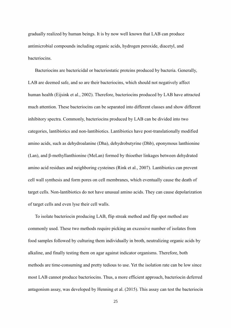
3.3.2 Effect of initial pH of bottom media
Elliker medium with the combination of sodium phosphate dibasic and monobasic was
used as bottom media. The pH of bottom media was adjusted to 5.5, 6, 6.5 and 6.9,
respectively. Their effects on the diameter of inhibition zone are shown in Figure 5. Those
bacteriocin producing strains showed inhibition zones in the bottom media from pH 5.5 to
6.9. However, bacteriocin non-producing strains could also form inhibition zones at pH 5.5.,
which indicates that the bottom media at pH 5.5 should not be used in deferred antagonism
assay. The diameter of inhibition zones created by the three bacteriocin producing
Lactococcus lactis strains increased when the pH of bottom media rose. Figure 6 shows the
inhibition zones formed by bacteriocin-producers and bacteriocin non-producers in the
bottom media from pH 5.5 to 6.9. The pH value 6.9 was chosen for the bottom media in
deferred antagonism assay.
35
Figure 5. The effects of pH of the bottom media on the diameter of inhibition zones

36
Figure 6. The inhibition zones formed by representative bacteriocin-
producers and bacteriocin non-producers at different initial medium pH.
Bacteriocin-producers (A, C, E, G) and bacteriocin non-producers (B, D, F,
H) on Elliker medium at pH 5.5 (A, B), 6.0 (C, D), 6.5 (E, F) or 6.9 (G, H).
37
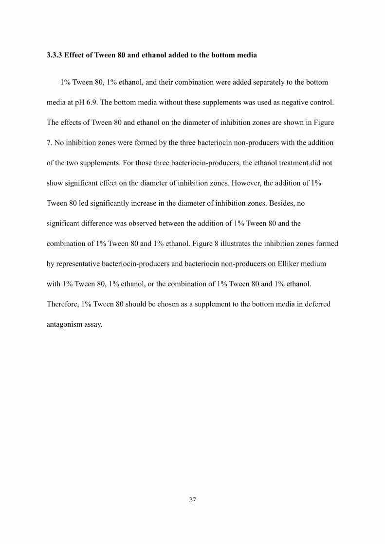
3.3.3 Effect of Tween 80 and ethanol added to the bottom media
1% Tween 80, 1% ethanol, and their combination were added separately to the bottom
media at pH 6.9. The bottom media without these supplements was used as negative control.
The effects of Tween 80 and ethanol on the diameter of inhibition zones are shown in Figure
7. No inhibition zones were formed by the three bacteriocin non-producers with the addition
of the two supplements. For those three bacteriocin-producers, the ethanol treatment did not
show significant effect on the diameter of inhibition zones. However, the addition of 1%
Tween 80 led significantly increase in the diameter of inhibition zones. Besides, no
significant difference was observed between the addition of 1% Tween 80 and the
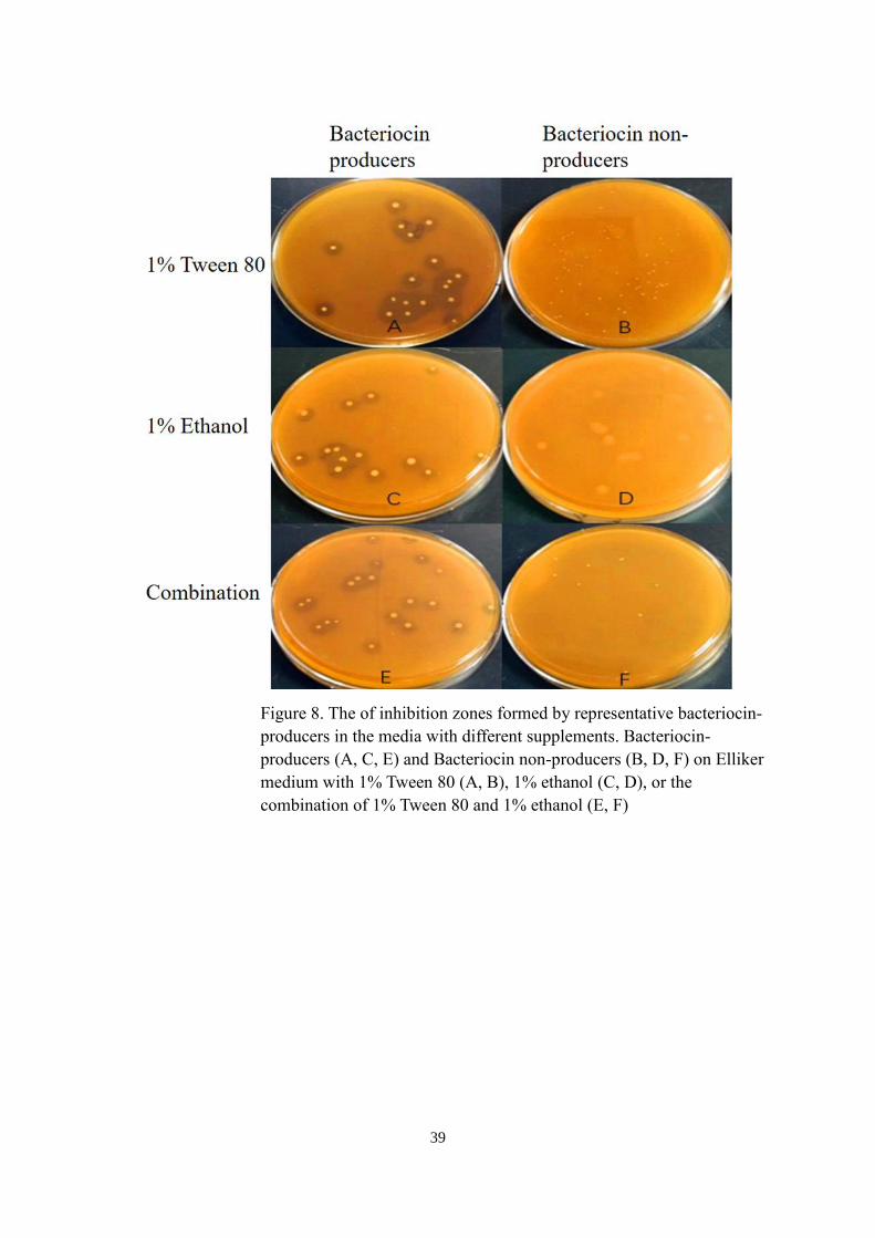
combination of 1% Tween 80 and 1% ethanol. Figure 8 illustrates the inhibition zones formed
by representative bacteriocin-producers and bacteriocin non-producers on Elliker medium
with 1% Tween 80, 1% ethanol, or the combination of 1% Tween 80 and 1% ethanol.
Therefore, 1% Tween 80 should be chosen as a supplement to the bottom media in deferred
antagonism assay.

38
Figure 7. The effects of Tween 80 and/or ethanol on the diameter of inhibition zones
39
Figure 8. The of inhibition zones formed by representative bacteriocin-
producers in the media with different supplements. Bacteriocin-
producers (A, C, E) and Bacteriocin non-producers (B, D, F) on Elliker
medium with 1% Tween 80 (A, B), 1% ethanol (C, D), or the
combination of 1% Tween 80 and 1% ethanol (E, F)

40
3.4 Discussions
These results demonstrated the effects of the type and initial pH of bottom media, Tween
80, and ethanol on the diameter of inhibition zones caused by bacteriocin-producers in the
deferred antagonism assay. The phenomenon that inhibition zones could be formed by
bacteriocin non-producers when MRS agar was used as the bottom media was probably one of
main reasons for the isolation of many non-specific inhibition LAB in deferred antagonism
assay. MRS medium might promote the production of non-bacteriocin antimicrobial substances,
like lactic acid. Naveena et al. (2005) reported that triammonium citrate, an ingredient only
used in MRS agar but not the other two tested media, can significantly increase the production
of lactic acid by Lactobacillus casei. This kind of substance can interfere with the detection of
bacteriocin-producers. Although Vignolo et al., (1995) reported that MRS medium caused
higher bacteriocin production than Elliker medium and M17 medium, this was not the case
with bacteriocin producing Lactococcus lactis strains in this test. In deferred antagonism assay,
bacteriocins need to diffuse through sandwich layer to inhibit the growth of Listeria
monocytogenes in indicator layer, which might decrease the sensitivity of indicator organisms
to bacteriocins. Elliker medium was more suitable for those bacteriocin producing Lactococcus
lactis to produce bacteriocins, which yielded larger inhibition zones than M17 medium in
deferred antagonism assay. Vignolo et al. (1995) and Geis et al. (1983) reported some
bacteriocin producing LAB showed low bacteriocin activity after they grew in M17 medium.
Therefore, M17 medium might interfere with the synthesis of bacteriocins by those LAB.
When Elliker agar and M17 agar were separately used as bottom media, both β-
glycerophosphate and the combination of sodium phosphate monobasic and dibasic were able
41
to eliminate the interference of acids in deferred antagonism assay. Overall, the two buffering
salts did not significantly affect the diameter of inhibition zones, indicating that they have
similar buffering capacity. However, Shankar and Davies (1977) reported that 0.15% β-
glycerophosphate could inhibited the growth of Lactobacillus bulgaricus; and many
Lactobacillus strains cannot grow in 1.5% β-glycerophosphate. Considering the possible
occurrence of bacteriocin producing Lactobacillus strains in the sample, the combination of
sodium phosphate dibasic and monobasic is more suitable to be used as buffering salts to isolate
bacteriocin producing LAB.
The three tested bacteriocin producing Lactococcus lactis strains showed larger diameter
of inhibition zones with an increase in bottom media pH, which might suggest that those strains
have higher bacteriocin production at initial pH 6.9. The reason for the inhibition zones formed
by the bacteriocin non-producers at initial pH 5.5 was that those LAB might reduce their
surrounding pH of bottom media to too low after 24 hours of incubation, which could inhibit
the growth of the indicator strain Listeria monocytogenes. According to the test results, the
appropriate initial pH of bottom media used in deferred antagonism assay should be 6.9.
However, there are specific bacteriocin-producers that have different preferred pH. For
example, the highest production of Lactocin S by Lactobacillus sake L45 was observed when
it was grown at pH 5.0; the production of Lactocin S was only one-tenth of the highest level
when the producer was grown at pH 6.0 (Mortvedt-Abildgaa et al., 1995). Parente et al. (1994)
reported that the highest production of lactococcin 140 was observed at initial pH 5.5. To isolate
those bacteriocin-producers from foods and prevent the isolation of bacteriocin non-producers,
the bottom media at initial pH 6.0 is also required in deferred antagonism assay.

42
The addition of 1% Tween 80 significantly increased the diameter of inhibition zones,
which means 1% Tween 80 can increase the bacteriocin production. Ravi et al. (2017), Jung et
al. (1992), and Vignolo et al. (1995) also reported that Tween 80 could remarkably increase the
production of bacteriocins by LAB. Vignolo et al. (1995) reported that Tween 80 was a critical
factor in lactocin 705 production, which works as a surfactant on the cell membranes of its
producer and can accelerate diffusion of lactocin 705. Jung et al. (1992) stated that Tween 80
may counteract the adsorption of the bacteriocin by proteins, thus making possible the
inhibition of microorganisms. As for ethanol, although Mortvedt-Abildgaa et al. (1995)
reported 1% ethanol can increase the bacteriocin yield, there was no significant difference in
the diameter of inhibition zone caused by bacteriocin-producers tested in this study.
In conclusion, adding 1% Tween 80 into Elliker medium with the combination of sodium
phosphate dibasic and monobasic at initial pH 6.9 or pH 6.0 can enhance the possibility of
isolating bacteriocin producing LAB in the deferred antagonism assay. For future research,
other factors, like the concentration of glucose, which can increase the production of
bacteriocins by LAB should be considered and tested based on this study. There is a high
demand for potent bacteriocins and strong bacteriocin-producers isolated from foods.

43
Chapter 4
Isolation, identification and characterization of bacteriocin producing lactic acid
bacteria from foods
4.1 Introduction
Bacteriocins are ribosomally synthesized proteins produced by bacteria which are
bactericidal or bacteriostatic. Bacteriocin production is a widespread phenomenon among
lactic acid bacteria (LAB), having been observed among lactobacilli, lactococci, pediococci,
and leuconostocs (Klaenhammer, 1988). In an environment with mixed bacterial populations,
the production of bacteriocins is a competitive strategy which is advantageous for producing
organisms to get more nutrients and space to grow. LAB are a critical microbial group for
industrial applications They have been commonly utilized as starter cultures in fermented
products sold on marketplaces, such as cheese, yogurt, and buttermilk. As a result,
bacteriocins produced by LAB, used in fermented foods, are deemed novel, safe and natural
food preservatives. Bacteriocins are sensitive to digestive protease and do not induce changes
in the organoleptic properties of the foods (Cabo et al., 2001). Therefore, bacteriocin
producing LAB can be isolated from fermented foods and applied to enhance food safety and
quality.
Although bacteriocins can inhibit bacteria closely related to the bacteriocin-producers,
studies have shown that when bacteriocinogenic strains are added to food, their antimicrobial
effect is often less than expected (Mortvedt-Abildgaa et al., 1995). The reason is that the
environment of those bacteriocinogenic strains in foods may reduce their production of
bacteriocins. Hence, the growth condition of bacteria is important for bacteriocin production.

44
Maximal bacteriocin production can be achieved by using carefully selected culture
medium with optimized pH and certain supplements, such as ethanol and Tween 80. Geis et
al. (1983) reported that Elliker medium is better than M17 or milk for bacteriocin production.
Plantaricin S, produced by Lactobacillus plantarum LPCO-10, which is involved in the
fermentation of olives, reached a peak of antimicrobial activity in MRS broth (Mortvedt-
Abildgaa, et al., 1995).
As for pH, Vignolo et al. (1995) reported that the production of lactocin 705 by
Lactobacillus casei CRL 705 isolated from dry sausages reached the maximum at pH 6.5-7.5
in MRS broth. Meera and Devi (2017) showed that isolated bacteriocin producing LAB
displayed maximum bacteriocin activity at pH 6 in MRS broth. Turgis et al. (2016) showed
that the maximum nisin production of Lactococcus lactis MM19 was achieved at pH 9; the
maximum pediocin production of Pediococcus acidilactici MM33 was achieved at pH 6.
Mortvedt-Abildgaa et al. (1995) reported Lactobacillus sake L45 produced more bacteriocin
at pH 5 than at pH 6.
Some supplements also have significant effects on the production of bacteriocin. Tween
80 is one of the surfactants which can provide essential oleate to metabolically active LAB
(Jacques et al., 1980). The addition of Tween 80 (0.5%-2%) to growth medium can increase
the production of lactocin 705, with the maximum bacteriocin activity at 1% Tween 80
(Vignolo et al., 1995). Ravi et al. (2017) also reported that the 1% Tween 80 can increase the
production of bacteriocin from the LAB isolated from mango. Besides favorable components,
specific bacteriocin production can be enhanced under unfavorable conditions such as low
level of ethanol. De Vuyst et al. (1996) reported 1% ethanol can increase the production of

45
amylovorin L471, a bacteriocin produced by Lactobacillus amylovorus, by two-fold.
Mortvedt-Abildgaa et al. (1995) showed that the addition of 1% ethanol to growth medium
can cause two- to four-fold increase in the production of lactocin S.
In this study, a variety of fermented foods were used to isolate bacteriocin producing
LAB via the improved deferred antagonism assay. After the isolation, the species 16S rRNA
and the bacteriocin encoding genes carried by the bacteriocin producing LAB were identified.
The main objective was to identify bacteriocin producing LAB that have inhibitory activity
against foodborne pathogens. In addition, the influence of growth conditions of isolated LAB
on their production of bacteriocins was determined, which included the culture medium,
initial pH, and medium supplements.
4.2 Materials and Methods
4.2.1 Sample collection
Fermented kimchee, sauerkraut, yoghurt, and kefir were collected from local food stores.
All food samples were stored at 7℃ prior to be subjected to microbiological experiments in
the laboratory.
4.2.2 Isolation of bacteriocin producing bacteria from foods
Ten grams of each sample were mixed with 90 ml 0.1% peptone water in sterile stomach
bags before being homogenized at 200 rpm for 1.5 minutes. After that, the homogenates were
serially 10-fold diluted with 9 ml peptone water. 100 μl of sample diluents were spread plated
on MRS agar to identify appropriate dilutions at 250-2,500 CFU/ml. The bacteriocin

46
producing capability of bacteria in the sample was checked by the improved deferred
antagonism assay described in Chapter 3. Appropriate sample dilutions were spread onto
buffered Elliker medium (1% Na2HPO4 and 0.35% NaH2PO4) with 1% Tween 80. The pH of
the medium was adjusted to 6.9 and 6.0 respectively by using 3 M hydrochloric acid. Plated
samples were immediately covered with the same buffered Elliker medium and incubated at
32 ℃ for 24 to 48 hours until the colony showed up. Then, the indicator strain, listeria
monocytogenes (107 CFU), was mixed with molten BHI agar (0.75% agar) and covered on
the second agar layer. During incubation at 32℃ for 24 hours, indicator strain grew to
compete with the bacteria from fermented food samples. Therefore, bacteriocin producing
bacteria would show inhibition zones surround them. To confirm the production of
bacteriocin, the colonies which showed the inhibition zones were picked by using sterile
needles. Then, those isolates were streaked on MRS agar to get the purified single colony.
Each isolate was grown in MRS broth at 32℃ for 24 hours. The pure cultures were
centrifuged at 4,000 rpm for 10 min, and their supernatants were neutralized to pH 5.8 by
using 1 M sodium hydroxide. The neutralized supernatants were filter-sterilized. 100 μl of
108 CFU/ml Listeria monocytogenes were added into each cell-free neutralized supernatant.
The supernatants which kept clear after incubation at 32℃ for 24 hours represented the
containment of bacteriocins.
4.2.3 Extraction of DNA from the isolates
Five hundred microliters of the pure cultures were pipetted into sterile 1.7-milliliter tubes.
The tubes were centrifuged at 14,000 rpm for 2 min, and resulting pellets were used for DNA

47
extraction. DNA extraction was performed using the DNeasy Tissue kit (Qiagen, Maryland,
USA) following the protocol for Gram-positive bacteria.
4.2.4 Identification of the isolates by 16S rDNA amplification and sequencing
To identify the isolates, their DNA extracts were amplified with universal primers 16S
rRNA-F (5’-GGA GAG TTT GAT CCT GGC TCA G-3’) and 16S rRNA-R (5’-TAT TAC
CGC GGC TGC TGG CAC-3’) targeting the eubacterial 16s rRNA gene (Vilegen et al.,
2006). A total of 25 μl reaction mixture contained 12 μl Master mix (Promega, WI), 10 μl
nuclease-free water, 0.5 μl each of forward and reverse primers, and 2 μl of sample DNA.
The PCR reaction included an initial denaturation at 95℃ for 5 min, followed by 30 cycles of
94℃ for 30 s (denaturation), 62℃ for 30 s (annealing), 72℃ for 45 s (extension), and a final
extension step at 72℃ for 10 min. The amplified samples were held at 10℃. The
amplification products of PCR reaction were checked by gel electrophoresis. The PCR
products were run in a 1.5% agarose gel in 1X TAE buffer (0.04 M Tris-acetate, 0.001 M
EDTA, pH 8.0) at 80V for 30 min. The gels were then stained with SYBR Green for 30 min
and photographed by using the FOTO/Analyst investigator system. All PCR reactions in this
study included a negative control containing the same PCR mixture without template DNA,
which was run in parallel to determine potential occurrence of false positive results.
The amplified DNA was cleaned of excess primers and dNTPs with ExoSAP-IT before
sequencing. 5 μl PCR products were mixed with 2 μl of ExoSAP-IT reagent completely. The
mixture was held at 37℃ for 15 min, followed by inactivation of ExoSAP-IT via heating at
80℃ for 15 min. 5 μl of purified amplified DNA from each isolate were mixed with 0.5 μl of

48
the forward or reverse primer and 6.5 μl nuclease-free water. The mixture was submitted to
the Biotechnology Core Facility of the University of Hawaii at Manoa for sequencing. The
sequences were identified using the National Center for Biotechnology Information (NCBI)
Nucleotide BLAST Program.
4.2.5 Differentiation of the isolates via RAPD-PCR
Although PCR reaction and sequencing can identify the isolates to the species level, the
genetic distinction of individual strain still needed to be determined by using random
amplification of polymorphic DNA (RAPD)-PCR. The M13 primer (5’-GAG GGT GGC
GGT TCT-3’) was used for amplification of template DNA in RAPD-PCR according to the
protocol previously described by Pulido et al. (2005). A volume of 25 μl of reaction mixture
contained 12.5 μl Master mix, 10 μl nuclease-free water, 0.5 μl of 50 mM M13 primer, and 2
μl DNA extracted from the isolated LAB. The amplification program included an initial
denaturation at 95℃ for 5 min, followed by 35 cycles of 94℃ for 1 min (denaturation), 40℃
for 20 s (annealing), ramp to 72℃ at 0.6℃/s, and hold at 72℃ for 2 min (extension). A final
extension step was carried out at 72℃ for 10 min. 5 μl of RAPD-PCR products were
analyzed by gel electrophoresis. The amplified products were run in a 1.5% agarose gel in
0.5X TAE buffer (0.04 M Tris-acetate, 0.001 M EDTA, pH 8.0) at 55V for 10 hours. The 100
bp Plus DNA ladder was used as a molecular weight marker for reference. The gels were then
stained with SYBR Green for 30 min and photographed by using the FOTO/Analyst
investigator system. RAPD-PCR banding patterns of all LAB isolates were obtained and
analyzed. Isolates with the same banding pattern were considered to be the same strain.

49
4.2.6 Detection of bacteriocin genes of the isolates
A bacteriocin-specific PCR array using eight pairs of Lactococcus lactis bacteriocin gene
primers, designed by Macwana et al. (2012), and three pairs of Lactobacillus plantarum
bacteriocin gene primers, described by Barbosa et al. (2016), was employed to determine
bacteriocin genes of the isolates (Table 2). A total 25 μl of reaction mixture contained 12.5 μl
Master mix, 10 μl nuclease-free water, 0.25 μl of forward and reverse primers from an
individual pair, and 2 μl DNA extracted from the isolated LAB. The PCR reactions with
positive results were run one more time to confirm. The amplification program included an
initial denaturation at 94℃ for 5 min, followed by 30 cycles of 94℃ for 30 s (denaturation),
annealing at the temperature shown in table 2 for 30 s, and hold at 72℃ for 30s (extension).
A final extension step was carried out at 72℃ for 5 min. Amplicons showing positive results
through the agarose gel electrophoresis analysis were sent out for sequencing in both
directions. Amplicon sequences were analyzed to determine their similarity to bacterocin-
encoding gene sequences available in the NCBI GenBank.
4.2.7 Determination of the inhibitory spectrum of bacteriocins produced by the isolates
100 μl of 108 CFU/ml pathogenic bacteria, including Salmonella Typhimurium,
Staphylococcus aureus and E. coli O157:H7, were individually evaluated for their sensitivity
to selected LAB isolates via the improved deferred antagonism assay.

50
Table 2. List of primers used for the bacteriocin-specific PCR targeting LAB
Organism Primers Sequences Annealing
Temp (ºC)
Size
(bp)
Lactococcus lactis subsp.
lactis
IclA-F aaaccaagtctctcgtattggc 60 200
IclA-R ggcacgttgtgtatccttacct
Lactococcus lactis LtnA-F ccagttacatggttggaagaag 56.4 150
LtnA-R tttacaccaagccatacattca
Lactococcus lactis subsp.
lactis
IcnB-F agttaatggaggaagcttgcag 57.7 156
IcnB-R tagtggaatgtttttccccatc
Lactococcus lactis LtnB-F caattgggaaaataccttgaaga 56.4 152
LtnB-R caagcacgtgtacattttgttgt
Lactococcus lactis subsp.
lactis
LacA-F agtgctattcaaaattctggcg 57.7 217
LacA-R taatccaacctccggaataaga
Lactococcus lactis subsp.
cremoris
IcpJ-F tggaccttattttaggtgcaaaa 57.7 100
IcpJ-R gagcagcaagtaaatacaaagttcc
Lactococcus lactis NisZ-F atgagtacaaaagattttaacttgg 56.4 174
NisZ-R ttatttgcttacgtgaatactaca
Lactococcus lactis KF147 Bac147-F accaatgtttggtttgaactgta 54 314
Bac147-R aaaattacttgtttcaacacttatcct
Lactobacillus plantarum
LPCO 10
PlnS-F gccttaccagcgtaatgccc 58 450
PlnS-R ctggtcatgcaatggttagttt
Lactobacillus plantarum PlnNC8-F ggtctgcgtataagcatcgc 51 207
PlnNC8-R aaattgaacatatgggtgctttaaaat
cc
Lactobacillus plantarum PlnW-F tcacacgaaatattcca 50 165
PlnW-R ggcaagcgtaagaaataaatgag

51
4.2.8 Optimal conditions for bacteriocin production by representative isolates
Representative strains from distinct groups of the isolates determined by RAPD PCR
banding patterns were grown at 32℃ for 24 hours in three types of medium, including MRS
broth, M17 broth, and Elliker broth. The fresh cultures were centrifuged at 4,000 rpm for 10
min. Then the supernatants were neutralized to pH 5.8 with 1 M sodium hydroxide and filter-
sterilized. The antimicrobial activity of bacteriocins produced by representative isolates was
quantified using a micro-titer plate assay system described by Mørtvedt et al (1991). 100 μl
of bacteriocin extractions were serially diluted with 100 μl of broth, the same type as the one
used in culturing the strain, in 0.65 ml centrifuge tubes. 20 μl of Listeria monocytogenes
culture mixed with 80 μl of the same broth were also added to the centrifuge tubes. The tubes
were incubated at 32℃ for 24 hours, and the mixtures were transferred into 12X8 micro-titer
plates. Growth inhibition of Listeria monocytogenes was measured spectrophotometrically at
600 nm. One bacteriocin unit (BU) was defined as the amount of bacteriocin which inhibited
the growth of Listeria monocytogenes by 50% (50% of the turbidity of the control culture
without bacteriocin) under standard assay conditions. The antimicrobial activities of
bacteriocins produced by each representative isolate grown in three types of broth were
determined and compared. After the most appropriate medium was identified, the influence of
initial medium pH on the bacteriocin production was determined. Finally, 1% Tween and 1%
ethanol were added individually and in combination to the medium with optimal pH. The
bacteriocin production of each representative isolate in these supplemented media was
measured and compared.

52
4.3 Results
4.3.1 Isolation of bacteriocin-producers from foods
The tested food samples yielded colonies forming inhibition zones in the deferred
antagonism assay (Figure 9). A total of 10 colonies were picked and cultivated in MRS broth.
Their cell-free neutralized supernatants remained clear after being inoculated with Listeria
monocytogenes cells and cultured for 24 h.
Figure 9. Inhibition zones caused by lactic acid bacteria isolated from the fermented foods

53
4.3.2 Identification of isolated LAB
PCR amplification of DNA from the 10 isolates with the 16S RNA gene primers
generated amplicons of 500 bp (data not shown), which was the same as expected size.
Sequencing analysis disclosded eight isolates belonged to Lactococcus lactis and the other
two belonged to Lactobacillus plantarum (Table 3).
Table 3. Sequencing information for bacteriocin producing LAB isolates
Isolate ID Closest Relative % Identify GenBank No.
#1, #2, #3, #4, #5 Lactococcus lactis
subsp. lactis
99% LC311732.1
#6 Lactococcus lactis 98% MF185375.1
#7, #8 Lactococcus lactis
subsp. lactis
98% MF369874.1
#9, #10 Lactobacillus
plantarum
99% CP024413.1

54
4.3.3 Differentiation of the bacteriocin producing LAB isolates
The gel of RAPD PCR is exhibited in Figure 10. The amplicons from 10 isolates were
run at the same time. Isolates 1 to 8 were Lactococcus lactis, and the last two were
Lactobacillus plantarum. The profiles of all LAB isolates were compared to one another in
hope of differentiating isolates with unique binding patterns and grouping those with identical
profiles as the same strain. The group 1 included isolates 1, 2, 4, and 5 which showed the
same four amplicons of 1400 to 2200 bp. These four isolates are considered the same strain.
The group 2 was isolates 3 which displayed three amplicons of 1400 to 2000 bp. The group 3
was isolates 6 which showed two amplicons of 1400 to 2000 bp. The group 4 included
isolates 7 and 8 which had four amplicons of 1400 to 2200 bp. The group 5 included isolates
9 and 10 with 7 DNA amplicons of 1000 to 3200 bp. Therefore, a total of four types of
Lactococcus lactis and one type of Lactobacillus plantarum were isolated from the fermented
foods. The isolate #1, #3, #6, #7 and #9 were chosen as representative strains and used in the
following tests.
55
Figure 10. RAPD-PCR gel profiles for bacteriocin producing LAB isolates
from the fermented foods

56
4.3.4 Identification of bacteriocin genes of representative isolates
For all four types of Lactococcus lactis isolates, two sets of primers, nisin Z and Bac 147,
showed positive results in PCR amplification. They yielded about 200 bp and 300 bp
products, respectively (Figure 11A). The sequencing results confirmed that they covered the
nisin Z and lactococcin 972 genes (Table 4). As to the Lactobacillus plantarum, the
plantaricin S primers could bind to its DNA during annealing. PCR amplification generated a
450 bp amplicon (Figure 11.B). When the DNA sequence generated by the primer set PlnS
was compared with that of Lactobacillus plantarum LPCO10, which was the strain used to
design the primers, there were obvious differences between the intra-primer regions.
However, high homology of the amplicon to the genome of Lactobacillus plantarum strain
PC520 was observed (Table 4).

57
A
B
Figure 11. Agarose gel electrophoresis of PCR amplicons generated from DNA of
Lactococcus lactis isolates with primer sets Nis Z and Bac 147 (A), and DNA of
Lactobacillus plantarum isolate with primer set Pln S (B)
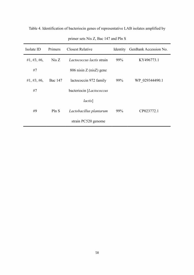
58
Table 4. Identification of bacteriocin genes of representative LAB isolates amplified by
primer sets Nis Z, Bac 147 and Pln S
Isolate ID Primers Closest Relative Identity GenBank Accession No.
#1, #3, #6,
#7
Nis Z Lactococcus lactis strain
806 nisin Z (nisZ) gene
99% KY496773.1
#1, #3, #6,
#7
Bac 147 lactococcin 972 family
bacteriocin [Lactococcus
lactis]
99% WP_029344490.1
#9 Pln S Lactobacillus plantarum
strain PC520 genome
99% CP023772.1

59
4.3.5 The inhibition spectrum of representative isolates
Lactococcus lactis isolates #1, #3, and #7 also showed antimicrobial activity against
Staphylococcus aureus. However, isolated #6 Lactococcus lactis and #9 Lactobacillus
plantarum could not inhibit the growth of Staphylococcus aureus. None of the isolates
showed inhibition against gram-negative pathogens Salmonella Typhimurium and E. coli
O157:H7.
4.3.6 The effect of growth conditions on bacteriocin production of representative isolates
The influence of culture medium on the bacteriocin production was demonstrated in
Figure 8. For Lactococcus lactis isolates, the highest bacteriocin activity was observed when
they were grown in MRS broth (Figure 12A). The bacteriocin production of isolates #1, #3
and #6 was doubled in MRS broth compated to Elliker broth. When those Lactococcus lactis
isolates were grown in M17 broth, their bacteriocin production was significantly lower. As
for the isolated Lactobacillus plantarum #9, its antimicrobial activity to Listeria
monocytogenes was much lower than that of the Lactococcus lactis isolates, which only
showed around 13 BU/ml (Figure 12B). There was no significant difference in its
antimicrobial activity when the isolate was grown in those different culture media.
The initial pH of MRS broth showed remarkable effects on the production of bacteriocins
(Figure 13A and Figure 13B). All tested representative isolates had higher bacteriocin
production with increasing pH. Highest bacteriocin production of isolate #6 was observed at
pH 6.9 (1059 BU/ml) which was 5-fold higher than its bacteriocin production at pH 5.5. The
bacteriocin production of isolated Lactobacillus plantarum #9 had no significant change until

60
the medium pH reached 6.9, which was more than double of the production at pH 6.0.
As shown in Figure 14, supplements added to MRS broth at pH 6.9 could affect the
production of bacteriocins. The addition of 1% Tween 80 promoted bacteriocin production by
all representative isolates. The highest bacteriocin production (1316 BU/ml) was achieved by
isolate #6 with 1% Tween 80. The bacteriocin production of isolated Lactococcus lactis #1,
#3, #6 and #7 did not show significant change when 1% ethanol was added to the culture
medium (Figure 14A). However, 1% ethanol noticeably increased the bacteriocin yield of
isolated Lactobacillus plantarum #9 by 7-fold, which was higher than the effect of 1% Tween
80 (Figure 14B). Besides, adding the combination of 1% Tween 80 and 1% ethanol had no
increased effect than only adding 1% Tween 80 on the bacteriocin production of Lactococcus
lactis isolates or 1% ethanol on the bacteriocin production of Lactobacillus plantarum.

61
A
B
Figure 12. Influences of medium on bacteriocin production (BU/ml) by Lactococcus
lactis isolates (A) and Lactobacillus platarum isolates (B)

62
A
B
Figure 13. Influences of initial medium pH on bacteriocin production (BU/ml) by
Lactococcus lactis isolates (A) and Lactobacillus platarum isolates (B)

63
A
B
Figure 14. Influences of supplements on bacteriocin production (BU/ml) by Lactococcus
lactis isolates (A) and Lactobacillus platarum isolates (B)

64
4.4 Discussions
A total of 10 bacteriocin-producers were isolated from the food samples, demonstrating
the effectiveness of the improved deferred antagonism assay. As Figure 6 shows, the
bacteriocin-producers did not dominate the microbiota of fermented foods. The PCR
amplification and DNA sequencing suggest that those bacteriocin-producers belonged to
Lactococcus lactis and Lactobacillus plantarum, which were common starter cultures used in
fermented foods. Four different types of Lactococcus lactis and one type of Lactobacillus
plantarum were identified among those 10 isolates by using RAPD PCR. The reason why the
amplicons of RAPD PCR had longer size than those generated in general PCR was that its
extension time was over 3-fold longer than that in general PCR. The amplification of the
representative isolates with bacteriocin gene primers showed that all Lactococcus lactis isolates
carried the nisin Z and lactococcin 972 genes, and isolated Lactobacillus plantarum carried the
plantaricin S gene. The lactococcin 972 had a narrow inhibition spectrum, effective only
against sensitive lactococci (Bacillus 1998). Therefore, the antimicrobial effect of isolated
Lactococcus lactis on Listeria monocytogenes was due to the secretion of nisin Z. The isolates
#1, #3, and #7 could also inhibit the growth of Staphylococcus aureus, which was probably due
to the expression of nisin Z gene (Pinto et al., 2011). However, the nisin Z gene carrier, isolate
#6, could not inhibit Staphylococcus aureus, suggesting that bacteria possessing the same
bacteriocin gene may not necessarily express the same antimicrobial effect.
According to the results, culture medium had significant effects on the bacteriocin
production of representative isolates. Isolated Lactococcus lactis generated more bacteriocins
in MRS broth than Elliker broth or M17 broth. The possible reason was that MRS broth

65
contains 0.1% Tween 80 which acts as a surfactant and can increase the permeability of cell
membrane to promote bacteriocin production (Vignolo et al., 1995). Jung et al. (1992) also
reported an enhanced effect on nisin activity in milk by emulsifier Tween 80, which
counteracted the absorption of the bacteriocin by proteins, thus increased the effect of nisin. In
addition, the concentration of nitrogen sources, peptone and yeast extract, are higher in MRS
broth than in M17 broth. Those nitrogen sources can promote the production of nisin (Cheigh
et al., 2002). As for higher bacteriocin production of L. lactis in Elliker broth than in M17 broth,
the abundant carbohydrate source in Elliker broth might make significant contributions.
Although the concentration of glucose in Elliker broth is the same as M17 broth, there are extra
0.5% lactose and 0.5% sucrose in Ellker broth. Cheigh et al. (2002) confirmed that Lactococcus
lactis produce more bacteriocin in M17 broth supplemented with lactose than the original M17
broth. Kim et al. (2006) also reported the addition of lactose to growth medium can increase
the production of bacteriocins.
For Lactobacillus plantarum, Todorov and Dicks (2005) confirmed that their bacteriocin
production was higher in MRS broth than in M17 broth. That was not observed with the isolate
in this study. One reason could be the bacteriocin produced by the isolated Lactobacillus
plantarum was not inhibitive enough to the indicator organism Listeria monocytogenes, which
might interfere with determining the influence of culture medium on the production of
plantaricin S.
In this study, increasing initial pH of culture medium could enhance the bacteriocin
production of all representative isolates. When the initial pH reached 6.9, the highest
bacteriocin production of isolated Lactococcus lactis and Lactobacillus plantarum was

66
observed. For Lactococcus lactis, this result was different from the finding of Matsusaki et al.
(1996). Matsusaki et al. reported that the optimal nisin production pH was around 5 to 5.5.
However, Turgis et al. (2016) reported that the optimal nisin production pH was in the range
from 7 to 9. The production of plantaricin S had the same problem. Todorov et al. (1999)
announced that the highest production of plantaricin S was achieved at pH 6. However, in 2005,
Todorov reported that there was no difference observed for plantaricin S production at pH 5.5,
6 and 6.5. Therefore, the same species of LAB isolated from different food samples may have
different pH requirements for bacteriocin production.
The results showed that adding 1% Tween 80 into culture medium can increase the
production of nisin and plantaricin S. This is similar to the findings of Vignolo et al. (1995),
Jung et al. (1992), and Malheiros et al. (2015). In comparison, the addition of 1% ethanol only
increased the production of plantaricin S. The reason was that low concentration of ethanol
could cause slightly unfavorable growth conditions which might increase the bacteriocin
production (De Vuyst et al., 1996) and/or prevent the bacteriocin aggregation (Mortvedt-
Abildgaa et al., 1995). However, there was no significant influence of 1% ethanol on the nisin
production. This might be explained by the fact that Lactococcus lactis has lower ethanol
tolerance. The maximum ethanol tolerance of Lactococcus lactis is about 7% (Hviid et al.,
2017). But Lactobacillus plantarum can still proliferate with 13% ethanol (G-Alegría et al.,
2004). The high ethanol tolerance of Lactobacillus plantarum ensures that 1% ethanol just
generates slightly stressful conditions and even increases the permeability of its cell membrane
(Hviid et al., 2017). However, 1% ethanol may inhibit the growth of Lactococcus lactis, and
higher ethanol concentration can decrease its production of nisin (Müller‐Auffermann et al.,

67
2015).
In conclusion, the improved deferred antagonism assay effectively isolated five types of
bacateriocin-producing LAB from the fermented foods. The isolated Lactococcus lactis strains
carry the nisin Z and lactococcin 972 gene, and can inhibit Listeria monocytogenes and
Staphylococcus aureus by producing nisin Z. The isolated Lactobacillus plantarum can also
inhibit Listeria monocytogenes by producing plantaricin S. Bacteriocin production by the LAB
isolates was significantly affected by the type and acidity of broth and supplements. Increasing
the medium pH can enhance the bacteriocin production of all isolates. The yield of bacterions
produced by Lactococcus Lactis and Lactobacillus plantarum significantly increased with the
addition of 1% Tween 80. The supplement 1% ethanol only increased the bacteriocin
production by Lactobacillus plantarum.
Lactococcus Lactis and Lactobacillus plantarum have been extensively applied in
fermented food as starter cultures. Those bacteriocin-producers isolated in this study should be
considered safe strains to use in fermented foods. Moreover, those isolates hold high potential
to be developed as probiotics with valuable application in human health and animal production.
In food manufacture, the isolated LAB and bacteriocins they produce can also be utilized as
part of the hurdle technology to improve the quality and safety of food.

68
Chapter 5
Conclusions
There have been increased consumer demands for natural food preservatives. The
discovery of bacteriocins meets the need since they may not only inhibit undesirable bacteria
in food but also enhance human gut health. Research is progressing to find new bacteriocin-
producers and explore their applications. Although there have been several approaches to
isolating bacteriocin producing LAB, those methods are inefficient and may be interfered by
certain bacteriocin non-producers which can produce other antimicrobial compounds.
In this study, the bacteriocin-producer isolation method, deferred antagonism assay, was
improved by optimizing the bottom media composition. The replacement of MRS agar with
Elliker agar as the bottom media in deferred antagonism assay eliminated the possibility of
isolating bacteriocin non-producing LAB. The inhibition zones formed by bacteriocin-
producers enlarged by adding 1% Tween 80 to the bottom media and increasing media initial
pH. This improved method can decrease the interference of lactic acid produced by
bacteriocin non-producing LAB and increase the effect of bacteriocins in the inhibition test.
Hence, it would enhance the chance of isolating true bacteriocin-producers from foods.
By using the improved deferred antagonism assay, four types of bacteriocin producing
Lactococcus lactis and one type of bacteriocin producing Lactobacillus plantarum were
isolated from fermented foods including yoghurt, kefir, sauerkraut, and kimchee. All
Lactococcus lactis isolates carry the nisin Z and lactococcin 972 genes, and the Lactobacillus
plantarum isolate may carry a novel plantaricin S gene. Three types of isolated Lactococcus
lactis can inhibit the growth of indicator pathogen, Listeria monocytogenes, and also show

69
antimicrobial activity against Staphylococcus aureus.
The bacteriocin production of the LAB isolates was affected by the type and acidity of
broth medium and supplements. The maximum bacteriocin production of isolated
Lactococcus lactis strains was achieved when they were cultivated in MRS broth with 1%
Tween 80 at pH 6.9. As for Lactobacillus plantarum, culture medium did not show a
significant effect on its bacteriocin production. The production of plantaricin S was more
effective with increasing pH and supplements of 1% Tween 80 or 1% ethanol. 1% ethanol
showed more significant effect on the plantaricin S production than 1% Tween 80.
The isolated LAB are commonly found in fermented foods. L. lactis subsp. lactis and L.
lactis subsp. cremoris are important starter cultures in dairy fermentation (Hayes et al., 2006).
Lactobacillus plantarum is one kind of beneficial bacteria found in Kimchee, sauerkraut,
cultured vegetables and even human saliva. Further research is required on these isolates
before they can be directly used in foods. Although these isolates are naturally occurring
bacteria and have been utilized in food fermentation without known negative effects, it would
be wise to evaluate them for potential effect on humans. Upon confirmation that these
isolates and their bacteriocins are safe, they could be utilized in foods to extend shelf life and
improve quality. Moreover, the bacteriocin producing LAB isolates also hold a great potential
to be applied as probiotics to improve gut health by secreting antimicrobial metabolites to
fight off pathogens in the intestine. These isolated LAB should be tested for their capability
to adhere to the human distal ileum and colon to elicit their health beneficial effects, and keep
non-invasive to the harmless human commensal microbiota (Naidu et al., 1999).
Additional testing on the application of these LAB isolates is demanded as well. Their

70
antimicrobial influence on pathogens when they are added into foods or utilized to ferment
foods has not been investigated. The effect of combinations of bacteriocins and other food
preservation processes on pathogens, particularly gram-negative bacteria which are naturally
resistant to the action of bacteriocins produced by gram-positive bacteria, is an interesting topic
to investigate (Prudêncio et al., 2015).
This research has facilitated efficient isolation of bacteriocin producing LAB from
fermented foods, identified five distinct bacteriocin-producers, and revealed important factors
affecting their bacteriocin production. Research will continue on the application of the LAB
isolates with a goal of confirming their safety and exploring their potential as probiotics. Their
bacteriocins may one day be used as natural preservatives in food production.

71
Appendix A
Diameter of inhibition zones formed by the testing strains in Chapter 3
Table A.1 The effects of bottom media composition on the diameter of inhibition zones
formed by bacteriocin-producing strains (Ki, L7-3 and B) and bacteriocin non-producing
strains (R7-4, LGG and LBP)
The composition of
bottom media
The diameters of inhibition zone (mm) of test strains
Ki L7-3 B R7-4 LGG LBP
MRS + Na2HPO4 and
NaH2PO4
3.25±0.27a 5.10±0.26ab 4.19±0.23a 1.75±0.35a 1.25±0.61a 1.81±0.60a
MRS + β-
glycerophosphate
3.29±0.23a 5.16±0.19a 4.23±0.37a 1.63±0.70a 1.31±0.56a 1.88±0.78a
M17 + Na2HPO4 and
NaH2PO4
3.13±0.95a 4.63±0.52b 3.75±0.27b ND* ND* ND*
M17 + β-
glycerophosphate
2.94±0.42a 3.75±0.65c 3.31±0.46c ND* ND* ND*
Elliker + Na2HPO4 and
NaH2PO4
3.38±0.69a 5.31±0.53a 4.17±0.63a ND* ND* ND*
Elliker + β-
glycerophosphate
3.56±0.32a 5.00±0.53ab 4.31±0.26a ND* ND* ND*
*No inhibition zone detected

72
Table A.2 The effects of pH of bottom media on the diameter of inhibition zones formed
by bacteriocin-producing strains (Ki, L7-3 and B) and bacteriocin non-producing strains (R7-
4, LGG and LBP)
The initial pH of
bottom media
The diameters of inhibition zone (mm) of test strains
Ki L7-3 B R7-4 LGG LBP
pH 5.5 2.19±0.80b 4.06±0.42b 2.81±0.59b 1.51±0.08 1.69±0.43 1.88±0.22
pH 6.0 2.63±0.44b 4.00±0.46b 3.06±0.50b ND* ND* ND*
pH 6.5 2.88±0.58ab 4.81±0.65a 3.69±0.46a ND* ND* ND*
pH 6.9 3.38±0.44a 5.31±0.53a 4.18±0.64a ND* ND* ND*
*No inhibition zone detected
Table A.3 The effects of tween 80 and/or ethanol on the diameter of inhibition zones
formed by bacteriocin-producing strains (Ki, L7-3 and B) and bacteriocin non-producing
strains (R7-4, LGG and LBP)
*No inhibition zone detected
The supplement of
bottom media
The diameters of inhibition zone (mm) of test strains
Ki L7-3 B R7-4 LGG LBP
No Tween 80 or Ethanol
(negative control)
3.38±0.70b 5.31±0.53b 4.18±0.64b ND* ND* ND*
1% Tween 80 4.39±0.66a 6.09±0.32a 5.48±0.43a ND* ND* ND*
1% Ethanol 3.31±0.53b 5.25±0.46b 4.13±0.59b ND* ND* ND*
1% Tween 80 and Ethanol 4.18±0.51a 5.81±0.26a 4.69±0.27a ND* ND* ND*

73
Appendix B
Bacteriocin units of different isolates in Chapter 4
Table B.1 Influences of medium on bacteriocin production (BU/ml) by representative isolates
Type of
medium
The bacteriocin units of representative isolates
#1 #3 #6 #7 #9
MRS 463.23±68.02a 567.12±55.51a 821.18±155.47a 382.04±81.68a 12.04±4.61a
Elliker 201.56±14.44b 210.47±31.16b 200.29±31.25b 212.39±69.32b 13.31±3.12a
M17 31.01±5.17c 49.31±26.51c 55.39±11.91c 51.88±22.72c 14.36±7.46a
Table B.2 Influences of initial medium pH on bacteriocin production (BU/ml) by
representative isolates
Initial
medium
pH
The bacteriocin units of representative isolate
#1 #3 #6 #7 #9
6.9 771.65±13.60a 729.2±69.82a 1058.96±157.40a 645.45±125.88a 32.76±19.14a
6.5 463.23±68.02b 567.12±55.51b 821.18±155.47b 382.04±81.68b 12.04±4.61b
6 387.18±98.21b 324.08±58.64c 256.24±73.74c 251.64±19.28c 11.75±4.10b
5.5 222.75±69.57c 108.49±35.94d 183.11±48.92c 141.19±26.33d 8.76±3.70b

74
Table B.3 Influences of supplements on bacteriocin production (BU/ml) by representative
isolates
Supplements The bacteriocin units of representative isolate
#1 #3 #6 #7 #9
Control 463.23±68.02c 567.12±55.51c 821.18±155.47c 382.04±81.68b 12.04±4.61c
1% Tween 955.84±136.1
7a
1023.68±184.56a 1315.8±142.94a 709.99±138.32a 55.32±6.52b
1% Ethanol 524.20±55.73c 587.69±60.52c 711.19±120.57c 484.51±67.15b 88.59±27.79a
1% Tween
and 1%
Ethanol
749.93±72.30
b
745.03±29.92b 1050.1±182.46b 639.55±35.82a 64.13±10.63b

75
References
Abriouel, H., Franz, C. M., Omar, N. B., & Gálvez, A. (2010). Diversity and applications of
Bacillus bacteriocins. FEMS microbiology reviews, 35(1), 201-232.
Adams, M. R., & Hall, C. J. (1988). Growth inhibition of food‐borne pathogens by lactic and
acetic acids and their mixtures. International Journal of Food Science & Technology, 23(3),
287-292.
Ayad, E. H., Verheul, A., de Jong, C., Wouters, J. T., & Smit, G. (1999). Flavour forming
abilities and amino acid requirements of Lactococcus lactis strains isolated from artisanal and
non-dairy origin. International Dairy Journal, 9(10), 725-735.
Bacillus, M. (1998). Detection and characterization of a bacteriocin produced by Lactococcus
lactis subsp. cremoris R isolated from radish. Letters in applied microbiology, 26, 297-304.
Bandell, M., Lhotte, M. E., Marty-Teysset, C., Veyrat, A., Prevost, H., Dartois, V., ... &
Lolkema, J. S. (1998). Mechanism of the Citrate Transporters in Carbohydrate and Citrate
Cometabolism in Lactococcus andLeuconostoc Species. Applied and environmental
microbiology, 64(5), 1594-1600.
Barbosa, M. S., Todorov, S. D., Ivanova, I. V., Belguesmia, Y., Choiset, Y., Rabesona, H., ... &
Franco, B. D. G. M. (2016). Characterization of a two-peptide plantaricin produced by
Lactobacillus plantarum MBSa4 isolated from Brazilian salami. Food Control, 60, 103-112.
Beresford, T. P., Fitzsimons, N. A., Brennan, N. L., & Cogan, T. M. (2001). Recent advances
in cheese microbiology. International Dairy Journal, 11(4), 259-274.
Bergan, T., Solberg, R., & Solberg, O. (1984). 6 Fatty Acid and Carbohydrate Cell Composition
in Pediococci and Aerococci, and Identification of Related Species. Methods in
microbiology, 16, 179-211.
Blaser, M. (2011). Antibiotic overuse: stop the killing of beneficial bacteria. Nature, 476(7361),
393-394.
Brink, B. T., Minekus, M., Van der Vossen, J. M. B. M., Leer, R. J., & Veld, J. H. J. (1994).
Antimicrobial activity of lactobacilli: preliminary characterization and optimization of
production of acidocin B, a novel bacteriocin produced by Lactobacillus acidophilus
M46. Journal of Applied Microbiology, 77(2), 140-148.
Cabo, M. L., Murado, M. A., aP González, M., & Pastoriza, L. (2001). Effects of aeration and
pH gradient on nisin production.: A mathematical model. Enzyme and Microbial
Technology, 29(4), 264-273.
Cai, Y., Kumai, S., Ogawa, M., Benno, Y., & Nakase, T. (1999). Characterization and

76
Identification ofPediococcus Species Isolated from Forage Crops and Their Application for
Silage Preparation. Applied and environmental microbiology, 65(7), 2901-2906.
Callewaert, R., Holo, H., Devreese, B., Van Beeumen, J., Nes, I., & De Vuyst, L. (1999).
Characterization and production of amylovorin L471, a bacteriocin purified from Lactobacillus
amylovorus DCE 471 by a novel three-step method. Microbiology, 145(9), 2559-2568.
Castro MP, Palavecino NZ, Herman CO et al. (2011) Lactic acid bacteria isolated from
artisanal dry sausages: Characterization of antibacterial compounds and study of the factors
affecting bacteriocin production. Meat Sci 87:321–329
Cheigh, C. I., Choi, H. J., Park, H., Kim, S. B., Kook, M. C., Kim, T. S., ... & Pyun, Y. R.
(2002). Influence of growth conditions on the production of a nisin-like bacteriocin by
Lactococcus lactis subsp. lactis A164 isolated from kimchi. Journal of Biotechnology, 95(3),
225-235.
Cogan, T. M., Barbosa, M., Beuvier, E., BIANCHI-SALVADORI, B. R. U. N. A.,
COCCONCELLI, P. S., FERNANDES, I., ... & MEDINA, M. (1997). Characterization of the
lactic acid bacteria in artisanal dairy products. Journal of Dairy Research, 64(3), 409-421.
Cotter, P. D., Hill, C., & Ross, R. P. (2005). Bacteriocins: developing innate immunity for
food. Nature Reviews Microbiology, 3(10), 777-788.
Cotter, P. D., Stanton, C., Ross, R. P., & Hill, C. (2012). The impact of antibiotics on the gut
microbiota as revealed by high throughput DNA sequencing. Discovery medicine, 13(70), 193-
199.
De Carvalho, A. A. T., Vanetti, M. C. D., & Mantovani, H. C. (2008). Bovicin HC5 reduces
thermal resistance of Alicyclobacillus acidoterrestris in acidic mango pulp. Journal of applied
microbiology, 104(6), 1685-1691.
De Vuyst, L., Callewaert, R., & Crabbe, K. (1996). Primary metabolite kinetics of bacteriocin
biosynthesis by Lactobacillus amylovorus and evidence for stimulation of bacteriocin
production under unfavourable growth conditions. Microbiology, 142(4), 817-827.
Drider, D., Fimland, G., Héchard, Y., McMullen, L. M., & Prévost, H. (2006). The continuing
story of class IIa bacteriocins. Microbiology and molecular biology reviews, 70(2), 564-582.
Eijsink, V. G., Axelsson, L., Diep, D. B., Håvarstein, L. S., Holo, H., & Nes, I. F. (2002).
Production of class II bacteriocins by lactic acid bacteria; an example of biological warfare and
communication. Antonie Van Leeuwenhoek, 81(1-4), 639-654.
Ennahar, S., Cai, Y., & Fujita, Y. (2003). Phylogenetic diversity of lactic acid bacteria

77
associated with paddy rice silage as determined by 16S ribosomal DNA analysis. Applied and
Environmental Microbiology, 69(1), 444-451.
Ennahar, S., Sashihara, T., Sonomoto, K., & Ishizaki, A. (2000). Class IIa bacteriocins:
biosynthesis, structure and activity. FEMS microbiology reviews, 24(1), 85-106.
G-Alegría, E., López, I., Ruiz, J. I., Sáenz, J., Fernández, E., Zarazaga, M., ... & Ruiz-Larrea,
F. (2004). High tolerance of wild Lactobacillus plantarum and Oenococcus oeni strains to
lyophilisation and stress environmental conditions of acid pH and ethanol. FEMS microbiology
letters, 230(1), 53-61.
Garneau, S., Martin, N. I., & Vederas, J. C. (2002). Two-peptide bacteriocins produced by lactic
acid bacteria. Biochimie, 84(5), 577-592.
Geis, A., Singh, J., & Teuber, M. (1983). Potential of lactic streptococci to produce
bacteriocin. Applied and Environmental Microbiology, 45(1), 205-211.
Gibson, G. R., Saavedra, J. M., MacFarlane, S., & MacFarlane, G. T. (1997). Probiotics and
intestinal infections. In Probiotics 2 (pp. 10-39). Springer Netherlands.
Gibson, L., Paba, A., Pes, M., & Scintu, M. F. (2000). Genotypic and phenotypic heterogeneity
among lactococci isolated from traditional Pecorino Sardo cheese. Journal of applied
microbiology, 89(2), 191-197.
Goepfert, J. M., & Hicks, R. U. T. H. A. N. N. (1969). Effect of volatile fatty acids on
Salmonella typhimurium. Journal of bacteriology, 97(2), 956.
Golnazarian, C. A., DONNELLY, C. W., PINTAURO, S. J., & HOWARD, D. B. (1989).
Comparison of infectious dose of Listeria monocytogenes F5817 as determined for normal
versus compromised C57B1/6J mice. Journal of food protection, 52(10), 696-701.
Grande, M. J., Lopez, R. L., Abriouel, H., Valdivia, E., Ben Omar, N., Maqueda, M., ... &
Galvez, A. (2007). Treatment of vegetable sauces with enterocin AS-48 alone or in combination
with phenolic compounds to inhibit proliferation of Staphylococcus aureus. Journal of Food
Protection, 70(2), 405-411.
Guarner, F., & Schaafsma, G. J. (1998). Probiotics. International journal of food
microbiology, 39(3), 237-238.
Haines, W. C., & Harmon, L. G. (1973). Effect of selected lactic acid bacteria on growth of
Staphylococcus aureus and production of enterotoxin. Applied microbiology, 25(3), 436-441.
Hardie, J. M., & Whiley, R. A. (1994). Recent developments in streptococcal taxonomy: their
relation to infections. Reviews in Medical Microbiology, 5(3), 151-162.

78
Hardie, J. M., & Whiley, R. A. (1995). The genus Streptococcus. In The genera of lactic acid
bacteria (pp. 55-124). Springer US.
Hawes, S. E., Hillier, S. L., Benedetti, J., Stevens, C. E., Koutsky, L. A., Wølner-Hanssen, P.,
& Holmes, K. K. (1996). Hydrogen peroxide—producing lactobacilli and acquisition of
vaginal infections. Journal of Infectious Diseases, 174(5), 1058-1063.
Hayes, M., Coakley, M., O'sullivan, L., & Stanton, C. (2006). Cheese as a delivery vehicle for
probiotics and biogenic substances. Australian Journal of Dairy Technology, 61(2), 132.
Heng, N. C., Wescombe, P. A., Burton, J. P., Jack, R. W., & Tagg, J. R. (2007). The diversity
of bacteriocins in Gram-positive bacteria. In Bacteriocins (pp. 45-92). Springer Berlin
Heidelberg.
Henning, C., Vijayakumar, P., Adhikari, R., Jagannathan, B., Gautam, D., & Muriana, P. M.
(2015). Isolation and taxonomic identity of bacteriocin-producing lactic acid bacteria from
retail foods and animal sources. Microorganisms, 3(1), 80-93.
Hillier, S. L., Krohn, M. A., Klebanoff, S. J., & Eschenbach, D. A. (1992). The relationship of
hydrogen peroxide-producing lactobacilli to bacterial vaginosis and genital microflora in
pregnant women. Obstetrics & Gynecology, 79(3), 369-373.
Hirsch, A., Grinsted, E., Chapman, H. R., & Mattick, A. T. R. (1951). 446. A note on the
inhibition of an anaerobic sporeformer in Swiss-type cheese by a nisin-producing
streptococcus. Journal of Dairy Research, 18(2), 205-206.
Hoover, D. G., & Steenson, L. R. (Eds.). (2014). Bacteriocins of lactic acid bacteria. Academic
Press.
Hviid, A. M. M., Ruhdal-Jensen, P., & Kilstrup, M. (2017). Butanol is cytotoxic to Lactococcus
lactis while ethanol and hexanol are cytostatic. Microbiology, 163(4), 453-461.
Jacobsen, T., Budde, B. B., & Koch, A. G. (2003). Application of Leuconostoc carnosum for
biopreservation of cooked meat products. Journal of Applied Microbiology, 95(2), 242-249.
JACQUES, N. A., HARDY, L., KNOX, K. W., & WICKEN, A. J. (1980). Effect of Tween 80
on the morphology and physiology of Lactobacillus salivarius strain IV CL-37 grown in a
chemostat under glucose limitation. Microbiology, 119(1), 195-201.
Johnson-Henry, K. C., Mitchell, D. J., Avitzur, Y., Galindo-Mata, E., Jones, N. L., & Sherman,
P. M. (2004). Probiotics reduce bacterial colonization and gastric inflammation in H. pylori-
infected mice. Digestive diseases and sciences, 49(7), 1095-1102.
Jung, D. S., Bodyfelt, F. W., & Daeschel, M. A. (1992). Influence of Fat and Emulsifiers on the
Efficacy of Nisin in Inhibiting Listeria monocytogenes in Fluid Milk1. Journal of Dairy

79
Science, 75(2), 387-393.
Keymanesh, K., Soltani, S., & Sardari, S. (2009). Application of antimicrobial peptides in
agriculture and food industry. World Journal of Microbiology and Biotechnology, 25(6), 933-
944.
Kim, D. S., Thomas, S., & Fogler, H. S. (2000). Effects of pH and trace minerals on long-term
starvation of Leuconostoc mesenteroides. Applied and environmental microbiology, 66(3),
976-981.
Kim, M. H., Kong, Y. J., Baek, H., & Hyun, H. H. (2006). Optimization of culture conditions
and medium composition for the production of micrococcin GO5 by Micrococcus sp.
GO5. Journal of biotechnology, 121(1), 54-61.
Klaenhammer, T. R. (1993). Genetics of bacteriocins produced by lactic acid bacteria. FEMS
microbiology reviews, 12(1-3), 39-85.
Klaenhammer, T.R. (1988) Bacteriocins of lactic acid bacteria. Btochimie 70,337-349.
Kniel, K. E., Montville, T. J., & Matthews, K. R. (2012). Food Microbiology: An Introduction
(3rd Edition). ASM Press
Kong, S., & Davison, A. J. (1980). The role of interactions between O2, H2O2,· OH, e− and
O2− in free radical damage to biological systems. Archives of biochemistry and
biophysics, 204(1), 18-29.
König, H., & Fröhlich, J. (2017). Lactic acid bacteria. In Biology of Microorganisms on Grapes,
in Must and in Wine (pp. 3-41). Springer, Cham.
Kouakou, P., Ghalfi, H., Dortu, C., Evrard, P., & Thonart, P. (2010). Combined use of
bacteriocin‐producing strains to control Listeria monocytogenes regrowth in raw pork
meat. International journal of food science & technology, 45(5), 937-943.
Lyon, W. J., & Glatz, B. A. (1993). Isolation and purification of propionicin PLG-1, a
bacteriocin produced by a strain of Propionibacterium thoenii. Applied and environmental
microbiology, 59(1), 83-88.
Macwana, S. J., & Muriana, P. M. (2012). A ‘bacteriocin PCR array’for identification of
bacteriocin-related structural genes in lactic acid bacteria. Journal of microbiological
methods, 88(2), 197-204.
Malheiros, P. S., Sant’Anna, V., Todorov, S. D., & Franco, B. D. (2015). Optimization of growth
and bacteriocin production by Lactobacillus sakei subsp. sakei2a. Brazilian Journal of
Microbiology, 46(3), 825-834.

80
Martinez, F. A. C., Domínguez, J. M., Converti, A., & de Souza Oliveira, R. P. (2015).
Production of bacteriocin-like inhibitory substance by Bifidobacterium lactis in skim milk
supplemented with additives. Journal of Dairy Research, 82(3), 350-355.
Martínez-Viedma, P., Abriouel, H., Omar, N. B., Valdivia, E., López, R. L., & Gálvez, A. (2008).
Inactivation of exopolysaccharide and 3-hydroxypropionaldehyde-producing lactic acid
bacteria in apple juice and apple cider by enterocin AS-48. Food and chemical
toxicology, 46(3), 1143-1151.
Martley, F. G., & Crow, V. L. (1993). Interactions between non-starter microorganisms during
cheese manufacture and repening. International Dairy Journal, 3(4-6), 461-483.
Mathara, J. M., Schillinger, U., Guigas, C., Franz, C., Kutima, P. M., Mbugua, S. K., ... &
Holzapfel, W. H. (2008). Functional characteristics of Lactobacillus spp. from traditional
Maasai fermented milk products in Kenya. International journal of food microbiology, 126(1),
57-64.
Matsusaki, H., Endo, N., Sonomoto, K., & Ishizaki, A. (1996). Lantibiotic nisin Z fermentative
production by Lactococcus lactis IO-1: relationship between production of the lantibiotic and
lactate and cell growth. Applied Microbiology and Biotechnology, 45(1), 36-40.
Mendes-Soares, H., Suzuki, H., Hickey, R. J., & Forney, L. J. (2014). Comparative functional
genomics of Lactobacillus spp. reveals possible mechanisms for specialization of vaginal
lactobacilli to their environment. Journal of bacteriology, 196(7), 1458-1470.
Midolo, P. D., Lambert, J. R., Hull, R., Luo, F., & Grayson, M. L. (1995). In vitro inhibition of
Helicobacter pylori NCTC 11637 by organic acids and lactic acid bacteria. Journal of Applied
Bacteriology, 79(4), 475-479.
Molinos, A. C., Abriouel, H., López, R. L., Omar, N. B., Valdivia, E., & Gálvez, A. (2008).
Inhibition of Bacillus cereus and Bacillus weihenstephanensis in raw vegetables by application
of washing solutions containing enterocin AS-48 alone and in combination with other
antimicrobials. Food microbiology, 25(6), 762-770.
MORRIS, B. J., IWAMOTO, H. S., & REID, I. A. (1979). Localization of angiotensinogen in
rat liver by immunocytochemistry. Endocrinology, 105(3), 796-800.
Mørtvedt, C. I., Nissen-Meyer, J., Sletten, K., & Nes, I. F. (1991). Purification and amino acid
sequence of lactocin S, a bacteriocin produced by Lactobacillus sake L45. Applied and
environmental microbiology, 57(6), 1829-1834.
Mortvedt-Abildgaa, C. I., Nissen-Meyer, J., Jelle, B., Grenov, B., Skaugen, M., & Nes, I. F.
(1995). Production and pH-Dependent Bactericidal Activity of Lactocin S, a Lantibiotic from
Lactobacillus sake L45. Applied and Environmental Microbiology, 61(1), 175-179.

81
Müller‐Auffermann, K., Grijalva, F., Jacob, F., & Hutzler, M. (2015). Nisin‐producing
microorganisms and their implementation in brewers' wort. Journal of the Institute of
Brewing, 121(3), 320-331.
Muriana, P. M., & Klaenhammer, T. R. (1987). Conjugal transfer of plasmid-encoded
determinants for bacteriocin production and immunity in Lactobacillus acidophilus 88. Applied
and Environmental Microbiology, 53(3), 553-560.
Murphy, M. G., & Condon, S. (1984). Correlation of oxygen utilization and hydrogen peroxide
accumulation with oxygen induced enzymes in Lactobacillus plantarum cultures. Archives of
microbiology, 138(1), 44-48.
Naidu, A. S., Bidlack, W. R., & Clemens, R. A. (1999). Probiotic spectra of lactic acid bacteria
(LAB). Critical reviews in food science and nutrition, 39(1), 13-126.
Naveena, B. J., Altaf, M., Bhadriah, K., & Reddy, G. (2005). Selection of medium components
by Plackett–Burman design for production of L (+) lactic acid by Lactobacillus amylophilus
GV6 in SSF using wheat bran. Bioresource technology, 96(4), 485-490.
Nesbakken, T., Kapperud, G., & Caugant, D. A. (1996). Pathways of Listeria monocytogenes
contamination in the meat processing industry. International Journal of Food
Microbiology, 31(1-3), 161-171.
Nissen-Meyer, J., & Nes, I. F. (1997). Ribosomally synthesized antimicrobial peptides: their
function, structure, biogenesis, and mechanism of action. Archives of microbiology, 167(2), 67-
77.
Parente, E., Ricciardi, A., & Addario, G. (1994). Influence of pH on growth and bacteriocin
production byLactococcus lactis subsp. lactis 14ONWC during batch fermentation. Applied
Microbiology and Biotechnology, 41(4), 388-394.
Patterson, M. J. (1996). Streptococcus.
Piard, J. C., & Desmazeaud, M. (1992). Inhibiting factors produced by lactic acid bacteria. 2.
Bacteriocins and other antibacterial substances. Le lait, 72(2), 113-142.
Piard, J. C., Delorme, F., Giraffa, G., Commissaire, J., & Desmazeaud, M. J. (1990). Evidence
for a bacteriocin produced by Lactococcus lactis CNRZ 481. Netherlands Milk and Dairy
Journal, 44(3-4), 143-158.
Pinto, M. S., de Carvalho, A. F., dos Santos Pires, A. C., Souza, A. A. C., da Silva, P. H. F.,
Sobral, D., ... & de Lima Santos, A. (2011). The effects of nisin on Staphylococcus aureus count
and the physicochemical properties of Traditional Minas Serro cheese. International dairy

82
journal, 21(2), 90-96.
Prudêncio, C. V., Dos Santos, M. T., & Vanetti, M. C. D. (2015). Strategies for the use of
bacteriocins in Gram-negative bacteria: relevance in food microbiology. Journal of food
science and technology, 52(9), 5408-5417.
Pulido, R. P., Omar, N. B., Abriouel, H., López, R. L., Cañamero, M. M., & Gálvez, A. (2005).
Microbiological study of lactic acid fermentation of caper berries by molecular and culture-
dependent methods. Applied and environmental microbiology, 71(12), 7872-7879.
Radha, K. R., & Padmavathi, T. (2017). Statistical optimization of bacteriocin produced from
Lactobacillus delbrueckii subsp bulgaricus isolated from yoghurt. International Food Research
Journal, 24(2).
Ravi, V., Prabhu, M., & Subramanyam, D. (2017). Isolation of bacteriocin producing bacteria
from mango pulp and its antimicrobial activity. Journal of Microbiology and Biotechnology
Research, 1(2), 54-63.
Rink, R., Wierenga, J., Kuipers, A., Kluskens, L. D., Driessen, A. J., Kuipers, O. P., & Moll, G.
N. (2007). Production of dehydroamino acid-containing peptides by Lactococcus
lactis. Applied and environmental microbiology, 73(6), 1792-1796.
Sabia, C., De Niederhäusern, S., Messi, P., Manicardi, G., & Bondi, M. (2003). Bacteriocin-
producing Enterococcus casseliflavus IM 416K1, a natural antagonist for control of Listeria
monocytogenes in Italian sausages (“cacciatore”). International Journal of Food
Microbiology, 87(1), 173-179.
Salminen, S., & Gueimonde, M. (2004). Human studies on probiotics: what is scientifically
proven. Journal of food science, 69(5).
Sanders, M. E. (1993). Effect of consumption of lactic cultures on human health. Advances in
food and nutrition research, 37, 67-130.
Savadogo, A., Ouattara, A. C., Bassole, H. I., & Traore, S. A. (2006). Bacteriocins and lactic
acid bacteria-a minireview. African journal of biotechnology, 5(9).
Shareck, J., Choi, Y., Lee, B., & Miguez, C. B. (2004). Cloning vectors based on cryptic
plasmids isolated from lactic acid bacteria: their characteristics and potential applications in
biotechnology. Critical reviews in biotechnology, 24(4), 155-208.
SKINNER, F. A., & QUESNEL, L. B. (1978). Streptococci. The Society for Applied
Bacteriology Symposium Ser. In Streptococci. The Society for Applied Bacteriology
Symposium Ser. (No. 7). Academic Press Inc.(London) Ltd. 24-28 Oval Road London
NW17DX..

83
Souza, E. C., Azevedo, P. O. D. S. D., Domínguez, J. M., Converti, A., & Oliveira, R. P. D. S.
(2017). Influence of temperature and pH on the production of biosurfactant, bacteriocin and
lactic acid by Lactococcus lactis CECT-4434. CyTA-Journal of Food, 1-6.
SPELHAUG, S. R., & HARLANDER, S. K. (1989). Inhibition of foodborne bacterial
pathogens by bacteriocins from Lactococcus lactis and Pediococcus pentosaceous. Journal of
Food Protection, 52(12), 856-862.
Todorov, S. D., & Dicks, L. M. T. (2005). Effect of growth medium on bacteriocin production
by Lactobacillus plantarum ST194 BZ, a strain isolated from boza. Food Technology and
Biotechnology, 43(2), 165-173.
Todorov, S. D., Wachsman, M., Tomé, E., Dousset, X., Destro, M. T., Dicks, L. M. T., ... &
Drider, D. (2010). Characterisation of an antiviral pediocin-like bacteriocin produced by
Enterococcus faecium. Food Microbiology, 27(7), 869-879.
Todorov, S., Onno, B., Sorokine, O., Chobert, J. M., Ivanova, I., & Dousset, X. (1999).
Detection and characterization of a novel antibacterial substance produced by Lactobacillus
plantarum ST 31 isolated from sourdough. International Journal of Food Microbiology, 48(3),
167-177.
Turgis, M., Vu, K. D., Millette, M., Dupont, C., & Lacroix, M. (2016). Influence of
environmental factors on bacteriocin production by human isolates of Lactococcus lactis
MM19 and Pediococcus acidilactici MM33. Probiotics and antimicrobial proteins, 8(1), 53-
59.
Vignolo, G. M., Kairuz, M. N., Holgado, A. A., & Oliver, G. (1995). Influence of growth
conditions on the production of lactocin 705, a bacteriocin produced by Lactobacillus casei
CRL 705. Journal of Applied Microbiology, 78(1), 5-10.
Vliegen, I., Jacobs, J. A., Beuken, E., Bruggeman, C. A., & Vink, C. (2006). Rapid
identification of bacteria by real-time amplification and sequencing of the 16S rRNA
gene. Journal of microbiological methods, 66(1), 156-164.
Walker, S. J., Archer, P., & Banks, J. G. (1990). Growth of Listeria monocytogenes at
refrigeration temperatures. Journal of applied microbiology, 68(2), 157-162.
Whiley, R. A., Beighton, D., Winstanley, T. G., Fraser, H. Y., & Hardie, J. M. (1992).
Streptococcus intermedius, Streptococcus constellatus, and Streptococcus anginosus (the
Streptococcus milleri group): association with different body sites and clinical
infections. Journal of clinical microbiology, 30(1), 243-244.
Yamauchi, Y., Ishii, S., Toyoda, S., & Ahiko, K. (1996). U.S. Patent No. 5,527,505. Washington,
84
DC: U.S. Patent and Trademark Office.
Yang, E., Fan, L., Jiang, Y., Doucette, C., & Fillmore, S. (2012). Antimicrobial activity of
bacteriocin-producing lactic acid bacteria isolated from cheeses and yogurts. AMB
Express, 2(1), 48.
Zottola, E. A. (1994). Food Micro'93—15th International Symposium: the International
Committee on Food Microbiology and Hygiene: Bingen, Germany, 31 August–3 September
1993. Food Control, 5(1), 49.