Optic neuritis - New Zealand Brain Research Institute
17
www.thelancet.com/neurology Vol 13 January 2014 83 Review Optic neuritis Ahmed T Toosy, Deborah F Mason, David H Miller Acute optic neuritis is the most common optic neuropathy affecting young adults. Exciting developments have occurred over the past decade in understanding of optic neuritis pathophysiology, and these developments have been translated into treatment trials. In its typical form, optic neuritis presents as an inflammatory demyelinating disorder of the optic nerve, which can be associated with multiple sclerosis. Atypical forms of optic neuritis can occur, either in association with other inflammatory disorders or in isolation. Differential diagnosis includes various optic nerve and retinal disorders. Diagnostic investigations include MRI, visual evoked potentials, and CSF examination. Optical coherence tomography can show retinal axonal loss, which correlates with measures of persistent visual dysfunction. Treatment of typical forms with high-dose corticosteroids shortens the period of acute visual dysfunction but does not affect the final visual outcome. Atypical forms can necessitate prolonged immunosuppressive regimens. Optical coherence tomography and visual evoked potential measures are suitable for detection of neuroaxonal loss and myelin repair after optic neuritis. Clinical trials are underway to identify potential neuroprotective or remyelinating treatments for acutely symptomatic inflammatory demyelinating CNS lesions. Introduction Optic neuritis is an inflammation of the optic nerve (panel 1). It occurs throughout the world and has many causes. In temperate latitudes and white populations it is commonly associated with multiple sclerosis (MS). However, the differential diagnosis is extensive, and prognosis and treatment depend on the cause. The incidence of unilateral optic neuritis around the world ranges from 0∙94 to 2∙18 per 100 000 per year. 1–4 Rates in Japan (1∙6 per 100 0000) are similar to those in Sweden (1∙46 per 100 000) and the UK (1 per 100 000). 5,6 Incidence studies universally show a female pre- ponderance, although the ratio of men to women in the Japanese cohort (1:1∙22) is greater than in northern European cohorts (1:3), suggesting that racial differences exist. 5–7 Results of a meta-analysis of optic neuritis in the northern hemisphere showed rates to be greater at higher latitudes, during spring, and in people of north European ancestry. 8 Similar findings have been reported in Australia. 3 There is also an association between incidence rates and serological evidence of past Epstein- Barr virus infection, and an additive interaction with HLA-DRB1*1501 status, 9 suggesting an association between risk factors for MS and cause of optic neuritis in areas of the world where MS is common. Conversely, in regions of low MS prevalence, optic neuritis is probably less frequently associated with MS and has different risk- factor profiles. In adults the incidence of bilateral simultaneous optic neuritis in white populations is low 10 and, as in all children with bilateral simultaneous optic neuritis, the risk of developing MS is low. 11 In recurrent optic neuritis, both visual recovery and neurological prognosis are more variable than in isolated occurrences. This variability is probably due to the broader differential diagnosis and the background population risk of these conditions. Substantial developments have occurred in diagnostic work-up, understanding of pathophysiology, and treatment approaches in optic neuritis. In this Review we provide an update of these developments. Optic neuritis and the risk of MS Optic neuritis is the presenting symptom of MS in 25% of cases and occurs during the disease in about 70%, usually in the relapsing–remitting phase. Long-term follow-up studies before MRI reported conversion to clinically definite MS in 34–75% of patients presenting with optic neuritis in the UK 12 and USA. 13 MRI studies in the same regions identified disseminated white-matter lesions suggestive of demyelination in 50% 14 of patients in the USA and 61% 15 in the UK. Clinically silent MRI lesions predispose to future clinical events, leading to clinically definite MS; in the North American Optic Neuritis Treatment Trial (ONTT), 16 72% of patients with an abnormal brain scan converted to MS after 15 years, compared with 25% with a normal scan. Brain MRI abnormalities in optic neuritis are less frequent in regions where MS is uncommon (eg, in Japan 17 ), and, in these regions, optic neuritis is more likely to be associated with other disorders, such as neuromyelitis optica. MRI criteria have been developed that predict the conversion to clinically definite MS with high sensitivity and specificity in optic neuritis and other clinically isolated syndromes. 18,19 MRI evidence of dissemination in space and time can now enable MS diagnosis at presentation in some patients with acute optic neuritis (panel 2). 20,21 Diagnosis, differential diagnosis, and investigations Clinical features of typical optic neuritis Typical optic neuritis presents with subacute monocular visual loss associated with pain during eye movement. Visual loss usually develops during hours or days. 22 Most patients report diffuse blurring or fogging of vision. Severity varies widely and tends to reach its nadir within 2 weeks. Dyschromatopsia occurs early and has a variable spectral pattern. Investigators of the ONTT described mostly mixed defects (red–green and blue–yellow), but blue–yellow defects were slightly more common in the acute phase, and red–green more common at 6 months. 23,24 Defect type was not associated with severity Lancet Neurol 2014; 13: 83–99 Queen Square Multiple Sclerosis Centre (A T Toosy PhD, Prof D H Miller FMedSci), Department of Brain Repair and Rehabilitation (A T Toosy), and Department of Neuroinflammation (Prof D H Miller), UCL Institute of Neurology, University College London, London, UK; Department of Neurology, Christchurch Hospital, Christchurch, New Zealand (D F Mason FRACP); and New Zealand Brain Research Institute, University of Otago, Christchurch, New Zealand (Prof D H Miller) Correspondence to: Dr Ahmed Toosy, Queen Square Multiple Sclerosis Centre, Department of Brain Repair and Rehabilitation, UCL Institute of Neurology, University College London, Queen Square, London WC1N 3BG, UK [email protected]
-
Upload
khangminh22 -
Category
Documents
-
view
1 -
download
0
Transcript of Optic neuritis - New Zealand Brain Research Institute
www.thelancet.com/neurology Vol 13 January 2014 83
Review
Optic neuritisAhmed T Toosy, Deborah F Mason, David H Miller
Acute optic neuritis is the most common optic neuropathy aff ecting young adults. Exciting developments have occurred over the past decade in understanding of optic neuritis pathophysiology, and these developments have been translated into treatment trials. In its typical form, optic neuritis presents as an infl ammatory demyelinating disorder of the optic nerve, which can be associated with multiple sclerosis. Atypical forms of optic neuritis can occur, either in association with other infl ammatory disorders or in isolation. Diff erential diagnosis includes various optic nerve and retinal disorders. Diagnostic investigations include MRI, visual evoked potentials, and CSF examination. Optical coherence tomography can show retinal axonal loss, which correlates with measures of persistent visual dysfunction. Treatment of typical forms with high-dose corticosteroids shortens the period of acute visual dysfunction but does not aff ect the fi nal visual outcome. Atypical forms can necessitate prolonged immunosuppressive regimens. Optical coherence tomography and visual evoked potential measures are suitable for detection of neuroaxonal loss and myelin repair after optic neuritis. Clinical trials are underway to identify potential neuroprotective or remyelinating treatments for acutely symptomatic infl ammatory demyelinating CNS lesions.
IntroductionOptic neuritis is an infl ammation of the optic nerve (panel 1). It occurs throughout the world and has many causes. In temperate latitudes and white populations it is commonly associated with multiple sclerosis (MS). However, the diff erential diagnosis is extensive, and prognosis and treatment depend on the cause.
The incidence of unilateral optic neuritis around the world ranges from 0∙94 to 2∙18 per 100 000 per year.1–4 Rates in Japan (1∙6 per 100 0000) are similar to those in Sweden (1∙46 per 100 000) and the UK (1 per 100 000).5,6 Incidence studies universally show a female pre-ponderance, although the ratio of men to women in the Japanese cohort (1:1∙22) is greater than in northern European cohorts (1:3), suggesting that racial diff erences exist.5–7 Results of a meta-analysis of optic neuritis in the northern hemisphere showed rates to be greater at higher latitudes, during spring, and in people of north European ancestry.8 Similar fi ndings have been reported in Australia.3 There is also an association between incidence rates and serological evidence of past Epstein-Barr virus infection, and an additive interaction with HLA-DRB1*1501 status,9 suggesting an association between risk factors for MS and cause of optic neuritis in areas of the world where MS is common. Conversely, in regions of low MS prevalence, optic neuritis is probably less frequently associated with MS and has diff erent risk-factor profi les.
In adults the incidence of bilateral simultaneous optic neuritis in white populations is low10 and, as in all children with bilateral simultaneous optic neuritis, the risk of developing MS is low.11 In recurrent optic neuritis, both visual recovery and neurological prognosis are more variable than in isolated occurrences. This variability is probably due to the broader diff erential diagnosis and the background population risk of these conditions.
Substantial developments have occurred in diagnostic work-up, understanding of pathophysiology, and treatment approaches in optic neuritis. In this Review we provide an update of these developments.
Optic neuritis and the risk of MSOptic neuritis is the presenting symptom of MS in 25% of cases and occurs during the disease in about 70%, usually in the relapsing–remitting phase. Long-term follow-up studies before MRI reported conversion to clinically defi nite MS in 34–75% of patients presenting with optic neuritis in the UK12 and USA.13 MRI studies in the same regions identifi ed disseminated white-matter lesions suggestive of demyelination in 50%14 of patients in the USA and 61%15 in the UK. Clinically silent MRI lesions predispose to future clinical events, leading to clinically defi nite MS; in the North American Optic Neuritis Treatment Trial (ONTT),16 72% of patients with an abnormal brain scan converted to MS after 15 years, compared with 25% with a normal scan. Brain MRI abnormalities in optic neuritis are less frequent in regions where MS is uncommon (eg, in Japan17), and, in these regions, optic neuritis is more likely to be associated with other disorders, such as neuromyelitis optica. MRI criteria have been developed that predict the conversion to clinically defi nite MS with high sensitivity and specifi city in optic neuritis and other clinically isolated syndromes.18,19 MRI evidence of dissemination in space and time can now enable MS diagnosis at presentation in some patients with acute optic neuritis (panel 2).20,21
Diagnosis, diff erential diagnosis, and investigationsClinical features of typical optic neuritisTypical optic neuritis presents with subacute monocular visual loss associated with pain during eye movement. Visual loss usually develops during hours or days.22 Most patients report diff use blurring or fogging of vision. Severity varies widely and tends to reach its nadir within 2 weeks. Dyschromatopsia occurs early and has a variable spectral pattern. Investigators of the ONTT described mostly mixed defects (red–green and blue–yellow), but blue–yellow defects were slightly more common in the acute phase, and red–green more common at 6 months.23,24 Defect type was not associated with severity
Lancet Neurol 2014; 13: 83–99
Queen Square Multiple Sclerosis Centre (A T Toosy PhD, Prof D H Miller FMedSci), Department of Brain Repair and Rehabilitation (A T Toosy), and Department of Neuroinfl ammation (Prof D H Miller), UCL Institute of Neurology, University College London, London, UK; Department of Neurology, Christchurch Hospital, Christchurch, New Zealand (D F Mason FRACP); and New Zealand Brain Research Institute, University of Otago, Christchurch, New Zealand (Prof D H Miller)
Correspondence to:Dr Ahmed Toosy, Queen Square Multiple Sclerosis Centre, Department of Brain Repair and Rehabilitation, UCL Institute of Neurology, University College London, Queen Square, London WC1N 3BG, [email protected]
84 www.thelancet.com/neurology Vol 13 January 2014
Review
of visual loss. Other studies mostly show both red–green and blue–yellow colour defects with neither type dominating.25–27 Results of one study showed that greater foveal fi eld depression was more likely to be associated
with red–green than with blue–yellow defects, but in patients with visual fi elds dominated by perifoveal defects the converse was recorded.25 Periocular pain, exacerbated by eye movement, is usually mild but present in most patients and usually settles within days. It can precede or begin with the onset of visual dysfunction. Optic neuritis lesions posterior to the orbit are less likely to cause pain.28 Other described symptoms include the presence of phosphenes,22,29 Uhthoff ’s phenomenon, and the Pulfrich eff ect (panel 3).
Early clinical signs include reduced visual and contrast acuities. Findings from the ONTT showed variable visual fi eld defi cits with static perimetry (panel 3) that included focal and diff use defi cits; central, centrocaecal, altitudinal, arcuate, and nasal step defects; and even hemianopic defects.30 Central defects were more common than peripheral ones.31 Results of other studies using kinetic or higher resolution static perimetry have shown central scotomas to be prevalent,32,33 the discordance in fi ndings from the ONTT potentially explained by methodological diff erences—eg, a diff use defect assigned in the ONTT (testing the central 30 degrees of vision) could actually have been a large central scotomas if tested with larger fi eld perimetry. A relative aff erent pupillary defect is usually seen, although involvement of the other optic nerve might mask it. Diff use optic disc swelling is present in a third of cases but the optic disc is normal in two-thirds (retrobulbar optic neuritis).22 Retinal periphlebitis (perivenous sheathing) is occasionally observed and could indicate a greater risk of conversion to MS.34
Asymptomatic concomitant visual dysfunction can occur in the other eye.22,35 In the ONTT, these abnormalities were not associated with a previous MS history or brain MRI lesion load, but did take several months to recover, suggesting that subclinical acute contralateral optic nerve demyelination could be the cause.35
Recovery from typical optic neuritis usually begins within the fi rst few weeks of symptom onset. An initial rapid recovery is followed by a slow improvement that can continue for up to a year after onset, with more than 90% of patients making a good visual recovery (20/40 acuity or better).36,37 After a fi nal 15 year ONTT analysis of 294 patients (65% of the original cohort), investigators reported that 72% of patients had acuities of 20/20 or better whereas 8∙7% had acuities of 20/40 or worse in the aff ected eye. Six patients (2%) had a visual acuity of 20/40 or worse and only three (1%) had a visual acuity of 20/200 or worse in both eyes.38 Visual improvement is slightly correlated with initial degree of visual loss,22 although patients with severe visual loss can still recover well. In the ONTT, 64% of patients with perception of light only or worse recovered to acuity of 20/40 or better.36 Poor acuity (20/200 or worse), contrast sensitivity (<1∙0 log units), or visual mean deviation (≤−15 dB) at 1 month, but not baseline, can predict poor vision at 6 months.39
Although recovery is usually good, persistent residual defi cits can include disturbances of visual acuity
Panel 1: Terminology for optic neuritis
There is little consensus about a systematic nosology for optic neuritis. Research studies generally use diff erent classifi cation systems, which can lead to confusion in interpretation of their fi ndings. Optic neuritis is traditionally divided on clinical grounds into typical and atypical forms, with the understanding that typical optic neuritis is generally associated with multiple sclerosis (MS) or is regarded as a demyelinating clinically isolated syndrome at risk of conversion to MS in white populations. An alternative method classifi es optic neuritis by cause. On this basis, in this Review we describe immune-mediated optic neuritis, which itself can be subclassifi ed into several types including MS-associated optic neuritis (MS-ON), optic neuritis associated with neuromyelitis optica (NMO-ON), optic neuritis associated with systemic disorders (connective tissue disease, granulomatous disease, infective conditions), and other idiopathic optic neuritis without systemic disease (recurrent isolated optic neuritis, chronic relapsing infl ammatory optic neuropathy, solitary isolated optic neuritis). The term demyelinating optic neuritis has been also been used as a pathology-based defi nition, although this term is also not ideal because both MS-ON and NMO-ON cause demyelination and are managed diff erently. In this Review we tend to classify optic neuritis on clinical grounds—ie, as typical or atypical, for which typical optic neuritis is associated with MS and clinically isolated syndromes and atypical optic neuritis with non-MS immune-mediated causes (eg, neuromyelitis optica and systemic disorders, etc) because this is highly relevant for approach to clinical management. When appropriate—ie, when specifi c research has been undertaken—in relevant sections of this Review we allude to cause-based defi nitions (particularly MS-ON, NMO-ON).
Panel 2: Diagnosis of MS in MS-optic neuritis (2010 McDonald MRI criteria)21
Diagnosis requires dissemination in space and timeDissemination in space• At least one lesion visible on T2-weighted scan in at least
two of four locations: juxtacortical, periventricular, infratentorial, and spinal cord
Dissemination in time• A new T2 lesion or gadolinium-enhancing lesion visible
on a follow-up MRI scan when compared with a previous scan (which is thought to be the baseline scan) obtained at any time after the onset of symptoms; or
• An MRI scan showing both gadolinium-enhancing and non-enhancing lesions that do not cause clinical signs (ie, asymptomatic lesions)
www.thelancet.com/neurology Vol 13 January 2014 85
Review
(15–30%), contrast sensitivity (63–100%), colour vision (33–100%), visual fi eld (62–100%), stereopsis (89%), pupillary reaction (55–92%), and visual evoked potentials (VEPs) (63–100%).40 Results of psychophysical studies have shown variable contributions of parvocellular and magnocellular pathway damage to visual defi cit,41,42 and persistent defi cits in motion perception.43 Optic disc pallor, especially involving the temporal aspect, often develops even when recovery is excellent.
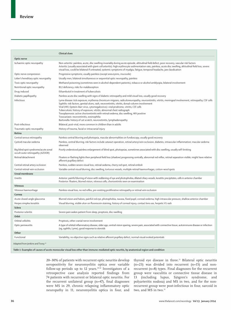
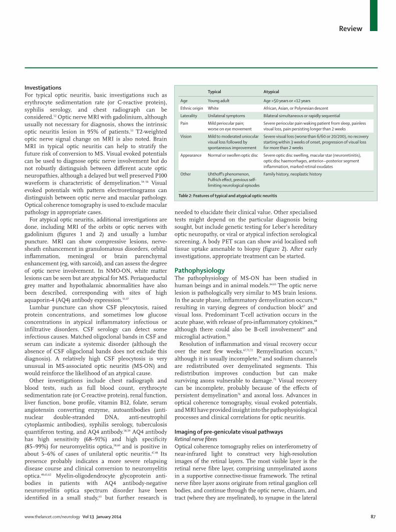
Diff erential diagnosisDiagnosis of optic neuritis can be made clinically. Patient history and neuro-ophthalmic examination can be used to look for other causes of acute monocular visual loss (table 1). After diagnosis of optic neuritis, a clinical distinction should be made between typical and atypical forms (table 2). Patients with atypical optic
neuritis can be classifi ed into those with systemic disease and those without (table 3). Optic neuritis without systemic disease includes patients with neuromyelitis optica-optic neuritis (NMO-ON) (panel 4), and corticosteroid-dependent chronic relapsing infl ammatory optic neuropathy.46 Systemic disorders associated with atypical optic neuritis include sarcoidosis, connective tissue diseases (eg, lupus), and vasculitis (eg, Wegener’s granulomatosis). Many patients who are not corticosteroid-dependent could have isolated optic neuritis without systemic or neurological disease. These patients are diagnosed retrospectively, after extended follow-up, with solitary optic neuritis or recurrent optic neuritis.47 Solitary optic neuritis is rarely associated with neuromyelitis optica seropositivity (about 5%) and tends to be a common retrospective diagnosis when brain MRI is normal.48
• Phosphenes are bright, fl eeting fl ashes of light that, in optic neuritis, tend to be connected to eye movement. The symptom of phosphenes should be clinically distinguished from a scintillating scotomas, which are usually associated with visual aura of migraine. This aura tends to appear as a blind region surrounded by a margin of sparkling lights that can change shape or move over a period of time, typically 15–30 min.
• Uhthoff ’s phenomenon is a worsening of vision provoked by small increases in body temperature, typically attributed to exercise, hot baths or showers, or hot weather conditions.
• The Pulfrich eff ect describes anomalous stereoscopic perception of objects in motion due to asymmetrical conduction between optic nerves.
• Visual acuity measures the spatial resolution ability of the visual system. Traditionally it is measured with Snellen charts and expressed in fractional notation, with the numerator denoting the actual distance (20 feet or 6 m) from the chart and the denominator denoting the distance at which a person with normal eyesight can see the line of letters—eg, an acuity of 20/40 means that a person viewing the chart at 20 feet can read letters that normal eyesight can distinguish at 40 feet. 20/20 or 6/6 vision is normal; 20/200 or its equivalent 6/60 signifi es very poor vision. Research studies such as the North American Optic Neuritis Treatment Trial (ONTT) tend to use logMAR scores, requiring a retro-illuminated Early Treatment Diabetic Retinopathy Study (ETDRS) chart. A standard ETDRS chart measures the logarithmic (base 10) minimum angle of resolution at 4 m and provides a linearly continuous variable amenable to parametric statistics. LogMAR scores can be converted to Snellen equivalent scores, and vice versa.
• Low-contrast acuity charts are more sensitive than standard-contrast acuity charts at detection of visual dysfunction in optic neuritis. Several types of chart exist, but the Pelli-Robson chart has been commonly used in research studies (including the ONTT). This chart comprises eight lines of six
letters, each arranged in two triplets per line. With each successive triplet, the contrast decreases in logarithmic steps by 0·15 log units. The patient is asked to read along and down the chart until the detection limit is reached, scored from 0 to about 2 log units. High scores indicate better contrast sensitivity, measured at the peak of the contrast sensitivity function (about one to two cycles per degree).
• Colour vision is conventionally measured in clinical practice with Ishihara pseudoisochromatic plates. The patient is asked to distinguish diff erent coloured numbers, but this test is designed for defi ciencies of the red–green axis. In research, the Farnsworth-Munsell 100-hue test provides a comprehensive assessment (used in the ONTT). The patient is asked to grade 85 coloured caps according to perceived hue, from which an error score is established. The resulting data are amenable to parametric statistics and indicate the type of spectral defi ciency.
• Visual fi elds can be measured with static or dynamic perimetry. The ONTT used the Humphrey fi eld perimetry (static), which can test diff erent fi eld sizes in an automated fashion but typically tests the central 30 degrees of vision. The patient has to acknowledge luminant stimuli briefl y presented in diff erent locations. The stimuli are randomly repeated at various luminances to assess reliability and luminance threshold. The output can be quantitatively summarised as a score ranging usually from 0 to –30 decibels, where 0 is normal vision and –30 is severe visual-fi eld loss. Goldmann perimetry is dynamic and relies on the patient detecting a luminant stimulus moving in from the peripheral fi eld. The test is done by a trained operator who uses targets of diff erent luminances and sizes to create a fi eld map. Its advantages over Humphrey perimetry are that patient compliance can be assessed and the whole visual fi eld can be mapped; however, its output is more qualitative and scotomas can be missed if not properly assessed.
Panel 3: Visual phenomena and measurements in optic neuritis
86 www.thelancet.com/neurology Vol 13 January 2014
Review
20–30% of patients with recurrent optic neuritis develop seropositivity for neuromyelitis optica over variable follow-up periods up to 12 years.49,50 Investigators of a retrospective case analysis reported fi ndings from 74 patients with recurrent or bilateral optic neuritis. For the recurrent unilateral group (n=47), fi nal diagnoses were MS in 29, chronic relapsing infl ammatory optic neuropathy in 11, neuromyelitis optica in four, and
thyroid eye disease in three.51 Bilateral optic neuritis (n=23) was divided into recurrent (n=15) and non-recurrent (n=8) types. Final diagnoses for the recurrent group were vasculitis or connective tissue disease in 13 (including lupus, Sjögren’s syndrome, and polyarteritis nodosa) and MS in two, and for the non-recurrent group were post-infectious in four, sarcoid in two, and MS in two.51
Clinical clues
Optic nerve
Ischaemic optic neuropathy Non-arteritic: painless, acute, disc swelling invariably during acute episode, altitudinal fi eld defect, poor recovery, vascular risk factorsArteritic (usually associated with giant-cell arteritis): high erythrocyte sedimentation rate, painless, acute disc swelling, altitudinal fi eld loss, severe visual loss, could be bilateral if untreated, systemic symptoms of myalgia, fatigue, temporal headache, jaw claudication
Optic nerve compression Progressive symptoms, usually painless (except aneurysms, mucocele)
Leber’s hereditary optic neuropathy Usually men, bilateral simultaneous or sequential optic neuropathy, painless
Toxic optic neuropathy Methanol poisoning (sometimes seen in alcohol-dependent patients), tobacco or alcohol amblyopia, bilateral involvement
Nutritional optic neuropathy B12 defi ciency, risks for malabsorption
Drug-induced Ethambutol in treatment of tuberculosis
Diabetic papillopathy Painless acute disc swelling with signs of diabetic retinopathy and mild visual loss, usually good recovery
Infectious Lyme disease: tick exposure, erythema chronicum migrans, radiculoneuropathy, neuroretinitis, vitritis, meningeal involvement, retinopathy, CSF cellsSyphilis: risk factors, genital ulcers, rash, neuroretinitis, vitritis, dorsal-column involvementViral (HIV, Epstein-Barr virus, cytomegalovirus): viral prodrome, vitritis, CSF cellsTuberculosis: history of exposure, vitritis, abnormal chest radiographToxoplasmosis: active chorioretinitis with retinal oedema, disc swelling, HIV positiveToxocariasis: neuroretinitis, eosinophiliaBartonella: history of cat scratch, neuroretinitis, lymphadenopathy
Post-infectious Bilateral, post-viral, more common in children than in adults
Traumatic optic neuropathy History of trauma, facial or intracranial injury
Retina
Central serous retinopathy Painless central blurring and photopsia, macular abnormalities on fundoscopy, usually good recovery
Cystoid macular oedema Painless, central blurring; risk factors include cataract operation, retinal artery/vein occlusion, diabetes, intraocular infl ammation; macular oedema observed
Big blind spot syndrome/acute zonal occult outer retinopathy (AZOOR)
Poorly understood painless enlargement of blind spot, photopsias, sometimes associated with disc swelling, usually self-limiting
Retinal detachment Floaters or fl ashing lights then peripheral fi eld loss (shadow) progressing centrally, abnormal red refl ex, retinal separation visible; might have relative aff erent pupillary defect
Central retinal artery occlusion Painless, sudden severe visual loss, retinal oedema, cherry red spot, retinal emboli
Central retinal vein occlusion Variable central visual blurring, disc swelling, tortuous vessels, multiple retinal haemorrhages, cotton-wool spots
Uveal membrane
Uveitis Anterior: painful blurring of vision with reddening of eye and photophobia, dilated ciliary vessels, keratitic precipitates, cells in anterior chamberPosterior: fl oaters, blurred vision, vitreous cells, chorioretinits seen on examination
Vitreous
Vitreous haemorrhage Painless visual loss, no red refl ex, pre-existing proliferative retinopathy or retinal vein occlusion
Cornea
Acute closed-angle glaucoma Blurred vision and haloes, painful red eye, photophobia, nausea, fi xed pupil, corneal oedema, high intraocular pressure, shallow anterior chamber
Herpes simplex keratitis Visual blurring, visible ulcer on fl uorescein staining, history of corneal injury, contact lens use, herpetic V1 rash
Sclera
Posterior scleritis Severe pain wakes patient from sleep, proptosis, disc swelling
Orbit
Orbital cellulitis Proptosis, other cranial nerve involvement
Optic perineuritis A type of orbital infl ammatory disease, older age, central vision sparing, severe pain, associated with connective tissue, autoimmune disease or infection (eg, syphilis, Lyme), good response to steroids
Other
Functional Variability, no objective signs such as relative aff erent pupillary defect, normal visual evoked potentials
Adapted from Jenkins and Toosy.44
Table 1: Examples of causes of acute monocular visual loss other than immune-mediated optic neuritis, by anatomical region and condition
www.thelancet.com/neurology Vol 13 January 2014 87
Review
InvestigationsFor typical optic neuritis, basic investigations such as erythrocyte sedimentation rate (or C-reactive protein), syphilis serology, and chest radiograph can be considered.52 Optic nerve MRI with gadolinium, although usually not necessary for diagnosis, shows the intrinsic optic neuritis lesion in 95% of patients.53 T2-weighted optic nerve signal change on MRI is also noted. Brain MRI in typical optic neuritis can help to stratify the future risk of conversion to MS. Visual evoked potentials can be used to diagnose optic nerve involvement but do not robustly distinguish between diff erent acute optic neuropathies, although a delayed but well preserved P100 waveform is characteristic of demyelination.54–56 Visual evoked potentials with pattern electroretinograms can distinguish between optic nerve and macular pathology. Optical coherence tomography is used to exclude macular pathology in appropriate cases.
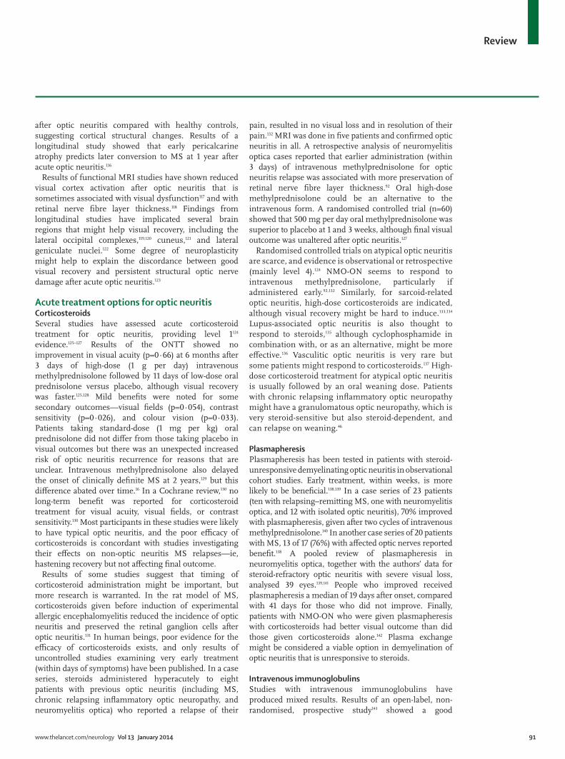
For atypical optic neuritis, additional investigations are done, including MRI of the orbits or optic nerves with gadolinium (fi gures 1 and 2) and usually a lumbar puncture. MRI can show compressive lesions, nerve-sheath enhancement in granulomatous disorders, orbital infl ammation, meningeal or brain parenchymal enhancement (eg, with sarcoid), and can assess the degree of optic nerve involvement. In NMO-ON, white matter lesions can be seen but are atypical for MS. Periaqueductal grey matter and hypothalamic abnormalities have also been described, corresponding with sites of high aquaporin-4 (AQ4) antibody expression.55,57
Lumbar puncture can show CSF pleocytosis, raised protein concentrations, and sometimes low glucose concentrations in atypical infl ammatory infectious or infi ltrative disorders. CSF serology can detect some infectious causes. Matched oligoclonal bands in CSF and serum can indicate a systemic disorder (although the absence of CSF oligoclonal bands does not exclude this diagnosis). A relatively high CSF pleocytosis is very unusual in MS-associated optic neuritis (MS-ON) and would reinforce the likelihood of an atypical cause.
Other investigations include chest radiograph and blood tests, such as full blood count, erythrocyte sedimentation rate (or C-reactive protein), renal function, liver function, bone profi le, vitamin B12, folate, serum angiotensin converting enzyme, autoantibodies (anti-nuclear double-stranded DNA, anti-neutrophil cytoplasmic antibodies), syphilis serology, tuberculosis quantiferon testing, and AQ4 antibody.58,59 AQ4 antibody has high sensitivity (68–91%) and high specifi city (85–99%) for neuromyelitis optica,58,60 and is positive in about 5–6% of cases of unilateral optic neuritis.47,48 Its presence probably indicates a more severe relapsing disease course and clinical conversion to neuromyelitis optica.48,61,62 Myelin-oligodendrocyte glycoprotein anti-bodies in patients with AQ4 antibody-negative neuromyelitis optica spectrum disorder have been identifi ed in a small study,63 but further research is
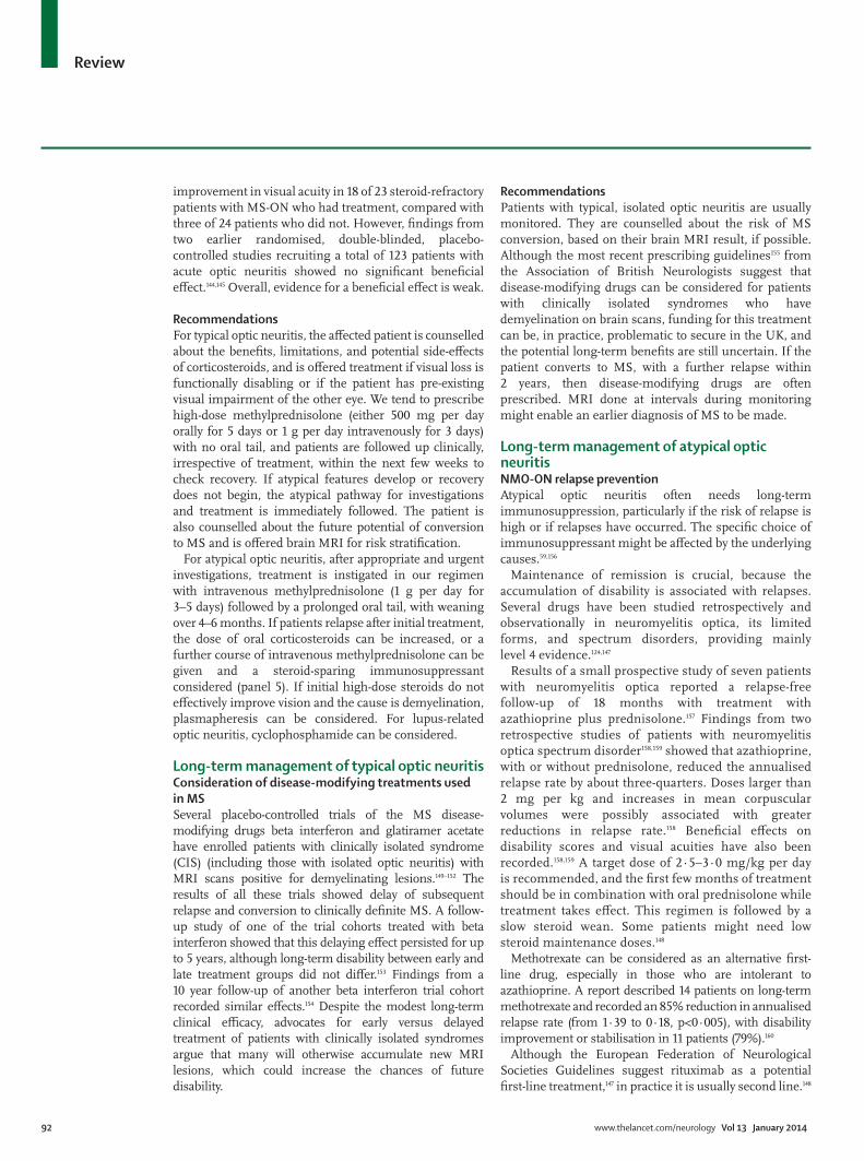
needed to elucidate their clinical value. Other specialised tests might depend on the particular diagnosis being sought, but include genetic testing for Leber’s hereditary optic neuropathy, or viral or atypical infection serological screening. A body PET scan can show avid localised soft tissue uptake amenable to biopsy (fi gure 2). After early investigations, appropriate treatment can be started.
PathophysiologyThe pathophysiology of MS-ON has been studied in human beings and in animal models.64,65 The optic nerve lesion is pathologically very similar to MS brain lesions. In the acute phase, infl ammatory demyelination occurs,66 resulting in varying degrees of conduction block67 and visual loss. Predominant T-cell activation occurs in the acute phase, with release of pro-infl ammatory cytokines,68 although there could also be B-cell involvement69 and microglial activation.70
Resolution of infl ammation and visual recovery occur over the next few weeks.67,71,72 Remyelination occurs,73 although it is usually incomplete,74 and sodium channels are redistributed over demyelinated segments. This redistribution improves conduction but can make surviving axons vulnerable to damage.75 Visual recovery can be incomplete, probably because of the eff ects of persistent demyelination76 and axonal loss. Advances in optical coherence tomography, visual evoked potentials, and MRI have provided insight into the pathophysiological processes and clinical correlations for optic neuritis.
Imaging of pre-geniculate visual pathwaysRetinal nerve fi bresOptical coherence tomography relies on interferometry of near-infrared light to construct very high-resolution images of the retinal layers. The most visible layer is the retinal nerve fi bre layer, comprising unmyelinated axons in a supportive connective-tissue framework. The retinal nerve fi bre layer axons originate from retinal ganglion cell bodies, and continue through the optic nerve, chiasm, and tract (where they are myelinated), to synapse in the lateral
Typical Atypical
Age Young adult Age >50 years or <12 years
Ethnic origin White African, Asian, or Polynesian descent
Laterality Unilateral symptoms Bilateral simultaneous or rapidly sequential
Pain Mild periocular pain; worse on eye movement
Severe periocular pain waking patient from sleep, painless visual loss, pain persisting longer than 2 weeks
Vision Mild to moderated uniocular visual loss followed by spontaneous improvement
Severe visual loss (worse than 6/60 or 20/200), no recovery starting within 3 weeks of onset, progression of visual loss for more than 2 weeks
Appearance Normal or swollen optic disc Severe optic disc swelling, macular star (neuroretinitis), optic disc haemorrhages, anterior—posterior segment infl ammation, marked retinal exudates
Other Uhthoff ’s phenomenon, Pulfrich eff ect, previous self-limiting neurological episodes
Family history, neoplastic history
Table 2: Features of typical and atypical optic neuritis
88 www.thelancet.com/neurology Vol 13 January 2014
Review
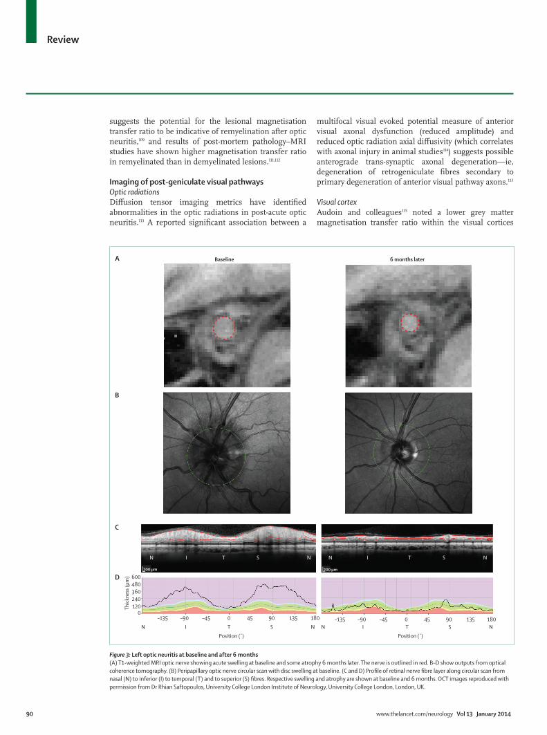
geniculate bodies. The retinal nerve fi bre layer measurements using optical coherence tomography are typically made from a circular arc through the retinal nerve fi bre layer 3∙4 mm from the centre of the optic disc. In acute optic neuritis, retinal nerve fi bre layer thickness increases occur with optic nerve swelling.77 Subsequent reductions in retinal nerve fi bre layer thickness indicate signifi cant axonal loss after acute optic neuritis (fi gure 3).78,79 Investigators of two serial optical coherence tomography studies identifi ed a median 20% decrease in retinal nerve fi bre layer thickness after 6 months in two cohorts of 54 and 23 patients with optic neuritis, respectively,79,80 although retinal nerve fi bre layer loss varied greatly between patients (0–50%).80 Consistent with these fi ndings, results of a study of 90 patients with MS showed a mean 20% decrease in retinal nerve fi bre layer thickness in eyes previously aff ected by optic neuritis.81 Retinal nerve fi bre layer thinning correlates with visual dysfunction,78,79,82 optic nerve MRI-detected atrophy,83 and radial diff usivity (water
diff usivity perpendicular to the main axis of diff usion along the nerve).84 Impaired colour vision is particularly correlated with thinning of the retinal nerve fi bre layer in both cross-sectional and prospective cohorts.85,86
Supported by data (obtained in an animal model of optic neuritis) showing an association between visual evoked potential latency and demyelination,56 clinical studies have acquired both visual evoked potential and optical coherence tomography measures to investigate the association between myelination and axonal loss. Both visual evoked potential amplitude and latency were correlated with optical coherence tomography measures of axonal loss in a clinical cross-sectional study of post-acute optic neuritis.87 In another clinical study,88 sectoral retinal nerve fi bre layer thickness correlated with the multifocal visual evoked potential amplitude from the corresponding part of the visual fi eld, suggestive of a structural–functional association, whereby the largest reductions in retinal nerve fi bre layer thickness aff ected the temporal disc and the largest reductions in multifocal visual evoked potential amplitude aff ected the central fi eld.88 Results of a longitudinal clinical study showed that the improvement in latency delay in acute optic neuritis tends to be greatest in the fi rst 6 months.73 From 1 to 3 years an ongoing small reduction in retinal nerve fi bre layer thickness did not correlate with the change in multifocal visual evoked potential latency, suggesting no association between optic nerve demyelination and ongoing axonal loss.88
Clinical optical coherence tomography studies have used intraretinal layer segmentation to examine the retinal ganglion cell and inner plexiform layers. Measurements of retinal ganglion cell layer (or retinal ganglion cell and inner plexiform layers combined, because the two layers are diffi cult to discriminate) are not aff ected by disc swelling and should be more specifi c to axonal pathology. Thin retinal ganglion cell and inner
Features
No systemic disease
Multiple sclerosis-associated optic neuritis
Typical symptoms of optic neuritis, usually disseminated white-matter brain lesions suggestive of demyelination, CSF-positive oligoclonal bands (unmatched); if fi rst episode can be called demyelinating clinically isolated syndrome
Solitary isolated optic neuritis Diagnosed after extended follow-up; normal brain MRI, isolated optic neuritis
Neuromyelitis optica-associated optic neuritis
Positive antibodies to aquaporin 4 or myelin-oligodendrocytes, longitudinally extensive cord lesion (myelitis), CSF pleocytosis, negative oligoclonal bands, normal MRI brain or abnormalities atypical for MS (hypothalamus, third ventricle, medulla)
Chronic relapsing infl ammatory optic neuropathy
Tendency to relapse when off steroids, normal MRI brain, optic nerve sheath enhancement, might become bilateral, needs chronic immunosuppression
Recurrent isolated optic neuritis Diagnosed after extended follow-up; normal brain MRI, no other neurological sequelae
Acute disseminated encephalomyelitis Enhancing brain lesions, severe bilateral optic neuritis, more common in children than in adults
Systemic disease
Sarcoid Other signs of intraocular infl ammation, optic nerve sheath enhancement, white matter brain lesions, meningeal enhancement, respiratory symptoms, abnormal chest radiograph, CSF pleocytosis, matched oligoclonal bands
Connective tissue disease (eg, lupus) Skin rash, arthritis, alopecia, positive autoantibodies (double-stranded DNA for lupus), raised infl ammatory markers
Vasculitis (eg, polyarteritis nodosa, Wegener’s granulomatosis)
Ischaemic presentation if pure vasculitic; compressive presentation if sino–nasal diseasePositive anti-neutrophil cytoplasmic antibodies
Table 3: Main causes of immune-mediated optic neuritis
Panel 4: Neuromyelitis optica spectrum
• Neuromyelitis optica• Some forms of neuromyelitis optica
• Idiopathic single or recurrent events of longitudinally extensive myelitis (≥three vertebral segment spinal cord lesions seen on MRI)
• Optic neuritis: recurrent or simultaneous bilateral• Optic neuritis or longitudinally extensive myelitis
associated with systemic autoimmune disease• Optic neuritis or myelitis associated with brain lesions
typical of neuromyelitis optica (hypothalamic, corpus callosal, periventricular, or brainstem)
Modifi ed from Wingerchuk and colleagues.45
www.thelancet.com/neurology Vol 13 January 2014 89
Review
plexiform layers have been recorded in patients with MS, particularly those aff ected by previous optic neuritis, and the depths of the layers are associated with visual function.89 Such thinning has also been recorded in longitudinal studies of optic neuritis.90
Greater retinal nerve fi bre layer thinning is seen in NMO-ON eyes than in MS-ON eyes,91,92 with the greatest thinning in the superior and inferior retinal nerve fi bre layer quadrants.92,93 Results of one study suggested that retinal nerve fi bre layer thickness loss greater than 15 μm in patients without MS should prompt investigations for neuromyelitis optica spectrum disorder.91 Early administration of high-dose steroids preserved retinal nerve fi bre layer thickness in an uncontrolled, retrospective study.92 Longitudinal reductions in retinal ganglion cell and inner plexiform layer thickness have also been recorded in NMO-ON eyes.90 Thickness of the inner nuclear layer could be increased in neuromyelitis optica,94,95 although in-vivo quantifi cation of the inner nuclear layer has also included the outer plexiform layer; thus the specifi city of the observation for the inner nuclear layer itself is uncertain. A qualitative abnormality called microcystic macular oedema has been observed in the inner neuronal layer of the retina in 20% of patients with neuromyelitis optica96 and about 5% of those with MS.95,97 Microcystic macular oedema has been defi ned as cystic, lacunar areas of hyporefl ectivity with clear boundaries, evident on at least two contiguous B scans, or visible in a comparable region on at least two separate acquisitions, and has been associated with more severe MS.98 Microcystic macular oedema might be indicative of a greater degree of neuroinfl ammation, rather than being more specifi c to neuromyelitis optica. An alternative mechanism for the cystic spaces might be tissue loss due to neurodegeneration.
Optic nervesThe acute infl ammatory lesion is detectable on MRI with gadolinium enhancement.53,67,99 Optic nerve atrophy often develops and has been associated with disease duration and visual function (fi gure 3),83,100 thinner retinal nerve fi bre layer, and lower visual evoked potential amplitude.83 Results of two studies showed that the NMO-ON lesion is most likely to aff ect the posterior optic nerve, including the chiasm,101,102 although the distinction is not absolute, and chiasmal involvement can be seen in MS-ON.
Optic-nerve diff usion tensor imaging measures water diff usion to provide microstructural information.103 Diff usion tensor imaging markers of tissue disruption are present in the aff ected optic nerves after optic neuritis and are associated with visual function and visual evoked potentials.104,105 Low axial diff usivities in the acute phase (suggesting greater axonal damage) are associated with worse vision at 6 months.106
Magnetisation transfer imaging distinguishes between free and macromolecular bound protons, and the magnetisation transfer ratio is aff ected by myelination and axonal loss and is altered from normal in optic
neuritis107–109 and MS, decreasing in the initial post-acute phase.110 A time-dependent association between visual evoked potential latency and magnetisation transfer ratio
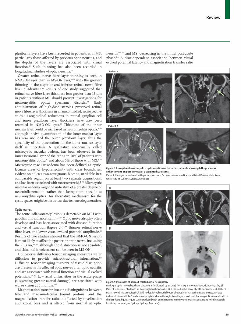
Figure 1: Examples of neuromyelitis optica-optic neuritis in two patients showing left optic nerve enhancement on post-contrast T1-weighted MRI scansPatient 1 images reproduced with permission from Dr Lynette Masters (Brain and Mind Research Institute, University of Sydney, Sydney, Australia).
Patient 1
Patient 2
LR
LR
LR
Figure 2: Two cases of sarcoid-related optic neuropathy(A) Right optic nerve sheath enhancement (indicated by arrows) from a granulomatous optic neuropathy. (B) Patient who presented with an acute right optic neuritis. MRI showed optic nerve sheath enhancement. FDG-PET scan showed hilar/mediastinal avid nodes. Lymph node biopsy showed non-caseating granulomata. Arrows indicate FDG-avid hilar/mediastinal lymph nodes in the right-hand fi gure, and to enhancing optic nerve sheath in the left-hand fi gure. Figure 2A reproduced with permission from Dr Lynette Masters (Brain and Mind Research Institute, University of Sydney, Sydney, Australia).
LR
A
B
90 www.thelancet.com/neurology Vol 13 January 2014
Review
suggests the potential for the lesional magnetisation transfer ratio to be indicative of remyelination after optic neuritis,109 and results of post-mortem pathology–MRI studies have shown higher magnetisation transfer ratio in remyelinated than in demyelinated lesions.111,112
Imaging of post-geniculate visual pathwaysOptic radiationsDiff usion tensor imaging metrics have identifi ed abnormalities in the optic radiations in post-acute optic neuritis.113 A reported signifi cant association between a
multifocal visual evoked potential measure of anterior visual axonal dysfunction (reduced amplitude) and reduced optic radiation axial diff usivity (which correlates with axonal injury in animal studies114) suggests possible anterograde trans-synaptic axonal degeneration—ie, degeneration of retrogeniculate fi bres secondary to primary degeneration of anterior visual pathway axons.113
Visual cortexAudoin and colleagues115 noted a lower grey matter magnetisation transfer ratio within the visual cortices
Figure 3: Left optic neuritis at baseline and after 6 months(A) T1-weighted MRI optic nerve showing acute swelling at baseline and some atrophy 6 months later. The nerve is outlined in red. B–D show outputs from optical coherence tomography. (B) Peripapillary optic nerve circular scan with disc swelling at baseline. (C and D) Profi le of retinal nerve fi bre layer along circular scan from nasal (N) to inferior (I) to temporal (T) and to superior (S) fi bres. Respective swelling and atrophy are shown at baseline and 6 months. OCT images reproduced with permission from Dr Rhian Saftopoulos, University College London Institute of Neurology, University College London, London, UK.
600
240360480
1200
–135 –90 –45 0 45 90 135 180 –135 –90 –45 0 45 90 135 180
N I T S N N I T S N
N I T S N N I T S N
Position (˚) Position (˚)
Thick
ness
(μm
)
A
B
C
D
Baseline 6 months later
200 µm 200 µm
www.thelancet.com/neurology Vol 13 January 2014 91
Review
after optic neuritis compared with healthy controls, suggesting cortical structural changes. Results of a longitudinal study showed that early pericalcarine atrophy predicts later conversion to MS at 1 year after acute optic neuritis.116
Results of functional MRI studies have shown reduced visual cortex activation after optic neuritis that is sometimes associated with visual dysfunction117 and with retinal nerve fi bre layer thickness.118 Findings from longitudinal studies have implicated several brain regions that might help visual recovery, including the lateral occipital complexes,119,120 cuneus,121 and lateral geniculate nuclei.122 Some degree of neuroplasticity might help to explain the discordance between good visual recovery and persistent structural optic nerve damage after acute optic neuritis.123
Acute treatment options for optic neuritisCorticosteroidsSeveral studies have assessed acute corticosteroid treatment for optic neuritis, providing level 1124 evidence.125–127 Results of the ONTT showed no improvement in visual acuity (p=0∙66) at 6 months after 3 days of high-dose (1 g per day) intravenous methylprednisolone followed by 11 days of low-dose oral prednisolone versus placebo, although visual recovery was faster.125,128 Mild benefi ts were noted for some secondary outcomes—visual fi elds (p=0∙054), contrast sensitivity (p=0∙026), and colour vision (p=0∙033). Patients taking standard-dose (1 mg per kg) oral prednisolone did not diff er from those taking placebo in visual outcomes but there was an unexpected increased risk of optic neuritis recurrence for reasons that are unclear. Intravenous methylprednisolone also delayed the onset of clinically defi nite MS at 2 years,129 but this diff erence abated over time.16 In a Cochrane review,130 no long-term benefi t was reported for corticosteroid treatment for visual acuity, visual fi elds, or contrast sensitivity.130 Most participants in these studies were likely to have typical optic neuritis, and the poor effi cacy of corticosteroids is concordant with studies investigating their eff ects on non-optic neuritis MS relapses—ie, hastening recovery but not aff ecting fi nal outcome.
Results of some studies suggest that timing of corticosteroid administration might be important, but more research is warranted. In the rat model of MS, corticosteroids given before induction of experimental allergic encephalomyelitis reduced the incidence of optic neuritis and preserved the retinal ganglion cells after optic neuritis.131 In human beings, poor evidence for the effi cacy of corticosteroids exists, and only results of uncontrolled studies examining very early treatment (within days of symptoms) have been published. In a case series, steroids administered hyperacutely to eight patients with previous optic neuritis (including MS, chronic relapsing infl ammatory optic neuropathy, and neuromyelitis optica) who reported a relapse of their
pain, resulted in no visual loss and in resolution of their pain.132 MRI was done in fi ve patients and confi rmed optic neuritis in all. A retrospective analysis of neuromyelitis optica cases reported that earlier administration (within 3 days) of intravenous methylprednisolone for optic neuritis relapse was associated with more preservation of retinal nerve fi bre layer thickness.92 Oral high-dose methylprednisolone could be an alternative to the intravenous form. A randomised controlled trial (n=60) showed that 500 mg per day oral methylprednisolone was superior to placebo at 1 and 3 weeks, although fi nal visual outcome was unaltered after optic neuritis.127
Randomised controlled trials on atypical optic neuritis are scarce, and evidence is observational or retrospective (mainly level 4).124 NMO-ON seems to respond to intravenous methylprednisolone, particularly if administered early.92,132 Similarly, for sarcoid-related optic neuritis, high-dose corticosteroids are indicated, although visual recovery might be hard to induce.133,134 Lupus-associated optic neuritis is also thought to respond to steroids,135 although cyclo phosphamide in combination with, or as an alternative, might be more eff ective.136 Vasculitic optic neuritis is very rare but some patients might respond to corticosteroids.137 High-dose corticosteroid treatment for atypical optic neuritis is usually followed by an oral weaning dose. Patients with chronic relapsing infl ammatory optic neuropathy might have a granulomatous optic neuropathy, which is very steroid-sensitive but also steroid-dependent, and can relapse on weaning.46
PlasmapheresisPlasmapheresis has been tested in patients with steroid-unresponsive demyelinating optic neuritis in observational cohort studies. Early treatment, within weeks, is more likely to be benefi cial.138,139 In a case series of 23 patients (ten with relapsing–remitting MS, one with neuromyelitis optica, and 12 with isolated optic neuritis), 70% improved with plasmapheresis, given after two cycles of intravenous methylprednisolone.140 In another case series of 20 patients with MS, 13 of 17 (76%) with aff ected optic nerves reported benefi t.138 A pooled review of plasmapheresis in neuromyelitis optica, together with the authors’ data for steroid-refractory optic neuritis with severe visual loss, analysed 39 eyes.139,141 People who improved received plasmapheresis a median of 19 days after onset, compared with 41 days for those who did not improve. Finally, patients with NMO-ON who were given plasmapheresis with corticosteroids had better visual outcome than did those given corticosteroids alone.142 Plasma exchange might be considered a viable option in demyelination of optic neuritis that is unresponsive to steroids.
Intravenous immunoglobulinsStudies with intravenous immunoglobulins have produced mixed results. Results of an open-label, non-randomised, prospective study143 showed a good
92 www.thelancet.com/neurology Vol 13 January 2014
Review
improvement in visual acuity in 18 of 23 steroid-refractory patients with MS-ON who had treatment, compared with three of 24 patients who did not. However, fi ndings from two earlier randomised, double-blinded, placebo-controlled studies recruiting a total of 123 patients with acute optic neuritis showed no signifi cant benefi cial eff ect.144,145 Overall, evidence for a benefi cial eff ect is weak.
RecommendationsFor typical optic neuritis, the aff ected patient is counselled about the benefi ts, limitations, and potential side-eff ects of corticosteroids, and is off ered treatment if visual loss is functionally disabling or if the patient has pre-existing visual impairment of the other eye. We tend to prescribe high-dose methylprednisolone (either 500 mg per day orally for 5 days or 1 g per day intravenously for 3 days) with no oral tail, and patients are followed up clinically, irrespective of treatment, within the next few weeks to check recovery. If atypical features develop or recovery does not begin, the atypical pathway for investigations and treatment is immediately followed. The patient is also counselled about the future potential of conversion to MS and is off ered brain MRI for risk stratifi cation.
For atypical optic neuritis, after appropriate and urgent investigations, treatment is instigated in our regimen with intravenous methylprednisolone (1 g per day for 3–5 days) followed by a prolonged oral tail, with weaning over 4–6 months. If patients relapse after initial treatment, the dose of oral corticosteroids can be increased, or a further course of intravenous methylprednisolone can be given and a steroid-sparing immunosuppressant considered (panel 5). If initial high-dose steroids do not eff ectively improve vision and the cause is demyelination, plasmapheresis can be considered. For lupus-related optic neuritis, cyclophosphamide can be considered.
Long-term management of typical optic neuritisConsideration of disease-modifying treatments used in MS Several placebo-controlled trials of the MS disease-modifying drugs beta interferon and glatiramer acetate have enrolled patients with clinically isolated syndrome (CIS) (including those with isolated optic neuritis) with MRI scans positive for demyelinating lesions.149–152 The results of all these trials showed delay of subsequent relapse and conversion to clinically defi nite MS. A follow-up study of one of the trial cohorts treated with beta interferon showed that this delaying eff ect persisted for up to 5 years, although long-term disability between early and late treatment groups did not diff er.153 Findings from a 10 year follow-up of another beta interferon trial cohort recorded similar eff ects.154 Despite the modest long-term clinical effi cacy, advocates for early versus delayed treatment of patients with clinically isolated syndromes argue that many will otherwise accumulate new MRI lesions, which could increase the chances of future disability.
RecommendationsPatients with typical, isolated optic neuritis are usually monitored. They are counselled about the risk of MS conversion, based on their brain MRI result, if possible. Although the most recent prescribing guidelines155 from the Association of British Neurologists suggest that disease-modifying drugs can be considered for patients with clinically isolated syndromes who have demyelination on brain scans, funding for this treatment can be, in practice, problematic to secure in the UK, and the potential long-term benefi ts are still uncertain. If the patient converts to MS, with a further relapse within 2 years, then disease-modifying drugs are often prescribed. MRI done at intervals during monitoring might enable an earlier diagnosis of MS to be made.
Long-term management of atypical optic neuritisNMO-ON relapse preventionAtypical optic neuritis often needs long-term immunosuppression, particularly if the risk of relapse is high or if relapses have occurred. The specifi c choice of immunosuppressant might be aff ected by the underlying causes.59,156
Maintenance of remission is crucial, because the accumulation of disability is associated with relapses. Several drugs have been studied retrospectively and observationally in neuromyelitis optica, its limited forms, and spectrum disorders, providing mainly level 4 evidence.124,147
Results of a small prospective study of seven patients with neuromyelitis optica reported a relapse-free follow-up of 18 months with treatment with azathioprine plus prednisolone.157 Findings from two retrospective studies of patients with neuromyelitis optica spectrum disorder158,159 showed that azathioprine, with or without prednisolone, reduced the annualised relapse rate by about three-quarters. Doses larger than 2 mg per kg and increases in mean corpuscular volumes were possibly associated with greater reductions in relapse rate.158 Benefi cial eff ects on disability scores and visual acuities have also been recorded.158,159 A target dose of 2∙5–3∙0 mg/kg per day is recommended, and the fi rst few months of treatment should be in combination with oral prednisolone while treatment takes eff ect. This regimen is followed by a slow steroid wean. Some patients might need low steroid maintenance doses.148
Methotrexate can be considered as an alternative fi rst-line drug, especially in those who are intolerant to azathioprine. A report described 14 patients on long-term methotrexate and recorded an 85% reduction in annualised relapse rate (from 1∙39 to 0∙18, p<0∙005), with disability improvement or stabilisation in 11 patients (79%).160
Although the European Federation of Neurological Societies Guidelines suggest rituximab as a potential fi rst-line treatment,147 in practice it is usually second line.148
www.thelancet.com/neurology Vol 13 January 2014 93
Review
It is an anti-CD20 chimeric monoclonal antibody administered by intermittent infusion, while the CD19 count is monitored.157,148 The three largest retrospective studies (recruiting 23, 35, and 30 patients, respectively)161–163 all described reductions in median annualised relapse rate by at least 90%, and stabilisation or improvement of disability in at least 80% of cases.
Mycophenolate is gaining favour as an eff ective immunosuppressant, despite the scarcity of evidence in neuromyelitis optica.147 The authors of a retrospective analysis of 24 patients reported an improvement in median annualised relapse rate of 93% (1∙3 before treatment; 0∙09 after treatment, p<0∙001) and stabilisation or improvement of disability in 91% of cases.164
Eculizumab has shown promising results, albeit in a small open-label pilot study.165 Median annualised relapse rate fell from 3 (range 2–4) to 0 (range 0–1) after a year’s treatment in 14 patients (p<0∙001). Median Expanded Disability Status Scale score improved from 4∙3 to 3∙5 (p=0∙0078). Further studies are warranted.
Low-dose maintenance corticosteroids could have a role in maintenance of remission.166 Evidence is scarce or mixed for other treatments used in relapse prevention, including mitoxantrone,167 cyclophosphamide,168 pulsed plasmapheresis,169 cyclosporin A,170 tacrolimus,148 intravenous immunoglobulins,171 and tocilizumab.172
Use of interferon beta, fi ngolimod, and natalizumab should be avoided in neuromyelitis optica and neuromyelitis optica spectrum disorders because some evidence suggests that clinical disease does not improve or can worsen with these treatments.173–177
Optic neuritis with systemic disease or chronic relapsing infl ammatory optic neuropathyMost immunosuppressants used to maintain clinical remission in systemic infl ammatory disease (eg, sarcoid, connective tissue disorder, or vasculitis) are the same as those described for neuromyelitis optica. Specifi c treatments should be tailored towards the underlying disorder, individual clinical course, and the
For the immunosuppressants listed here, the treatment advice is not defi nitive and the reader is encouraged to refer to local guidelines for more details about monitoring and use. Most patients will also need pre-treatment screening investigations.
• Corticosteroids can be started at 0·75–1 mg/kg per day (after intravenous doses given in an acute relapse) and then tapered slowly over about 6 months, with close clinical monitoring for atypical optic neuritis. Many side-eff ects are associated with corticosteroid use. The main ones include mood disturbances, glucose intolerance or diabetes, osteoporosis, proximal myopathy, Cushing’s syndrome, adrenal suppression, increased risk of infections (eg, varicella zoster, tuberculosis reactivation), hypertension, glaucoma, cataracts, electrolyte imbalance, neutrophilia, lymphopenia, peptic ulceration, and avascular necrosis. Patients on long-term corticosteroids in the UK should carry a steroid treatment card (providing information about reduction of risk and dose details) and should not discontinue treatment abruptly because of the risk of acute adrenal insuffi ciency. Local guidelines should be implemented for the prevention and treatment of osteoporosis, and patients should be monitored for diabetes and hypertension.
• Azathioprine treatment usually has a maintenance dose of 2·5–3·0 mg/kg per day, assuming that thiopurine methyltransferase concentrations are normal. It can be commenced at 25 mg daily and increased in 50 mg steps every week as an outpatient. Side-eff ects include bone-marrow suppression, hypersensitivity reactions, gastrointestinal reactions (nausea and vomiting), liver dysfunction, increased infection risk, and, rarely, pancreatitis. Full blood count and liver function tests should be monitored
frequently during early treatment while dose changes are occurring, and less frequently after reaching maintenance dose. A high mean corpuscular volume or lymphopenia tends to suggest a treatment eff ect.
• Mycophenolate treatment typically starts at 500 mg daily, increasing every week in 500 mg steps to a maintenance dose of 1 g twice daily. Main side-eff ects include hypersensitivity reactions, bone-marrow suppression, gastrointestinal reactions, liver dysfunction, renal dysfunction, potential risk of lymphoma and skin malignancy. Blood-test monitoring should include full blood count, urea and electrolytes, and liver function, and should be done frequently during the early treatment phase.
• Methotrexate treatment can aim for a maintenance dose of initially 15 mg once weekly. Doses start at 7·5 mg weekly (with folate supplementation) and then slowly increase in 2·5 mg steps each week. Side-eff ects include bone-marrow suppression, liver toxicity, pulmonary fi brosis, gastrointestinal symptoms, hypersensitivity reactions, and increased infection risk. Blood-test monitoring is mandatory (usually full blood count and liver function tests). Yearly chest radiographs are advised.
• Rituximab treatment is usually 1 g intravenously on days 1 and 14, repeated every 6 months or when the CD19 count (which is measured monthly) begins to rise.146 Side-eff ects include allergic reactions, hypotension, and exacerbation of cardiac disease. Infections can occur in 30% of rituximab-treated patients and are severe in 1–2%.147 Pre-infusion blood tests are recommended (full blood count, urea and electrolytes, and liver function).
Modifi ed from Palace and colleagues.148
Panel 5: Considerations for immunosuppressant use in atypical optic neuritis
94 www.thelancet.com/neurology Vol 13 January 2014
Review
degree of multiorgan involvement. Available evidence is usually level 4.124
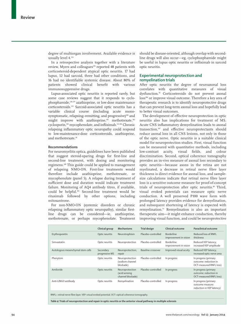
In a retrospective analysis together with a literature review, Myers and colleagues178 reported 48 patients with corticosteroid-dependent atypical optic neuritis. 17 had lupus, 12 had sarcoid, three had other conditions, and 16 had no identifi able systemic disease. About 80% of patients showed clinical benefi t with various immunosuppressive drugs.
Lupus-associated optic neuritis is reported rarely, but some case reviews suggest that it responds to cyclo-phosphamide,138,179 azathioprine, or low-dose maintenance corticosteroids.135 Sarcoid-associated optic neuritis has a variable clinical course (including acute mono-symptomatic, relapsing–remitting, and pro gressive)180 and might improve with azathioprine,181 methotrexate,182 cyclosporin,183 mycophenolate, and infl i xi mab.133,184 Chronic relapsing infl ammatory optic neuropathy could respond to low-maintenance-dose corticosteroids, azathioprine, and methotrexate.46
RecommendationsFor neuromyelitis optica, guidelines have been published that suggest steroid-sparing drugs for fi rst-line and second-line treatment, with dosing and monitoring regimens.148 This guide could be applied to management of relapsing NMO-ON. First-line treatments would therefore include azathioprine, methotrexate, or mycophenolate (panel 5). A relapse during treatment of suffi cient dose and duration would indicate treatment failure. Monitoring of AQ4 antibody titres, if available, could be helpful.185 Second-line treatment would be rituximab followed by other options, including mitoxantrone.
For non-NMO-ON (systemic disorders or chronic relapsing infl ammatory optic neuropathy), similar fi rst-line drugs can be considered—ie, azathioprine, methotrexate, or perhaps mycophenolate. Treatment
should be disease-oriented, although overlap with second-line drugs will also occur—eg, cyclophosphamide might be useful in lupus optic neuritis or infl iximab in sarcoid optic neuritis.
Experimental neuroprotection and remyelination trialsAfter optic neuritis the degree of neuroaxonal loss correlates with quantitative measures of visual dysfunction.78 Corticosteroids do not prevent axonal loss186 or improve visual outcome. Therefore a key area of therapeutic research is to identify neuroprotective drugs that can prevent long-term axonal loss and hopefully lead to better visual outcomes.
The development of eff ective neuroprotection in optic neuritis also has implications for treatment of MS. Acute CNS infl ammatory demyelination leads to axonal transection,187 and eff ective neuroprotectants should reduce axonal loss in all CNS lesions, not only in those of the optic nerve. Optic neuritis is a suitable clinical model for neuroprotection studies. First, visual function can be measured with quantitative methods, including low-contrast acuity, visual fi elds, and colour discrimination. Second, optical coherence tomography provides an in-vivo measure of axonal loss secondary to optic neuritis—because axons in the retina are not myelinated, a decrease in retinal nerve fi bre layer thickness is direct evidence for axonal loss, and sample-size calculations indicate that retinal nerve fi bre layer loss is a sensitive outcome measure for proof-of-concept trials of neuroprotection after optic neuritis.80 Third, visual evoked potentials can measure optic nerve conduction. A well preserved P100 wave form with prolonged latency provides evidence for demyelination, and subsequent shortening of latency is expected with remyelination.73 Remyelination is also an important therapeutic aim—it might enhance conduction, thereby improving visual function, and could be neuroprotective
Clinical group Mechanisms Trial design Clinical outcome Paraclinical outcome
Erythropoietin Optic neuritis Neurotrophism Placebo-controlled Borderline improvement in vision
Reduced loss of RNFL thickness
Simvastatin Optic neuritis Neuroprotection Placebo-controlled Borderline improvement in vision
Reduced VEP latency; increased VEP amplitude
Autologous mesenchymal stem cells Secondary progressive MS
Neuroprotection; repair
Baseline crossover Improved visual acuity Reduced VEP latency; increased optic nerve area
Phenytoin Optic neuritis Neuroprotection (sodium channel blockade)
Placebo-controlled In progress In progress (primary outcome: reduction in OCT-measured RNFL loss)
Amiloride Optic neuritis Neuroprotection (acid sensing channel blockade)
Placebo-controlled In progress In progress (primary outcome: reduction in OCT-measured RNFL loss)
Anti-LINGO antibody Optic neuritis Remyelination Placebo-controlled In progress In progress (primary outcome measure: reduction in VEP latency)
RNFL= retinal nerve fi bre layer. VEP=visual evoked potential. OCT=optical coherence tomography.
Table 4: Trials of neuroprotection and repair in optic neuritis or the anterior visual pathway in multiple sclerosis
www.thelancet.com/neurology Vol 13 January 2014 95
Review
by reducing the vulnerability of axons to adverse eff ects associated with infl ammation and demyelination.86
Some trials have investigated neuroprotection or remyelination in the anterior visual pathway in optic neuritis and MS (table 4). Results of a placebo-controlled trial of erythropoietin showed smaller decreases in retinal nerve fi bre layer thickness in the aff ected nerve with erythropoietin when compared with placebo,188 but were complicated by retinal nerve fi bre layer swelling during the acute phase of optic neuritis. Swelling indicates acute infl ammation, and the decrease in retinal nerve fi bre layer thickness is a combination of axonal loss and resolution of infl ammation. More specifi c evidence for axonal protection should be obtained by comparison of the fi nal thickness of the aff ected optic nerve with that of the unaff ected nerve.80 Another randomised controlled trial investigated simvastatin in acute optic neuritis.189 Signifi cant benefi ts were noted with visual evoked potential amplitude (p=0∙01) and latency (p=0∙01), and borderline eff ects for contrast sensitivity (p=0∙06). A small baseline crossover trial of autologous stem cells in secondary progressive MS identifi ed shortening of visual evoked potential latency and increase in optic nerve area after treatment, which could indicate remyelination.190
ConclusionsTo clinically distinguish typical optic neuritis from atypical forms in the acute phase is crucial; this distinction will then guide further management. The most common form is typical optic neuritis, probably demyelinating and closely associated with MS, although sometimes occurring in isolation. Typical optic neuritis resolves spontaneously, and provides researchers with a useful in-vivo model with which to study mechanisms of localised damage and recovery due to infl ammatory demyelination in the CNS, including the study of neuroprotective and remyelination strategies. If untreated, atypical optic neuritis can lead to irreversible visual loss, and often needs urgent treatment with corticosteroids, with slow wean and, sometimes, chronic immunosuppression.
ContributorsATT, DFM, and DHM were involved in planning, writing, critical
reviewing, and revision of the Review. DHM was involved in conception
of the manuscript.
Confl icts of interestATT has received honoraria from Sereno Symposia International
Foundation and Bayer. DHM has received honoraria through payments
to his employer, UCL Institute of Neurology, for Advisory Committee or
consultancy advice in MS studies from Biogen Idec, GlaxoSmithKline,
Novartis, Merck, Chugai, Mitsubishi Pharma Europe, and Bayer
Schering Pharma. He has also received compensation through payments
to his employer for doing central MRI analysis of MS trials from
GlaxoSmithKline, Biogen Idec, Novartis, and Merck. DFM has received
honoraria from Biogen.
AcknowledgmentsATT is funded by the Higher Education Funding Council for England.
This work was partly done at University College London Hospitals or
University College London, which received a proportion of funding
from the Department of Health’s National Institute for Health
Research and Biomedical Research Centres funding scheme. We thank
Dr Rhian Raftopoulos (University College London Institute of
Neurology, University College London, London, UK) and
Lynette Masters (Brain and Mind Research Institute, University of
Sydney, Sydney, Australia) for providing some of the fi gures used in
this Review. The NMR Research Unit at University College London
Institute of Neurology is supported by the UK MS Society and
University College London-University College London Hospitals
Biomedical Research Centre.
References1 Wikstrom J. The epidemiology of optic neuritis in Finland.
Acta Neurol Scand 1975; 52: 196–206.
2 Loncarek K, Brajac I, Petricek I, Stalekar H, Cerovski B, Pokupe R. Epidemiology of monosymptomatic optic neuritis in Rijeka County, Croatia: meteorological aspects. Coll Antropol 2005; 29: 309–13.
3 Taylor BV, Lucas RM, Dear K, et al. Latitudinal variation in incidence and type of fi rst central nervous system demyelinating events. Mult Scler 2010; 16: 398–405.
4 Lucas RM, Ponsonby AL, Dear K, et al. Sun exposure and vitamin D are independent risk factors for CNS demyelination. Neurology 2011; 76: 540–48.
5 Wakakura M, Ishikawa S, Oono S, et al. Incidence of acute idiopathic optic neuritis and its therapy in Japan. Optic Neuritis Treatment Trial Multicenter Cooperative Research Group (ONMRG). Nippon Ganka Gakkai Zasshi 1995; 99: 93–97.
6 Jin YP, Pedro-Cuesta J, Soderstrom M, Stawiarz L, Link H. Incidence of optic neuritis in Stockholm, Sweden 1990–1995: I. Age, sex, birth and ethnic-group related patterns. J Neurol Sci 1998; 159: 107–14.
7 MacDonald BK, Cockerell OC, Sander JW, Shorvon SD. The incidence and lifetime prevalence of neurological disorders in a prospective community-based study in the UK. Brain 2000; 123: 665–76.
8 Jin Y, Pedro-Cuesta J, Soderstrom M, Stawiarz L, Link H. Seasonal patterns in optic neuritis and multiple sclerosis: a meta-analysis. J Neurol Sci 2000; 181: 56–64.
9 Lucas RM, Ponsonby AL, Dear K, et al. Current and past Epstein-Barr virus infection in risk of initial CNS demyelination. Neurology 2011; 77: 371–79.
10 de la Cruz J, Kupersmith MJ. Clinical profi le of simultaneous bilateral optic neuritis in adults. Br J Ophthalmol 2006; 90: 551–54.
11 Parkin PJ, Hierons R, McDonald WI. Bilateral optic neuritis. A long-term follow-up. Brain 1984; 107: 951–64.
12 Francis DA, Compston DA, Batchelor JR, McDonald WI. A reassessment of the risk of multiple sclerosis developing in patients with optic neuritis after extended follow-up. J Neurol Neurosurg Psychiatry 1987; 50: 758–65.
13 Rizzo JF III, Lessell S. Risk of developing multiple sclerosis after uncomplicated optic neuritis: a long-term prospective study. Neurology 1988; 38: 185–90.
14 Jacobs L, Kinkel PR, Kinkel WR. Silent brain lesions in patients with isolated idiopathic optic neuritis. A clinical and nuclear magnetic resonance imaging study. Arch Neurol 1986; 43: 452–55.
Search strategy and selection criteria
We searched PubMed for articles published in English from 1970 to July, 2013, with the general search term “optic neuritis” combined with more specifi c search terms related to the subheadings—eg, “optical coherence tomography”, “corticosteroid”, “plasmapheresis”, and “magnetic resonance imaging”. References from identifi ed studies were checked and included if deemed appropriate, relevant, and scientifi cally important. We considered articles in other languages if referenced in a selected English article. We also searched references from our own fi les. We preferentially selected articles published within the past 10 years, although we also included older references that were important.
96 www.thelancet.com/neurology Vol 13 January 2014
Review
15 Ormerod IE, McDonald WI, du Boulay GH, et al. Disseminated lesions at presentation in patients with optic neuritis. J Neurol Neurosurg Psychiatry 1986; 49: 124–27.
16 Optic neuritis study group. Multiple sclerosis risk after optic neuritis: fi nal optic neuritis treatment trial follow-up. Arch Neurol 2008; 65: 727–32.
17 Swanton JK, Fernando K, Dalton CM, et al. Is the frequency of abnormalities on magnetic resonance imaging in isolated optic neuritis related to the prevalence of multiple sclerosis? A global comparison. J Neurol Neurosurg Psychiatry 2006; 77: 1070–72.
18 Swanton JK, Rovira A, Tintore M, et al. MRI criteria for multiple sclerosis in patients presenting with clinically isolated syndromes: a multicentre retrospective study. Lancet Neurol 2007; 6: 677–86.
19 Rovira A, Swanton J, Tintore M, et al. A single, early magnetic resonance imaging study in the diagnosis of multiple sclerosis. Arch Neurol 2009; 66: 587–92.
20 Polman CH, Reingold SC, Banwell B, et al. Diagnostic criteria for multiple sclerosis: 2010 revisions to the McDonald criteria. Ann Neurol 2011; 69: 292–302.
21 Miller DH, Chard DT, Ciccarelli O. Clinically isolated syndromes. Lancet Neurol 2012; 11: 157–69.
22 Optic Neuritis Study Group The clinical profi le of optic neuritis. Experience of the Optic Neuritis Treatment Trial. Arch Ophthalmol 199; 109: 1673–78.
23 Schneck ME, Haegerstrom-Portnoy G. Color vision defect type and spatial vision in the optic neuritis treatment trial. Invest Ophthalmol Vis Sci 1997; 38: 2278–89.
24 Katz B. The dyschromatopsia of optic neuritis: a descriptive analysis of data from the optic neuritis treatment trial. Trans Am Ophthalmol Soc 1995; 93: 685–708.
25 Silverman SE, Hart WM Jr, Gordon MO, Kilo C. The dyschromatopsia of optic neuritis is determined in part by the foveal/perifoveal distribution of visual fi eld damage. Invest Ophthalmol Vis Sci 1990; 31: 1895–902.
26 Mullen KT, Plant GT. Colour and luminance vision in human optic neuritis. Brain 1986; 109: 1–13.
27 Moura AL, Teixeira RA, Oiwa NN, et al. Chromatic discrimination losses in multiple sclerosis patients with and without optic neuritis using the Cambridge Colour Test. Vis Neurosci 2008; 25: 463–68.
28 Fazzone HE, Lefton DR, Kupersmith MJ. Optic neuritis: correlation of pain and magnetic resonance imaging. Ophthalmology 2003; 110: 1646–49.
29 McDonald WI, Barnes D. The ocular manifestations of multiple sclerosis. 1. Abnormalities of the aff erent visual system. J Neurol Neurosurg Psychiatry 1992; 55: 747–52.
30 Optic Neuritis Study Group. Keltner JL, Johnson CA, Spurr JO, Beck RW. Baseline visual fi eld profi le of optic neuritis. The experience of the optic neuritis treatment trial. Arch Ophthalmol 1993; 111: 231–34.
31 Keltner JL, Johnson CA, Spurr JO, Beck RW. Comparison of central and peripheral visual fi eld properties in the optic neuritis treatment trial. Am J Ophthalmol 1999; 128: 543–53.
32 Gerling J, Meyer JH, Kommerell G. Visual fi eld defects in optic neuritis and anterior ischemic optic neuropathy: distinctive features. Graefes Arch Clin Exp Ophthalmol 1998; 236: 188–92.
33 Nevalainen J, Krapp E, Paetzold J, et al. Visual fi eld defects in acute optic neuritis—distribution of diff erent types of defect pattern, assessed with threshold-related supraliminal perimetry, ensuring high spatial resolution. Graefes Arch Clin Exp Ophthalmol 2008; 246: 599–607.
34 Lightman S, McDonald WI, Bird AC, et al. Retinal venous sheathing in optic neuritis. Its signifi cance for the pathogenesis of multiple sclerosis. Brain 1987; 110: 405–14.
35 Beck RW, Kupersmith MJ, Cleary PA, Katz B. Fellow eye abnormalities in acute unilateral optic neuritis. Experience of the optic neuritis treatment trial. Ophthalmology 1993; 100: 691–97.
36 Beck RW, Cleary PA, Backlund JC. The course of visual recovery after optic neuritis. Experience of the Optic Neuritis Treatment Trial. Ophthalmology 1994; 101: 1771–78.
37 Beck RW, Cleary PA. Optic neuritis treatment trial. One-year follow-up results. Arch Ophthalmol 1993; 111: 773–75.
38 Visual function 15 years after optic neuritis: a fi nal follow-up report from the Optic Neuritis Treatment Trial. Ophthalmology 2008; 115: 1079–82.
39 Kupersmith MJ, Gal RL, Beck RW, Xing D, Miller N. Visual function at baseline and 1 month in acute optic neuritis: predictors of visual outcome. Neurology 2007; 69: 508–14.
40 Beck RW. Optic Neuritis. In: Miller NR, Newman NJ, eds. Walsh and Hoyt’s clinical neuro-ophthalmology, 5th edn. Baltimore: Williams and Wilkins, 1998. Chapter 12: 599–647.
41 Flanagan P, Markulev C. Spatio-temporal selectivity of loss of colour and luminance contrast sensitivity with multiple sclerosis and optic neuritis. Ophthalmic Physiol Opt 2005; 25: 57–65.
42 Phillips ML, Foster DH, Honan WP, Edgar GK, Heron JR. Optic neuritis. Diff erential losses of luminance and chromatic function near a scotoma. Brain 1994; 117: 767–73.
43 Raz N, Dotan S, Benoliel T, Chokron S, Ben-Hur T, Levin N. Sustained motion perception defi cit following optic neuritis: Behavioral and cortical evidence. Neurology 2011; 76: 2103–11.
44 Jenkins TM, Toosy AT. New developments in the treatment of optic neuritis. Eye and Brain 2010; 2: 83–94.
45 Wingerchuk DM, Lennon VA, Lucchinetti CF, Pittock SJ, Weinshenker BG. The spectrum of neuromyelitis optica. Lancet Neurol 2007; 6: 805–15.
46 Kidd D, Burton B, Plant GT, Graham EM. Chronic relapsing infl ammatory optic neuropathy (CRION). Brain 2003; 126: 276–84.
47 Petzold A, Pittock S, Lennon V, Maggiore C, Weinshenker BG, Plant GT. Neuromyelitis optica-IgG (aquaporin-4) autoantibodies in immune mediated optic neuritis. J Neurol Neurosurg Psychiatry 2010; 81: 109–11.
48 Jarius S, Frederikson J, Waters P, et al. Frequency and prognostic impact of antibodies to aquaporin-4 in patients with optic neuritis. J Neurol Sci 2010; 298: 158–62.
49 de Seze J, Arndt C, Jeanjean L, et al. Relapsing infl ammatory optic neuritis: is it neuromyelitis optica? Neurology 2008; 70: 2075–76.
50 Matiello M, Lennon VA, Jacob A, et al. NMO-IgG predicts the outcome of recurrent optic neuritis. Neurology 2008; 70: 2197–200.
51 Kurne A, Karabudak R, Yalcin-Cakmakli G, et al. Recurrent optic neuritis: clues from a long-term follow up study of recurrent and bilateral optic neuritis patients. Eye and Brain 2010; 2: 15–20.
52 Plant G, Acheson J, Clarke C, Graham E, Howard R, Shorvon S. Neuro-ophthalmology. In: Clarke C, Howard R, Rossor M, Shorvon S, eds. Neurology: a queen square textbook. Wiley-Blackwell, 2011. Chapter 13: 489–532.
53 Kupersmith MJ, Alban T, Zeiff er B, Lefton D. Contrast-enhanced MRI in acute optic neuritis: relationship to visual performance. Brain 2002; 125: 812–22.
54 Halliday AM, McDonald WI, Mushin J. Delayed visual evoked response in optic neuritis. Lancet 1972; 299: 982–85.
55 Hickman SJ, Dalton CM, Miller DH, Plant GT. Management of acute optic neuritis. Lancet 2002; 360: 1953–62.
56 You Y, Klistorner A, Thie J, Graham SL. Latency delay of visual evoked potential is a real measurement of demyelination in a rat model of optic neuritis. Invest Ophthalmol Vis Sci 2011; 52: 6911–18.
57 Pittock SJ, Weinshenker BG, Lucchinetti CF, Wingerchuk DM, Corboy JR, Lennon VA. Neuromyelitis optica brain lesions localized at sites of high aquaporin 4 expression. Arch Neurol 2006; 63: 964–68.
58 Papadopoulos MC, Verkman AS. Aquaporin 4 and neuromyelitis optica. Lancet Neurol 2012; 11: 535–44.
59 Jindahra P, Plant GT. Update on neuromyelitis optica: natural history and management. Eye and Brain 2012; 4: 27–41.
60 Lennon VA, Wingerchuk DM, Kryzer TJ, et al. A serum autoantibody marker of neuromyelitis optica: distinction from multiple sclerosis. Lancet 2004; 364: 2106–12.
61 Akman-Demir G, Tuzun E, Waters P, et al. Prognostic implications of aquaporin-4 antibody status in neuromyelitis optica patients. J Neurol 2011; 258: 464–70.
62 Jarius S, Ruprecht K, Wildemann B, et al. Contrasting disease patterns in seropositive and seronegative neuromyelitis optica: a multicentre study of 175 patients. J Neuroinfl ammation 2012; 9: 14.
63 Kitley J, Woodhall M, Waters P, et al. Myelin-oligodendrocyte glycoprotein antibodies in adults with a neuromyelitis optica phenotype. Neurology 2012; 79: 1273–77.
64 Smith KJ, McDonald WI. The pathophysiology of multiple sclerosis: the mechanisms underlying the production of symptoms and the natural history of the disease. Philos Trans R Soc Lond B Biol Sci 1999; 354: 1649–73.
www.thelancet.com/neurology Vol 13 January 2014 97
Review
65 Kolappan M, Henderson AP, Jenkins TM, et al. Assessing structure and function of the aff erent visual pathway in multiple sclerosis and associated optic neuritis. J Neurol 2009; 256: 305–19.
66 Guy J, Fitzsimmons J, Ellis EA, Beck B, Mancuso A. Intraorbital optic nerve and experimental optic neuritis. Correlation of fat suppression magnetic resonance imaging and electron microscopy. Ophthalmology 1992; 99: 720–25.
67 Youl BD, Turano G, Miller DH, et al. The pathophysiology of acute optic neuritis. An association of gadolinium leakage with clinical and electrophysiological defi cits. Brain 1991; 114: 2437–50.
68 Roed H, Frederiksen J, Langkilde A, Sorensen TL, Lauritzen M, Sellebjerg F. Systemic T-cell activation in acute clinically isolated optic neuritis. J Neuroimmunol 2005; 162: 165–72.
69 Soderstrom M, Link H, Xu Z, Fredriksson S. Optic neuritis and multiple sclerosis: anti-MBP and anti-MBP peptide antibody-secreting cells are accumulated in CSF. Neurology 1993; 43: 1215–22.
70 Matsunaga Y, Kezuka T, An X, et al. Visual functional and histopathological correlation in experimental autoimmune optic neuritis. Invest Ophthalmol Vis Sci 2012; 53: 6964–71.
71 Hickman SJ, Toosy AT, Jones SJ, et al. A serial MRI study following optic nerve mean area in acute optic neuritis. Brain 2004; 127: 2498–505.
72 Hickman SJ, Toosy AT, Miszkiel KA, et al. Visual recovery following acute optic neuritis: a clinical, electrophysiological and magnetic resonance imaging study. J Neurol 2004; 251: 996–1005.
73 Klistorner A, Arvind H, Garrick R, Yiannikas C, Paine M, Graham SL. Remyelination of optic nerve lesions: spatial and temporal factors. Mult Scler 2010; 16: 786–95.
74 Felts PA, Baker TA, Smith KJ. Conduction in segmentally demyelinated mammalian central axons. J Neurosci 1997; 17: 7267–77.
75 Smith KJ. Sodium channels and multiple sclerosis: roles in symptom production, damage and therapy. Brain Pathol 2007; 17: 230–42.
76 Raz N, Dotan S, Chokron S, Ben-Hur T, Levin N. Demyelination aff ects temporal aspects of perception: an optic neuritis study. Ann Neurol 2012; 71: 531–38.
77 Kupersmith MJ, Mandel G, Anderson S, Meltzer DE, Kardon R. Baseline, one and three month changes in the peripapillary retinal nerve fi ber layer in acute optic neuritis: relation to baseline vision and MRI. J Neurol Sci 2011; 308: 117–23.
78 Trip SA, Schlottmann PG, Jones SJ, et al. Retinal nerve fi ber layer axonal loss and visual dysfunction in optic neuritis. Ann Neurol 2005; 58: 383–91.
79 Costello F, Coupland S, Hodge W, et al. Quantifying axonal loss after optic neuritis with optical coherence tomography. Ann Neurol 2006; 59: 963–69.
80 Henderson AP, Altmann DR, Trip AS, et al. A serial study of retinal changes following optic neuritis with sample size estimates for acute neuroprotection trials. Brain 2010; 133: 2592–602.
81 Fisher JB, Jacobs DA, Markowitz CE, et al. Relation of visual function to retinal nerve fi ber layer thickness in multiple sclerosis. Ophthalmology 2006; 113: 324–32.
82 Henderson AP, Trip SA, Schlottmann PG, et al. An investigation of the retinal nerve fi bre layer in progressive multiple sclerosis using optical coherence tomography. Brain 2008; 131: 277–87.
83 Trip SA, Schlottmann PG, Jones SJ, et al. Optic nerve atrophy and retinal nerve fi bre layer thinning following optic neuritis: evidence that axonal loss is a substrate of MRI-detected atrophy. Neuroimage 2006; 31: 286–93.
84 Naismith RT, Xu J, Tutlam NT, et al. Disability in optic neuritis correlates with diff usion tensor-derived directional diff usivities. Neurology 2009; 72: 589–94.
85 Villoslada P, Cuneo A, Gelfand J, Hauser SL, Green A. Color vision is strongly associated with retinal thinning in multiple sclerosis. Mult Scler 2012; 18: 991–99.
86 Henderson AP, Altmann DR, Trip SA, et al. Early factors associated with axonal loss after optic neuritis. Ann Neurol 2011; 70: 955–63.
87 Klistorner A, Arvind H, Nguyen T, et al. Axonal loss and myelin in early optic neuritis loss in postacute optic neuritis. Ann Neurol 2008; 64: 325–31.
88 Klistorner A, Arvind H, Nguyen T, et al. Multifocal VEP and OCT in optic neuritis: a topographical study of the structure-function relationship. Doc Ophthalmol 2009; 118: 129–37.
89 Walter SD, Ishikawa H, Galetta KM, et al. Ganglion cell loss in relation to visual disability in multiple sclerosis. Ophthalmology 2012; 119: 1250–57.
90 Syc SB, Saidha S, Newsome SD, et al. Optical coherence tomography segmentation reveals ganglion cell layer pathology after optic neuritis. Brain 2012; 135: 521–33.
91 Ratchford JN, Quigg ME, Conger A, et al. Optical coherence tomography helps diff erentiate neuromyelitis optica and MS optic neuropathies. Neurology 2009; 73: 302–08.
92 Nakamura M, Nakazawa T, Doi H, et al. Early high-dose intravenous methylprednisolone is eff ective in preserving retinal nerve fi ber layer thickness in patients with neuromyelitis optica. Graefes Arch Clin Exp Ophthalmol 2010; 248: 1777–85.
93 Naismith RT, Tutlam NT, Xu J, et al. Optical coherence tomography diff ers in neuromyelitis optica compared with multiple sclerosis. Neurology 2009; 72: 1077–82.
94 Fernandes DB, Raza AS, Nogueira RG, et al. Evaluation of inner retinal layers in patients with multiple sclerosis or neuromyelitis optica using optical coherence tomography. Ophthalmology 2013; 120: 387–94.
95 Sotirchos ES, Saidha S, Byraiah G, et al. In vivo identifi cation of morphologic retinal abnormalities in neuromyelitis optica. Neurology 2013; 80: 1406–14.
96 Gelfand JM, Cree BA, Nolan R, Arnow S, Green AJ. Microcystic inner nuclear layer abnormalities and neuromyelitis optica. JAMA Neurol 2013; 70: 629–33.
97 Gelfand JM, Nolan R, Schwartz DM, Graves J, Green AJ. Microcystic macular oedema in multiple sclerosis is associated with disease severity. Brain 2012; 135: 1786–93.
98 Saidha S, Sotirchos ES, Ibrahim MA, et al. Microcystic macular oedema, thickness of the inner nuclear layer of the retina, and disease characteristics in multiple sclerosis: a retrospective study. Lancet Neurol 2012; 11: 963–72.
99 Hickman SJ, Toosy AT, Miszkiel KA, et al. Visual recovery following acute optic neuritis—a clinical, electrophysiological and magnetic resonance imaging study. J Neurol 2004; 251: 996–1005.
100 Hickman SJ, Brierley CM, Brex PA, al. Continuing optic nerve atrophy following optic neuritis: a serial MRI study. Mult Scler 2002; 8: 339–42.
101 Khanna S, Sharma A, Huecker J, Gordon M, Naismith RT, van Stavern GP. Magnetic resonance imaging of optic neuritis in patients with neuromyelitis optica versus multiple sclerosis. J Neuroophthalmol 2012; 32: 216–20.
102 Storoni M, Davagnanam I, Radon M, Siddiqui A, Plant GT. Distinguishing optic neuritis in neuromyelitis optica spectrum disease from multiple sclerosis: a novel magnetic resonance imaging scoring system. J Neuroophthalmol 2013; 33: 123–27.
103 Basser PJ, Pierpaoli C. Microstructural and physiological features of tissues elucidated by quantitative-diff usion-tensor MRI. J Magn Reson B 1996; 111: 209–19.
104 Kolbe S, Chapman C, Nguyen T, et al. Optic nerve diff usion changes and atrophy jointly predict visual dysfunction after optic neuritis. Neuroimage 2009; 45: 679–86.
105 Naismith RT, Xu J, Tutlam NT, Trinkaus K, Cross AH, Song SK. Radial diff usivity in remote optic neuritis discriminates visual outcomes. Neurology 2010; 74: 1702–10.
106 Naismith RT, Xu J, Tutlam NT, et al. Diff usion tensor imaging in acute optic neuropathies: predictor of clinical outcomes. Arch Neurol 2012; 69: 65–71.
107 Trip SA, Schlottmann PG, Jones SJ, et al. Optic nerve magnetization transfer imaging and measures of axonal loss and demyelination in optic neuritis. Mult Scler 2007; 13: 875–79.
108 Klistorner A, Chaganti J, Garrick R, Moff at K, Yiannikas C. Magnetisation transfer ratio in optic neuritis is associated with axonal loss, but not with demyelination. Neuroimage 2011; 56: 21–26.
109 Hickman SJ, Toosy AT, Jones SJ, et al. Serial magnetization transfer imaging in acute optic neuritis. Brain 2004; 127: 692–700.
110 Chen JT, Collins DL, Atkins HL, Freedman MS, Arnold DL. Magnetization transfer ratio evolution with demyelination and remyelination in multiple sclerosis lesions. Ann Neurol 2008; 63: 254–62.
111 Schmierer K, Scaravilli F, Altmann DR, Barker GJ, Miller DH. Magnetization transfer ratio and myelin in postmortem multiple sclerosis brain. Ann Neurol 2004; 56: 407–15.

98 www.thelancet.com/neurology Vol 13 January 2014
Review
112 Barkhof F, Bruck W, de Groot CJ, et al. Remyelinated lesions in multiple sclerosis: magnetic resonance image appearance. Arch Neurol 2003; 60: 1073–81.
113 Kolbe S, Bajraszewski C, Chapman C, et al. Diff usion tensor imaging of the optic radiations after optic neuritis. Hum Brain Mapp 2012; 33: 2047–61.
114 Budde MD, Kim JH, Liang HF, Russell JH, Cross AH, Song SK. Axonal injury detected by in vivo diff usion tensor imaging correlates with neurological disability in a mouse model of multiple sclerosis. NMR Biomed 2008; 21: 589–97.
115 Audoin B, Fernando KT, Swanton JK, Thompson AJ, Plant GT, Miller DH. Selective magnetization transfer ratio decrease in the visual cortex following optic neuritis. Brain 2006; 129: 1031–39.
116 Jenkins TM, Ciccarelli O, Atzori M, et al. Early pericalcarine atrophy in acute optic neuritis is associated with conversion to multiple sclerosis. J Neurol Neurosurg Psychiatry 2011; 82: 1017–21.
117 Langkilde AR, Frederiksen JL, Rostrup E, Larsson HB. Functional MRI of the visual cortex and visual testing in patients with previous optic neuritis. Eur J Neurol 2002; 9: 277–86.
118 Fuglo D, Kallenbach K, Tsakiri A, et al. Retinal atrophy correlates with fMRI response in patients with recovered optic neuritis. Neurology 2011; 77: 645–51.
119 Toosy AT, Hickman SJ, Miszkiel KA, et al. Adaptive cortical plasticity in higher visual areas after acute optic neuritis. Ann Neurol 2005; 57: 622–33.
120 Jenkins TM, Toosy AT, Ciccarelli O, et al. Neuroplasticity predicts outcome of optic neuritis independent of tissue damage. Ann Neurol 2010; 67: 99–113.
121 Jenkins T, Ciccarelli O, Toosy A, et al. Dissecting structure-function interactions in acute optic neuritis to investigate neuroplasticity. Hum Brain Mapp 2010; 31: 276–86.
122 Korsholm K, Madsen KH, Frederiksen JL, Skimminge A, Lund TE. Recovery from optic neuritis: an ROI-based analysis of LGN and visual cortical areas. Brain 2007; 130: 1244–53.
123 Klistorner A, Arvind H, Garrick R, Graham SL, Paine M, Yiannikas C. Interrelationship of optical coherence tomography and multifocal visual-evoked potentials after optic neuritis. Invest Ophthalmol Vis Sci 2010; 51: 2770–77.
124 Howick J, Chalmers I, Glasziou P, et al. OCEBM Levels of Evidence Working Group. The Oxford 2011 Levels of Evidence. Oxford: Oxford Centre for Evidence-Based Medicine, 2011.
125 Beck RW, Cleary PA, Anderson MM Jr, et al. The Optic Neuritis Study Group. A randomized, controlled trial of corticosteroids in the treatment of acute optic neuritis. N Engl J Med 1992; 326: 581–88.
126 Kapoor R, Miller DH, Jones SJ, et al. Eff ects of intravenous methylprednisolone on outcome in MRI-based prognostic subgroups in acute optic neuritis. Neurology 1998; 50: 230–37.
127 Sellebjerg F, Nielsen HS, Frederiksen JL, Olesen J. A randomized, controlled trial of oral high-dose methylprednisolone in acute optic neuritis. Neurology 1999; 52: 1479–84.
128 Beck RW, Gal RL. Treatment of acute optic neuritis: a summary of fi ndings from the optic neuritis treatment trial. Arch Ophthalmol 2008; 126: 994–95.
129 Beck RW, Cleary PA, Trobe JD, et al. The Optic Neuritis Study Group. The eff ect of corticosteroids for acute optic neuritis on the subsequent development of multiple sclerosis. N Engl J Med 1993; 329: 1764–69.
130 Gal RL, Vedula SS, Beck R. Corticosteroids for treating optic neuritis. Cochrane Database Syst Rev 2012; 4: CD001430.
131 Dutt M, Tabuena P, Ventura E, Rostami A, Shindler KS. Timing of corticosteroid therapy is critical to prevent retinal ganglion cell loss in experimental optic neuritis. Invest Ophthalmol Vis Sci 2010; 51: 1439–45.
132 Plant GT, Sibtain NA, Thomas D. Hyperacute corticosteroid treatment of optic neuritis at the onset of pain may prevent visual loss: a case series. Mult Scler Int 2011; 2011: 815068.
133 Pawate S, Moses H, Sriram S. Presentations and outcomes of neurosarcoidosis: a study of 54 cases. Q JM 2009; 102: 449–60.
134 Zajicek JP, Scolding NJ, Foster O, et al. Central nervous system sarcoidosis—diagnosis and management. Q JM 1999; 92: 103–17.
135 Lin YC, Wang AG, Yen MY. Systemic lupus erythematosus-associated optic neuritis: clinical experience and literature review. Acta Ophthalmol 2009; 87: 204–10.
136 Galindo-Rodriguez G, Avina-Zubieta JA, Pizaro S, et al. Cyclophosphamide pulse therapy in optic neuritis due to systemic lupus erythematosus: an open trial. Am J Med 1999; 106: 65–69.
137 Goodwin J. Autoimmune optic neuropathy. Curr Neurol Neurosci Rep 2006; 6: 396–402.
138 Trebst C, Reising A, Kielstein JT, Hafer C, Stangel M. Plasma exchange therapy in steroid-unresponsive relapses in patients with multiple sclerosis. Blood Purif 2009; 28: 108–15.
139 Bonnan M, Cabre P. Plasma exchange in severe attacks of neuromyelitis optica. Mult Scler Int 2012; 2012: 787630.
140 Roesner S, Appel R, Gbadamosi J, Martin R, Heesen C. Treatment of steroid-unresponsive optic neuritis with plasma exchange. Acta Neurol Scand 2012; 126: 103–08.
141 Bonnan M, Brasme H, Diaby MM, Vlaicu M, Le G, V, Zuber M. Severe bouts of neuromyelitis optica: dramatic improvement after plasma exchanges. Rev Neurol (Paris) 2009; 165: 479–81.
142 Merle H, Olindo S, Jeannin S, et al. Treatment of optic neuritis by plasma exchange (add-on) in neuromyelitis optica. Arch Ophthalmol 2012; 130: 858–62.
143 Tselis A, Perumal J, Caon C, et al. Treatment of corticosteroid refractory optic neuritis in multiple sclerosis patients with intravenous immunoglobulin. Eur J Neurol 2008; 15: 1163–67.
144 Noseworthy JH, O’Brien PC, Petterson TM, et al. A randomized trial of intravenous immunoglobulin in infl ammatory demyelinating optic neuritis. Neurology 2001; 56: 1514–22.
145 Roed HG, Langkilde A, Sellebjerg F, et al. A double-blind, randomized trial of IV immunoglobulin treatment in acute optic neuritis. Neurology 2005; 64: 804–10.
146 Greenberg BM, Graves D, Remington G, et al. Rituximab dosing and monitoring strategies in neuromyelitis optica patients: creating strategies for therapeutic success. Mult Scler 2012; 18: 1022–26.
147 Sellner J, Boggild M, Clanet M, et al. EFNS guidelines on diagnosis and management of neuromyelitis optica. Eur J Neurol 2010; 17: 1019–32.
148 Palace J, Leite MI, Jacob A. A practical guide to the treatment of neuromyelitis optica. Pract Neurol 2012; 12: 209–14.
149 Comi G, Filippi M, Barkhof F, et al. Eff ect of early interferon treatment on conversion to defi nite multiple sclerosis: a randomised study. Lancet 2001; 357: 1576–82.
150 Comi G, Martinelli V, Rodegher M, et al. Eff ect of glatiramer acetate on conversion to clinically defi nite multiple sclerosis in patients with clinically isolated syndrome (PreCISe study): a randomised, double-blind, placebo-controlled trial. Lancet 2009; 374: 1503–11.
151 Kappos L, Polman CH, Freedman MS, et al. Treatment with interferon beta-1b delays conversion to clinically defi nite and McDonald MS in patients with clinically isolated syndromes. Neurology 2006; 67: 1242–49.
152 Jacobs LD, Beck RW, Simon JH, et al. Intramuscular interferon beta-1a therapy initiated during a fi rst demyelinating event in multiple sclerosis. CHAMPS Study Group. N Engl J Med 2000; 343: 898–904.
153 Kappos L, Freedman MS, Polman CH, et al. Long-term eff ect of early treatment with interferon beta-1b after a fi rst clinical event suggestive of multiple sclerosis: 5-year active treatment extension of the phase 3 BENEFIT trial. Lancet Neurol 2009; 8: 987–97.
154 Kinkel RP, Dontchev M, Kollman C, Skaramagas TT, O’Connor PW, Simon JH. Association between immediate initiation of intramuscular interferon beta-1a at the time of a clinically isolated syndrome and long-term outcomes: a 10-year follow-up of the Controlled High-Risk Avonex Multiple Sclerosis Prevention Study in Ongoing Neurological Surveillance. Arch Neurol 2012; 69: 183–90.
155 Association of British Neurologists. Revised guidelines for prescribing in multiple sclerosis. 2009. http://www.theabn.org/abn/userfi les/fi le/ABN_MS_Guidelines_2009_Final(1).pdf (accessed Nov 4, 2013).
156 Pula JH, Macdonald CJ. Current options for the treatment of optic neuritis. Clin Ophthalmol 2012; 6: 1211–23.
157 Mandler RN, Ahmed W, Dencoff JE. Devic’s neuromyelitis optica: a prospective study of seven patients treated with prednisone and azathioprine. Neurology 1998; 51: 1219–20.
158 Costanzi C, Matiello M, Lucchinetti CF, et al. Azathioprine: tolerability, effi cacy, and predictors of benefi t in neuromyelitis optica. Neurology 2011; 77: 659–66.
www.thelancet.com/neurology Vol 13 January 2014 99
Review
159 Bichuetti DB, Lobato de Oliveira EM, Oliveira DM, Amorin de SN, Gabbai AA. Neuromyelitis optica treatment: analysis of 36 patients. Arch Neurol 2010; 67: 1131–36.
160 Kitley J, Elsone L, George J, et al. Methotrexate is an alternative to azathioprine in neuromyelitis optica spectrum disorders with aquaporin-4 antibodies. J Neurol Neurosurg Psychiatry 2013; 84: 918–21.
161 Bedi GS, Brown AD, Delgado SR, Usmani N, Lam BL, Sheremata WA. Impact of rituximab on relapse rate and disability in neuromyelitis optica. Mult Scler 2011; 17: 1225–30.
162 Kim SH, Kim W, Li XF, Jung IJ, Kim HJ. Repeated treatment with rituximab based on the assessment of peripheral circulating memory B cells in patients with relapsing neuromyelitis optica over 2 years. Arch Neurol 2011; 68: 1412–20.
163 Jacob A, Weinshenker BG, Violich I, et al. Treatment of neuromyelitis optica with rituximab: retrospective analysis of 25 patients. Arch Neurol 2008; 65: 1443–48.
164 Jacob A, Matiello M, Weinshenker BG, et al. Treatment of neuromyelitis optica with mycophenolate mofetil: retrospective analysis of 24 patients. Arch Neurol 2009; 66: 1128–33.
165 Pittock SJ, Lennon VA, McKeon A, et al. Eculizumab in AQP4-IgG-positive relapsing neuromyelitis optica spectrum disorders: an open-label pilot study. Lancet Neurol 2013; 12: 554–62.
166 Watanabe S, Misu T, Miyazawa I, et al. Low-dose corticosteroids reduce relapses in neuromyelitis optica: a retrospective analysis. Mult Scler 2007; 13: 968–74.
167 Cabre P, Olindo S, Marignier R, Jeannin S, Merle H, Smadja D. Effi cacy of mitoxantrone in neuromyelitis optica spectrum: clinical and neuroradiological study. J Neurol Neurosurg Psychiatry 2013; 84: 511–16.
168 Bichuetti DB, Oliveira EM, Boulos FC, Gabbai AA. Lack of response to pulse cyclophosphamide in neuromyelitis optica: evaluation of 7 patients. Arch Neurol 2012; 69: 938–39.
169 Khatri BO, Kramer J, Dukic M, Palencia M, Verre W. Maintenance plasma exchange therapy for steroid-refractory neuromyelitis optica. J Clin Apher 2012; 27: 183–92.
170 Kageyama T, Komori M, Miyamoto K, et al. Combination of cyclosporine A with corticosteroids is eff ective for the treatment of neuromyelitis optica. J Neurol 2013; 260: 627–34.
171 Wingerchuk DM. Neuromyelitis optica: potential roles for intravenous immunoglobulin. J Clin Immunol 2013; 33 (suppl 1): S33–37.
172 Kieseier BC, Stuve O, Dehmel T, et al. Disease amelioration with tocilizumab in a treatment-resistant patient with neuromyelitis optica: implication for cellular immune responses. JAMA Neurol 2013; 70: 390–93.
173 Palace J, Leite MI, Nairne A, Vincent A. Interferon beta treatment in neuromyelitis optica: increase in relapses and aquaporin 4 antibody titers. Arch Neurol 2010; 67: 1016–17.
174 Kleiter I, Hellwig K, Berthele A, et al. Failure of natalizumab to prevent relapses in neuromyelitis optica. Arch Neurol 2012; 69: 239–45.
175 Min JH, Kim BJ, Lee KH. Development of extensive brain lesions following fi ngolimod (FTY720) treatment in a patient with neuromyelitis optica spectrum disorder. Mult Scler 2012; 18: 113–15.
176 Barnett MH, Prineas JW, Buckland ME, Parratt JD, Pollard JD. Massive astrocyte destruction in neuromyelitis optica despite natalizumab therapy. Mult Scler 2012; 18: 108–12.
177 Kim SH, Kim W, Li XF, Jung IJ, Kim HJ. Does interferon beta treatment exacerbate neuromyelitis optica spectrum disorder? Mult Scler 2012; 18: 1480–83.
178 Myers TD, Smith JR, Wertheim MS, Egan RA, Shults WT, Rosenbaum JT. Use of corticosteroid sparing systemic immunosuppression for treatment of corticosteroid dependent optic neuritis not associated with demyelinating disease. Br J Ophthalmol 2004; 88: 673–80.
179 Rosenbaum JT, Simpson J, Neuwelt CM. Successful treatment of optic neuropathy in association with systemic lupus erythematosus using intravenous cyclophosphamide. Br J Ophthalmol 1997; 81: 130–32.
180 Koczman JJ, Rouleau J, Gaunt M, Kardon RH, Wall M, Lee AG. Neuro-ophthalmic sarcoidosis: the University of Iowa experience. Semin Ophthalmol 2008; 23: 157–68.
181 Gelwan MJ, Kellen RI, Burde RM, Kupersmith MJ. Sarcoidosis of the anterior visual pathway: successes and failures. J Neurol Neurosurg Psychiatry 1988; 51: 1473–80.
182 Maust HA, Foroozan R, Sergott RC, Niazi S, Weibel S, Savino PJ. Use of methotrexate in sarcoid-associated optic neuropathy. Ophthalmology 2003; 110: 559–63.
183 Bielory L, Frohman LP. Low-dose cyclosporine therapy of granulomatous optic neuropathy and orbitopathy. Ophthalmology 1991; 98: 1732–36.
184 Salama B, Gicquel JJ, Lenoble P, Dighiero PL. Optic neuropathy in refractory neurosarcoidosis treated with TNF-alpha antagonist. Can J Ophthalmol 2006; 41: 766–68.
185 Jarius S, Aboul-Enein F, Waters P, et al. Antibody to aquaporin-4 in the long-term course of neuromyelitis optica. Brain 2008; 131: 3072–80.
186 Hickman SJ, Kapoor R, Jones SJ, Altmann DR, Plant GT, Miller DH. Corticosteroids do not prevent optic nerve atrophy following optic neuritis. J Neurol Neurosurg Psychiatry 2003; 74: 1139–41.
187 Trapp BD, Peterson J, Ransohoff RM, Rudick R, Mork S, Bo L. Axonal transection in the lesions of multiple sclerosis. N Engl J Med 1998; 338: 278–85.
188 Suhs KW, Hein K, Sattler MB, et al. A randomized, double-blind, phase 2 study of erythropoietin in optic neuritis. Ann Neurol 2012; 72: 199–210.
189 Tsakiri A, Kallenbach K, Fuglo D, Wanscher B, Larsson H, Frederiksen J. Simvastatin improves fi nal visual outcome in acute optic neuritis: a randomized study. Mult Scler 2012; 18: 72–81.
190 Connick P, Kolappan M, Crawley C, et al. Autologous mesenchymal stem cells for the treatment of secondary progressive multiple sclerosis: an open-label phase 2a proof-of-concept study. Lancet Neurol 2012; 11: 150–56.