Open microscopy in the life sciences: Quo Vadis? - arXiv
28
Opinion paper Open microscopy in the life sciences: Quo Vadis? Johannes Hohlbein 1,2,* , Benedict Diederich 3,4 , Barbora Marsikova 3 , Emmanuel G. Reynaud 5 , Séamus Holden 6 , Wiebke Jahr 7 , Robert Haase 8 , and Kirti Prakash 9 1) Laboratory of Biophysics, Wageningen University & Research, Wageningen, the Netherlands 2) Microspectroscopy Research Facility, Wageningen University & Research, Wageningen, the Netherlands 3) Leibniz Institute for Photonic Technology, Jena, Germany 4) Institute for Physical Chemistry, Friedrich-Schiller University, Jena, Germany 5) School of Biomolecular and Biomedical Sciences, University College Dublin, Dublin, Ireland 6) Centre for Bacterial Cell Biology, Biosciences Institute, Newcastle University, Newcastle upon Tyne, UK 7) In-Vision Digital Imaging Optics GmbH, Austria 8) DFG Cluster of Excellence "Physics of Life", TU Dresden, Germany 9) National Physical Laboratory, Teddington, UK Contacts: [email protected] (https://orcid.org/0000-0001-7436-2221), [email protected] (https://orcid.org/0000-0003-0453-6286), [email protected] (https://orcid.org/0000-0002-8206-1549), [email protected] (https://orcid.org/0000-0003-1502-661X), [email protected] (https://orcid.org/0000-0002-7169-907X), [email protected] (https://orcid.org/0000-0003-0201-2315), [email protected] (https://orcid.org/0000-0001-5949-2327), [email protected] (https://orcid.org/0000-0002-0325-9988) * corresponding author keywords: open microscopy, open source, open science, open hardware
-
Upload
khangminh22 -
Category
Documents
-
view
0 -
download
0
Transcript of Open microscopy in the life sciences: Quo Vadis? - arXiv
Opinion paper
Open microscopy in the life sciences: Quo Vadis?
Johannes Hohlbein1,2,*, Benedict Diederich3,4, Barbora Marsikova3, Emmanuel G. Reynaud5,
Séamus Holden6, Wiebke Jahr7, Robert Haase8, and Kirti Prakash9
1) Laboratory of Biophysics, Wageningen University & Research, Wageningen, the Netherlands 2) Microspectroscopy Research Facility, Wageningen University & Research, Wageningen, the
Netherlands 3) Leibniz Institute for Photonic Technology, Jena, Germany 4) Institute for Physical Chemistry, Friedrich-Schiller University, Jena, Germany 5) School of Biomolecular and Biomedical Sciences, University College Dublin, Dublin, Ireland 6) Centre for Bacterial Cell Biology, Biosciences Institute, Newcastle University, Newcastle upon
Tyne, UK 7) In-Vision Digital Imaging Optics GmbH, Austria
8) DFG Cluster of Excellence "Physics of Life", TU Dresden, Germany 9) National Physical Laboratory, Teddington, UK
Contacts:
[email protected] (https://orcid.org/0000-0001-7436-2221),
[email protected] (https://orcid.org/0000-0003-0453-6286),
[email protected] (https://orcid.org/0000-0002-8206-1549),
[email protected] (https://orcid.org/0000-0003-1502-661X),
[email protected] (https://orcid.org/0000-0002-7169-907X),
[email protected] (https://orcid.org/0000-0003-0201-2315),
[email protected] (https://orcid.org/0000-0001-5949-2327),
[email protected] (https://orcid.org/0000-0002-0325-9988)
*corresponding author
keywords: open microscopy, open source, open science, open hardware
Abstract 3
A. Introduction 3
B. On the purpose of open science and open microscopy 4
B.1. Open microscopy is just good scientific practice 5
B.2. Open microscopy enables flexible and powerful platforms for different user groups 7
C. Current and future challenges 10
Open microscopy: diverse but united 10
C.1. Accessibility, availability, safety, and time versus money 11
C.2. Standards and continuing proliferation: it’s not a bug, it’s a feature! 12
C.3. Generating shareable hardware files 13
C.4. Connecting open-source software to open hardware 13
C.5. Strategies enabling long term support of open-microscopy projects 14
C.6. Commercialization of open-source projects: Why and how? 16
C.7. Continuing training and education 18
D. Conclusion 19
Additional resources 20
Author contributions 20
Acknowledgements 20
References 20
Figures 27
Figure 1 27
Figure 2 28
Abstract
Light microscopy allows observing cellular features and objects with sub-micrometer
resolution. As such, light microscopy has been playing a fundamental role in the life sciences for
more than a hundred years. Fueled by the availability of mass-produced electronics and
hardware, publicly shared documentation and building instructions, open-source software,
wide access to rapid prototyping and 3D printing, and the enthusiasm of contributors and users
involved, the concept of open microscopy has been gaining incredible momentum, bringing
new sophisticated tools to an expanding user base. Here, we will first discuss the ideas behind
open science and open microscopy before highlighting recent projects and developments in
open microscopy. We argue that the availability of well-designed open hardware and software
solutions targeting broad user groups or even non-experts, will increasingly be relevant to cope
with the increasing complexity of cutting-edge imaging technologies. We will then extensively
discuss the current and future challenges of open microscopy.
A. Introduction
Light microscopy has been pivotal in the life sciences to study small features and objects
otherwise hidden to the naked eye. Simple microscopes such as the Foldscope or smartphone
auxiliary lenses are available for a few Euros and form the basis of citizen science projects,
scientific education, and medical diagnosis1,2.
Driven by societal and academic shifts towards a culture of open science and open
technology, more and more information on modalities for advanced and high-end microscopy
has become openly available to interested researchers and private enthusiasts. Compared to
the open software movement, however, the development of open hardware frameworks for
microscopy accelerated only rather recently with the increasing accessibility and affordability of
suitable components. With 3D printers, rapid reproduction of designs and prototyping moved
from professional machine shops to the living room table. Web shops deliver sophisticated CNC
milled designs within days to the doorsteps. Mass-produced electronics such as light-emitting
or laser diodes, microcontrollers, optical parts, and (mobile phone) cameras have significantly
levelled the playing field for high-end instrumentation. Open microscopy does not
automatically imply that solutions are cheap. Some of the frameworks further discussed below
feature higher-priced components such as microscope objectives, laser engines, and cameras
often costing tens of thousands of Euros. As such, these components maximize performance
but can surprisingly often be adequately replaced by cheaper alternatives that better benefit
from the economies of scale9.
Supported by their accessible documentation, all open projects empower scientists to
adopt solutions - even if only as a source of inspiration. In academia, open hardware and
software projects are lifesavers at a time of fierce competition for funding and a decline in
government investment in science. Projects that have developed strong communities enable
researchers to receive support within minutes in public support forums and over social media.
By its very nature, microscopy is interdisciplinary combining aspects from physics,
engineering, informatics, biology, and data science, often with a hint of breakthrough
technologies. As educational resources, open microscopy frameworks further provide an entry
to the world of coding, tinkering and science with minimal thresholds to overcome.
In an age of seemingly unlimited educational and technical possibilities, what does that
mean to the general field of optical light microscopy and its high-end applications? What are
the remaining challenges and where lie potential issues? Here, after providing a brief overview
of current open-hardware and open-software microscopy projects, we highlight several goals
and challenges that we consider important to ensure the growth and future success of open
microscopy.
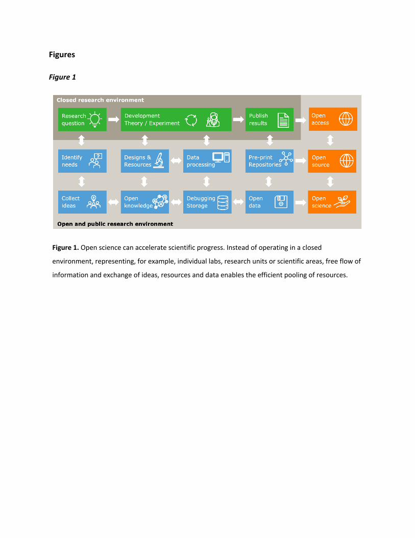
B. On the purpose of open science and open microscopy
Open science, open source, open access, open hardware, open everything is now the
motto for many communities and has become the obligatory reference. In general, open
science seeks to improve transparency, reproducibility, inclusiveness, and accessibility of
research and innovation as, for example, discussed in the UNESCO draft recommendation on
open science10. For scientific research, the conventional setting of an academic environment
kept the science largely behind closed doors; although results were published, information on
the methodology, the experimental settings or access to raw data was, and unfortunately often
still is, hard to come by (Figure 1). Open science is addressing these issues by providing a myriad
of additional interaction points between researchers and more broadly between citizens,
starting from the
collection of ideas over reusing hardware designs and open-source software to the exploration
of publicly available data by anyone interested. For scientific data, the FAIR principle (Findable,
Accessible, Interoperable, Reusable)11 provides guidelines for moving towards what science
should be all about: shared knowledge accessible to all. With that in mind, we here define open
microscopy as scientific data which contains information on (i) how to build, maintain and use
(light) microscopes, (ii) how to prepare, handle and measure samples and (iii) how to analyze,
store and distribute experimental data and models.
Figure 1. Open science can accelerate scientific progress. Instead of operating in a closed
environment, representing, for example, individual labs, research units or scientific areas, free flow of
information and exchange of ideas, resources and data enables the efficient pooling of resources.
B.1. Open microscopy is just good scientific practice
For open hardware, recent work highlights general opportunities and best practices12,13.
For light microscopy, this implies making new imaging technologies accessible and easily
reproducible. In the past, new methods were often published with only a vague cartoon of the
optical setup and without access to any software or electrical circuit diagrams. Even for
specialist labs, implementation of published modalities often required extensive reverse
engineering and tinkering for many years. Commercial microscopes, on the other hand, are
often built on closed-source methods and compound the reproducibility problem by making
key steps in the process of biological discovery dependent on an ill-characterized black box.
Open microscopy can address this problem by ensuring that any new imaging method, both
hardware and software, is sufficiently documented and open to allow straightforward
reproduction by other specialists, and without information being hidden behind restrictive
Material Transfer Agreements (MTA) or Non-Disclosure Agreements (NDA). Documenting
assembly and manufacturing (e.g., 3D printing, CNC machining) guides, bill of materials or even
providing video tutorials that help others recreating a setup without additional help by the
authors, acts as an amplifier, in which knowledge is multiplied and can be used at remote
places outside the originating lab. It can be very motivating to see one's work being replicated
and used by others, even if current scientific scoring systems may only recognize formal
citations and not pre-publication replications.
Since prototype microscopes usually require extensive further development to make
them production-ready and user-friendly, this does not preclude their co-existence with
commercial devices. The need for users to receive comprehensive product support and a
turnkey solution is not necessarily a requirement of the Open Microscopy movement.
Generally, we argue that open microscopy should be considered an essential requirement for
any scientific publication of a new microscopy method to meet modern standards of scientific
reproducibility. Detailed documentation, including why something was implemented in the
suggested way, is also key for others to learn about the given technique and to discover
potential optimization steps. For small hardware or software components, this could further
imply making drawings or source code available under an open-source license. In general,
documentation can be written in the form of citable scientific publications and thus, is well
compatible with the academic incentive system. Editors from the optics and scientific imaging
fields, however, should increase efforts in supporting scientists, who are willing to share their
designs openly, to improve the reproducibility of publications and science in general.
Reproducibility in the context of data analysis implies that both the experimental or
simulated data as well as the analysis software is openly available. We therefore argue that
reproducibility is the key to advancing science as without rigorous verification of results and
discoveries true scientific progress is impossible.
Academic researchers should be aware that, by default, everything developed and
created is the property of the research institute, meaning that researchers leaving the institute
may lose both rights and access to their unpublished intellectual contributions. To permit the
use and further development of open microscopy projects by anyone, regardless of location or
affiliation, appropriate licenses such as CERN Open Hardware License, MIT, GPL v3 or Creative
Commons must be chosen. This also addresses the issue posed by active patents that
theoretically prohibit the replication and productive use of protected setups in the laboratory
as discussed here14. We recommend scientists and developers to make themselves aware of
the regulations and possibilities with the institutional IP handling offices as early as possible.
While not an intrinsic feature of open source, we encourage developers to use version
control tools like git (GitHub, Gitlab) at any stage of the project to document the process and
track individual contributions that can help to clarify the contribution of individual authors.
B.2. Open microscopy enables flexible and powerful platforms for different user groups
Until recently, microscopy hardware developers seeking to develop new optical
methods faced the choice of either retrofitting new hardware onto an existing commercial
microscope (body) or designing and building an entire bespoke microscope from individual
optical components. Neither fits the requirements of a hardware developer perfectly. Although
using a commercial body has the advantage of starting with a working system that usually lacks
serious optical aberrations and offers a stable mechanical base, critical optical planes required
in the microscope development process are often hidden, unknown or inaccessible within the
monolithic body. Furthermore, some features implemented for user-friendliness and safety
(eyepieces, safety interlocks, dedicated software) can potentially prevent the hardware
developer from easily modifying a setup. Customized designs, on the other hand, offer full
control and often lower costs at the expense of significant development time and the need to
re-implement basic components and features, such as focusing or sample positioning. One
drawback of custom microscopy solutions is that they are often less user-friendly compared to
their commercial counterparts that offer safe housing or one-click software solutions. In the
end, commercial bodies and self-designed systems may target different audiences but, as we
will discuss in the following, the gap has been closing rapidly.
Rather than classifying open hardware frameworks in terms of their apparent
complexity, we suggest looking at potential user groups. We foresee disruptive potential of
extremely simplified, minimalist open microscopes (FlyPi15, OpenFlexure3, UC2 system4,
µCube16, Octopi17) as they change advanced microscopy from a scarce resource shared
between many labs to everyday tools of life scientists and hobby enthusiasts all over the world
(Figure 2). These microscopes are designed to be affordable, adaptable, reproducible, and
exchangeable or easily repairable, for example using 3D printed parts instead of specialist
components as recently reviewed for light microscopy in reference18. Time-intensive
experiments such as live cell microscopy or automated high-content screening could be made
easily accessible, creating new scientific opportunities similar to how low-cost (whole-genome)
DNA sequencing enabled the widespread use of genomics in biological research.
For developmental biologists interested in volumetric imaging, the OpenSPIM (SPIM:
Selective- Plane Illumination Microscopy) project has been a successful example of an open
microscopy platform that required an entire redesign of microscopy hardware and software19.
Starting in 2010, OpenSPIM enabled many labs as well as imaging platforms to build and apply
light-sheet microscopy at a time when commercial solutions were neither accessible nor
affordable. It is estimated that more than 80 systems have been built with at least 6 in facility
settings. Similarly, the mesoSPIM initiative targets the imaging of large samples and provides a
very comprehensive open-source documentation20. According to the project website
(https://mesospim.org/), at least 12 builds are in operation and follow-up work provides
detailed protocols for tissue clearing21. Further, an open platform for scanned oblique plane
illumination microscopy (SOPi), was introduced that features open hardware assembly, clear
alignment protocol, and control software for sub-micron resolution single objective light-sheet
microscopy22.
A number of open microscopy frameworks have been developed that more closely
resemble the layout of conventional upright commercial systems but offer a higher degree of
modularity and customizability. Several platforms for cost-effective single-molecule localization
microscopy visualize biological structures with a resolution of well below 50 nm (WOSM23,
liteTIRF24) or even characterize protein-DNA-RNA interactions in live bacteria (miCube25).
Additional frameworks focus on conventional epifluorescence microscope (LFSM26), high-
throughput screening and tracking of microorganisms (Squid27, cell biology28), diffusion-based
confocal for analyzing conformational dynamics of biomolecules29 or detection of protein
aggregation30, two-photon Ca²⁺ deep tissue imaging31, and structured illumination for sub-
diffraction resolved (live) cell imaging32,33 (Figure 2). Depending on exact implementations, the
cost of these microscopes can be considerably lower than for commercial systems, although, as
discussed below, the costs due to expert time investment for both build and maintenance
should not be underestimated.
We note that every published technical solution can potentially contribute to simplifying
the development of future microscopy techniques. Open microscopy software is also constantly
evolving. ImageJ6 and Fiji7 have been pivotal for analyzing images and movies by life scientists
for many years. Their microscope control sibling µManager35 and counterparts in the Python
world36–39 have been changing the way how the community analyses and displays images in the
microscopy context for many years, with new plugins and functionalities for data acquisition
being constantly released40,41. Smooth transfer of images acquired on one system to another
was key to create this ecosystem for bioimage analysis that now flourishes beyond the
manufacturer formats, especially due to the efforts of the Open Microscopy Environment
(OME) initiative42. The field of image data analysis has exploded in recent years now involving
deep learning techniques for image quality improvements, segmentation and overall data
analysis (e.g., CARE48, StarDist49, CellPose50, QuPath51, ZeroCostDL4Mic8). Modern Graphics
Processing Units (GPUs) do not just execute deep learning algorithms in the shortest amount of
time, the technology also supports post-processing of data in real-time such as multiview
deconvolution52, deconvolution and fusion53, and general-purpose image processing54.
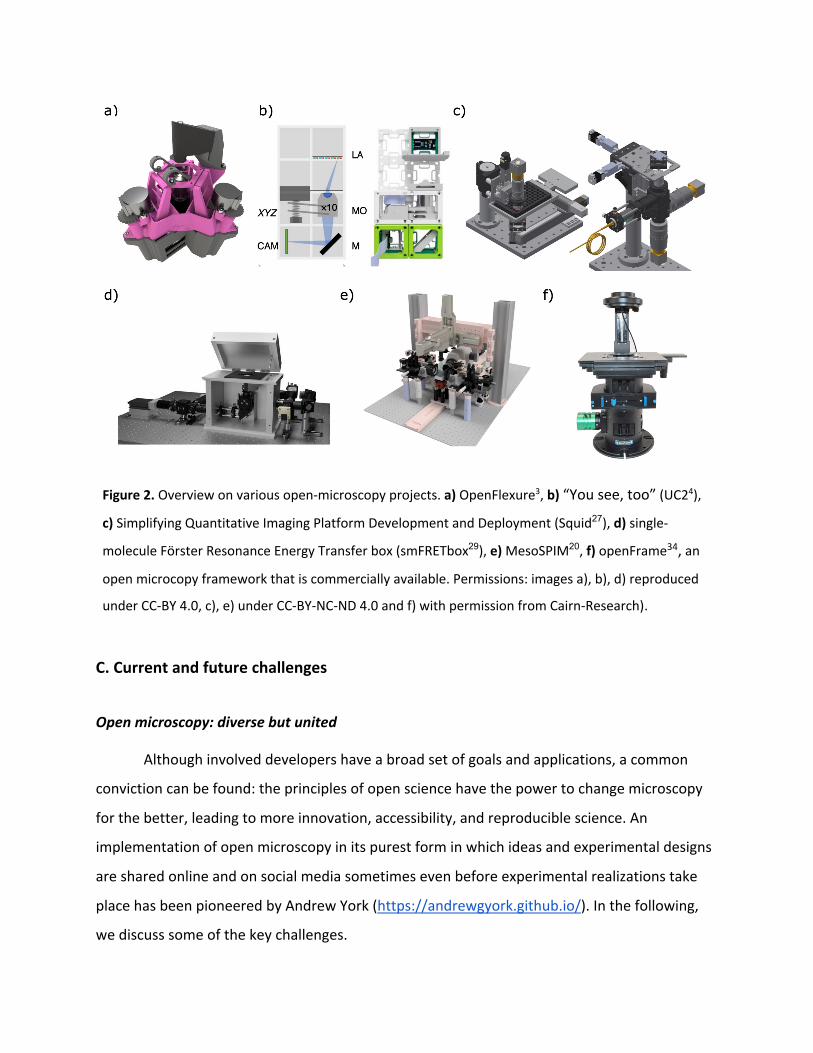
Figure 2. Overview on various open-microscopy projects. a) OpenFlexure3, b) “You see, too” (UC24),
c) Simplifying Quantitative Imaging Platform Development and Deployment (Squid27), d) single-
molecule Förster Resonance Energy Transfer box (smFRETbox29), e) MesoSPIM20, f) openFrame34, an
open microcopy framework that is commercially available. Permissions: images a), b), d) reproduced
under CC-BY 4.0, c), e) under CC-BY-NC-ND 4.0 and f) with permission from Cairn-Research).
C. Current and future challenges
Open microscopy: diverse but united
Although involved developers have a broad set of goals and applications, a common
conviction can be found: the principles of open science have the power to change microscopy
for the better, leading to more innovation, accessibility, and reproducible science. An
implementation of open microscopy in its purest form in which ideas and experimental designs
are shared online and on social media sometimes even before experimental realizations take
place has been pioneered by Andrew York (https://andrewgyork.github.io/). In the following,
we discuss some of the key challenges.
C.1. Accessibility, availability, safety, and time versus money
The reasons for working on and with open microscopy projects are as diverse as the
people involved. Some might enjoy the tinkering aspects most (the tinkerer). Others see open
software and technology primarily as tools to address their scientific questions as affordable
and fitting as possible (the end-user). Microscope developers and end-users, and all the
researchers falling somewhere in between, may have a very different vision for open
microscopy and should be aware of each other. The end-user is likely to prefer more polished
software or hardware, sometimes even willing to sacrifice additional features for stability and
ease of use. In fact, the end-user might have less time to build or adapt complete solutions and
would rather prefer to buy them. Both sides ultimately depend on each other like in a classical
“supply and demand” situation in which a growing request for innovative solutions can support
people working on them. Along the same line, many projects require substantial training and
build-up of expertise before productive data acquisition can be achieved. Although proper
documentation and design can support the perspective of the end-user, the availability of
personalized training and education is needed. As we will discuss later, education is an inherent
feature of open microscopy since parts to build microscopes can easily be sourced.
One often-used statement in the open microscopy field is that an instrument was
designed for (part-) costs that are ten times cheaper than the price of a comparable
commercially available instrument. We consider such statements at best misleading and at
worst plainly wrong as neither the costs of development nor the time spent to build the
instrument is accounted for in most cases. We also point out that any company must fulfil a
minimum of conformity with health, safety, and environmental protection standards (e.g., CE,
FCC, TÜV or others) for their products and provide customer support. In open projects, even
when building up with commercially available components, the sole responsibility on safety is
shifted to the user. Additionally, customer support depends largely on the goodwill of the
developer.
C.2. Standards and continuing proliferation: it’s not a bug, it’s a feature!
As outlined above, the number of open microscopy frameworks has hugely increased
over the last years. An interesting example of proliferation is the variety of software packages
available for single-molecule localization microscopy that fit data from individual fluorescent
emitters to obtain their position with sub-pixel accuracy. Recently, over 30 different software
packages were compared using a diverse set of metrics43. Although such wide choice can be
intimidating at first, the robust assessment itself, indicating strengths and weaknesses of
individual software packages, is excellent proof of the benefits of open microscopy. As new
packages and developments can be directly compared to previous ones, the super-resolution
field was able to quickly reach a high degree of maturity in terms of data analysis. It became
equally apparent that user-friendly implementations of the best approaches are critical to
reaching end-users.
Decentralized hardware development in which experts design solutions for specific
problems cannot often be integrated in other frameworks. Here, interfaces such as device
adaptors in µManager35 or mechanical adaptors as provided by the modular optical toolbox
UC24 can help to bridge multiple different hardware and software projects. Recently, a series of
documents suggest the implementation of standards in open hardware and software
development44 as well as data provenance and quality control in microscopy45,46. We suggest
that these best practices are requested and followed by scientists, reviewers, and editors to
ensure and enable long-lasting inter-device operability. We note, however, that hardware- and
software development significantly differ in the way how solutions are distributed. Ready-to-
install software packages require only a limited knowledge about the underlying system
integrity. Well-formulated building instructions still require someone who gathers all necessary
parts (e.g., 3D printed or from external sources), assembles these parts and invests a fair
amount of time to finally get the system running - no matter how well the documentation is
described. The equivalent to a ready-to-install software package could be assorted hardware
kits (UC24), but the non-zero cost of duplicating hardware poses a challenge for widespread
distribution. It remains important to implement and use calibration control points and sample
standards both for hardware as well as software.
C.3. Generating shareable hardware files
Open microscopy projects are shared using different files and data formats. Whereas
many formats are open and suitable viewers are freely available, this is not necessarily the case
for designs that feature Computer Aided Design (CAD). For CAD, buying suitable software
licenses can be prohibitively expensive leading to limited accessibility and unknown design
history. For 3D printing, 3D models can be exported, for example, in the *.stl format which
describes only the surface geometry of a three-dimensional object without any scale thereby
inhibiting any modifications on the design. Alternatives, such as sharing links to cloud-based
CAD software (e.g., Fusion360, Tinkercad) or relying on open-source CAD models (e.g.,
openSCAD, Python-based FreeCAD) can help to distribute design files across many different
development environments. Ultimately, publishers and developers should ensure that design
files are available as (vectorized) raw data-file as well as processed files. The open source
hardware association (OSHWA) offers a guideline to maintain a good quality of openness in
projects (https://www.oshwa.org/sharing-best-practices/). Whilst protein structures, DNA
sequences and other bioinformatic data can be deposited in well-established databases (e.g.,
PDB, EBI), no dedicated repositories for microscopy hardware are currently available.
C.4. Connecting open-source software to open hardware
The close connection between open hardware and software is inevitable for complex
microscopy projects. Projects such as µManager35, Pycro-Manager36 and Python-microscopy37,47
have been playing a key part in connecting setup control, data acquisition and data analysis.
When it comes to hardware control, the availability of open-source device drivers and adapters
is crucial. The software architecture used in µManager, for example, standardizes how
hardware devices can be controlled from diverse software components via a plug-in
mechanism. If this mechanism is well-designed and easy to use, developers from all over the
world will contribute plugins. As a case in point, the µManager community managed to collect
hundreds of device adapters (https://micro-manager.org/Device_Support).
Many of the earlier introduced approaches for computational microscopy have the
potential to lower the quality requirements for microscopy hardware. Efficient denoising
algorithms, for example, allow the use of lower laser powers for fluorescence excitation or the
use of cheaper cameras without compromising final image quality.
Such advanced analysis techniques, however, have not been integrated widely and
reliably into any open microscope control software yet. Combining open software solutions for
microscope control, image processing and data analysis is hugely challenging, requiring many
developers from different backgrounds closely working together to optimize signal and data
streams. First promising steps have been made by the smart microscopy software autopilot55
and by combining OpenFlexure, ImJoy and UC256. These scenarios highlight how important
interdisciplinary collaboration is when it comes to solving problems in biology using
experimental imaging and software approaches. Overall, plugin-based software projects that
provide a reusable development platform can help integrating, e.g., graphical user interfaces
(see Fiji, napari, ImJoy). Plugins can be easily shared with the community; algorithms and code
can thus be used without much prior knowledge, which leads to a significantly increased
acceptance by users. Open-source licenses enable the free (re-)use of software and thus
contribute to accelerated research, as solutions are not always created from scratch.
C.5. Strategies enabling long term support of open-microscopy projects
From our experience, many microscopy projects are initially driven by one or two
people and have a limited time as scientific advancements can render ideas and concepts
obsolete very quickly. Some projects, however, develop into large community-driven projects
with specific requirements to gain and maintain long-lasting relevance and impact.
When it comes to questions regarding long-term support, clear communication between
the developer and the potential target audience is advised to keep expectations aligned and in
check. Developers should indicate as soon as possible whether their project is intended as a
research platform for others that could turn into a community-driven project or whether the
developer is mainly interested in using their hardware or software to promote their research.
Gathering a broad community can push projects forward and active communities often
generate new ideas that developers can incorporate. A good example is the OpenFlexure
community, which has a Helpdesk/Forum where users can exchange problems, present results,
and share ideas (https://openflexure.discourse.group/). For the primary developer, providing
this kind of service plus managing the contributions of others comes at substantial costs, which
are often difficult to cover in the current academic incentive system and can put a strain
especially on smaller groups. Although funding agencies widely propagate the idea of open
science, institutional support or open calls that are explicitly dedicated to the development of
open hardware, software, and knowledge exchange projects are rare. The Chan Zuckerberg
Initiative and NASA are notable exceptions providing substantial funding to support open
science.
Furthermore, interacting with the community, selecting issues to work on and
motivating others to support an open hardware initiative requires a substantial investment of
time and effort. We recommend developers who want to develop community standards to
think about these aspects carefully and identify sources and people for support early, noting
that follow-up costs, both in time and money, cannot be paid by a single PhD student or
postdoc no matter how enthusiastic they are.
What are the requirements for microscopy projects to succeed as community
standards? First and foremost, we think that a clear need should be identified by the
developer/community. As a recent example, the need for an adaptable image viewer
interacting with the Python environment kickstarted the impressive and community-driven
development of napari (https://zenodo.org/record/3555620). Second, some uniqueness to the
approach differentiating it sufficiently from existing solutions should exist. If uniqueness is not
sufficiently given, we recommend contributing to the other project. Third, one or more core
developers with sufficient resources in terms of time, money, or appreciation are required to
ensure continuity. Fourth, create and maintain an active user based on all levels of involvement
ranging from “use as is”, “test and report bugs”, “request features” to “fix bugs and implement
small features”. The “miniscope”-community offers a great example how the base assembly, a
miniaturized fluorescence microscope that enables in vivo brain imaging of mice, is used by the
community to add a variety of soft- and hardware addons, such as two-photon imaging of one-
shot 3D imaging using lightfields further reviewed in reference57. We note, however, that "open
source" should not be translated as "free support". People already involved in a project have to
talk to other potential users, help them, create examples, code/develop for them, work in close
contact with them, find out what they want, create a product what they are willing to use
without extra effort, give them the feeling of "YOU DID IT!". Fifth, merging expertise by means
of adapting hardware or software designs from different projects, such as taking advantage of
the microscopy OpenFlexure stage within the UC2 system can speed up development
processes. Sixth, developers should strive for device interoperability by means of openly
developed interfaces. Here, the software community gives a nice example of how object-
oriented and packaged code enables simple reuse and hence speeds up development. Seventh,
extensive high-quality documentation should be seen as a core priority. This documentation
allows new users and developers to easily join and continue a project even if initial contributors
left or initial investments have run dry.
We note that larger imaging facilities are well suited to support developers and users.
We hope that universities and funders recognize the potential value of having a wide portfolio
of maintained open microscopy projects.
C.6. Commercialization of open-source projects: Why and how?
We think that it can be advantageous especially for hardware projects if (parts of the)
assemblies could be made commercially available. We see an increasing demand for affordable
and proven high-quality microscopy solutions by end-users who are not interested in building
scientific instrumentation. In the simplest case, 3D printed or CNC-milled entities (e.g.,
OpenFlexure or miCube) can be sold directly, or prototypes can be made available that still
require further adjustments to function to the user’s need in the form of do-it-yourself kits. In
other cases, microscopy solutions could become refined and user-ready products. For this
route, however, there are many challenges to overcome. First, investors required to finance the
transition from a prototype to a full product generally prefer solutions that are patent
protected or protectable. Second, within universities, huge overhead costs often make the
exploration of commercialization expensive and time-consuming. Third, the size of the market
might be too small to get sufficient return on investment to keep the business viable. Fourth,
there is the risk that potential patent infringement is targeted aggressively by established
companies as soon as patented technology leaves the realm of pure academic use. Fifth, there
is often a lack of knowledge of academics in the areas of economics and experience of how a
project is turned from a prototype into a product. Sixth, the reluctance of some academics to
devote part of their time to setting up a business.
One recent example of open microscopy hitting the shelves is the openFrame
microscope developed by the French group and commercially available via Cairn research
(https://www.cairn-research.co.uk/product/openframe-microscope/). Other examples of
successful companies that rely on open hardware are Opentrons or Prusa 3D printers, which
show that open source and economic efficiency are not mutually exclusive.
We hope that universities and their technology transfer units can develop solutions that
reach the market with minimal bureaucratic and financial overhead for involved researchers.
One potential route is involving external companies specializing in the commercialization of
academic ideas and products. Some business models and companies even permit the
production and sale of open-source hardware under open-source hardware licenses such as the
CERN Open Hardware License (OHL). For a discussion on potential business models, the reader
is referred to Josuah Pearce’s essay58.
When thinking about routes towards commercialization, another business opportunity
could be to provide services related to specific open microscopy projects. Scientists who prefer
to work with open solutions may neither have the experience nor the time to do these
modifications and extensions themselves. Inviting a specialised and independent open
hardware or software developer as a guest scientist or consultant might be more effective than
hiring a postdoc. Such a job profile, however, still needs to be established and supported by
research institutions.
In general, the field strongly requires role models; people that go from open source to
commercialization and talk about it. Conferences, as well as journals, should invite people to
talk and write about these important topics showing that (open source) business models can be
sustainable.
C.7. Continuing training and education
The increasing complexity of methods and tools required for research in the life sciences
requires continuing training and education. The financial investment necessary for hands-on
training in optics and related fields has been substantially reduced with open instrumentation
and simplified hardware demonstrating basic physical principles while mainly preserving quality
in terms of resolution, usability, and stability. Moreover, in the interdisciplinary area of
microscopy, project-based courses encourage creativity and the development of new
approaches to solving individual user problems. Open education in microscopy further
improves hardware projects via bidirectional exchange of knowledge and experience.
With the widespread use of digital teaching and learning platforms, and the possibility
of printing or building the microscope yourself or converting a smartphone into one, training no
longer has to take place at one location. Like in the flipped classroom concept, the tasks are
discussed first, possibly online, solved individually outside the classroom and the results are
discussed afterwards. During the SARS-CoV-2 pandemic, for example, the possibility of
distributing UC2 boxes offered a hands-on practical course at times at which in-person lectures
and lab work were not possible.
Low-cost microscopes like the Foldscope and the associated worldwide dissemination of
small microscopes in places where they would otherwise not be found can lead to discoveries
that can be shared and discussed both in class and with the wider community, e.g., on social
networks. The associated ease of access to these tools, which are available to virtually
everyone, makes education more inclusive. These novel ways of education also support the
growing interest in STEM subjects. Training young interdisciplinary professionals with the help
of open-source tools promote and create international cooperation. An important element for
the future is making the resources comprehensive to reduce the burden on the educators and
provide the easiest possible access for direct use in the classroom.
D. Conclusion
In the past, advanced optical light microscopy was seen as an expensive specialist
endeavor. Open microscopy, similar to the open-source movement as a whole, breaks down
barriers in microscopy at all levels. We are convinced that this applies just as much to cutting-
edge microscopy-driven research as it does to applications of simpler microscopy methods in
areas such as healthcare and education.
Method developers working to expand the absolute technical limits of the field benefit
from open microscopy as they build upon the latest, most sophisticated system designs. Instead
of expending limited research time reproducing ill-documented systems, they can focus on the
genuine novelty in their project. Thus, the complete and detailed documentation inherent to
open microscopy drives the development of more sophisticated, more powerful new
microscope technologies. Any new microscope development project will strongly benefit from
the availability and accessibility of smart and open solutions for hardware, software, and
assays. In fact, we do not expect any future cutting edge microscope development to be
possible without using open science in one way or another.
For the large pool of microscopists for whom biological discovery is the key driver, the
goal is not to apply the method with the best resolution on paper. Rather, the aim is to find or
develop the most suitable technique, or combination of techniques, that work within the
constraints of a specific biological question. These researchers benefit from the modular nature
of open microscopy when they rapidly test, prototype, and tailor different microscopy
approaches for their specific system, and often combine multiple advanced microscopy
techniques in a way that simply would not be feasible in either commercial or traditional home-
built systems. This allows researchers to use the best microscopy tool for their project, instead
of being limited by what is available in their local facility or needing to embark on multi-year
fundraising efforts. Above all, open microscopy opens up the black box of technology-driven
device development and makes it more accessible to those who use it. Creative thinking that
emerges from this can produce new methods and a fully interdisciplinary approach to research.
Despite the progress and promise of open microscopy, long term support for open
microscopy projects, accessibility of more complex designs for non-specialists, and incomplete
availability of documentation and designs remain outstanding problems in the field. To solve
these challenges, we outlined several possible solutions. Sharing microscope technology using
openly accessible and modifiable documentation provides the opportunity to create
communities that can ensure the long-term stability and continuing development that end-
users are hoping for when investing time and resources in open microscopy. Outsourcing or
commercialization of open microscopy hardware could largely solve the sustainability problem
and increase accessibility for the end-user.
At its best open microscopy empowers scientific curiosity, creativity, and collaboration.
For this reason alone, it is worth investing time and money into its bright future.
Additional resources
A list of hardware and software projects, repositories and additional resources can be found on
https://github.com/HohlbeinLab/OpenMicroscopy. The authors welcome contributions to
make the list comprehensive and keep the list up to date.
Author contributions
J.H. and K.P. initiated the manuscript. All authors provided sections and contributed to the final
version of the text.
Acknowledgements
The authors thank their colleagues and SciTwitter for inspiration and discussions. We thank
Lothar Schermelleh for discussions and suggestions; Nikita Vladimirov for his kind help with the
MesoSPIM figure. R.H. acknowledges support by the Deutsche Forschungsgemeinschaft under
Germany’s Excellence Strategy - EXC2068 - Cluster of Excellence Physics of Life of TU Dresden.
We thank Dimitrios Tsikritsis for providing valuable feedback to the manuscript.
References
1. Cybulski, J. S., Clements, J. & Prakash, M. Foldscope: Origami-Based Paper Microscope.
PLOS ONE 9, e98781 (2014).
2. Naqvi, A. et al. Evaluating the performance of a low-cost mobile phone attachable
microscope in cervical cytology. BMC Womens Health 20, 60 (2020).
3. Collins, J. T. et al. Robotic microscopy for everyone: the OpenFlexure microscope. Biomed.
Opt. Express 11, 2447–2460 (2020).
4. Diederich, B. et al. A versatile and customizable low-cost 3D-printed open standard for
microscopic imaging. Nat. Commun. 11, 5979 (2020).
5. Ouyang, W., Mueller, F., Hjelmare, M., Lundberg, E. & Zimmer, C. ImJoy: an open-source
computational platform for the deep learning era. Nat. Methods 16, 1199–1200 (2019).
6. Schneider, C. A., Rasband, W. S. & Eliceiri, K. W. NIH Image to ImageJ: 25 years of image
analysis. Nat. Methods 9, 671–675 (2012).
7. Schindelin, J. et al. Fiji: an open-source platform for biological-image analysis. Nat. Methods
9, 676–682 (2012).
8. von Chamier, L. et al. Democratising deep learning for microscopy with ZeroCostDL4Mic.
Nat. Commun. 12, 2276 (2021).
9. Diekmann, R. et al. Characterization of an industry-grade CMOS camera well suited for
single molecule localization microscopy – high performance super-resolution at low cost. Sci.
Rep. 7, 14425 (2017).
10. https://plus.google.com/+UNESCO. UNESCO Recommendation on Open Science.
UNESCO https://en.unesco.org/science-sustainable-future/open-science/recommendation
(2020).
11. Wilkinson, M. D. et al. The FAIR Guiding Principles for scientific data management and
stewardship. Sci. Data 3, 160018 (2016).
12. Open Hardware: A Key for Accelerating Science and Technology Towards the U.n.
Sustainable Development Goals (sdgs). Gathering for Open Science Hardware
https://openhardware.science/2021/09/02/open-hardware-a-key-for-accelerating-science-
and-technology-towards-the-u-n-sustainable-development-goals-sdgs/.
13. Chagas, A. M. Haves and have nots must find a better way: The case for open scientific
hardware. PLOS Biol. 16, e3000014 (2018).
14. Patents and academic research – Labrigger.
https://labrigger.com/blog/2018/04/18/patents-and-academic-research/.
15. Chagas, A. M., Prieto-Godino, L. L., Arrenberg, A. B. & Baden, T. The €100 lab: A 3D-
printable open-source platform for fluorescence microscopy, optogenetics, and accurate
temperature control during behaviour of zebrafish, Drosophila, and Caenorhabditis elegans.
PLOS Biol. 15, e2002702 (2017).
16. Delmans, M. & Haseloff, J. μCube: A Framework for 3D Printable Optomechanics. J.
Open Hardw. 2, (2018).
17. Li, H., Soto-Montoya, H., Voisin, M., Valenzuela, L. F. & Prakash, M. Octopi: Open
configurable high-throughput imaging platform for infectious disease diagnosis in the field.
bioRxiv 684423 (2019) doi:10.1101/684423.
18. Rosario, M. D., Heil, H. S., Mendes, A., Saggiomo, V. & Henriques, R. The Field Guide
to 3D Printing in Microscopy. (2021) doi:10.20944/preprints202105.0352.v1.
19. Pitrone, P. G. et al. OpenSPIM: an open-access light-sheet microscopy platform. Nat.
Methods 10, 598–599 (2013).
20. Voigt, F. F. et al. The mesoSPIM initiative: open-source light-sheet microscopes for
imaging cleared tissue. Nat. Methods 16, 1105–1108 (2019).
21. Weiss, K. R., Voigt, F. F., Shepherd, D. P. & Huisken, J. Tutorial: practical
considerations for tissue clearing and imaging. Nat. Protoc. 16, 2732–2748 (2021).
22. Kumar, M., Kishore, S., McLean, D. & Kozorovitskiy, Y. Crossbill: an open access single
objective light-sheet microscopy platform. bioRxiv 2021.04.30.442190 (2021)
doi:10.1101/2021.04.30.442190.
23. WOSM. http://wosmic.org/.
24. Auer, A. et al. Nanometer-scale Multiplexed Super-Resolution Imaging with an Economic
3D-DNA-PAINT Microscope. ChemPhysChem 19, 3024–3034 (2018).
25. Martens, K. J. A. et al. Visualisation of dCas9 target search in vivo using an open-
microscopy framework. Nat. Commun. 10, 1–11 (2019).
26. Prakash, K. Laser-free super-resolution microscopy. Philos. Trans. R. Soc. Math. Phys.
Eng. Sci. 379, 20200144 (2021).
27. Li, H. et al. Squid: Simplifying Quantitative Imaging Platform Development and
Deployment. bioRxiv 2020.12.28.424613 (2020) doi:10.1101/2020.12.28.424613.
28. Katunin, P. et al. An Open-Source Framework for Automated High-Throughput Cell
Biology Experiments. Front. Cell Dev. Biol. 9, 2507 (2021).
29. Ambrose, B. et al. The smfBox is an open-source platform for single-molecule FRET.
Nat. Commun. 11, 5641 (2020).
30. Brown, J. W. P. et al. Single-molecule detection on a portable 3D-printed microscope.
Nat. Commun. 10, 1–7 (2019).
31. Rosenegger, D. G., Tran, C. H. T., LeDue, J., Zhou, N. & Gordon, G. R. A High
Performance, Cost-Effective, Open-Source Microscope for Scanning Two-Photon Microscopy
that Is Modular and Readily Adaptable. PLOS ONE 9, e110475 (2014).
32. Markwirth, A. et al. Video-rate multi-color structured illumination microscopy with
simultaneous real-time reconstruction. Nat. Commun. 10, 1–11 (2019).
33. Sandmeyer, A. et al. Cost-Effective Live Cell Structured Illumination Microscopy with
Video-Rate Imaging. ACS Photonics 8, 1639–1648 (2021).
34. openFrame. Imperial College London http://www.imperial.ac.uk/a-z-
research/photonics/research/biophotonics/instruments--software/fluorescence-
microscopy/openframe/.
35. Edelstein, A., Amodaj, N., Hoover, K., Vale, R. & Stuurman, N. Computer Control of
Microscopes Using µManager. in Current Protocols in Molecular Biology (John Wiley & Sons,
Inc., 2010).
36. Pinkard, H. et al. Pycro-Manager: open-source software for customized and reproducible
microscope control. Nat. Methods 18, 226–228 (2021).
37. Barentine, A. E. S. et al. 3D Multicolor Nanoscopy at 10,000 Cells a Day. 606954
https://www.biorxiv.org/content/10.1101/606954v2 (2019) doi:10.1101/606954.
38. Pinto, D. M. S. et al. Python-Microscope: High performance control of arbitrarily complex
and scalable bespoke microscopes. bioRxiv 2021.01.18.427171 (2021)
doi:10.1101/2021.01.18.427171.
39. Moreno, X. C., Al-Kadhimi, S., Alvelid, J., Bodén, A. & Testa, I. ImSwitch: Generalizing
microscope control in Python. J. Open Source Softw. 6, 3394 (2021).
40. Deschamps, J. & Ries, J. EMU: reconfigurable graphical user interfaces for Micro-
Manager. BMC Bioinformatics 21, 456 (2020).
41. Pinkard, H., Stuurman, N., Corbin, K., Vale, R. & Krummel, M. F. Micro-Magellan: open-
source, sample-adaptive, acquisition software for optical microscopy. Nat. Methods 13, 807–
809 (2016).
42. Swedlow, J. R. The Open Microscopy Environment: A Collaborative Data Modeling and
Software Development Project for Biological Image Informatics. in Imaging Cellular and
Molecular Biological Functions (eds. Shorte, S. L. & Frischknecht, F.) 71–92 (Springer,
2007). doi:10.1007/978-3-540-71331-9_3.
43. Sage, D. et al. Super-resolution fight club: assessment of 2D and 3D single-molecule
localization microscopy software. Nat. Methods 16, 387–395 (2019).
44. Bonvoisin, J., Molloy, J., Häuer, M. & Wenzel, T. Standardisation of Practices in Open
Source Hardware. J. Open Hardw. 4, 2 (2020).
45. Huisman, M. et al. A perspective on Microscopy Metadata: data provenance and quality
control. ArXiv191011370 Cs Q-Bio (2021).
46. Nelson, G. et al. QUAREP-LiMi: A community-driven initiative to establish guidelines for
quality assessment and reproducibility for instruments and images in light microscopy. J.
Microsc. 284, 56–73 (2021).
47. Marin, Z. et al. PYMEVisualize: an open-source tool for exploring 3D super-resolution
data. Nat. Methods 18, 582–584 (2021).
48. Weigert, M. et al. Content-aware image restoration: pushing the limits of fluorescence
microscopy. Nat. Methods 15, 1090 (2018).
49. Weigert, M., Schmidt, U., Haase, R., Sugawara, K. & Myers, G. Star-convex Polyhedra
for 3D Object Detection and Segmentation in Microscopy. ArXiv190803636 Cs (2019).
50. Stringer, C., Wang, T., Michaelos, M. & Pachitariu, M. Cellpose: a generalist algorithm
for cellular segmentation. Nat. Methods 18, 100–106 (2021).
51. Bankhead, P. et al. QuPath: Open source software for digital pathology image analysis.
Sci. Rep. 7, 16878 (2017).
52. Schmid, B. & Huisken, J. Real-time multi-view deconvolution. Bioinformatics 31, 3398–
3400 (2015).
53. Guo, M. et al. Rapid image deconvolution and multiview fusion for optical microscopy.
Nat. Biotechnol. 38, 1337–1346 (2020).
54. Haase, R. et al. CLIJ: GPU-accelerated image processing for everyone. Nat. Methods
17, 5–6 (2020).
55. McDole, K. et al. In Toto Imaging and Reconstruction of Post-Implantation Mouse
Development at the Single-Cell Level. Cell 175, 859-876.e33 (2018).
56. Ouyang, W. et al. An Open-Source Modular Framework for Automated Pipetting and
Imaging Applications. 2021.06.24.449732
https://www.biorxiv.org/content/10.1101/2021.06.24.449732v1 (2021)
doi:10.1101/2021.06.24.449732.
57. Aharoni, D. & Hoogland, T. M. Circuit Investigations With Open-Source Miniaturized
Microscopes: Past, Present and Future. Front. Cell. Neurosci. 13, 141 (2019).

58. Pearce, J. M. Emerging Business Models for Open Source Hardware. J. Open Hardw. 1,
(2017).
Figures
Figure 1
Figure 1. Open science can accelerate scientific progress. Instead of operating in a closed
environment, representing, for example, individual labs, research units or scientific areas, free flow of
information and exchange of ideas, resources and data enables the efficient pooling of resources.
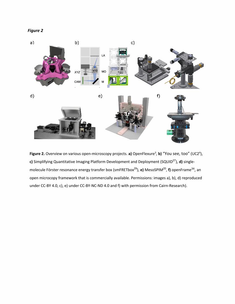
Figure 2
Figure 2. Overview on various open-microscopy projects. a) OpenFlexure3, b) “You see, too” (UC24),
c) Simplifying Quantitative Imaging Platform Development and Deployment (SQUID27), d) single-
molecule Förster resonance energy transfer box (smFRETbox29), e) MesoSPIM20, f) openFrame34, an
open microcopy framework that is commercially available. Permissions: images a), b), d) reproduced
under CC-BY 4.0, c), e) under CC-BY-NC-ND 4.0 and f) with permission from Cairn-Research).