Oosome formation in two ichneumonid wasps
8
TISSUE AND CELL, 1993 25 (1) 121-128 0 1993 Longman Group UK Ltd. JERZY KLAG and SZCZEPAN BILIhlSKI OOSOME FORMATION IN TWO ICHNEUMONID WASPS Keywords: Oosome. ovariole, Hymenoptcra. trophocyte. v,itellogencsis ABSTRACT. During oogenesis of two hymenopteran species. Cosrnvconru meridionuror and Lissonora carenafor, the place of origin and the ways of transport of two oosome components have been shown. The first constituent is produced in the trophocytes cytoplasm as the nuage- mitochondria complexes (NM complexes). The NM complexes subsequently are transported uia intercellular bridges to the oocyte and along the oolcmma to its posterior pole. At the same time, near the posterior pole, the other structures connected with the oosome formation appear. Most probably they are produced in the ooplasm and have the appearance of wage of moderate density (NMD). The two structures are composed of proteins and do not contain RNA (toluidine blue test). The NM complexes as well as the NMD disappear as the definite oosorne forms at the posterior pole of the oocyte. At the same time the oosome becomes RNA positive. It is suggested that the oocytc nucleus is not involved in the process of the oosome formation Introduction At the posterior pole of many insect eggs, especially those formed in polytrophic ovari- oles a characteristic structure termed oosome or pole plasm (in many dipterans e.g. Dro- sophila, composed of granules called polar granules) has been described. The structure was already known in the 19th century and was believed to be involved in germ cell determination (Beams and Kessel, 1974; Eddy, 1975; Nieuwkoop and Satasuria, 1981). Recent biochemical and genetic work has confirmed these suppositions and shows that there are several components needed for germ cells to be successfully determined (Ueda and Okada, 1982; Okada, 1986,198s; Kobayashi et al., 1988; Okada and Kobay- ashi, 1987 a, b; Junquera, 1983; Togasi et al., 1986). These components are synthesized during oogenesis and asymmetrically seg- regated within the oocyte to the posterior egg pole (Mahowald, 1971 a, b, 1972, 1975; Boswell etal., 1991; Nusslein-Volhard, 1991). In the case of polytrophic ovarioles there Department of Systematic Zoology, Jagiellonian Uni- versity, lngardena 6, 30-060 Krakow, Poland. Received 26 June 1992 Revised 7 July 1992 121 are many facts pointing to the possibility that oosome constituents are produced in the trophocytes and subsequently transferred to the oocyte (Mahowald, 1975, 1977; Mahowald et al., 1979; Gutzeit, 1985; Hay el al., 1988 a, b). Recently, the abundant genetic work on Drosophila maternal genes has confirmed the trophocytes’ involvement in both production and localization of the pole plasm con- stituents (Wang and Lehmann 1991; Lehmann and Nusslein-Volhard, 1986, 1991; Kim-Ha et al., 1991; Lasko and Ashburner. 1990). Many authors also studied oogenesis in polytrophic ovarioles of other Diptera (Bier, 1963 a, b; Mahowald and Stoiber, 1974; Gut- zeit, 1985; Hay et ul., 1988b) using electron microscope. They have shown that in all species studied the nurse cell nuclear envel- opes are surrounded by large accumulations of fibrous material. The fine structure of this material is similar to that of the oosome. It has been shown. for example, that in the nurse cell cytoplasm of Miustor (Mahowald and Stoiber, 1974), the fibrous material becomes attached to the mitochondria. Simi- lar complexes were observed during later stages in the oosome region. During oog- enesis of Brudysiu tritici, the formation of the oosome begins only after the phase of
-
Upload
independent -
Category
Documents
-
view
1 -
download
0
Transcript of Oosome formation in two ichneumonid wasps

TISSUE AND CELL, 1993 25 (1) 121-128 0 1993 Longman Group UK Ltd.
JERZY KLAG and SZCZEPAN BILIhlSKI
OOSOME FORMATION IN TWO ICHNEUMONID WASPS
Keywords: Oosome. ovariole, Hymenoptcra. trophocyte. v,itellogencsis
ABSTRACT. During oogenesis of two hymenopteran species. Cosrnvconru meridionuror and Lissonora carenafor, the place of origin and the ways of transport of two oosome components have been shown. The first constituent is produced in the trophocytes cytoplasm as the nuage- mitochondria complexes (NM complexes). The NM complexes subsequently are transported uia intercellular bridges to the oocyte and along the oolcmma to its posterior pole. At the same time, near the posterior pole, the other structures connected with the oosome formation appear. Most probably they are produced in the ooplasm and have the appearance of wage of moderate density (NMD). The two structures are composed of proteins and do not contain RNA (toluidine blue test). The NM complexes as well as the NMD disappear as the definite oosorne forms at the posterior pole of the oocyte. At the same time the oosome becomes RNA positive. It is suggested that the oocytc nucleus is not involved in the process of the oosome formation
Introduction
At the posterior pole of many insect eggs, especially those formed in polytrophic ovari- oles a characteristic structure termed oosome or pole plasm (in many dipterans e.g. Dro- sophila, composed of granules called polar granules) has been described. The structure was already known in the 19th century and was believed to be involved in germ cell determination (Beams and Kessel, 1974; Eddy, 1975; Nieuwkoop and Satasuria, 1981). Recent biochemical and genetic work has confirmed these suppositions and shows that there are several components needed for germ cells to be successfully determined (Ueda and Okada, 1982; Okada, 1986,198s; Kobayashi et al., 1988; Okada and Kobay- ashi, 1987 a, b; Junquera, 1983; Togasi et al., 1986). These components are synthesized during oogenesis and asymmetrically seg- regated within the oocyte to the posterior egg pole (Mahowald, 1971 a, b, 1972, 1975; Boswell etal., 1991; Nusslein-Volhard, 1991). In the case of polytrophic ovarioles there
Department of Systematic Zoology, Jagiellonian Uni- versity, lngardena 6, 30-060 Krakow, Poland.
Received 26 June 1992 Revised 7 July 1992
121
are many facts pointing to the possibility that oosome constituents are produced in the trophocytes and subsequently transferred to the oocyte (Mahowald, 1975, 1977; Mahowald et al., 1979; Gutzeit, 1985; Hay el al., 1988 a, b).
Recently, the abundant genetic work on Drosophila maternal genes has confirmed the trophocytes’ involvement in both production and localization of the pole plasm con- stituents (Wang and Lehmann 1991; Lehmann and Nusslein-Volhard, 1986, 1991; Kim-Ha et al., 1991; Lasko and Ashburner. 1990).
Many authors also studied oogenesis in polytrophic ovarioles of other Diptera (Bier, 1963 a, b; Mahowald and Stoiber, 1974; Gut- zeit, 1985; Hay et ul., 1988b) using electron microscope. They have shown that in all species studied the nurse cell nuclear envel- opes are surrounded by large accumulations of fibrous material. The fine structure of this material is similar to that of the oosome.
It has been shown. for example, that in the nurse cell cytoplasm of Miustor (Mahowald and Stoiber, 1974), the fibrous material becomes attached to the mitochondria. Simi- lar complexes were observed during later stages in the oosome region. During oog- enesis of Brudysiu tritici, the formation of the oosome begins only after the phase of

122
maximal nurse cell activity when the single nurse cell has largely regressed. By autoradiography Gutzeit (1985)) has shown that in the species studied the oosome material is synthesised in the trophocyte cyto- plasm long before the oosome is formed. In the subsequent stages of oogenesis the labelled material was incorporated into the oosome. The same results were obtained by Bier (1963, a: b) studying Musca domestica oogenesis.
The plausible hypothesis is that this material was transported to the oocyte and then accumulated at the posterior tip of the egg. What is more, the oosome failed to form in the presence of a drug disturbing the cytoskeleton. However, it was not possible to show any direct morphological evidence for transport of this material to the posterior pole of the oocyte.
Apparently, the eggs of Diptera are not very suitable for studying oosome formation. Large amounts of yolk accumulated at the same time in the oocyte cytoplasm makes it impossible to follow such components as small accumulations of nuage material.
Instead, Meng (1968), describing the for- mation of the oosome in the hymenopteran Pimpla turionellae, found by light micro- scopy, a thin stream of the material starting midway in the oocyte and stretching to the oosome region at the posterior tip. This find- ing suggested hymenopterans as more suit- able for search for morphological traces of this kind of transport.
For this reason, we have chosen for our studies two hymenopteran species of the fam- ily Ichneumonidae. The members of this fam- ily lay their eggs in the bodies of other insects and therefore the eggs accumulate very little yolk. Thanks to this, they constitute very convenient material to study differentiation of the egg cytoplasm during oogenesis.
KLAG AND BILI&‘SKI
acetone, the material was embedded in Epon 812. To facilitate precise sectioning, prior to embedding in the resin the ovaries were divided into individual ovarioles. Semithin Epon sections were stained with 1% methyl- ene blue in 1% borax. For RNA test the material was embedded in JB4 resin and cut into 5,um sections which were eventually stained with toluidine blue in pH5.0. Both Epon and JB4 sections were examined in a Jenalumar (Zeiss Jena) microscope. Ultra- thin sections were contrasted with uranyl acetate and lead citrate and examined in a Tesla BS 500 or JEOL 1OOC electron micro- scopes.
Materials and Methods
Specimens of Cosmoconus meridionator and Lissonota catenator were collected in Bieszczady National Park in June and July. Dissected ovaries were fixed in 2.5% glutar- aldehyde in 0.1 M phosphate buffer, pH 7.4, at room temperature. The specimens were then rinsed and post-fixed in 1% osmium tetroxide in the same buffer. After dehy- dration in a graded series of ethanol and
Results
The ovaries of Ichneumonidae are of the meroistic polytrophic type. They are com- posed of a dozen or so tube-like ovarioles. Each ovariole is differentiated into an anterior germarium and a posterior vit- ellarium which contains &lo egg chambers in a linear arrangement. The individual egg chamber contains an oocyte and 31 tro- phocytes (nurse cells) connected to the oocyte via 5 intercellular bridges. With the exception of the youngest chambers the con- tact of the oocyte with accompanying nurse cells is ensured by a cytoplasmic process, named by Cassidy and King (1972) the ‘nutri- tive appendix’. Each egg chamber is sur- rounded by a layer of follicular cells which do not show any regional differentiation throughout the oogenesis.
To facilitate the description of oogenesis Bilidski (1991) defined, for C. meridionator, six stages easily recognizable under the light microscope. Stages 1-3, 4-5, and 6, cor- respond to previtellogenic growth, yolk uptake (vitellogenesis) and the formation of egg envelopes, respectively. In the present paper the same division has been accepted for C. meridionator and L. catenator. However, we shall terminate the description at stage 5 because the formation of the oosome is already completed by this stage. Germarial pro-oocytes and pro-trophocytes are not considered in this staging.
The oogenesis and oosome formation in C. meridionator and L. catenator are so simi- lar that the description of these processes concerns both species unless clearly speci- fied.

OOSOME FORMATION
Stage I. Early previtellogenesis
The egg chambers in this stage are charac- terized by 31 small trophocytes and a slightly larger oocyte. The trophocytes are iso- diametric, with lucent cytoplasm and round nuclei. The cisternae of endoplasmic retic- ulum, mitochondria, dictyosomes are not numerous but the trophocyte cytoplasm also contains free ribosomes and polyribosomes. In the vicinity of nuclei small masses of elec- tron-opaque substances, not bounded by a membrane usually referred to as ‘nuage’ ( for the morphology, composition and origin of this structure see Beams and Kessel 1974, Eddy 1975) can be seen (Fig. 1).
The oocyte has a pear-like shape and is tightly surrounded by a layer of follicle cells. A comparatively large, spherical nucleus (germinal vesicle) occupies the oocyte centre. In the oocyte cytoplasm numerous, heavily stained bodies of various dimensions can be observed under the light microscope. The bodies are scattered throughout all the cytoplasm (Fig. 2).
The heavily stained bodies have the appearance of lysosomes and multilamellar bodies (Fig. 7). Beside the lysosomal bodies there is a small number of mitochondria, ribosomes and few dictyosomes scattered in the cytoplasm. There is no ‘nuage’ in the oocyte cytoplasm at this stage.
1113
Stage 3. Late previtellogenesis
The trophocytes are now larger and have larger nuclei surrounded by abundant masses of ‘nuage’ of different density. In the trophocyte cytoplasm the NM complexes have become rare.
At the same time, displacement of the organelles in the oocyte cytoplasm continues. The number of lipid droplets has greatly increased and these now occupy the posterior half of the oocyte. They are located per- ipherally and surround comparatively empty ‘central’ ooplasm. The dense granules accumulate at the posterior pole where they form a lens-shaped aggregation. Single lipid droplets are intermingled with the dense bodies (Fig. 7). In contrast. the anterior half of the oocyte contains only elements of rough endoplasmic reticulum, mitochondria and scarce, isolated lipid droplets. Moreover, at the beginning of this stage, in the cytoplasm of the nutritive appendix and later in the vicinity of the oocyte surface, the NM com- plexes appear. The oocyte nucleus lies dor- sally, roughly in the mid-length of the oocyte (Fig. 4). In the karyoplasm, structures of varying shapes and density are observed. The nuclear envelop is blebbing, and most prob- ably producing accessory nuclei. However. at no stage did it appear that nuage was exiting the oocyte nucleus or situated in its vicinity.
Sections stained with toluidine blue show no traces of RNA at the posterior pole of the oocytes in this stage. The accumulation of the dense bodies is apparently RNA negative
(Fig. 4).
Stage 2. Mid previtellogenesis
The nuclei of the trophocytes become larger and lose their regular round shape. In the vicinity of the nuclei the ‘nuage’ masses have similar appearance as in the previous stage. The only difference is that the complexes of ‘nuage’ and mitochondria (NM) appear in these cells (Fig. 5). The complexes are never found in the vicinity of nuclei but, a certain distance from them.
In each oocyte at this stage, the nucleus has shifted from the centre to the periphery of the cytoplasm. At the anterior pole facing the nurse cells the nutritive appendix dev- elops and the whole oocyte acquires a more spherical shape. The distribution of the cyto- plasmic organelles does not change but the dense bodies shift from the anterior part of the oocyte to the posterior, and few lipid droplets appear among the dense bodies (Fig. 3).
Stage 4. Vitellogenesis
At the beginning of vitellogenesis the tro- phocytes attain their maximal sizes. Their nuclei are very large and contain prominent nucleoli. They are surrounded by great masses of nuage of different density. However, the NM complexes disappear com- pletely from the trophocytes’ cytoplasm.
During vitellogenesis the pronounced elongation of the anteroposterior axis of the oocyte and the flattening of its dorsal side is taking place. The oocyte nucleus migrates towards the anterior pole and eventually attains its specific anterodorsal position. Ar the beginning of this stage, the dense gran- ules shift sidewards from the posterior pole
KLAG AND BILIrjSKI
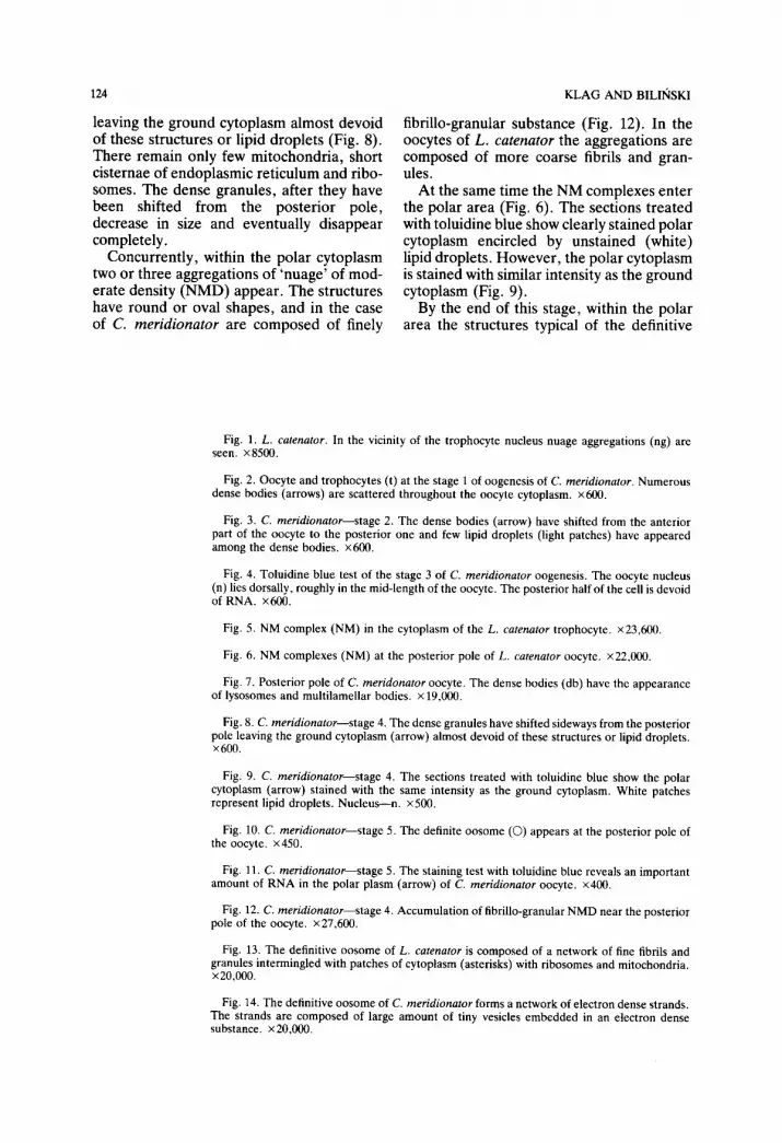
fibrillo-granular substance (Fig. 12). In the oocytes of L. catenator the aggregations are composed of more coarse fibrils and gran- ules.
At the same time the NM complexes enter the polar area (Fig. 6). The sections treated with toluidine blue show clearly stained polar cytoplasm encircled by unstained (white) lipid droplets. However, the polar cytoplasm is stained with similar intensity as the ground cytoplasm (Fig. 9).
By the end of this stage, within the polar area the structures typical of the definitive
leaving the ground cytoplasm almost devoid of these structures or lipid droplets (Fig. 8). There remain only few mitochondria, short cisternae of endoplasmic reticulum and ribo- somes. The dense granules, after they have been shifted from the posterior pole, decrease in size and eventually disappear completely.
Concurrently, within the polar cytoplasm two or three aggregations of ‘nuage’ of mod- erate density (NMD) appear. The structures have round or oval shapes, and in the case of C. meridionator are composed of finely
Fig. 1. L. cafenator. In the vicinity of the trophocyte nucleus nuage aggregations (ng) arc seen. x8500.
Fig. 2. Oocyte and trophocytes (t) at the stage 1 of oogenesis of C. meridionamr. Numerous dense bodies (arrows) are scattered throughout the oocyte cytoplasm. x600.
Fig. 3. C. meridionator-stage 2. The dense bodies (arrow) have shifted from the anterior part of the oocyte to the posterior one and few lipid droplets (light patches) have appeared among the dense bodies. x600.
Fig. 4. Toluidine blue test of the stage 3 of C. meridionator oogenesis. The oocyte nucleus (n) lies dorsally, roughly in the mid-length of the oocyte. The posterior half of the cell is devoid of RNA. x600.
Fig. 5. NM complex (NM) in the cytoplasm of the L. cakmafor trophocyte. ~23,600.
Fig. 6. NM complexes (NM) at the posterior pole of L. cntenator oocyte. x22,ooO.
Fig. 7. Posterior pole of C. meridonntor oocyte. The dense bodies (db) have the appearance of lysosomes and multilamellar bodies. x 19,ooO.
Fig. 8. C. meridionaror-stage 4. The dense granules have shifted sideways from the posterior pole leaving the ground cytoplasm (arrow) almost devoid of these structures or lipid droplets. x600.
Fig. 9. C. meridionator-stage 4. The sections treated with toluidine blue show the polar cytoplasm (arrow) stained with the same intensity as the ground cytoplasm. White patches represent lipid droplets. Nucleus-n. x500.
Fig. 10. C. meridionator-stage 5. The definite oosome (0) appears at the posterior pole of the oocyte. x450.
Fig. 11. C. meridionafor-stage 5. The staining test with toluidine blue reveals an important amount of RNA in the polar plasm (arrow) of C. men’dionator oocyte. x400.
Fig. 12. C. meridionator--stage 4. Accumulation of librillo-granular NMD near the posterior pole of the oocyte. ~27,600.
Fig. 13. The definitive oosome of L. cafenamr is composed of a network of fine fibrils and granules intermingled with patches of cytoplasm (asterisks) with ribosomes and mitochondria. x20,000.
Fig. 14. The definitive oosome of C. meridionator forms a network of electron dense strands. The strands are composed of large amount of tiny vesicles embedded in an electron dense substance. x20,ooO.


126
oosome appear. In the case of C. meri- dionator this comprises strands of electron opaque material filled with tiny vesicles. In the case of L. catenator it is an extensive network of thin fibrils intermingled with granules.
It must be pointed out that in C. meri- dionator as in L. catenator, at the beginning of oosome formation, both the NMD, along with NM complexes completely disappear from the oocytes.
Stage 5. Termination of vitellogenesis
At this stage, the nutritive appendix is being constricted by surrounding follicular cells and the nurse cells are separated from the ooplasm. Soon afterwards the nurse cells undergo progressive degeneration. The fol- licular cells begin the deposition of egg envel- opes around the oocyte surface.
At this stage the oosomes in the eggs of both species studied are fully developed. They form spherical structures occupying the most posterior pole of the eggs (Fig. 10). In C. meridionator, the oosome constitutes a network of electron dense strands. The strands are composed of large amount of tiny vesicles immersed in an electron dense substance. Within the network there are spaces filled with ground cytoplasm with numerous ribosomes and ordinary mito- chondria (Fig. 14). Occasionally, isolated lipid droplets can be seen. In the eggs of L. catenator the oosome has a more compact appearance. It is formed by a continuous mass of tiny fibres associated with numerous tiny granules. Within the mass there are sev- eral isolated islands of the ground cytoplasm with many ribosomes, mitochondria but no lipid droplets (Fig. 13).
Staining with toluidine blue reveals an important increase in the RNA content in the polar plasm (Fig. 11).
KLAG AND BILIhKI
of the nutritive appendix and in the cortical cytoplasm of the oocyte. At the beginning of vitellogenesis they are gathered in fairly large numbers within the polar cytoplasm which appears at that time already devoid of the dense granules.
The second component which probably takes part in the formation of the oosome in the oocytes of the species studied is the NMD. Accumulations of this substance appear at the beginning of vitellogenesis in the polar cytoplasm. They differ in appear- ance both from the NM complexes and from the definitive oosome material. Most prob- ably they originate in the oocyte cytoplasm but not in the oocyte nucleus since, despite careful observation of the latter, we were not able to notice any traces of ‘nuage’ leaving the oocyte nucleus.
Autoradiographic studies of oosome for- mation (Meng, 1970; Gutzeit, 1985) have shown that some of the oosome constituents must be synthesized in the oocyte cytoplasm since the oosome became labelled with tri- tiated aminoacids, even after very short labelling periods. In the case of the two species studied the NMD would correspond to the above component synthesized in the oocyte cytoplasm.
It is noteworthy that when both NM com- plexes and NMD are present within the polar cytoplasm, the staining test for RNA does not show the amount of this substance higher than in the other parts of the ooplasm. As the NM complexes arise in the trophocyte cytoplasm, and NMD in the oocyte cyto- plasm far from the nuclei, they probably constitute only the protein component of the oosome.
Instead, the intensive RNA staining appears in the polar plasm only during vit- ellogenesis when the oosome assumes its definite aspect. We can suppose that by this time the remaining component(s), which we were not able to detect, join the polar plasm and complete the creation of the oosome. Most probably, it is one of the different cat- egories of ‘nuage’ produced in large quan- tities by the trophocyte nuclei and which can be observed around them throughout the oogenesis.
The late appearance of the RNA within the oosome is in agreement with numerous earlier and recent findings (Illmensee, 1976; Mahowald, 1971; Mahowald et al., 1979;
Discussion
In the present paper, for the first time, it was possible to show the site of formation and the ways of transport of at least one of the oosome constituents, namely the NM com- plexes. They appear in the cytoplasm of the trophocytes in the beginning of previt- ellogenic growth of the oocyte. Then they are seen in large numbers in the cytoplasm

OOSOME FORMATION
Golumbeski et al., 1991; St Johnston et al., 1991).
The behaviour of the dense bodies scat- tered throughout the whole cytoplasm of the youngest oocytes is of special interest. Dur- ing oogenesis they gather at the posterior pole just in the place where the future oosome will be formed. Subsequently, the dense granules move apart leaving the ground cytoplasm free of any inclusions. On the basis of present data it is not possible to elucidate the role and ultimate fate of the granules. However, it may be supposed that they represent autolysosomes containing aged organelles in the process of degra- dation. The phenomenon of digestion of cytoplasm constituents in young oocytes is believed to be related to the elimination of the damaged molecules accumulated during embryonic and larva1 life (Medvedev, 1981). The process is considered to be very impor- tant in rejuvenation of the oocyte cytoplasm. before intensive cytoplasmic growth starts.
In conclusion, we describe some obser- vations pointing to the possibility that the
127
first two components of the oosome are pro- teins. One is produced in the trophocyte cytoplasm and transported through nutritive appendix and cortical layer of the oocyte. to the posterior pole of the oocyte in the form of NM complexes. The second protein com- ponent (NMD) is probably produced within the oocyte cytoplasm and segregated to its posterior pole. Both components subsequently take part in the formation of the oosome. Later an RNA component(s) is added during vitellogenesis. and the oosome takes up its definitive appearance. We were not able to follow the transport of an RNA component from the trophocytes but are con- vinced that it does not originate in the oocyte nucleus.
Acknowledgements
This work was supported by Research Grant PB 1868/4/91 from KBN.
References
Beams II. W.. Kcssel R. G. 1974. The problem of germ cell dctcrminants. Inl. Reu. Crfo[. , 39, 413-479.
Bier K. 1963~1. Synthcsc, intercellul%en Transport und Ahbau van Ribonukleinsaurc im Ovar dcr Stubenfliege Mww
domestica. J. Cell Biol.. 16, 436-440.
Bier K. 1963b. Autoradiographische Untersuchungen iibcr die Letstungen dcs Follikclcpithcls und dcr Nahrrellen hcl
der Dotterbildung und EiwciBsynthese im Fliegenovar. Wilhelm Roux’ Arch. 154, 5%S7S.
Bililiski Sz. M. 1991. Morphological markers of anteroposterior and dorsovcntral polarity in developing oocyto ot the
hymenopteran Cosmoconur meridionator (Ichneumonidae). Roux’ Arch. Dro. Biol., 200, 330-335.
Boswell R. E., Prout M., Steichen J. C. lY91, Mutations in a newly idcntttied Drosophila melano~mter gcnc. maqo
nashi. disrupt germ cell formation and result in the formation of mirror-image symmetrical double ahdomcn
embryos. Deoelopment, 113, 373-384.
Cassidy J. D., King R. C. 1972. Ovarian development in Habrobracon ju$mdis (Ashmead) (H~menoptrrut Brrr~w-
idm): I. The origin and differentiation of the oocyte now cell complex. Biol. Bull.. 143, 4X3-505.
Eddy E. M. 1975. Germ plasm and the differentiation of the germ cell line. fnf. Reu. Cyt.. 43, 220-2X1).
Golumbeski G. S.. Bardsley A., Tax F., Bowel1 R. E. 1991, tudor, a posterior-group gent of Drosophdu melmo,qu.wr.
encodes a novel protein and an mRNA localized during mid oogencsis. Genes Dew, 5, 2Mo2070.
Gutzeit H. 0. 1985. Oosome formation during in vitro oogenesis in Bradwin tritici (syn. Sciuru oce//uri.s) Rou.~’ .Irr+~.
Deu. Biol.. 194, 404-410.
[lay B.. Jan L. Y.. Jan Y. N. 198%. A protein component of Drosophila polar granules is encoded by uu.w ,tnd has
extensive sequence similarity to ATP-dependent helicases. Cell, 55, 577-.5X7.
IIay B., Ackerman L.. Barbel S., Jan L. Y., Jan Y. N. 198%. Identification of a component of Drovophi/u polar
granules. Deuelopment. 103, 625-640.
Illmensee K. 1976. Nuclear and cytoplasmic transplantation in Drosophila. In Insect Deoelopment. (cd. P. .A. Lawrence 1 pp. 76-96 Blackwell Sci. Publ., Oxford, London, Melbourne.
Junquera P. 1983. Polar plasm and pole cell formation in an insect egg developing with or without folicular epithelium:
an ultrastructural study. J. Expd Zool., 227, 44-452.
Kim-Ha J., Smith J. L.. Macdonald P. M. 1991. oskar mRNA is localized to the poatcrior pole of the Drosophila
oocytc. Cell, 66, 2%35.

128 KLAG AND BILIr;lSKI
Kobayashi S., Mizuno H.. Okada M. 1988. Accumulation and spatial distribution of poly (A)+ RNA in oocytes and
early embryos of Drosophila melanogaster. Deuelop. Growth Differ., 30, 251-260.
Lasko P. F.. Ashburner M. 1990. Posterior localization of vasa protein correlates with, but is not sufficient for, pole
cell development Genes Deuelop., 4, 905-921.
Lehman R., Nusslein-Volhard C. 1986. Abdominal segmentation. pole cell formation and embryonic polarity require
the localized activity of oskar. a maternal gene in Drosophila. Cell, 47, 141-115.
Lehmann R., Nusslein-Volhard C. 1991. The maternal gene nanos has a central role in posterior pattern formation of
the Drosophila embryo. Deuelopment. 112, 679-691.
Mahowald A. P. 1971a. Origin and continuity of polar granules. In Origin and cominuify of cell organelles (eds. .I.
Reinert and H. Ursprung). pp. 158-169. Springer Verlag, New York.
Mahowald A. P. 1971b. Polar granules of Drosophila. III. The continuity of polar granules during the life cycle of
Drosophila. J. Expd Zoo/. , 176, 329-344.
Mahowald A. P. 1972. Oogenesis. In Developmental systems: Insects (eds. S. J. Counce and C. H. Waddington), pp. 1-47. Academic Press. London and New York.
Mahowald A. P. 1975. Ultrastructural changes in the germ plasm during the life cycle of Miasror (Cecidomyidae, Diptera). Wilhelm Roux’ Archio, 176, 223-240.
Mahowald A. P. 1977. The germ plasm of Drosophila: an experimental system for the analysis of determination.
Amer. Zool., 17, 551-563.
Mahowald A. P., Stoiber D. 1974. The origin of the nurse chamber in ovaries of Miastor (Diptera: Cecidomyidae) Wilhelm Row;’ Archiu. 176, 159-166.
Mahowald A. P., Allis C.. Karrer K., Underwood E., Waring G. 1979. Germ plasm and poll cells of Drosophikz. In
Determinanrs ofsparial orgonisarion (eds. S. Subtelny and I. Konigsberg. pp. 127-146. Academic Press. London
and New York.
Medvedev Z. 1981. On the immortality of the germ line: genetic and biochemical mechanisms. A review. Mech. Ageing
Deu., 17, 31-359.
Meng C. 1968. Strukturwandel und histochemische Befunde inbesondere am Oosom wahrend der Oogencse und nach
der Ablagc des Eies van Pimpia turionellae L. (Hymenop~era. Ichneumonidae). Wilhelm Roux’ Arch., 161, 162- 208.
Meng C. 1970 Autoradiographische Untcrsuchungen am Oosome im der Oocyte van Pimpb rurronellae L. (Hyme- noprera). Wilhelm Roux’ Arch., 165, 35-52.
Nieuwkoop P., Sytasurya L. 1981. Primordial germ cells in the invertebrates. Cambridge University Press.
Nusslein-Volhard C. 1991. Determination of the embryonic axes of Drosophikt. Deoebpment Supplemenr, 1, I-10.
Okada M. 1986. Cytoplasmic function segregating germ line in Drosophila embryogenesis. Zoo/. Sci., 3, 573-583.
Okada M. 1988. Developmental and molecular analysis of ooplasmic factors required for pole cell formation. Zoo/.
Sci. 5, 1191.
Okada M., Kobayashi S. 1987a. Regulation of pole cell formation and germ cell determination by cytoplasmic factors
in Drosophila embryogenesis. Gunma Symposia on Endocrinology. 24, 40. Okada M., Kobayashi S. 1987b. Maternal messenger RNA as a determinant of pole cell formation in Drosophib
embryos. Develop. Growth. Differ., 29, 185-192. St Johnston D., Beuchle D., Nusslein-Volhard C. 1991. sraufen, a gene required to localize maternal RNAs in the
Drosophila egg. cell, 46, 51-63. Togasi S., Kobayashi S.. Okada M. 1986. Functions of maternal mRNA as a cytoplasmic factor responsible for pole
cell formation in Drosophila embryos. Deal Eiol. 118, 352-360.
Ueda R. and Okada M. 1982. Induction of pole cells in sterilized Drosophila embryos by injection of subcellular
fraction from eggs. Proc. Nad. Acad. Sci. USA 79, 694&6950. Wang C., Lehman” R. 1991. nn~xx is the localized posterior determinant in Drosophila Cell, 66, 637-647.