One Hundred Consecutive Patients Treated with Magnetic Sphincter Augmentation for Gastroesophageal...
9
One Hundred Consecutive Patients Treated with Magnetic Sphincter Augmentation for Gastroesophageal Reflux Disease: 6 Years of Clinical Experience from a Single Center Luigi Bonavina, MD, FACS, Greta Saino, MD, Davide Bona, MD, Andrea Sironi, MD, Veronica Lazzari, MD BACKGROUND: This study was undertaken to evaluate our clinical experience during a 6-year period with an implantable device that augments the lower esophageal sphincter for gastroesophageal reflux disease (GERD). The device uses magnetic sphincter augmentation (MSA) to strengthen the antireflux barrier. STUDY DESIGN: In a single-center, prospective case series, 100 consecutive patients underwent laparoscopic MSA for GERD between March 2007 and February 2012. Clinical outcomes for each patient were tracked post implantation and compared with presurgical data for esophageal pH measurements, symptom scores, and proton pump inhibitor (PPI) use. RESULTS: Median implant duration was 3 years (range 378 days to 6 years). Median total acid exposure time was reduced from 8.0% before implant to 3.2% post implant (p < 0.001). The median GERD Health Related Quality of Life score at baseline was 16 on PPIs and 24 off PPIs and improved to a score of 2 (p < 0.001). Freedom from daily dependence on PPIs was achieved in 85% of patients. There have been no long-term complications, such as device migrations or erosions. Three patients had the device laparoscopically removed for persistent GERD, odynophagia, or dysphagia, with subsequent resolution of symptoms. CONCLUSIONS: Magnetic sphincter augmentation for GERD in clinical practice provides safe and long-term reduction of esophageal acid exposure, substantial symptom improvement, and elimination of daily PPI use. For candidates of antireflux surgery who have been carefully evaluated before surgery to confirm indication for MSA, MSA has become a standard treatment at our institution because control of reflux symptoms and pH normalization can be achieved with minimal side effects and preservation of gastric anatomy. (J Am Coll Surg 2013;217: 577e585. Ó 2013 by the American College of Surgeons) Gastroesophageal reflux disease (GERD) is a common foregut disorder that increases health care use and adversely impacts quality of life by disturbing sleep, reducing productivity, and interfering with daily activities. 1-3 Ther- apeutic approaches include medical management with acid suppression therapy, which reduces esophageal acid expo- sure to alleviate acid reflux symptoms; and antireflux surgery, which corrects the mechanically defective lower esophageal sphincter (LES) to improve the antireflux barrier and prevent acidic and nonacidic gastric juices from refluxing into the esophagus. 4 Antireflux surgery is an alternative to long-term medical therapy and has been shown to be more effective at controlling reflux symp- toms. 5,6 Laparoscopic fundoplication is the accepted stan- dard of antireflux surgery, but it relies on considerable dissection and remodeling of the gastric and hiatal anatomy to restore competence to the LES. 7 Magnetic sphincter augmentation (MSA) with the LINX Reflux Management System (Torax Medical) is a minimally invasive alternative to fundoplication and is indicated for patients with abnormal esophageal acid exposure Disclosure Information: Dr Bonavina received fees as a consultant for Torax Medical, Inc. All other authors have nothing to declare. This study was funded in part by Torax Medical, Inc. Received March 8, 2013; Revised April 30, 2013; Accepted April 30, 2013. From the Department of Biomedical Sciences for Health, University of Milano Medical School, Milan, Italy. Correspondence address: Luigi Bonavina, MD, FACS, Department of Biomedical Sciences for Health, University of Milano Medical School, Division of General Surgery, IRCCS Policlinico San Donato, Via Morandi 30 20097, San Donato Milanese, Milan, Italy. email: luigi.bonavina@ unimi.it 577 ª 2013 by the American College of Surgeons ISSN 1072-7515/13/$36.00 Published by Elsevier Inc. http://dx.doi.org/10.1016/j.jamcollsurg.2013.04.039
Transcript of One Hundred Consecutive Patients Treated with Magnetic Sphincter Augmentation for Gastroesophageal...
One Hundred Consecutive Patients Treatedwith Magnetic Sphincter Augmentation forGastroesophageal Reflux Disease: 6 Yearsof Clinical Experience from a Single Center
Luigi Bonavina, MD, FACS, Greta Saino, MD, Davide Bona, MD, Andrea Sironi, MD,Veronica Lazzari, MD
BACKGROUND: This study was undertaken to evaluate our clinical experience during a 6-year period with animplantable device that augments the lower esophageal sphincter for gastroesophageal refluxdisease (GERD). The device uses magnetic sphincter augmentation (MSA) to strengthen theantireflux barrier.
STUDY DESIGN: In a single-center, prospective case series, 100 consecutive patients underwent laparoscopicMSA for GERD between March 2007 and February 2012. Clinical outcomes for each patientwere tracked post implantation and compared with presurgical data for esophageal pHmeasurements, symptom scores, and proton pump inhibitor (PPI) use.
RESULTS: Median implant duration was 3 years (range 378 days to 6 years). Median total acid exposuretime was reduced from 8.0% before implant to 3.2% post implant (p < 0.001). The medianGERD Health Related Quality of Life score at baseline was 16 on PPIs and 24 off PPIs andimproved to a score of 2 (p < 0.001). Freedom from daily dependence on PPIs was achievedin 85% of patients. There have been no long-term complications, such as device migrationsor erosions. Three patients had the device laparoscopically removed for persistent GERD,odynophagia, or dysphagia, with subsequent resolution of symptoms.
CONCLUSIONS: Magnetic sphincter augmentation for GERD in clinical practice provides safe and long-termreduction of esophageal acid exposure, substantial symptom improvement, and elimination ofdaily PPI use. For candidates of antireflux surgery who have been carefully evaluated beforesurgery to confirm indication for MSA, MSA has become a standard treatment at ourinstitution because control of reflux symptoms and pH normalization can be achieved withminimal side effects and preservation of gastric anatomy. (J Am Coll Surg 2013;217:577e585. � 2013 by the American College of Surgeons)
Gastroesophageal reflux disease (GERD) is a commonforegut disorder that increases health care use and adverselyimpacts quality of life by disturbing sleep, reducingproductivity, and interfering with daily activities.1-3 Ther-apeutic approaches include medical management with acid
Disclosure Information: Dr Bonavina received fees as a consultant forTorax Medical, Inc. All other authors have nothing to declare. This studywas funded in part by Torax Medical, Inc.
Received March 8, 2013; Revised April 30, 2013; Accepted April 30, 2013.From the Department of Biomedical Sciences for Health, University ofMilano Medical School, Milan, Italy.Correspondence address: Luigi Bonavina, MD, FACS, Department ofBiomedical Sciences for Health, University of Milano Medical School,Division of General Surgery, IRCCS Policlinico San Donato, Via Morandi30 20097, San Donato Milanese, Milan, Italy. email: [email protected]
577ª 2013 by the American College of Surgeons
Published by Elsevier Inc.
suppression therapy, which reduces esophageal acid expo-sure to alleviate acid reflux symptoms; and antirefluxsurgery, which corrects the mechanically defective loweresophageal sphincter (LES) to improve the antirefluxbarrier and prevent acidic and nonacidic gastric juicesfrom refluxing into the esophagus.4 Antireflux surgery isan alternative to long-term medical therapy and has beenshown to be more effective at controlling reflux symp-toms.5,6 Laparoscopic fundoplication is the accepted stan-dard of antireflux surgery, but it relies on considerabledissection and remodeling of the gastric and hiatalanatomy to restore competence to the LES.7 Magneticsphincter augmentation (MSA) with the LINX RefluxManagement System (Torax Medical) is a minimallyinvasive alternative to fundoplication and is indicatedfor patients with abnormal esophageal acid exposure
ISSN 1072-7515/13/$36.00
http://dx.doi.org/10.1016/j.jamcollsurg.2013.04.039

Abbreviations and Acronyms
GEJ ¼ gastroesophageal junctionGERD ¼ gastroesophageal reflux diseaseHRQL ¼ Health Related Quality of LifeLES ¼ lower esophageal sphincterMSA ¼ magnetic sphincter augmentationPPI ¼ proton pump inhibitor
578 Bonavina et al Magnetic Sphincter Augmentation J Am Coll Surg
confirmed by esophageal pH testing and with chronicsymptoms despite maximum medical therapy. The device,a series of magnetic beads connected to each other by inde-pendent wires, is implanted via standard laparoscopic tech-niques around the external tubular esophagus in the area ofthe LES using minimal dissection technique and withoutaltering the gastric anatomy. We have been using MSAin our clinical practice for 6 years.8,9 We report the clinicaloutcomes for 100 consecutive patients treated at a singlecenter to provide insights from our long-term experience.
METHODS
Study population
Between March 2007 and February 2012, 100 patientsunderwent MSA for GERD via laparoscopic surgery atour institution. Patients 1 through 30 (30%) underwentthe implantation procedure between March 2007 andMay 2008 as part of a multicenter pilot study.10 Patients31 through 100 (70%) underwent the implantation proce-dure between December 2009 and February 2012 as partof a registry. Patient selection for the pilot study was perprotocol, and patient selection in clinical practice wasguided by the manufacturer’s labeling and our experiencefrom the pilot study. Patients were considered for MSA ifthey were older than18 years of age, had GERD for at least6 months, had persistent reflux symptoms despite dailyproton pump inhibitors (PPIs), and pathologic refluxwas confirmed by ambulatory esophageal pH monitoring.In the pilot study, patients were excluded if presurgicalscreening revealed a hernia �3 cm between the diaphrag-matic impression and the top of the gastric rugal folds;erosive esophagitis grade B, C, or D (Los Angeles Classifi-cation); body mass index >35; Barrett’s esophagus;motility disorders; gross esophageal anatomic abnormali-ties; or a known allergy to titanium, stainless steel, nickel,or ferrous materials. Exclusion criteria from the pilot studywere largely applied in clinical practice. The protocol forthe pivotal study of MSA allowed hiatal hernias of up to3 cm as well as grade A and B esophagitis, which we adop-ted as part of our screening criteria in clinical practice.Before surgery, all patients completed esophageal pH
testing off PPIs, upper endoscopy, manometry/motility,and a barium esophagram. Patients were excluded from
device implant in the pilot study and in clinical practiceif manometry showed effective swallows were <70%and the distal amplitude was <35 mmHg. Patientswith a known allergy to titanium, stainless steel, nickel,or ferrous materials are contraindicated for MSA.
Study design
This single-center, prospective case series of consecutivepatients evaluated MSA for GERD. Patients were fol-lowed after surgery to evaluate the device’s treatmenteffect and safety. Clinical outcomes at annual intervalsfor each patient were tracked post implant and comparedwith presurgical data for esophageal pH measurements,symptom scores, and PPI use. Study evaluations wereapproved by the Ethics Committee at our institution,and written informed consent was obtained from thepatients.
Patient assessment
All baseline and follow-up data were collected bya surgeon during a telephone call with the patient orduring a visit. The assessments for PPI use and non-GERD Health Related Quality of Life (HRQL) symp-toms were captured on standardized data collectionforms. The GERD-HRQL, a validated assessment tool,was used to evaluate symptoms before and after surgery.11
This questionnaire consists of a total of 10 questionsscored on a scale of 0 to 5 (ranging from 0 ¼ no symp-toms to 5 ¼ incapacitating symptoms) and 1 nonscoredquestion about satisfaction with present condition (satis-fied, neutral, or dissatisfied). Six questions pertain specif-ically to heartburn; 3 questions pertain to the side effectsof dysphagia, odynophagia, and gas bloat; and 1 questionpertains to medication use (Fig. 1). At baseline, 30patients completed the GERD-HRQL when off PPIsand 70 completed it when on PPIs. After surgery, theGERD-HRQL score off PPIs at last follow-up wascompared with the total score at baseline and determinedto be significant if at least a 50% reduction in score hadbeen achieved.All patients required daily PPIs; therefore, discontinu-
ation of daily PPIs after surgery was considered a measureof success. Additionally, patients were queried before andafter surgery for the presence of regurgitation, extra-esophageal symptoms, and for the ability to belch orvomit. The pilot study required esophageal pH testingat multiple time points after surgery. Results are reportedas of the last follow-up the patient completed for esoph-ageal pH monitoring.A 13-item questionnaire designed to collect informa-
tion about the referral pathway, patient’s perception,and patient’s satisfaction was administered. The survey

Figure 1. GastroEsophageal Reflux DiseaseeHealth RelatedQuality of Life questions and scoring scale.
Vol. 217, No. 4, October 2013 Bonavina et al Magnetic Sphincter Augmentation 579
was done in cooperation with the clinical psychology unitat our institution and was administered only to theregistry patients (n ¼ 70). There were 5 specific questions(out of the 13-item questionnaire) that related to thisstudy:
1. Why did you decide to undergo surgery?2. Where did you learn about LINX, who sent you to us?3. Would you undergo the operation again?4. Would you recommend it to a friend? and5. What have you heard about Nissen fundoplication?
Figure 2. Magnetic sphincter augmentation. Deannular configuration.
Adverse events and complications considered to bepossiblyrelated to the device or procedure were tracked and docu-mented. An adverse event was considered serious if it resultedin death, was life threatening, required prolongation of exist-ing hospitalization or required re-hospitalization, resulted inpersistent or significant disability, required intervention toprevent permanent impairment or damage, or resulted in fetaldistress/death or congenital anatomy or birth defect.
Procedure
All patients underwent MSA using the LINX RefluxManagement System during a laparoscopic procedure.Patients 1 to 30were treatedwith the first-generation devicethat used aTi-Knot Replacement System (LSI Solutions) tosecure the ends of the device around the esophagus. Patients31 to 100 were treated with the second-generation device.The second-generation device is the same as the first-generation device except that the ends of the device aresecured with a clasp instead of suture (Fig. 2). Additionally,sizing of the esophagus for the first 30 patients used a color-coded sizing device of connected beads as described previ-ously.10 With the introduction of the second-generationdevice, a laparoscopic sizing tool was introduced. The prin-ciples of sizing the esophagus remained the same, with theonly difference being the tool used.All procedures were performed by 2 surgeons (LB, GS).
The standard laparoscopic approach to the gastroesophagealjunction (GEJ)was used.Weperformedminimal dissectionto create the space where the device would encircle the LESwhen implanted. The first step sufficiently exposed the GEJby applying PEEP and traction on the stomach. Only theperitoneal reflection should be divided, and the mediastinal
vice with clasp closure to secure ends in
580 Bonavina et al Magnetic Sphincter Augmentation J Am Coll Surg
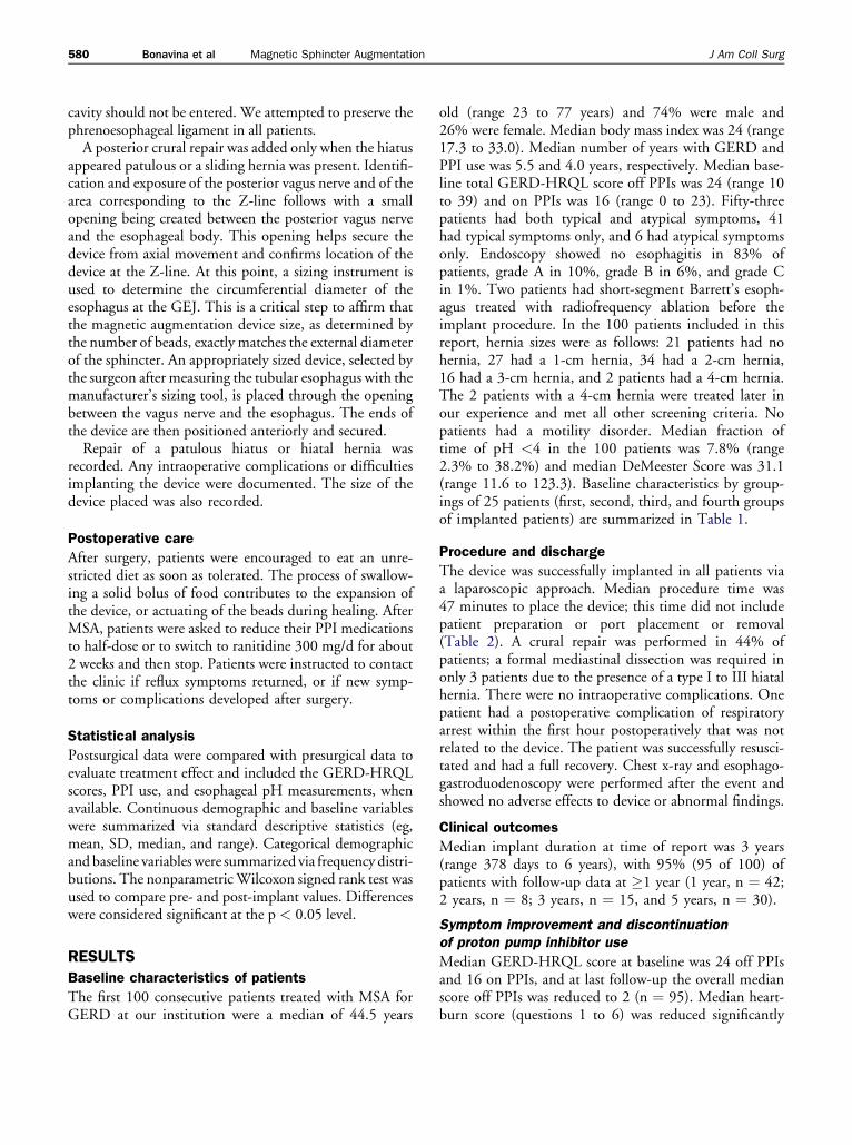
cavity should not be entered. We attempted to preserve thephrenoesophageal ligament in all patients.A posterior crural repair was added only when the hiatus
appeared patulous or a sliding hernia was present. Identifi-cation and exposure of the posterior vagus nerve and of thearea corresponding to the Z-line follows with a smallopening being created between the posterior vagus nerveand the esophageal body. This opening helps secure thedevice from axial movement and confirms location of thedevice at the Z-line. At this point, a sizing instrument isused to determine the circumferential diameter of theesophagus at the GEJ. This is a critical step to affirm thatthe magnetic augmentation device size, as determined bythe number of beads, exactly matches the external diameterof the sphincter. An appropriately sized device, selected bythe surgeon after measuring the tubular esophagus with themanufacturer’s sizing tool, is placed through the openingbetween the vagus nerve and the esophagus. The ends ofthe device are then positioned anteriorly and secured.Repair of a patulous hiatus or hiatal hernia was
recorded. Any intraoperative complications or difficultiesimplanting the device were documented. The size of thedevice placed was also recorded.
Postoperative care
After surgery, patients were encouraged to eat an unre-stricted diet as soon as tolerated. The process of swallow-ing a solid bolus of food contributes to the expansion ofthe device, or actuating of the beads during healing. AfterMSA, patients were asked to reduce their PPI medicationsto half-dose or to switch to ranitidine 300 mg/d for about2 weeks and then stop. Patients were instructed to contactthe clinic if reflux symptoms returned, or if new symp-toms or complications developed after surgery.
Statistical analysis
Postsurgical data were compared with presurgical data toevaluate treatment effect and included the GERD-HRQLscores, PPI use, and esophageal pH measurements, whenavailable. Continuous demographic and baseline variableswere summarized via standard descriptive statistics (eg,mean, SD, median, and range). Categorical demographicandbaseline variableswere summarized via frequency distri-butions. The nonparametricWilcoxon signed rank test wasused to compare pre- and post-implant values. Differenceswere considered significant at the p < 0.05 level.
RESULTS
Baseline characteristics of patients
The first 100 consecutive patients treated with MSA forGERD at our institution were a median of 44.5 years
old (range 23 to 77 years) and 74% were male and26% were female. Median body mass index was 24 (range17.3 to 33.0). Median number of years with GERD andPPI use was 5.5 and 4.0 years, respectively. Median base-line total GERD-HRQL score off PPIs was 24 (range 10to 39) and on PPIs was 16 (range 0 to 23). Fifty-threepatients had both typical and atypical symptoms, 41had typical symptoms only, and 6 had atypical symptomsonly. Endoscopy showed no esophagitis in 83% ofpatients, grade A in 10%, grade B in 6%, and grade Cin 1%. Two patients had short-segment Barrett’s esoph-agus treated with radiofrequency ablation before theimplant procedure. In the 100 patients included in thisreport, hernia sizes were as follows: 21 patients had nohernia, 27 had a 1-cm hernia, 34 had a 2-cm hernia,16 had a 3-cm hernia, and 2 patients had a 4-cm hernia.The 2 patients with a 4-cm hernia were treated later inour experience and met all other screening criteria. Nopatients had a motility disorder. Median fraction oftime of pH <4 in the 100 patients was 7.8% (range2.3% to 38.2%) and median DeMeester Score was 31.1(range 11.6 to 123.3). Baseline characteristics by group-ings of 25 patients (first, second, third, and fourth groupsof implanted patients) are summarized in Table 1.
Procedure and discharge
The device was successfully implanted in all patients viaa laparoscopic approach. Median procedure time was47 minutes to place the device; this time did not includepatient preparation or port placement or removal(Table 2). A crural repair was performed in 44% ofpatients; a formal mediastinal dissection was required inonly 3 patients due to the presence of a type I to III hiatalhernia. There were no intraoperative complications. Onepatient had a postoperative complication of respiratoryarrest within the first hour postoperatively that was notrelated to the device. The patient was successfully resusci-tated and had a full recovery. Chest x-ray and esophago-gastroduodenoscopy were performed after the event andshowed no adverse effects to device or abnormal findings.
Clinical outcomes
Median implant duration at time of report was 3 years(range 378 days to 6 years), with 95% (95 of 100) ofpatients with follow-up data at �1 year (1 year, n ¼ 42;2 years, n ¼ 8; 3 years, n ¼ 15, and 5 years, n ¼ 30).
Symptom improvement and discontinuationof proton pump inhibitor use
Median GERD-HRQL score at baseline was 24 off PPIsand 16 on PPIs, and at last follow-up the overall medianscore off PPIs was reduced to 2 (n ¼ 95). Median heart-burn score (questions 1 to 6) was reduced significantly
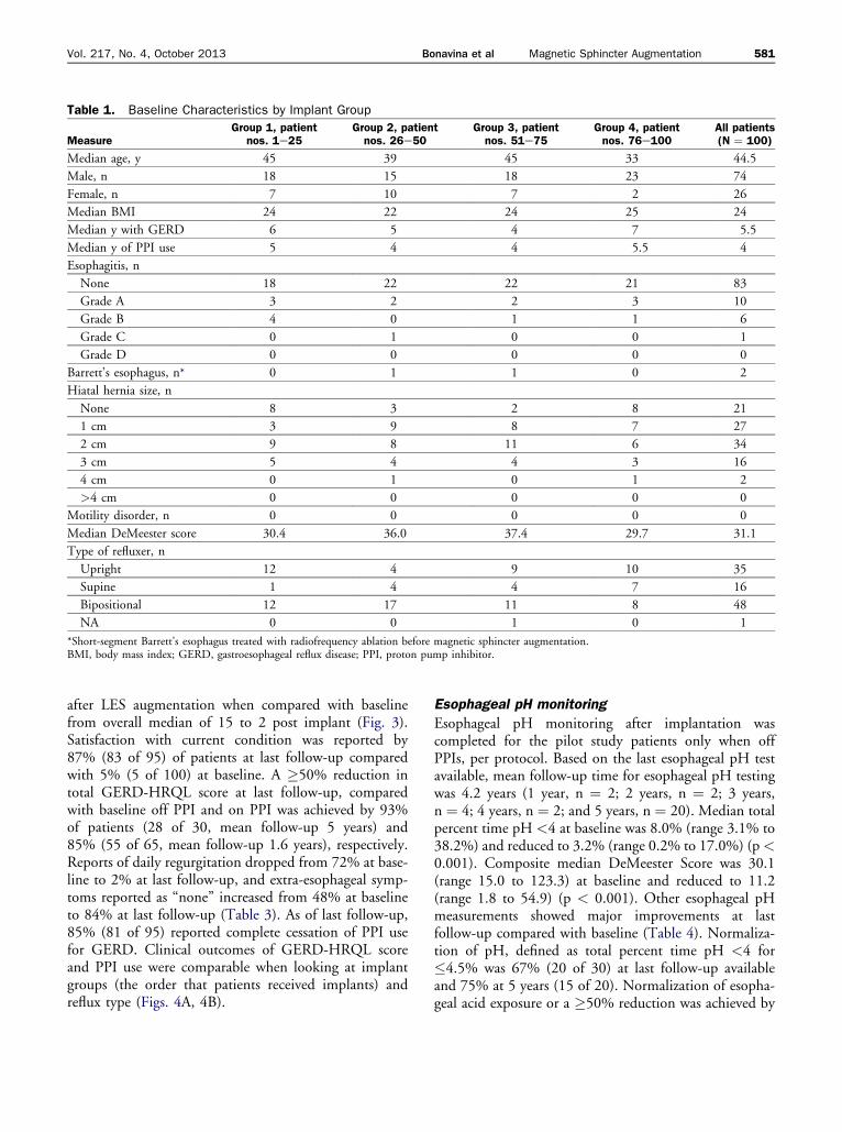
Table 1. Baseline Characteristics by Implant Group
MeasureGroup 1, patient
nos. 1e25Group 2, patientnos. 26e50
Group 3, patientnos. 51e75
Group 4, patientnos. 76e100
All patients(N ¼ 100)
Median age, y 45 39 45 33 44.5
Male, n 18 15 18 23 74
Female, n 7 10 7 2 26
Median BMI 24 22 24 25 24
Median y with GERD 6 5 4 7 5.5
Median y of PPI use 5 4 4 5.5 4
Esophagitis, n
None 18 22 22 21 83
Grade A 3 2 2 3 10
Grade B 4 0 1 1 6
Grade C 0 1 0 0 1
Grade D 0 0 0 0 0
Barrett’s esophagus, n* 0 1 1 0 2
Hiatal hernia size, n
None 8 3 2 8 21
1 cm 3 9 8 7 27
2 cm 9 8 11 6 34
3 cm 5 4 4 3 16
4 cm 0 1 0 1 2
>4 cm 0 0 0 0 0
Motility disorder, n 0 0 0 0 0
Median DeMeester score 30.4 36.0 37.4 29.7 31.1
Type of refluxer, n
Upright 12 4 9 10 35
Supine 1 4 4 7 16
Bipositional 12 17 11 8 48
NA 0 0 1 0 1
*Short-segment Barrett’s esophagus treated with radiofrequency ablation before magnetic sphincter augmentation.BMI, body mass index; GERD, gastroesophageal reflux disease; PPI, proton pump inhibitor.
Vol. 217, No. 4, October 2013 Bonavina et al Magnetic Sphincter Augmentation 581
after LES augmentation when compared with baselinefrom overall median of 15 to 2 post implant (Fig. 3).Satisfaction with current condition was reported by87% (83 of 95) of patients at last follow-up comparedwith 5% (5 of 100) at baseline. A �50% reduction intotal GERD-HRQL score at last follow-up, comparedwith baseline off PPI and on PPI was achieved by 93%of patients (28 of 30, mean follow-up 5 years) and85% (55 of 65, mean follow-up 1.6 years), respectively.Reports of daily regurgitation dropped from 72% at base-line to 2% at last follow-up, and extra-esophageal symp-toms reported as “none” increased from 48% at baselineto 84% at last follow-up (Table 3). As of last follow-up,85% (81 of 95) reported complete cessation of PPI usefor GERD. Clinical outcomes of GERD-HRQL scoreand PPI use were comparable when looking at implantgroups (the order that patients received implants) andreflux type (Figs. 4A, 4B).
Esophageal pH monitoring
Esophageal pH monitoring after implantation wascompleted for the pilot study patients only when offPPIs, per protocol. Based on the last esophageal pH testavailable, mean follow-up time for esophageal pH testingwas 4.2 years (1 year, n ¼ 2; 2 years, n ¼ 2; 3 years,n ¼ 4; 4 years, n ¼ 2; and 5 years, n ¼ 20). Median totalpercent time pH <4 at baseline was 8.0% (range 3.1% to38.2%) and reduced to 3.2% (range 0.2% to 17.0%) (p<0.001). Composite median DeMeester Score was 30.1(range 15.0 to 123.3) at baseline and reduced to 11.2(range 1.8 to 54.9) (p < 0.001). Other esophageal pHmeasurements showed major improvements at lastfollow-up compared with baseline (Table 4). Normaliza-tion of pH, defined as total percent time pH <4 for�4.5% was 67% (20 of 30) at last follow-up availableand 75% at 5 years (15 of 20). Normalization of esopha-geal acid exposure or a �50% reduction was achieved by
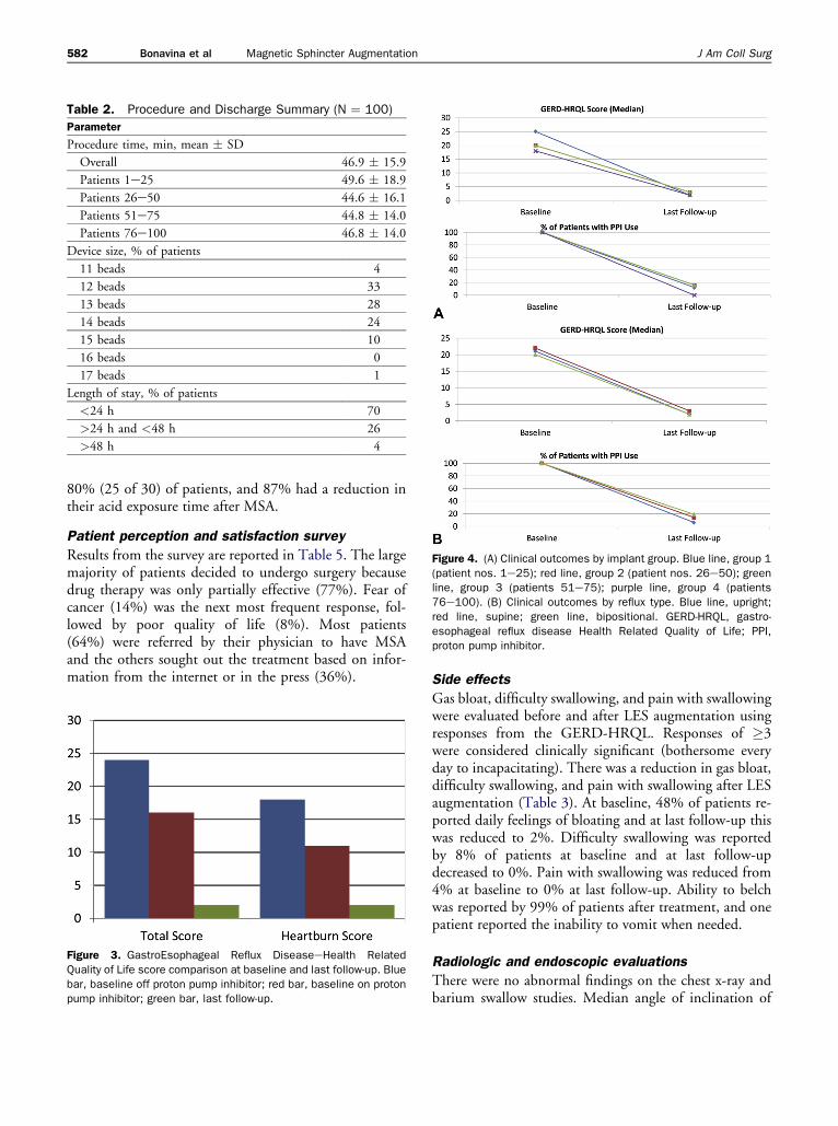
Figure 4. (A) Clinical outcomes by implant group. Blue line, group 1(patient nos. 1e25); red line, group 2 (patient nos. 26e50); greenline, group 3 (patients 51e75); purple line, group 4 (patients76e100). (B) Clinical outcomes by reflux type. Blue line, upright;red line, supine; green line, bipositional. GERD-HRQL, gastro-esophageal reflux disease Health Related Quality of Life; PPI,proton pump inhibitor.
Table 2. Procedure and Discharge Summary (N ¼ 100)
Parameter
Procedure time, min, mean � SD
Overall 46.9 � 15.9
Patients 1e25 49.6 � 18.9
Patients 26e50 44.6 � 16.1
Patients 51e75 44.8 � 14.0
Patients 76e100 46.8 � 14.0
Device size, % of patients
11 beads 4
12 beads 33
13 beads 28
14 beads 24
15 beads 10
16 beads 0
17 beads 1
Length of stay, % of patients
<24 h 70
>24 h and <48 h 26
>48 h 4
582 Bonavina et al Magnetic Sphincter Augmentation J Am Coll Surg
80% (25 of 30) of patients, and 87% had a reduction intheir acid exposure time after MSA.
Patient perception and satisfaction survey
Results from the survey are reported in Table 5. The largemajority of patients decided to undergo surgery becausedrug therapy was only partially effective (77%). Fear ofcancer (14%) was the next most frequent response, fol-lowed by poor quality of life (8%). Most patients(64%) were referred by their physician to have MSAand the others sought out the treatment based on infor-mation from the internet or in the press (36%).
Figure 3. GastroEsophageal Reflux DiseaseeHealth RelatedQuality of Life score comparison at baseline and last follow-up. Bluebar, baseline off proton pump inhibitor; red bar, baseline on protonpump inhibitor; green bar, last follow-up.
Side effects
Gas bloat, difficulty swallowing, and pain with swallowingwere evaluated before and after LES augmentation usingresponses from the GERD-HRQL. Responses of �3were considered clinically significant (bothersome everyday to incapacitating). There was a reduction in gas bloat,difficulty swallowing, and pain with swallowing after LESaugmentation (Table 3). At baseline, 48% of patients re-ported daily feelings of bloating and at last follow-up thiswas reduced to 2%. Difficulty swallowing was reportedby 8% of patients at baseline and at last follow-updecreased to 0%. Pain with swallowing was reduced from4% at baseline to 0% at last follow-up. Ability to belchwas reported by 99% of patients after treatment, and onepatient reported the inability to vomit when needed.
Radiologic and endoscopic evaluations
There were no abnormal findings on the chest x-ray andbarium swallow studies. Median angle of inclination of
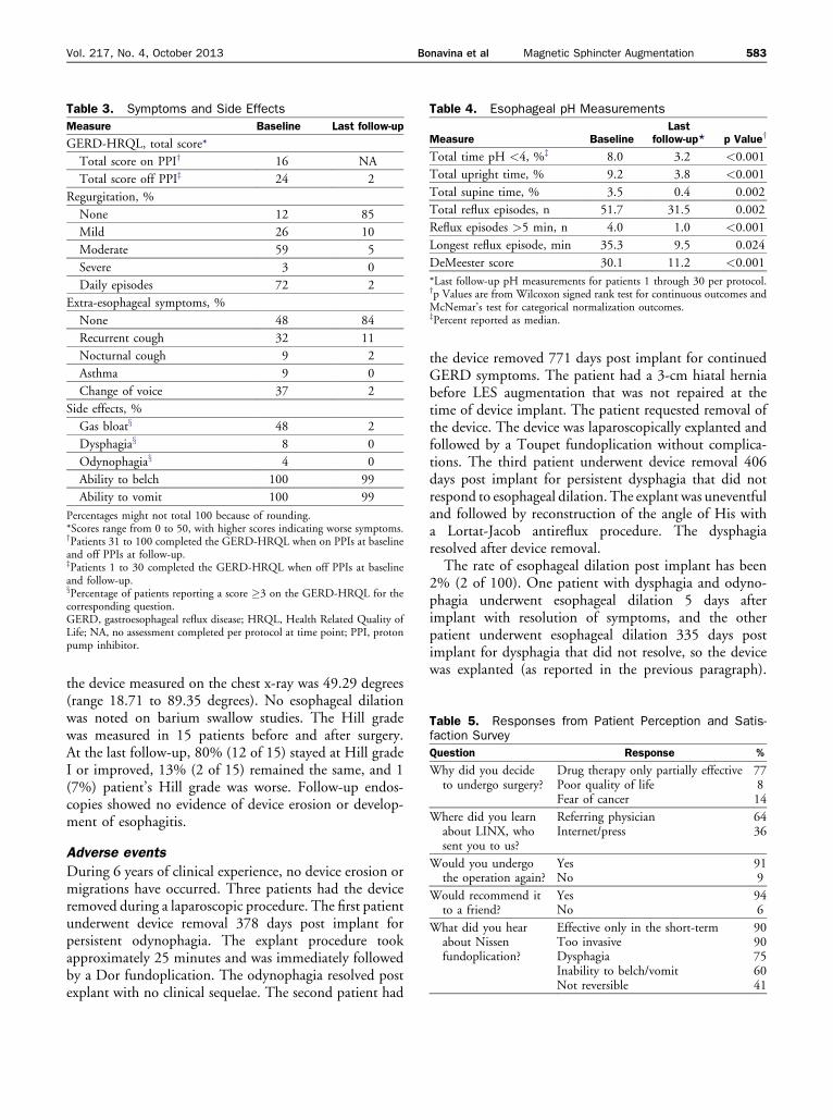
Table 3. Symptoms and Side Effects
Measure Baseline Last follow-up
GERD-HRQL, total score*
Total score on PPIy 16 NA
Total score off PPIz 24 2
Regurgitation, %
None 12 85
Mild 26 10
Moderate 59 5
Severe 3 0
Daily episodes 72 2
Extra-esophageal symptoms, %
None 48 84
Recurrent cough 32 11
Nocturnal cough 9 2
Asthma 9 0
Change of voice 37 2
Side effects, %
Gas bloatx 48 2
Dysphagiax 8 0
Odynophagiax 4 0
Ability to belch 100 99
Ability to vomit 100 99
Percentages might not total 100 because of rounding.*Scores range from 0 to 50, with higher scores indicating worse symptoms.yPatients 31 to 100 completed the GERD-HRQL when on PPIs at baselineand off PPIs at follow-up.zPatients 1 to 30 completed the GERD-HRQL when off PPIs at baselineand follow-up.xPercentage of patients reporting a score �3 on the GERD-HRQL for thecorresponding question.GERD, gastroesophageal reflux disease; HRQL, Health Related Quality ofLife; NA, no assessment completed per protocol at time point; PPI, protonpump inhibitor.
Table 4. Esophageal pH Measurements
Measure BaselineLast
follow-up* p Valuey
Total time pH <4, %z 8.0 3.2 <0.001
Total upright time, % 9.2 3.8 <0.001
Total supine time, % 3.5 0.4 0.002
Total reflux episodes, n 51.7 31.5 0.002
Reflux episodes >5 min, n 4.0 1.0 <0.001
Longest reflux episode, min 35.3 9.5 0.024
DeMeester score 30.1 11.2 <0.001
*Last follow-up pH measurements for patients 1 through 30 per protocol.yp Values are from Wilcoxon signed rank test for continuous outcomes andMcNemar’s test for categorical normalization outcomes.zPercent reported as median.
Table 5. Responses from Patient Perception and Satis-faction Survey
Question Response %
Why did you decideto undergo surgery?
Drug therapy only partially effectivePoor quality of lifeFear of cancer
77814
Where did you learnabout LINX, whosent you to us?
Referring physicianInternet/press
6436
Would you undergothe operation again?
YesNo
919
Would recommend itto a friend?
YesNo
946
What did you hearabout Nissenfundoplication?
Effective only in the short-termToo invasiveDysphagiaInability to belch/vomitNot reversible
9090756041
Vol. 217, No. 4, October 2013 Bonavina et al Magnetic Sphincter Augmentation 583
the device measured on the chest x-ray was 49.29 degrees(range 18.71 to 89.35 degrees). No esophageal dilationwas noted on barium swallow studies. The Hill gradewas measured in 15 patients before and after surgery.At the last follow-up, 80% (12 of 15) stayed at Hill gradeI or improved, 13% (2 of 15) remained the same, and 1(7%) patient’s Hill grade was worse. Follow-up endos-copies showed no evidence of device erosion or develop-ment of esophagitis.
Adverse events
During 6 years of clinical experience, no device erosion ormigrations have occurred. Three patients had the deviceremoved during a laparoscopic procedure. The first patientunderwent device removal 378 days post implant forpersistent odynophagia. The explant procedure tookapproximately 25 minutes and was immediately followedby a Dor fundoplication. The odynophagia resolved postexplant with no clinical sequelae. The second patient had
the device removed 771 days post implant for continuedGERD symptoms. The patient had a 3-cm hiatal herniabefore LES augmentation that was not repaired at thetime of device implant. The patient requested removal ofthe device. The device was laparoscopically explanted andfollowed by a Toupet fundoplication without complica-tions. The third patient underwent device removal 406days post implant for persistent dysphagia that did notrespond to esophageal dilation. The explant was uneventfuland followed by reconstruction of the angle of His witha Lortat-Jacob antireflux procedure. The dysphagiaresolved after device removal.The rate of esophageal dilation post implant has been
2% (2 of 100). One patient with dysphagia and odyno-phagia underwent esophageal dilation 5 days afterimplant with resolution of symptoms, and the otherpatient underwent esophageal dilation 335 days postimplant for dysphagia that did not resolve, so the devicewas explanted (as reported in the previous paragraph).

584 Bonavina et al Magnetic Sphincter Augmentation J Am Coll Surg
The patient who underwent dilation on postoperative day5 was dilated to assess compliance of the GEJ and reassurethe patient that the device was functioning properly. Thedilation was performed under fluoroscopy to enable visu-alization of bead actuation during dilation, and it wasconfirmed that the device responded appropriately whensubjected to dilation. In this particular patient, it isunclear if dilation at this early postoperative time pointcontributed to the resolution of dysphagia.Four patients reported mild chest pain/odynophagia
that improved within 4 months after surgery. Threepatients had unscheduled office visits for increased belch-ing that required no intervention or medication for reso-lution of symptoms. A total of 5 patients had surgery forreasons unrelated to the device or implant procedure: 2patients had coronary artery bypass surgery, 1 had chole-cystectomy, 1 had vocal surgery, and another had repairof an aortic aneurysm.
Pregnancy and magnetic resonance imaging
Three patients have become pregnant with the deviceimplanted. One patient uneventfully delivered her thirdchild and was, for the first time, asymptomatic duringher pregnancy. Another patient also carried the baby tofull term with no complications or adverse events, andhad good control of reflux symptoms during the preg-nancy. A third patient is currently pregnant at the timeof this report and has not had any issues.During follow-up visits, a total of 8 patients reported
undergoing MRI. No serious injuries occurred. All but2 patients were asymptomatic during and after MRI.The 2 patients reported discomfort during the MRI.The first patient had dysphagia and odynophagia for 3months that resolved, and the second reports occasionalheartburn post MRI. Chest x-ray for both of thesepatients showed the device in a more open geometry.
DISCUSSIONWhen offered alongside fundoplication, all patients but oneelected to have MSA instead of fundoplication, showingacceptance of this new treatment. Data collected by ques-tionnaire provided insight into the perceptions held bypatients about fundoplication: 90% of patients reportedconcerns about long-term failure of fundoplication, 75%were concerned about dysphagia, and 41%were concernedabout the difficulties reversing a fundoplication if needed.Patient perception of overall success is an importantoutcome variable.12,13 It has been reported that 59%of dissatisfied patients report new symptoms after fundopli-cation, such as dysphagia, chest pain, gas bloating, inabilityto belch and vomit, and nausea, which appear to over-shadow the simultaneous improvements in heartburn
severity.14 In our experience with the magnetic sphincterdevice, these side effects were minimal and less than whatis expected after fundoplication.An important limitation of the fundoplication proce-
dure has been lack of standardization of the procedureitself. Fundoplications involve a complete or partialwrap, with variations in tightness of the wrap and itslength; anatomically, the type of dissection varies bysurgeon, including how the vagus nerves and short gastricvessels are managed. The wide variability in technique andthe lack of standardized calibration methods contribute tothe variable outcomes based on surgeon skill and experi-ence.15 As a consequence, these disadvantages have beencited as the reason adult Nissen fundoplication operationshave steadily dropped in the last 10 years in the UnitedStates.16
Placement of the magnetic device at the GEJ can beuniformly performed by proper dissection of the tunneland objective sizing of the distal esophagus, thereby elim-inating many of the variables associated with fundoplica-tion. With the standardization of MSA, it is possible thatthe variability in outcomes observed with traditional anti-reflux surgery could be reduced substantially. From ourexperience gained over several years, performing a limiteddissection of the GEJ is paramount for optimal implanta-tion of the magnetic sphincter device. The technical skillsrequired to perform minimal dissection of the GEJ are notinferior to the larger, standard dissection associated withNissen fundoplication, and therefore should be viewed asequivalent in complexity. It could be speculated thatonce mastered with appropriate proctorship, the MSAprocedure has the potential to become much more stan-dardized than other antireflux procedures.The clinical outcomes seen in our clinical practice were
comparable with the results from a multicenter, prospec-tive trial for MSA, indicating that a reduction in acidexposure, improvement in quality of life, and eliminationof PPI can be achieved in clinical practice.17,18 The consis-tency of the results among these independent studiessupports MSA in selected patients as an importantaddition to the treatment options available for antirefluxsurgery.
CONCLUSIONSMagnetic sphincter augmentation represents the firstmajor advancement for antireflux surgery in decades,and is a much needed alternative treatment for patientswith reflux disease refractory to medical therapy. Patientselection is an important consideration and has contrib-uted to our successful results. Patients with large hernias,motility disorders, advanced esophagitis, previous surgeryat the GEJ, and Barrett’s esophagus have not been studied

Vol. 217, No. 4, October 2013 Bonavina et al Magnetic Sphincter Augmentation 585
under a controlled protocol, so no recommendations orconclusions can be made about safety and efficacy in thesepatients. The majority of our patients achieved a reductionin distal esophageal acid exposure, sustained symptomimprovement, and discontinuation of PPIs with minimalor no side effects, and no substantial or new safety issueshave been identified. Patients who are candidates for anti-reflux surgery should be provided with the option ofeither MSA or fundoplication when appropriate.
Author Contributions
Study conception and design: Bonavina, SainoAcquisition of data: Bonavina, Saino, Bona, Sironi,Lazzari
Analysis and interpretation of data: Bonavina, Saino,Bona, Sironi, Lazzari
Drafting of manuscript: Bonavina, Saino, Bona, Sironi,Lazzari
Critical revision: Bonavina, Saino
REFERENCES
1. Dent J, El-Serag HB, Wallander MA, Johansson S. Epidemi-ology of gastroesophageal reflux disease: a systematic review.Gut 2005;54:710e717.
2. Liker H, Hungin P, Wiklund I. Managing gastroesophagealreflux disease in primary care: the patient perspective. J AmBoard Family Pract 2005;18:393e400.
3. Toghanian S, Johnson DA, Stalhammar NO, Zerbib F.Burden of gastro-oesophageal reflux disease in patients withpersistent and intense symptoms despite proton pump inhib-itor therapy: a post hoc analysis of the 2007 national healthand wellness survey. Clin Drug Investig 2011;31:703e715.
4. Stein HJ, Barlow AP, DeMeester TR, Hinder RA. Compli-cations of gastroesophageal reflux disease. Role of the loweresophageal sphincter, esophageal acid and acid/alkaline expo-sure, and duodenogastric reflux. Ann Surg 1992;216:35e43.
5. Stefanidis D, Hope W, Kohn G, et al. Guidelines for SurgicalTreatment of Gastroesophageal Reflux Disease. Society ofAmerican Gastrointestinal Endoscopic Surgeons (SAGES)
2010;Feb:1e46. Available at: http://www.sages.org/publication/id/22/. Accessed April 26, 2013.
6. Wileman SM, McCann S, Grant AM, et al. Medical versussurgical management for gastro-oesophageal reflux disease(GORD) in adults. Cochrane Database Syst Rev 2010;13:332e341.
7. Catarci M, Gentileschi P, Papi C, et al. Evidence-basedappraisal of antireflux fundoplication. Ann Surg 2004;23:325e337.
8. Bonavina L, DeMeester TR, Fockens P, et al. Laparoscopicsphincter augmentation device eliminates reflux symptomsand normalizes esophageal acid exposure: one- and 2-yearresults of a feasibility trial. Ann Surg 2010;252:857e862.
9. Lipham JC, DeMeester TR, Ganz RA, et al. The LINX�reflux management system: confirmed safety and efficacynow at 4 years. Surg Endosc 2012;26:2944e2949.
10. Bonavina L, Saino GI, Bona D, et al. Magnetic augmentationof the lower esophageal sphincter: results of a feasibility clinicaltrial. J Gastrointest Surg 2008;12:2133e2140.
11. Velanovich V. Comparison of generic (SF-36) vs. diseasespecific quality-of-life (GERD-HRQL) scales for gastroesoph-ageal disease. J Gastrointest Surg 1998;2:141e145.
12. Oelschlager BK, Ma KC, Soares RV, et al. A broad assessmentof clinical outcomes after laparoscopic antireflux surgery. AnnSurg 2012;256:87e94.
13. Richter JE. Gastroesophageal reflux disease treatment: sideeffects and complications of fundoplication. Clin Gastroen-terol Hepatol 2013;11:465e471.
14. Humphries LA, Hernandez JM, Whale C, et al. Causes ofdissatisfaction after laparoscopic fundoplication: the impactof new symptoms, recurrent symptoms, and the patient expe-rience. Surg Endosc 2013;27:1537e1545.
15. Dunnington GL, DeMeester TR. Outcome effect of adherenceto operative principles of Nissen fundoplication by multiplesurgeons. Am J Surg 1993;166:654e658.
16. Finks JF, Wei Y, Brinkmeyer JD. The rise and fall of antirefluxsurgery in the United States. Surg Endosc 2006;20:1698e1701.
17. Ganz RA, Peters JH, Horgan S, et al. Esophageal sphincterdevice for gastroesophageal reflux disease. N Engl J Med2013;368:719e727.
18. US Food and Drug Administration. Summary of safety andeffectiveness data: LINX Reflux Management System. Availableat http://www.accessdata.fda.gov/cdrh_docs/pdf10/P100049b.pdf. Accessed February 28, 2013.