On the origin of proteins in human drusen
112
Accepted Manuscript On the origin of proteins in human drusen: The meet, greet and stick hypothesis Arthur A. Bergen, Swati Arya, Céline Koster, Matthew G. Pilgrim, Dagmara Wiatrek- Moumoulidis, Peter van der Spek, Stefanie M. Hauck, Camiel J.F. Boon, Eszter Emri, Alan J. Stewart, Imre Lengyel PII: S1350-9462(18)30041-7 DOI: https://doi.org/10.1016/j.preteyeres.2018.12.003 Reference: JPRR 752 To appear in: Progress in Retinal and Eye Research Received Date: 26 July 2018 Revised Date: 11 December 2018 Accepted Date: 12 December 2018 Please cite this article as: Bergen, A.A., Arya, S., Koster, Cé., Pilgrim, M.G., Wiatrek-Moumoulidis, D., van der Spek, P., Hauck, S.M., Boon, C.J.F., Emri, E., Stewart, A.J., Lengyel, I., On the origin of proteins in human drusen: The meet, greet and stick hypothesis, Progress in Retinal and Eye Research (2019), doi: https://doi.org/10.1016/j.preteyeres.2018.12.003. This is a PDF file of an unedited manuscript that has been accepted for publication. As a service to our customers we are providing this early version of the manuscript. The manuscript will undergo copyediting, typesetting, and review of the resulting proof before it is published in its final form. Please note that during the production process errors may be discovered which could affect the content, and all legal disclaimers that apply to the journal pertain.
-
Upload
khangminh22 -
Category
Documents
-
view
0 -
download
0
Transcript of On the origin of proteins in human drusen
Accepted Manuscript
On the origin of proteins in human drusen: The meet, greet and stick hypothesis
Arthur A. Bergen, Swati Arya, Céline Koster, Matthew G. Pilgrim, Dagmara Wiatrek-Moumoulidis, Peter van der Spek, Stefanie M. Hauck, Camiel J.F. Boon, Eszter Emri,Alan J. Stewart, Imre Lengyel
PII: S1350-9462(18)30041-7
DOI: https://doi.org/10.1016/j.preteyeres.2018.12.003
Reference: JPRR 752
To appear in: Progress in Retinal and Eye Research
Received Date: 26 July 2018
Revised Date: 11 December 2018
Accepted Date: 12 December 2018
Please cite this article as: Bergen, A.A., Arya, S., Koster, Cé., Pilgrim, M.G., Wiatrek-Moumoulidis, D.,van der Spek, P., Hauck, S.M., Boon, C.J.F., Emri, E., Stewart, A.J., Lengyel, I., On the origin of proteinsin human drusen: The meet, greet and stick hypothesis, Progress in Retinal and Eye Research (2019),doi: https://doi.org/10.1016/j.preteyeres.2018.12.003.
This is a PDF file of an unedited manuscript that has been accepted for publication. As a service toour customers we are providing this early version of the manuscript. The manuscript will undergocopyediting, typesetting, and review of the resulting proof before it is published in its final form. Pleasenote that during the production process errors may be discovered which could affect the content, and alllegal disclaimers that apply to the journal pertain.

MANUSCRIP
T
ACCEPTED
ACCEPTED MANUSCRIPT
On the Origin of Proteins in Human Drusen: The Meet, Greet and Stick Hypothesis
Arthur A. Bergena,b,c,*, Swati Aryad, Céline Kostera, Matthew G. Pilgrime, Dagmara Wiatrek-
Moumoulidisd, Peter van der Spekf, Stefanie M. Hauckg, Camiel J. F. Boonb,h, Eszter Emrii,
Alan J. Stewartd and Imre Lengyele,i
Affiliations:
Departments of a. Clinical Genetics and b. Ophthalmology, Amsterdam UMC, University of
Amsterdam, Meibergdreef 9, 1105 AZ, Amsterdam, The Netherlands (NL). c. Netherlands Institute for Neuroscience (NIN-KNAW), Amsterdam, NL. d. School of Medicine, University of St Andrews, St Andrews, United Kingdom (UK). e. UCL Institute of Ophthalmology, University College London, London, UK. Division of
Biomaterials and Tissue Engineering, UCL Eastman Dental Institute, UCL, London, UK. f. Dept. of Pathology, division Clinical Bioinformatics, Erasmus MC, Rotterdam, NL. g. Research Unit Protein Science, Helmholtz Zentrum München, German Research Center for
Environmental Health GmbH, Neuherberg, Germany. h. Department of Ophthalmology, Leiden University Medical Center, Leiden, NL. i. Centre for Experimental Medicine, School of Medicine, Dentistry and Biomedical Sciences,
Queen's University, Belfast, Northern Ireland, UK
*Corresponding author:
Meibergdreef 9, 1105 AZ, Amsterdam, The Netherlands
Email address: [email protected] (A.A. Bergen).
Key words: Drusen proteins, Retinal Pigment Epithelium (RPE), Bruch’s membrane, Blood,
Age-related Macular Degeneration (AMD), Alzheimer’s disease.
Acknowledgments:
This research was part-supported by de Algemene Nederlandse Vereniging ter Voorkoming
van Blindheid (ANVVB), de Stichting Blinden-Penning, de Gelderse Blinden Stichting, de
Landelijke Stichting voor Blinden en Slechtzienden (LSBS), Stichting Oogfonds Nederland,
Stichting MD Fonds and Stichting Retina Nederland Fonds (represented by Uitzicht, grants
2011-6 and 2014-7 to A.A.B.), de Rotterdamse Stichting Blindenbelangen (RSB), de Haagse
Stichting Blindenhulp, Stichting Lijf en Leven, Stichting Ooglijders (to A.A.B.); ZonMW grant
nr 446001002 (to A.A.B. and C.K.) ; the Bill Brown Charitable Trust, Moorfields Eye Hospital
Special Trustees, Mercer Fund from Fight for Sight, the Eye-Risk project funded by the
European Union’s Horizon 2020 research and innovation programme under grant
agreement No 634479 (I.L. and E.E.), Fight for Sight project grant (I.L. and A.S.), the Bright
Focus Foundation grant nr M2015370 (to SMH). The authros thank Dr J Booij for partly
unpublished data and the reviewers for their invaluable commentsto improve the
manuscript.
Conflict of interest statement:
The authors do not have any competing financial interest to declare.

MANUSCRIP
T
ACCEPTED
ACCEPTED MANUSCRIPT
1
Title: On the Origin of Proteins in Human Drusen: The Meet, Greet and Stick Hypothesis 1
2
Abstract: Retinal drusen formation is not only a clinical hallmark for the development of 3
age-related macular degeneration (AMD) but also for other disorders, such as 4
Alzheimer’s disease and renal diseases. The initiation and growth of drusen is poorly 5
understood. Attention has focused on lipids and minerals, but relatively little is known 6
about the origin of drusen-associated proteins and how they are retained in the space 7
between the basal lamina of the retinal pigment epithelium and the inner collagenous 8
layer space (sub-RPE-BL space). While some authors suggested that drusen proteins are 9
mainly derived from cellular debris from processed photoreceptor outer segments and 10
the RPE, others suggest a choroidal cell or blood origin. 11
Here, we reviewed and supplement the existing literature on the molecular composition 12
of the retina/choroid complex, to gain a more complete understanding of the sources of 13
proteins in drusen. These “drusenomics” studies showed that a considerable proportion 14
of currently identified drusen proteins is uniquely originating from the blood. A smaller, 15
but still large fraction of drusen proteins comes from both blood and/or RPE. Only a 16
small proportion of drusen proteins is uniquely derived from the photoreceptors or 17
choroid. We next evaluated how drusen components may “meet, greet and stick” to each 18
other and/or to structures like hydroxyapatite spherules to form macroscopic deposits 19
in the sub-RPE-BL space. Finally, we discuss implications of our findings with respect to 20
the previously proposed homology between drusenogenesis in AMD and plaque 21
formation in atherosclerosis. 22

MANUSCRIP
T
ACCEPTED
ACCEPTED MANUSCRIPT
2
Table of Contents 23 24 1. Drusen. 25 26 2. Functional annotation of drusen proteins. 27 2.1. Biological or disease motifs and canonical pathways. 28 2.2. Molecular networks. 29 2.2.1. Network 1.1, 1.2, 1.3: Complement, collagens and crystallins. 30 2.2.2. Network 2.4: development, genetics ophthalmic disorders. 31 2.2.3. Network 3.5: Immunological response. 32
2.2.4. Network 4.6: Cell-to-cell signaling and systemic involvement, lipid 33 metabolism. 34
35 3. Drusenomics, part I: Where do drusen proteins come from: the literature. 36 3.1. The neural side of drusen. 37 3.1.1. Histopathological, retinal imaging observations. 38 3.1.2. Proteomic level observations. 39 3.2. The systemic side of drusen. 40 3.2.1. Bruch’s membrane. 41 3.2.2. Choroidal capillaries. 42 3.2.3. Contribution of blood proteins. 43 44 4. Selection of transcriptomic and proteomic datasets to determine the origin of 45 drusen proteins. 46
4.1. Selection criteria and considerations. 47 4.2. Description of expression datasets used for drusenomics. 48
4.3. Functional annotation of the photoreceptor (cPR-ET) and choroidal (cChor-49 ET) datasets. 50
5. Drusenomics, part II: Qualitative analysis. 51 5.1. Comparative study design considerations. 52 5.2. Where do proteins in drusen come from? A qualitative comparison. 53 5.2.1. Network 1.1: The complement gene cluster. 54 5.2.2. Network 1.2: The collagen cluster. 55 5.2.3. Network 1.3: The crystallin cluster. 56 5.2.4. Network 2.4: Genetic and developmental ophthalmic disorders. 57 5.2.5. Network 3. 5: Injury, inflammation and dermatological disease. 58 5.2.6. Network 4.6: Cell to cell signaling; systemic involvement. 59 60 6. Drusenomics, part III: A quantitative approach. 61 6.1. Quantitative analysis and curation of datasets. 62 6.1.1. Ten out of 89 drusen proteins originate uniquely from the PR/RPE. 63 6.1.2. Twenty-three of 89 drusen proteins originate from both the “neural 64

MANUSCRIP
T
ACCEPTED
ACCEPTED MANUSCRIPT
3
and systemic side” of drusen. 65 6.2. Nineteen drusen proteins out of 89 were not assigned. 66 6.3. Blood proteins are an important source of drusen proteins. 67 68
7. Drusen and hydroxyapatite. 69 70 8. Drusen and plaques: age-related macular degeneration and atherosclerosis. 71
8.1. Clinical and epidemiological studies. 72 8.2. Histological and pathobiological similarities. 73 8.3. Genetics and molecular biology. 74 75
9. Future directions and conclusions. 76

MANUSCRIP
T
ACCEPTED
ACCEPTED MANUSCRIPT
4
1. Drusen. 77
78
Drusen are extracellular deposits of bio-materials underneath the retinal pigment 79
epithelium (RPE) in the eye (Farkas et al., 1971b; Sarks, 1976). They are considered 80
clinical hallmarks for a number of diseases, including age-related macular degeneration 81
(AMD) (Hogan, 1965; Sarks, 1976; Hageman et al., 2001; Khan et al., 2016), Alzheimer 82
disease (AD) (Csincsik et al., 2018) and dense deposit disease (DDD) (Duvall-Young et 83
al., 1989; Mullins et al., 2000; Boon et al., 2009). AMD is the leading cause of severe 84
visual impairment, affecting 4% of the population over 60 years old (de Jong, 2006). AD 85
is the biggest cause of dementia, affecting millions of people in the western world. DDD 86
is a relatively rare juvenile disease characterized by kidney malfunction (Ito et al., 2017; 87
Wang et al., 2017; Cunningham and Kotagiri, 2018). Despite the potential relevance for 88
diseases, little is known about the composition of drusen and how and why biomaterials 89
accumulate these deposits. 90
Drusen are heterogeneous in terms of size, shape, color on retinal imaging, retinal 91
location and molecular content (Sarks, 1976; Sarks et al., 1980; Sarks et al., 1999; Crabb 92
et al., 2002; Khan et al., 2016). In the clinic, drusen can be identified as yellow spots on 93
funduscopy and color fundus images or dome shaped objects of different sizes under the 94
RPE on Optical Coherence Tomography (OCT)(Marshall et al., 1992; Bird et al., 1995; 95
Loeffler and Lee, 1998; Khan et al., 2016). Histopathological examination of drusen 96
showed that are located between the basal lamina of the RPE cells and the Inner 97
collagenous layer of the Bruch’s membrane, a space that had been termed recently as 98
sub-RPE-BL space (Balaratnasingam et al., 2016; Li et al., 2018). Clinical definition of 99
drusen depends on size, color, auto fluorescence, and retinal location (Sarks, 1976; Bird 100
et al., 1995) (Figure 1). Drusen may appear in the macula, peri-macular area or in the 101
mid-and/or far periphery (Lengyel et al., 2015; Domalpally et al., 2017; Csincsik et al., 102
2018). A particular druse can be termed as “hard”, when it’s appearance is small, round 103
and well demarcated, with a size of <63 μm. “Intermediate” drusen have a size of 104
approximately 63-125 μm, while “soft” drusen are >125 μm in size, and frequently have 105
more ill-defined edges (Bird et al., 1995). A few (<5) small hard (sub-clinical) drusen in 106
the macula does not raise alarm bells, but when numbers of hard drusen increase, or the 107
size of drusen increases such that they become “intermediate” and/or- “soft” drusen, the 108
likelihood to progression to AMD is increased significantly (Bird et al., 1995). Drusen 109
MANUSCRIP
T
ACCEPTED
ACCEPTED MANUSCRIPT
5
should be distinguished from reticular pseudodrusen (or subretinal drusenoid deposits) 110
that occur between the RPE and photoreceptor (PR) in the subretinal space (Zweifel et 111
al., 2010; Spaide et al., 2018). Relatively little is known about pseudo-drusen and as 112
such, they are excluded from this review. Drusen are formed in the sub-RPE-BL space, 113
between the basement membrane of the RPE and the inner collagenous layer of Bruch’s 114
membrane (BrM). 115
The RPE is a multifunctional single neuro-epithelial cell-layer that act as a metabolic 116
interface between the choroid and the neurosensory retina (Strauss, 2005). The RPE 117
cells are connected by intercellular tight junctions, together forming the outer blood-118
retina barrier. On the apical side, the photoreceptor cells line the RPE. On the basal side 119
the interposing BrM separates the basement membrane of the RPE from the choroidal 120
micro-vasculature (choriocapillaris). The choroidal capillaries are fenestrated, and not 121
surrounded by pericytes or smooth muscle cells. The BrM consists of three interleaved 122
layersː the inner and outer collagenous layers with an elastic layer in between them 123
(Booij et al., 2010a). Often, the basement membranes of the endothelium and the 124
epithelium are classified as part of the BrM but we will refer here to the BrM structure 125
as tri-laminar (rather than as penta-laminar). Embedded in the BrM are macromolecules 126
such as proteins and proteoglycans to help remodeling the extra cellular matrix (with 127
age)(Guo et al., 1999; Guymer et al., 1999; Del Priore et al., 2006; Beattie et al., 2010; 128
Booij et al., 2010a; Hussain et al., 2011). The diffuse thickening of BrM is also a 129
characteristic age-related feature (Hogan, 1965; Sarks et al., 1999). This is largely due to 130
the entrapment of proteins and lipids within the ECM (Curcio et al., 2011; Curcio and 131
Johnson, 2012). The diffuse build-up of extracellular bio-materials between the 132
basement membrane of the RPE and the inner collagenous layer of the BrM is called 133
basal linear deposits while the deposit formation between the basement membrane and 134
the cell membrane of the RPE are called basal laminar deposits (Sarks, 1976; Sarks et al., 135
1980; van der Schaft et al., 1993; Abdelsalam et al., 1999; Curcio and Millican, 1999; 136
Spraul et al., 1999). Due to the lack of information of the composition of these deposits, 137
these specific classifications are excluded from our analysis. The deposits in BrM result 138
in a decline in the conductivity of the membrane creating in a diffusion barrier that 139
further enhances the accumulation of biomaterials (Green and Enger, 1993; Moore et al., 140
1995; Starita et al., 1997; Curcio and Millican, 1999; Curcio et al., 2011; Curcio, 2018b). 141
MANUSCRIP
T
ACCEPTED
ACCEPTED MANUSCRIPT
6
This phenomenon may be a general “passive” pathophysiological process that resembles 142
plaque formation in disorders such as AD or atherosclerosis. 143
Even more detailed insights into sub-RPE-BL space deposits originated from molecular 144
and histochemical studies on isolated drusen material. Recent investigations have 145
shown that drusen contain lipids, trace elements, including zinc, iron and calcium, as 146
well as a wide array of different proteins (Crabb et al., 2002; Curcio et al., 2011; 147
Thompson et al., 2015; van Leeuwen et al., 2018). The distribution of these components 148
is not uniform, neither within nor between drusen, further emphasizing the 149
heterogeneous nature of the deposits (Thompson et al., 2015). 150
Oxidative modification of lipids and proteins may result in the cross-linking of these 151
molecules and may contribute to deposit formation and drusenogenesis. Subsequently, 152
local cellular damage at the very early onset of AMD, via the complement cascade attack 153
on drusen compounds and the NLRP3 inflammasome (Edwards and Malek, 2007; Yuan 154
et al., 2010; Doyle et al., 2012), can lead to retinal damage and more advanced AMD. 155
Relatively few studies addressed the origin of proteins in the initiation and progression 156
of drusen (Mullins et al., 2000; Nordgaard et al., 2006; Cryan and O'Brien, 2008; Wang et 157
al., 2010; Crabb, 2014). A number of studies (Johnson et al., 2011; Kunchithapautham et 158
al., 2014) have yielded conflicting data as to where drusen proteins originate from, and 159
whether the accumulation of this apparent depositioning of biomaterials in BrM is a 160
passive or an active process. Several questions remain, which include: to what extent do 161
proteins in drusen originate from photoreceptors, RPE, choroidal endothelium or even 162
the circulating blood? How do drusen form and how are drusen components recruited 163
and deposited in the sub-RPE-BL space? What is the extent of the (molecular) 164
heterogeneity that exists within and between drusen? Here, we will review and combine 165
data from the existing literature, and supplement these with our own (new and recently 166
published) data from subretinal transcriptomic, proteomic and immunohistochemical 167
staining experiments. To enable this, we have functionally annotated a compiled list of 168
drusen proteins and compared these proteins with those identified in specific 169
transcriptomic and proteomic datasets derived from cells and tissues of the various 170
relevant compartments. These include both subretinal and choroidal tissues, as well as 171
the plasma proteome. Collectively, these analyses increase our understanding of 172
drusenogenesis, which may provide clues for the prevention of drusen formation and, 173
ultimately, for the prevention of drusen associated disorders (Khan et al., 2016) 174

MANUSCRIP
T
ACCEPTED
ACCEPTED MANUSCRIPT
7
2. Functional annotation of drusen proteins. 175
176
One of the key aims of this study is to identify the most likely original sources of drusen 177
proteins. More specifically, do drusen proteins only come from the neural tissues 178
(photoreceptors and RPE) or is there also a choroidal or systemic component? In the 179
next chapters, we try to answer this question through a literature search and by using a 180
variety of qualitative and quantitative transcriptomics and proteomics meta-analyses of 181
the relevant genes and proteins involved. 182
We did not distinguish between various drusen types, sizes and/or drusen locations, 183
since little –omics data are available for each drusen subtype. Essentially, we followed 184
the (sub-clinical) drusen type description used by Crabb and coworkers (Crabb et al., 185
2002) who defined drusen to appear as opaque, 0 to 250 μm spherical to irregular 186
deposits that remained attached to BrM after removing the RPE from human donor 187
globes, both in the macular and the retinal periphery. 188
Based on relevant studies in the literature (Mullins et al., 2000; Crabb et al., 2002; Wang 189
et al., 2010), we curated a list of 89 drusen proteins (Table 1). This was achieved by 190
combining the published datasets and removing incomplete, duplicate or ambiguous 191
entries. Several entries did not correspond to a single full-length cDNA annotated in the 192
knowledge database Ingenuity (www.ingenuity.com) and were left out. Since the 193
complement gene pathway is likely the best and most extensively studied pathway 194
(compared to other pathways) we only added a few complement proteins to the list, to 195
avoid bias toward one pathway and the “winner’s curse”. In addition, we also searched 196
the literature for confirmatory immunohistochemistry (IHC) studies and manually 197
added proteins from such smaller-scale studies. We realize this list may not be complete. 198
For example, individual entries like the locally produced vitronectin (Hageman et al., 199
1999; Wasmuth et al., 2009) present in drusen is missing in Table 1 and an entry like 200
elastin may be present as contamination of the BrM rather than a “specific” drusen 201
protein. The problem with selecting these proteins lies with the heterogeneity of drusen 202
(one protein may be present in one drusen but not in the other), lack of uniform criteria 203
“what is drusen-specific (?)”, lack of uniformity in healthy or diseased stage of examined 204
samples and overall, how much evidence is needed to assign proteins to drusen (see also 205
discussion section). Nonetheless, we believe that, for the purposes of this study, our 206

MANUSCRIP
T
ACCEPTED
ACCEPTED MANUSCRIPT
8
selection of 89 proteins, largely based on the proteomic study of Crabb and colleagues 207
(Crabb et al., 2002) provides us with a sufficient representative drusen protein dataset 208
for the purpose of this study. 209
We used the 89 drusen protein data set first to investigate the molecular aggregation 210
and the functional annotation of drusen proteins. A similar study was previously carried 211
out by Crabb and coworkers (Crabb et al., 2002; Crabb, 2014). However, here we used a 212
slightly different list of drusen proteins and subjected this to additional, advanced 213
bioinformatics analysis. Consequently, we ran an Ingenuity knowledge database core 214
analysis using our list of drusen components (Table 1) which yielded biological motifs, 215
canonical pathways and molecular structural or functional networks. A summary of the 216
results of this analysis is shown in Table 2. 217
2.1. Biological or disease motifs and canonical pathways. 218
The functional annotation of the 89 drusen proteins (Table 2), revealed that these 219
proteins (motifs or aggregates) can be associated with a number of functional or disease 220
entities, such as “hereditary disorders”, “ophthalmic disease”, “organismal injury or 221
abnormalities” and “metabolic disease and developmental disorders”. Although these 222
annotation categories are broad and not very specific, they do point to a wide range of 223
potential sources of drusen components from both local and systemic origin. 224
Ingenuity analysis also yielded a number of canonical pathways. A canonical pathway is 225
the simplest linear representation of an established chain of biochemically related 226
molecules in a given system or cellular environment. The software recognizes enriched 227
canonical pathways specific for “acute phase response signaling”, the “retinoid- and 228
farnesoid X receptors (LXR/RXR and FXR/RXR) response”, “atherosclerosis signaling” 229
and “IL-12 signaling in macrophages” in the drusen dataset. 230
“The acute phase response” is a fast-systemic inflammatory response triggered by 231
infection, tissue injury and/or immunological disease (Serhan et al., 2015). The response 232
is mediated by the hypothalamus and several acute phase plasma proteins. These 233
proteins have a broad-working spectrum: they kill micro-organisms and modulate 234
complement activation, enzyme activity and the immune response. How and why these 235
proteins potentially end up in drusen is not clear (Johnson et al., 2000). Although not 236
undisputed, Despriet and coworkers found independently, that AMD is associated with 237
MANUSCRIP
T
ACCEPTED
ACCEPTED MANUSCRIPT
9
acute phase plasma protein levels and with genetic variation in C-reactive protein (CRP), 238
one of the principal acute phase proteins (Despriet et al., 2006). Chirco and Potempa 239
showed that CRP protein acts as a mediator of complement activation and inflammatory 240
signaling in AMD (Chirco and Potempa, 2018). It is generally assumed that acute phase 241
proteins are present in the blood; suggesting that some drusen proteins can originate 242
from this pathway and have a systemic origin. While choroidal CRP apparently correlate 243
to serum levels (Chirco et al., 2018), it cannot be said with certainty that these proteins 244
are not (transiently and/or locally) produced by the choroid as well in cases of (nearby) 245
low-grade inflammation. 246
“The retinoid X receptors (RXRs)” are nuclear retinoid receptors that regulate, via the 247
ligand LXR, lipid and cholesterol metabolism as well as inflammation (Hiebl et al., 2018). 248
Cholesterol metabolism is essential for many retinal functions (Pikuleva and Curcio, 249
2014), while ocular (para-) inflammation is crucial for maintaining retinal homeostasis 250
(Xu et al., 2009). In the eye, retinoid X receptor activation contributes to retinal 251
photoreceptor differentiation, survival, and disease (Forrest and Swaroop, 2012), and 252
more specifically, for docosahexaenoic acid-mediated protection of photoreceptors 253
(German et al., 2013). The presence of this protein signature in drusen points toward a 254
local cellular origin of this protein. LXR can form heterodimers with “the farnesoid X 255
receptor (FXR)” which is also a nuclear receptor, and is an important regulator of a 256
variety of bile acid, glucose and lipid-related metabolic pathways, including the removal 257
of cholesterol (Tu et al., 2000; Hiebl et al., 2018). FXR protein was detected in a variety 258
of tissues, including heart, ovary, thymus and eye. Both LXR and FXR may be involved in 259
cholesterol homeostasis in RPE and retina (Zheng et al., 2015). The presence of these 260
receptor proteins in drusen points toward a local cellular origin. 261
“Atherosclerosis signaling”: Atherosclerosis is a low grade chronic inflammatory 262
disorder characterized by local plaque deposition in the vessel wall, formed by a local 263
accumulation of modified plasma lipoproteins and macrophage activation. The major 264
cause of coronary events is rupture and thrombosis. Interestingly, clinical, 265
epidemiological, pathobiological and molecular evidence suggest that an overlap exists 266
between drusen in AMD and plaque formation in atherosclerosis. Indeed, like AMD, 267
atherosclerosis is now considered as a low-grade chronic inflammatory process 268
resulting from interaction (in) between plasma lipoproteins and the vascular wall 269
(Mullins et al., 2000). In AMD, not only plasma lipoproteins, but also local lipoproteins 270
MANUSCRIP
T
ACCEPTED
ACCEPTED MANUSCRIPT
10
are involved. In section 8 of this manuscript, we describe the potential molecular and 271
pathobiological overlap between drusen/AMD and vascular plaques in detail. Taken 272
together, the homology between drusen and atherosclerotic plaques points toward a 273
systemic origin of some drusen proteins. 274
“IL-12 Signaling and Production in Macrophages”: The production of the cytokine IL-12 275
by activated (incoming) macrophages in damaged or diseased retinal tissue, is well 276
known (Zamiri et al., 2006; Chen et al., 2013). However, IL-12 exerts an autocrine effect 277
since macrophages and dendritic cells also respond to IL-12 by producing interferons 278
that stimulates T-helper cell differentiation. The RPE is apparently able to suppress 279
inflammation by modulating IL-12 production (Zamiri et al., 2006). Cao and coworkers 280
showed that cultured RPE cell in vitro secrete several cytokines, including IL-12, under 281
conditions of oxidative stress and replicative senescence (Cao et al., 2013). Therefore, 282
the molecules identified in this category (IL-12 signaling) can originate from both the 283
circulation as well as from the local cellular environment. 284
2.2. Molecular networks. 285
Molecular networks in Ingenuity are built up from a myriad of relevant literature 286
connections and they are formed on the basis of most likely physical or functional 287
interactions between (input) genes and/or proteins. For example, see molecular 288
network 1 in Figure 2a. Based on millions of experimentally verified and curated data 289
points, these networks represent the most likely functional associations between 290
components of the “biological soup” in the context of the input molecules. Structural, 291
functional and mixed molecular networks exist. Structural networks contain primarily 292
networks of structurally and physically interacting entries. Functional networks are 293
dominated by functional relationship between participating molecules. A third, “mixed” 294
network, contains both structural and functional associations. The molecular network 295
analysis of drusen proteins yielded 4 significant networks, with 6 distinct functional 296
clusters. Note that the networks are not a priori built through their possible relationship 297
with drusen or AMD per se. 298
2.2.1. Network 1.1, 1.2 and 1.3: Complement, collagens and crystallins. 299
The most significant network formed in the data-driven Ingenuity drusen analysis, is 300
presented in Figure 2a. This network consists of three functionally more or less specific 301
molecular clusters: the complement protein cluster, the collagen protein cluster and the 302

MANUSCRIP
T
ACCEPTED
ACCEPTED MANUSCRIPT
11
crystallin heat shock protein cluster (Table 3). The presence of complement proteins in 303
drusen (and the choriocapillaris) was previously shown in older and AMD affected eyes 304
through immunohistochemistry, long before the genetic involvement of CFH and other 305
complement factors in drusen formation and AMD became genetically apparent 306
(Johnson et al., 2000; Hageman et al., 2001; Edwards et al., 2005; Hageman et al., 2005; 307
Haines et al., 2005; Klein et al., 2005). Analyzing the drusen proteome, we confirmed the 308
involvement of the terminal complement protein complex by identifying the 309
complement factors C7, C8A, C8B, C8G, and the membrane attack complex (MAC) in 310
drusen. Of note, the MAC was initially identified in drusen from unspecified retinal 311
locations, but was later shown not to be present in macular drusen (Johnson et al., 2000; 312
Mullins et al., 2014). The MAC is the final downstream event of the complement cascade. 313
It results from the binding of C5b to blood plasma complement proteins C6, C7, C8, and 314
C9, forming transmembrane pores that leads to cell lysis and death. In the same cluster, 315
we found the Prolyl endopeptidase-like protein (PRELP), a small leucine-rich 316
proteoglycan (SLRP) (Hultgardh-Nilsson et al., 2015), which among others, is involved in 317
the inhibition of complement activation (Warwick et al., 2014). The main complement 318
cascade regulator CFH is another member of the complement cascade that is present in 319
the drusen proteome. Genetic variation in CFH may regulate complement activation on 320
RPE cells (Radu et al., 2014). Of note, is that a certain degree of low-grade complement 321
activation and para-inflammation is always present in healthy aging eyes, to maintain 322
local health (Xu et al., 2009). In a recent review, Warwick et al. concluded that 323
complement deposition in the retina could be of local and/or systemic origin (Warwick 324
et al., 2014). The majority of complement genes are expressed in the liver, resulting in 325
an abundance of complement proteins in the blood. However, the RPE expresses several 326
key complement genes which may modulate the complement attack on RPE and drusen 327
(Chen et al., 2007; Kim et al., 2009; Pao et al., 2018). Interestingly, locally produced CFH 328
is, at least in cultured RPE cells, secreted apically, and not basally (Kim et al., 2009; Pao 329
et al., 2018). Consequently, the potential regulating role of locally produced CFH in vivo, 330
and other complement factors, potentially involved in the complement attack on drusen, 331
needs to be further investigated. A diversity of collagen proteins, such as COLA1, A2, 332
6A1, 6A2 and 8A1 were previously consistently identified in basal laminar deposits and 333
basal linear deposits, and, occasionally, in drusen (Newsome et al., 1987; Booij et al., 334
2010a; Curcio and Johnson, 2012). Newsome and coworkers (1987) noted that the 335

MANUSCRIP
T
ACCEPTED
ACCEPTED MANUSCRIPT
12
involvement of extracellular matrix components in drusen is variable but these findings 336
have not been confirmed in other studies. These molecules may primarily present as a 337
remnant from the (ab)normal turnover of BrM components (Newsome et al., 1987). 338
Alternatively, they may be secreted by the RPE in response to challenges presented by 339
drusen or by the conditions that lead to drusen formation. Interestingly, collagen IV is 340
not present in our curated drusen dataset, despite the fact that collagen IV 341
accumulations are found in autosomal dominant radiant drusen in Doyne Honeycomb 342
Retinal Dystrophy caused by 343
EFEMP1(EGF-containing fibulin extracellular matrix protein 1) mutations (Sohn et al., 344
2015). In fact, in the absence of detailed electron microscopic examination it is not clear 345
whether these are drusen or basal laminal deposits. 346
The presence of crystallin proteins in drusen had been shown by Crabb and co-workers 347
(Crabb et al., 2002) and functionally studied by Nakata et al. (Nakata et al., 2005). These 348
authors found that BrM, drusen and part of the choroidal connective tissue, when 349
affected by AMD, showed higher immunoreactivity for α- and β-crystallins than healthy 350
control tissues. Retinal crystallins are also up-regulated in a variety of other retinal 351
pathologies, including diabetic retinopathy, ischemia, mechanical injury and uveitis. The 352
α-crystallin family plays a crucial role in neuroprotection and inflammation (Fort and 353
Lampi, 2011), while the β- and γ-crystallins are small proteins with a possible ganglion 354
cell protective role in glaucoma (Anders et al., 2017) and a role in retinal tissue 355
remodeling and repair (Thanos et al., 2014). Consequently, the presence of these 356
proteins in drusen points to a local cellular origin. 357
2.2.2. Network 2.4 development, genetics of ophthalmic disorders. 358
The fourth cluster in our drusen protein analysis is actually similar to the entire network 359
2 which is functionally annotated as “network of genetic and developmental disorders”. 360
The components of this cluster are functionally presented in detail in Figure 2b; Table 3. 361
This network contains annexin A2 (ANXA2), a relatively small calcium and 362
phospholipid-binding protein involved in multiple intra-cellular transport functions. The 363
RPE secretion data set, called RPE-IVS (Table 3; STable 1) reveals that this protein is 364
indeed secreted basally by the RPE (Pao et al., 2018). The protein was initially assigned 365
to drusen (Crabb et al., 2002). However, the same authors showed, using IHC in a 366

MANUSCRIP
T
ACCEPTED
ACCEPTED MANUSCRIPT
13
number of human donor eyes, that ANXA2 is not associated with the interior of drusen, 367
but with the basal lamina of the RPE close to the drusen surface. 368
2.2.3. Network 3.5: Immunological response. 369
The fifth functional cluster in our drusen protein analysis represents network three: 370
“Injury and inflammatory response; dermatological disease” (Figure 2c). The 371
components are given in Table 3. This network contains, among others, annexin A1 372
(ANXA1). ANXA1 antibodies intensely stained whole drusen, but also the BrM and 373
choroid (Rayborn et al., 2006). Given its positive staining in entire drusen, we consider it 374
here as a drusen protein. Apolipoprotein E (APOE) is also present in this group. APOE is 375
classically thought of as a cholesterol carrier. Risk alleles of the APOE gene were 376
associated with a variety of diseases including AMD, AD and atherosclerosis (Klaver et 377
al., 1998; Ashford, 2004; Song et al., 2004; Tikellis et al., 2007). Its presence in laminar 378
deposits and drusen was initially established by Klaver and colleagues (Klaver et al., 379
1998) and later confirmed by Anderson and Malek (Anderson et al., 2001; Malek et al., 380
2003). Interestingly, in an RPE cell culture model that mimics drusen formation, Pao 381
(2018) and coworkers found that APOE is secreted basally by these cells. Subsequent 382
exposure of these cultures to human serum led to heterogeneous sub-RPE-BL space 383
deposits, some of which were rich in serum-derived proteins such as vimentin, clusterin 384
and amyloid P (Pao et al., 2018). In addition to ANXA1 and APOE, the serum amyloid 385
proteins S100A7, S100A8 and S100A9 are part of this functional cluster and the drusen 386
proteome. S100 proteins are a family of small calcium-binding proteins, produced in the 387
nucleus and cytoplasm of a wide variety of cells (Gross et al., 2014; Narumi et al., 2015; 388
Cunden et al., 2017). 389
2.2.4. Network 4.6 Cell-to-cell signaling and systemic involvement, lipid metabolism. 390
The sixth cluster “cell-to cell signaling and systemic involvements” (Figure 2d; Table 3) 391
points to proteins which come from an extracellular environment. For example, The 392
APOA1, APOA4 and SAA1 lipoproteins and S, and the protein-groups related to the LDL, 393
HDL, VLDL metabolism (that have been added by Ingenuity to construct a meaningful 394
network) are most likely derived from the blood, and not from the retina. However, 395
cautious interpretation of these general data is warranted, since the RPE is also capable 396
of secreting a number of lipoproteins, such as APOB (Li et al., 2005b). The mechanisms 397
MANUSCRIP
T
ACCEPTED
ACCEPTED MANUSCRIPT
14
of biogenesis of lipid-laden soft drusen has been recently reviewed elsewhere (Curcio, 398
2018a, b) as has the role of lipids in AMD (van Leeuwen et al., 2018). 399
In at least two blood proteomics datasets (Table 3) the ORM1 (acute phase plasma 400
protein of unknown function; www.genecards.org) and the SERPINA1 (serine protease 401
inhibitor; www.genecards.org) proteins occur, which point also at a systemic origin of 402
these drusen proteins. Furthermore, in this cluster we see the drusen protein clusterin 403
(CLU), which is expressed in many cell types, including photoreceptors or RPE, and is 404
also present in blood (Garcia-Aranda et al., 2018). The presence of annexin 6 (ANXA6) in 405
drusen (and BrM) was previously confirmed using immunohistochemistry (Rayborn et 406
al., 2006). Finally, we observe also the presence of the (systemic) HRG protein, which is 407
extensively discussed in section 7 of this manuscript. 408
MANUSCRIP
T
ACCEPTED
ACCEPTED MANUSCRIPT
15
3. Drusenomics, part I: Where do drusen proteins come from: the literature. 409
Multiple epidemiological, genetic, biochemical and pathophysiological studies in the 410
literature address the origin of drusen. While many studies address the origins of metal 411
ions or lipids in drusen, here we focus on the likely source of proteins. Drusen proteins 412
could originate from either the neural side of drusen (Photoreceptors, RPE), the 413
systemic side (BrM, choroid complex, blood) BrM, or both (Penfold et al., 2001; Curcio 414
and Johnson, 2012). 415
3.1. The neural side of drusen. 416
Theories on drusen accumulation from the neural side vary: proteins may either come 417
from dying PR and RPE cells, or from (basal) secretion of proteins generated by the 418
normal functions of the RPE (Crabb et al., 2002; Kinnunen et al., 2012). Respectively, 419
cellular debris or secreted proteins may get trapped in BrM or drusen. Evidence for 420
these origins was gathered from histopathological investigation, retinal imaging, and 421
proteomics studies. 422
3.1.1. Histopathological and retinal imaging observations. 423
Drusen formation goes hand in hand with hypo- or hyperpigmentation (Curcio et al., 424
1998) of the RPE, especially in the early stages of AMD. Indeed, retinal cells overlying 425
drusen exhibit numerous irregular structural and molecular abnormalities which are 426
confined to areas directly internal to drusen (Farkas et al., 1971b; Hogan, 1972; Burns 427
and Feeney-Burns, 1980; The Eye Disease Case-Control Study, 1992; Johnson et al., 428
2003). Deflection and shortening of rod inner and outer segments of rod photoreceptors 429
have been postulated to contribute to sub-RPE deposit formation (Farkas et al., 1971a). 430
Drusen have been also associated with more indirect changes, such as alterations in the 431
synaptic terminals of photoreceptor cells and an increase in vimentin and glial fibrillary 432
acidic (GFAP) protein within Müller cells (Johnson et al., 2003). Other retinal cells, such 433
as bipolar, horizontal, amacrine and ganglion cells are most likely unaffected by 434
drusenogenesis (Johnson et al., 2003). 435
Using immunohistochemical, molecular biological and biochemical approaches, 436
Hageman and coworkers found that RPE cell loss is correlated with increasing drusen 437
density (Hageman et al., 2001). More recent OCT studies, focusing on the integrity of the 438
RPE layer directly internal to drusen showed that 41.3% of all drusen coincided with an 439

MANUSCRIP
T
ACCEPTED
ACCEPTED MANUSCRIPT
16
intact overlying RPE, and that in 28.1% of cases, the RPE was irregular but continuous 440
(Schlanitz et al., 2018). In 30.6% of cases, the RPE layer adjacent to drusen was 441
discontinuous. Larger drusen were associated with higher probability of RPE loss 442
(Schlanitz et al., 2018). Taken together, these results suggest that RPE or PR cell death is 443
associated with drusenogenesis. However, it is not clear whether the observed cellular 444
damage is a cause or consequence of sub-RPE deposit formation. 445
The presence of cytoplasmic (Burns and Feeney-Burns, 1980), fibrous and 446
membranous/lipoid material (Fine, 1981; Young, 1987; Green and Enger, 1993; Loeffler 447
and Lee, 1998; Curcio and Millican, 1999) in drusen suggest that deposits are formed 448
after cellular degeneration. According to Coats, small colloid bodies derived from 449
degenerated RPE cells, develop into larger drusen due to uptake of biomolecules 450
through a defective BrM (Coats, 1905) and clinical support was provided for the 451
existence of these bodies (Pauleikhoff et al., 1990). Later, necrotic RPE cells were 452
presumed to be incorporated into existing drusen (Young, 1987). However, these 453
findings also did not distinguish between cause or consequence of deposit formation. To 454
complicate matters further, there are a number of reports in the literature describing 455
drusen regression; in an experimental study after laser photocoagulation and in clinical 456
studies using fluorescein angiograms (FAs) fundus photography (Bressler et al., 1995) 457
and OCT (Yehoshua et al., 2011). A similar observation were done in rhesus monkeys 458
(Duvall and Tso, 1985) in APOE mice with thickened BrM as well as AMD patients 459
(Jobling et al., 2015). This intriguing phenomenon may be linked to transiently 460
increasing the RPE-mediated release of active MMP enzymes that alter the turnover of 461
BrM (Zhang et al., 2012). 462
3.1.2 Proteomic level observations. 463
Proteomics studies into drusenogenesis can be divided into studies on (archived) 464
human post-mortem eyes, in vitro RPE culture, and proteomic studies on retinas of 465
animal models. A variety of techniques, such as 2D gels and LC-MS/MS analysis have 466
been used. To date, up to over 500 healthy and AMD-affected post-mortem human eye 467
tissue specimens (numerous contributions of Sarks, Hageman, Mullins, Lutty, Bergen, 468
Lengyel, and Curcio) have been examined by light, confocal, or electron microscopy, in 469
conjunction with proteomics and with antibodies to specific drusen-associated proteins 470
(Curcio et al., 2017). These studies emphasize the heterogeneity of drusen, a concept 471

MANUSCRIP
T
ACCEPTED
ACCEPTED MANUSCRIPT
17
initially developed by Sarks and coworkers (Sarks et al., 1980; Sarks et al., 1994; Sarks et 472
al., 1999) and strongly suggest that chronic local inflammation at the level of BrM is an 473
important contributor to drusenogenesis. 474
In vitro, the transcriptome and proteome of RPE cells, such as cultured primary retinal 475
cells (fetal or from postmortem human donor eyes) (Alge et al., 2003; Oshikawa et al., 476
2011; Pao et al., 2018) has been determined. Stable isotope labeling of amino acids 477
showed that these cells secrete a variety of extracellular matrix proteins, complement 478
factors, and protease inhibitors, that have also been reported to be major constituents of 479
drusen (An et al., 2006). In addition, abnormal protein secretion by human primary RPE 480
cultures derived from AMD patients has been observed compared to age-matched 481
controls (An et al., 2006). However, the fact that major components of drusen can be 482
reproduced by RPE cells without the need for PR outer segments, supports a crucial role 483
of RPE in drusen formation (Pilgrim et al., 2017). At the same time, it suggests that PRs 484
may contribute but are not essential for drusenogenesis. Off note, it is important to 485
emphasize that cells in culture were treated with heat-inactivated serum, and that the 486
contribution of components from this material to drusenogenesis, as “dietary” 487
contribution, is highly likely (Bretillon et al., 2008; Pikuleva and Curcio, 2014; Pilgrim et 488
al., 2017). 489
Wang and coworkers found that, after simultaneous mass spectrometry analysis of both 490
archived drusen and RPE material, similar protein profiles, but with higher intensities 491
and greater variability in the drusen. Within the limits of unavoidable sample 492
contamination, these data suggest that other than RPE alone, additional local cells or 493
tissues contribute to formation of debris in the sub-RPE-BL space (Wang et al., 2010). 494
3.2. The systemic side of drusen. 495
Drusenogenesis theories have focused on the role of lipids and immune-mediated 496
effects. Lipoproteins, neutral lipids (Curcio et al., 2011), complement-activating 497
molecules and other immune mediators as well as monocyte-derived cellular processes 498
have been identified within drusen (Hageman et al., 2001; Penfold et al., 2001; Anderson 499
et al., 2010; Molins et al., 2018), which indicates the biogenesis or propagation of drusen 500
from the systemic side. 501
3.2.1. Bruch’s membrane. 502

MANUSCRIP
T
ACCEPTED
ACCEPTED MANUSCRIPT
18
The main functions of BrM are structural, to support the RPE, and to regulate the 503
transport of fluid, ions and biomolecules from the choroid to the RPE, and vice versa 504
(Curcio and Johnson, 2012). BrM thickening and decline of hydraulic conductivity have 505
been observed during aging (Hussain et al., 2010; Cankova et al., 2011). Studies suggest 506
diffuse thickening of the inner aspect of BrM is associated with retinal pigment epithelial 507
hypopigmentation, focal atrophy, and soft (large) drusen formation (Bressler et al., 508
1994). A variety of extracellular matrix components have been detected in diffuse 509
thickenings of BrM (Fernandez-Godino et al., 2016). Immunohistochemical reactivity of 510
BrM showed age-related accumulation of type I collagen and localized changes 511
associated with some drusen (Newsome et al., 1987; Curcio and Johnson, 2012). The 512
tissue inhibitor of metalloproteinases-3 (TIMP-3) protein, a major component of the 513
drusen proteome, showed high immune-reactivity in human drusen and in BrM (Fariss 514
et al., 1997). The continuous turnover of BrM during life could provide a continuous 515
local supply of BrM proteins. Some of the remnants may be cleared to the blood but 516
some of them might end up in drusen. Please note, that most studies on the aspects of 517
BrM thickening have been performed by light microscopy on paraffin sections. In future 518
studies, it will require TEM or high resolution light microscopy to confirm the majority 519
of these findings, and to distinguish, for example, between “BrM thickening” and basal 520
laminar deposits. 521
3.2.2. Choroidal capillaries. 522
The choriocapillaris is located directly underneath the RPE and BrM. It is composed of a 523
unique vascular network which provides nutrients and fluid for the RPE and the retina 524
(Bernstein and Hollenberg, 1965). The abundance of fenestrations on the RPE aspect of 525
the choriocapillaris endothelium makes this vascular bed much leakier than non-526
fenestrated vessels (Bernstein and Hollenberg, 1965). A compromised interface can 527
result in various abnormalities such as choroidal neovascularization (CNV) and AMD 528
(Lutty et al., 2010). 529
With age and in AMD, the choroid thins. The choriocapillaris loses density and covers an 530
increasingly smaller portion of BrM. At the same time, increased drusen deposition 531
occurs, as witnessed by histopathological evidence (Ramrattan et al., 1994; Ida et al., 532
2004). OCT Angiography (OCTA) showed atrophy of choriocapillaris underneath and 533
beyond the region of photoreceptors and RPE loss (Wakatsuki et al., 2015; Moreira-Neto 534

MANUSCRIP
T
ACCEPTED
ACCEPTED MANUSCRIPT
19
et al., 2018), in agreement with previous and parallel histopathological studies (McLeod 535
et al., 2009; Biesemeier et al., 2014). In human macular sections, histopathological 536
evaluation of the sub-RPE-BL deposits together with potential vascular changes, showed 537
that vascular density was inversely correlated with sub-RPE-BL deposit density 538
(Biesemeier et al., 2014). Curcio and coworkers observed that modest endothelial cell 539
loss in the choriocapillaris also occurred directly adjacent to basal linear deposits and 540
subretinal drusenoid deposits (Curcio et al., 2013). Sub-RPE-BL deposits showed a 541
positive correlation with the number of ghost vessels in the choroid, suggesting that 542
vascular endothelial cell loss could contribute to deposit formation (Mullins et al., 2011). 543
It has also been shown that the presence of complement components and specifically, 544
MAC, in the choroid increases with aging, and increases even more in AMD-affected eyes 545
(Mullins et al., 2014; Chirco et al., 2016). In fact, C5b-9 complement complexes are 546
present in hard drusen, BrM, and extend to the choriocapillaris in some cases (Johnson 547
et al., 2000; Anderson et al., 2002). C5b-9 complexes were not observed in soft drusen 548
(Mullins et al., 2014). 549
On whole-mount hydrated preparations of the choroid and BrM, (hard) drusen were 550
located to the intercapillary pillars of the choroid, suggesting a close relationship 551
between drusen formation and the capillary bed (Lengyel et al., 2004). This was 552
observed in earlier studies, but not systematically examined (Friedman et al., 1963). It 553
was suggested that drusen are a manifestation of (a) disturbed transport mechanism(s) 554
of substances across the capillary wall or BrM (Penfold et al., 2001). Whether this 555
indicates that drusen deposition is the result of slower clearance at the intercapillary 556
pillars or a manifestation of a disturbed transport mechanism of substances across the 557
capillary wall, or both, needs additional investigation. Of note, further pathological 558
compromise of the vascular bed and BrM leads eventually to the development of 559
subretinal neovascularization and wet AMD. 560
3.2.3. Contribution of blood proteins. 561
Penfold and coworkers suggested that breakdown of the normal choroidal vascular 562
function allows the movement of plasma proteins to the sub-RPE-BL space and this 563
leakiness is one of the cause of initiating the progression to AMD (Penfold et al., 2001). 564
Another study involved the analysis of age-related changes in various proteins and lipids 565
in the BrM using multiplexed Raman spectroscopy and found age dependent change in 566

MANUSCRIP
T
ACCEPTED
ACCEPTED MANUSCRIPT
20
heme signals (Beattie et al., 2010). However, there are no detailed and definitive studies 567
how these plasma molecules end up in the sub-RPE-BL space. Involvement of 568
fenestrations, breakdown of tight junctions, active vesicle transport (caveola) and 569
receptor-mediated endocytosis (for macromolecules) have been suggested. 570
Fenestrations are found predominantly on the endothelial vessel wall closest to the RPE 571
(Bernstein and Hollenberg, 1965; Pino, 1985; Mancini et al., 1986). Rodent studies 572
suggested that the number of fenestrae initially increases with age; but in advanced age 573
and in AMD the number of fenestrae decreases (Burns and Hartz, 1992; McLeod et al., 574
2009). Transport through fenestrae is likely to be tightly regulated but it is not yet fully 575
characterized (Pino and Essner, 1981; Essner and Gordon, 1983). Tight junctions of the 576
choroidal capillaries show a tendency to become leaky with age, and lack transport 577
regulation which may facilitate movement of plasma proteins from the choroid towards 578
to the RPE (Nakanishi et al., 2016) (Aiello et al., 1998). Finally, vesicle- or receptor-579
mediated transport of proteins also exist in the choroid. (Smith et al., 1989). Taken 580
together, transport of proteins at the choroid/BrM interface is complex and warrants 581
further investigation. 582
583
It has long been speculated that both blood plasma and incomplete digestion of 584
photoreceptor outer segments contribute to the buildup of drusen material (Farkas et 585
al., 1971a). It has also been suggested that drusen formation in the retina may be similar 586
to plaque formation in arterial walls (Curcio et al., 2001), which, again, suggests that the 587
contribution of blood proteins may be more important than previously thought (see 588
section 8 on “drusen and plaques”). However, there is a paucity of information as to 589
what extent proteins from the blood really contribute to drusen formation. It is thus 590
plausible that some molecules exit the choroidal vessels into the extracellular space 591
adjacent to the RPE, especially as the barriers in place to prevent such an event from 592
happening, become compromised with age. 593

MANUSCRIP
T
ACCEPTED
ACCEPTED MANUSCRIPT
21
4. Selection of transcriptomic and proteomic datasets to determine the origin of 594
drusen proteins 595
4.1. Exclusion criteria and considerations. 596
One of the main goals of this study was to compare subretinal cellular transcriptomics 597
and proteomics as well as the blood proteome with proteins that are present in drusen. 598
To achieve this, we made use of a subset of studies from the literature as well as our own 599
data. Apart from the drusen protein studies, which date back to 2002, we only 600
considered here mRNA and protein studies published over the last 8 years; we did not 601
include retinal microRNA studies, non-coding RNA, metabolomics, imprinting studies 602
and data from (differences in) single-cell expression studies, simply because there are 603
relatively few confirmed and validated studies for the various types of retinal tissues 604
available yet. 605
Multiple excellent transcriptomics and proteomics studies have been published on 606
different layers of the retina/RPE/choroid complex, these are reviewed by a number of 607
authors recently (Skeie and Mahajan, 2014; Tian et al., 2015; Zhang et al., 2015a). 608
However, the studies currently available differ in many aspects, including study design, 609
retinal area and retinal cell type examined, sample source selection, sample handling, 610
sample numbers investigated, probe labeling methodology, microarray- or RNA 611
sequencing- methodology as well as the platform, quality and type of bioinformatics 612
programs used for analysis. It is not our goal here to describe and compare all the retinal 613
transcriptomic or proteomic data in the literature. Nonetheless, if one wants to compare 614
different sources (subretinal transcriptomics and proteomics, blood proteomics) and/or 615
outcomes (drusen proteins), similarity of the components and parameters of the 616
comparison(s) is obviously, highly desirable (Ahmad et al., 2018). 617
In the relevant transcriptomics literature, at least three phases can be observed: studies 618
before and after the introduction of the MIAME (Minimum Information About a 619
Microarray Experiment) quality guidelines studies (Brazma et al., 2001); studies before 620
and after the introduction of whole genome microarrays (at least 22000 genes (22 K or 621
more)) and studies before and after the introduction of RNA-Seq and GTex criteria. Over 622
time, a similar technological development has taken place in the proteomics field: from 623
2-D gels to high pressure liquid chromatography columns coupled and high throughput 624

MANUSCRIP
T
ACCEPTED
ACCEPTED MANUSCRIPT
22
mass-spectrometry-based studies (Geyer et al., 2016). In principle, the quality of large-625
scale transcriptomics and proteomics studies has continued to improve, and better and 626
more complete datasets may become available in time that may change some of the 627
interpretations described here. 628
There are several obvious differences between transcriptomics and proteomics studies. 629
In principle, transcriptomics techniques are highly sensitive and highly quantitative, but 630
as such, highly susceptible to RNA contamination or degradation. In addition, 631
transcriptome changes may not equate with changes on coded proteins and as such are 632
further away from biological function. 633
Proteomics studies, however, are usually less sensitive and quantitation can only be 634
achieved under certain circumstances, but proteomes per se are closer to function. 635
During disease progression, transcriptomics and proteomics profiles of a tissue can 636
change rapidly depending on disease stage. Also, a single tissue under study can be 637
affected by two or more consecutive disease stages at the same time. For example, in 638
AMD, new hard drusen continue to appear in the sub-RPE-BL space, while other drusen 639
in the same tissue already become confluent, and perhaps part of the same retina is 640
already prone to neovascularization. Consequently, for a disease like AMD, where the 641
RPE is subject to consecutive, insidious and overlapping disease stages, it is very difficult 642
to sift out useful and consistent healthy and disease stage specific expression profiles for 643
this cell layer. 644
Obviously, transcriptomics and proteomics studies cannot be translated one-to-one, due 645
to, for example, differences in RNA and protein synthesis and turnover rates. The sound 646
interpretation of both transcriptomics and proteomics is highly dependent on the use of 647
advanced bioinformatics and knowledge databases, which combine millions of data-648
points from human, mouse, and rat studies. Nonetheless, it is the investigator, with 649
knowledge of disease pathology, molecular biology and bioinformatics alike, who can 650
make the difference. 651
There are two goals with most transcriptomics (or proteomics) studies: One type of 652
study aims to find a complete molecular blueprint of the cells or tissues of interest; these 653
studies usually yield an enriched expression data set for the cell of interest. This type of 654
study usually includes both genes specifically expressed in the cell type of interest, but 655
also genes expressed in similar cell types. For example, the RPE is probably defined by a 656
few hundred RPE-specifically expressed genes, a few thousand neural cell-type 657

MANUSCRIP
T
ACCEPTED
ACCEPTED MANUSCRIPT
23
expressed genes, many expressed housekeeping genes for basic functions, as well as 658
many genes which are on “standby”. The genes that are on “standby” have a very low 659
(leaky) expression if the cell in is a state of homeostasis. However, if the environment 660
changes, these very low expressed genes can rapidly be expressed to adapt the cell to a 661
changing environment. For example, the RPE shares most likely the RNA expression of a 662
large portion of its transcriptome: neural cell type genes, the household genes, and low-663
level expressed genes, with the other (neural) cell types in the retina (own 664
observations). Finally, there are many specific non-expressed genes in a certain cell-665
type. An example of expression studies which aim to find a molecular blueprint of the 666
cell is the uncurated RPE expression dataset, RPE-ET (Table 3), which contains 10% of 667
the biologically highest expressed genes in the RPE (Booij et al., 2009). 668
The other type of study aims to find only a maximum of genes specifically expressed in 669
only the cells or tissue of interest. These few hundred genes, in the context of the more 670
generally expressed genes, give the cells of interest their specific cell type-associated 671
functionalities. An example is the dataset, RPE-ST (Bennis et al., 2015), which contains 672
170 RPE-specific expressed genes derived from previous RPE expression studies (Booij 673
et al., 2009; Booij et al., 2010b; Strunnikova et al., 2010) (STable 2). 674
4.2. Description of expression datasets used for drusenomics. 675
Apart from the 89 drusen protein data set, we used in this review 11 additional 676
subretinal and blood data-sets derived from previous transcriptomics and proteomics 677
studies; this is summarized in Table 3. We found that these transcriptomics and 678
proteomics databases complement each other and, together, give a more complete 679
overview of relevant expressed genes/proteins per tissue investigated. A common 680
feature of all high throughput studies is that they generate, by default, a small 681
percentage of misidentifications. This is due to cellular or molecular contaminations, or 682
mis-representation due to experimental sample handling. Therefore, individual gene 683
findings usually need to be confirmed by at least a second technique which focuses on 684
the analysis of single genes or proteins. 685
We used pure, enriched and curated cellular expression datasets. Pure datasets are 686
those without possible contaminations of other cell types while enriched datasets are 687
those datasets that have a certain degree of contamination of adjacent cell types. Finally, 688
curated datasets are those which are manually enriched either by bioinformatics or by 689

MANUSCRIP
T
ACCEPTED
ACCEPTED MANUSCRIPT
24
literature search to remove inevitable contaminations or irrelevant data as much as 690
possible. The curation strategies employed are presented in Figure 3. 691
Most of the (non-curated) data were used for qualitative studies, have been published 692
and analyzed elsewhere, and are mentioned below for reference. For the quantitative 693
studies, we used curated datasets. The photoreceptors and choroidal transcriptome 694
datasets, cPR-ET and cChor/ET (Booij et al., 2010b) (GEO database accession number 695
GSE20191) have not been fully published before and therefore, their description will 696
receive a little more attention here. 697
698
First of all, we used (1) a combined data set for drusen proteins, curated by hand as 699
described above (Table 1). Furthermore, we used (2) a photoreceptor outer segment 700
proteomics dataset published by Kiel and coworkers (Kiel et al., 2011), which contains 701
proteins reflecting a multiscale signaling network associated with rhodopsin, the major 702
protein component of rod photoreceptor outer segments. It was constructed by 703
combining relevant proteomics datasets, structural and functional literature mining and 704
bioinformatics approaches (Table 3; STable3). Most likely, this database listing contains 705
some contamination from adjacent cell types, the RPE and choroid. Therefore, a curated 706
list was used for the quantitative studies: we subtracted the most highly expressed 707
sequences of the choroid (top 10% chor Booij; Chor-ET; and the uniquely expressed 708
sequences of the RPE (RPE-ST, Bennis)) from this database listing. The acronym used for 709
this dataset in this manuscript is PRos-EP (Photoreceptor outer segment-enriched 710
proteomics). The annotation of the curated version (c) of this dataset is cPRos-EP. 711
(3) The RPE-specific database with 170 entries was constructed by bioinformatic 712
curating and combining other (highly) enriched RPE gene expression databases (Booij et 713
al., 2010b; Strunnikova et al., 2010; Bennis et al., 2015). This database listing should be 714
viewed as a minimal number of RPE-specific expressed genes based on previous –omics 715
studies; the acronym used here is RPE-ST (Specific Transcriptomics) (Table 3; STable 2). 716
(4) The RPE secretome data from (Pao et al., 2018) that was published recently. RPE 717
cells were grown in vitro to confluency while adding various amounts of zinc to the 718
culture medium. Both the apically and basally secreted RPE proteomes were 719
determined. Here, we use the basal secretome proteomics listing which contains 276 720
entries. (Table 3; STable 1). Due to its nature, this dataset does not contain 721
contamination from other cell types but may contain contaminants from the culture 722
MANUSCRIP
T
ACCEPTED
ACCEPTED MANUSCRIPT
25
medium. In addition, its in vitro basis may not be fully representative of the in vivo 723
situation, particularly in the disease state. The acronym for this database in this study is 724
RPE-IVS (in vitro secreted) (Table 3). 725
(5) The RPE/choroid proteomics dataset from Zhang and coworkers that contain 726
proteins extracted from RPE/choroid tissues of eyes from five individuals, fractionated 727
and separated using SDS-PAGE and analyzed using mass spectrometry (Zhang et al., 728
2016). In the RPE/choroid the authors identified 2755 non-redundant proteins. This 729
dataset is rather large in components and is likely to contain entries from multiple cell-730
types (RPE, choroid, blood and possibly PR), and not only (RPE/choroid), given the 731
inevitable contaminations of the PR sample with RPE and vice versa, and the 732
contamination of the choroid with blood. The authors deposited their data to the 733
ProteomeXchange Consortium via the PRIDE partner repository with the dataset 734
identifiers PXD001424 and PXD002194. The acronym for this database in this 735
manuscript is RPE/chor-EP (RPE/choroid-enriched proteomics) Table 3. 736
(6) The blood proteome listing by Geyer and coworkers was produced by a new efficient 737
plasma proteome profiling pipeline (Geyer et al., 2016). Using a modified mass 738
spectrometry-based workflow they were able to identify and quantify at least 1000 739
plasma proteins. Given the nature of the samples, it is unlikely to contain other retinal 740
cells or proteins as contamination. The acronym for this database in this study is BL-SP1 741
(Blood plasma-specific proteomics; no 1) (Table 3; STable 4) (Geyer et al., 2016). 742
(7) The blood proteome dataset by Farrah and coworkers contains a non-redundant set 743
of 1929 protein sequences from human plasma detected by tandem MS (Farrah et al., 744
2011). The full data are available via PeptideAtlas, a large, international database of 745
publicly accessible peptides identified in tandem MS experiments in a multitude of 746
organisms. This is also a “pure” database listing. The original dataset contains 747
endogenous chemicals, which we removed for our analyses. The acronym for this 748
database in this study is BL-SP2 (Blood plasma-specific proteomics, no 2); (Table 3). 749
(8) The BL-PHP blood proteome dataset consists of 262 HAP binding proteins from AMD 750
patients and controls, as recently described (Arya et al., 2018). Plasma samples were 751
taken from 23 individuals aged 65-90 with late stage AMD, each displaying drusen and 752
choroidal neovascularization in clinical images and attending the anti-VEGF injection 753
clinic at Moorfields Eye Hospital, London (STable 5). 754

MANUSCRIP
T
ACCEPTED
ACCEPTED MANUSCRIPT
26
(9) The atherosclerosis plaque proteomics dataset contains 3196 entries based on a 755
comprehensive review of the literature in this field (Bleijerveld et al., 2013). The 756
acronym used in this study is AS-EP (Atherosclerosis-enriched proteomics) (Table 3). 757
The (large) dataset is available as supplementary file to the authors’ publication. 758
(10-12) Transcriptomics datasets of the photoreceptor (acronym: PR-ET: 759
Photoreceptor; enriched transcriptomics), the choroid (acronym: Chor-ET: Choroid-760
enriched transcriptomics.) (Table 3), and RPE (acronym: RPE-ET: RPE-enriched 761
transcriptomics) were produced using the same Agilent methodology and platform. For 762
functional annotation and quantitative analyses, curated versions of these databases 763
were constructed, named, respectively, cPR-ET (STable 6) and cChor-ET (STable 7). The 764
(c)RPE(-ET) database has been extensively published elsewhere (Booij et al., 2009; Booij 765
et al., 2010b). 766
4.3. Functional annotation photoreceptor (cPR-ET) and choroidal (cChor-ET) datasets. 767
The PR-ET and Chor-ET datasets contain, respectively, the averaged top 10% highest 768
expressed genes in the photoreceptor and choroid. The isolation methods, study design 769
and methodological issues for these datasets have been extensively discussed elsewhere 770
(Booij et al., 2009; Booij et al., 2010b). These raw datasets were used for the qualitative 771
studies in this manuscript. The experimental studies were performed in agreement with 772
the declaration of Helsinki concerning the use of human material for research and 773
followed both MIAME and GTex criteria (Brazma et al., 2001; Consortium, 2013). 774
We curated both datasets PR-ET and Chor-ET according to scheme C in Figure 3. In 775
order to obtain cell-specific datasets for photoreceptor and choroid, which are useful for 776
both cell-specific functional annotation and for quantitative studies described elsewhere 777
in this manuscript. Consequently, we removed from the PR-ET and Chor-ET datasets 778
all expressed genes that overlap between them (either contaminations or truly shared 779
gene expression). This resulted in two smaller curated datasets. Subsequently, we also 780
removed all potentially present RPE-expressed unique sequences (RPE-ST dataset) to 781
generate the cPR-ET and cChor-ET datasets. Thus, the resulting cPR-ET and cChor-ET 782
datasets contain less, but highly cell-specific entries compared to PR-ET and Chor-ET. 783
Hence, we ended up with a highly photoreceptor-enriched gene expression dataset 784
consisting of 745 genes (STable 6) and a highly enriched expression dataset for the 785
choroid of 848 entries (STable 7). 786
MANUSCRIP
T
ACCEPTED
ACCEPTED MANUSCRIPT
27
We ran an Ingenuity core analysis (www.ingenuity.com) on both cPR-ET and cChor-ET 787
datasets. This type of analysis typically yields data-driven functional annotations (i.e. it 788
produces biological motifs, canonical pathways and molecular networks enriched in the 789
dataset). The results of the cPR-ET analysis are presented in Table 4; PDF summary. We 790
found some very basic and very specific functional features related to established 791
photoreceptor function. The basic annotations included “cancer”, “cellular function and 792
maintenance” as well as “tissue morphology”. One could speculate that these relate to 793
the unique shape of the photoreceptor cell, and its unique ability to renew its 794
photoreceptor outer segments. More specific (highly ranked) annotations included 795
“photo transduction cascade”, “visual system development and function” and 796
“neurological disease”. These data-driven results clearly fit with reported specific 797
photoreceptor functionalities from the literature (Diamond, 2017; Musser and Arendt, 798
2017; Fain and Sampath, 2018). 799
The choroidal transcriptomics dataset cChor-ET was generated in a similar way to the 800
photoreceptor cPR-ET dataset described above. Obviously, the choroid is not a single 801
tissue, but consists of multiple cell types, including endothelial cells, fibroblast cells, 802
melanocytes, macrophages, and resident lymphocytes. The choroid is unavoidably 803
contaminated with blood cells and proteins. Nevertheless, after curation, we obtained 804
848 genes with a highly enriched choroidal expression in the cChor-ET dataset (STable 805
7). Following Ingenuity core analysis, the resulting functional picture of the choroid is, as 806
expected, completely different from that of the photoreceptors (Table 5; PDF summary). 807
We found that two of the top five biological motifs (“inflammatory response” and 808
“inflammatory disease”) and three canonical pathways (“antigen presentation pathway”, 809
“acute phase response signaling” and “complement system”) are all involved with the 810
immune system. This confirms the crucial role of the choroid and blood in external 811
immune surveillance of the eye (Dick, 2017). The second highlight of this analysis was 812
the canonical pathway “atherosclerosis signaling” which again points to an important 813
resemblance between healthy or disease processes going on at the BrM (choroidal-RPE 814
interface) and the vessel walls (see also section 8). 815
Finally, both the canonical pathways and the highest ranked networks identified in this 816
cChor-ET analysis indicate tissue damage and injury. One possible explanation is that 817
this damage refers to early molecular complement attack already present or setting in, 818
which may be well before any morpholocial changes or damage may be visible. 819
MANUSCRIP
T
ACCEPTED
ACCEPTED MANUSCRIPT
28
Alternatively, although we used data from healthy post-mortem eyes, performed the 820
studies according to MIAME and GTex guidelines, 3’ primer design which avoids 821
potential problems due to 5’ directed degradation, as well as very stringent RNA quality 822
controls, the tissue damage and injury might still be due to post-mortem damage. 823
Detailed data relating to both cPR-ET and cChor-ET analyses are available on request. 824

MANUSCRIP
T
ACCEPTED
ACCEPTED MANUSCRIPT
29
5. Drusenomics, part II: Qualitative analysis. 825
5.1. Comparative study design considerations. 826
From the previous sections, it has become clear that a systematic investigation into the 827
origin of proteins in drusen is lacking in the literature. Authors suggest a variety of 828
protein sources, frequently on the basis of single observations. Systematic investigation 829
of this phenomenon is hampered by the heterogeneity of source samples, methodology, 830
and analysis. Large scale transcriptomics or proteomics studies frequently end up with a 831
rather abstract annotation analysis, allowing a certain error rate and lack of detail; small 832
scale studies frequently lack sufficient technical, methodological or biological replicates. 833
In a first attempt to investigate the origin of drusen-proteins systematically, we used the 834
presence of functional protein clusters identified in drusen (described in section 2). 835
Subsequently, we investigated whether expression of entities in these clusters also 836
(partly) occurs in the various non-curated expression database sets selected for this 837
study. An overview of this comparison is presented in Table 3. This comparison serves 838
two purposes: Firstly, the presence or absence of clusters in subcellular databases or 839
blood may give a qualitative indication of the origin of (the proteins in) the cluster and 840
secondly, it gives an indication of the completeness, quality, and contamination in each 841
of the databases listed. 842
We argued above (section 4.2.) in detail that all individual transcriptomic and 843
proteomics database lists used here (and those in the literature) are incomplete and, as 844
a rule, have a degree of RNA/protein/cellular contamination due to original mixed cell 845
sampling. How do we compare incomplete, contaminated datasets? 846
First of all, one should have some knowledge of the study-design and character of the 847
dataset under study, to understand why certain entries do, or do not, appear. The 848
characteristics of the databases used are described in section 4.2 above. As an example, 849
in the study of RPE/Chor-EP expression dataset (Table 3) we systematically identified 850
that a large number of functional cluster queries/entries are indeed present. However, 851
this is most likely due to the fact that the RPE/Chor-EP study contains proteins from 852
photoreceptor, RPE, choroid and blood. Thus, we decided to use this dataset as a positive 853
control (i.e., a dataset where almost all genes relevant to the study are 854
expressed/present). Similarly, we used either the unique sequences from the RPE-ST 855

MANUSCRIP
T
ACCEPTED
ACCEPTED MANUSCRIPT
30
dataset, or the consequent absence of an entry in a set of similar dataset listings, as a 856
negative control. 857
In our qualitative comparison, the incompleteness and potential contaminations of the 858
various non-curated datasets (Table 3) may be largely overcome by considering similar 859
data from different studies at the same time. For example, a specific query may be 860
present in all photoreceptor studies, and, at the same time be absent from all blood 861
proteomics studies. 862
5.2. Where do proteins in drusen come from? A qualitative comparison. 863
We will now turn to the interpretation of the highlights of the comparative study results 864
presented in Table 3. In the first column (top to bottom) the functional gene clusters 865
from the molecular networks of drusen (Figure 2a-2d) are presented together with their 866
individual gene content (column 2) and functional annotation (column 3). On the top 867
rows (fourth column onward), subretinal transcriptomics and proteomics as well as 868
blood proteomics data(sets) from the literature are given. For full length names of the 869
abbreviated gene/proteins, see Table 1. 870
5.2.1. Network 1.1: The complement gene cluster. 871
The first functional cluster (Table 3; Figure 2a), in our analysis consists of the 872
complement end proteins: C7, C8 isoforms as part of the/and the Membrane Attack 873
(MAC) protein group in general as well as the multifunctional PRELP protein. Mutations 874
in PRELP cause myasthenic syndrome (Engel, 2018). Among other functions, PRELP is 875
involved in regulation of the complement cascade (Engel, 2018). As expected, we 876
observed that the (alternative pathway) complement gene transcripts/proteins are 877
absent from all photoreceptor and/or RPE transcription and proteomics datasets (PRos-878
EP, PR-ET, RPE-ET, RPE-ST, RPE-IVS). The only possible exception is the presence of 879
these proteins in the RPE/chor-EP dataset (positive control), where they most likely 880
originate from the choroid/blood component of the sample (please note again that the 881
choroid sample is inevitably contaminated with blood). In contrast, in 2 out of 3 blood-882
plasma datasets (BLP-SP1 and BLPHP) this cluster is present, except for PRELP. The 883
latter entry is apparently uniquely present in the Chor-ET listing (and in the positive 884
control RPE/Chor-EP) and is probably produced in the choroid. Interestingly, this leads 885
us to suggest that the systemic driven complement attack from the blood is locally 886
regulated by PRELP produced by choroidal cells (Happonen et al., 2012). 887

MANUSCRIP
T
ACCEPTED
ACCEPTED MANUSCRIPT
31
888
5.2.2. Network 1.2: The collagen cluster. 889
The second functional cluster is that of the collagens and related molecules (Table 3; 890
Figure 2a). Here, the picture directly becomes, perhaps understandably, more 891
complicated. Collagen proteins are likely to be produced (RNA, protein) by the basal 892
secretion of the RPE and apical secretion of choroidal cells, and become part of the a-893
cellular BrM (Booij et al., 2010a) and, at least theoretically, by the apical RPE and by the 894
PR for the interphotoreceptor matrix (IPM). Indeed, proteomics of apical RPE secretion 895
in vitro (Fort and Lampi, 2011) and functional annotation of the in vivo human RPE and 896
photoreceptor predicted secretomes (based on transcriptomics of the RPE and 897
photoreceptor cells; Bergen, unpublished), suggest that several specific collagen 898
proteins may (transiently) be present in the IPM, although their presence was never 899
detected by immunohistochemistry (yet). 900
During life, there is a constant turnover of ECM’s, resulting in a mix of newly synthesized 901
and (partly) digested collagen fragments shuttling around the subretinal area. 902
Interestingly, the unique RPE-ST database (170 entries) does not contain any collagen 903
related entries, thereby confirming that, if the RPE produces collagen, none of these 904
collagens are made exclusively by the RPE (but also by adjacent tissues like the choroid). 905
Further to this, it is of interest to note that collagen (type 8A1) is produced or present in 906
the PR-ET, RPE-ET, Chor-ET and the RPE/CHOR-ET datasets, which supports the 907
hypothesis that the (two collagenous layers of the) BrM, at least in part, are built from 908
both the RPE and choroid sides side (Booij et al., 2010a). The data in Table 3 further 909
suggest that the BrM proteins COL1A2, COLA1 and COLA2 are produced or are present 910
exclusively (or at least mainly) in the Chor/blood, and not in the PR and RPE datasets. 911
COL6A1 is secreted basally by the RPE in vitro (Pao et al., 2018) (Table 3), and is part of 912
the BrM (Booij et al., 2010a). Consequently, the protein may end up in the drusen 913
dataset either as a contamination, or as a remnant of the turnover of BrM components. 914
The proteins THBS4 and TNC occur only in one of the blood proteomics datasets (BL-915
SP1). The presence of other entities (RBP3, EFEMP1, PLG, GPNMB, SEMA3B, TBHS4) 916
from this cluster in multiple PR/RPE and Chor/blood listings suggest that these 917
genes/proteins can be derived from different sources. 918
5.2.3. Network 1.3: The crystallin cluster. 919

MANUSCRIP
T
ACCEPTED
ACCEPTED MANUSCRIPT
32
The third cluster of drusen proteins to be discussed are the crystallins (Crabb et al., 920
2002; Nakata et al., 2005) (Figure 2a), frequently referred to as heat-shock proteins, 921
which act as chaperones to prevent or reduce protein degradation in stressed or aging 922
cells. Although they may have a more structural role, it is possible that the expression of 923
crystallins is increased only in those studies in which cells or tissues have been exposed 924
to a relatively large amount of stress. This would mean that further systematic and 925
methodological analysis of consistent (differences in) expression does not make sense. It 926
is remarkable however, that the CRYBB2 protein is present in the PRos-PT, RPE-IVS, and 927
the RPE/Chor-EP studies and in drusen. Consequently, this protein may originate from 928
the PR outer segments, processed, transported and secreted by the RPE and then 929
accumulates in drusen. To our knowledge, this is the only photoreceptor protein known 930
to possibly make it through phagocytosis and lysosomal processing in the RPE and end 931
up in drusen (Feeney-Burns et al., 1988; Hoppe et al., 2001). 932
5.2.4. Network 2.4: Genetic and developmental ophthalmic disorders. 933
The fourth functional gene cluster (Network 2, Figure 2b) can be considered as a 934
pathobiological cluster of developmental and ocular disease. The comparative analysis 935
(Table 3) shows that ATP5F1B, ACTB and annexin2 (ANXA2), are present in a number of 936
PR/RPE and Chor/blood expression datasets. These entries may thus be expressed in 937
multiple cell types or blood. ANXA2 is included here “within brackets”, since it was 938
initially assigned to drusen using proteomics, but later the same authors stained for 939
ANXA2 in human donor eyes and concluded it was not present in drusen (Crabb et al., 940
2002; Nakata et al., 2005). The CRYAB, ENO2 and SPTAN1 proteins cannot be clearly 941
assigned, but appear to be of a local cellular origin (PR, RPE, or Chor) and not from the 942
blood. The subcellular/systemic assignment of the FN1 and MYH9 entries are not clear. 943
The BFSBP1 and BFSP2 proteins neither occur in the subretinal datasets, the blood 944
proteomics lists, nor the positive control (RPE/Chor-EP; Table 3). According to the 945
literature, both are structural proteins that specifically form filaments in the 946
cytoskeleton of lens-cells (www.ingenuity.com). We therefore conclude that these are 947
very weakly expressed genes which express proteins that build up slowly and/or with a 948
long half-life. The only other explanation that could be offered is that they are 949
contaminations within the drusen dataset. 950
5.2.5. Network 3.5: Injury, inflammation and dermatological disease. 951
MANUSCRIP
T
ACCEPTED
ACCEPTED MANUSCRIPT
33
The fifth functional drusen cluster (Network 3; Figure 2c) is related to interacting genes 952
and proteins involved in injury, inflammation and dermatological disease. A substantial 953
number of entries of this group seem to have both a cellular as well as a systemic 954
presence or origin since they are present in at least two database listings from PR/RPE 955
and Chor/blood category. These include ANXA1, ANXA5, CKB, GAPDH, PRDX1 and 956
S100A8. 957
The SERPINA3 and ALDH1A1 proteins appear only in at least two of the RPE-Chor-EP, 958
Chor-ET, BL-SP1, BL-SP2 and the BL-PHP datasets, but not in the PR/RPE dataset. Thus, 959
both proteins appear to come from the systemic side of drusen. FRZB (SFRP3) 960
(www.genecard.org) is present in 3 PR/RPE listings (Table 3), including the RPE-specific 961
listing (RPE-ST), and in only one choroid-enriched list (Chor-ET). We tentatively assign 962
this drusen protein primarily to the RPE, and as a contamination in the (PR and/or) Chor 963
database listings. S100A7 is only once present in the PRos-EP proteomics dataset. 964
Assignment of S100A9, TYRP1, LAMB2, APOE, and FrzB or LUM to a single source cannot 965
be done on the basis of this comparison. 966
5.2.6. Network 4.6: Cell to cell signaling; systemic involvement. 967
As can be expected from the functional annotation “cell to cell signaling; systemic 968
involvement”, almost all of the entries of this category of drusen proteins, appear in the 969
Chor/blood datasets (Table 3). The exceptions are clusterin (CLU), ANXA6 and HRG. 970
From the literature, we know that CLU is a ubiquitously expressed gene that is 971
expressed in all cell types (Wilson and Zoubeidi, 2017). It is therefore not surprising that 972
it features in both the PR/RPE as well as the Chor/blood listings. The final assignment of 973
ANXA6 and HRG, on the basis of this comparison is not clear. The role of the systemically 974
derived HRG drusen protein is discussed in detail below (section 7). Of particular 975
interest is the expression of CFH, given its central regulatory role in the complement 976
attack on (chemically modified) drusen components. There is compelling evidence in the 977
literature that CFH is present in the blood, the neural retina and that it is also expressed 978
by the RPE (Li et al., 2014; Mullins et al., 2014; Whitmore et al., 2014; Chirco et al., 2016; 979
Chirco and Potempa, 2018; Toomey et al., 2018). The presence of CFH protein in the 980
blood corresponds with the data and proteomics listings of blood in Table 3. What is not 981
entirely clear is why CFH does not pop up in the RPE listings. This can perhaps be 982
explained as follows: The enriched RPE-ET transcriptomics list contains only the highest 983

MANUSCRIP
T
ACCEPTED
ACCEPTED MANUSCRIPT
34
10% expressed genes in the RPE. Apparently, CFH is somewhat lower expressed and so 984
does not belong to this group (Warwick et al., 2014). Also, the RPE-ST specific listing 985
only contains 170 entries uniquely expressed by the RPE; whilst CFH is produced in 986
other cells or blood as well. Finally, CFH does not occur in the RPE-IVS basal secretion 987
proteomics listing, which is not entirely unexpected as recent evidence suggests that 988
CFH is secreted apically, not basically by the RPE (Kim et al., 2009; Pao et al., 2018). 989
5.2.7: Conclusion. 990
On the basis of our qualitative comparison, we suggest that a number of (functional 991
clusters of) drusen proteins come from the blood, while others come from a subretinal 992
cellular compartment. These results are in line with the findings in the literature. 993
However, it is not clear yet how many of the drusen proteins come from each particular 994
compartment. The latter may be estimated by a more quantitative analysis, which is the 995
subject of the next section. 996
997

MANUSCRIP
T
ACCEPTED
ACCEPTED MANUSCRIPT
35
6. Drusenomics, part III: A quantitative approach 998
999
6.1. Quantitative analysis and curation of datasets. 1000
In this chapter, we quantitatively compare drusen proteins with transcripts and proteins 1001
from adjacent retinal compartments (photoreceptor, RPE, choroid) and blood. As 1002
described in section 2 above, the drusen protein list was compiled manually according 1003
to the curation strategy presented in Figure 3, scheme A. Similar to the qualitative 1004
studies, the quantitative analysis of the origin of drusen proteins is also hampered by 1005
two problems: (a) most large scale cellular transcriptomics and proteomics datasets 1006
contain some contamination (both RNA and/or protein) from adjacent cells or tissues, 1007
and (b) most of the datasets are incomplete due to differences in the study design and 1008
methodology used in contributing studies. In other words, we need to use a quantitative 1009
comparison strategy that maximizes the signal (number of entries to be compared) and 1010
minimizes the noise (number of contaminations in datasets). We overcame the 1011
incompleteness of various datasets by pooling the entries from various similar (cell-type 1012
specific) studies, to get a more complete numerical picture (Figure 3, scheme 3A). 1013
With regard to possible contaminations, we used two types of datasets. The first 1014
category includes datasets that, by definition or by previous curation in the literature, 1015
contain cell-specific expressed entries only, such as the RPE-ST, RPE-IVS, BL-SP1 and the 1016
BL-SP2 datasets (section 4.2 and/or Table 3). . The curation of datasets PR-ET and Chor-1017
ET into cPR-ET (STable 6) and cChor-ET (STable 7) was already described above 1018
(section 4.2). The other category datasets used (PRos-EP, PR-ET, Chor-ET) were newly 1019
curated, as presented in Figure 3 and 4, in such a way that they, after curation, also only 1020
contained cell-type specific entries 1021
The PRos-EP dataset was curated according to the curation strategy presented in Figure 1022
3, scheme 3C: We removed from the PRos-EP dataset (in principle containing 1023
photoreceptor outer segment expressed genes only) all choroidal highly expressed 1024
genes (from Chor-ET) as well as potentially present uniquely RPE expressed entries 1025
(from RPE-ST) resulting in the curated cPRos-EP dataset (STable 6). The removed 1026
choroidal and RPE entries were (potentially) present in the PRos-EP dataset due to truly 1027
overlapping gene expression between these different cell types and/or due to 1028
contaminations in the original cell sample. Of note, cPRos-EP does still contain entries 1029
from both photoreceptor and RPE since contamination between these two is inevitable. 1030

MANUSCRIP
T
ACCEPTED
ACCEPTED MANUSCRIPT
36
Together, the database listings cPRos-EP, cPR-ET, RPE-ST and RPE-IVS form the neural 1031
side of drusen database listings (Figure 4). Similarly, the cChor-ET, the BL-SP1 and the 1032
BL-SP2 constitute the systemic side of proteins found in the drusen dataset. In summary, 1033
we ended up with three large datasets suitable for further robust analysis: The drusen 1034
protein dataset, the “neural source of drusen” database, and the ’systemic source” of 1035
drusen dataset (Figure 4). 1036
Next, we compared the entries present in the “neural source listing” and in “the systemic 1037
source listing” with the proteins present in drusen. The result of this analysis is 1038
summarized in the Venn-diagram in Figure 5. The comparison revealed that 10 proteins 1039
appeared to be uniquely derived from the neural side (Table 6) and 37 proteins are 1040
derived from the systemic drusen side (STable 8). In addition, there were 23 proteins 1041
that come (potentially) from both the neural and systemic side (STable 9). For 19 drusen 1042
proteins (out of the 89), the origin remained unclear as they were neither present in the 1043
“neural source” nor in the “systemic source” expression datasets (STable 10). 1044
6.1.1. Ten out of 89 drusen proteins originate uniquely from the PR/RPE. 1045
Our analysis yielded 10 drusen proteins that originate from the PR or RPE (Figure 5; 1046
Table 6). They are both uniquely present in the drusen proteomics dataset and the 1047
neural source of drusen database listing. We traced these proteins back to their original 1048
source(s), and we observed that three of them (FRZB, RDH5 and RGR) originally came 1049
from the unique entries in the RPE-ST dataset, five came from the RPE-IVS dataset 1050
(CRYBA1, CRYBA4, CRYBB2, ENO2 and TUBB3), and the remainder from the cPRos; cPR-1051
ET datasets. Taken together, 8 out of the 89 drusen proteins originated uniquely from 1052
the RPE, while 2 came from the curated PR/RPE database listings (cPROS; CPR-ET) 1053
(Table 3). Finally, we also reviewed the psychochemical properties and molecular 1054
weight (Mw) of these 10 proteins (Table 6). We do not know the HAP binding properties 1055
of these proteins, but they do not occur in the BH-PLP HAP-binders’ dataset (STable 5). 1056
In conclusion,, we did not observe any common signatures of these proteins that would 1057
explain why they in particular are trapped in the sub-RPE-BL space (whilst other 1058
proteins are not). 1059
6.1.2. Twenty-three of 89 drusen proteins originate from both neural and systemic sources. 1060
From our analysis, 23 drusen proteins were present in both the “neural source” as the 1061
“systemic source” datasets (Figure 5; STable 9). From these, only 1 protein (S100A9) 1062

MANUSCRIP
T
ACCEPTED
ACCEPTED MANUSCRIPT
37
falls in the curated PR/RPE category (Figure 4). Additional groups of two and twenty 1063
proteins come from the unique RPE RPE-ST and the RPE-IVS datasets, respectively. At 1064
the same time, all 23 of these proteins are also present in the blood. Remarkably, in this 1065
shared category the majority of proteins are either secreted basally by the RPE or 1066
present in a soluble form in the blood plasma. We hypothesize that the proteins in this 1067
category enter the sub-RPE-BL from both sides, where they “meet, greet and stick”, i.e. 1068
form aggregates that cannot be cleared and therefore contribute to drusenogenesis. 1069
Functional and pathobiological annotation of (combinations of) these proteins can be 1070
found in STable 9a. 1071
6.1.3. Thirty-seven out of 89 drusen proteins originate from the choroid/blood. 1072
We found that 37 out of 89 drusen proteins uniquely originate from choroid or blood 1073
datasets (STable 8). From these, 31 proteins came from the plasma-proteomics datasets 1074
(BL1-SP1 and BL-SP2). The remaining six entries (ANXA6, FLBN5, HLA-DRA, MFAP4, 1075
PRELP, SEMA3B) are present in cChor-ET database listing and thus originate from either 1076
the choroid or blood. Functional and pathobiological annotation of (combinations of) 1077
these proteins can be found in STable 8a. 1078
In summary, we again observed a large proportion of drusen proteins are most likely 1079
originating from the blood. If we take this unique category (31 proteins from plasma) 1080
and the shared contribution of plasma (23 proteins) from the previous paragraph into 1081
account, we can conclude that as many as 54 out of 89 drusen proteins (>60 %) are (co-) 1082
derived from blood plasma. 1083
6.2. Nineteen drusen proteins out of 89 were not assigned. 1084
We can, in the end, still not determine the possible origin of 19 out of 89 drusen proteins 1085
(STable 10) Why is this not possible? Do these proteins have a number of characteristics 1086
in common that prevents us to determine their origin? To attempt to answer these 1087
questions we need to take a closer look at these remaining drusen proteins. The 1088
functional annotation of these proteins is presented in STable 10a and they can be 1089
divided in five groups: (1) a gamma-crystallin group; (2) a histone cluster group; (3) 1090
(remnants from) BrM turnover group (4) a beaded filament group; (5) a rest group 1091
containing a variety of proteins that do not belong to a specific functional group. 1092
In group one, we observed several gamma-crystallin-isoforms in drusen, which have not 1093
been assigned to a specific source (as yet). Crystallins are commonly found in the lens 1094
MANUSCRIP
T
ACCEPTED
ACCEPTED MANUSCRIPT
38
but are also present in soluble form in the retina (Jones et al., 1999) and probably act as 1095
chaperone proteins after (oxidative) stress. Indeed, in the mouse retina, crystallin 1096
expression has a binary nature in which either they are highly upregulated, or their 1097
expression is extremely low (Templeton et al., 2013). Gamma-crystallins may have a 1098
neuroprotective role (Thanos et al., 2014). At least one specific type of crystalline 1099
(alphaB type) is known to be secreted by the RPE through micro-vesicle release (Kannan 1100
et al., 2016). In conclusion, the source assignment of gamma-crystallin isoforms in 1101
drusen, in our comparison, may be hampered by this binary expression. More 1102
specifically, it will simply be absent from a number of subretinal expression datasets 1103
and, as such, too little evidence exists to make a definite assignment. 1104
Next, we found a group consisting of HIST1H1E, HIST1H2BJ, HIST1H2BL and 1105
HIST2H2BE. Histones are highly basic proteins that have an essential role in the 1106
maintenance of nuclear DNA structure and gene transcription. HIST1H1E is a 219-amino 1107
acid protein that binds to the linker DNA stretch between nucleosomes, while HIST2, 1108
together with HIST3 and HIST 4, are part of the nucleosome core (Tessarz and 1109
Kouzarides, 2014). Damaged or dying cells (potentially RPE or endothelial cells of the 1110
choroid) can release cellular as well as nuclear fragments that may contain histones. 1111
Alternatively, high concentrations of serum histones have been detected in several 1112
human diseases (Yang et al., 2015). These extracellular histones may get trapped in BrM 1113
and drusen. Interestingly, extracellular histones trigger activation of multiple signaling 1114
pathways related to cell death, growth and inflammation and may play a role in auto-1115
immunity, aging and disease (Allam et al., 2014; Kalbitz et al., 2015; Zhang et al., 2015b). 1116
Why these specific histone proteins (and not others) are trapped in drusen and cannot 1117
be assigned to a source remains to be elucidated. 1118
The third drusen protein group with an as yet unassigned source contains elastin (ELN), 1119
collagen 8A1 (COL8A1), biglycan (BGN) and tissue inhibitor of metalloproteinase 3 1120
(TIMP-3) proteins. These proteins may come from an as yet little considered drusen 1121
protein source: the BrM and its (turnover) components (Booij et al., 2010a; Curcio and 1122
Johnson, 2012). TIMP-3 is expressed in the RPE (Ruiz et al., 1996) and is crucial for the 1123
maintenance of BrM. Mutations in TIMPO-3 caused Sorsby Fundus dystrophy, a 1124
monogenic disease that resembles the phenotype of AMD (Weber et al., 1994). Indeed, 1125
as described previously, BrM is dynamic in nature, not only in a physiological sense, but 1126

MANUSCRIP
T
ACCEPTED
ACCEPTED MANUSCRIPT
39
its composition and properties vary with age. Proteins involved in BrM and its turnover 1127
may be absent from (some of) our subretinal transcriptomics and proteomics datasets, if 1128
the relative expression levels of such entries are low. The middle layer of the BrM 1129
consists of elastin so it is conceivable that the RPE and/or choroidal cells make this 1130
protein. Within the BrM, elastin turnover might be relatively low, thus little “new” 1131
elastin is needed. While elastin protein fragments (tropo-elastin) might be present in 1132
some drusen as a remnant from BrM-turnover, there is little evidence in the literature 1133
that they accumulate in drusen. 1134
Fourth, two members of the beaded filament structural protein family, BFSP1 and 1135
BFSP2, remain unassigned. Similar to crystallins, these proteins were initially 1136
discovered as lens fiber proteins. To our knowledge, it is not clear whether they also 1137
play a role in retina/RPE or maybe even the BrM. How these proteins end up in drusen 1138
and their origin remains unclear. 1139
The fifth, yet unassigned group contains a number of, apparently, unrelated proteins, 1140
including retinol binding protein 3 (RBP3), tyrosinase protein-like 1 (TYRP1), spectrin 1141
alpha, non-erythrocytic 1 (SPTAN1), disco interacting protein homologue (DIP2C), 1142
forkhead-associated phosphor peptide (FHAD1), and scavenger receptor class B 1143
member 2 (SCARB2). 1144
The RBP3 gene is transcribed in the PR and its protein is located in the 1145
interphotoreceptor matrix (IPM). It binds to retinoids which are shuttled from the PR to 1146
the RPE, and vice versa (Gonzalez-Fernandez et al., 1993). The TYRP1 gene belongs to 1147
the tyrosinase family transcribed in the RPE and encodes an enzyme in the melanin 1148
biosynthetic pathway (Lai et al., 2018). Mutations in this gene are one of the causes of 1149
albinism (Kamaraj and Purohit, 2014; Kruijt et al., 2018). Both RBP3 and TYRP1 genes 1150
may be absent from our datasets given their relatively low, transient or binary 1151
expression in the relevant tissues. 1152
Finally, FHAD1 is a small protein that recognizes phosphorylated epitopes on a wide 1153
range of proteins as part of an evolutionarily ancient mechanism enabling assembly of 1154
protein complexes (Durocher and Jackson, 2002). The expression of FHAD1 is very low 1155
in many tissues including the retina but is high in the testis and lungs. Since the 1156
expression is very low, the transcript and protein production or presence may go 1157
undetected in the subretinal and blood transcriptomics and proteomics studies we used 1158

MANUSCRIP
T
ACCEPTED
ACCEPTED MANUSCRIPT
40
in this study. However, once the FHAD1 protein has accumulated, as apparently in 1159
drusen, it may be (more) detectable there. DIPC2 is a ubiquitously expressed protein 1160
that shares homology with a Drosophila protein that interacts with the transcription 1161
factor disco (www.ncbi.nlm.nih.gov). It is possible that the expression of this type of 1162
protein is transient or binary; and it may go undetected in our retinal compartment and 1163
blood transcriptomics and proteomics studies for that reason. 1164
Finally, the last two proteins, SPTAN1 and SCARB2 may have related functionalities. The 1165
SPTAN1 protein is a part of the cytoskeletal spectrin protein family that is involved in 1166
stabilizing membranes of both cell and organelles (Tohyama et al., 2015). It is highly 1167
expressed in the brain, and still expressed to a significant level in multiple other tissues. 1168
SCARB2 is a ubiquitously expressed gene that encodes a lysosomal type III plasma 1169
membrane glycoprotein (Gonzalez et al., 2014). Given the involvement of this type of 1170
proteins in the lysosomal digestion of cellular material, it is tempting to speculate that 1171
these proteins come from (transiently present in high numbers) lysosomal membrane 1172
fragments basally secreted by the RPE. 1173
6.3. Blood proteins are an important source of drusen proteins. 1174
If we summarize the combined data from our literature search and our qualitative and 1175
quantitative analyses, we conclude that blood proteins are an important protein source 1176
for drusen development. Further studies are needed to confirm and enhance our data, 1177
especially on the single-protein level. Given the apparent contribution of blood to the 1178
formation of drusen, the next chapters will discuss the role of hydroxyapatite as a 1179
retainer of blood proteins during drusen formation, and the similarities that exist 1180
between drusen and atherosclerotic plaques, which occur exclusively in the vasculature. 1181

MANUSCRIP
T
ACCEPTED
ACCEPTED MANUSCRIPT
41
7. Drusen and hydroxyapatite 1182
Our analyses provide strong evidence that proteins in drusen come from multiple 1183
sources. The next logical step was to consider how proteins arrive and how they are 1184
retained in the sub-RPE-BL space. One possibility is that proteins may bind to 1185
constituents of the BrM (Tabas et al., 2007). Another is the formation of large oligomers 1186
in the sub-RPE-BL space in the presence of the high concentration of trace metals 1187
(Lengyel et al., 2007; Nan et al., 2013; Flinn et al., 2014). In addition, it was recently 1188
hypothesized that proteins might be retained in the sub-RPE-BL space due to their 1189
binding to hydroxyapatite spherules recently identified in human drusen (Thompson et 1190
al., 2015) (Figure 6). Since this hypothesis is relatively new, it is described in more detail 1191
below. 1192
Using confocal microscopy and hydroxyapatite (HAP)-specific fluorescent dyes, small 1193
hollow spherical structures ranging from 0.5 µm to 20 µm in diameter were identified 1194
within sub-RPE-BL deposits in retinal tissue sections of human cadaveric eyes 1195
(Thompson et al., 2015). The HAP spherules were present in all deposits examined 1196
(Thompson et al., 2015). Protein constituents of drusen, such as amyloid-beta, 1197
vitronectin and complement factor H, were localized to the surface of the HAP spherules, 1198
either individually or in combination (Thompson et al., 2015). Although not all 1199
investigated drusen proteins appeared to bind to the surface of HAP (Thompson et al., 1200
2015), this finding proved that the retention of proteins can, at least partly, occur 1201
through this protein-HAP interaction. These results also suggested that the binding of 1202
proteins to HAP spherules is a wide ranging, though selective, process and thus 1203
understanding which proteins can bind to HAP might be important. The plasma protein-1204
binding capacity and selectivity of HAP was recently examined using a quantitative 1205
proteomic approach called Sequential Window Acquisition of all theoretical fragment-1206
ion spectra-Mass Spectrometry (SWATH-MS) (Arya et al., 2018). Using this approach, 1207
242 proteins with the propensity to binding HAP were identified and quantified (Table 1208
3; STable 5) (Arya et al., 2018). Taking advantage of the quantitative nature of the 1209
analysis the binding of samples from participants with wild type and the AMD associated 1210
high risk CFH variant, T1277C were compared. Quantitative differences in the 1211
abundance of at least 34 proteins were identified, suggesting that the genetic 1212
background is likely to affect the protein composition of drusen “simply” due to the 1213
MANUSCRIP
T
ACCEPTED
ACCEPTED MANUSCRIPT
42
availability of proteins in the blood. This approach also highlighted that there are 1214
proteins, whose presence and potential role in sub-RPE deposit formation and in AMD 1215
had not previously been explored. One such example is the pregnancy zone protein 1216
(Arya et al., 2018), a plasma protein whose levels are known to increase in pregnancy 1217
and some disease states such as AD (Nijholt et al., 2015). 1218
It appears therefore that while drusen deposition is a hallmark of AMD, HAP deposition 1219
is a hallmark of drusen formation. The study by Thompson and coworkers was not the 1220
first to identify calcified components of drusen (Thompson et al., 2015). Spherical 1221
particles of similar size were previously identified within drusen and BLinD (Green and 1222
Key, 1977), and electron microscopy (Ulshafer et al., 1987; van der Schaft et al., 1992; 1223
van der Schaft et al., 1993; Thompson et al., 2015). Particles size observed in these 1224
studies ranged from 0.5 µm to 10 µm in diameter and contained calcium and phosphate 1225
as determined by elemental analysis (Ulshafer et al., 1987). More recent studies using 1226
von Kossa staining, a silver enhancement technique that identifies phosphates salts also 1227
indicated that calcium phosphates were present within deposits in the sub-RPE-BL 1228
space (Suzuki et al., 2015). 1229
The precipitation of calcium phosphate from an aqueous solution is a complex process 1230
(Kani et al., 1983; Tas, 2000; Jang et al., 2014). At neutral pH, HAP is considered the most 1231
thermodynamically stable form of calcium phosphate. In fact, it is possible that HAP is 1232
stable enough that once the lipid or protein components of the drusen regress (Sallo et 1233
al., 2009; Toy et al., 2013; Novais et al., 2015), HAP still remains and continues to 1234
interact with its environment. This may suggest that HAP interactions, not only with the 1235
BrM and the RPE but also with the remanence of photoreceptor cells or other parts of 1236
the neurosensory retina (Bird et al., 2014) may require further investigation. 1237
1238
Figure 7 summarizes the model of HAP associated deposit initiation. Under normal 1239
circumstances, there is a physiological exchange of material between the RPE and the 1240
choroidal circulation (Fig.7A and A’), and this includes the exchange of lipid particles 1241
(Curcio et al., 2011). With age and disease lipid particles start accumulating in the sub-1242
RPE space including the BrM (Fig.7B and B’) (Curcio et al., 2011). In the presence of lipid 1243
droplets and homeostatic changes in calcium and phosphate availability in the sub-RPE-1244
BL space, HAP can precipitate on the surface of the lipid droplets (Fig.7C and C’). Then, 1245
on the surface of the HAP spherules drusen proteins can accumulate (Fig.7D and D’) via 1246

MANUSCRIP
T
ACCEPTED
ACCEPTED MANUSCRIPT
43
directly interacting with HAP (Arya et al., 2018). Based on fluorescence labeling of HAP 1247
in human eyes, it is appeared that HAP spherules can exist without drusen (Fig.7C’), but 1248
drusen have not been seen without HAP spherules (Fig.7D and D’) (when specifically 1249
looked for) thus far (Thompson et al., 2015). Based on these observations it was 1250
proposed that HAP deposition is a seeding point for drusen formation (Thompson et al., 1251
2015). 1252
The next obvious question to ask is where the HAP spherules are originating from? 1253
Could they be blood or RPE derived? Do they exist as spherules only in the sub-RPE-BL 1254
space or is the material present in the surrounding tissues? Spherules have not been 1255
detected in any of the cellular or intercellular spaces although the calcium phosphate 1256
crystals had been showed in mitochondria (Carafoli, 2010). Therefore, it appears that 1257
HAP spherules are deposited in the retina exclusively in the sub-RPE-BL space. In fact, it 1258
had been shown that HAP deposition can occur in primary RPE cell models which 1259
showed that HAP deposition can be initiated by the RPE alone, although contribution of 1260
the culture medium cannot be ruled out (Pilgrim et al., 2017). Whether spherical 1261
structures can develop in a cell culture system that are co-cultured with endothelial cells 1262
and/or fed with photoreceptor outer segments will need to be investigated. 1263
HAP mineralization in the retina clearly differs from classical mineralization in bone, but 1264
it may, or may not, share some key features with general soft tissue/elastin calcification 1265
(Figure 8). Obviously, the retina lacks extracellular matrix forming osteoblasts. Also, no 1266
relationship has been found between spherule mineralization and general HAP 1267
deposition on elastin and/or collagen. However, systemic driven HAP deposition can 1268
take place in the BrM, as reported before (Gorgels et al., 2012). Indeed, a systemic lack of 1269
inorganic PPi in the blood (Jansen et al., 2013) may be involved in local HAP deposition 1270
in BrM, facilitated by local conditions, such as oxidative stress (Mungrue et al., 2011). 1271
Interestingly, investigation of the ultrastructure and composition of vascular micro-1272
calcifications associated with uremia showed the presence of spherical particles in the 1273
media of the kidney, with internal structures comparable to those observed in the 1274
human eye (Schlieper et al., 2010). Similarly, the loose stroma of the choroid plexus of 1275
the aging or Alzheimer’s disease brain contain psammoma bodies, which are entities 1276
with distinct HAP cores and multiple concentric rings or swirls of collagen wrapped 1277
around it (Alcolado et al., 1986). More recently, similarly structured spherules were also 1278
MANUSCRIP
T
ACCEPTED
ACCEPTED MANUSCRIPT
44
identified within patients with osteoporosis and in cardiovascular disease (Bertazzo et 1279
al., 2013; Shah et al., 2017). Thus, comparable mineralization mechanisms in a variety of 1280
non-osseous tissues appear to be associated with a number of different disease 1281
conditions. 1282
Alternatively, transcriptomic data suggests that part of the elements of the physiological 1283
mineralization process are (also) present in the RPE cells (Booij et al., 2009), or at the 1284
RPE/choroid interface (Whitmore et al., 2014). This evidence suggests that the 1285
molecular machinery required for general physiological mineralization (depicted in 1286
Figure 8) could (also in part) be assembled in the outer retina. Given that the bulk of the 1287
calcium is extracellular, while phosphate is mainly localized intracellularly, the 1288
conditions that allow mineralization to happen could be present locally, in the sub-RPE-1289
BL space. This concept is novel and has not been investigated previously but may lead to 1290
HAP-based treatment strategies and/or new early detection mechanisms. 1291

MANUSCRIP
T
ACCEPTED
ACCEPTED MANUSCRIPT
45
8. Drusen and plaques: age-related macular degeneration and atherosclerosis 1292
1293
The finding that a substantial number of drusen proteins are blood-borne prompted us 1294
to re-summarize a possible relationship between initiation and propagation of drusen 1295
and atherosclerotic plaques. A possible link between these two diseases was previously 1296
suggested based on (controversial) epidemiological evidence, the involvement of similar 1297
lipoproteins in the formation of extracellular deposits in AMD and atherosclerosis and 1298
structural commonalities between the vessel wall and Bruch’s membrane (Curcio et al., 1299
2001; Sivaprasad et al., 2005). 1300
AMD is a disease starting with (multiple macular) drusen formation. Drusen consist of 1301
(oxidatively-modified) lipids and proteins as well as minerals (Sarks et al., 1988; Green 1302
and Enger, 1993; Curcio and Millican, 1999; Crabb, 2014; Flinn et al., 2014; Handa et al., 1303
2017; Pilgrim et al., 2017; Spaide et al., 2018). Drusen constituents most likely invoke a 1304
complement attack and sustain a continuous low-grade inflammation, which leads to 1305
serious events such as RPE cell loss, neovascularization and ultimately, central vision 1306
loss (Bird et al., 1995; de Jong, 2006). 1307
Atherosclerosis is a disease associated with the build-up of plaques also composed of 1308
(oxidatively-modified) lipids, proteins as well as minerals in the vessel wall of arteries 1309
that, via complement attack and low-grade inflammation, can lead to serious events 1310
including heart attack, stroke or aneurysm (Simmons et al., 2016). 1311
There are a number of clear differences between AMD and atherosclerosis, such as 1312
location of the deposition, the local metabolic physiology (Stefansson et al., 2011), 1313
involvement of other (different) genes or molecules and obviously, aspects of the 1314
pathological consequences of deposition build up (Hageman et al., 2001; Hopkins, 2013). 1315
For example, other than in plaques in atherosclerosis, deposit (drusen) formation in 1316
AMD most likely blocks the exchange of biomolecules between the retina and the 1317
choroid. It is thought that this interferes with the “nourishment” of the sensory cells in 1318
the retina causing them to die. Over time the cells cannot be replaced leading to a loss of 1319
vision, typically within the macula, which progressively deteriorates over time 1320
contributing to the AMD pathology (Bhutto and Lutty, 2012). 1321
Nonetheless, a relatively large number of commonalities have been found between both 1322
drusen and atherosclerotic plaque formation and their associated diseases. Available 1323
MANUSCRIP
T
ACCEPTED
ACCEPTED MANUSCRIPT
46
evidence comes from clinical, epidemiological, genetic, histological and pathobiological 1324
investigations (see below). 1325
8.1. Clinical and epidemiological studies. 1326
Verhoeff and Grosmann were the first to suggest a relationship between vascular 1327
disease and AMD (Verhoeff and Grossman, 1937). Except for a few reports (Gass, 1967; 1328
Kornzweig, 1977), this observation was largely ignored for over forty years when 1329
Maltzman and Hyman (Maltzman et al., 1979; Hyman et al., 1983) pioneered a plethora 1330
of subsequent epidemiological studies on the subject (Vidaurri et al., 1984; Vingerling et 1331
al., 1995; Snow and Seddon, 1999). Some studies between atherosclerosis (and similar 1332
diseases) and AMD showed positive associations (The Eye Disease Case-Control Study, 1333
1992; Klein et al., 1993), while others did not (Hyman et al., 1983). These controversial 1334
results, especially in the early investigations, were partly due to differences in 1335
description of the clinical phenotype, use of different end phenotypes, study design, 1336
population size, lack of suitable replication populations, and insufficient knowledge of 1337
possible confounders. Nonetheless, this issue has not been resolved up until today. 1338
A wide range of epidemiological studies have also suggested that there are certain risk 1339
factors which are common to both AMD and atherosclerosis (-associated cardiovascular 1340
disease). These most consistently include environmental factors, such as age and 1341
tobacco smoking (Woodell and Rohrer, 2014). These studies indicate that, 1342
mechanistically, oxidative stress and potentially lipid metabolism may play an important 1343
role in both disorders (Serban and Dragan, 2014; Gehlbach et al., 2016; George et al., 1344
2018; van Leeuwen et al., 2018; Wilson et al., 2018). 1345
8.2. Histological and pathobiological similarities. 1346
Histological and pathobiological similarities between drusen and atherosclerotic 1347
plaques and their associated diseases include similarities of lipid and mineral content 1348
and structural similarities between the BrM and the vascular wall, endothelial cell 1349
dysfunction, and proteoglycan turnover. Curcio and coworkers proposed, for the first 1350
time, a relationship between drusen and atherosclerotic plaques since both contain 1351
similar neutral lipids and both accumulate cholesterol esters (Curcio et al., 2001). This 1352
finding was confirmed by others (Chung et al., 2005; Wang et al., 2010). These and 1353
subsequent studies made clear that diseases related to this type of accumulations may 1354
be mediated by genetic variation, oxidative stress and inflammation. The accumulation 1355

MANUSCRIP
T
ACCEPTED
ACCEPTED MANUSCRIPT
47
of lipids in drusen and atherosclerosis has recently been reviewed elsewhere in detail 1356
(Pikuleva and Curcio, 2014; van Leeuwen et al., 2018; Xu et al., 2018). 1357
Sivaprasad and coworkers observed that the BrM and the vascular intima share a 1358
number of common structural modalities, and age-related changes (Sivaprasad et al., 1359
2005). Indeed, similar to the vessel wall, and given the presence of local fenestrated 1360
choroidal capillaries, BrM acts a collagen and elastin rich physical barrier for the blood. 1361
Both the BrM and vascular intima thicken through accumulation of extracellular lipids 1362
and other debris and become less flexible with age (Chung et al., 2005; Curcio and 1363
Johnson, 2012). 1364
Another important feature of the ECM of both the vessel wall and BrM are the presence 1365
and turnover of a variety of proteoglycans. In BrM, the ratio between several 1366
proteoglycan types, most notably heparan sulfate and chondroitin sulfate, changes 1367
dramatically during aging and the development of AMD (Barzegar-befroei et al., 2012). 1368
In atherosclerosis and AMD, (oxidatively) modified proteoglycans may bind and retain 1369
specific apolipoproteins from the circulation in, respectively the artery wall (Williams 1370
and Tabas, 1995; Tabas et al., 2007) and the BrM (Curcio et al., 2009; Al Gwairi et al., 1371
2016) BrM. Indeed, proteoglycans may play an, as so far underestimated, role in 1372
regulating the complement response and the development of both AMD and 1373
atherosclerosis pathology (Tate et al., 1993; Toomey et al., 2018). Happonen and 1374
coworkers (2012) recently showed that small proteoglycans, such as PRELP, are 1375
regulators of the complement cascade (Happonen et al., 2012). Please note that 1376
choroidal cells produce PRELP (as suggested in the current study) and that this protein 1377
apparently accumulates in drusen. There are a few reports which have established the 1378
different patterns of distribution of large (Clark et al., 2011) and small proteoglycans 1379
(Keenan et al., 2012); the latter including biglycan, decorin, fibromodulin, lumican, 1380
mimecan, opticin, and prolargin in post-mortem eye or vascular tissue. 1381
Both atherosclerosis and AMD patients may suffer from endothelial cell dysfunction. 1382
Accumulating evidence suggests that endothelial cell dysfunction may be the initiating 1383
step in atherosclerosis (Miteva et al., 2018). In their AMD studies, Schaumberg and 1384
coworkers provided epidemiological evidence that at least one marker for endothelial 1385
dysfunction and inflammation, sICAM-1 is linked to drusen formation and 1386
neovascularization (Schaumberg et al., 2007). Interestingly, higher levels of circulating 1387
endothelial cells (CECs), a biomarker for a diversity of systemic complications, including 1388

MANUSCRIP
T
ACCEPTED
ACCEPTED MANUSCRIPT
48
vascular disorders, were found in AMD patients compared to controls (Machalinska et 1389
al., 2011). 1390
While these studies focused on common risk factors and parallel development of drusen 1391
and plaques, the possibility that atherosclerosis plays a direct role in the development of 1392
AMD cannot be ruled out. Using FA, a slow filling of the choroidal capillaries over time 1393
has been observed in AMD patients (Pauleikhoff et al., 1990). This may be due to (a 1394
combination of) thickening of the BrM, a declining function of the RPE or by decreased 1395
atherosclerosis-driven perfusion of these capillaries. Reduced capillary blood flow could 1396
directly enhance the initiation of drusen or development of AMD. 1397
As with drusen, deposition of calcified mineral, that includes hydroxyapatite (Lee et al., 1398
2012) is associated with the formation of atherosclerotic plaques (Doherty et al., 2003). 1399
Such mineral is readily quantifiable using radiography and even serves as a marker for 1400
atherosclerosis. It has been reported that the presence of mineral in cardiovascular soft 1401
tissue can be used to predict mortality (Okuno et al., 2007; Kestenbaum et al., 2009), and 1402
morbidity of cardiovascular disease in various forms (Arad et al., 2000; Keelan et al., 1403
2001). The specific molecular mechanisms underlying mineral formation in such tissues 1404
remains to be fully elucidated. However, both AMD and atherosclerosis are associated 1405
with low grade inflammation in the respective affected tissues (Hansson et al., 2006; 1406
Kauppinen et al., 2016), and it has been proposed that soft tissue mineralization may be 1407
best conceptualized as a convergence of bone biology with inflammatory pathobiology 1408
(Doherty et al., 2003). 1409
8.3. Genetics and molecular biology. 1410
Early candidate gene association studies found an association between genetic variation 1411
in APOE in both AMD (Klaver et al., 1998; Toops et al., 2016) and atherosclerosis (Zhang 1412
et al., 2018), thereby implicating lipid metabolism and transport in both disorders. 1413
Genetic variations in apolipoproteins and complement factors showed strong 1414
associations with AMD and CVD conditions. For example, polymorphisms in the CFH and 1415
a number of other complement factor genes confer at-risk genotypes for AMD (Klein et 1416
al., 2005), whilst similar associations between complement C5 and the complement 1417
receptor 1 genes confer an increased risk of atherosclerosis (Hoke et al., 2012; de Vries 1418
et al., 2017). Of note, although the same genes may be frequently associated with both 1419
(or other) diseases, different alleles are frequently implicated in the associations found 1420

MANUSCRIP
T
ACCEPTED
ACCEPTED MANUSCRIPT
49
between these disorders. A well-known example is the APOE4 allele, that increases the 1421
risk of Alzheimer’s disease, and perhaps atherosclerosis (Mahley, 2016), but is 1422
protective in age-related macular degeneration (Klaver et al., 1998). Indeed, these 1423
observations were confirmed and extended by large GWAS studies that implicated 1424
regulation of lipid metabolism, extracellular matrix remodeling and the immune system 1425
low-grade inflammation in both AMD and atherosclerosis (Fritsche et al., 2016; 1426
Schunkert et al., 2018). A recent study by the International AMD consortium explored 1427
the overlap between 34 AMD-associated loci with other complex diseases (Grassmann et 1428
al., 2017). Surprisingly, the authors found that an increased risk of AMD correlates with 1429
a reduced risk for cardiovascular disease. 1430
A key similarity between atherosclerotic plaque and drusen formation are the molecular 1431
components involved. Both types of deposit have a significant lipid component 1432
(including cholesterol and neutral fats) and mineral content, as described above. It has 1433
also been reported that drusen contains a number of proteins that are also common to 1434
atherosclerotic deposits (Mullins et al., 2000; Klein et al., 2005; Booij et al., 2010a). To 1435
gain information as to the degree of this overlap in proteins contributing to these 1436
pathologies we compared a dataset of 3196 proteins known to be present in 1437
atherosclerotic plaques from Bleijerveld and coworkers (Table 3) with our drusen data 1438
set, as shown in Figure 9a (Bleijerveld et al., 2013). The resultant Venn-diagram 1439
revealed that out the 89 drusen-associated proteins, 64 of these (72%) were also 1440
present in atherosclerotic plaques. Indeed, 50 out of 60 drusen proteins derived from 1441
blood are also present in atherosclerotic plaques (Figure 9b). Details of proteins found 1442
to be common to both plaques and drusen can be found in STable 11 and STable 11a. 1443
Closer inspection of proteins common to both atherosclerotic plaques and drusen as 1444
defined in this manuscript revealed a number of functional classes of protein in this 1445
group including apolipoproteins (APOA1, A2 and E), complement factors (C7, C8A, C8B, 1446
C8G and complement factor H), as well as lipid- and Ca2+-binding annexins (annexins-1, 1447
2, 5 and 6). Obviously, our analysis may not be fully comprehensive, since it is limited to 1448
the entries which are present in both database listings. Another limitation is that similar 1449
proteins still may originate from different sources. For example, the previously 1450
suggested presence of APOB as principal protein of LDL in both sub-RPE-BL deposits 1451
and cardiovascular plaques (Curcio et al., 2001) is missing from the current overlap, 1452
MANUSCRIP
T
ACCEPTED
ACCEPTED MANUSCRIPT
50
since detailed investigation of the (presence and origin) of this lipoprotein (Li et al., 1453
2005a) suggested that APOB isolated from BrM thickenings is (also) present in a 1454
distinct, non-LDL lipid profile. Consequently, it was suggested that APOB in BrM 1455
thickenings is made locally, while APOB in plaques is probably from systemic origin. 1456
Cytoskeletal proteins (actinin α1, tubulin α1c and tubulin β3) as well as extracellular 1457
matrix proteins such as collagens (type 1 α2, type 6 α1, type 6 α2 and type 8 α1), 1458
tenascin C, microfibril-associated protein 4 and vimentin were found to be present in 1459
both BlamD deposits and atherosclerotic plaques (Fernandez-Godino et al., 2016; 1460
Pelisek et al., 2016). Analysis of proteins common to both plaques and drusen in 1461
biological processes revealed significant contribution of this group of proteins to other 1462
diseases and processes including various cancers, development of the vasculature, cell 1463
movement and AD (tauopathy and amyloidosis; see STable 11a). The involvement of 1464
these proteins in AD is particularly interesting as it is another disorder of which 1465
extracellular deposits are a feature (Figure 9c). Furthermore, drusen reside on the 1466
interface between the neural and cardiovascular system, so it may share properties of 1467
both types of atherosclerosis and Alzheimer’s plaques (Booij et al., 2010a). 1468
MANUSCRIP
T
ACCEPTED
ACCEPTED MANUSCRIPT
51
9. Future directions and conclusions. 1469
Our review of the literature and the qualitative and quantitative meta-analysis of retinal 1470
and blood transcriptomic and proteomic data all point in the same direction: proteins in 1471
drusen originate from multiple sources. Based on the data we have available, the largest 1472
number of protein contribution from a single source appears to be the blood. The 1473
second-most prominent source of number of specific proteins in drusen is from the RPE, 1474
while the contribution from the choroid and the photoreceptors appears to be relatively 1475
modest. However, the varying number of proteins cannot be directly translated to 1476
concentration. There is the possibility that a relatively small number of proteins 1477
contribute the bulk of proteins in drusen. 1478
How proteins get recruited to and retained in the sub-RPE-BL space is still not fully 1479
understood. In vivo and in vitro BrM conductance studies suggest that human proteins of 1480
average size, such as proteins of 53 kDa (source: NCBI) , can readily diffuse through 1481
healthy BrM, while macromolecular migration through BrM is slower and/or limited 1482
(Curcio and Johnson, 2012). Moore and Clover found that proteins of 200 kDa could 1483
readily cross young BrM (Moore and Clover, 2001). More recent work suggested that the 1484
transport exclusion size limit in healthy young BrM can be as high as 180-500 kDa, well 1485
over the size of macro molecules like HDL (Hussain et al., 2010; Cankova et al., 2011). In 1486
our current study, we found that 9 out of 10 drusen proteins that are uniquely derived 1487
from the RPE had a Mw of less than 50 kDa (Table 6). Moreover, we took a random 1488
sample of 30 proteins from the RPE-IVS basal secretion dataset (Table 3), containing 1489
proteins which are likely to encounter BrM in vivo, and determined their average Mw: 95 1490
kDa). After taking three extremely large proteins out (APOB, AHNAK, and C4B; proteins 1491
that we did not identify in drusen in this study) that average dropped to 60 kDa (data 1492
not shown). Six of these 30 proteins are present in drusen (ALB, ANXA1, ANXA2, APOA4, 1493
APOE, ATP5F1B) and have a MW <66.5 kDa. Although the overall transport capability 1494
BrM decreases substantially in the AMD-affected and aging retina (Hussain et al., 2010; 1495
Cankova et al., 2011; Curcio and Johnson, 2012; Lee et al., 2015), older BrM was found to 1496
be still permeable to proteins in excess of 100 kDa (Moore and Clover, 2001). Taken 1497
together, entrapment of proteins in the sub-RPE-BL space is unlikely to be due to size if 1498
single molecules. They might become entrapped by forming aggregates that are no 1499
longer capable of leaving through BrM, as was suggested for CFH (Nan et al., 2008; Nan 1500

MANUSCRIP
T
ACCEPTED
ACCEPTED MANUSCRIPT
52
et al., 2011; Nan et al., 2013). Therefore, drusen proteins, especially the ones that come 1501
from multiple sources, “meet, greet and stick” to form sub-RPE-BL space deposits. 1502
There are several ways proteins can interact in BrM to form larger aggregates: they can 1503
interact among themselves, with other lipids, proteins and/or mineral deposits, or stick 1504
to the ECM of BrM itself. These interactions may be enhanced by chemical modification, 1505
(Blaum et al., 2010) including oxidative damage and glycosylation of lipids, proteins and 1506
carbohydrates (Crabb et al., 2002; Hollyfield et al., 2010) and they may be further 1507
facilitated by the structure and dynamic nature of BrM (Booij et al., 2010a). Over time, 1508
several changes in BrM occur, that may hinder protein clearance from the sub-RPE-BL 1509
space. Remodeling of BrM ECM takes place, including proteoglycan changes and 1510
turnover, elastin changes and eventually mineralization takes place. BrM becomes laden 1511
with lipids to form a hydrophobic barrier (“lipid wall”) and accumulates other debris 1512
(Curcio and Johnson, 2012). Consequently, the role of the structure and function of BrM 1513
and the chemical state of the sub-RPE-BL space may be even more important in sub-1514
RPE-BL space deposit formation than its exact protein composition. 1515
1516
An important source of entrapment of proteins in BrM may be the formation of HAP 1517
surfaces in the sub-RPE-BL space (Thompson et al., 2015). In our current study, at least 1518
30% of the 89 drusen proteins can bind to HAP. This percentage increases towards at 1519
least 50% if only the blood borne proteins are counted (data not shown). Thus, HAP 1520
readily binds a substantial number, but not all drusen proteins (Arya et al., 2018). 1521
The finding that blood proteins are seemingly the most important contributors to 1522
drusen formation provides a new target to prevent the initiation and propagation of sub-1523
RPE-BL space deposits. Reducing the concentration of blood proteins that interact with 1524
HAP may lead to a reduction of the source of drusen components and ultimately 1525
postpone, or potentially even stop, the progression to AMD. 1526
Finally, it is also important to mention that the non-specific interaction of proteins with 1527
HAP will also affect their ability to carry out their physiological function. For example, 1528
once CFH binds to the HAP surface it may not be able to regulate the alternative 1529
complement pathway. Therefore, this interaction with HAP could be a double whammy: 1530
it increases the bulk of sub-RPE-BL space deposits and stops the local protein function. It 1531
will be important to understand the role of the blood-derived proteins in the sub-RPE-1532
BL space, if any. The study and potential modification of these interactions is now 1533

MANUSCRIP
T
ACCEPTED
ACCEPTED MANUSCRIPT
53
possible and could lead to intervention strategies through modified diet, 1534
supplementation or through manipulation of retinal molecular or cellular processes. 1535
An important specific question in the context of this study that needs still to be resolved 1536
is how plasma proteins find their way into the sub-RPE-BL space. Apart from the 1537
mechanisms already described above (chemical modification of interacting 1538
biomolecules, dynamic structure and functional changes BrM, and HAP-binding) it is 1539
tempting to speculate that not only blood composition but also blood pressure plays a 1540
role. Why blood pressure? It was previously shown that a relationship exists between 1541
drusen location and choriocapillary pillars. Indeed, by investigating retina whole 1542
mounts, initially Friedman, and subsequently, Lengyel and coworkers concluded that 1543
drusen deposition is the result of a lower clearance at the choroidal intercapillary pillars 1544
(Friedman et al., 1963; Lengyel et al., 2004). Thus, in other words, higher clearance of 1545
sub-RPE-BL space debris corresponds with the vascular lumen, through which the blood 1546
flows and directly encounters BrM. Much in line with the reflections of Penfold and 1547
others (Penfold et al., 2001) , we hypothesize that the pulsating blood pushes debris 1548
through endothelial fenestrations into BrM, through relatively open BrM pores; and at 1549
the same time, clears debris which was already present in BrM. One could compare that, 1550
by analogy, with the sea bringing and taking, wave after wave, debris to and from the 1551
beach. Changes in blood composition, choroidal endothelial cell compromise and rising 1552
blood pressure with age (Pinto, 2007) may negatively change the dynamics of this 1553
proposed “debris-exchange”. 1554
Our review further underlines the importance of comparative studies between drusen 1555
deposition and atherosclerosis plaque formation. Clinical, (genetic) epidemiological 1556
pathobiological and molecular similarities between these two disorders have been 1557
highlighted previously (see section 8). Such similarities include that both are 1558
extracellular lipid/protein/mineral-based depositions that invoke a low-grade immune 1559
response leading to further disease. Several molecular similarities between drusen and 1560
plaques have also been described. We currently add the observation that most drusen 1561
(and plaque) components are blood-borne. Therefore, the genesis of drusen and plaques 1562
may be similar, and should be subject of further multidisciplinary studies. 1563
MANUSCRIP
T
ACCEPTED
ACCEPTED MANUSCRIPT
54
While studying the literature for this review, we have made a number of additional 1564
observations that may guide future research directions: First, while the number of 1565
retinal (cell-type-specific) transcriptomics studies are large and proteomics information 1566
is emerging, there are very few proteomics studies on different types of (human) sub-1567
RPE-BL space deposits (types). For example, additional proteomics studies of hard 1568
versus soft drusen or macular versus peripheral drusen might improve our 1569
understanding of deposition formation in the sub-RPE-BL space and their association 1570
with different disorders or disease stages. Next, transcriptomics, proteomics, and 1571
immunohistochemical studies have their own conceptual and technical advantages and 1572
limitations. However, in the literature, the description of these strengths and 1573
weaknesses are not always clear and standardization is lacking. International 1574
agreements such as MIAME and MISFISHIE (minimum information specification for in 1575
situ hybridization and immunohistochemistry experiments) guidelines (Deutsch et al., 1576
2008) are a step in the right direction, but must be seen as initial steps for further 1577
standardization. A few examples for illustration: How many confirmatory 1578
transcriptomics or proteomics studies should be performed before a definite subcellular 1579
assignment can be made? How do we define cellular specificity and cellular enrichment? 1580
How many drusen types should be screened and how many different antibodies should 1581
be used before proteins are clearly assigned as drusen proteins (or as sub-types). When 1582
should we designate labeling drusen specific? Do we consider staining of the border of 1583
hydroxyapatite or drusen important; or is only the staining of the whole inner mass of 1584
drusen relevant? Given the heterogeneity of drusen: what is the exact location of the 1585
drusen under study and its appearance? Indeed, in line with recent similar calls by 1586
Curcio and co-workers (Curcio, 2018a, b) we call here for better considerations, 1587
agreements and definitions of these issues. 1588
Last but not least, it will be interesting to understand whether drusen heterogeneity is a 1589
direct feature of a disease or a reflection of the change in the (micro-) environment that 1590
results in initiation and growth of the deposits. While drusen deposition clearly is a 1591
hallmark of AMD and is associated with a number of other diseases (Khan et al., 2016), 1592
its actual composition might reflect the disease state at the RPE/choroid interface more 1593
than (cause) the disease. The identification of why and not necessarily what proteins 1594

MANUSCRIP
T
ACCEPTED
ACCEPTED MANUSCRIPT
55
and lipids are deposited in the sub-RPE-BL space might therefore an important question 1595
to consider for future studies. 1596

MANUSCRIP
T
ACCEPTED
ACCEPTED MANUSCRIPT
56
References 1597 1598 1. Abdelsalam, A., Del Priore, L., Zarbin, M.A., 1999. Drusen in age-related macular degeneration: 1599 pathogenesis, natural course, and laser photocoagulation-induced regression. Surv Ophthalmol 44, 1-1600 29. 1601 2. Ahmad, M.T., Zhang, P., Dufresne, C., Ferrucci, L., Semba, R.D., 2018. The Human Eye Proteome 1602 Project: Updates on an Emerging Proteome. Proteomics 18, e1700394. 1603 3. Aiello, L.P., Gardner, T.W., King, G.L., Blankenship, G., Cavallerano, J.D., Ferris, F.L., 3rd, Klein, R., 1604 1998. Diabetic retinopathy. Diabetes Care 21, 143-156. 1605 4. Al Gwairi, O., Thach, L., Zheng, W., Osman, N., Little, P.J., 2016. Cellular and Molecular Pathology of 1606 Age-Related Macular Degeneration: Potential Role for Proteoglycans. J Ophthalmol 2016, 2913612. 1607 5. Alcolado, J.C., Moore, I.E., Weller, R.O., 1986. Calcification in the human choroid plexus, 1608 meningiomas and pineal gland. Neuropathol Appl Neurobiol 12, 235-250. 1609 6. Alge, C.S., Suppmann, S., Priglinger, S.G., Neubauer, A.S., May, C.A., Hauck, S., Welge-Lussen, U., 1610 Ueffing, M., Kampik, A., 2003. Comparative proteome analysis of native differentiated and cultured 1611 dedifferentiated human RPE cells. Investigative ophthalmology & visual science 44, 3629-3641. 1612 7. Allam, R., Kumar, S.V., Darisipudi, M.N., Anders, H.J., 2014. Extracellular histones in tissue injury 1613 and inflammation. J Mol Med (Berl) 92, 465-472. 1614 8. An, E., Lu, X., Flippin, J., Devaney, J.M., Halligan, B., Hoffman, E.P., Strunnikova, N., Csaky, K., 1615 Hathout, Y., 2006. Secreted proteome profiling in human RPE cell cultures derived from donors with 1616 age related macular degeneration and age matched healthy donors. J Proteome Res 5, 2599-2610. 1617 9. Anders, F., Liu, A., Mann, C., Teister, J., Lauzi, J., Thanos, S., Grus, F.H., Pfeiffer, N., Prokosch, V., 1618 2017. The Small Heat Shock Protein alpha-Crystallin B Shows Neuroprotective Properties in a 1619 Glaucoma Animal Model. Int J Mol Sci 18. 1620 10. Anderson, D.H., Mullins, R.F., Hageman, G.S., Johnson, L.V., 2002. A role for local inflammation in 1621 the formation of drusen in the aging eye. Am J Ophthalmol 134, 411-431. 1622 11. Anderson, D.H., Ozaki, S., Nealon, M., Neitz, J., Mullins, R.F., Hageman, G.S., Johnson, L.V., 2001. 1623 Local cellular sources of apolipoprotein E in the human retina and retinal pigmented epithelium: 1624 implications for the process of drusen formation. Am J Ophthalmol 131, 767-781. 1625 12. Anderson, D.H., Radeke, M.J., Gallo, N.B., Chapin, E.A., Johnson, P.T., Curletti, C.R., Hancox, L.S., 1626 Hu, J., Ebright, J.N., Malek, G., Hauser, M.A., Rickman, C.B., Bok, D., Hageman, G.S., Johnson, L.V., 1627 2010. The pivotal role of the complement system in aging and age-related macular degeneration: 1628 hypothesis re-visited. Prog Retin Eye Res 29, 95-112. 1629 13. Arad, Y., Spadaro, L.A., Goodman, K., Newstein, D., Guerci, A.D., 2000. Prediction of coronary 1630 events with electron beam computed tomography. J Am Coll Cardiol 36, 1253-1260. 1631 14. Arya, S., Emri, E., Synowsky, S.A., Shirran, S.L., Barzegar-Befroei, N., Peto, T., Botting, C.H., 1632 Lengyel, I., Stewart, A.J., 2018. Quantitative analysis of hydroxyapatite-binding plasma proteins in 1633 genotyped individuals with late-stage age-related macular degeneration. Exp Eye Res 172, 21-29. 1634 15. Ashford, J.W., 2004. APOE genotype effects on Alzheimer's disease onset and epidemiology. J Mol 1635 Neurosci 23, 157-165. 1636 16. Balaratnasingam, C., Hoang, Q.V., Inoue, M., Curcio, C.A., Dolz-Marco, R., Yannuzzi, N.A., Dhrami-1637 Gavazi, E., Yannuzzi, L.A., Freund, K.B., 2016. Clinical Characteristics, Choroidal Neovascularization, 1638 and Predictors of Visual Outcomes in Acquired Vitelliform Lesions. Am J Ophthalmol 172, 28-38. 1639 17. Barzegar-befroei, N., Peto, T., Bergen, A.A., Lengyel, I., 2012. Understanding the Role of Bruch's 1640 Membrane in Cnv. Retinal Physician 9, 20-25. 1641 18. Beattie, J.R., Pawlak, A.M., Boulton, M.E., Zhang, J., Monnier, V.M., McGarvey, J.J., Stitt, A.W., 1642 2010. Multiplex analysis of age-related protein and lipid modifications in human Bruch's membrane. 1643 FASEB journal : official publication of the Federation of American Societies for Experimental Biology 1644 24, 4816-4824. 1645 19. Bennis, A., Gorgels, T.G., Ten Brink, J.B., van der Spek, P.J., Bossers, K., Heine, V.M., Bergen, A.A., 1646 2015. Comparison of Mouse and Human Retinal Pigment Epithelium Gene Expression Profiles: 1647 Potential Implications for Age-Related Macular Degeneration. PLoS One 10, e0141597. 1648

MANUSCRIP
T
ACCEPTED
ACCEPTED MANUSCRIPT
57
20. Bernstein, M.H., Hollenberg, M.J., 1965. Fine structure of the choriocappillaris and retinal 1649 capillaries. Invest Ophthalmol 4, 1016-1025. 1650 21. Bertazzo, S., Gentleman, E., Cloyd, K.L., Chester, A.H., Yacoub, M.H., Stevens, M.M., 2013. Nano-1651 analytical electron microscopy reveals fundamental insights into human cardiovascular tissue 1652 calcification. Nat Mater 12, 576-583. 1653 22. Bhutto, I., Lutty, G., 2012. Understanding age-related macular degeneration (AMD): relationships 1654 between the photoreceptor/retinal pigment epithelium/Bruch's membrane/choriocapillaris complex. 1655 Mol Aspects Med 33, 295-317. 1656 23. Biesemeier, A., Taubitz, T., Julien, S., Yoeruek, E., Schraermeyer, U., 2014. Choriocapillaris 1657 breakdown precedes retinal degeneration in age-related macular degeneration. Neurobiol Aging 35, 1658 2562-2573. 1659 24. Bird, A.C., Bressler, N.M., Bressler, S.B., Chisholm, I.H., Coscas, G., Davis, M.D., de Jong, P.T., 1660 Klaver, C.C., Klein, B.E., Klein, R., et al., 1995. An international classification and grading system for 1661 age-related maculopathy and age-related macular degeneration. The International ARM 1662 Epidemiological Study Group. Surv Ophthalmol 39, 367-374. 1663 25. Bird, A.C., Phillips, R.L., Hageman, G.S., 2014. Geographic atrophy: a histopathological 1664 assessment. JAMA Ophthalmol 132, 338-345. 1665 26. Blaum, B.S., Deakin, J.A., Johansson, C.M., Herbert, A.P., Barlow, P.N., Lyon, M., Uhrin, D., 2010. 1666 Lysine and arginine side chains in glycosaminoglycan-protein complexes investigated by NMR, cross-1667 linking, and mass spectrometry: a case study of the factor H-heparin interaction. J Am Chem Soc 132, 1668 6374-6381. 1669 27. Bleijerveld, O.B., Zhang, Y.N., Beldar, S., Hoefer, I.E., Sze, S.K., Pasterkamp, G., de Kleijn, D.P., 1670 2013. Proteomics of plaques and novel sources of potential biomarkers for atherosclerosis. 1671 Proteomics Clin Appl 7, 490-503. 1672 28. Booij, J.C., Baas, D.C., Beisekeeva, J., Gorgels, T.G., Bergen, A.A., 2010a. The dynamic nature of 1673 Bruch's membrane. Prog Retin Eye Res 29, 1-18. 1674 29. Booij, J.C., ten Brink, J.B., Swagemakers, S.M., Verkerk, A.J., Essing, A.H., van der Spek, P.J., 1675 Bergen, A.A., 2010b. A new strategy to identify and annotate human RPE-specific gene expression. 1676 PLoS One 5, e9341. 1677 30. Booij, J.C., van Soest, S., Swagemakers, S.M., Essing, A.H., Verkerk, A.J., van der Spek, P.J., 1678 Gorgels, T.G., Bergen, A.A., 2009. Functional annotation of the human retinal pigment epithelium 1679 transcriptome. BMC Genomics 10, 164. 1680 31. Boon, C.J., van de Kar, N.C., Klevering, B.J., Keunen, J.E., Cremers, F.P., Klaver, C.C., Hoyng, C.B., 1681 Daha, M.R., den Hollander, A.I., 2009. The spectrum of phenotypes caused by variants in the CFH 1682 gene. Mol Immunol 46, 1573-1594. 1683 32. Brazma, A., Hingamp, P., Quackenbush, J., Sherlock, G., Spellman, P., Stoeckert, C., Aach, J., 1684 Ansorge, W., Ball, C.A., Causton, H.C., Gaasterland, T., Glenisson, P., Holstege, F.C., Kim, I.F., 1685 Markowitz, V., Matese, J.C., Parkinson, H., Robinson, A., Sarkans, U., Schulze-Kremer, S., Stewart, J., 1686 Taylor, R., Vilo, J., Vingron, M., 2001. Minimum information about a microarray experiment (MIAME)-1687 toward standards for microarray data. Nat Genet 29, 365-371. 1688 33. Bressler, N.M., Munoz, B., Maguire, M.G., Vitale, S.E., Schein, O.D., Taylor, H.R., West, S.K., 1995. 1689 Five-year incidence and disappearance of drusen and retinal pigment epithelial abnormalities. 1690 Waterman study. Arch Ophthalmol 113, 301-308. 1691 34. Bressler, N.M., Silva, J.C., Bressler, S.B., Fine, S.L., Green, W.R., 1994. Clinicopathologic correlation 1692 of drusen and retinal pigment epithelial abnormalities in age-related macular degeneration. Retina 1693 14, 130-142. 1694 35. Bretillon, L., Thuret, G., Gregoire, S., Acar, N., Joffre, C., Bron, A.M., Gain, P., Creuzot-Garcher, 1695 C.P., 2008. Lipid and fatty acid profile of the retina, retinal pigment epithelium/choroid, and the 1696 lacrimal gland, and associations with adipose tissue fatty acids in human subjects. Exp Eye Res 87, 1697 521-528. 1698 36. Burns, M.S., Hartz, M.J., 1992. The retinal pigment epithelium induces fenestration of endothelial 1699 cells in vivo. Curr Eye Res 11, 863-873. 1700
MANUSCRIP
T
ACCEPTED
ACCEPTED MANUSCRIPT
58
37. Burns, R.P., Feeney-Burns, L., 1980. Clinico-morphologic correlations of drusen of Bruch's 1701 membrane. Trans Am Ophthalmol Soc 78, 206-225. 1702 38. Cankova, Z., Huang, J.D., Kruth, H.S., Johnson, M., 2011. Passage of low-density lipoproteins 1703 through Bruch's membrane and choroid. Exp Eye Res 93, 947-955. 1704 39. Cao, S., Walker, G.B., Wang, X., Cui, J.Z., Matsubara, J.A., 2013. Altered cytokine profiles of human 1705 retinal pigment epithelium: oxidant injury and replicative senescence. Mol Vis 19, 718-728. 1706 40. Carafoli, E., 2010. The fateful encounter of mitochondria with calcium: how did it happen? 1707 Biochim Biophys Acta 1797, 595-606. 1708 41. Chen, M., Forrester, J.V., Xu, H., 2007. Synthesis of complement factor H by retinal pigment 1709 epithelial cells is down-regulated by oxidized photoreceptor outer segments. Exp Eye Res 84, 635-1710 645. 1711 42. Chen, M., Hombrebueno, J.R., Luo, C., Penalva, R., Zhao, J., Colhoun, L., Pandi, S.P., Forrester, J.V., 1712 Xu, H., 2013. Age- and light-dependent development of localised retinal atrophy in CCL2(-/-1713 )CX3CR1(GFP/GFP) mice. PLoS One 8, e61381. 1714 43. Chirco, K.R., Flamme-Wiese, M.J., Wiley, J.S., Potempa, L.A., Stone, E.M., Tucker, B.A., Mullins, 1715 R.F., 2018. Evaluation of serum and ocular levels of membrane attack complex and C-reactive protein 1716 in CFH-genotyped human donors. Eye (Lond). 1717 44. Chirco, K.R., Potempa, L.A., 2018. C-Reactive Protein As a Mediator of Complement Activation 1718 and Inflammatory Signaling in Age-Related Macular Degeneration. Front Immunol 9, 539. 1719 45. Chirco, K.R., Tucker, B.A., Stone, E.M., Mullins, R.F., 2016. Selective accumulation of the 1720 complement membrane attack complex in aging choriocapillaris. Exp Eye Res 146, 393-397. 1721 46. Chung, B.H., Franklin, F., Liang, P., Doran, S., Cho, B.H., Curcio, C.A., 2005. Phosphatidylcholine-1722 rich acceptors, but not native HDL or its apolipoproteins, mobilize cholesterol from cholesterol-rich 1723 insoluble components of human atherosclerotic plaques. Biochim Biophys Acta 1733, 76-89. 1724 47. Clark, S.J., Keenan, T.D., Fielder, H.L., Collinson, L.J., Holley, R.J., Merry, C.L., van Kuppevelt, T.H., 1725 Day, A.J., Bishop, P.N., 2011. Mapping the differential distribution of glycosaminoglycans in the adult 1726 human retina, choroid, and sclera. Investigative ophthalmology & visual science 52, 6511-6521. 1727 48. Coats, G., 1905. The structure of the membrane of Bruch, and its relation to the formation of 1728 colloid excrescences. J. & A. Churchill. 1729 49. Consortium, c.G., 2013. The Genotype-Tissue Expression (GTEx) project. Nat Genet 45, 580-585. 1730 50. Coppen, E.M., van der Grond, J., Hart, E.P., Lakke, E., Roos, R.A.C., 2018. The visual cortex and 1731 visual cognition in Huntington's disease: An overview of current literature. Behav Brain Res 351, 63-1732 74. 1733 51. Crabb, J.W., 2014. The proteomics of drusen. Cold Spring Harb Perspect Med 4, a017194. 1734 52. Crabb, J.W., Miyagi, M., Gu, X., Shadrach, K., West, K.A., Sakaguchi, H., Kamei, M., Hasan, A., Yan, 1735 L., Rayborn, M.E., Salomon, R.G., Hollyfield, J.G., 2002. Drusen proteome analysis: an approach to the 1736 etiology of age-related macular degeneration. Proc Natl Acad Sci U S A 99, 14682-14687. 1737 53. Cryan, L.M., O'Brien, C., 2008. Proteomics as a research tool in clinical and experimental 1738 ophthalmology. Proteomics Clin Appl 2, 762-775. 1739 54. Csincsik, L., MacGillivray, T.J., Flynn, E., Pellegrini, E., Papanastasiou, G., Barzegar-Befroei, N., 1740 Csutak, A., Bird, A.C., Ritchie, C.W., Peto, T., Lengyel, I., 2018. Peripheral Retinal Imaging Biomarkers 1741 for Alzheimer's Disease: A Pilot Study. Ophthalmic Res 59, 182-192. 1742 55. Cunden, L.S., Brophy, M.B., Rodriguez, G.E., Flaxman, H.A., Nolan, E.M., 2017. Biochemical and 1743 Functional Evaluation of the Intramolecular Disulfide Bonds in the Zinc-Chelating Antimicrobial 1744 Protein Human S100A7 (Psoriasin). Biochemistry 56, 5726-5738. 1745 56. Cunningham, A., Kotagiri, A., 2018. A long history of dense deposit disease. BMC Ophthalmol 18, 1746 228. 1747 57. Curcio, C.A., 2018a. Antecedents of Soft Drusen, the Specific Deposits of Age-Related Macular 1748 Degeneration, in the Biology of Human Macula. Investigative ophthalmology & visual science 59, 1749 Amd182-amd194. 1750 58. Curcio, C.A., 2018b. Soft Drusen in Age-Related Macular Degeneration: Biology and Targeting Via 1751 the Oil Spill Strategies. Investigative ophthalmology & visual science 59, Amd160-amd181. 1752

MANUSCRIP
T
ACCEPTED
ACCEPTED MANUSCRIPT
59
59. Curcio, C.A., Johnson, M., 2012. Structure, function, and pathology of Bruch's membrane, in: S. J. 1753 Ryan, A.P.S., C. P. Wilkinson, D. R. Hinton, S. Sadda, P. Wiedemann (Ed.), Retina, Londen. Elsevier, pp. 1754 465-481. 1755 60. Curcio, C.A., Johnson, M., Huang, J.D., Rudolf, M., 2009. Aging, age-related macular degeneration, 1756 and the response-to-retention of apolipoprotein B-containing lipoproteins. Prog Retin Eye Res 28, 1757 393-422. 1758 61. Curcio, C.A., Johnson, M., Rudolf, M., Huang, J.D., 2011. The oil spill in ageing Bruch membrane. 1759 The British journal of ophthalmology 95, 1638-1645. 1760 62. Curcio, C.A., Medeiros, N.E., Millican, C.L., 1998. The Alabama Age-Related Macular Degeneration 1761 Grading System for donor eyes. Investigative ophthalmology & visual science 39, 1085-1096. 1762 63. Curcio, C.A., Messinger, J.D., Sloan, K.R., McGwin, G., Medeiros, N.E., Spaide, R.F., 2013. 1763 Subretinal drusenoid deposits in non-neovascular age-related macular degeneration: morphology, 1764 prevalence, topography, and biogenesis model. Retina 33, 265-276. 1765 64. Curcio, C.A., Millican, C.L., 1999. Basal linear deposit and large drusen are specific for early age-1766 related maculopathy. Arch Ophthalmol 117, 329-339. 1767 65. Curcio, C.A., Millican, C.L., Bailey, T., Kruth, H.S., 2001. Accumulation of cholesterol with age in 1768 human Bruch's membrane. Investigative ophthalmology & visual science 42, 265-274. 1769 66. Curcio, C.A., Zanzottera, E.C., Ach, T., Balaratnasingam, C., Freund, K.B., 2017. Activated Retinal 1770 Pigment Epithelium, an Optical Coherence Tomography Biomarker for Progression in Age-Related 1771 Macular Degeneration. Investigative ophthalmology & visual science 58, Bio211-bio226. 1772 67. de Jong, P.T., 2006. Age-related macular degeneration. N Engl J Med 355, 1474-1485. 1773 68. De, S., Rabin, D.M., Salero, E., Lederman, P.L., Temple, S., Stern, J.H., 2007. Human retinal 1774 pigment epithelium cell changes and expression of alphaB-crystallin: a biomarker for retinal pigment 1775 epithelium cell change in age-related macular degeneration. Arch Ophthalmol 125, 641-645. 1776 69. de Vries, M.A., Trompet, S., Mooijaart, S.P., Smit, R.A., Bohringer, S., Castro Cabezas, M., Jukema, 1777 J.W., 2017. Complement receptor 1 gene polymorphisms are associated with cardiovascular risk. 1778 Atherosclerosis 257, 16-21. 1779 70. Del Priore, L.V., Tezel, T.H., Kaplan, H.J., 2006. Maculoplasty for age-related macular 1780 degeneration: reengineering Bruch's membrane and the human macula. Prog Retin Eye Res 25, 539-1781 562. 1782 71. Despriet, D.D., Klaver, C.C., Witteman, J.C., Bergen, A.A., Kardys, I., de Maat, M.P., Boekhoorn, 1783 S.S., Vingerling, J.R., Hofman, A., Oostra, B.A., Uitterlinden, A.G., Stijnen, T., van Duijn, C.M., de Jong, 1784 P.T., 2006. Complement factor H polymorphism, complement activators, and risk of age-related 1785 macular degeneration. Jama 296, 301-309. 1786 72. Deutsch, E.W., Ball, C.A., Berman, J.J., Bova, G.S., Brazma, A., Bumgarner, R.E., Campbell, D., 1787 Causton, H.C., Christiansen, J.H., Daian, F., Dauga, D., Davidson, D.R., Gimenez, G., Goo, Y.A., 1788 Grimmond, S., Henrich, T., Herrmann, B.G., Johnson, M.H., Korb, M., Mills, J.C., Oudes, A.J., 1789 Parkinson, H.E., Pascal, L.E., Pollet, N., Quackenbush, J., Ramialison, M., Ringwald, M., Salgado, D., 1790 Sansone, S.A., Sherlock, G., Stoeckert, C.J., Jr., Swedlow, J., Taylor, R.C., Walashek, L., Warford, A., 1791 Wilkinson, D.G., Zhou, Y., Zon, L.I., Liu, A.Y., True, L.D., 2008. Minimum information specification for 1792 in situ hybridization and immunohistochemistry experiments (MISFISHIE). Nat Biotechnol 26, 305-1793 312. 1794 73. Diamond, J.S., 2017. Inhibitory Interneurons in the Retina: Types, Circuitry, and Function. Annu 1795 Rev Vis Sci 3, 1-24. 1796 74. Dick, A.D., 2017. Doyne lecture 2016: intraocular health and the many faces of inflammation. Eye 1797 (Lond) 31, 87-96. 1798 75. Doherty, T.M., Asotra, K., Fitzpatrick, L.A., Qiao, J.H., Wilkin, D.J., Detrano, R.C., Dunstan, C.R., 1799 Shah, P.K., Rajavashisth, T.B., 2003. Calcification in atherosclerosis: bone biology and chronic 1800 inflammation at the arterial crossroads. Proc Natl Acad Sci U S A 100, 11201-11206. 1801 76. Domalpally, A., Clemons, T.E., Danis, R.P., Sadda, S.R., Cukras, C.A., Toth, C.A., Friberg, T.R., Chew, 1802 E.Y., 2017. Peripheral Retinal Changes Associated with Age-Related Macular Degeneration in the Age-1803 Related Eye Disease Study 2: Age-Related Eye Disease Study 2 Report Number 12 by the Age-Related 1804

MANUSCRIP
T
ACCEPTED
ACCEPTED MANUSCRIPT
60
Eye Disease Study 2 Optos PEripheral RetinA (OPERA) Study Research Group. Ophthalmology 124, 1805 479-487. 1806 77. Doyle, S.L., Campbell, M., Ozaki, E., Salomon, R.G., Mori, A., Kenna, P.F., Farrar, G.J., Kiang, A.S., 1807 Humphries, M.M., Lavelle, E.C., O'Neill, L.A., Hollyfield, J.G., Humphries, P., 2012. NLRP3 has a 1808 protective role in age-related macular degeneration through the induction of IL-18 by drusen 1809 components. Nat Med 18, 791-798. 1810 78. Durocher, D., Jackson, S.P., 2002. The FHA domain. FEBS Lett 513, 58-66. 1811 79. Duvall, J., Tso, M.O., 1985. Cellular mechanisms of resolution of drusen after laser coagulation. An 1812 experimental study. Arch Ophthalmol 103, 694-703. 1813 80. Duvall-Young, J., MacDonald, M.K., McKechnie, N.M., 1989. Fundus changes in (type II) 1814 mesangiocapillary glomerulonephritis simulating drusen: a histopathological report. The British 1815 journal of ophthalmology 73, 297-302. 1816 81. Edwards, A.O., Malek, G., 2007. Molecular genetics of AMD and current animal models. 1817 Angiogenesis 10, 119-132. 1818 82. Edwards, A.O., Ritter, R., 3rd, Abel, K.J., Manning, A., Panhuysen, C., Farrer, L.A., 2005. 1819 Complement factor H polymorphism and age-related macular degeneration. Science 308, 421-424. 1820 83. Engel, A.G., 2018. Congenital Myasthenic Syndromes in 2018. Curr Neurol Neurosci Rep 18, 46. 1821 84. Essner, E., Gordon, S.R., 1983. Observations on the permeability of the choriocapillaris of the eye. 1822 Cell Tissue Res 231, 571-577. 1823 85. Fain, G., Sampath, A.P., 2018. Rod and cone interactions in the retina. F1000Res 7. 1824 86. Fariss, R.N., Apte, S.S., Olsen, B.R., Iwata, K., Milam, A.H., 1997. Tissue inhibitor of 1825 metalloproteinases-3 is a component of Bruch's membrane of the eye. Am J Pathol 150, 323-328. 1826 87. Farkas, T.G., Sylvester, V., Archer, D., 1971a. The ultrastructure of drusen. Am J Ophthalmol 71, 1827 1196-1205. 1828 88. Farkas, T.G., Sylvester, V., Archer, D., Altona, M., 1971b. The histochemistry of drusen. Am J 1829 Ophthalmol 71, 1206-1215. 1830 89. Farrah, T., Deutsch, E.W., Omenn, G.S., Campbell, D.S., Sun, Z., Bletz, J.A., Mallick, P., Katz, J.E., 1831 Malmstrom, J., Ossola, R., Watts, J.D., Lin, B., Zhang, H., Moritz, R.L., Aebersold, R., 2011. A high-1832 confidence human plasma proteome reference set with estimated concentrations in PeptideAtlas. 1833 Mol Cell Proteomics 10, M110.006353. 1834 90. Feeney-Burns, L., Gao, C.L., Berman, E.R., 1988. The fate of immunoreactive opsin following 1835 phagocytosis by pigment epithelium in human and monkey retinas. Investigative ophthalmology & 1836 visual science 29, 708-719. 1837 91. Fernandez-Godino, R., Pierce, E.A., Garland, D.L., 2016. Extracellular Matrix Alterations and 1838 Deposit Formation in AMD. Adv Exp Med Biol 854, 53-58. 1839 92. Fine, B.S., 1981. Lipoidal degeneration of the retinal pigment epithelium. Am J Ophthalmol 91, 1840 469-473. 1841 93. Flinn, J.M., Kakalec, P., Tappero, R., Jones, B., Lengyel, I., 2014. Correlations in distribution and 1842 concentration of calcium, copper and iron with zinc in isolated extracellular deposits associated with 1843 age-related macular degeneration. Metallomics 6, 1223-1228. 1844 94. Forrest, D., Swaroop, A., 2012. Minireview: the role of nuclear receptors in photoreceptor 1845 differentiation and disease. Mol Endocrinol 26, 905-915. 1846 95. Fort, P.E., Lampi, K.J., 2011. New focus on alpha-crystallins in retinal neurodegenerative diseases. 1847 Exp Eye Res 92, 98-103. 1848 96. Friedman, E., Smith, T.R., Kuwabara, T., 1963. Senile choroidal vascular patterns and drusen. Arch 1849 Ophthalmol 69, 220-230. 1850 97. Fritsche, L.G., Igl, W., Bailey, J.N., Grassmann, F., Sengupta, S., Bragg-Gresham, J.L., Burdon, K.P., 1851 Hebbring, S.J., Wen, C., Gorski, M., Kim, I.K., Cho, D., Zack, D., Souied, E., Scholl, H.P., Bala, E., Lee, 1852 K.E., Hunter, D.J., Sardell, R.J., Mitchell, P., Merriam, J.E., Cipriani, V., Hoffman, J.D., Schick, T., 1853 Lechanteur, Y.T., Guymer, R.H., Johnson, M.P., Jiang, Y., Stanton, C.M., Buitendijk, G.H., Zhan, X., 1854 Kwong, A.M., Boleda, A., Brooks, M., Gieser, L., Ratnapriya, R., Branham, K.E., Foerster, J.R., 1855 Heckenlively, J.R., Othman, M.I., Vote, B.J., Liang, H.H., Souzeau, E., McAllister, I.L., Isaacs, T., Hall, J., 1856

MANUSCRIP
T
ACCEPTED
ACCEPTED MANUSCRIPT
61
Lake, S., Mackey, D.A., Constable, I.J., Craig, J.E., Kitchner, T.E., Yang, Z., Su, Z., Luo, H., Chen, D., 1857 Ouyang, H., Flagg, K., Lin, D., Mao, G., Ferreyra, H., Stark, K., von Strachwitz, C.N., Wolf, A., Brandl, C., 1858 Rudolph, G., Olden, M., Morrison, M.A., Morgan, D.J., Schu, M., Ahn, J., Silvestri, G., Tsironi, E.E., 1859 Park, K.H., Farrer, L.A., Orlin, A., Brucker, A., Li, M., Curcio, C.A., Mohand-Said, S., Sahel, J.A., Audo, I., 1860 Benchaboune, M., Cree, A.J., Rennie, C.A., Goverdhan, S.V., Grunin, M., Hagbi-Levi, S., Campochiaro, 1861 P., Katsanis, N., Holz, F.G., Blond, F., Blanche, H., Deleuze, J.F., Igo, R.P., Jr., Truitt, B., Peachey, N.S., 1862 Meuer, S.M., Myers, C.E., Moore, E.L., Klein, R., Hauser, M.A., Postel, E.A., Courtenay, M.D., 1863 Schwartz, S.G., Kovach, J.L., Scott, W.K., Liew, G., Tan, A.G., Gopinath, B., Merriam, J.C., Smith, R.T., 1864 Khan, J.C., Shahid, H., Moore, A.T., McGrath, J.A., Laux, R., Brantley, M.A., Jr., Agarwal, A., Ersoy, L., 1865 Caramoy, A., Langmann, T., Saksens, N.T., de Jong, E.K., Hoyng, C.B., Cain, M.S., Richardson, A.J., 1866 Martin, T.M., Blangero, J., Weeks, D.E., Dhillon, B., van Duijn, C.M., Doheny, K.F., Romm, J., Klaver, 1867 C.C., Hayward, C., Gorin, M.B., Klein, M.L., Baird, P.N., den Hollander, A.I., Fauser, S., Yates, J.R., 1868 Allikmets, R., Wang, J.J., Schaumberg, D.A., Klein, B.E., Hagstrom, S.A., Chowers, I., Lotery, A.J., 1869 Leveillard, T., Zhang, K., Brilliant, M.H., Hewitt, A.W., Swaroop, A., Chew, E.Y., Pericak-Vance, M.A., 1870 DeAngelis, M., Stambolian, D., Haines, J.L., Iyengar, S.K., Weber, B.H., Abecasis, G.R., Heid, I.M., 2016. 1871 A large genome-wide association study of age-related macular degeneration highlights contributions 1872 of rare and common variants. Nat Genet 48, 134-143. 1873 98. Garcia-Aranda, M., Serrano, A., Redondo, M., 2018. Regulation of Clusterin Gene Expression. Curr 1874 Protein Pept Sci 19, 612-622. 1875 99. Gass, J.D., 1967. Pathogenesis of disciform detachment of the neuroepithelium. Am J Ophthalmol 1876 63, Suppl:1-139. 1877 100. Gehlbach, P., Li, T., Hatef, E., 2016. Statins for age-related macular degeneration. Cochrane 1878 Database Syst Rev, Cd006927. 1879 101. George, A.K., Singh, M., Homme, R.P., Majumder, A., Sandhu, H.S., Tyagi, S.C., 2018. A 1880 hypothesis for treating inflammation and oxidative stress with hydrogen sulfide during age-related 1881 macular degeneration. Int J Ophthalmol 11, 881-887. 1882 102. German, O.L., Monaco, S., Agnolazza, D.L., Rotstein, N.P., Politi, L.E., 2013. Retinoid X receptor 1883 activation is essential for docosahexaenoic acid protection of retina photoreceptors. J Lipid Res 54, 1884 2236-2246. 1885 103. Geyer, P.E., Kulak, N.A., Pichler, G., Holdt, L.M., Teupser, D., Mann, M., 2016. Plasma Proteome 1886 Profiling to Assess Human Health and Disease. Cell Syst 2, 185-195. 1887 104. Gonzalez, A., Valeiras, M., Sidransky, E., Tayebi, N., 2014. Lysosomal integral membrane protein-1888 2: a new player in lysosome-related pathology. Mol Genet Metab 111, 84-91. 1889 105. Gonzalez-Fernandez, F., Kittredge, K.L., Rayborn, M.E., Hollyfield, J.G., Landers, R.A., Saha, M., 1890 Grainger, R.M., 1993. Interphotoreceptor retinoid-binding protein (IRBP), a major 124 kDa 1891 glycoprotein in the interphotoreceptor matrix of Xenopus laevis. Characterization, molecular cloning 1892 and biosynthesis. J Cell Sci 105 ( Pt 1), 7-21. 1893 106. Gorgels, T.G., Teeling, P., Meeldijk, J.D., Nillesen, S.T., van der Wal, A.C., van Kuppevelt, T.H., 1894 Bergen, A.A., 2012. Abcc6 deficiency in the mouse leads to calcification of collagen fibers in Bruch's 1895 membrane. Exp Eye Res 104, 59-64. 1896 107. Grassmann, F., Kiel, C., Zimmermann, M.E., Gorski, M., Grassmann, V., Stark, K., Heid, I.M., 1897 Weber, B.H., 2017. Genetic pleiotropy between age-related macular degeneration and 16 complex 1898 diseases and traits. Genome Med 9, 29. 1899 108. Green, W.R., Enger, C., 1993. Age-related macular degeneration histopathologic studies. The 1900 1992 Lorenz E. Zimmerman Lecture. Ophthalmology 100, 1519-1535. 1901 109. Green, W.R., Key, S.N., 3rd, 1977. Senile macular degeneration: a histopathologic study. . Retina 1902 25, 180-250; discussion 250-184. 1903 110. Gross, S.R., Sin, C.G., Barraclough, R., Rudland, P.S., 2014. Joining S100 proteins and migration: 1904 for better or for worse, in sickness and in health. Cell Mol Life Sci 71, 1551-1579. 1905 111. Guo, L., Hussain, A.A., Limb, G.A., Marshall, J., 1999. Age-dependent variation in 1906 metalloproteinase activity of isolated human Bruch's membrane and choroid. Investigative 1907 ophthalmology & visual science 40, 2676-2682. 1908

MANUSCRIP
T
ACCEPTED
ACCEPTED MANUSCRIPT
62
112. Guymer, R., Luthert, P., Bird, A., 1999. Changes in Bruch's membrane and related structures with 1909 age. Prog Retin Eye Res 18, 59-90. 1910 113. Hageman, G.S., Anderson, D.H., Johnson, L.V., Hancox, L.S., Taiber, A.J., Hardisty, L.I., Hageman, 1911 J.L., Stockman, H.A., Borchardt, J.D., Gehrs, K.M., Smith, R.J., Silvestri, G., Russell, S.R., Klaver, C.C., 1912 Barbazetto, I., Chang, S., Yannuzzi, L.A., Barile, G.R., Merriam, J.C., Smith, R.T., Olsh, A.K., Bergeron, J., 1913 Zernant, J., Merriam, J.E., Gold, B., Dean, M., Allikmets, R., 2005. A common haplotype in the 1914 complement regulatory gene factor H (HF1/CFH) predisposes individuals to age-related macular 1915 degeneration. Proc Natl Acad Sci U S A 102, 7227-7232. 1916 114. Hageman, G.S., Luthert, P.J., Victor Chong, N.H., Johnson, L.V., Anderson, D.H., Mullins, R.F., 1917 2001. An integrated hypothesis that considers drusen as biomarkers of immune-mediated processes 1918 at the RPE-Bruch's membrane interface in aging and age-related macular degeneration. Prog Retin 1919 Eye Res 20, 705-732. 1920 115. Hageman, G.S., Mullins, R.F., Russell, S.R., Johnson, L.V., Anderson, D.H., 1999. Vitronectin is a 1921 constituent of ocular drusen and the vitronectin gene is expressed in human retinal pigmented 1922 epithelial cells. FASEB journal : official publication of the Federation of American Societies for 1923 Experimental Biology 13, 477-484. 1924 116. Haines, J.L., Hauser, M.A., Schmidt, S., Scott, W.K., Olson, L.M., Gallins, P., Spencer, K.L., Kwan, 1925 S.Y., Noureddine, M., Gilbert, J.R., Schnetz-Boutaud, N., Agarwal, A., Postel, E.A., Pericak-Vance, 1926 M.A., 2005. Complement factor H variant increases the risk of age-related macular degeneration. 1927 Science 308, 419-421. 1928 117. Handa, J.T., Cano, M., Wang, L., Datta, S., Liu, T., 2017. Lipids, oxidized lipids, oxidation-specific 1929 epitopes, and Age-related Macular Degeneration. Biochim Biophys Acta Mol Cell Biol Lipids 1862, 1930 430-440. 1931 118. Hansson, G.K., Robertson, A.K., Soderberg-Naucler, C., 2006. Inflammation and atherosclerosis. 1932 Annu Rev Pathol 1, 297-329. 1933 119. Happonen, K.E., Furst, C.M., Saxne, T., Heinegard, D., Blom, A.M., 2012. PRELP protein inhibits 1934 the formation of the complement membrane attack complex. J Biol Chem 287, 8092-8100. 1935 120. Hiebl, V., Ladurner, A., Latkolik, S., Dirsch, V.M., 2018. Natural products as modulators of the 1936 nuclear receptors and metabolic sensors LXR, FXR and RXR. Biotechnol Adv. 1937 121. Hogan, M.J., 1965. MACULAR DISEASES: PATHOGENESIS. ELECTRON MICROSCOPY OF BRUCH'S 1938 MEMBRANE. Trans Am Acad Ophthalmol Otolaryngol 69, 683-690. 1939 122. Hogan, M.J., 1972. Role of the retinal pigment epithelium in macular disease. Trans Am Acad 1940 Ophthalmol Otolaryngol 76, 64-80. 1941 123. Hoke, M., Speidl, W., Schillinger, M., Minar, E., Zehetmayer, S., Schonherr, M., Wagner, O., 1942 Mannhalter, C., 2012. Polymorphism of the complement 5 gene and cardiovascular outcome in 1943 patients with atherosclerosis. Eur J Clin Invest 42, 921-926. 1944 124. Hollyfield, J.G., Perez, V.L., Salomon, R.G., 2010. A hapten generated from an oxidation fragment 1945 of docosahexaenoic acid is sufficient to initiate age-related macular degeneration. Mol Neurobiol 41, 1946 290-298. 1947 125. Hollyfield, J.G., Salomon, R.G., Crabb, J.W., 2003. Proteomic approaches to understanding age-1948 related macular degeneration. Adv Exp Med Biol 533, 83-89. 1949 126. Hopkins, P.N., 2013. Molecular biology of atherosclerosis. Physiol Rev 93, 1317-1542. 1950 127. Hoppe, G., Marmorstein, A.D., Pennock, E.A., Hoff, H.F., 2001. Oxidized low density lipoprotein-1951 induced inhibition of processing of photoreceptor outer segments by RPE. Investigative 1952 ophthalmology & visual science 42, 2714-2720. 1953 128. Hultgardh-Nilsson, A., Boren, J., Chakravarti, S., 2015. The small leucine-rich repeat 1954 proteoglycans in tissue repair and atherosclerosis. J Intern Med 278, 447-461. 1955 129. Hussain, A.A., Lee, Y., Zhang, J.J., Marshall, J., 2011. Disturbed matrix metalloproteinase activity 1956 of Bruch's membrane in age-related macular degeneration. Investigative ophthalmology & visual 1957 science 52, 4459-4466. 1958
MANUSCRIP
T
ACCEPTED
ACCEPTED MANUSCRIPT
63
130. Hussain, A.A., Starita, C., Hodgetts, A., Marshall, J., 2010. Macromolecular diffusion 1959 characteristics of ageing human Bruch's membrane: implications for age-related macular 1960 degeneration (AMD). Exp Eye Res 90, 703-710. 1961 131. Hyman, L.G., Lilienfeld, A.M., Ferris, F.L., 3rd, Fine, S.L., 1983. Senile macular degeneration: a 1962 case-control study. Am J Epidemiol 118, 213-227. 1963 132. Ida, H., Ishibashi, K., Reiser, K., Hjelmeland, L.M., Handa, J.T., 2004. Ultrastructural aging of the 1964 RPE-Bruch's membrane-choriocapillaris complex in the D-galactose-treated mouse. Investigative 1965 ophthalmology & visual science 45, 2348-2354. 1966 133. Ito, N., Ohashi, R., Nagata, M., 2017. C3 glomerulopathy and current dilemmas. Clin Exp Nephrol 1967 21, 541-551. 1968 134. Jang, H.L., Jin, K., Lee, J., Kim, Y., Nahm, S.H., Hong, K.S., Nam, K.T., 2014. Revisiting whitlockite, 1969 the second most abundant biomineral in bone: nanocrystal synthesis in physiologically relevant 1970 conditions and biocompatibility evaluation. ACS Nano 8, 634-641. 1971 135. Jansen, R.S., Kucukosmanoglu, A., de Haas, M., Sapthu, S., Otero, J.A., Hegman, I.E., Bergen, 1972 A.A., Gorgels, T.G., Borst, P., van de Wetering, K., 2013. ABCC6 prevents ectopic mineralization seen 1973 in pseudoxanthoma elasticum by inducing cellular nucleotide release. Proc Natl Acad Sci U S A 110, 1974 20206-20211. 1975 136. Jobling, A.I., Guymer, R.H., Vessey, K.A., Greferath, U., Mills, S.A., Brassington, K.H., Luu, C.D., 1976 Aung, K.Z., Trogrlic, L., Plunkett, M., Fletcher, E.L., 2015. Nanosecond laser therapy reverses 1977 pathologic and molecular changes in age-related macular degeneration without retinal damage. 1978 FASEB journal : official publication of the Federation of American Societies for Experimental Biology 1979 29, 696-710. 1980 137. Johnson, L.V., Forest, D.L., Banna, C.D., Radeke, C.M., Maloney, M.A., Hu, J., Spencer, C.N., 1981 Walker, A.M., Tsie, M.S., Bok, D., Radeke, M.J., Anderson, D.H., 2011. Cell culture model that mimics 1982 drusen formation and triggers complement activation associated with age-related macular 1983 degeneration. Proc Natl Acad Sci U S A 108, 18277-18282. 1984 138. Johnson, L.V., Ozaki, S., Staples, M.K., Erickson, P.A., Anderson, D.H., 2000. A potential role for 1985 immune complex pathogenesis in drusen formation. Exp Eye Res 70, 441-449. 1986 139. Johnson, P.T., Lewis, G.P., Talaga, K.C., Brown, M.N., Kappel, P.J., Fisher, S.K., Anderson, D.H., 1987 Johnson, L.V., 2003. Drusen-associated degeneration in the retina. Investigative ophthalmology & 1988 visual science 44, 4481-4488. 1989 140. Jones, S.E., Jomary, C., Grist, J., Makwana, J., Neal, M.J., 1999. Retinal expression of gamma-1990 crystallins in the mouse. Investigative ophthalmology & visual science 40, 3017-3020. 1991 141. Kalbitz, M., Grailer, J.J., Fattahi, F., Jajou, L., Herron, T.J., Campbell, K.F., Zetoune, F.S., Bosmann, 1992 M., Sarma, J.V., Huber-Lang, M., Gebhard, F., Loaiza, R., Valdivia, H.H., Jalife, J., Russell, M.W., Ward, 1993 P.A., 2015. Role of extracellular histones in the cardiomyopathy of sepsis. FASEB journal : official 1994 publication of the Federation of American Societies for Experimental Biology 29, 2185-2193. 1995 142. Kamaraj, B., Purohit, R., 2014. Mutational analysis of oculocutaneous albinism: a compact 1996 review. Biomed Res Int 2014, 905472. 1997 143. Kamei, M., Hollyfield, J.G., 1999. TIMP-3 in Bruch's membrane: changes during aging and in age-1998 related macular degeneration. Investigative ophthalmology & visual science 40, 2367-2375. 1999 144. Kani, T., Kani, M., Moriwaki, Y., Doi, Y., 1983. Microbeam x-ray diffraction analysis of dental 2000 calculus. J Dent Res 62, 92-95. 2001 145. Kannan, R., Sreekumar, P.G., Hinton, D.R., 2016. Alpha crystallins in the retinal pigment 2002 epithelium and implications for the pathogenesis and treatment of age-related macular 2003 degeneration. Biochim Biophys Acta 1860, 258-268. 2004 146. Kauppinen, A., Paterno, J.J., Blasiak, J., Salminen, A., Kaarniranta, K., 2016. Inflammation and its 2005 role in age-related macular degeneration. Cell Mol Life Sci 73, 1765-1786. 2006 147. Keelan, P.C., Bielak, L.F., Ashai, K., Jamjoum, L.S., Denktas, A.E., Rumberger, J.A., Sheedy, I.P., 2007 Peyser, P.A., Schwartz, R.S., 2001. Long-term prognostic value of coronary calcification detected by 2008 electron-beam computed tomography in patients undergoing coronary angiography. Circulation 104, 2009 412-417. 2010

MANUSCRIP
T
ACCEPTED
ACCEPTED MANUSCRIPT
64
148. Keenan, T.D., Clark, S.J., Unwin, R.D., Ridge, L.A., Day, A.J., Bishop, P.N., 2012. Mapping the 2011 differential distribution of proteoglycan core proteins in the adult human retina, choroid, and sclera. 2012 Investigative ophthalmology & visual science 53, 7528-7538. 2013 149. Kestenbaum, B.R., Adeney, K.L., de Boer, I.H., Ix, J.H., Shlipak, M.G., Siscovick, D.S., 2009. 2014 Incidence and progression of coronary calcification in chronic kidney disease: the Multi-Ethnic Study 2015 of Atherosclerosis. Kidney Int 76, 991-998. 2016 150. Khan, K.N., Mahroo, O.A., Khan, R.S., Mohamed, M.D., McKibbin, M., Bird, A., Michaelides, M., 2017 Tufail, A., Moore, A.T., 2016. Differentiating drusen: Drusen and drusen-like appearances associated 2018 with ageing, age-related macular degeneration, inherited eye disease and other pathological 2019 processes. Prog Retin Eye Res 53, 70-106. 2020 151. Kiel, C., Vogt, A., Campagna, A., Chatr-aryamontri, A., Swiatek-de Lange, M., Beer, M., Bolz, S., 2021 Mack, A.F., Kinkl, N., Cesareni, G., Serrano, L., Ueffing, M., 2011. Structural and functional protein 2022 network analyses predict novel signaling functions for rhodopsin. Mol Syst Biol 7, 551. 2023 152. Kim, Y.H., He, S., Kase, S., Kitamura, M., Ryan, S.J., Hinton, D.R., 2009. Regulated secretion of 2024 complement factor H by RPE and its role in RPE migration. Graefes Arch Clin Exp Ophthalmol 247, 2025 651-659. 2026 153. Kinnunen, K., Petrovski, G., Moe, M.C., Berta, A., Kaarniranta, K., 2012. Molecular mechanisms 2027 of retinal pigment epithelium damage and development of age-related macular degeneration. Acta 2028 Ophthalmol 90, 299-309. 2029 154. Klaver, C.C., Kliffen, M., van Duijn, C.M., Hofman, A., Cruts, M., Grobbee, D.E., van Broeckhoven, 2030 C., de Jong, P.T., 1998. Genetic association of apolipoprotein E with age-related macular 2031 degeneration. Am J Hum Genet 63, 200-206. 2032 155. Klein, R., Klein, B.E., Franke, T., 1993. The relationship of cardiovascular disease and its risk 2033 factors to age-related maculopathy. The Beaver Dam Eye Study. Ophthalmology 100, 406-414. 2034 156. Klein, R.J., Zeiss, C., Chew, E.Y., Tsai, J.Y., Sackler, R.S., Haynes, C., Henning, A.K., SanGiovanni, 2035 J.P., Mane, S.M., Mayne, S.T., Bracken, M.B., Ferris, F.L., Ott, J., Barnstable, C., Hoh, J., 2005. 2036 Complement factor H polymorphism in age-related macular degeneration. Science 308, 385-389. 2037 157. Kobayashi, H., Okamoto, H., Murakami, A., Iwata, T., 2014. Plasma proteome analysis on 2038 cynomolgus monkey (Macaca fascicularis) pedigrees with early onset drusen formation. Exp Anim 63, 2039 305-310. 2040 158. Kornzweig, A.L., 1977. Changes in the choriocapillaris associated with senile macular 2041 degeneration. Ann Ophthalmol 9, 753-756, 759-762. 2042 159. Kruijt, C.C., de Wit, G.C., Bergen, A.A., Florijn, R.J., Schalij-Delfos, N.E., van Genderen, M.M., 2043 2018. The Phenotypic Spectrum of Albinism. Ophthalmology. 2044 160. Kunchithapautham, K., Atkinson, C., Rohrer, B., 2014. Smoke exposure causes endoplasmic 2045 reticulum stress and lipid accumulation in retinal pigment epithelium through oxidative stress and 2046 complement activation. J Biol Chem 289, 14534-14546. 2047 161. Lai, X., Wichers, H.J., Soler-Lopez, M., Dijkstra, B.W., 2018. Structure and Function of Human 2048 Tyrosinase and Tyrosinase-Related Proteins. Chemistry 24, 47-55. 2049 162. Lee, J.S., Morrisett, J.D., Tung, C.H., 2012. Detection of hydroxyapatite in calcified cardiovascular 2050 tissues. Atherosclerosis 224, 340-347. 2051 163. Lee, Y., Hussain, A.A., Seok, J.H., Kim, S.H., Marshall, J., 2015. Modulating the Transport 2052 Characteristics of Bruch's Membrane With Steroidal Glycosides and its Relevance to Age-Related 2053 Macular Degeneration (AMD). Investigative ophthalmology & visual science 56, 8403-8418. 2054 164. Lengyel, I., Csutak, A., Florea, D., Leung, I., Bird, A.C., Jonasson, F., Peto, T., 2015. A Population-2055 Based Ultra-Widefield Digital Image Grading Study for Age-Related Macular Degeneration-Like 2056 Lesions at the Peripheral Retina. Ophthalmology 122, 1340-1347. 2057 165. Lengyel, I., Flinn, J.M., Peto, T., Linkous, D.H., Cano, K., Bird, A.C., Lanzirotti, A., Frederickson, 2058 C.J., van Kuijk, F.J., 2007. High concentration of zinc in sub-retinal pigment epithelial deposits. Exp 2059 Eye Res 84, 772-780. 2060

MANUSCRIP
T
ACCEPTED
ACCEPTED MANUSCRIPT
65
166. Lengyel, I., Tufail, A., Hosaini, H.A., Luthert, P., Bird, A.C., Jeffery, G., 2004. Association of drusen 2061 deposition with choroidal intercapillary pillars in the aging human eye. Investigative ophthalmology & 2062 visual science 45, 2886-2892. 2063 167. Li, C.M., Chung, B.H., Presley, J.B., Malek, G., Zhang, X., Dashti, N., Li, L., Chen, J., Bradley, K., 2064 Kruth, H.S., Curcio, C.A., 2005a. Lipoprotein-like particles and cholesteryl esters in human Bruch's 2065 membrane: initial characterization. Investigative ophthalmology & visual science 46, 2576-2586. 2066 168. Li, C.M., Presley, J.B., Zhang, X., Dashti, N., Chung, B.H., Medeiros, N.E., Guidry, C., Curcio, C.A., 2067 2005b. Retina expresses microsomal triglyceride transfer protein: implications for age-related 2068 maculopathy. J Lipid Res 46, 628-640. 2069 169. Li, M., Dolz-Marco, R., Messinger, J.D., Wang, L., Feist, R.M., Girkin, C.A., Gattoussi, S., Ferrara, 2070 D., Curcio, C.A., Freund, K.B., 2018. Clinicopathologic Correlation of Anti-Vascular Endothelial Growth 2071 Factor-Treated Type 3 Neovascularization in Age-Related Macular Degeneration. Ophthalmology 125, 2072 276-287. 2073 170. Li, M., Jia, C., Kazmierkiewicz, K.L., Bowman, A.S., Tian, L., Liu, Y., Gupta, N.A., Gudiseva, H.V., 2074 Yee, S.S., Kim, M., Dentchev, T., Kimble, J.A., Parker, J.S., Messinger, J.D., Hakonarson, H., Curcio, 2075 C.A., Stambolian, D., 2014. Comprehensive analysis of gene expression in human retina and 2076 supporting tissues. Human molecular genetics 23, 4001-4014. 2077 171. Loeffler, K.U., Lee, W.R., 1998. Terminology of sub-RPE deposits: do we all speak the same 2078 language? The British journal of ophthalmology 82, 1104-1105. 2079 172. Lutty, G.A., Hasegawa, T., Baba, T., Grebe, R., Bhutto, I., McLeod, D.S., 2010. Development of the 2080 human choriocapillaris. Eye (Lond) 24, 408-415. 2081 173. Machalinska, A., Safranow, K., Dziedziejko, V., Mozolewska-Piotrowska, K., Paczkowska, E., Klos, 2082 P., Pius, E., Grymula, K., Wiszniewska, B., Karczewicz, D., Machalinski, B., 2011. Different populations 2083 of circulating endothelial cells in patients with age-related macular degeneration: a novel insight into 2084 pathogenesis. Investigative ophthalmology & visual science 52, 93-100. 2085 174. Mahley, R.W., 2016. Apolipoprotein E: from cardiovascular disease to neurodegenerative 2086 disorders. J Mol Med (Berl) 94, 739-746. 2087 175. Malek, G., Li, C.M., Guidry, C., Medeiros, N.E., Curcio, C.A., 2003. Apolipoprotein B in 2088 cholesterol-containing drusen and basal deposits of human eyes with age-related maculopathy. Am J 2089 Pathol 162, 413-425. 2090 176. Maltzman, B.A., Mulvihill, M.N., Greenbaum, A., 1979. Senile macular degeneration and risk 2091 factors: a case-control study. Ann Ophthalmol 11, 1197-1201. 2092 177. Mancini, M.A., Frank, R.N., Keirn, R.J., Kennedy, A., Khoury, J.K., 1986. Does the retinal pigment 2093 epithelium polarize the choriocapillaris? Investigative ophthalmology & visual science 27, 336-345. 2094 178. Marshall, G.E., Konstas, A.G., Reid, G.G., Edwards, J.G., Lee, W.R., 1992. Type IV collagen and 2095 laminin in Bruch's membrane and basal linear deposit in the human macula. The British journal of 2096 ophthalmology 76, 607-614. 2097 179. McLeod, D.S., Grebe, R., Bhutto, I., Merges, C., Baba, T., Lutty, G.A., 2009. Relationship between 2098 RPE and choriocapillaris in age-related macular degeneration. Investigative ophthalmology & visual 2099 science 50, 4982-4991. 2100 180. Miteva, K., Madonna, R., De Caterina, R., Van Linthout, S., 2018. Innate and adaptive immunity 2101 in atherosclerosis. Vascul Pharmacol. 2102 181. Molins, B., Romero-Vazquez, S., Fuentes-Prior, P., Adan, A., Dick, A.D., 2018. C-Reactive Protein 2103 as a Therapeutic Target in Age-Related Macular Degeneration. Front Immunol 9, 808. 2104 182. Moore, D.J., Clover, G.M., 2001. The effect of age on the macromolecular permeability of 2105 human Bruch's membrane. Investigative ophthalmology & visual science 42, 2970-2975. 2106 183. Moore, D.J., Hussain, A.A., Marshall, J., 1995. Age-related variation in the hydraulic conductivity 2107 of Bruch's membrane. Investigative ophthalmology & visual science 36, 1290-1297. 2108 184. Moreira-Neto, C.A., Moult, E.M., Fujimoto, J.G., Waheed, N.K., Ferrara, D., 2018. Choriocapillaris 2109 Loss in Advanced Age-Related Macular Degeneration. J Ophthalmol 2018, 8125267. 2110
MANUSCRIP
T
ACCEPTED
ACCEPTED MANUSCRIPT
66
185. Mullins, R.F., Johnson, M.N., Faidley, E.A., Skeie, J.M., Huang, J., 2011. Choriocapillaris vascular 2111 dropout related to density of drusen in human eyes with early age-related macular degeneration. 2112 Investigative ophthalmology & visual science 52, 1606-1612. 2113 186. Mullins, R.F., Russell, S.R., Anderson, D.H., Hageman, G.S., 2000. Drusen associated with aging 2114 and age-related macular degeneration contain proteins common to extracellular deposits associated 2115 with atherosclerosis, elastosis, amyloidosis, and dense deposit disease. FASEB journal : official 2116 publication of the Federation of American Societies for Experimental Biology 14, 835-846. 2117 187. Mullins, R.F., Schoo, D.P., Sohn, E.H., Flamme-Wiese, M.J., Workamelahu, G., Johnston, R.M., 2118 Wang, K., Tucker, B.A., Stone, E.M., 2014. The membrane attack complex in aging human 2119 choriocapillaris: relationship to macular degeneration and choroidal thinning. Am J Pathol 184, 3142-2120 3153. 2121 188. Mungrue, I.N., Zhao, P., Yao, Y., Meng, H., Rau, C., Havel, J.V., Gorgels, T.G., Bergen, A.A., 2122 MacLellan, W.R., Drake, T.A., Bostrom, K.I., Lusis, A.J., 2011. Abcc6 deficiency causes increased infarct 2123 size and apoptosis in a mouse cardiac ischemia-reperfusion model. Arterioscler Thromb Vasc Biol 31, 2124 2806-2812. 2125 189. Musser, J.M., Arendt, D., 2017. Loss and gain of cone types in vertebrate ciliary photoreceptor 2126 evolution. Dev Biol 431, 26-35. 2127 190. Nakanishi, M., Grebe, R., Bhutto, I.A., Edwards, M., McLeod, D.S., Lutty, G.A., 2016. Albumen 2128 Transport to Bruch's Membrane and RPE by Choriocapillaris Caveolae. Investigative ophthalmology & 2129 visual science 57, 2213-2224. 2130 191. Nakata, K., Crabb, J.W., Hollyfield, J.G., 2005. Crystallin distribution in Bruch's membrane-2131 choroid complex from AMD and age-matched donor eyes. Exp Eye Res 80, 821-826. 2132 192. Nan, R., Farabella, I., Schumacher, F.F., Miller, A., Gor, J., Martin, A.C., Jones, D.T., Lengyel, I., 2133 Perkins, S.J., 2011. Zinc binding to the Tyr402 and His402 allotypes of complement factor H: possible 2134 implications for age-related macular degeneration. J Mol Biol 408, 714-735. 2135 193. Nan, R., Gor, J., Lengyel, I., Perkins, S.J., 2008. Uncontrolled zinc- and copper-induced 2136 oligomerisation of the human complement regulator factor H and its possible implications for 2137 function and disease. J Mol Biol 384, 1341-1352. 2138 194. Nan, R., Tetchner, S., Rodriguez, E., Pao, P.J., Gor, J., Lengyel, I., Perkins, S.J., 2013. Zinc-induced 2139 self-association of complement C3b and Factor H: implications for inflammation and age-related 2140 macular degeneration. J Biol Chem 288, 19197-19210. 2141 195. Narumi, K., Miyakawa, R., Ueda, R., Hashimoto, H., Yamamoto, Y., Yoshida, T., Aoki, K., 2015. 2142 Proinflammatory Proteins S100A8/S100A9 Activate NK Cells via Interaction with RAGE. J Immunol 2143 194, 5539-5548. 2144 196. Newsome, D.A., Hewitt, A.T., Huh, W., Robey, P.G., Hassell, J.R., 1987. Detection of specific 2145 extracellular matrix molecules in drusen, Bruch's membrane, and ciliary body. Am J Ophthalmol 104, 2146 373-381. 2147 197. Nijholt, D.A., Ijsselstijn, L., van der Weiden, M.M., Zheng, P.P., Sillevis Smitt, P.A., Koudstaal, P.J., 2148 Luider, T.M., Kros, J.M., 2015. Pregnancy Zone Protein is Increased in the Alzheimer's Disease Brain 2149 and Associates with Senile Plaques. J Alzheimers Dis 46, 227-238. 2150 198. Nordgaard, C.L., Berg, K.M., Kapphahn, R.J., Reilly, C., Feng, X., Olsen, T.W., Ferrington, D.A., 2151 2006. Proteomics of the retinal pigment epithelium reveals altered protein expression at progressive 2152 stages of age-related macular degeneration. Investigative ophthalmology & visual science 47, 815-2153 822. 2154 199. Novais, E.A., Badaro, E., Regatieri, C.V., Duker, J., de Oliveira Bonomo, P.P., 2015. Regression of 2155 drusen after combined treatment using photodynamic therapy with verteporfin and ranibizumab. 2156 Ophthalmic Surg Lasers Imaging Retina 46, 275-278. 2157 200. Okuno, S., Ishimura, E., Kitatani, K., Fujino, Y., Kohno, K., Maeno, Y., Maekawa, K., Yamakawa, T., 2158 Imanishi, Y., Inaba, M., Nishizawa, Y., 2007. Presence of abdominal aortic calcification is significantly 2159 associated with all-cause and cardiovascular mortality in maintenance hemodialysis patients. Am J 2160 Kidney Dis 49, 417-425. 2161

MANUSCRIP
T
ACCEPTED
ACCEPTED MANUSCRIPT
67
201. Oshikawa, M., Tsutsui, C., Ikegami, T., Fuchida, Y., Matsubara, M., Toyama, S., Usami, R., 2162 Ohtoko, K., Kato, S., 2011. Full-length transcriptome analysis of human retina-derived cell lines ARPE-2163 19 and Y79 using the vector-capping method. Investigative ophthalmology & visual science 52, 6662-2164 6670. 2165 202. Pao, P.J., Emri, E., Abdirahman, S.B., Soorma, T., Zeng, H.H., Hauck, S.M., Thompson, R.B., 2166 Lengyel, I., 2018. The effects of zinc supplementation on primary human retinal pigment epithelium. 2167 J Trace Elem Med Biol 49, 184-191. 2168 203. Pauleikhoff, D., Chen, J.C., Chisholm, I.H., Bird, A.C., 1990. Choroidal perfusion abnormality with 2169 age-related Bruch's membrane change. Am J Ophthalmol 109, 211-217. 2170 204. Pelisek, J., Wendorff, H., Wendorff, C., Kuehnl, A., Eckstein, H.H., 2016. Age-associated changes 2171 in human carotid atherosclerotic plaques. Ann Med 48, 541-551. 2172 205. Penfold, P.L., Madigan, M.C., Gillies, M.C., Provis, J.M., 2001. Immunological and aetiological 2173 aspects of macular degeneration. Prog Retin Eye Res 20, 385-414. 2174 206. Pikuleva, I.A., Curcio, C.A., 2014. Cholesterol in the retina: the best is yet to come. Prog Retin 2175 Eye Res 41, 64-89. 2176 207. Pilgrim, M.G., Lengyel, I., Lanzirotti, A., Newville, M., Fearn, S., Emri, E., Knowles, J.C., Messinger, 2177 J.D., Read, R.W., Guidry, C., Curcio, C.A., 2017. Subretinal Pigment Epithelial Deposition of Drusen 2178 Components Including Hydroxyapatite in a Primary Cell Culture Model. Investigative ophthalmology 2179 & visual science 58, 708-719. 2180 208. Pino, R.M., 1985. Restriction to endogenous plasma proteins by a fenestrated capillary 2181 endothelium: an ultrastructural immunocytochemical study of the choriocapillary endothelium. Am J 2182 Anat 172, 279-289. 2183 209. Pino, R.M., Essner, E., 1981. Permeability of rat choriocapillaris to hemeproteins. Restriction of 2184 tracers by a fenestrated endothelium. J Histochem Cytochem 29, 281-290. 2185 210. Pinto, E., 2007. Blood pressure and ageing. Postgrad Med J 83, 109-114. 2186 211. Radu, R.A., Hu, J., Jiang, Z., Bok, D., 2014. Bisretinoid-mediated complement activation on retinal 2187 pigment epithelial cells is dependent on complement factor H haplotype. J Biol Chem 289, 9113-2188 9120. 2189 212. Rakoczy, P.E., Sarks, S.H., Daw, N., Constable, I.J., 1999. Distribution of cathepsin D in human 2190 eyes with or without age-related maculopathy. Exp Eye Res 69, 367-374. 2191 213. Ramrattan, R.S., van der Schaft, T.L., Mooy, C.M., de Bruijn, W.C., Mulder, P.G., de Jong, P.T., 2192 1994. Morphometric analysis of Bruch's membrane, the choriocapillaris, and the choroid in aging. 2193 Investigative ophthalmology & visual science 35, 2857-2864. 2194 214. Rayborn, M.E., Sakaguchi, H., Shadrach, K.G., Crabb, J.W., Hollyfield, J.G., 2006. Annexins in 2195 Bruch's membrane and drusen. Adv Exp Med Biol 572, 75-78. 2196 215. Ronchetti, I., Boraldi, F., Annovi, G., Cianciulli, P., Quaglino, D., 2013. Fibroblast involvement in 2197 soft connective tissue calcification. Front Genet 4, 22. 2198 216. Ruiz, A., Brett, P., Bok, D., 1996. TIMP-3 is expressed in the human retinal pigment epithelium. 2199 Biochemical and biophysical research communications 226, 467-474. 2200 217. Sakaguchi, H., Miyagi, M., Shadrach, K.G., Rayborn, M.E., Crabb, J.W., Hollyfield, J.G., 2002. 2201 Clusterin is present in drusen in age-related macular degeneration. Exp Eye Res 74, 547-549. 2202 218. Sallo, F.B., Rechtman, E., Peto, T., Stanescu-Segall, D., Vogt, G., Bird, A.C., Fitzke, F.W., 2009. 2203 Functional aspects of drusen regression in age-related macular degeneration. The British journal of 2204 ophthalmology 93, 1345-1350. 2205 219. Sarks, J.P., Sarks, S.H., Killingsworth, M.C., 1988. Evolution of geographic atrophy of the retinal 2206 pigment epithelium. Eye (Lond) 2 ( Pt 5), 552-577. 2207 220. Sarks, J.P., Sarks, S.H., Killingsworth, M.C., 1994. Evolution of soft drusen in age-related macular 2208 degeneration. Eye (Lond) 8 ( Pt 3), 269-283. 2209 221. Sarks, S.H., 1976. Ageing and degeneration in the macular region: a clinico-pathological study. 2210 The British journal of ophthalmology 60, 324-341. 2211

MANUSCRIP
T
ACCEPTED
ACCEPTED MANUSCRIPT
68
222. Sarks, S.H., Arnold, J.J., Killingsworth, M.C., Sarks, J.P., 1999. Early drusen formation in the 2212 normal and aging eye and their relation to age related maculopathy: a clinicopathological study. The 2213 British journal of ophthalmology 83, 358-368. 2214 223. Sarks, S.H., Van Driel, D., Maxwell, L., Killingsworth, M., 1980. Softening of drusen and subretinal 2215 neovascularization. Trans Ophthalmol Soc U K 100, 414-422. 2216 224. Schaumberg, D.A., Christen, W.G., Buring, J.E., Glynn, R.J., Rifai, N., Ridker, P.M., 2007. High-2217 sensitivity C-reactive protein, other markers of inflammation, and the incidence of macular 2218 degeneration in women. Arch Ophthalmol 125, 300-305. 2219 225. Schlanitz, F., Baumann, B., Sacu, S., Baumann, L., Pircher, M., Hitzenberger, C.K., Schmidt-2220 Erfurth, U.M., 2018. Impact of drusen and drusenoid retinal pigment epithelium elevation size and 2221 structure on the integrity of the retinal pigment epithelium layer. The British journal of 2222 ophthalmology. 2223 226. Schlieper, G., Aretz, A., Verberckmoes, S.C., Kruger, T., Behets, G.J., Ghadimi, R., Weirich, T.E., 2224 Rohrmann, D., Langer, S., Tordoir, J.H., Amann, K., Westenfeld, R., Brandenburg, V.M., D'Haese, P.C., 2225 Mayer, J., Ketteler, M., McKee, M.D., Floege, J., 2010. Ultrastructural analysis of vascular 2226 calcifications in uremia. J Am Soc Nephrol 21, 689-696. 2227 227. Schunkert, H., von Scheidt, M., Kessler, T., Stiller, B., Zeng, L., Vilne, B., 2018. Genetics of 2228 coronary artery disease in the light of genome-wide association studies. Clin Res Cardiol 107, 2-9. 2229 228. Serban, C., Dragan, S., 2014. The relationship between inflammatory and oxidative stress 2230 biomarkers, atherosclerosis and rheumatic diseases. Curr Pharm Des 20, 585-600. 2231 229. Serhan, C.N., Dalli, J., Colas, R.A., Winkler, J.W., Chiang, N., 2015. Protectins and maresins: New 2232 pro-resolving families of mediators in acute inflammation and resolution bioactive metabolome. 2233 Biochim Biophys Acta 1851, 397-413. 2234 230. Shah, F.A., Lee, B.E.J., Tedesco, J., Larsson Wexell, C., Persson, C., Thomsen, P., Grandfield, K., 2235 Palmquist, A., 2017. Micrometer-Sized Magnesium Whitlockite Crystals in Micropetrosis of 2236 Bisphosphonate-Exposed Human Alveolar Bone. Nano Lett 17, 6210-6216. 2237 231. Simmons, R.D., Kumar, S., Thabet, S.R., Sur, S., Jo, H., 2016. Omics-based approaches to 2238 understand mechanosensitive endothelial biology and atherosclerosis. Wiley Interdiscip Rev Syst Biol 2239 Med 8, 378-401. 2240 232. Sivaprasad, S., Bailey, T.A., Chong, V.N., 2005. Bruch's membrane and the vascular intima: is 2241 there a common basis for age-related changes and disease? Clin Exp Ophthalmol 33, 518-523. 2242 233. Skeie, J.M., Mahajan, V.B., 2014. Proteomic landscape of the human choroid-retinal pigment 2243 epithelial complex. JAMA Ophthalmol 132, 1271-1281. 2244 234. Smith, S.S., Pino, R.M., Thouron, C.L., 1989. Binding and transport of transthyretin-gold by the 2245 endothelium of the rat choriocapillaris. J Histochem Cytochem 37, 1497-1502. 2246 235. Snow, K.K., Seddon, J.M., 1999. Do age-related macular degeneration and cardiovascular disease 2247 share common antecedents? Ophthalmic Epidemiol 6, 125-143. 2248 236. Sohn, E.H., Wang, K., Thompson, S., Riker, M.J., Hoffmann, J.M., Stone, E.M., Mullins, R.F., 2015. 2249 Comparison of drusen and modifying genes in autosomal dominant radial drusen and age-related 2250 macular degeneration. Retina 35, 48-57. 2251 237. Song, Y., Stampfer, M.J., Liu, S., 2004. Meta-analysis: apolipoprotein E genotypes and risk for 2252 coronary heart disease. Ann Intern Med 141, 137-147. 2253 238. Spaide, R.F., Ooto, S., Curcio, C.A., 2018. Subretinal Drusenoid Deposits AKA Pseudodrusen. Surv 2254 Ophthalmol. 2255 239. Spraul, C.W., Lang, G.E., Grossniklaus, H.E., Lang, G.K., 1999. Histologic and morphometric 2256 analysis of the choroid, Bruch's membrane, and retinal pigment epithelium in postmortem eyes with 2257 age-related macular degeneration and histologic examination of surgically excised choroidal 2258 neovascular membranes. Surv Ophthalmol 44 Suppl 1, S10-32. 2259 240. Starita, C., Hussain, A.A., Patmore, A., Marshall, J., 1997. Localization of the site of major 2260 resistance to fluid transport in Bruch's membrane. Investigative ophthalmology & visual science 38, 2261 762-767. 2262

MANUSCRIP
T
ACCEPTED
ACCEPTED MANUSCRIPT
69
241. Stefansson, E., Geirsdottir, A., Sigurdsson, H., 2011. Metabolic physiology in age related macular 2263 degeneration. Prog Retin Eye Res 30, 72-80. 2264 242. Strauss, O., 2005. The retinal pigment epithelium in visual function. Physiol Rev 85, 845-881. 2265 243. Strunnikova, N.V., Maminishkis, A., Barb, J.J., Wang, F., Zhi, C., Sergeev, Y., Chen, W., Edwards, 2266 A.O., Stambolian, D., Abecasis, G., Swaroop, A., Munson, P.J., Miller, S.S., 2010. Transcriptome 2267 analysis and molecular signature of human retinal pigment epithelium. Human molecular genetics 2268 19, 2468-2486. 2269 244. Suzuki, M., Curcio, C.A., Mullins, R.F., Spaide, R.F., 2015. REFRACTILE DRUSEN: Clinical Imaging 2270 and Candidate Histology. Retina 35, 859-865. 2271 245. Tabas, I., Williams, K.J., Boren, J., 2007. Subendothelial lipoprotein retention as the initiating 2272 process in atherosclerosis: update and therapeutic implications. Circulation 116, 1832-1844. 2273 246. Tas, A.C., 2000. Synthesis of biomimetic Ca-hydroxyapatite powders at 37 degrees C in synthetic 2274 body fluids. Biomaterials 21, 1429-1438. 2275 247. Tate, D.J., Jr., Oliver, P.D., Miceli, M.V., Stern, R., Shuster, S., Newsome, D.A., 1993. Age-2276 dependent change in the hyaluronic acid content of the human chorioretinal complex. Arch 2277 Ophthalmol 111, 963-967. 2278 248. Templeton, J.P., Wang, X., Freeman, N.E., Ma, Z., Lu, A., Hejtmancik, F., Geisert, E.E., 2013. A 2279 crystallin gene network in the mouse retina. Exp Eye Res 116, 129-140. 2280 249. Tessarz, P., Kouzarides, T., 2014. Histone core modifications regulating nucleosome structure 2281 and dynamics. Nat Rev Mol Cell Biol 15, 703-708. 2282 250. Thanos, S., Bohm, M.R., Meyer zu Horste, M., Prokosch-Willing, V., Hennig, M., Bauer, D., 2283 Heiligenhaus, A., 2014. Role of crystallins in ocular neuroprotection and axonal regeneration. Prog 2284 Retin Eye Res 42, 145-161. 2285 251. The Eye Disease Case-Control Study, C., 1992. Risk factors for neovascular age-related macular 2286 degeneration. Arch Ophthalmol 110, 1701-1708. 2287 252. Thompson, R.B., Reffatto, V., Bundy, J.G., Kortvely, E., Flinn, J.M., Lanzirotti, A., Jones, E.A., 2288 McPhail, D.S., Fearn, S., Boldt, K., Ueffing, M., Ratu, S.G., Pauleikhoff, L., Bird, A.C., Lengyel, I., 2015. 2289 Identification of hydroxyapatite spherules provides new insight into subretinal pigment epithelial 2290 deposit formation in the aging eye. Proc Natl Acad Sci U S A 112, 1565-1570. 2291 253. Tian, L., Kazmierkiewicz, K.L., Bowman, A.S., Li, M., Curcio, C.A., Stambolian, D.E., 2015. 2292 Transcriptome of the human retina, retinal pigmented epithelium and choroid. Genomics 105, 253-2293 264. 2294 254. Tikellis, G., Sun, C., Gorin, M.B., Klein, R., Klein, B.E., Larsen, E.K., Siscovick, D.S., Hubbard, L.D., 2295 Wong, T.Y., 2007. Apolipoprotein e gene and age-related maculopathy in older individuals: the 2296 cardiovascular health study. Arch Ophthalmol 125, 68-73. 2297 255. Tohyama, J., Nakashima, M., Nabatame, S., Gaik-Siew, C., Miyata, R., Rener-Primec, Z., Kato, M., 2298 Matsumoto, N., Saitsu, H., 2015. SPTAN1 encephalopathy: distinct phenotypes and genotypes. J Hum 2299 Genet 60, 167-173. 2300 256. Toomey, C.B., Johnson, L.V., Bowes Rickman, C., 2018. Complement factor H in AMD: Bridging 2301 genetic associations and pathobiology. Prog Retin Eye Res 62, 38-57. 2302 257. Toops, K.A., Tan, L.X., Lakkaraju, A., 2016. Apolipoprotein E Isoforms and AMD. Adv Exp Med 2303 Biol 854, 3-9. 2304 258. Toy, B.C., Krishnadev, N., Indaram, M., Cunningham, D., Cukras, C.A., Chew, E.Y., Wong, W.T., 2305 2013. Drusen regression is associated with local changes in fundus autofluorescence in intermediate 2306 age-related macular degeneration. Am J Ophthalmol 156, 532-542.e531. 2307 259. Tu, H., Okamoto, A.Y., Shan, B., 2000. FXR, a bile acid receptor and biological sensor. Trends 2308 Cardiovasc Med 10, 30-35. 2309 260. Ulshafer, R.J., Allen, C.B., Nicolaissen, B., Jr., Rubin, M.L., 1987. Scanning electron microscopy of 2310 human drusen. Investigative ophthalmology & visual science 28, 683-689. 2311 261. van der Schaft, T.L., de Bruijn, W.C., Mooy, C.M., Ketelaars, D.A., de Jong, P.T., 1992. Element 2312 analysis of the early stages of age-related macular degeneration. Arch Ophthalmol 110, 389-394. 2313

MANUSCRIP
T
ACCEPTED
ACCEPTED MANUSCRIPT
70
262. van der Schaft, T.L., Mooy, C.M., de Bruijn, W.C., de Jong, P.T., 1993. Early stages of age-related 2314 macular degeneration: an immunofluorescence and electron microscopy study. The British journal of 2315 ophthalmology 77, 657-661. 2316 263. van Leeuwen, E.M., Emri, E., Merle, B.M.J., Colijn, J.M., Kersten, E., Cougnard-Gregoire, A., 2317 Dammeier, S., Meester-Smoor, M., Pool, F.M., de Jong, E.K., Delcourt, C., Rodrigez-Bocanegra, E., 2318 Biarnes, M., Luthert, P.J., Ueffing, M., Klaver, C.C.W., Nogoceke, E., den Hollander, A.I., Lengyel, I., 2319 2018. A new perspective on lipid research in age-related macular degeneration. Prog Retin Eye Res. 2320 264. Verhoeff, F.H., Grossman, H.P., 1937. The Pathogenesis of Disciform Degeneration of the 2321 Macula. Trans Am Ophthalmol Soc 35, 262-294. 2322 265. Vidaurri, J.S., Pe'er, J., Halfon, S.T., Halperin, G., Zauberman, H., 1984. Association between 2323 drusen and some of the risk factors for coronary artery disease. Ophthalmologica 188, 243-247. 2324 266. Vingerling, J.R., Dielemans, I., Bots, M.L., Hofman, A., Grobbee, D.E., de Jong, P.T., 1995. Age-2325 related macular degeneration is associated with atherosclerosis. The Rotterdam Study. Am J 2326 Epidemiol 142, 404-409. 2327 267. Wakatsuki, Y., Shinojima, A., Kawamura, A., Yuzawa, M., 2015. Correlation of Aging and 2328 Segmental Choroidal Thickness Measurement using Swept Source Optical Coherence Tomography in 2329 Healthy Eyes. PLoS One 10, e0144156. 2330 268. Wang, J., Gu, B.J., Masters, C.L., Wang, Y.J., 2017. A systemic view of Alzheimer disease - insights 2331 from amyloid-beta metabolism beyond the brain. Nat Rev Neurol 13, 612-623. 2332 269. Wang, L., Clark, M.E., Crossman, D.K., Kojima, K., Messinger, J.D., Mobley, J.A., Curcio, C.A., 2333 2010. Abundant lipid and protein components of drusen. PLoS One 5, e10329. 2334 270. Warwick, A., Khandhadia, S., Ennis, S., Lotery, A., 2014. Age-Related Macular Degeneration: A 2335 Disease of Systemic or Local Complement Dysregulation? J Clin Med 3, 1234-1257. 2336 271. Wasmuth, S., Lueck, K., Baehler, H., Lommatzsch, A., Pauleikhoff, D., 2009. Increased vitronectin 2337 production by complement-stimulated human retinal pigment epithelial cells. Investigative 2338 ophthalmology & visual science 50, 5304-5309. 2339 272. Weber, B.H., Vogt, G., Pruett, R.C., Stohr, H., Felbor, U., 1994. Mutations in the tissue inhibitor 2340 of metalloproteinases-3 (TIMP3) in patients with Sorsby's fundus dystrophy. Nature genetics 8, 352-2341 356. 2342 273. Whitmore, S.S., Wagner, A.H., DeLuca, A.P., Drack, A.V., Stone, E.M., Tucker, B.A., Zeng, S., 2343 Braun, T.A., Mullins, R.F., Scheetz, T.E., 2014. Transcriptomic analysis across nasal, temporal, and 2344 macular regions of human neural retina and RPE/choroid by RNA-Seq. Exp Eye Res 129, 93-106. 2345 274. Williams, K.J., Tabas, I., 1995. The response-to-retention hypothesis of early atherogenesis. 2346 Arterioscler Thromb Vasc Biol 15, 551-561. 2347 275. Wilson, C.J., Das, M., Jayaraman, S., Gursky, O., Engen, J.R., 2018. Effects of Disease-Causing 2348 Mutations on the Conformation of Human Apolipoprotein A-I in Model Lipoproteins. Biochemistry. 2349 276. Wilson, M.R., Zoubeidi, A., 2017. Clusterin as a therapeutic target. Expert Opin Ther Targets 21, 2350 201-213. 2351 277. Woodell, A., Rohrer, B., 2014. A mechanistic review of cigarette smoke and age-related macular 2352 degeneration. Adv Exp Med Biol 801, 301-307. 2353 278. Xu, H., Chen, M., Forrester, J.V., 2009. Para-inflammation in the aging retina. Prog Retin Eye Res 2354 28, 348-368. 2355 279. Xu, Q., Cao, S., Rajapakse, S., Matsubara, J.A., 2018. Understanding AMD by analogy: systematic 2356 review of lipid-related common pathogenic mechanisms in AMD, AD, AS and GN. Lipids Health Dis 17, 2357 3. 2358 280. Yang, J., Xu, Z., Sui, M., Han, J., Sun, L., Jia, X., Zhang, H., Han, C., Jin, X., Gao, F., Liu, Y., Li, Y., 2359 Cao, J., Ling, H., Zhang, F., Ren, H., 2015. Co-Positivity for Anti-dsDNA, -Nucleosome and -Histone 2360 Antibodies in Lupus Nephritis Is Indicative of High Serum Levels and Severe Nephropathy. PLoS One 2361 10, e0140441. 2362 281. Yehoshua, Z., Wang, F., Rosenfeld, P.J., Penha, F.M., Feuer, W.J., Gregori, G., 2011. Natural 2363 history of drusen morphology in age-related macular degeneration using spectral domain optical 2364 coherence tomography. Ophthalmology 118, 2434-2441. 2365
MANUSCRIP
T
ACCEPTED
ACCEPTED MANUSCRIPT
71
282. Young, R.W., 1987. Pathophysiology of age-related macular degeneration. Surv Ophthalmol 31, 2366 291-306. 2367 283. Yuan, X., Gu, X., Crabb, J.S., Yue, X., Shadrach, K., Hollyfield, J.G., Crabb, J.W., 2010. Quantitative 2368 proteomics: comparison of the macular Bruch membrane/choroid complex from age-related macular 2369 degeneration and normal eyes. Mol Cell Proteomics 9, 1031-1046. 2370 284. Zamiri, P., Masli, S., Streilein, J.W., Taylor, A.W., 2006. Pigment epithelial growth factor 2371 suppresses inflammation by modulating macrophage activation. Investigative ophthalmology & visual 2372 science 47, 3912-3918. 2373 285. Zhang, J.J., Sun, Y., Hussain, A.A., Marshall, J., 2012. Laser-mediated activation of human retinal 2374 pigment epithelial cells and concomitant release of matrix metalloproteinases. Investigative 2375 ophthalmology & visual science 53, 2928-2937. 2376 286. Zhang, P., Dufresne, C., Turner, R., Ferri, S., Venkatraman, V., Karani, R., Lutty, G.A., Van Eyk, J.E., 2377 Semba, R.D., 2015a. The proteome of human retina. Proteomics 15, 836-840. 2378 287. Zhang, P., Kirby, D., Dufresne, C., Chen, Y., Turner, R., Ferri, S., Edward, D.P., Van Eyk, J.E., 2379 Semba, R.D., 2016. Defining the proteome of human iris, ciliary body, retinal pigment epithelium, and 2380 choroid. Proteomics 16, 1146-1153. 2381 288. Zhang, Y., Wen, Z., Guan, L., Jiang, P., Gu, T., Zhao, J., Lv, X., Wen, T., 2015b. Extracellular 2382 histones play an inflammatory role in acid aspiration-induced acute respiratory distress syndrome. 2383 Anesthesiology 122, 127-139. 2384 289. Zhang, Z., Chen, X.Y., Baum, L., Ng, H.K., Mok, V., Wong, K.S., 2018. Association Between the 2385 Apolipoprotein E Gene Polymorphism and Atherosclerotic Middle Cerebral Artery Stenosis. 2386 Neurologist 23, 47-50. 2387 290. Zheng, W., Mast, N., Saadane, A., Pikuleva, I.A., 2015. Pathways of cholesterol homeostasis in 2388 mouse retina responsive to dietary and pharmacologic treatments. J Lipid Res 56, 81-97. 2389 291. Zweifel, S.A., Spaide, R.F., Curcio, C.A., Malek, G., Imamura, Y., 2010. Reticular pseudodrusen are 2390 subretinal drusenoid deposits. Ophthalmology 117, 303-312.e301. 2391
2392

MANUSCRIP
T
ACCEPTED
ACCEPTED MANUSCRIPT
72
Legends to Figures and (S)Tables. 2393
Figure legends: 2394
2395
Figure 1. Heterogeneity of drusen. Imaging of drusen and drusen content with various 2396
clinical and laboratory methods. On color fundus images, the yellow spots identify 2397
drusen (A). On OCT image the elongated RPE is reflective. Drusen appear as 2398
homogeneous and hyper-reflective sub-RPE-BL space focal entities. (B); scale bar: 500 2399
µm. On hematoxylin-eosin staining, drusen appears between the brown pigments of the 2400
RPE and the Bruch’s membrane (C); scale bar: 10 µm. Note the inclusions without 2401
staining. Drusen contain numerous von Kossa positive spherule structures identifying 2402
hydroxyapatite spherules (D); scale bar: 10 µm. Auto-fluorescence of the more or less 2403
circular drusen is indicative of the protein and lipid accumulation. Auto-fluorescence of 2404
the RPE is more intense and yellowish. Auto-fluorescence of the BrM adjacent to the RPE 2405
is greenish (E); scale bar: 10 µm. 2406
2407
Figure 2 a-d. Functional molecular network analysis (4 networks). For all four networks: 2408
Molecular network analysis of physical or functional interactions between 89 drusen 2409
genes/proteins using Ingenuity. The knowledge database generated four most likely 2410
functional networks from the given input (drusen proteins). The functionalities in the 2411
figures are generated based on a combination of available molecular and cellular 2412
experimental data in human, mouse, rat, and in vitro data. In each network the circles, 2413
squares and other symbols represent proteins from a homologue from either human, rat 2414
or mouse. The systematic name of the gene/protein is printed on each symbol. Double 2415
circles in a single symbol denotes a group or /family of entries with a specific function 2416
and are sometimes introduced by the knowledge database to make networks possible or 2417
to simplify them. Solid lines represent strong physical or functional interaction between 2418
the entries, taken from published peer reviewed literature and/or transcriptomics and 2419
proteomics databases. Dotted lines represent weaker, associated relationships between 2420
the genes/proteins based on published peer reviewed experimental data (for example 2421
co-upregulation of expression in an in vitro experiment). The lines represent thus 2422
functionalities found in either, both, or all experiments on human, rat or mice (tissues) 2423
and in vitro findings. For example, the functional relationship between molecules A, B, C, 2424
could possibly be defined as follows A-functional mouse finding-B-functional human 2425
MANUSCRIP
T
ACCEPTED
ACCEPTED MANUSCRIPT
73
finding-C. The underlying hypothesis is that the functionalities in human, mouse and rat 2426
are very similar. One can also generate networks of human (or rat or mice) functional 2427
data separately, but they are frequently quite similar, but less extensive. 2428
In the first network (Figure 2a), clearly three functional clusters of closely related 2429
entries can be recognized: The complement cluster, the collagen cluster and the 2430
crystallin cluster. The second network (Figure 2b) is much more complicated and 2431
heterogeneous and is a network of genes and proteins related to development and 2432
genetic or ophthalmic disorders. The common theme of the third network (Figure 2c) is 2433
the immune response. Finally, the fourth and last network (Figure 2d) presents 2434
functional and structural relationships between entries involved in cell-cell interactions 2435
and systemic involvement. 2436
2437
Figure 3. Schematic overview of various strategies used for dataset curation. This figure 2438
shows several ways to curate (pre-existing) transcriptomics or proteomics datasets to 2439
form an improved, thorough or more specific dataset. For example, in (A) several 2440
datasets are merged into a cumulative new one by simply combining the datasets. The 2441
possible overlap is counted only once in the new merged database. One dataset was 2442
deleted, because it did not adhere to quality standard or had a different signature as the 2443
other ones. Strategy (B) has been published before (Booij et al., 2010b). In this case the 2444
original enriched RPE database contains 10% of the highest expressed genes in the cell. 2445
Some of the expressed genes in the RPE10% dataset overlap with the genes expressed in 2446
the adjacent tissues (photoreceptors and/or choroid). These “overlapping expressed 2447
genes” are therefore not specific of the RPE. Thus, to obtain a more specific (smaller) 2448
dataset, we discard of all the “overlapping expressed genes” in the RPE dataset, to obtain 2449
a highly enriched RPE dataset. Curation strategy C shows the breakdown of two datasets 2450
into desired subfamilies: The overlap between datasets X and Z is Y. Dataset Y can be 2451
used if overlap between X and Z is desired. Dataset X minus Y can be used to obtain 2452
unique entries from X (compared to set Z). 2453
2454
Figure 4. Scheme of the relationships between the respective transcriptomic and 2455
proteomic datasets used for quantitative studies. The curated drusen protein dataset 2456
represents 89 proteins known to be present in drusen/sub-RPE-BL space deposits 2457
(black box in the middle). These were compared with the entries present or produced 2458
MANUSCRIP
T
ACCEPTED
ACCEPTED MANUSCRIPT
74
uniquely from the neural side of drusen (photoreceptor/RPE neural source), and with 2459
entries uniquely from the systemic side of drusen (blood /choroid basal source). 2460
The “neural source” and “systemic source” merged data-sets each consist of non-curated 2461
datasets and curated datasets. The non-curated (pure) dataset contain, by virtue of their 2462
nature or previous curation in the literature) only entries from, respectively, the neural 2463
(RPE-ST; RPE-IVS) and systemic side (BL-SP1; BL-SP2) of drusen. The curated datasets 2464
(cPRos; cPR-ET and cChor-ET)) contained, before curation, a number of entries 2465
expressed/present in both the neural and systemic side (see PRos; PR-ET and Chor-ET). 2466
Thus, we removed all overlapping expressed genes between the PRos; PR-ET and Chor-2467
ET datasets, to obtain unique datasets from both sides of drusen. 2468
2469
Figure 5. Potential contribution of neural and systemically expressed/present proteins 2470
to drusen formation. Venn diagram showing overlap between (A) neural RPE and 2471
photoreceptor-derived proteins, (B) systemically derived choroid and blood proteins 2472
and (C) drusen-associated proteins. 2473
2474
Figure 6. Hydroxyapatite spherules can retain proteins originating from blood in the 2475
sub-RPE-BL space. (A) Immunocytochemical labelling of histidine-rich glycoprotein 2476
(HRG) using a specific anti-HRG primary antibody (green) on the surface of a HAP 2477
spherule labelled by LiCor680 (magenta); scale bar: 10 µm. (B) Binding of purified 2478
human HRG to HAP-coated magnetic beads. Binding assays were performed using 0.3 2479
mg beads per sample. HAP-beads were washed with 50 mM Tris, 140 mM NaCl, pH 7.4 2480
and incubated with 400 µl of 0-1µM human HRG for an hour at room temperature. The 2481
protein-bound beads were washed with the same buffer twice followed by blocking with 2482
1% BSA for an hour. Rabbit anti-human HRG antibody (1:1000 dilution) and HRP-2483
conjugated anti-rabbit antibody (1:10000 dilution) were respectively used as primary 2484
and secondary antibodies. Detection was done at 492 nm using o-phenylenediamine 2485
dihydrochloride (OPD, Sigma Aldrich) substrate. 2486
2487
Figure 7. A model for drusen formation. Top row (A-E) is adopted from the schematic 2488
diagram proposed for sub-RPE-BL space deposit formation by Thompson and colleagues 2489
(Thompson et al., 2015). (A) Healthy eyes show no sub-RPE-BL space deposit formation. 2490
(B) At Stage 1 lipid droplets are retained in the sub-RPE-BL space (black dot). (C) At 2491

MANUSCRIP
T
ACCEPTED
ACCEPTED MANUSCRIPT
75
Stage 2 mineralization occurs surrounding the lipid droplets (magenta ring). (D) At 2492
Stage 3 proteins bind to the HAP surfaces (blue ring). (E) At Stage 4 proteins and lipids 2493
start accumulating around the “seed” (yellow material). The bottom row (A’-E’) shows 2494
morphological evidence for the prediction in the top row. (A’) Retinal pigment 2495
epithelium forms a monolayer along the inner collagenous layer of the Bruch’s 2496
membrane in healthy eyes (scanning electron microscopic image); scale bar: 10 µm. (B’) 2497
Transmission electron micrograph of lipid droplets that accumulate in the sub-RPE-BL 2498
space; reproduced with permission from Curcio and Millican (Curcio and Millican, 2499
1999); scale bar: 2 µm. (C’) Scanning electron microscopic identification of a single 2500
spherule located between the RPE basement membrane and the inner collagenous layer 2501
of Bruch’s membrane; scale bar: 2 µm. (D’) Immunofluorescent labelling of HRG (green) 2502
on the surface of a HAP spherule (magenta); scale bar: 2 µm. (E’) An immunofluorescent 2503
labelling of complement factor H on a spherule surrounded by the autofluorescence of 2504
drusen (green) and RPE cells (yellow) (blue is DAPI staining the cell nuclei); scale bar: 2505
10 µm. 2506
2507
Figure 8. Schematic showing factors that are identified to contribute to mineralization of 2508
soft tissues and may contribute to HAP deposition in the sub-RPE-BL space. 2509
Abbreviations: ABCC6, ATP binding cassette subfamily C member 6; ANKH, ankylosis 2510
protein homolog; ATP, adenosine triphosphate; BrMP2, bone morphogenetic protein-2; 2511
BrMP2R, bone morphogenetic protein-2 receptor; BSP, bone sialoprotein; Ca, calcium; 2512
Cbfa-1, core-binding factor alpha-1; ENPP1, ectonucleotide 2513
pyrophosphatase/phosphodiesterase; Glu- and Gla-MGP, uncarboxylated- and 2514
carboxylated-matrix Gla protein; OPG, osteoprotegerin; OPN, osteopontin; Pi, inorganic 2515
phosphate; Pit-1, phosphate transporter-1; PPi, pyrophosphate; RANKL, receptor 2516
activator of nuclear factor kappa-B ligand; TNAP, tissue non-specific alkaline 2517
phosphatase. Figure adapted from (Ronchetti et al., 2013). 2518
2519
Figure 9. Proteins present in atherosclerotic plaques and drusen. A. Venn diagram 2520
showing 64 out of 89 drusen proteins overlap with the atherosclerotic plaque proteome, 2521
while 25 entries are unique to drusen in this comparison. B. Venn diagram showing 50 2522
out of 60 proteins (from the 89 drusen proteins) that come from blood (as unique 2523
source or shared with the PR/RPE) are actually present in atherosclerotic plaques. C. 2524

MANUSCRIP
T
ACCEPTED
ACCEPTED MANUSCRIPT
76
Venn diagram displaying the uniqueness and overlap of proteins between drusen (C.A), 2525
Alzheimer plaque proteins (C.B.) and atherosclerotic plaque proteins (C.C). The 2526
corresponding STable 11 and STable 11a present the corresponding entries in detail. 2527
2528
Table Legends: 2529
2530
Table 1. List of proteins present in the curated drusen dataset. We assembled a list of 89 2531
drusen proteins, mostly derived from the macular area, from the literature. For each 2532
entry the Gene symbol, Entrez gene name, location and type, human 2533
immunohistochemistry source and literature references are provided based on 2534
information found via the Ingenuity knowledge database (Qiagen, all rights reserved), 2535
relevant literature (PubMed searches) and other public databases, such as Genecard 2536
(www.genecard.org) and DAVID (https://david.ncifcrf.gov/) 2537
Crabb 2002 (Crabb et al., 2002); Wang 2002 (Wang et al., 2010); Entries with *: although 2538
assigned to drusen by proteomics, IHC studies suggest a more likely protein location 2539
around or directly external from drusen. Further detailed investigation is warranted for 2540
these entries. **First detected in cynomolgus monkeys, afterwards in human drusen. 2541
2542
Table 2. Summary of Ingenuity knowledge database core analysis of 89 proteins present 2543
in the curated drusen protein dataset. Summary of enriched motifs present in the 2544
dataset presented as top disease and biological functions, canonical pathways and 2545
discrete molecular networks. Note that these functional annotations types relate to 2546
either cellular (LRX/RXR/FXR activation; macrophages) or systemic (acute phase, 2547
atherosclerosis) entities. In the top disease and biological functions, we see that the 2548
dataset is enriched for hereditary disorders, ophthalmic disease, injury, metabolic 2549
diseases and developmental disorders. Finally, in the top functional or structural 2550
molecular networks, we find combinations of very basic functions (cancer and cellular) 2551
to more specific pathobiological ones (ophthalmic and neurological disease etc. 2552
2553
Table 3. Summary of datasets used in this study and their respective functional clusters. 2554
Table displaying various datasets used in this study, along with their characteristics. In 2555
the first column, the result of the Ingenuity network analysis of drusen proteins is given 2556
in 4 significant molecular networks (N=1-4) corresponding to the networks shown in 2557

MANUSCRIP
T
ACCEPTED
ACCEPTED MANUSCRIPT
77
Figure 2a-2d. Within these networks, six functional molecule clusters can be observed. 2558
For example, Network 1 (N1) contains 3 functional clusters: the complement (Network 2559
1. cluster 1), the collagen (1.2) and the crystallines (heatshock) (1.3). Network 2 consist 2560
of 1 large cluster (2.4) being genetic and developmental ophthalmic disorders. Network 2561
3 can be viewed as a cluster (# 5) of injury and inflammatory response and 2562
dermatological disease. Network 4 (N4) contains a cluster (4.6) of cell-to cell-signaling 2563
and systemic involvement. Column B gives the actual gene/protein names in these 2564
clusters. Column C states the overall functional annotation of these clusters. The first 2565
row of the Table from column D onward states the compartment of the datasets to be 2566
compared with drusen proteins in the functional clusters (within brackets, the number 2567
of entire in each dataset are given). In row 2 (acronym) from column D onward, the 2568
short and systematic acronym of each dataset is given. Row 3 (reference) contains from 2569
column D onward, the reference where the dataset can be found. Row 4 (methodology), 2570
from column D onward, contains the method by which the data were generated 2571
(transcriptomics, proteomics). Row five (source), from column D onward, contains the 2572
primary author who submitted the data or who can be contacted to obtain further 2573
information. The remaining boxes contains information which entries of the functional 2574
cluster are present both in drusen as well as in the transcriptomics or proteomics 2575
dataset(s). Combined analysis of the clusters in different datasets gives a qualitative 2576
idea from which cell type(s) drusen protein are derived. 2577
2578
Table 4. Summary of Ingenuity knowledge database core analysis of the curated 2579
photoreceptor gene expression (cPR-ET) dataset. Functional annotation of the curated 2580
and highly enriched photoreceptor cPR-ET database using the ingenuity knowledge 2581
database. The data driven top canonical pathways are highly relevant for photoreceptor 2582
function: Phototransduction pathway, glutamate receptor signaling, cholesterol 2583
biosynthesis and Wnt/Ca2+ signaling. The only surprise in our data-driven analysis 2584
could be the Huntinton disease signaling pathway. However, it has recently become 2585
clear that in Huntington’s disease (HD), an inherited neurodegenerative disorder 2586
resulting in motor disturbances, cognitive and behavioral changes, deficits in retinal and 2587
visual processing function are significantly present (Coppen et al., 2018). Although we 2588
curated the PR database quite extensively, and thus selected for specific photoreceptor 2589
molecular signature and function, it is interesting to see that these motifs occur also in a 2590

MANUSCRIP
T
ACCEPTED
ACCEPTED MANUSCRIPT
78
number of other (top) diseases and functions, such as cancer, organismal injury, 2591
gastrointestinal disease, Hepatic disease and reproductive system disease. This may 2592
reflect the accumulating evidence that a substantial number of genetic or metabolic 2593
disease are also affect photoreceptor function. Similar to the canonical pathways and the 2594
biological motifs, the functional annotation of the photoreceptor selected molecular 2595
machinery apparently reflects a broad spectrum of biological and disease processes. 2596
2597
Table 5. Summary of Ingenuity knowledge database core analysis of the curated choroid, 2598
cChor-ET datasets. In this Table, we present the summary of the functional annotation of 2599
the choroid. Of course, the choroid is not a single tissue, but contains multiple cell types 2600
(endothelial cells, fibroblasts, macrophages, etc.) and the sample is inevitably 2601
contaminated with the blood. Within these limitations, data driven analysis of this 2602
specifically curated data set yielded a number of interesting enriched motifs, which 2603
indeed can be contributed to the choroid or blood: The canonical pathways indicate 2604
enriched immunological themes, such as the complement system, acute phase response 2605
signaling, and antigen-presenting cells, which is confirmed by several biological motifs 2606
(inflammatory disease and response, injury). Further, the canonical pathways generated, 2607
suggest an overlap between the molecular machinery of the choroid and atherosclerosis 2608
signaling. Indeed, in this manuscript, we devoted a whole section (8) to the 2609
pathobiological and molecular similarities 2610
between drusen and atherosclerotic plaques, and their –in time-associated diseases: 2611
AMD and atherosclerosis. Finally, a homology between hepatic function and choroid was 2612
observed. Indeed, there are a number of reports in the literature of cross-talk between 2613
liver and choroidal function, but that potential relationship remains to be elucidated. 2614
The final biological motifs are cancer and connective tissue disorders. Cancer, is of 2615
course very broad and frequently relates to blood vessel metabolism or (abnormal) cell 2616
division, while the connective tissue motif may relate to the action of local fibroblasts. 2617
The choroidal networks, show, again a very broad spectrum of molecular interactions, 2618
but this spectrum is quite distinct from the functional annotation of the photoreceptor 2619
networks presented in Table 5. 2620
2621
Table 6. Drusen proteins expressed or present in the PR/RPE and their characteristics. 2622
Overview and characteristics of ten drusen proteins, which most likely originate from 2623

MANUSCRIP
T
ACCEPTED
ACCEPTED MANUSCRIPT
79
the neural side of drusen (namely PR and Chor). In the first column (A), general used 2624
abbreviations (according Gen bank) for gene/protein names are given. In column B, C, D 2625
respectively systematic Entrez number, cellular location and protein type corresponding 2626
to these proteins are presented. Column E and F contain the amino acid (aa) size and 2627
Molecular weight (Mw) of the proteins. Further the isoelectric point (pI; column G), the 2628
number of negative and positive charged aa residues (column H), the protein instability 2629
Index number (column I); the Alipathic index for solubility (J), and the GRAVY 2630
(hydrophobicity and hydrophilicity index). These are all standard characteristic of 2631
proteins which can be found in the Ingenuity database (Qiagen all right reserved) and 2632
public databases such as DAVID, (https://david.ncifcrf.gov), SWISS-prot 2633
(https://www.ebi.ac.uk/uniprot), Genecards (www.genecard.org) and/or the data 2634
shows that these entries apparently do not have specific characteristics, except perhaps 2635
for their ability to interact with one another, that could explain why they would get 2636
stuck in BrM as a drusen protein. We conclude that, if it is not the proteins that explain 2637
this, it must be the structure of BrM. 2638
2639
Supplementary Table Legends: 2640
2641
Table S1. List of 276 proteins present in the RPE-IVS dataset. For each entry the gene 2642
symbol, Entrez gene name, location and type are provided based on information found 2643
in the Ingenuity knowledge database. 2644
2645
Table S2. List of 170 proteins present in the RPE-ST dataset. For each entry the gene 2646
symbol, Entrez gene name, location and type are provided based on information found 2647
in the Ingenuity knowledge database. 2648
2649
Table S3. List of 412 proteins present in the Pros-EP dataset. For each entry the gene 2650
symbol, Entrez gene name, location and type are provided based on information found 2651
in the Ingenuity knowledge database. 2652
2653
Table S4. List of 995 proteins present in the BLP-SP1 dataset. For each entry the gene 2654
symbol, Entrez gene name, location and type are provided based on information found 2655
in the Ingenuity knowledge database. 2656
MANUSCRIP
T
ACCEPTED
ACCEPTED MANUSCRIPT
80
2657
Table S5. List of 262 HAP binding proteins in the BL-PHP blood proteome dataset. For 2658
each entry the gene symbol, Entrez gene name, location and type are provided based on 2659
information found in the Ingenuity knowledge database. 2660
2661
Table S6. List of 754 expressed genes present in the cPR-ET dataset. For each entry the 2662
gene symbol, Entrez gene IDs for human and mouse are provided. 2663
2664
Table S7. List of 848 expressed genes present in the cChor-ET dataset. For each entry the 2665
gene symbol, Entrez gene IDs for human and mouse are provided. 2666
2667
Table S8. Annotation of 37 drusen proteins (out of 89) that may uniquely originate from 2668
the blood. For each entry, the gene symbol. Entrez gene IDs for human and mouse are 2669
presented. 2670
2671
Table S8a Functional annotation of 37 drusen proteins that may originate from the 2672
blood. Combinations of genes/proteins in this group makes up specific functional 2673
categories associated with biological function or disease. 2674
2675
Table S9. Annotation of 23 drusen proteins that may originate either from the neural or 2676
from the systemic side, using Ingenuity. For each entry its functional category, specific 2677
associated disease or function, p-value, gene names of associated proteins and number 2678
of proteins in each category are provided. 2679
2680
Table S9a Functional annotation of 23 drusen proteins that may originate from either 2681
the neural or the systemic side of drusen using Ingenuity. Combinations of 2682
genes/proteins in this group makes up specific functional categories associated with 2683
biological function or disease. 2684
2685
Table S10 Annotation of 19 drusen proteins of unclear origin. Entrez gene IDs for human 2686
and mouse are presented. 2687
2688

MANUSCRIP
T
ACCEPTED
ACCEPTED MANUSCRIPT
81
Table S10a Functional annotation of 19 drusen proteins of unclear origin using 2689
Ingenuity. For each entry its functional category, specific associated disease or function, 2690
p-value, gene names of associated proteins and number of proteins in each category are 2691
provided. 2692
2693
Table S11 List of 64 proteins common to both drusen and atherosclerotic plaques. For 2694
each entry, the gene symbol and Entrez Gene IDs for human and mouse and are 2695
provided. 2696
2697
Table S11a Functional annotation of 64 proteins common to both drusen and 2698
atherosclerotic plaques. For each entry its functional category, specific associated 2699
disease or function, p-value, gene names of associated proteins and number of proteins 2700
in each category are provided. 2701
2702
2703
2704
2705
2706
2707
2708
2709
2710
2711
2712
2713
2714
2715
2716
2717
2718
2719
2720
Next page : Table 1: 89 drusen proteins used in this study. 2721

MANUSCRIP
T
ACCEPTED
ACCEPTED MANUSCRIPT
82
ID/Symbol Entrez Gene Name Location Type(s) Source Human IHC ref
ACTB Actin beta Cytoplasm other (Crabb et al., 2002)
ACTN1 Actinin alpha 1 Cytoplasm transcription
regulator (Crabb et al., 2002)
ALB Albumin Extracellular
Space transporter (Crabb et al., 2002) (Hollyfield et al., 2003)
ALDH1A1 Aldehyde dehydrogenase 1 family, A1 Cytoplasm enzyme (Crabb et al., 2002)
AMBP Alpha-1-microglobulin/bikufinbulin 5
drusennin precursor
Extracellular
Space transporter (Crabb et al., 2002)
ANXA1 Annexin A1 Plasma Membrane enzyme (Crabb et al., 2002) (Rayborn et al., 2006)
ANXA2 Annexin A2* Plasma Membrane other (Crabb et al., 2002) (Crabb et al., 2002)*
ANXA5 Annexin A5 Plasma Membrane transporter (Crabb et al., 2002)
ANXA6 Annexin A6 Plasma Membrane ion channel (Crabb et al., 2002) (Crabb et al., 2002);
(Rayborn et al., 2006)
APCS Amyloid P component, serum Extracellular
Space other (Crabb et al., 2002) (Mullins et al., 2000)
APOA1 Apolipoprotein A1 Extracellular
Space transporter (Crabb et al., 2002) (Mullins et al., 2000)
APOA4 Apolipoprotein A4 Extracellular
Space transporter (Crabb et al., 2002)
APOE Apolipoprotein E Extracellular
Space transporter (Crabb et al., 2002) (Mullins et al., 2000)
ATP5A1 ATP synth., H+ transp., mitochondr. F1
compl., alpha sub. 1, cardiac muscle Cytoplasm transporter (Crabb et al., 2002)
ATP5B ATP synth., H+ transp., mitochondr. F1
compl., beta pp Cytoplasm transporter (Wang et al., 2010)
BFSP1 Beaded filament structural protein 1 Cytoplasm enzyme (Crabb et al., 2002)
BFSP2 Beaded filament structural protein 2 Cytoplasm other (Crabb et al., 2002)
BGN Biglycan Extracellular
Space other (Crabb et al., 2002)
C7 Complement C7 Extracellular other (Crabb et al., 2002)

MANUSCRIP
T
ACCEPTED
ACCEPTED MANUSCRIPT
83
Space
C8A Complement C8 alpha chain Extracellular
Space other (Crabb et al., 2002) (Wang et al., 2010)
C8B Complement C8 beta chain Extracellular
Space other (Crabb et al., 2002) (Wang et al., 2010)
C8G Complement C8 gamma chain Extracellular
Space transporter (Crabb et al., 2002) (Wang et al., 2010)
CFH Complement factor H Extracellular
Space other (Wang et al., 2010) (Arya et al., 2018)
CKB Creatine kinase B Cytoplasm kinase (Crabb et al., 2002)
CLU Clusterin Cytoplasm other (Crabb et al., 2002) (Sakaguchi et al., 2002)
COL1A2 Collagen type I alpha 2 chain Extracellular
Space other (Crabb et al., 2002) (Newsome et al., 1987)
COL6A1 Collagen type VI alpha 1 chain Extracellular
Space other (Crabb et al., 2002)
COL6A2 Collagen type VI alpha 2 chain Extracellular
Space other (Crabb et al., 2002)
COL8A1 Collagen type VIII alpha 1 chain Extracellular
Space other (Crabb et al., 2002)
CRYAB Crystallin alpha B* Nucleus other (Crabb et al., 2002) (De et al., 2007)*
CRYBA1 Crystallin beta A1 Other other (Crabb et al., 2002)
CRYBA4 Crystallin beta A4 Other other (Crabb et al., 2002)
CRYBB1 Crystallin beta B1 Other other (Crabb et al., 2002)
CRYBB2 Crystallin beta B2 Other other (Crabb et al., 2002)
CRYGB Crystallin gamma B Nucleus other (Crabb et al., 2002)
CRYGC Crystallin gamma C Cytoplasm other (Crabb et al., 2002)
CRYGD Crystallin gamma D Cytoplasm other (Crabb et al., 2002)
CRYGS Crystallin gamma S Other other (Crabb et al., 2002)
CTSD Cathepsin D* Cytoplasm peptidase (Crabb et al., 2002) (Rakoczy et al., 1999)*
DIP2C Disco interacting protein 2 homolog C Other other (Crabb et al., 2002)

MANUSCRIP
T
ACCEPTED
ACCEPTED MANUSCRIPT
84
EFEMP1 EGF containing fibulin like ECM
protein 1
Extracellular
Space enzyme (Crabb et al., 2002)
ELN Elastin* Extracellular
Space other (Crabb et al., 2002)*
ENO2 Enolase 2 Cytoplasm enzyme (Wang et al., 2010)
FBLN5 Fibulin 5 Extracellular
Space other (Crabb et al., 2002) (Mullins et al., 2000)
FGG Fbrinogen gamma chain Extracellular
Space other (Crabb et al., 2002) (Mullins et al., 2000)
FHAD1 Forkhead ass.phosphopept.bind. dom.
1 Other other (Wang et al., 2010)
FN1 Fibronectin 1 Extracellular
Space enzyme (Crabb et al., 2002) (Newsome et al., 1987)
FRZB Frizzled-related protein Extracellular
Space other (Crabb et al., 2002)
GAPDH Glyceraldehyde-3-phosphate
dehydrogenase Cytoplasm enzyme (Crabb et al., 2002)
GPNMB Glycoprotein nmb Plasma Membrane enzyme (Crabb et al., 2002)
HIST1H1E Histone cluster 1 H1 family member e Nucleus other (Crabb et al., 2002)
HIST1H2BJ Histone cluster 1 H2B family member j Nucleus other (Crabb et al., 2002)
HIST1H2B
L Histone cluster 1 H2B family member l Nucleus other (Crabb et al., 2002)
HIST2H2B
E
Histone cluster 2 H2B family member
e Nucleus other (Crabb et al., 2002)
HLA-DRA Major histocompatibility complex,
class II, DR alpha Plasma Membrane
transmembrane
receptor (Wang et al., 2010)
HRG Histidine rich glycoprotein Extracellular
Space other
(Kobayashi et al.,
2014)** Figure 6 and 7 (this study)
LAMB2 Laminin subunit beta 2 Extracellular
Space enzyme (Crabb et al., 2002) (Newsome et al., 1987)
LTF Lactotransferrin Extracellular
Space peptidase (Crabb et al., 2002)
MANUSCRIP
T
ACCEPTED
ACCEPTED MANUSCRIPT
85
LUM Lumican Extracellular
Space other (Crabb et al., 2002)
MFAP4 Microfibril associated protein 4 Extracellular
Space other (Crabb et al., 2002)
MYH9 Myosin heavy chain 9 Cytoplasm enzyme (Crabb et al., 2002)
OGN Osteoglycin Extracellular
Space growth factor (Crabb et al., 2002)
ORM1 Orosomucoid 1 Extracellular
Space other (Crabb et al., 2002)
PLG Plasminogen Extracellular
Space peptidase (Crabb et al., 2002)
PRDX1 peroxiredoxin 1 Cytoplasm enzyme (Crabb et al., 2002)
PRELP Pro, Arg rich end Leu rich repeat
protein
Extracellular
Space other (Crabb et al., 2002)
PSMB5 Proteasome subunit beta 5 Cytoplasm peptidase (Crabb et al., 2002)
RBP3 Retinol binding protein 3 Extracellular
Space transporter (Crabb et al., 2002)
RDH5 Retinol dehydrogenase 5 Cytoplasm enzyme (Wang et al., 2010)
RGR Retinal G protein coupled receptor Plasma Membrane G-protein
coupled recept. (Crabb et al., 2002)
RNASE4 Ribonuclease A family member 4 Extracellular
Space enzyme (Crabb et al., 2002)
S100A7 S100 calcium binding proteA7 Cytoplasm other (Crabb et al., 2002) (Crabb et al., 2002)
S100A8 S100 calcium binding protein A8 Cytoplasm other (Crabb et al., 2002) (Crabb et al., 2002)
S100A9 S100 calcium binding protein A9 Cytoplasm other (Crabb et al., 2002) (Crabb et al., 2002)
SAA1 Serum amyloid A1 Extracellular
Space transporter (Crabb et al., 2002)
SCARB2 Scavenger receptor class B member 2 Plasma Membrane other (Wang et al., 2010)
SEMA3B Semaphorin 3B Extracellular
Space other (Crabb et al., 2002)
SERPINA1 Serpin family A member 1 Extracellular other (Crabb et al., 2002)
MANUSCRIP
T
ACCEPTED
ACCEPTED MANUSCRIPT
86
Space
SERPINA3 Serpin family A member 3 Extracellular
Space other (Crabb et al., 2002)
SERPINF1 Serpin family F member 1 Extracellular
Space other (Crabb et al., 2002)
SPP2 Secreted phosphoprotein 2 Extracellular
Space other (Crabb et al., 2002)
SPTAN1 Spectrin alpha, non-erythrocytic 1 Plasma Membrane other (Crabb et al., 2002)
THBS4 Thrombospondin 4 Extracellular
Space other (Crabb et al., 2002)
TIMP3 TIMP metallopeptidase inhibitor 3* Extracellular
Space other (Crabb et al., 2002)
(Kamei and Hollyfield,
1999)*
TNC Tenascin C Extracellular
Space other (Crabb et al., 2002)
TUBA1C Tubulin alpha 1c Cytoplasm other (Crabb et al., 2002)
TUBB3 Tubulin beta 3 class III Cytoplasm other (Crabb et al., 2002)
TYRP1 Tyrosinase related protein 1 Cytoplasm enzyme (Crabb et al., 2002)
VIM Vimentin* Cytoplasm other (Crabb et al., 2002) (Johnson et al., 2003)*
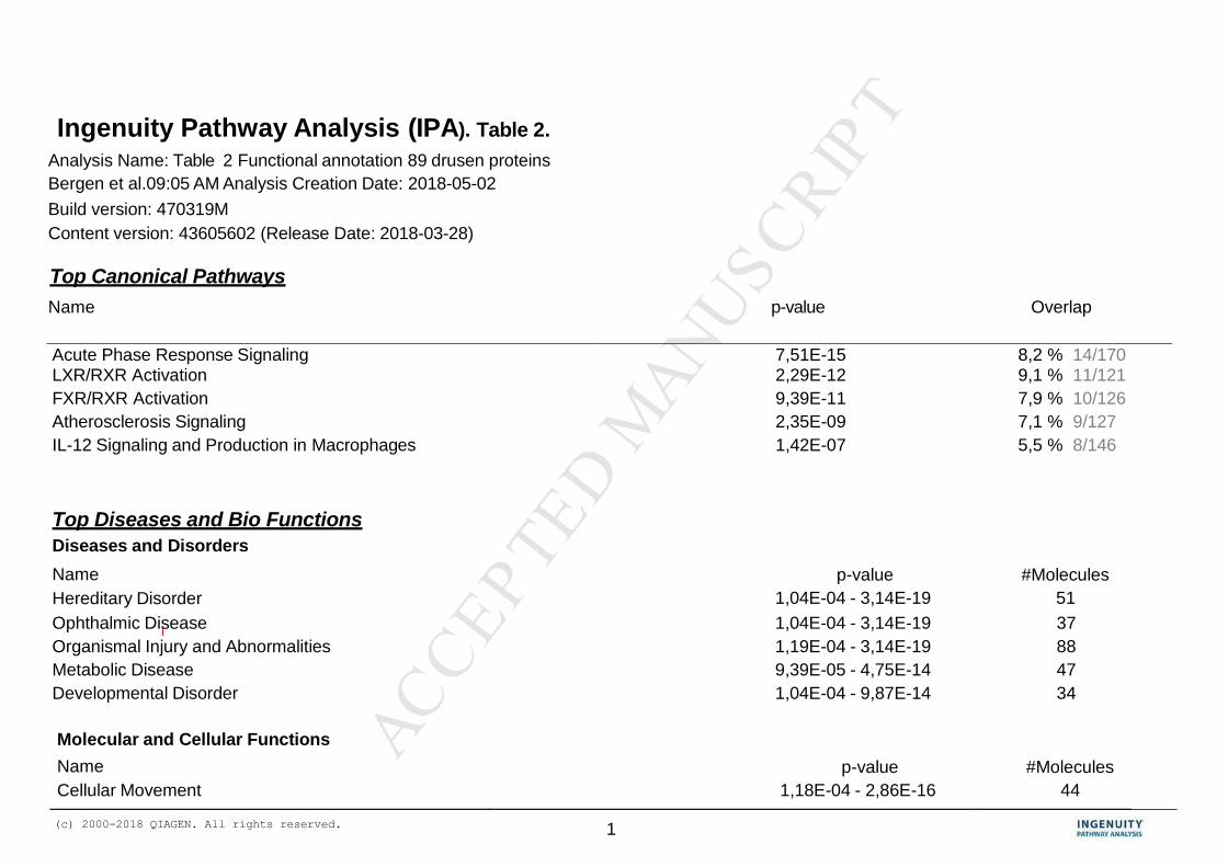
MANUSCRIP
T
ACCEPTED
ACCEPTED MANUSCRIPT
(c) 2000-2018 QIAGEN. All rights reserved. 1
Ingenuity Pathway Analysis (IPA). Table 2.
Analysis Name: Table 2 Functional annotation 89 drusen proteins
Bergen et al.09:05 AM Analysis Creation Date: 2018-05-02
Build version: 470319M
Content version: 43605602 (Release Date: 2018-03-28)
Top Canonical Pathways
Name p-value Overlap
Acute Phase Response Signaling 7,51E-15 8,2 % 14/170 LXR/RXR Activation 2,29E-12 9,1 % 11/121
FXR/RXR Activation 9,39E-11 7,9 % 10/126
Atherosclerosis Signaling 2,35E-09 7,1 % 9/127
IL-12 Signaling and Production in Macrophages 1,42E-07 5,5 % 8/146
Top Diseases and Bio Functions
Diseases and Disorders
Name
Hereditary Disorder
p-value
1,04E-04 - 3,14E-19
#Molecules
51
Ophthalmic Disease 1,04E-04 - 3,14E-19 37
Organismal Injury and Abnormalities 1,19E-04 - 3,14E-19 88
Metabolic Disease 9,39E-05 - 4,75E-14 47
Developmental Disorder 1,04E-04 - 9,87E-14 34
Molecular and Cellular Functions
Name
Cellular Movement
p-value
1,18E-04 - 2,86E-16
#Molecules
44
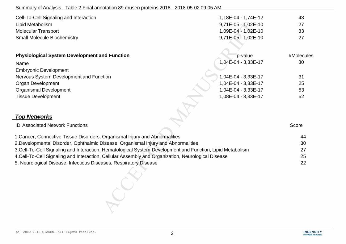
MANUSCRIP
T
ACCEPTED
ACCEPTED MANUSCRIPT
(c) 2000-2018 QIAGEN. All rights reserved. 2
Summary of Analysis - Table 2 Final annotation 89 drusen proteins 2018 - 2018-05-02 09:05 AM
Cell-To-Cell Signaling and Interaction 1,18E-04 - 1,74E-12 43
Lipid Metabolism 9,71E-05 - 1,02E-10 27
Molecular Transport 1,09E-04 - 1,02E-10 33
Small Molecule Biochemistry 9,71E-05 - 1,02E-10 27
Physiological System Development and Function
Name
Embryonic Development
p-value
1,04E-04 - 3,33E-17
#Molecules
30
Nervous System Development and Function 1,04E-04 - 3,33E-17 31
Organ Development 1,04E-04 - 3,33E-17 25
Organismal Development 1,04E-04 - 3,33E-17 53
Tissue Development 1,08E-04 - 3,33E-17 52
Top Networks
ID Associated Network Functions Score
1.Cancer, Connective Tissue Disorders, Organismal Injury and Abnormalities 44
2.Developmental Disorder, Ophthalmic Disease, Organismal Injury and Abnormalities 30
3.Cell-To-Cell Signaling and Interaction, Hematological System Development and Function, Lipid Metabolism 27
4.Cell-To-Cell Signaling and Interaction, Cellular Assembly and Organization, Neurological Disease 25
5. Neurological Disease, Infectious Diseases, Respiratory Disease 22

MANUSCRIP
T
ACCEPTED
ACCEPTED MANUSCRIPT
Network(N).
functional
cluster (#
entries)
Participating drusen
entries in functional
clusters
Functional
annotation underl.
Mol. Network
Phot OS
enriched
(412)
Phot enriched
(2293)
RPE enriched
(2284)
RPE spec.
(170)
Basal RPE
secreted
enriched
(276)
RPE/chor
enriched
(2680)
Chor enriched
(2387)
Blood plasma
(995)
Blood plasma
(405)
Blood post (242) Atheroscl plaque
enriched (3196)
Acronym PRos-EP PR-ET RPE-ET RPE-ST RPE-IVS RPE/Chor-EP Chor-ET BLP-SP1 BL-SP2 BL-PHP AS-EP
Reference STable3 GEOdat.base
GSE20191
GEOdat.base
GSE20191
STable2 STable1 PXD001424
PXD002194
GEOdat.base
GSE20191
STable4 Swiss-Prot
2010–04 + IPI
v3.71 + Ensembl
v54.37
STable 5 ST bleijerveld
Methodology proteomics transcript. transcript. transcript. secretome proteomics transcript. proteomics proteomics proteomics proteomics
Source Kiel Booij Booij Bennis Hauck Zhang Booij Geyer Farrah Stewart Bleijerveld
N1.1 (7) C7; C8A; C8B; C8G; MAC;
PRELP
Complement end 0 0 0 0 0 all PREPL C7, C8A, C8B,
C8G, MAC
0 C7, C8A, C8B, C8G,
MAC
All
N1.2 (18) akt; ATP synth; COL(col,
alpha 1, typeIII, Type VI,
1A2, 6A1, 6A2, 8A1);
GPNMB; RBP3; SEMA3;
EFEMP1; F1ATPase;
GPNMB; Pdi; PLG; RBP3;
SEMA3B; THBS4; TNC
Collagen cluster RBP3 COL8A1;
RBP3;GPNMB;
COL8A1;
RBP3;GPNMB;
SEMA3B;
EFEMP1
0 COL6A1; PLG COL(1A2,
6A1,6A2,8A1);
GPNMB; PLG;
RBP3; THBS4;
EFEMP1
COL(1A2,
6A1,8A1);
GPNMB; RBP3;
SEMA3B;
EFEMP1
COL6A1;
COL6A2;
GPNMB; PLG;
THBS4; TNC
COL1A2 PLG COL(1A2,
6A1,6A2,8A1);
GPNMB; PLG; TNC
N1.3 (7) CRYB(A1, A4); CRYB(B2, B4);
CRYG(B,C,D)
Crystallins CRYBB2 0 0 0 CRYBA(1,4);
CRYBB2
all 0 0 0 0 0
N2.4, all (13) CTB; ACTN1; (ANXA2);
ATP5B; (BSFP1); (BSFP2);
CRYAB; ENO2; FN1, MYH9;
PSMB5; SCARB2; SPTAN1
Genetic and
developmental
ophthalmic
disorders
ACTN1;
(ANXA2);
CRYAB; ENO2;
SPTAN1
ACTB; ATP5B;
CRYAB; ENO2;
SPTAN1
ACTB; ATP5B;
(ANXA2); CRYAB;
ENO2; SPTAN1
0 (ANXA2);
ATP5B;
CRYAB; ENO2;
FN1; MYH9
all, except
(BFSP1) and
(BSFP2)
ACTB; (ANXA2);
ATP5B; CRYAB;
ENO2; FN1,
SPTAN1
ACTB;
(ANXA2);
ATP5B; FN1;
MYH9; PSMB5;
0 ACTB; FN1 all, except ACTB
and PSMB5
N3.5 all (15) ALDH1A1; ANXA1; ANXA5;
APOE; CKB; GAPDH;
LAMB2; LUM; FRZB; PRDX1;
s100A7; S100A8;S100A9;
SERPINA3; TYRP1
Injury and
inflammatory
respons;
dermatological
disease
ANXA1; ANXA5;
CKB; GAPDH;
PRDX1; S100A7;
S100A8; S100A9
ANXA5; CKB;
FRZB; GAPDH;
PRDX1; TYRP1
ANXA5; APOE;
CKB; FRZB;
GAPDH; LAMB2;
PRDX1; S100A8;
TYRP1
FRZB ANXA1;
APOE; CKB;
GAPDH; LUM;
PRDX1
all ALDH1A1;
ANXA1; ANXA5;
APOE; CKB;
GAPDH; LAMB2;
FRZB; PRDX1;
S100A8;
SERPINA3; TYRP1
ALDH1A1;
ANXA1;
ANXA5; APOE;
CKB; GAPDH;
LAMB2; LUM;
PRDX1;
S100A8;
S100A9;
SERPINA3;
SERPINA3 APOE; LUM;
S100A9; SERPINA3;
ALDH1A1; ANXA1;
ANXA5; APOE; CKB;
LAMB2; FRZB;
GAPDH; PRDX1;
LUM; S100A9;
SERPINA3;
N4.6 all (14) AMPB; ANXA6; APCS;
APOA1; BGN, CFH; CLU;
HIST1H2BJ; ELN; HRG;
ORM1; SAA1; SERPINA1;
SERPIN1f
Cell to cell
signalling; systemic
involvement
ANXA6 CLU CLU 0 HRG AMPB; ANXA6;
APCS; APOA1;
BGN, CFH;
HRG; ORM1;
SAA1;
SERPINA1
ANXA6, CFH,
CLU, SAA1
AMPB; APCS;
APOA1; CFH;
CLU; HRG;
ORM1; SAA1;
SERPIN1
SERPINA1 AMPB; APCS;
APOA1; CFH; CLU;
HRG; ORM1; SAA1;
SERPINA1
AMPB; ANXA6;
APCS; APOA1; BGN,
CFH; CLU; ELN;
HRG; SERPINA1

MANUSCRIP
T
ACCEPTED
ACCEPTED MANUSCRIPT
(c) 2000-2018 QIAGEN. All rights reserved. 1
Ingenuity Pathway Analysis (IPA). Table 4. Analysis Name:Table 4 Summary photoreceptor core annotation analysis (745); Bergen et al.
2018-05-07 10:07 PM Analysis Creation Date: 2018-05-07
Build version: 470319M
Content version: 43605602 (Release Date: 2018-03-28)
Top Canonical Pathways
Name p-value Overlap
Phototransduction Pathway 7,98E-11 28,3 % 15/53
Huntington's Disease Signaling 2,04E-04 8,0 % 20/250
Glutamate Receptor Signaling 4,79E-04 14,0 % 8/57
Superpathway of Cholesterol Biosynthesis 1,87E-03 17,9 % 5/28
Wnt/Ca+ pathway 4,20E-03 11,1 % 7/63
Top Diseases and Bio Functions
Diseases and Disorders
Name
Cancer
p-value
1,31E-02 - 6,87E-28
#Molecules
680
Organismal Injury and Abnormalities 1,33E-02 - 6,87E-28 685
Gastrointestinal Disease 1,21E-02 - 4,26E-22 629
Hepatic System Disease 2,47E-03 - 8,92E-15 474
Reproductive System Disease 1,27E-02 - 3,21E-08 420
Molecular and Cellular Functions
Name
Cellular Assembly and Organization
p-value
1,28E-02 - 1,06E-08
#Molecules
165
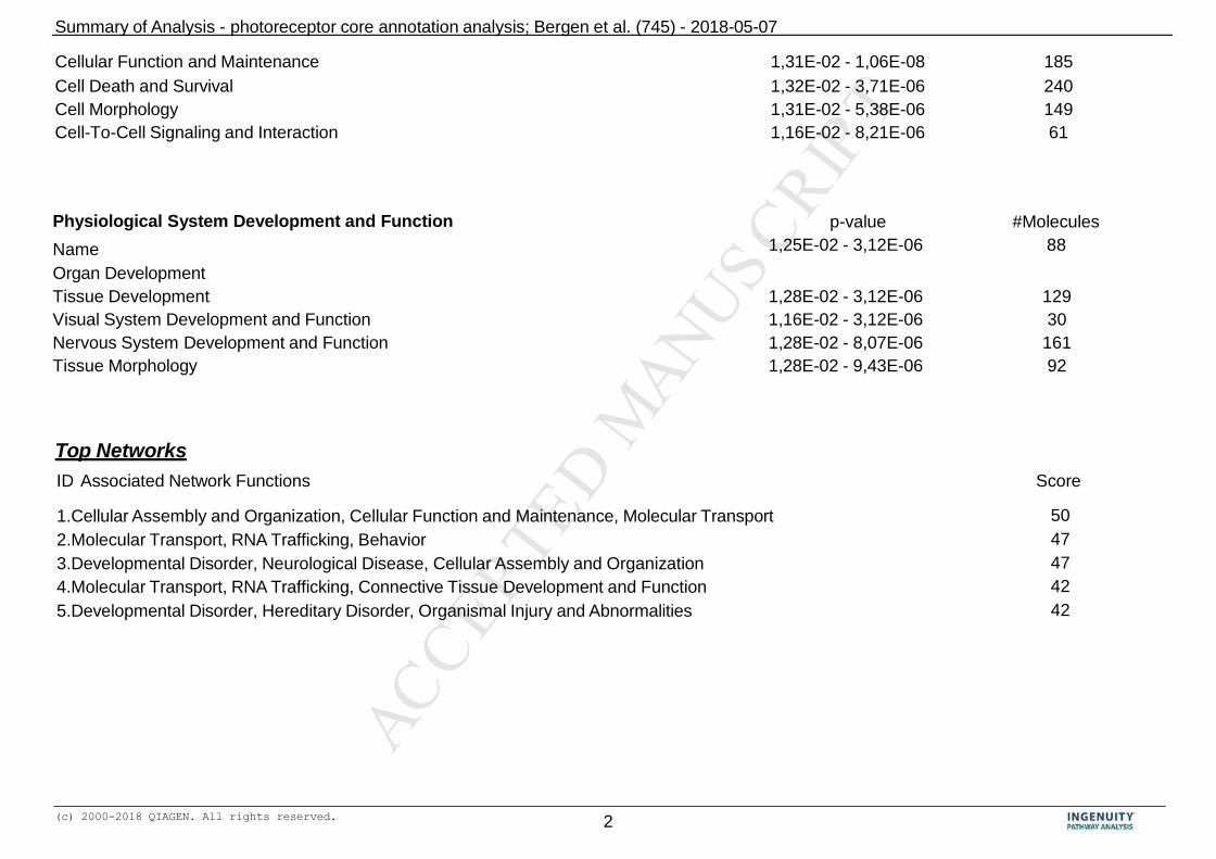
MANUSCRIP
T
ACCEPTED
ACCEPTED MANUSCRIPT
(c) 2000-2018 QIAGEN. All rights reserved. 2
Summary of Analysis - photoreceptor core annotation analysis; Bergen et al. (745) - 2018-05-07
Cellular Function and Maintenance 1,31E-02 - 1,06E-08 185
Cell Death and Survival 1,32E-02 - 3,71E-06 240
Cell Morphology 1,31E-02 - 5,38E-06 149
Cell-To-Cell Signaling and Interaction 1,16E-02 - 8,21E-06 61
Physiological System Development and Function
Name
Organ Development
p-value
1,25E-02 - 3,12E-06
#Molecules
88
Tissue Development 1,28E-02 - 3,12E-06 129
Visual System Development and Function 1,16E-02 - 3,12E-06 30
Nervous System Development and Function 1,28E-02 - 8,07E-06 161
Tissue Morphology 1,28E-02 - 9,43E-06 92
Top Networks
ID Associated Network Functions Score
1.Cellular Assembly and Organization, Cellular Function and Maintenance, Molecular Transport 50
2.Molecular Transport, RNA Trafficking, Behavior 47
3.Developmental Disorder, Neurological Disease, Cellular Assembly and Organization 47
4.Molecular Transport, RNA Trafficking, Connective Tissue Development and Function 42
5.Developmental Disorder, Hereditary Disorder, Organismal Injury and Abnormalities 42

MANUSCRIP
T
ACCEPTED
ACCEPTED MANUSCRIPT
(c) 2000-2018 QIAGEN. All rights reserved. 1
Diseases and Disorders
Ingenuity Pathway Analysis (IPA). Table 5.
Analysis Name: Table 5 Summary Functional Annotation Choroid- Bergen et al
Analysis Creation Date: 2018-05-07
Build version: 470319M
Content version: 43605602 (Release Date: 2018-03-28)
Top Canonical Pathways
Name p-value Overlap
Antigen Presentation Pathway 7,58E-12 36,8 % 14/38
Atherosclerosis Signaling 3,26E-11 18,0 % 23/128
Hepatic Fibrosis / Hepatic Stellate Cell Activation 3,88E-11 14,7 % 28/191
Acute Phase Response Signaling 5,19E-10 14,5 % 25/172
Complement System 1,90E-09 31,6 % 12/38
Top Diseases and Bio Functions
Diseases and Disorders
Name
Cancer
p-value
1,44E-06 - 4,74E-32
#Molecules
730
Organismal Injury and Abnormalities 1,48E-06 - 4,74E-32 746
Inflammatory Response 1,09E-06 - 1,87E-21 263
Connective Tissue Disorders 1,48E-06 - 1,46E-17 192
Inflammatory Disease 4,57E-08 - 1,46E-17 174
Molecular and Cellular Functions
Name
Cellular Movement
p-value
1,26E-06 - 1,81E-32
#Molecules
257

MANUSCRIP
T
ACCEPTED
ACCEPTED MANUSCRIPT
(c) 2000-2018 QIAGEN. All rights reserved. 2
Summary of Analysis -
Cell Death and Survival 1,47E-06 - 4,17E-21 323
Cell-To-Cell Signaling and Interaction 1,50E-06 - 9,12E-15 218
Cellular Development 1,49E-06 - 1,14E-14 326
Cellular Function and Maintenance 8,75E-07 - 3,23E-12 274
Physiological System Development and Function
Name
Cardiovascular System Development and Function
p-value
9,30E-07 - 1,46E-28
#Molecules
192
Organismal Development 1,47E-06 - 1,38E-24 325
Immune Cell Trafficking 1,29E-06 - 1,10E-23 158
Hematological System Development and Function 1,29E-06 - 3,90E-23 236
Organismal Survival 5,54E-08 - 1,79E-22 244
Top Networks
ID Associated Network Functions Score
1. Organ Morphology, Organismal Injury and Abnormalities, Renal Atrophy 49
2. Organismal Injury and Abnormalities, Skeletal and Muscular Disorders, Developmental Disorder 41
3. Molecular Transport, Nucleic Acid Metabolism, Small Molecule Biochemistry 38
4. Tissue Development, Cellular Movement, Hair and Skin Development and Function 37
5. Cell Cycle, Gene Expression, Cellular Growth and Proliferation 36
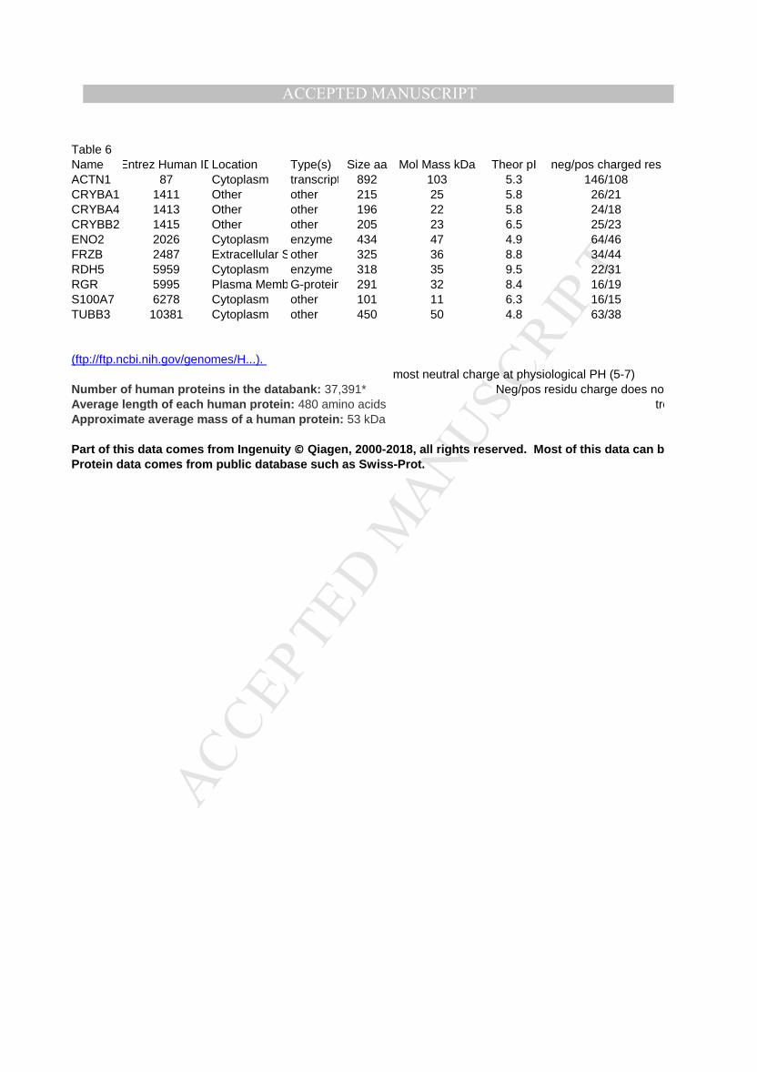
MANUSCRIP
T
ACCEPTED
ACCEPTED MANUSCRIPT
Table 6
Name Entrez Human IDLocation Type(s) Size aa Mol Mass kDa Theor pI neg/pos charged res
ACTN1 87 Cytoplasm transcription regulator892 103 5.3 146/108
CRYBA1 1411 Other other 215 25 5.8 26/21
CRYBA4 1413 Other other 196 22 5.8 24/18
CRYBB2 1415 Other other 205 23 6.5 25/23
ENO2 2026 Cytoplasm enzyme 434 47 4.9 64/46
FRZB 2487 Extracellular Spaceother 325 36 8.8 34/44
RDH5 5959 Cytoplasm enzyme 318 35 9.5 22/31
RGR 5995 Plasma MembraneG-protein coupled receptor291 32 8.4 16/19
S100A7 6278 Cytoplasm other 101 11 6.3 16/15
TUBB3 10381 Cytoplasm other 450 50 4.8 63/38
(ftp://ftp.ncbi.nih.gov/genomes/H...).
most neutral charge at physiological PH (5-7)
Number of human proteins in the databank: 37,391* Neg/pos residu charge does not play role
Average length of each human protein: 480 amino acids trend: (unstable)
Approximate average mass of a human protein: 53 kDa
Part of this data comes from Ingenuity © Qiagen, 2000-2018, all rights reserved. Most of this data can be compiled from public databases as well, such as DAVID
Protein data comes from public database such as Swiss-Prot.

MANUSCRIP
T
ACCEPTED
ACCEPTED MANUSCRIPT
Instability I Alipathic index GRAVY Comment
42.3 (u) 82.7 neg -0.6 Many interactive domains, calcium sensitive
50.2 (u) 50.8 neg -0.9 Oligomerisation, interact strongly with CRYBB2
42.8 (u) 54.7 neg -0.6 Oligomerisation, interact strongly with CRYBB2
34.2 (s) 58.4 neg -0.9 Oligomerisation, interact strongly with CRYBA1 and CRYBA4
35.3 (s) 92.6 neg -0.2
48.6 (u) 84.7 neg -0.3 Interacts with MYOC
43.4 (u) 106.8 0.18
40.9 (u) 102.3 0.414 Calcium binding
41.3 (u) 59.0 neg -0.8 interacts with RNABP9, calcium binding
39.6 (s) 71.7 neg -0.4 forms dimers with other tubulins.
most proteins little hydophilic
Neg/pos residu charge does not play role
trend: (unstable)
the AI index appears not to play a role.
Part of this data comes from Ingenuity © Qiagen, 2000-2018, all rights reserved. Most of this data can be compiled from public databases as well, such as DAVID

MANUSCRIP
T
ACCEPTED
ACCEPTED MANUSCRIPT
https://string-db.org
https://string-db.org
https://string-db.org
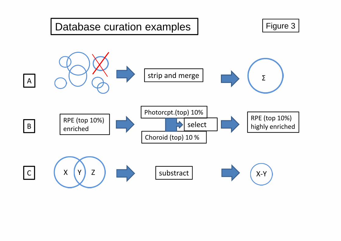
MANUSCRIP
T
ACCEPTED
ACCEPTED MANUSCRIPT
Database curation examples
RPE (top 10%)
enriched
Photorcpt.(top) 10%
Choroid (top) 10 %
strip and merge
substract
select
X Y Z X-Y
Σ
RPE (top 10%)
highly enriched
A
B
C
Figure 3

MANUSCRIP
T
ACCEPTED
ACCEPTED MANUSCRIPT
MANUSCRIP
T
ACCEPTED
ACCEPTED MANUSCRIPT
Figure 5
MANUSCRIP
T
ACCEPTED
ACCEPTED MANUSCRIPT
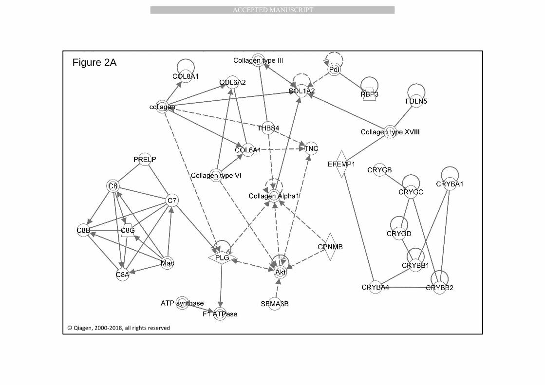
MANUSCRIP
T
ACCEPTED
ACCEPTED MANUSCRIPT
Figure 2A
© Qiagen, 2000-2018, all rights reserved

MANUSCRIP
T
ACCEPTED
ACCEPTED MANUSCRIPT
Figure 2B

MANUSCRIP
T
ACCEPTED
ACCEPTED MANUSCRIPT
Figure 2C

MANUSCRIP
T
ACCEPTED
ACCEPTED MANUSCRIPT
Figure 2D

MANUSCRIP
T
ACCEPTED
ACCEPTED MANUSCRIPT
Apical source of drusen -omics database
Basal source of drusen -omics database
Blood –omics(BL-SP1; BL-SP2)
Curated Chor/blood –omics(cChor-ET)
RPE –omics(RPE-ST; RPE-IVS)
Curated Phot/RPE –omics(cPRos-EP; cPR-ET)
BM
Curated database
drusen proteomics
Dr
Non-Curated Phot/RPE –omics(PRos-EP; PR-ET)
Non-Curated Chor/blood –omics(Chor-ET)
Figure 4

MANUSCRIP
T
ACCEPTED
ACCEPTED MANUSCRIPT
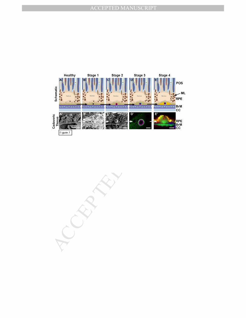
MANUSCRIP
T
ACCEPTED
ACCEPTED MANUSCRIPT

MANUSCRIP
T
ACCEPTED
ACCEPTED MANUSCRIPT
Figure 9A Figure 9B Figure 9C

MANUSCRIP
T
ACCEPTED
ACCEPTED MANUSCRIPT
On the origin of human drusen. Article highlights:
-A substantial amount of human drusen proteins is derived from blood.
-The second-best source of drusen proteins is the RPE
-Only few drusen proteins come uniquely from photoreceptor, RPE, or choroid.
-Drusen/blood protein identification may open up prevention for drusen formation.
-BM structure, HAP spherules and blood (pressure) play a role in drusen formation.

MANUSCRIP
T
ACCEPTED
ACCEPTED MANUSCRIPT
Authors’ statement
All authors have seen and approved the final version of the manuscript being submitted. They warrant
that the article is the authors' original work, hasn't received prior publication and isn't under
consideration for publication elsewhere.
None of the authors has a financial or personal conflicts of interest defined as a set of conditions in
which professional judgment concerning a primary interest, such as the validity of research, may be
influenced by a secondary interest, such as financial gain.
The work on this manuscript is approximately divided as follows:
AAB 28 %, SA 5 %, CK 5 %, MP 5 %, DW 4 %, PvdS 4%, SMH 4 %, CJFB 4 %, EE 5 %, AJS 18 %, IL 18 %