The Gi-coupled P2Y12 Receptor Regulates Diacylglycerol-mediated Signaling in Human Platelets
Upload
independentCategory
view
1download
0
Coppinger et al Characterization of the platelet releasate.
1
Characterization of the proteins released from activated platelets leads to
localization of novel platelet proteins in human atherosclerotic lesions.
Short Title for Running Head: Characterization of the platelet releasate.
Judith A. Coppinger*, Gerard Cagney*, Sinead Toomey*, Thomas Kislinger†, Orina Belton*,
James P. McRedmond*, Dolores J. Cahill*, Andrew Emili†, ‡, Desmond J. Fitzgerald* and
Patricia B. Maguire*§.
*Department of Clinical Pharmacology, Royal College of Surgeons in Ireland, 123 St.
Stephen’s Green, Dublin 2, Ireland.†Banting and Best Department of Medical Research, University of Toronto, Ontario, Canada.‡Department of Molecular and Medical Genetics, University of Toronto, Ontario, Canada.
The work was in part funded by a fellowship to PBM from Enterprise Ireland. PBM, OB and
DJF acknowledge research grants from the Health Research Board of Ireland. DJF, GC and
DJC acknowledge funding by the Higher Education Authority of Ireland. GC and DJC are
recipients of Science Foundation Ireland awards [Foundation Grant Nos. 02/IN.1/B117 and
02/CE.1/B141]. TK and AE are supported by Research Council of Canada.
§Corresponding author: Patricia B. Maguire,
Department of Clinical Pharmacology,
Royal College of Surgeons in Ireland,
123 St. Stephen’s Green, Dublin 2, Ireland.
Phone: +353-1-402-2420
Fax: +353-1-402-2453
Email: [email protected]
Word Count: Abstract: 165, Total Text: 3751.
Scientific Heading: Hemostasis, Thrombosis, and Vascular Biology.
Copyright (c) 2003 American Society of Hematology
Blood First Edition Paper, prepublished online November 20, 2003; DOI 10.1182/blood-2003-08-2804

Coppinger et al Characterization of the platelet releasate.
2
Abstract
Proteins secreted by activated platelets can adhere to the vessel wall and promote the
development of atherosclerosis and thrombosis. Despite this biological significance however,
the complement of proteins comprising the platelet releasate is largely unknown. Using a
proteomics approach, we have identified over 300 proteins released by human platelets
following thrombin activation. Many of the proteins identified were not previously attributed to
platelets, including secretogranin-III, a potential monocyte chemoattractant precursor;
cyclophilin A, a vascular smooth muscle cell growth factor; calumenin, an inhibitor of the
vitamin K epoxide reductase-warfarin interaction; as well as proteins of unknown function that
map to expressed sequence tags. Secretogranin-III, cyclophilin A and calumenin were
confirmed to localize in platelets and to be released upon activation. Furthermore, while absent
in normal vasculature, they were identified in human atherosclerotic lesions. Therefore, these
and other proteins released from platelets may contribute to atherosclerosis and to the
thrombosis that complicates the disease. Moreover, as soluble extracellular proteins, they may
prove suitable as novel therapeutic targets.
Coppinger et al Characterization of the platelet releasate.
3
Introduction
Atherosclerosis is a chronic inflammatory disease influenced by circulating cells, including
platelets1. Endothelial dysfunction, an early event in this process, leads to platelet adhesion,
which in turn leads to several steps in the development of atherosclerosis, including leukocyte
infiltration2. Furthermore, inhibition of platelet adhesion reduces leukocyte accumulation and
attenuates the progression of atherosclerotic lesions in the cholesterol-fed ApoE-/- mouse3.
Platelets may mediate such effects through products released following adhesion and activation.
Indeed, platelet-derived chemokines such as platelet factor-4 (PF4) are found in atherosclerotic
plaques where they express biological activities that may contribute to several aspects of the
disease3, 4.
Platelets contain a number of preformed, morphologically distinguishable storage granules - α-
granules, dense granules and lysosomes - the contents of which are released upon platelet
activation5. Platelets also release two distinct membrane vesicle populations during activation:
cell surface derived microvesicles and exosomes of endosomal origin6. Microvesicles have a
protein content similar to the activated plasma membrane and have procoagulant and
inflammatory functions7. On the other hand, exosomes are released following fusion of
subclasses of α-granules and multivesicular bodies (MVBs) and their function remains
unknown6. Exosomes are secreted by a multitude of other cells including those of hemopoietic
lineage, such as cytotoxic T cells8, antigen-presenting B cells9 and dendritic cells10 where they
play an immunoregulatory role11. Proteomic analysis of exosomes secreted from various cell
types has revealed the presence of ubiquitous proteins such as tubulin, actin and actin-binding
proteins as well as cell-type specific proteins12, 13.
Secreted platelet proteins act in an autocrine or paracrine fashion to modulate cell signalling.
Several of the proteins (for example, growth arrest specific gene 6 (GAS-6), factor V) are
prothrombotic, while others (for example, platelet derived growth factor (PDGF)), regulate cell
proliferation14. Platelets also release several immune modulators such as platelet basic protein
whose proteolytic product is neutrophil-activating peptide-2 (NAP-2)15, in addition to adhesion
proteins such as platelet endothelial cell adhesion molecule (PECAM) that may support

Coppinger et al Characterization of the platelet releasate.
4
leukocyte migration16. Thus, the platelet releasate contains factors of major significance in the
development of atherothrombosis. Here, we employed a comprehensive proteomics approach to
isolate, separate and identify the contents of the platelet releasate, a fraction highly enriched for
platelet granular and exosomal contents, and we demonstrate the presence of several of these
proteins in human atherosclerotic lesions.

Coppinger et al Characterization of the platelet releasate.
5
Methods
Platelet aggregation and isolation of supernatant fraction
Washed platelets were prepared and aggregations performed with 0.5 units (U) /ml thrombin, as
previously described17. Following aggregation, platelets were removed by centrifuging
sequentially twice at 1000g for 10 minutes and harvesting the supernatant. The supernatant was
further subjected to ultracentrifugation for one hour at 4°C at 50,000 x gmax using a 50.4 Ti
rotor (Beckman Instruments Inc., Fullerton, California) to remove microvesicles. The purity of
the releasate fraction was confirmed by the absence of platelet membrane specific protein αIIb
and the signalling protein focal adhesion kinase (FAK). (Results not shown).
SDS-PAGE and Western blotting
The methods for sodium dodecyl sulfate polyacrylamide gel electrophoresis (SDS-PAGE) and
western blotting were as previously described17. Protein concentration was measured according
to Bradford, and the same amount of protein (40µg) loaded onto each well. For total platelet
lysate this was equivalent to about 1.5 x 10⁷ platelets and for releasate about 8 x 10⁸ platelets.
The primary monoclonal antibody to thrombospondin Clone p10 (1:1000 dilution) and
polyclonal anti-goat (C-19) antibody to secretogranin III were purchased from Chemicon
International (Hampshire, UK) and Santa Cruz Biotech (Heidelberg, Germany), respectively.
The polyclonal anti-rabbit antibody to calumenin was a kind gift from Dr. Reidar Wallin, Wake
Forest University, North Carolina. The polyclonal anti-rabbit antibody against cyclophilin A
was from Upstate Biotech (Milton Keynes, UK). Anti-mouse, anti-goat and anti-rabbit
secondary horseradish peroxidase (HRP) antibodies were obtained from Pierce (Rockford,
Illinois). Fluorescence was detected using West Pico SupersignalTM chemiluminescent substrate
(Pierce, Rockford, Illinois).
Two-dimensional electrophoresis (2-DE)
2-DE was performed as previously delineated17, with the following minor modifications.
400µg of released protein (~8 x10⁹ platelets) were focused for 100,000-volt hours (Vh) on 18
cm, pI 3-10 immobiline dry strips using the Multiphor Isoelectric Focusing UnitTM (Amersham
Biosciences, Buckinghamshire, UK) and then sealed on top of 10% polyacrylamide gels (20 cm

Coppinger et al Characterization of the platelet releasate.
6
x 20 cm). Protein spots were visualized using a G-250 colloidal Coomassie blue dye (Sigma,
Dublin, Ireland).
MALDI-TOF mass spectrometry (MS)
In-gel digestion and matrix-assisted laser desorption/ionisation time-of-flight (MALDI-TOF)
MS was carried out as earlier outlined17, 18. Spectra were analysed by searching with the
publicly available search algorithm Mascot
(http://www.matrixscience.com/cgi/search_form.pl?FORMVER=2&SEARCH=PMF) using the
NCBI and SwissProt databases19.
Multidimensional protein identification technology (MudPIT) and Liquid
Chromatography (LC) -ion trap MS
Approximately 300µg of protein (~6 x 10⁹ platelets) was precipitated overnight with 5 volumes
of ice-cold acetone followed by centrifugation at 21,000g for 20 minutes. The pellet was
solubilized in 8 M urea, 50 mM Tris-HCl, pH 8.5 at 37°C for 2 hours and reduced by addition
of 1 mM dithiothreitol (DTT) for 1 hour at room temperature, followed by
carboxyamidomethylation with 5 mM iodoacetamide for 1 hour at 37 °C. The samples were
diluted to 4 M urea with 50 mM ammonium bicarbonate, pH 8.5 and digested with a 1/150
molar ratio of endoproteinase Lys-C at 37 °C overnight. The next day, the mixtures were
further diluted to 2 M urea with 50 mM ammonium bicarbonate, pH 8.5, supplemented with
calcium chloride to a final concentration of 1 mM, and incubated overnight with Poroszyme
immobilized trypsin beads at 30 °C with rotating. The resulting peptide mixtures were solid
phase extracted with SPEC-Plus PT C18 cartridges (Ansys Diagnostics; Lake Forest,
California) according to the manufacturer’s instructions and stored at –80 °C until further use.
A fully automated 7-cycle, 14 hour MudPIT chromatographic procedure was set up essentially
as described20, 21. Briefly, an HPLC quaternary pump was interfaced with an LCQ DECA XP
ion trap tandem mass spectrometer (ThermoFinnigan, San Jose, California). A 150 µm i.d.
fused silica capillary microcolumn (Polymicro Technologies, Phoenix, Arizona) was pulled to a
fine tip using a P-2000 laser puller (Sutter Instruments; Novato, California) and packed with 10
cm of 5 µm Zorbax Eclipse XDB-C18 resin (Agilent Technologies, Ontario, Canada) and then
Coppinger et al Characterization of the platelet releasate.
7
with 6cm of 5 µm Partisphere strong cation exchange resin (Whatman, Clifton, New Jersey).
Samples were loaded manually onto separate columns using a pressure vessel and
chromatography was performed as described22. The SEQUEST algorithm was used to identify
proteins from tandem mass spectra23. The ion state, XCorr, and DCn criteria to yield < 1% false
positive identification were used24.
Affymetrix RNA Analysis
Total RNA was isolated from highly purified platelet preparations, pooled and hybridised to the
Affymetrix HG-U95Av2 array, according to the manufacturer's instructions. Only samples that
were positive by PCR for platelet specific proteins (GPIIb, GPIIIa and the low abundance
CD151) and were negative for white cell markers (CD53 and delta chain of the T-cell antigen
receptor-associated T3 complex) were pooled for analysis. Average difference values were
scaled and transformed in accordance with the Gene Expression Atlas
(http://expression.gnf.org/cgi-bin/index.cgi)25. For comparison to proteomic data, Affymetrix
probesets were mapped to UniGene clusters (build 157, Nov 2002).
Confocal Microscopy
Glass slides were coated with 20 µg/ml of fibrinogen in buffer A (50 mM Tris pH 7.5, 150 mM
NaCl, 0.1 % Tween-20) at 37 °C for 2 hours and subsequently blocked with 1% bovine serum
albumin (BSA) for 1 hour. The platelets were resuspended in a modified Tyrodes buffer (130
mM NaCl, 10 mM trisodium citrate, 9 mM NaHCO3, 6 mM dextrose, 0.9 mM MgCl2, 0.81 mM
KH2PO4, 10 mM Tris pH 7.4) at a concentration of 20-30x105 /µl and allowed to adhere to the
blocked slides for the indicated time prior to fixation for 7 minutes in ice-cold methanol. Slides
were then permeabilized in ice-cold acetone for 2 minutes and blocked with normal goat serum
or BSA at room temperature for 30 minutes. The slides were incubated with primary antibody
(antibodies as for western blotting) for 45 minutes and then a fluorescent secondary antibody
(Alexa 488-conjugated goat anti-rabbit or mouse anti-goat) for 10 minutes. These slides were
visualised using an argon laser at 488 nm. For dual stained images, the slides were incubated
with anti-CD41 monoclonal antibody (IgG1; clone SZ22; Beckman Coulter, Buckinghamshire,
UK) for an additional 45 minutes and then a fluorescent Alexa 546-conjugated goat anti-mouse
secondary for 10 minutes. Control procedures included unstained cells to allow for

Coppinger et al Characterization of the platelet releasate.
8
autofluorescence, secondary antibody only and primary antibodies with two differentially
labelled secondaries to check for non-specific fluorescence. All images were acquired using a
Zeiss LSM 510 Confocal Microscope (Carl Zeiss, Germany).
Immunohistochemistry
Samples of arterial tissue were obtained from patients with atherosclerosis at the time of
surgery for carotid or peripheral vascular disease (n=5) and fixed in formal saline for
immunohistochemistry analysis26. The study was approved by the Irish Medicines Board and
the Ethics Committee of Beaumont Hospital, Dublin, and all patients gave written informed
consent. All patients were undergoing surgical revascularization for peripheral vascular disease
or carotid endarterectomy. Normal arterial sections were obtained from young individuals who
had no gross or microscopic evidence of atherosclerosis. Sections (5 µm) were stained with
hematoxylin and eosin and for proteins of interest, as described previously26. Primary
antibodies were as for western blotting and confocal microscopy. Anti-rabbit platelet factor 4
(PF4) polyclonal antibody (Ab1488P) was from Chemicon International (Temecula,
California). Monoclonal anti-α smooth muscle actin (mouse IgG2a isotype) was purchased
from Sigma Aldrich, Tallaght, Ireland26.
Coppinger et al Characterization of the platelet releasate.
9
Results
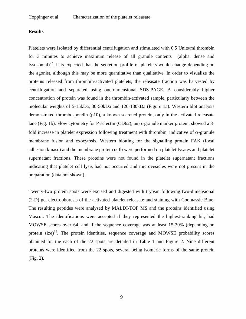
Platelets were isolated by differential centrifugation and stimulated with 0.5 Units/ml thrombin
for 3 minutes to achieve maximum release of all granule contents (alpha, dense and
lysosomal)27. It is expected that the secretion profile of platelets would change depending on
the agonist, although this may be more quantitative than qualitative. In order to visualize the
proteins released from thrombin-activated platelets, the releasate fraction was harvested by
centrifugation and separated using one-dimensional SDS-PAGE. A considerably higher
concentration of protein was found in the thrombin-activated sample, particularly between the
molecular weights of 5-15kDa, 30-50kDa and 120-180kDa (Figure 1a). Western blot analysis
demonstrated thrombospondin (p10), a known secreted protein, only in the activated releasate
lane (Fig. 1b). Flow cytometry for P-selectin (CD62), an α-granule marker protein, showed a 3-
fold increase in platelet expression following treatment with thrombin, indicative of α-granule
membrane fusion and exocytosis. Western blotting for the signalling protein FAK (focal
adhesion kinase) and the membrane protein αIIb were performed on platelet lysates and platelet
supernatant fractions. These proteins were not found in the platelet supernatant fractions
indicating that platelet cell lysis had not occurred and microvesicles were not present in the
preparation (data not shown).
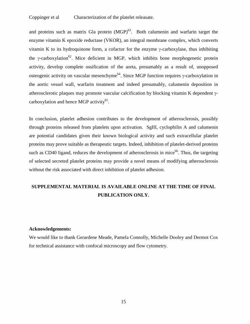
Twenty-two protein spots were excised and digested with trypsin following two-dimensional
(2-D) gel electrophoresis of the activated platelet releasate and staining with Coomassie Blue.
The resulting peptides were analysed by MALDI-TOF MS and the proteins identified using
Mascot. The identifications were accepted if they represented the highest-ranking hit, had
MOWSE scores over 64, and if the sequence coverage was at least 15-30% (depending on
protein size)28. The protein identities, sequence coverage and MOWSE probability scores
obtained for the each of the 22 spots are detailed in Table 1 and Figure 2. Nine different
proteins were identified from the 22 spots, several being isomeric forms of the same protein
(Fig. 2).

Coppinger et al Characterization of the platelet releasate.
10
We then used multidimensional liquid chromatography coupled with electrospray MS to
further characterize the platelet releasate. The released protein fraction from thrombin-activated
platelets was digested with trypsin and the resulting peptides loaded under pressure on to a
nanocapillary column containing both strong cation exchange and reverse phase materials29.
Seven successive salt elutions and HPLC cycles were used to separate the peptides (Fig. 3a-g).
The thrombin-activated releasates of pooled donors were analysed using identical cycle
conditions in three independent experiments. Over 300 proteins were identified, with 81
observed in two or three experiments (Table 2). Seventy percent of these 81 were identified in
all three experiments. Reproducibility of identification for a repeat analysis of a single sample
was ∼80%. This suggests that ∼20% of the variation in the results arise from missed
identifications due to saturation of the MudPIT analysis, while ∼10% may represent donor-to-
donor variation.
37% of the proteins identified were previously reported to be released from platelets including
thrombospondin30, platelet factor 4 (PF4)31, osteonectin32, metalloproteinase inhibitor 133 and
transforming growth factor34. Another 35% are known to be released from other secretory cells.
These include cofilin, profilin, 14-3-3 zeta and actin from dendritic cells12, peptidyl-prolyl-cis
isomerase (cyclophilin A) from smooth muscle cells35, phosphoglycerate kinase from
fibrosarcoma cells36, and beta-2 microglobulin37 and vitamin D binding protein38, from the
liver. The remaining proteins are not known to be released from any cell type with several
mapping to expressed sequence tags (ESTs) of unknown function (Table 2). Additionally, 75
of the 81 released proteins were matched to UniGene clusters, 68 of which had corresponding
Affymetrix probesets. Of these 68 array-comparable proteins, messages for 46 (68%) were
detected in the platelet mRNA (McRedmond et al, Manuscript submitted; Table 2).
We focused on three proteins, secretogranin III (SgIII), cyclophilin A and calumenin, which are
not known to be present in or released from platelets. SgIII (Fig. 4c), cyclophilin A (Fig. 4b)
and calumenin (Fig. 4a) were found by western blot in resting (PC) and thrombin activated
platelet lysates (PA), as well as in the supernatant of thrombin-activated platelets (RA) (Fig. 4).
Neither SgIII nor calumenin were detected in a crude leukocyte lysate (WBC), although

Coppinger et al Characterization of the platelet releasate.
11
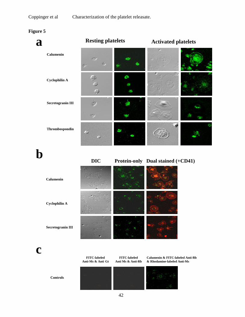
cyclophilin A was present in low amounts (Fig. 4). These three proteins were identified in P-
selectin positive platelets by flow cytometry (data not shown) and in CD41 positive platelets
adhering to fibrinogen-coated slides using confocal microscopy (Fig. 5b). No staining was
observed in platelets stained for secondary antibody only (Fig. 5c).
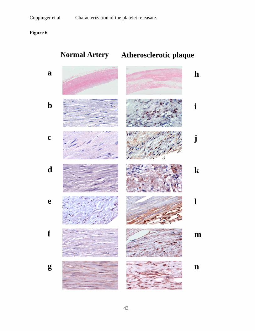
Sections of arterial tissue from five patients with atherosclerosis were examined using
immunohistochemistry26. The results from a representative patient are shown (Fig. 6). Low
power H & E staining of normal (a) and atherosclerotic plaque (h) are provided for orientation
of these sections. Atherosclerotic but not normal artery stained for the platelet specific proteins
PF4 (Fig. 6j) and CD41 (Fig. 6i). SgIII (Fig. 6l) and calumenin (Fig. 6m) were also expressed
widely in the plaque, while vascular smooth muscle cells in the lesion, identified by staining for
actin (6g,n), stained for cyclophilin A (Fig. 6k). None of the proteins were found in normal
artery (Fig. 6b-f).

Coppinger et al Characterization of the platelet releasate.
12
Discussion
We identified over 300 proteins in the platelet releasate; a fraction highly enriched for platelet
granular and exosomal contents. This list included proteins of relatively low (e.g.,
thrombospondin) and high (e.g., platelet glycoprotein V) isoelectric point, as well as low (e.g.,
PF4) and high (e.g., von Willebrand factor) molecular mass. Of the 81 proteins observed in two
or three repeat experiments, thirty (37%) are known to be released from platelets including
multimerin, thrombospondin and PF4. Integral membrane proteins, such as αIIb and known
signalling proteins were not represented, suggesting that the identified fraction is relatively
specific and enriched for secreted and exosomal proteins.
Fifty-one (63%) of the proteins identified were not known to be released from platelets. A
possible explanation for the presence of these proteins is contamination of the releasate with
intracellular platelet proteins released by platelet lysis. However, the absence of major
signalling proteins argues against this. Furthermore, twenty-eight (35%) of the identified
proteins are proteins that are released by other secretory cells (Table 2). For example, a number
of the platelet releasate proteins identified, including tubulin and adenylyl cyclase-associated
protein (CAP 1) are found in macrophage phagosomes39. Other proteins identified in the
releasate fraction may be involved in the process of exocytosis, for example calmodulin40.
Furthermore, neither vesicle-associated membrane protein (VAMP) nor syntaxins, which are
present in high amounts in platelets41, appear in the releasate.
Twenty-three (28%) proteins in the releasate are not reported to be released from any cell type.
Several map to expressed sequence tags (ESTs) of unknown function. However, out of 81
proteins found consistently in platelet releasate, 46 detected at the mRNA level in platelets
(McRedmond et al, Manuscript submitted; Table 2). Remarkably, 18 of the 50 most abundant
platelet messages were represented in the releasate. Protein synthesis in the platelet is limited42
and the platelet transcriptome may largely reflect that of the parent megakaryocyte43. The
messages for many secreted proteins may therefore, be transcribed in the megakaryocyte and
passed to the daughter platelet cells. Other proteins released that do not have a corresponding
mRNA may be endocytosed by platelets, for instance fibrinogen44 and albumin45. Although the

Coppinger et al Characterization of the platelet releasate.
13
detection of hemoglobin messages in platelet RNA might point to contamination of the platelet
preparations, our platelet RNA results are in close agreement with those of Gnatenko et al46,
whose comparison of the transcriptomes from platelets, erythrocytes and whole blood suggests
that mRNAs for hemoglobin are present in platelets.
A number of cytoskeletal and actin-binding proteins were found in the thrombin-activated
releasate. Changes in the actin cytoskeleton upon platelet activation play an important role in
granule movement and exocytosis and many ubiquitous actin-binding proteins interact with
endosomes and lysosomes47. Moreover, α-actinin and thymosin beta 4 are known to be released
from platelet α-granules48, where they bind thrombospondin and exhibit anti- microbial activity,
respectively49. Many of the other actin-binding proteins identified are released from other cells
including profilin, cofilin, actin and tubulin from exosomes of dendritic cells12. Interestingly,
the widely expressed intermediate filament protein vimentin is secreted by activated
macrophages where it is involved in bacterial killing and the generation of oxidative
metabolites50.
Five mitochondrial proteins were identified including pyruvate kinase, fructose biphosphate
aldolase, phosphoglycerate kinase, glyceraldehydes 3-phosphate dehydrogenase (GAPDH) and
superoxide dismutase. The most straightforward explanation for these proteins would be
mitochondrial contamination; however, two of the most abundant mitochondrial proteins
fumarate hydratase and aconitase are not present in our preparation51. Although there are no
reports of superoxide dismutase secretion, pyruvate kinase and GAPDH are released from B
cells13, fructose biphosphate aldolase is released from dendritic cells12 and phosphoglycerate
kinase is released from tumour cells36.
Three proteins, SgIII, cyclophilin A, and calumenin, not previously recognized to be present in
or released by platelets, were examined further as they are of potential interest in the
pathogenesis of atherosclerosis. These three proteins were localised to platelets by western
blotting, confocal microscopy, flow cytometry and, for cyclophilin A and calumenin, by
microarray analysis of mRNA. While absent from normal artery, these three proteins were
found in human atherosclerotic plaque. These lesions also stained for CD41 and PF4, two

Coppinger et al Characterization of the platelet releasate.
14
platelet-specific proteins. We have previously demonstrated CD41 staining as a marker for
platelets in the atherosclerosis of the ApoE-/- model52. In addition, PF4 and oxidised low-density
lipoprotein (LDL) have been found to co-localize in atherosclerotic lesions, especially in
macrophage-derived foam cells, as PF4 binds to oxidized LDL and may contribute to its
uptake53.
SgIII is a member of the chromogranin family of acidic secretory proteins, previously shown
only to be localized to storage vesicles of neuronal and endocrine cells54,55. Secreteogranin II, a
close homolog also found in our platelet releasate (n=1; see supplementary data), is present in
neuroendocrine storage vesicles, and is the precursor of the neuropeptide secretoneurin, which
has a tissue distribution and function similar to the proinflammatory neuropeptides, substance P
and neuropeptide Y. Secretoneurin stimulates monocyte adhesion to the vessel wall followed by
their trans-endothelial migration56. Whether cleavage products of SgIII play a similar role is
unknown.
Cyclophilins are peptidyl-propyl cis-trans-isomerases that act intracellularly both as catalysts
and chaperones in protein folding57 and have extracellular signaling functions such as the
induction of chemotaxis and adhesion of memory CD4 cells58. Recently, cyclophilin A was
found to be secreted by vascular smooth muscle cells in response to oxidative stress, where it
acted in an autocrine manner to stimulate extracellular signal-regulated kinase (ERK1/2)
activation and vascular smooth muscle proliferation35. Therefore, cyclophilin A released from
activated platelets may stimulate the migration and proliferation of smooth muscle cells, a
process implicated in the development of atherosclerosis.
The third protein, calumenin, belongs to the CREC family of calcium binding proteins and was
recently found to be secreted from early melanosomes, which are closely related to platelet
dense granules59. Calumenin has a chaperone function in the endoplasmic reticulum, but little is
known about its extracellular function60. It has however, been shown to bind to serum amyloid
P component, which is also released from platelets (Table 2)61. Calumenin inhibits the activity
of vitamin K-dependent γ-carboxylation62 responsible for the activation of coagulation factors
Coppinger et al Characterization of the platelet releasate.
15
and proteins such as matrix Gla protein (MGP)63. Both calumenin and warfarin target the
enzyme vitamin K epoxide reductase (VKOR), an integral membrane complex, which converts
vitamin K to its hydroquinone form, a cofactor for the enzyme γ-carboxylase, thus inhibiting
the γ-carboxylation62. Mice deficient in MGP, which inhibits bone morphogenetic protein
activity, develop complete ossification of the aorta, presumably as a result of, unopposed
osteogenic activity on vascular mesenchyme64. Since MGP function requires γ-carboxylation in
the aortic vessel wall, warfarin treatment and indeed presumably, calumenin deposition in
atherosclerotic plaques may promote vascular calcification by blocking vitamin K dependent γ-carboxylation and hence MGP activity65.
In conclusion, platelet adhesion contributes to the development of atherosclerosis, possibly
through proteins released from platelets upon activation. SgIII, cyclophilin A and calumenin
are potential candidates given their known biological activity and such extracellular platelet
proteins may prove suitable as therapeutic targets. Indeed, inhibition of platelet-derived proteins
such as CD40 ligand, reduces the development of atherosclerosis in mice66. Thus, the targeting
of selected secreted platelet proteins may provide a novel means of modifying atherosclerosis
without the risk associated with direct inhibition of platelet adhesion.
SUPPLEMENTAL MATERIAL IS AVAILABLE ONLINE AT THE TIME OF FINAL
PUBLICATION ONLY.
Acknowledgements:
We would like to thank Gerardene Meade, Pamela Connolly, Michelle Dooley and Dermot Cox
for technical assistance with confocal microscopy and flow cytometry.

Coppinger et al Characterization of the platelet releasate.
16
References:
1. Ross R. Atherosclerosis-an inflammatory disease. N Engl J Med. 1999;340:115-126.
2. Massberg S, Brand K, Gruner S, et al. A critical role of platelet adhesion in the initiation of
atherosclerotic lesion formation. J Exp Med. 2002;196:887-896.
3. Huo Y, Schober A, Forlow S, et al. Circulating activated platelets exacerbate atherosclerosis
in mice deficient in apolipoprotein E. Nat Med. 2003;9:61-67.
4. Chesterman CN, Berndt MC. Platelet and vessel wall interaction and the genesis of
atherosclerosis. Clin Haematol. 1986;15:323-353.
5. Fukami H, Holmsen H, Kowalska M, Niewiarowski S. Platelet. Secretion. In: Colman RW,
Hirsh J, Marder VJ, Clowes AW, George JN, eds. Haemostasis and Thrombosis: Basic
Principles and Clinical practice 4th Ed. Philadelphia, PA: Lippincott Williams & Wilkins.;
2001: 561-574.
6. Heijnen HF, Schiel AE, Fijnheer R, Geuze HJ, Sixma JJ. Activated platelets release two
types of membrane vesicles: microvesicles by surface shedding and exosomes derived from
exocytosis of multivesicular bodies and alpha-granules. Blood. 1999;94:3791-3799.
7. Barry O, FitzGerald G. Mechanisms of cellular activation by platelet microparticle. Thromb
Haemost. 1999;82:794-800.
8. Peters PJ, Geuze HJ, Van der Donk HA, et al. Molecules relevant for T cell-target cell
interaction are present in cytolytic granules of human T lymphocytes. Eur J Immunol.
1989;19:1469-1475.
9. Raposo G, Nijman HW, Stoorvogel W, et al. B lymphocytes secrete antigen-presenting
vesicles. J Exp Med. 1996;183:1161-1172.
Coppinger et al Characterization of the platelet releasate.
17
10. Zitvogel L, Regnault A, Lozier A, et al. Eradication of established murine tumors using a
novel cell-free vaccine: dendritic cell-derived exosomes. Nat Med. 1998;4:594-600.
11. Dimitris S, Hany G, Michele R, Salah M. Immunoregulatory properties of mast cell-
derived exosomes. Mol Immunol. 2002;38:1359-1362.
12. Thery C, Boussac M, Veron P, et al. Proteomic analysis of dendritic cell-derived exosomes:
a secreted subcellular compartment distinct from apoptotic vesicles. J Immunol.
2001;166:7309-7318.
13. Wubbolts R, Leckie R, Veenhuizen P, et al. Proteomic and biochemical analyses of human
B cell-derived exosomes: Potential implications for their function and multivesicular body
formation. J Biol Chem. 2003;278:10963-10970.
14. Angelillo-Scherrer A, de Frutos P, Aparicio C, et al.. Deficiency or inhibition of Gas6
causes platelet dysfunction and protects mice against thrombosis. Nat Med. 2001;7:215-221.
15. Castor C, Walz D, Ragsdale C, et al. Connective tissue activation. XXXIII. Biologically
active cleavage products of CTAP-III from human platelets. Biochem Biophys Res Commun.
1989;163:1071-1080.
16. Muller W. The role of PECAM-1 (CD31) in leukocyte emigration: studies in vitro and in
vivo. J Leukoc Biol. 1995;57:523-528.
17. Maguire PB, Wynne KJ, Harney DF, O'Donoghue NM, Stephens G, Fitzgerald DJ.
Identification of the phosphotyrosine proteome from thrombin activated platelets. Proteomics.
2002;2:642-648.
18. Shevchenko A, Wilm M, Vorm O, Mann M. Mass spectrometric sequencing of proteins
silver-stained polyacrylamide gels. Anal Chem. 1996;68:850-858.

Coppinger et al Characterization of the platelet releasate.
18
19. Fenyo D. Identifying the proteome: software tools. Curr Opin Biotechnol. 2000;11:391-
395.
20. Washburn MP, Wolters D, Yates JR, 3rd. Large-scale analysis of the yeast proteome by
multidimensional protein identification technology. Nat Biotech. 2001;19:242-247.
21. Wolters DA, Washburn MP, Yates JR, 3rd. An automated multidimensional protein
identification technology for shotgun proteomics. Anal Chem. 2001;73:5683-5690.
22. Peng J, Elias JE, Thoreen CC, Licklider LJ, Gygi SP. Evaluation of multidimensional
chromatography coupled with tandem mass spectrometry (LC/LC-MS/MS) for large-scale
protein analysis: the yeast proteome. J Proteome Res. 2003;2:43-50.
23. Eng JK, McCormack AL, Yates JR. An approach to correlate tandem mass spectral data of
peptides with amino acid sequences in a protein database. J Am Soc Mass Spectrom.
1994;5:976-989.
24. Kislinger T, Rahman K, Radulovic D, Cox B, Rossant J, Emili A. PRISM, a Generic Large
Scale Proteomic Investigation Strategy for Mammals. Mol Cell Proteomics. 2003;2:96-100.
25. Su A, Cooke M, Ching K, et al. Large-scale analysis of the human and mouse
transcriptomes. Proc. Natl. Acad. Sci. USA. 2002;99:4465-4470.
26. Belton O, Byrne D, Kearney D, Leahy A, Fitzgerald D. Cyclooxygenase-1 and -2-
dependent prostacyclin formation in patients with atherosclerosis. Circulation. 2000;102:840-
845.
27. Colman RW, Clowes AW, George JN, Hirsh J, Marder VJ. Overview of Hemostasis. In:
Colman RW, Hirsh J, Marder VJ, Clowes AW, George JN, eds. Haemostasis and Thrombosis:

Coppinger et al Characterization of the platelet releasate.
19
Basic Principles and Clinical practice. 4th Ed. Philadelphia, PA: Lippincott Williams & Wilkin;
2001: 3-16.
28. Lefkovits I, Kettman JR, Frey J. Global analysis of gene expression in cells of the immune
system I. Analytical limitations in obtaining sequence information on polypeptides in two-
dimensional gel spots. Electrophoresis. 2000;21:2688-2693.
29. Link AJ, Eng J, Schieltz DM, et al. Direct analysis of protein complexes using mass
spectrometry. Nat Biotech. 1999;17:676-682.
30. Baenziger NL, Brodie GN, Majerus PW. A thrombin-sensitive protein of human platelet
membranes. Proc Natl Acad Sci U S A. 1972;68:240-243.
31. Rucinski B, Poggi A, James P, Holt J, Niewiarowski S. Purification of two heparin-binding
proteins from porcine platelets and their homology with human secreted platelet proteins.
Blood. 1983;61:1072-1080.
32. Breton-Gorius J, Clezardin P, Guichard J, et al. Localization of platelet osteonectin at the
internal face of the alpha-granule membranes in platelets and megakaryocytes. Blood.
1992;79:936-941.
33. Kazes I, Elalamy I, Sraer JD, Hatmi M, Nguyen G. Platelet release of trimolecular complex
components MT1-MMP/TIMP2/MMP2: involvement in MMP2 activation and platelet
aggregation. Blood. 2000;96:3064-3069.
34. Fava R, Casey TT, Wilcox J, Pelton RW, Moses HL, Nanney LB. Synthesis of
transforming growth factor-beta 1 by megakaryocytes and its localization to megakaryocyte and
platelet alpha-granules. Blood. 1990;76:46-55.
35. Jin Z, Melaragno M, Liao D, et al. Cyclophilin A is a secreted growth factor induced by
oxidative stress. Circ Res. 2000;87:789-796.

Coppinger et al Characterization of the platelet releasate.
20
36. Lay A, Jiang XM, Kisker O, et al. Phosphoglycerate kinase acts in tumour angiogenesis as
a disulphide reductase. Nature. 2000;408:869-873.
37. Ramadori G, Mitsch A, Rieder H, Meyer zum Buschenfelde K. Alpha- and gamma-
interferon (IFN alpha, IFN gamma) but not interleukin-1 (IL-1) modulate synthesis and
secretion of beta 2-microglobulin by hepatocytes. Eur J Clin Invest. 1988;18:343-351.
38. Imawari M, Matsuzaki Y, Mitamura K, Osuga T. Synthesis of serum and cytosol vitamin
D-binding proteins by rat liver and kidney. J Biol Chem. 1982;257:8153-8170.
39. Garin J, Diez R, Kieffer S, et al. The phagosome proteome: Insight into phagosome
functions. J Cell Biol. 2001;152:165-180.
40. Quetglas S, Iborra C, Sasakawa N, et al. Calmodulin and lipid binding to synaptobrevin
regulates calcium-dependent exocytosis. EMBO J. 2002;21:3970-3979.
41. Polgar J, Chung S, Reed G. Vesicle-associated membrane protein 3 (VAMP-3) and
VAMP-8 are present in human platelets and are required for granule secretion. Blood.
2002;100:1081-1083.
42. Kieffer N, Guichard J, Farcet JP, Vainchenker W, Breton-Gorius J. Biosynthesis of major
platelet proteins in human blood platelets. Eur J Biochem. 1987;164:189-195.
43. Shaw T, Chesterman CN, Morgan FJ. In vitro synthesis of low molecular weight proteins
in human platelets: absence of labelled release products. Thromb Res. 1984;36:619-631.
44. Sixma J, Akkerman J, van Oost B, Gorter G. Intracellular localization of fibrinogen in
human blood platelets. Bibl Haematol. 1977;44:129-133.

Coppinger et al Characterization of the platelet releasate.
21
45. Gogstad G, Hagen I, Korsmo R, Solum N. Characterisation of the proteins of isolated
human platelet alpha granules. Evidence for a separate pool of the glycoproteins IIb and IIIa.
Biochem Biophys Acta. 1981;670:150-162.
46. Gnatenko DV, Dunn JJ, McCorkle SR, Weissmann D, Perrotta PL, Bahou WF. Transcript
profiling of human platelets using microarray and serial analysis of gene expression. Blood.
2003;101:2285-2293.
47. Cordonnier M, Dauzonne D, Louvard D, Coudrier E. Actin filaments and myosin I alpha
cooperate with microtubules for the movement of lysosomes. Mol Biol Cell. 2001;12:4013-
4029.
48. Dubernard V, Arbeille BB, Lemesle M, Legrand C. Evidence for an alpha-granular pool of
the cytoskeletal protein alpha-actinin in human platelets that redistributes with the adhesive
glycoprotein thrombospondin-1 during the exocytotic proces. Arterioscler Thromb Vasc Biol.
1997;17:2293-2305.
49. Tang Y, Yeaman M, Selsted M. Antimicrobial peptides from human platelets. Infect
Immun. 2002;70:6524-6533.
50. Mor-Vaknin N, Punturieri A, Sitwala K, Markovitz D. Vimentin is secreted by activated
macrophages. Nat Cell Biol. 2003;5:59-63.
51. Rabilloud T, Kieffer S, Procaccio V, et al. Two-dimensional electrophoresis of human
placental mitochondria and protein identification by mass spectrometry: toward a human
mitochondrial proteome. Electrophoresis. 1998;19:1006-1014.
52. Belton OA, Duffy A, Toomey S, Fitzgerald DJ. Cyclooxygenase Isoforms and Platelet
Vessel Wall Interactions in the ApoE Knockout Mouse Model of Atherosclerosis. Circulation.
In press.

Coppinger et al Characterization of the platelet releasate.
22
53. Nassar T, Sachais BS, Akkawi S, et al. Platelet factor 4 enhances the binding of oxidized
low-density lipoprotein to vascular wall cells. J Biol Chem. 2003;278:6187-6193.
54. Rong Y, Liu F, Zeng L, Ma W, Wei D, Han Z. Cloning and characterization of a novel
human secretory protein: secretogranin III. Sheng Wu Hua Xue Yu Sheng Wu Wu Li Xue Bao.
2002;34:411-417.
55. Taupenot L, Harper K, O'Connor D. The chromogranin-secretogranin family. N Engl J
Med. 2003;348:1134-1149.
56. Kahler C, Kaufmann G, Kahler S, Wiedermann C. The neuropeptide secretoneurin
stimulates adhesion of human monocytes to arterial and venous endothelial cells in vitro. Regul
Pept. 2002;110:65-73.
57. Gothel S, Marahiel M. Peptidyl-prolyl cis-trans isomerases, a superfamily of ubiquitous
folding catalysts. Cell Mol Life Sci. 1999;55:423-436.
58. Sherry B, Zybarth G, Alfano M, et al. Role of cyclophilin A in the uptake of HIV-1 by
macrophages and T lymphocytes. Proc Natl Acad Sci U S A. 1998;95:1758-1763.
59. Basrur V, Yang F, Kushimoto T, et al. Proteomic analysis of early melanosomes:
identification of novel melanosomal proteins. J Proteome Res. 2003;2:69-79.
60. Yabe D, Nakamura T, Kanazawa N, Tashiro K, Honjo T. Calumenin, a Ca2+-binding
protein retained in the endoplasmic reticulum with a novel carboxyl-terminal sequence, HDEF.
J Biol Chem. 1997;272:18232-18239.
61. Vorum H, Jacobsen C, Honore B. Calumenin interacts with serum amyloid P component.
FEBS Lett. 2000;465:129-134.
Coppinger et al Characterization of the platelet releasate.
23
62. Wallin R, Hutson S, Cain D, Sweatt A, Sane D. A molecular mechanism for genetic
warfarin resistance in the rat. FASEB J. 2001;15:2542-2544.
63. Hauschka PV, Lian JB, Cole DE, Gundberg CM. Osteocalcin and matrix Gla protein:
vitamin K-dependent proteins in bone. Physiol Rev. 1989;69:990-1047.
64. Luo G, Ducy P, McKee MD, et al. Spontaneous calcification of arteries and cartilage in
mice lacking matrix GLA protein. Nature. 1997;386:78-81.
65. Wallin R, Cain D, Sane DC. Matrix Gla protein synthesis and gamma-carboxylation in the
aortic vessel wall and proliferating vascular smooth muscle cells-a cell system which resembles
the system in bone cells. Thromb Haemost. 1999;82:1764-1767.
66. Schonbeck U, Sukhova GK, Shimizu K, Mach F, Libby P. Inhibition of CD40 signaling
limits evolution of established atherosclerosis in mice. Proc Natl Acad Sci U S A.
2000;97:7458-7463.

Coppinger et al Characterization of the platelet releasate.
24
Table 1: Proteins released from thrombin-activated platelets identified by MALDI-TOF
mass spectrometry. Protein identifications were generated from the MASCOT database. The
validity of the matches was quantified using MOWSE probability score. The percentage of the
protein sequence matched by the generated peptides (the sequence coverage) was also
documented.
Spot
no.
Protein
Identity
Accession
number
Molecular
weight
(Mr)
Isoelectric
point (pI)
Sequence
Coverage
MOWSE
Score
1 Apolipopotein
A1 fragment
CAA00975 28061 5.27 59% 232
2 Apolipopotein
A1 fragment
CAA00975 28061 5.27 46% 125
3 14-3-3 protein
zeta/delta
1QJBA 26297 4.99 38% 116
4 TMP4-ALK
fusion
oncoprotein type
2
Q9HBZ0 27570 4.77 29% 102
5 Haptoglobin AAC27432 38722 6.14 25% 91
6 Haptoglobin AAC27432 38722 6.14 36% 151
7 Haptoglobin AAC27432 38722 6.14 30% 137
8 Haptoglobin AAC27432 38722 6.14 36% 140
9 Actin CAA27396 39446 5.78 54% 158
10 Osteonectin O08953 35129 4.81 37% 127
11 Osteonectin O08953 35129 4.81 37% 119
12 Thrombospondin CAA32889 133261 4.71 15% 111
13 Alpha-1-
antitrypsin
CAA00206 44291 5.36 21% 120
14 Alpha-1- CAA00206 44291 5.36 21% 109

Coppinger et al Characterization of the platelet releasate.
25
antitrypsin
15 Alpha-1-
antitrypsin
CAA00206 44291 5.36 21% 120
16 Serum Albumin CAA00298 68588 5.67 30% 194
17 Serum Albumin 1AO6A 676090 5.63 27% 161
18 Serum Albumin CAA00298 68588 5.67 27% 159
19 Albumin CAA01216 68425 5.67 29% 154
20 Serum Albumin 1AO6A 67690 5.63 25% 164
21 Serum Albumin 1AO6A 67690 5.63 17% 90
22 Albumin AA64922 53416 5.69 33% 150

Coppinger et al Characterization of the platelet releasate.
26
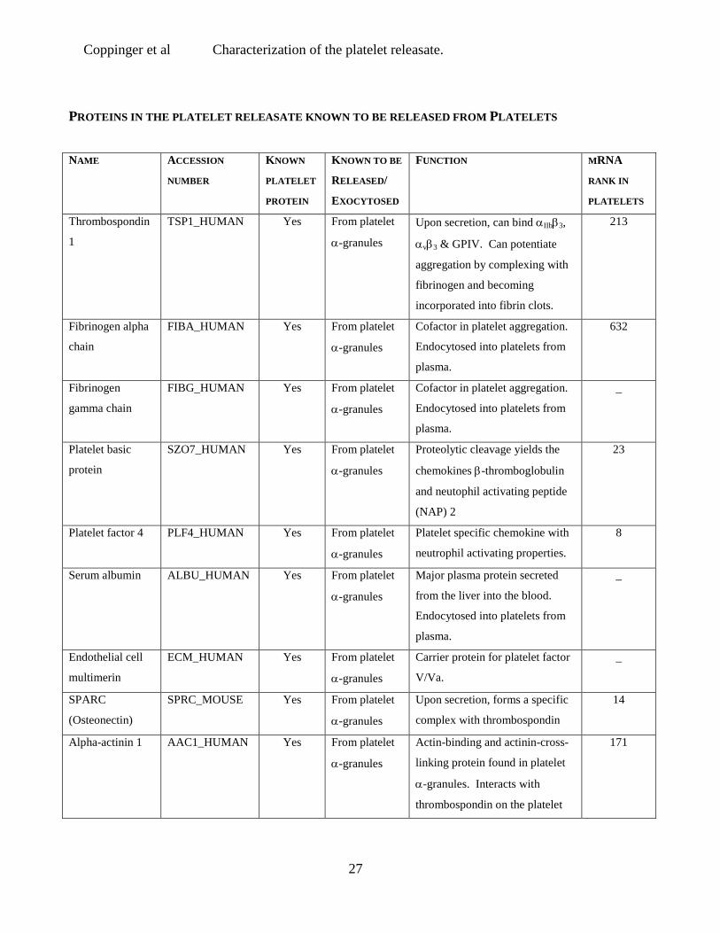
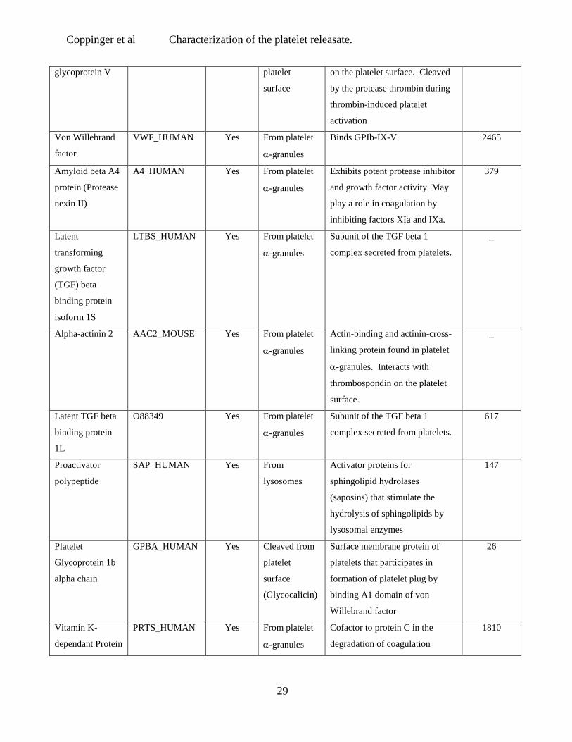
Table 2: Summary of 81 proteins from the thrombin-activated platelet releasate
identified using MudPIT. 81 proteins were identified using MudPIT from the thrombin
stimulated platelet supernatant fraction. Spectra were identified using the SEQUEST program
and a composite mouse and human database (NCBI July 2002 release) in three replicate
experiments. Information on their functions and whether they are secretory proteins are
provided. Also indicated is whether these proteins have a corresponding platelet mRNA. The
rank of abundance of the message is denoted numerically in the last column.
- Below the threshold for detection on the Affymetrix microarray.
\ Not present on Affymetrix microarray
Coppinger et al Characterization of the platelet releasate.
27
PROTEINS IN THE PLATELET RELEASATE KNOWN TO BE RELEASED FROM PLATELETS
NAME ACCESSION
NUMBER
KNOWN
PLATELET
PROTEIN
KNOWN TO BE
RELEASED/
EXOCYTOSED
FUNCTION MRNA
RANK IN
PLATELETS
Thrombospondin
1
TSP1_HUMAN Yes From platelet
α-granules
Upon secretion, can bind αIIbβ3,
αvβ3 & GPIV. Can potentiate
aggregation by complexing with
fibrinogen and becoming
incorporated into fibrin clots.
213
Fibrinogen alpha
chain
FIBA_HUMAN Yes From platelet
α-granules
Cofactor in platelet aggregation.
Endocytosed into platelets from
plasma.
632
Fibrinogen
gamma chain
FIBG_HUMAN Yes From platelet
α-granules
Cofactor in platelet aggregation.
Endocytosed into platelets from
plasma.
_
Platelet basic
protein
SZO7_HUMAN Yes From platelet
α-granules
Proteolytic cleavage yields the
chemokines β-thromboglobulin
and neutophil activating peptide
(NAP) 2
23
Platelet factor 4 PLF4_HUMAN Yes From platelet
α-granules
Platelet specific chemokine with
neutrophil activating properties.
8
Serum albumin ALBU_HUMAN Yes From platelet
α-granules
Major plasma protein secreted
from the liver into the blood.
Endocytosed into platelets from
plasma.
_
Endothelial cell
multimerin
ECM_HUMAN Yes From platelet
α-granules
Carrier protein for platelet factor
V/Va.
_
SPARC
(Osteonectin)
SPRC_MOUSE Yes From platelet
α-granules
Upon secretion, forms a specific
complex with thrombospondin
14
Alpha-actinin 1 AAC1_HUMAN Yes From platelet
α-granules
Actin-binding and actinin-cross-
linking protein found in platelet
α-granules. Interacts with
thrombospondin on the platelet
171

Coppinger et al Characterization of the platelet releasate.
28
surface.
Alpha_1-
antitrypsin
A1AT_HUMAN Yes From platelet
α-granules
Acute phase protein, similar to
complement, inhibits proteinases
_
Fibrinogen beta
chain
FIBB_HUMAN Yes From platelet
α-granules
Cofactor in platelet aggregation.
Endocytosed into platelets from
plasma.
_
Factor V FA5_HUMAN Yes From platelet
α-granules
Cofactor that participates with
Factor Xa to activate
prothrombin to thrombin.
_
Secretory granule
proteoglycan core
protein
PGSG_HUMAN Yes From platelet
α-granules
Function unknown. Associates
and co-released with
inflammatory mediators such as
platelet factor 4
343
Thymosin beta-4 TYB4_MOUSE Yes From platelets
α-granules
G-actin binding protein.
Functions as an antimicrobial
peptide when secreted.
5
Fructose
Biphosphate
Aldolase
ALFA_MOUSE Yes From platelets
& exosomes
from dendritic
cells
Glycolytic enzymes that converts
fructose 1,6-bis phosphate to
glyceraldeyde 3-phosphate and
dihydroxy acetone phosphate.
79
Clusterin CLUS_HUMAN Yes From platelet
α-granules
Not clear. Possibly platelet
derived apolipoprotein J
participates in short term wound
repair and chronic pathogenic
processes at vascular interface
1
Coagulation
factor XIIIA
chain
F13A_HUMAN Yes From platelet
α-granules
Coagulation protein involved in
the formation of the fibrin clots.
19
Metalloproteinase
inhibitor 1
TIM1_HUMAN Yes From platelet
α-granules
Interacts with metalloproteinases
and inactivates them. Stimulates
growth and differentiation of
erythroid progenitors, dependant
on disulfide bonds.
112
Platelet GPV_HUMAN Yes Cleaved from Part of the GPIb-IX-V complex
_
Coppinger et al Characterization of the platelet releasate.
29
glycoprotein V platelet
surface
on the platelet surface. Cleaved
by the protease thrombin during
thrombin-induced platelet
activation
Von Willebrand
factor
VWF_HUMAN Yes From platelet
α-granules
Binds GPIb-IX-V. 2465
Amyloid beta A4
protein (Protease
nexin II)
A4_HUMAN Yes From platelet
α-granules
Exhibits potent protease inhibitor
and growth factor activity. May
play a role in coagulation by
inhibiting factors XIa and IXa.
379
Latent
transforming
growth factor
(TGF) beta
binding protein
isoform 1S
LTBS_HUMAN Yes From platelet
α-granules
Subunit of the TGF beta 1
complex secreted from platelets.
_
Alpha-actinin 2 AAC2_MOUSE Yes From platelet
α-granules
Actin-binding and actinin-cross-
linking protein found in platelet
α-granules. Interacts with
thrombospondin on the platelet
surface.
_
Latent TGF beta
binding protein
1L
O88349 Yes From platelet
α-granules
Subunit of the TGF beta 1
complex secreted from platelets.
617
Proactivator
polypeptide
SAP_HUMAN Yes From
lysosomes
Activator proteins for
sphingolipid hydrolases
(saposins) that stimulate the
hydrolysis of sphingolipids by
lysosomal enzymes
147
Platelet
Glycoprotein 1b
alpha chain
GPBA_HUMAN Yes Cleaved from
platelet
surface
(Glycocalicin)
Surface membrane protein of
platelets that participates in
formation of platelet plug by
binding A1 domain of von
Willebrand factor
26
Vitamin K-
dependant Protein
PRTS_HUMAN Yes From platelet
α-granules
Cofactor to protein C in the
degradation of coagulation
1810

Coppinger et al Characterization of the platelet releasate.
30
S factors Va and VIIIA
Platelet factor 4
variant
PF4V_HUMAN Yes From platelet
α-granules
Platelet specific chemokines with
neutrophil activating properties.
346
Alpha-2
macroglobulin
A2MG_HUMAN Yes From platelet
α-granules
Acute phase protein, similar to
compliment, inhibits proteinases
\
Alpha-actinin 4 AAC4_HUMAN Yes From platelet
α-granules
Actin-binding and actinin-cross-
linking protein found in platelet
α-granules. Interacts with
thrombospondin on the platelet
surface.
467
SECRETORY PROTEINS IN THE PLATELET RELEASATE NOT PREVIOUSLY IDENTIFIED IN PLATELETS
NAME ACCESSION
NUMBER
KNOWN
PLATELET
PROTEIN
KNOWN TO BE
RELEASED/
EXOCYTOSED
FUNCTION MRNA
RANK IN
PLATELETS
Vitamin D-
binding protein
VTDB-HUMAN No From liver to
plasma
Carries Vitamin D Sterols.
Prevents actin polymerisation.
Has T Lymphocyte surface
association
_
Beta-2-
microglobulin
B2MG_HUMAN No Exosomes
from, dendritic
cells, B cells,
enterocytes,
tumour cells &
T cells
Is the beta-chain of the major
histocompatibility complex
(MHC) class I molecule.
3
Hemoglobin
alpha chain
HBA_HUMAN No Exosomes
from dendritic
cells &
Phagosomes in
macrophages
Oxygen transport. Potentiates
platelet aggregation through
thromboxane receptor
21
Plasminogen PLMN_HUMAN Yes From kidney
into plasma
Dissolves fibrin in blood clots,
proteolytic factor in tissue
remodelling, tumor invasion and
inflammation
_
Serotransferrin TRFE_HUMAN Yes From Liver
into plasma
Precursor to macromolecular
activators of phagocytosis
_

Coppinger et al Characterization of the platelet releasate.
31
(MAPP), which enhance
leukocyte phagocytosis via the
FcγRΙΙ receptor.
Pyruvate kinase,
M2 isozyme
KPY2_MOUSE Yes B cell
exosomes
Involved in final stage of
glycolysis. Presented as an
autoantigen by dendritic cells
61
Actin, aortic
smooth muscle
ACTA_HUMAN Yes Exosomes
from B cells,
dendritic cells,
enterocytes
and
mastocytes.
Major cytoskeletal protein. _
Actin ACTB_HUMAN Yes Exosomes
from B cells,
dendritic cells,
enterocytes
and
mastocytes.
Major cytoskeletal protein.
External function unknown.
11
14-3-3 protein
zeta/delta
143Z_MOUSE Yes Exosomes
from dendritic
cells &
Phagosomes in
macrophages
External function unknown.
Involved intracellularly in signal
transduction, however, may have
a role in regulating exocytosis.
63
Hemopexin HEMO_HUMAN No From liver to
plasma
Haem binding protein with
metallo proteinase domains
_
Hemoglobin beta
chain
HBB_HUMAN No From liver to
plasma &
Phagosomes
from
macrophages
Oxygen transport. 9
Peptidyl-prolyl-
cis isomerase A
(Cyclophilin A)
CYPH_MOUSE No From smooth
muscle cells
Cellular protein with isomerase
activity. Secreted vascular
smooth muscle cell growth factor
116
Calumenin CALU_MOUSE No From many
cells including
fibroblast and
An inhibitor of the Vitamin K
epoxide reductase-warfarin
interaction.
1816

Coppinger et al Characterization of the platelet releasate.
32
COS cells
Adenylyl cyclase-
associated protein
1 (CAP 1)
CAP1_MOUSE No Phagosomes
from
macrophages
Contains a WH2 actin-binding
domain (as beta-thymosin 4).
Known to regulate actin
dynamics. May mediate
endocytosis.
174
Tubulin TBA1_HUMAN Yes Exosomes
from dendritic
cells &
Phagosomes
from
macrophages
Cytoskeletal protein involved in
microtubule formation
33
Apolipoprotein
A-1
APA1_HUMAN Yes From liver to
plasma, From
monocytes &
exosomes of
dendritic cells.
Role in high density lipoprotein
binding to platelets
_
Compliment C3 CO3_HUMAN No From liver
cells and
monocytes
Activator of the compliment
system. Cleaved to α, β and γchains normally prior to
secretion, and is a mediator of the
local inflammatory response.
\
Transthyretin TTHY_HUMAN No From choroid
plexus into
cerebrospinal
fluid (CSF).
Thyroid hormone-binding protein
secreted from the choroid plexus
and the liver in to CSF and
plasma respectively.
_
Cofilin COF1_MOUSE Yes Exosome from
dendritic cells
Actin demolymerisation /
regulation in cytoplasm
31
Profilin PRO1_MOUSE Yes Exosome from
dendritic cells
Actin demolymerisation /
regulation in cytoplasm
70
Secretogranin III SG3_MOUSE No From neuronal
cells
Unclear possibly involved in
secretory granule biogenesis.
May be cleaved into active
inflammatory peptide like
secretogranin II.
\
Phosphoglycerate PGK1_MOUSE Yes From tumour Glycotic enzyme. Secreted from 378

Coppinger et al Characterization of the platelet releasate.
33
kinase cells tumour cells and involved in
angiogenesis
Alpha-IB
glycoprotein
A1BG_HUMAN No From many
cell including
white blood
cells
Found in plasma, not clear,
possibly involved in cell
recognition as a new member of
the immunoglobulin family.
\
Compliment C4
precursor
CO4_HUMAN No From many
cell including
white blood
cells
Activator of the compliment
system. Cleaved normally prior
to secretion, its products mediate
the local inflammatory response.
_
Prothrombin THRB_HUMAN No From liver to
plasma
Converts fibrinogen to fibrin and
activates coagulation factors
including factor V.
2143
Glyceraldehyde
3-phosphate
dehydrogenase
G3P2_HUMAN Yes From B cell
exosomes &
Phagosomes
from
macrophages
Mitochondrial enzyme involved
in glycolysis. May catalyse
membrane fusion.
60
Alpha-1-acid
glycoprotein
A1AH_HUMAN No From liver to
plasma
Modulates activity of the immune
system during the acute phase
reaction. Binds platelet surface.
_
Gelsolin GELS_HUMAN Yes Secreted
isoform
released from
liver and
adipocytes
Two isoforms, a cytoplasmic
actin modulating protein and a
secreted isoform involved in the
inflammatory response.
438
PROTEINS IN THE PLATELET RELEASATE NOT PREVIOUSLY REPORTED TO BE RELEASED FROM ANY
CELL
NAME ACCESSION
NUMBER
KNOWN
PLATELET
PROTEIN
KNOWN TO BE
RELEASED/
EXOCYTOSED
FUNCTION MRNA
RANK IN
PLATELETS
Calmodulin CALM_HUMAN Yes No evidence Known to regulate calcium-
dependent acrosomal exocytosis
in neuroendocrine cells
55
Pleckstrin PLEK_HUMAN Yes No evidence A substrate for protein kinase C, 532

Coppinger et al Characterization of the platelet releasate.
34
its phosphoryation is important
for platelet secretion
Nidogen NIDO_HUMAN Yes No evidence Glycoprotein found in basement
membranes,interacts with
laminin, collagen and integrin on
neutrophils.
_
Fibrinogen type
protein
Q8VCM7 No No evidence Similar to fibrinogen _
Rho GDP-
dissociation
inhibitor 2
GDIS_MOUSE Yes No evidence Regulates the GDP/GTP
exchange reaction of rho
proteins. Regulates platelet
aggregation. Involved in
exocytosis in mast cells
97
Rho GTPase
activating protein
Q92512 Yes No evidence Promotes the intrinsic GTP
hydrolysis activity of Rho family
proteins. Involved in regulating
myosin phosphorylation in
platelets
\
Transgelin TAG2_HUMAN No No evidence Actin-binding protein. Loss of
transgelin expression important
in early tumor progression. May
serve as a diagnostic marker for
breast and colon cancer
7
Vinculin VINC_HUMAN Yes No evidence Actin-binding protein 44
WD-repeat
protein
WDR1_HUMAN No No evidence Actin binding protein 127
Superoxide
Dismutase
(SOD)
SODC_HUMAN Yes No evidence Important enzyme in cellular
oxygen metabolism, role for
SOD-1 in inflammation
1730
78kDa glucose
related protein
GR78_MOUSE No No evidence Chaperone in the ER involved in
inhibition of secreted coagulation
factors thus reducing
prothrombotic potential of cell
_
Bromodomain
and PHD finger-
containing
BRF3_HUMAN No No evidence Unknown \

Coppinger et al Characterization of the platelet releasate.
35
protein 3
[Fragment]
Titin Q8WZ42 Yes No evidence Anchoring protein of
actinomyosin filaments. Role in
secretion of myostatin
_
Similar to
hepatocellular
carcinoma-
associated
antigen 59
Q99JW3 No No evidence Tumour Marker _
FKSG30 Q9BYX7 No No evidence Actin binding protein \
RNA binding
protein
Q9UQ35 No No evidence RNA binding protein _
Hypothetical
protein
Q9BTV9 No No evidence Unknown 1744
Intracellular
hyaluronan-
binding protein
p57
Q9JKS5 No No evidence Unknown _
Hypothetical
protein
Y586_HUMAN No No evidence Unknown \
Filamin fragment
(Hypothetical
54kDa protein)
Q99KQ2 Yes No evidence Unknown _
Filamin FLNA_HUMAN Yes No evidence Actin-binding protein. Essential
for GP1b alpha anchorage at high
shear. Substrate for caspase-3.
43
Talin TALI_HUMAN Yes No evidence Actin-binding protein that binds
to integrin beta 3 domain.
17
Zyxin ZYX_HUMAN Yes No evidence Associates with the actin
cytoskeleton near adhesion
plaques. Binds α-actinin and
VASP.
145

Coppinger et al Characterization of the platelet releasate.
36
Figure Legends
Figure 1 Proteins released by platelets following thrombin activation. (a) The proteins
from the supernatant of non-activated (C) and thrombin-activated platelets (A) were solubilized
in SDS reducing buffer, separated by one-dimensional SDS-PAGE (4-20 % gradient gel) and
stained with colloidal Coomassie Blue. (b) A duplicate of the gel was probed with a monoclonal
antibody to thrombospondin (Clone p10). Molecular weight markers are indicated.
Figure 2 Two-dimensional electrophoresis (2-DE) of the releasate fraction from
thrombin-activated platelets. 400µg of the releasate fraction from thrombin-activated platelets
was separated by 2-DE and stained with Coomassie Blue dye. Spots were excised and digested
with trypsin and the resulting peptides analysed by MALDI-TOF MS. A representative gel is
shown and the proteins identified listed (Table 1). Molecular weight markers and pI values are
indicated.
Figure 3. Seven-step Multidimensional Protein Identification Technology (MudPIT).
(a-g) The releasate from thrombin-activated platelets was digested with trypsin and the resulting
peptides separated using strong cation exchange and reverse phase chromatography before
introduction into an ion trap mass spectrometer. This figure displays the resulting
chromatograms from the 2D-LC tandem MS. Chromatograms a-g represent seven successive
salt elutions from a HPLC column. (h) A representative tandem MS spectrum is shown for a
peptide from thrombospondin that was identified using SEQUEST.
Figure 4 Western blot for calumenin, cyclophilin A and secretogranin III. The
presence of calumenin (a), cyclophilin (b) and secretogranin III (c) was confirmed in lysates
from control (platelet control, PC) and thrombin-activated (platelet activated, PA) platelets, as
well as the thrombin-activated releasate (releasate activated, RA). These proteins were not
found in the supernatant from unactivated platelets (releasate control, RC). In addition, SgIII
and calumenin were not detected in a crude leukocyte lysate (WBC), although cyclophilin A
was present in low amounts.

Coppinger et al Characterization of the platelet releasate.
37
Figure 5 Confocal Microscopy for calumenin, cyclophilin A and secretogranin III.
a) Platelets were adhered to fibrinogen coated slides for 5 (platelets resting) and 60 min
(platelets activated and spread). A granular staining pattern similar to thrombospondin was
observed for cyclophilin A and secretogranin III at both time points whereas a more diffuse
pattern was observed for calumenin. b) Activated platelets labeled with CD41 and a rhodamine
labeled secondary antibody, and dual stained with antibodies to SgIII, cyclophilin A and
calumenin (labelled with FITC-conjugated secondary antibodies). c) Secondary antibodies
alone.
Figure 6 Immunohistochemistry of normal and atherosclerotic tissue.
Immunohistochemistry for each of the three proteins was performed on sections of arterial
tissue from five patients with atherosclerosis. The results from one representative patient are
shown. Low power H & E staining of normal (a) and atherosclerotic plaque (h). Platelet
incorporation into the plaques was demonstrated by staining for the platelet specific proteins
PF4 (j) and CD41 (i). Secretogranin III (l) and calumenin (m) were widely expressed in the
plaque, while vascular smooth muscle cells in the lesion, identified by smooth muscle actin
staining (g & n), stained for cyclophilin A (k). No staining for secretogranin III, calumenin or
cyclophilin A was observed in sections of normal artery (b-f).

Coppinger et al Characterization of the platelet releasate.
38
Figure 1
C A
a b
66
116
45
205
29
C A
19
6

Coppinger et al Characterization of the platelet releasate.
39
Figure 2
3 4 5 6 7
66
45
36
29
19
1234
5678
91011
1213
1415
16171819
20 2122

Coppinger et al Characterization of the platelet releasate.
40
Figure 3
R T : 0 . 0 0 - 1 2 0 . 0 5
0 1 0 2 0 3 0 4 0 5 0 6 0 7 0 8 0 9 0 1 0 0 1 1 0 1 2 0T i m e ( m i n )
0
5
1 0
1 5
2 0
2 5
3 0
3 5
4 0
4 5
5 0
5 5
6 0
6 5
7 0
7 5
8 0
8 5
9 0
9 5
1 0 0
Re
lativ
e A
bu
nd
an
ce
0 . 7 2
1 . 1 4
9 2 . 7 8
1 0 8 . 9 59 3 . 6 9 1 0 9 . 3 1
9 2 . 2 5 1 0 6 . 6 18 6 . 9 1
8 0 . 0 67 7 . 8 61 . 9 4 7 4 . 3 9
6 7 . 1 06 2 . 0 33 3 . 2 02 8 . 7 9 5 1 . 4 93 9 . 5 06 . 0 0 1 8 . 7 5
N L :2 . 2 4 E 9
T I C M S T K _ 0 4 0 7 02 _ P L A T E LE T S _ A T _ ST E P 0 1
R T : 0 . 0 0 - 1 2 0 . 0 6
0 1 0 2 0 3 0 4 0 5 0 6 0 7 0 8 0 9 0 1 0 0 1 1 0 1 2 0T i m e ( m i n )
0
5
1 0
1 5
2 0
2 5
3 0
3 5
4 0
4 5
5 0
5 5
6 0
6 5
7 0
7 5
8 0
8 5
9 0
9 5
1 0 0
Re
lativ
e A
bund
anc
e
8 4 . 1 8
3 . 9 3
5 9 . 8 71 0 3 . 9 2
5 2 . 8 1
9 5 . 9 24 6 . 4 2
9 5 . 1 9
6 3 . 5 59 4 . 8 26 7 . 1 74 4 . 0 6 1 0 4 . 5 1
7 2 . 5 28 6 . 8 5
1 1 9 . 9 94 3 . 2 1 1 0 6 . 1 41 3 . 7 4 1 6 . 0 7 3 7 . 0 3
1 6 . 4 71 3 . 0 1 3 5 . 5 01 7 . 6 9 2 9 . 5 98 . 7 6
N L :1 . 2 4 E 9T I C M S T K _ 0 4 0 7 02 _ P l a t e l e t s_ A t _ s t e p 0 2
R T : 0 . 0 0 - 1 1 9 . 9 9
0 1 0 2 0 3 0 4 0 5 0 6 0 7 0 8 0 9 0 1 0 0 1 1 0T i m e ( m i n )
0
5
1 0
1 5
2 0
2 5
3 0
3 5
4 0
4 5
5 0
5 5
6 0
6 5
7 0
7 5
8 0
8 5
9 0
9 5
1 0 0
Rel
ativ
e A
bund
ance
6 9 . 7 1
7 2 . 4 2
8 4 . 6 07 6 . 5 3
5 5 . 3 7
6 3 . 2 78 5 . 7 05 0 . 3 2
9 9 . 3 44 3 . 0 4
9 0 . 2 79 9 . 9 02 5 . 3 2
2 6 . 2 06 . 9 3 1 1 4 . 3 21 5 . 2 0
1 0 2 . 5 8
N L :4 . 2 8 E 9
T I C M S T K _ 0 4 0 7 02 _ P l a t e l e t s_ A t _ s t e p 0 3
R T : 0 . 0 0 - 1 2 0 . 0 0
0 1 0 2 0 3 0 4 0 5 0 6 0 7 0 8 0 9 0 1 0 0 1 1 0T i m e ( m i n )
0
5
1 0
1 5
2 0
2 5
3 0
3 5
4 0
4 5
5 0
5 5
6 0
6 5
7 0
7 5
8 0
8 5
9 0
9 5
1 0 0
Rel
ativ
e A
bun
dan
ce
9 9 . 4 9 1 1 4 . 2 97 7 . 5 6
7 8 . 0 99 3 . 2 7
6 5 . 4 0
7 0 . 2 3 8 6 . 4 9
8 4 . 8 6
6 4 . 1 81 0 8 . 0 7
3 4 . 5 7
5 9 . 0 15 7 . 3 8
3 4 . 3 2
5 5 . 5 46 . 7 8 5 1 . 8 51 4 . 7 8
4 9 . 4 5
3 3 . 7 7 3 8 . 6 04 6 . 2 4
3 3 . 0 11 5 . 6 9 2 6 . 8 9
N L :1 . 0 2 E 9T I C M S T K _ 0 4 0 7 02 _ P l a t e l e t s_ A t _ s t e p 0 5
R T : 0 . 0 0 - 1 1 9 . 9 9
0 1 0 2 0 3 0 4 0 5 0 6 0 7 0 8 0 9 0 1 0 0 1 1 0T i m e ( m i n )
0
5
1 0
1 5
2 0
2 5
3 0
3 5
4 0
4 5
5 0
5 5
6 0
6 5
7 0
7 5
8 0
8 5
9 0
9 5
1 0 0
Re
lativ
e A
bund
anc
e
8 3 . 3 9
8 3 . 8 2
8 7 . 3 39 4 . 6 7
8 2 . 9 9
8 1 . 7 8
9 7 . 3 1
1 0 1 . 2 27 6 . 5 8
7 0 . 2 6 1 0 2 . 5 16 6 . 8 7
1 0 7 . 1 36 6 . 3 01 0 . 1 89 . 9 0 6 4 . 4 25 3 . 2 0
4 6 . 5 34 2 . 6 21 0 . 7 5 3 4 . 3 92 5 . 2 0
N L :7 . 2 6 E 8T I C M S T K _ 0 4 0 7 02 _ P l a t e l e t s_ A t _ s t e p 0 6
R T : 0 . 0 0 - 1 1 9 . 9 9
0 1 0 2 0 3 0 4 0 5 0 6 0 7 0 8 0 9 0 1 0 0 1 1 0T i m e ( m i n )
0
5
1 0
1 5
2 0
2 5
3 0
3 5
4 0
4 5
5 0
5 5
6 0
6 5
7 0
7 5
8 0
8 5
9 0
9 5
1 0 0
Re
lativ
e A
bund
anc
e
1 0 4 . 7 9 1 0 8 . 0 0
1 0 4 . 4 0
1 1 0 . 2 0
1 1 2 . 3 7
4 7 . 5 4
5 7 . 4 7
4 8 . 6 6 1 0 3 . 4 25 8 . 1 8
6 7 . 2 7
4 4 . 5 65 4 . 7 9 7 2 . 5 8 8 9 . 3 64 1 . 8 3
7 5 . 9 7 1 0 2 . 1 68 0 . 9 18 2 . 7 33 9 . 3 1
9 5 . 2 32 4 . 6 41 4 . 4 8 3 8 . 1 5
3 5 . 0 5
0 . 7 22 2 . 7 32 . 4 7
1 1 . 7 9
N L :3 . 3 6 E 7T I C M S T K _ 0 4 0 7 02 _ P L A T E LE T S _ A T _ ST E P 0 7
T K _ 0 4 0 7 0 2 _ P L A T E L E T S _ A T _ S T E P 0 3 # 4 7 1 5 R T : 1 0 3 . 2 6 A V : 1 N L : 2 . 0 2 E 4T : + c d F u l l m s 2 1 0 9 8 . 6 4 @ 3 5 . 0 0 [ 2 9 0 . 0 0 - 2 0 0 0 . 0 0 ]
4 0 0 6 0 0 8 0 0 1 0 0 0 1 2 0 0 1 4 0 0 1 6 0 0 1 8 0 0 2 0 0 0m / z
0
5
1 0
1 5
2 0
2 5
3 0
3 5
4 0
4 5
5 0
5 5
6 0
6 5
7 0
7 5
8 0
8 5
9 0
9 5
1 0 0
Rel
ativ
e A
bun
dan
ce
9 7 7 . 4 0
1 5 4 0 . 7 2
7 1 7 . 4 0
1 2 3 9 . 6 4
1 3 3 2 . 4 01 0 3 3 . 1 8
6 5 6 . 3 28 6 5 . 7 3
1 2 1 9 . 3 1 1 4 2 6 . 6 2 1 8 5 7 . 3 85 8 9 . 2 5 1 8 0 4 . 2 41 7 1 2 . 2 13 7 5 . 0 5 8 2 5 . 6 3
1 9 8 5 . 6 11 6 5 6 . 2 55 1 5 . 4 2
5 0 9 . 1 9
R T : 0 . 0 0 - 1 1 9 . 9 8
0 1 0 2 0 3 0 4 0 5 0 6 0 7 0 8 0 9 0 1 0 0 1 1 0T i m e ( m i n )
0
5
1 0
1 5
2 0
2 5
3 0
3 5
4 0
4 5
5 0
5 5
6 0
6 5
7 0
7 5
8 0
8 5
9 0
9 5
1 0 0
Re
lativ
e A
bund
anc
e
6 7 . 7 4
6 7 . 2 6
5 4 . 5 4
6 8 . 5 4
6 0 . 7 6
7 4 . 2 4
7 7 . 5 35 2 . 6 8
9 5 . 9 0
3 9 . 8 7 9 4 . 7 05 . 2 7 6 . 5 91 1 3 . 1 54 9 . 3 6 9 0 . 4 0
8 9 . 1 99 6 . 6 54 3 . 0 6 1 1 0 . 9 4
8 . 0 53 8 . 1 3
1 0 6 . 5 91 6 . 5 4 3 3 . 7 3
1 7 . 6 7
N L :5 . 0 1 E 8T I C M S T K _ 0 4 0 7 02 _ P L A T E LE T S _ A T _ ST E P 0 4
a
b
c
d
e
f
g
h

Coppinger et al Characterization of the platelet releasate.
41
Figure 4
c
a
b
RC RA PA PC WBC
Coppinger et al Characterization of the platelet releasate.
42
Figure 5
Calumenin
Cyclophilin A
Secretogranin III
Thrombospondin
Resting platelets Activated platelets
Calumenin
Cyclophilin A
Secretogranin III
DIC Protein-only Dual stained (+CD41)
Controls
FITC-labeled Anti-Ms & Anti Gt
FITC-labeled Anti Ms & Anti-Rb
Calumenin & FITC-labeled Anti-Rb& Rhodamine-labeled Anti-Ms
c
a
b
Coppinger et al Characterization of the platelet releasate.
43
Figure 6
Normal Artery Atherosclerotic plaque
a
c
b
d
f
e
g
i
k
j
l
m
n
h
Copyright © 2022 FDOKUMEN