Killer cell immunoglobulin-like receptor (KIR) genes and their HLA-C ligands in a Ugandan population

Hindawi Publishing CorporationEvidence-Based Complementary and Alternative MedicineVolume 2013, Article ID 563716, 17 pageshttp://dx.doi.org/10.1155/2013/563716
Review ArticleOn the G-Protein-Coupled Receptor Heteromers and TheirAllosteric Receptor-Receptor Interactions in the Central NervousSystem: Focus on Their Role in Pain Modulation
Dasiel O. Borroto-Escuela,1 Wilber Romero-Fernandez,1 Alicia Rivera,2
Kathleen Van Craenenbroeck,3 Alexander O. Tarakanov,4 Luigi F. Agnati,5 and Kjell Fuxe1
1 Department of Neuroscience, Karolinska Institutet, Retzius vag 8, 17177 Stockholm, Sweden2 Faculty of Science, University of Malaga, 29080 Malaga, Spain3 Laboratory of Eukaryotic Gene Expression and Signal Transduction (LEGEST), Ghent University-Gent, 9000 Ghent, Belgium4Russian Academy of Sciences, St. Petersburg Institute for Informatics and Automation, 193167 Saint Petersburg, Russia5 IRCCS, Lido Venice, 41100 Venice, Italy
Correspondence should be addressed to Kjell Fuxe; [email protected]
Received 27 February 2013; Revised 20 May 2013; Accepted 24 May 2013
Academic Editor: Yi-Hung Chen
Copyright © 2013 Dasiel O. Borroto-Escuela et al. This is an open access article distributed under the Creative CommonsAttribution License, which permits unrestricted use, distribution, and reproduction in any medium, provided the original work isproperly cited.
The modulatory role of allosteric receptor-receptor interactions in the pain pathways of the Central Nervous System and theperipheral nociceptors has become of increasing interest. As integrators of nociceptive and antinociceptive wiring and volumetransmission signals, with a major role for the opioid receptor heteromers, they likely have an important role in the pain circuitsand may be involved in acupuncture. The delta opioid receptor (DOR) exerts an antagonistic allosteric influence on the mu opioidreceptor (MOR) function in aMOR-DOR heteromer.This heteromer contributes to morphine-induced tolerance and dependence,since it becomes abundant and develops a reduced G-protein-coupling with reduced signaling mainly operating via 𝛽-arrestin2upon chronic morphine treatment. A DOR antagonist causes a return of the Gi/o binding and coupling to the heteromer and thebiological actions of morphine.The gender- and ovarian steroid-dependent recruitment of spinal cordMOR/kappa opioid receptor(KOR) heterodimers enhances antinociceptive functions and if impaired could contribute to chronic pain states in women.MOR1Dheterodimerizes with gastrin-releasing peptide receptor (GRPR) in the spinal cord, mediating morphine induced itch. Othermechanism for the antinociceptive actions of acupuncture along meridians may be that it enhances the cross-desensitization of theTRPA1 (chemical nociceptor)-TRPV1 (capsaicin receptor) heteromeric channel complexes within the nociceptor terminals locatedalong these meridians. Selective ionotropic cannabinoids may also produce cross-desensitization of the TRPA1-TRPV1 heteromericnociceptor channels by being negative allosteric modulators of these channels leading to antinociception and antihyperalgesia.
1. Introduction
The monoamine-peptide interactions have been of greatinterest [1]. How did they, in fact, interact at the molecularlevel? One possibility was that the monoamine and pep-tide signals became integrated through direct neuropeptide-monoamine receptor-receptor interactions in the plasmamembrane. We began to test this hypothesis in 1980-1981 inmembrane preparations of various Central Nervous System
(CNS) regions and found that neuropeptides could modulatethe binding characteristics, especially the affinity, of themonoamine receptors in a receptor subtype specific way[2, 3]. Thus, intramembrane receptor-receptor interactionsdid exist besides indirect actions via phosphorylation andchanges in membrane potential. The results were in linewith earlier findings by Limbird et al. in 1975, showingnegative cooperativity in 𝛽-adrenergic receptors, which maybe explained by the existence of receptor homodimers leading

2 Evidence-Based Complementary and Alternative Medicine
to orthosteric site-site interactions [4]. It was also clear thatadapter proteins can be involved in mediating the receptor-receptor interactions in brain membranes [5, 6].
In this paper we give a brief overview of the modulatoryrole of receptor heteromers in the pain pathways in the CNSand in peripheral nociceptors. They seem to be integratorsof nociceptive and antinociceptive wiring (WT) and volumetransmission (VT) signals with a major role for the opioidreceptor heteromers. Their relevance in the mechanisms forthe antinociceptive actions of acupuncture will be discussed.
2. Primary Afferents of the Dorsal HornMediating Nociception
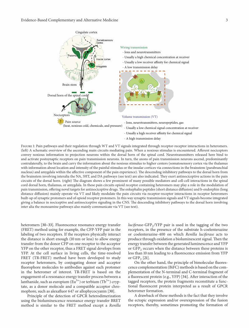
We have different types of nociceptors coming into the dorsalhorn (primary afferent fibres). One major class includesmedium diameter myelinated (A𝛿) afferents that mediatewell-localized fast pain. The second class of nociceptorincludes small diameter unmyelinated “C” fibers thatmediatepoorly localized, slow pain [7]. There exist a very preciselaminar organization of the dorsal horn of the spinal cord.Theunmyelinated, peptidergic C andmyelinatedA𝛿nocicep-tors terminate most superficially, synapsing upon large pro-jection neurons in lamina I and interneurons located in outerlamina II.Thus, primary afferent nociceptors convey noxiousinformation to projection neurons within the dorsal horn ofthe spinal cord (Figure 1, left).
3. Pain Pathways and TheirRegulation through WT and VT SignalsIntegrated through Receptor-ReceptorInteractions in Heteromers
A subset of these projection neurons transmits informationto the somatosensory cortex via the thalamus, providinginformation about the location and intensity of the painfulstimulus [7]. Other projection neurons reach the cingulateand insular cortices via relay stations in the lower brainstem(parabrachial nucleus) and amygdala, contributing to theemotional component of the pain experience (Figure 1, left).This ascending information also passes on to neurons of therostral ventral medulla, including the raphe and pararaphearea and the periaqueductal gray of the midbrain to activatedescending feedback systems like distinct bulbo-spinal sero-tonin (5-HT) and noradrenergic (NA) neurons [8] that regu-late the pain transmission output from the spinal cord, espe-cially the lateral paragigantocellular reticular 5-HT neuronsmay be involved in this process.
This important descending inhibitory control of noci-ception in the dorsal horn of the spinal cord (Figure 1, left)involves especially the periaqueductal gray-rostral ventrome-dial medulla, the dorsal reticular nucleus of the medulla, andthe ventrolateral medulla [9]. The descending bulbo-spinalNA and 5-HT neuron systems [10–12] play a substantial role[13–15]. Noxious stimulation excites the 5-HT neurons of thelateral paragigantocellular reticular and raphe magnus nuclei[16], building up group B3 of Dahlstroem and Fuxe [10] andthese 5-HT neuron systems produce strong analgesia and
the former also cardiovascular activation. Also descendingprojections to the dorsal horn from DA neurons in theposterior hypothalamus participate in control of pain [17].The major descending inhibitory bulbo-spinal systems areunder the control of high densities of mu opioid receptors[18, 19]. Blockade of the opioid receptors in the medullaryreticular nucleus dorsalis prevents analgesia produced bydiffuse noxious inhibitory controls [20].The anti-nociceptiveactions of the descending inhibitory pathways in the spinalcord involves the indirect activation of opioid receptors inthe dorsal horn, especially in the superficial layers. Themajor mode of communication in the descending inhibitorymonoamine pathways of nociception is extrasynaptic volumetransmission involvingmonoamine diffusion in the um range[21] and activation of their extrasynaptic receptors locatedon the nociceptors and their target nerve cells mainly foundin the superficial layers in the dorsal horn. However, wiringtransmission also exists in these descending systems throughformation of monoamine synapses. When neuropeptides arereleased from these descending inhibitory pathways longdistance volume transmission can develop in the order of 0.1–1mmand also involve CSF volume transmission provided thepeptides do not undergo rapid degradation [22].
In these pain circuits wewill describe howopioid receptorcontaining heteromers may play a role in the modulation ofpain transmission, offering novel targets for antinociceptivedrugs.Theymay be involved in conveying the antinociceptiveactions of acupuncture [23] since inter alia electroacupunc-ture has been shown to increase transcription and translationof enkephalins in the rostral ventrolateral medulla of rats,a region involved in regulation of not only circulation andrespiration but also pain [24]. The enkephalin peptides(short distance diffusion) and 𝛽-endorphin (long distancediffusion)mainly operate via VT [25–27] and likelymodulatethe pain circuits via receptor-receptor interactions in receptorheteromers built-up of synaptic protomers and of opioidreceptor protomers. In this way synaptic transmission signalsand VT signals become integrated giving a balance in noci-ceptive and antinociceptive signaling in the CNS (Figure 1).
4. Principal Features ofthe Receptor Heteromers
As mentioned in the introduction, the concept of intramem-brane receptor-receptor interactions in receptor heteromerswas born in the analysis of neuropeptide-monoaminereceptor-receptor interactions in 1980-81. The concept ofG protein-coupled receptor (GPCR) heteromers was laterconfirmed in 1998-1999 by studies reporting that two non-functional GPCR monomers, gamma amino butyric acid(GABA)B1 and GABAB2 receptors, can assemble in a sig-nalling heterodimer, the GABAB receptor at the cell surface[28, 29].
5. The Receptor-Receptor Interaction Toolbox
5.1. Fluorescence and Bioluminescence Resonance EnergyTransferMethods. Different resonance energy transfermeth-ods have been used to study the existence of GPCR
Evidence-Based Complementary and Alternative Medicine 3
Cingulate cortex
Wiring transmission- Ions and neurotransmitters- Usually a high chemical concentration at receiver- Usually a low receiver affinity for chemical signal- A low transmission delay
Brain stem
Dorsal horn of the spinal cord
Nociceptors Volume transmission (VT)
- Ions, neurotransmitters, neuropeptides, gas- Usually a low chemical signal concentration at receiver- Usually a high receiver affinity for chemical signal- A high transmission delay
(heat, noxious cold, chemicals, and pressure)Pain source
Figure 1: Pain pathways and their regulation through WT and VT signals integrated through receptor-receptor interactions in heteromers.(left) A schematic overview of the ascending main circuits mediating pain. When a noxious stimulus is encountered. Afferent nociceptorsconvey noxious information to projection neurons within the dorsal horn of the spinal cord. Neurotransmitters released here bind toand activate postsynaptic receptors on pain transmission neurons. In turn, the axons of pain transmission neurons ascend, predominantlycontralaterally, to the brain and carry the information about the noxious stimulus to higher centers (somatosensory cortex via the thalamuswith information about location and intensity of the painful stimulus or the insular cortices via connections in the brainstem (parabranchialnucleus) and amygdala within the affective component of the pain experience). The descending inhibitory pathways to the dorsal horn fromthe brainstem involving interalia the NA, 5HT, and DA pathways (see text) are also indicated. They exert antinociceptive actions in the paincircuits of the dorsal horn. (right) The diagram shows a few prominent of many possible mediators and cell-cell interactions in the spinalcord dorsal horn, thalamus, or amygdala. In these pain circuits opioid receptor containing heteromers may play a role in the modulation ofpain transmission, offering novel targets for antinociceptive drugs.The enkephalin peptides (short distance diffusion) and b-endorphin (longdistance diffusion) mainly operate via VT and likely modulate the pain circuits via receptor-receptor interactions in receptor heteromersbuilt-up of synaptic protomers and of opioid receptor protomers. In this way synaptic transmission signals and VT signals become integratedgiving a balance in nociceptive and antinociceptive signaling in the CNS. The descending inhibitory pathways to the dorsal horn involvinginter alia the monoamine pathways also mainly communicate via VT (see text).
heteromers [30–33]. Fluorescence resonance energy transfer(FRET) method using for example, the CFP-YFP pair in thelabeling of two receptors. If the receptors physically interactthe distance is short enough (10 nm or less) to allow energytransfer from the donor CFP on one receptor to the acceptorYFP on the other receptor, thus a FRET signal develops fromYFP. At the cell surface in living cells, the time-resolvedFRET (TR-FRET) method have been developed to studyreceptor heteromers, by conjugating donor and acceptorfluorophore molecules to antibodies against each protomerin the heteromer of interest. TR-FRET is based on theengagement of a resonance energy transfer process between alanthanide, such as europium (Eu3+) or terbium (Tb3+) cryp-tate, as a donor molecule and a compatible acceptor chro-mophore, such as alexafluor 647 or allophycocyanin [30].
Principle of the detection of GPCR heterodimerizationusing the bioluminescence resonance energy transfer BRETmethod is similar to the FRET method except a Renilla
luciferase-GFP2/YFP pair is used in the tagging of the two
receptors, in the presence of the substrate h-coelenterazineor coelenterazine-400 on which Renilla luciferase acts toproduce through oxidation a bioluminescent signal.Then theenergy transfer between the generated luminescence andYFPor GFP
2occurs when the distance between these proteins is
less than 10 nm leading to a fluorescence emission from YFPor GFP
2[21].
On the other hand, the principle of bimolecular fluores-cence complementation (BiFC)methods is based on the com-plementation of the N-terminal and C-terminal fragment ofa fluorescent protein (e.g., YFP) [34]. After interaction of thetagged receptors, the protein fragments reconstitute a func-tional fluorescent protein interpreted as a result of GPCRheteromer formation.
A drawback of these methods is the fact that they involvethe ectopic expression and/or overexpression of the fusionreceptors, thereby, sometimes promoting the formation of

4 Evidence-Based Complementary and Alternative Medicine
HO
N
O
OH
OOH
NH
O
NH
OO
NHON
HOHO
HON
[MOR][MOR][MOR] [MOR] [DOR]
Salvinorin A
DAMGO
MDAN-18 bivalent ligand
MOR monomer MOR homo- and heteromers
MOR agonist DOR antagonist
Intramolecular interactions Intermolecular receptor-receptor interactions
HO
NH2HH
O
O
OOO
OO
O O
O
OO
CH3
CH3
CH3
CH3
CH3
H3C
N
NNN
HH
H
NH2HO
OH
Figure 2: Intra- and intermolecular allosteric receptor-receptor interactions. Allosteric mechanisms make possible the integrative activitytaking place intramolecularly in monomers (left) or intermolecularly in homo-/heteromers (right). As one example of the intramolecularallosteric mechanisms is the allosteric binding of salvinorin A to the extracellular site of MOR, which partially affects the activity of theorthosteric MOR binding site via a conformational change [105]. Intermolecular allosteric mechanisms take place through the formationof different types of receptor homo-/heteromers and receptor/protein complexes which can change the function of an individual receptorpresent in a homomer or heteromer. Another example based on the intermolecular heteromer interactions is the use of heterobivalent ligandscontaining a MOR agonist and an DOR antagonist linked through a spacer of variable size which may function as useful molecular probesfor targeting the MOR-DOR heteromer and in this way counteracting the DOR antagonism onMOR function. Such compounds may have apotential use in pharmacotherapy of pain.
artefacts. It is therefore advisable, whenever possible, toconsider the physiological expression levels of the receptorpairs under study.
5.2. In Situ Proximity Ligation Assay. However, despite exten-sive experimental results supporting the formation of GPCRheteromers in heterologous systems (mainly by BRET andFRET methodologies), the existence of such heteromers inthe CNS and other tissues remains largely unknown, mostlybecause of the lack of appropriate methodology. Recently,a well-characterized in situ proximity ligation assay (in situPLA) has been adapted to confirm the existence of GPCRheteromers in brain slices ex vivo [35–39].
In situ PLA is based on a pair of antibodies that can bindto target proteins and to which oligonucleotides have beenattached. When the so-called proximity probes recognizea target, for example, the receptor heteromer, the attachedoligonucleotides are brought into a sufficiently close spatialproximity to allow them to join followed by ligation of the twolinear oligonucleotides into a circular DNA molecule. Thisnewly formed DNA circle strand can serve as a template forrolling circle amplification (RCA), resulting in a long single-stranded rolling circle product (RCP) attached to one of theproximity probes. Since the RCP is linked to the proximityprobe, it is attached at the site where the proximity probebound, which means that it can be used to reveal the locationof the receptor complex [40, 41]. The RCPs can then bedetected and quantified by hybridizing fluorescent oligonu-cleotides to the repeated sequences of the RCPs, renderingthem visible by fluorescence microscopy. With the in situPLA method the striatal adenosine 2A receptor (A2AR)-dopamine D2 receptor (D2R) heteromers and D2R-oxytocin
receptor heteromers have for example, been shown [35, 39].Also the hippocampal and the mesencephalic raphe FGFR1-5-HT1AR heteromers have recently been demonstrated [36,37]. The in situ PLA procedure represents a high selectivityand sensitivity assay to demonstrate GPCR heteromers inbrain [38].
6. Allosteric Receptor-Receptor Interactions
In the beginning, allosteric mechanisms were only discussedin terms of intramolecular interactions within a receptorbetween orthosteric and allosteric sites. This was the classicpharmacology (Figure 2, left).
Now we have moved into a novel pharmacology, whereintermolecular receptor-receptor interactions can occur andresults in novel receptor recognition, pharmacology andsignaling (Figure 2, right). Intermolecular allosteric mecha-nisms through the receptor interface produce these changesinvolving also receptor/protein complexes. An example ofthe novel pharmacology is the use of heterobivalent ligands[42–44] containing, for example, MOR agonist and DORantagonist pharmacophores linked through a spacer of vari-able size which may function as useful molecular probesfor targeting the MOR-DOR heteromer and in this waycounteracting the DOR antagonism on MOR function [43](Figure 2, right). Such compounds may have a potential usein pharmacotherapy of pain.
Allosteric mechanism causes a marked rise of the reper-toire of GPCR recognition, pharmacology, trafficking andsignaling of the participating protomers. This is achievedthrough changes in recognition, G protein selectivity, andsignaling cascades with among others switching from G

Evidence-Based Complementary and Alternative Medicine 5
proteins to 𝛽-arrestin or to calmodulin [33, 45]. Its functionmay also change by becoming linked to Receptor TyrosineKinases (RTKs) or to ion channel receptors [36, 37, 46].
The term moonlighting protein is used to describemultifunctional proteins in which several functions can befound in a single strand of amino acids unrelated to splicing,posttranslational changes, and so forth [47, 48]. In GPCRheteromers moonlighting is brought about by the allostericreceptor-receptor interactions altering the function of thereceptor protomers of the heteromer through conformationalchanges in single strands of amino acids [46].
7. The Receptor Interface
We are interested in the receptor interface since it can bea target for novel drugs by their ability to block or mimicthe allosteric receptor-receptor interactions [21, 49–51]. Theinterface in the A2AR-D2R heteromer can be given as anexample [50, 51]. It shows helix-helix interactions in theplasma membrane between A2AR TMIV and D2R-TMV.Intracellular electrostatic interactions between D2R IC3 andA2AR C-terminal tail involve positively charged argininesin the D2R IC3 and negatively charged residues in theA2AR especially phosphorylated serine [50–54]. Electrostaticinteractions may represent important hot spots in the recep-tor heteromer interface. The prototype was the A2AR-D2Rheteromer but it exists also in theA2AR-D3R andA2AR-D4Rinterface [55], giving an amazing stability of the heteromersbased on the arginine-phosphate bond [54].
Based on a mathematical approach developed by Dr.Tarakanov, we have deduced, based on 48 pairs of receptorsthat form or not form heterodimers, a set of triplet aminoacid homologies that may be critically involved in receptor-receptor interactions [56]. We call it the triplet puzzle. Weshowed how such triplets of amino acid residues and their“teams” may be used to construct a kind of code that helpdeterminewhich receptors should or should not formhetero-dimers.We propose a “guide-and clasp” manner for receptor-receptor interactions where “adhesive guides” may be thetriplet homologies [57–60].
The pro-triplet theory has recently became validated [61,62] underlining its impact on understanding the receptorinterface of the heteromers. On the other hand, the pro-posed contra-triplets, postulated to block the formation ofheteromers, still remain to be documented through experi-mental work. The lack of studies based on the specificities ofthe established heteromers hamper a proper prediction of theproposed contra-triplets, that now can be optimized throughnew experimental data.
8. On the Existence of MOR, DOR,and KOR and Their Participation inReceptor Heteromers
The MOR, DOR, and KOR and their heteromers are of spe-cial interest since they play a major role in mediating theantinociceptive transmission of the enkephalin and 𝛽-endo-rphin neurons in case of MOR and DOR and of the dynor-phin neurons in case of KOR (Figure 3). It is known from the
fine work of the Watson group that the DOR and especiallyMOR have a widespread distribution in the brain includingthe regions of the pain circuits [63]. There exist partialoverlaps in the brain and spinal cord of the distribution oftheMORandDOR systems.Awidespread distribution is truealso for KOR that partially overlaps with the MOR and DORdistribution in the brain and spinal cord. Pain control is allabout the balance of activity in the pain and anti-pain systemsin the spinal cord, the brainstem, the thalamus, the limbicsystem and the somato-sensory cortex.
The first opioid receptor heteromer to be discovered wasthe DOR-KOR heteromer in 1999 by Jordan and Devi [64].Then in 2000, the MOR-DOR heteromer was demonstratedby George et al. [65] and in 2010 the MOR-KOR heteromerwas identified in spinal cord membranes by Chakrabarti etal. [66] and found to be sex specific. They all participate inthe modulation of pain.
Together with the existence of opioid heteromers, alter-native splicing of the opioid receptor subtypes may help toreconcile the differences between pharmacological subtypesand the results by molecular cloning of only three opioidreceptor subtypes. However, also other mechanisms partici-pate [67]. The formation of different types of opioid receptorheteromers through allosteric mechanisms over the receptorinterface may contribute to the binding of receptor interact-ing proteins, producing additional pharmacological subtypes.This can involve, in the latter case, allosteric mechanisms inthe receptor-protein interface.
9. MOR-DOR Heteromers and TheirModulation of Pain Circuits
Earlier findings showed that there exist some MOR ago-nist/DOR antagonist interactions in morphine actions thatcan be explained by the existence and function of the MOR-DOR heteromer [68], namely, the following.
(i) Selective DOR blockade with a DOR antagonistreduces the development of morphine tolerance anddependence.
(ii) Chronic administration of morphine results in anupregulation of DORs in rats.
(iii) The intensity of the withdrawal syndrome afterchronic morphine treatment correlates with the levelof DOR binding sites.
(iv) An antisense oligodeoxynucleotide to DOR wasshown to prevent the development of morphinetolerance and dependence after chronic morphineadministration [69].
(v) In DOR knockout mice morphine retained its MOR-mediated analgesic activity without producing toler-ance with chronic administration.
All these findings can be explained by DOR exerting anantagonistic allosteric influence on the MOR function in aMOR-DOR heteromer.
In line with the results summarized above, acute invitro experiments on MOR-DOR heteromers in cell lines

6 Evidence-Based Complementary and Alternative Medicine
B-arrestin binding
Gi/o binding
internalization
Ovarian-steroid dependent antinociception
Opioid receptor homomers
MOR-DOR
MOR-KORERK1/2 cytosol
𝛼2AAR-MOR
Gi/o
b-arrestin2 Gi/o
binding
CB1-MOR
MOR-D4R
c-Fos, d-Fos, pCREB
MOR1D-GRPR
Internalization
MAPK Src-STAT3neuritogenesis
MOR-ORL1-Ntype Ca2+ channel
PLC-𝛽, IP3. Ca2+
N-type Ca2+ channel
+
+
+
+
+
+
+
−
−
−
−
−
−
−
Figure 3: Receptor-receptor interactions in different types of opioid receptor heteromers in the CNS and their potential role inpharmacotherapy of pain. The homo- and heterodimers would allow direct physical interactions between the receptors making possible theallosteric receptor receptor interactions between them.The functional balance between these oligomers determines the final functional outputand thus the eventual cellular response. The schematic representation depicts some of the principal, nonexclusive, molecular mechanisms bywhich opioid heteromers produce novel functions.
give evidence that the DOR antagonist enhances MORrecognition, Gi/o coupling and inhibition of cAMP levels.These actions correlatedwith potentiatedmorphine analgesia[70].
9.1. Hypothesis on the Role of the MOR-DOR Heteromer inOpioid-Induced Tolerance and Dependence. An interestingconcept was introduced by Rozenfeld et al. in 2007 [71] toshow the difference in the signaling of MOR homomersversus MOR-DOR heteromers upon repeated morphinetreatment. You have a mixture of MOR heteromers andhomomers in the plasma membrane and the MOR homod-imer activation leads to a rapid G protein-mediated ERK1/2phosphorylation. pERK1/2 goes to the nucleus, where it acti-vates transcription factors contributing to morphine inducedanalgesia. Instead in the MOR-DOR heterodimer increasing,
under chronicmorphine treatment, the allostericmechanismis different. It has switched the coupling from G protein to𝛽-arrestin2. You have instead a slow 𝛽-arrestin2-mediatedERK1/2 phosphorylation. pERK is retained in the cytoplasmand activates cytoplasmic substrates, such as p-p90srk(Ribosomal protein S6 kinase, 90 kDa) with reductions ofchanges in gene expression and reduction ofmorphine action[72].
Also a time course difference in the MOR homodimerversus MOR-DOR heterodimer mediated ERK phosphoryla-tion in primary dorsal root ganglion neurons was measured.Cotreatment of the heterodimer with a combination of aMOR agonist and a DOR antagonist after chronic treatmentwith morphine leads through altered allosteric receptor-receptor interaction to a dissociation of 𝛽-arrestin2 fromthe heteroreceptor complex and return of the Gi/o binding

Evidence-Based Complementary and Alternative Medicine 7
and coupling to the heteromer and the biological actions ofmorphine. It may also be associated with a certain disruptionof MOR-DOR heteromers into MOR homomers increasingthe MOR homomer/MOR-DOR ratio (Figure 3).
9.2. Experimental Evidence for Targeting the MOR-DOR Het-eromer as a Strategy in AntinociceptiveTherapy. Based on thehypothesis stated above of an increased formation of MOR-DOR heteromers upon chronic morphine treatment con-tributing to morphine induced tolerance and dependence,novel bivalent compounds with a MOR agonist pharma-cophore and DOR antagonist pharmacophore have beendeveloped [43]. Furthermore, opioid-induced tolerance anddependence in mice is modulated by the distance amongpharmacophores in a bivalent ligand series, several beingsubstantially more potent than morphine. It offers a newapproach for the development of analgesics devoid of tol-erance and dependence. One problem with some bridgedbivalent compounds is that they may reduce dissociationof the MOR-DOR heteromer and exert a negative allostericinfluence on MOR signaling in spite of the block producedby the delta opioid antagonist at the orthosteric site of theDOR.
9.3. Effects of Chronic Morphine Treatment on the MOR-DORHeteromer in the CNS. The above hypothesis is now alsosupported by new evidence. Gupta and colleagues have foundincreased abundance ofMOR-DOR heteromers after chronicmorphine administration [73]. Chronic, but not acute, mor-phine treatment caused an increase in the abundance ofMOR-DOR heteromers in key areas of the CNS that areimplicated in pain processing. Because of its distinct signalingproperties, theMOR-DORheteromermay, as outlined above,be a therapeutic target in the treatment of chronic pain andaddiction [73].
This fine piece of work was possible through a subtractiveimmunization strategy to generate antibodies that selectivelyrecognize the endogenous MOR-DOR heteromer but doesnot recognize either MORs or DORs [74]. Such heteromerspecific antibodies may also block or activate the heteromerwithout influencing the homomer adding to their use as toolsin the analysis of the function of these heteromers. Increasescould be observed in the rostral raphe region of the medullaoblongata, rich in 5-HT neurons projecting to the dorsalhorn, as well as in areas of relevance to reward andmood likeventral tegmental area, nucleus accumbens, prefrontal cortexand hippocampus.
All the data support a role of this heteromer in morphinetolerance and dependence, since this heteromer developsa reduced G protein-coupling with signaling mainly oper-ating via 𝛽-arrestin2 (see above). Treatment with DORantagonists reduces the 𝛽-arrestin2 coupling in the MOR-DOR heteromer and enhances MOR binding, signalingand morphine-induced antinociception; which may enhanceMOR-DOR disruption. It will be of substantial interest tostudy if acupuncture could favor the formation of MORhomomers versus MOR-DOR heteromers during chronicmorphine treatment.
10. MOR-KOR Heteromers andTheir Involvement in Gender andOvarian-Steroid DependentAntinociceptive Actions
The field of MOR-KOR heteromers is also exciting. Forma-tion of MOR-KOR heterodimer is gender-dependent andmediates female-specific opioid analgesia [66] (Figure 3).Spinal morphine antinociception in females, but not males,required the concomitant activation of spinalMORandKOR.The evidence shows that spinal cord expression of the MOR-KOR heterodimer is sexually dimorphic and dependent onthe stage of the estrous cycle. It is elevated in proestrus withhigh estrogen receptor (ER) levels as seen from coimmuno-precipitation studies obtained with anti KOR antibodies onthe spinal cord [66].
The evidence also shows that the contribution of dynor-phin/KOR (part of MOR-KOR heterodimer) to spinal mor-phine antinociception is dependent on the stage of theovarian cycle [66, 75]. Spinal morphine antinociceptionwas quantified using the tail-flick test during diestrus andproestrus. The dynorphin antibody and the KOR antago-nist counteracted the morphine-induced anti-nociception inproestrus but not in diestrus. Dynorphin was shown to belinked to the KOR protomer of the MOR-KOR heteromer. Itrepresents a molecular switch that shifts the function of KORand thereby endogenous dynorphin from pronociceptive toantinociceptive actions. Thus, KOR-MOR heteromer couldbe a novel molecular target for pain control in women.
Further work has indicated that spinal synthesis of estro-gen and concomitant signaling by membrane ER regulatespinal MOR-KOR heterodimerization and female-specificspinal morphine antinociception [76, 77]. There exists coex-pression of MOR-IR (immunoreactivity) with ER alpha (ER-alpha), GPR30 (a GPCR for estrogen), or KOR in the super-ficial dorsal horn. Colocation of MOR-IR and ER-alpha-IR,MOR- and GPR30-IR and MOR-KOR-IR is found in nervecell bodies and fibers in the superficial dorsal horn. Thus,MOR, KOR, ER-alpha, and GPR30 appear to be coexpressedin neurons of the spinal dorsal horn [76].
Biochemical and behavioral experiments suggest that ERswork in a cooperative manner as part of a macromolecularcomplex to increase KOR/MOR expression. Estradiol (E2)(spinally synthesized and ovarian derived) triggers the for-mation of a signaling complex that containsmultiple ERs andenhances heterodimerization of KOR and MOR. Transcrip-tional effects of progesterone (P4) are essential either for theformation of the ERs signaling complex and/or the hetero-dimerization of KOR with MOR [76, 77].
In our view the formation of receptormosaics of activatedmembrane ER complexes and MOR and/or KOR may takeplace markedly enhancing the formation of MOR-KOR het-eromers in the plasma membrane through allosteric changesin MOR and/or KOR. Possibly, progesterone may contributethrough transcriptional increases of opioid receptor inter-acting proteins that may be essential for the MOR-KORheteromer formation from the ERs-MOR and/or ERs-KORheteromer complexes.
8 Evidence-Based Complementary and Alternative Medicine
In summary, the gender- and ovarian steroid-dependentrecruitment of spinal cord MOR-KOR heterodimers wouldprovide a way to influence the balance between antinocicep-tive and pronociceptive functions of the spinal dynorphin/KOR opioid system. Impaired formation of MOR-KOR het-eromers could be a biological determinant of various typesof chronic pain states that are substantially more common inwomen than men [66].
11. Opioid Receptor-Like 1 ReceptorHeterodimerize with Other Members ofthe Opioid Receptor Family
The nociceptin receptor or opioid receptor-like receptor 1(ORL1) belong to the class of Gi/o-linked receptors [78, 79]and is activated by the endogenous 17 amino acid polypeptideligand orphanin FQ (nociceptin). ORL1 heterodimerize withthe other members of the opioid receptor family and cancointernalize each one of them upon agonist exposure.
Upon dimerization with opioid receptors, ORL1 regula-tion of N-type calcium channels is altered. ORL1 can functionas a molecular link that allows MORs to trigger N-type cal-cium channel internalization [80]. Thus, MOR-ORL1 hetero-dimers are shown to associate with N-type calcium channels,with activation ofMORs triggeringN-type channel internali-zation, but only in the presence of ORL1. Evans et al. [80]found that when coexpressed with the channels alone, ORL1could trigger internalization of the N-type channels in anociceptin dose-dependent manner. Without ORL1 expres-sion, activation of MORs by DAMGO did not affect N-type channel surface expression, consistent with a lack ofinternalization. However, when both MOR and ORL1 werecoexpressed,DAMGOapplication resulted in a dose-depend-ent loss of N-type channels from the cell surface but not asmarked as with ORL1 alone.These findings give the evidencethat ORL1 serves as a molecular link allowingMORs to regu-late N-type channel surface expression.
These results are of high interest since opioid and opioid-like receptors play a key role in controlling pain signalingin primary afferent terminals in the dorsal horn by two pri-mary mechanisms [81, 82]. These mechanisms are activationof G protein-coupled inwardly rectifying potassium chan-nels, and inhibition of N-type calcium channels in nerveterminals within the dorsal horn of the spinal cord, bothreducing neuronal excitability. In conclusion, formation ofopioid/ORL1 heterodimers exerts a profound effect on noci-ceptive processing [80].
Looking at the primary afferents to the dorsal horn clearlyactivated C and A𝛿 nociceptors release a variety of neuro-transmitters, activating output neurons in lamina I of thedorsal horn forming part of the pain pathways from the dorsalhorn. One important location of the opioid-ORL1 heteromersmay therefore be at the central terminals of these noci-ceptors to inhibit release of transmitters from them involv-ing increased internalization of the N-type Ca2+ channels aswell as activation of inwardly rectifying K+ channels. Again itwould be of high interest to explore how acupuncture wouldmodulate the formation of these receptor heteromeric com-plexes in the dorsal horn.
12. Do the Alpha-2A AdrenergicReceptor-MOR Heteromers Have a Rolein Pain Processing Pathways?
Early work indicates that agonists acting at the alpha-2A adrenergic receptor subtype (alpha-2AAR) and opioidreceptors have analgesic properties and act synergisticallywhen co-administered in the spinal cord. The alpha-2AARsubtype is the primary mediator of alpha2 adrenergic spinalanalgesia and is necessary for analgesic synergy with opioidsand feedback inhibition of capsaicin-induced hyperalgesia[83, 84]. Other findings also demonstrated that alpha-2AARpotentiated morphine analgesia. Thus, a mutual potentiationof anti-nociceptive effects of morphine (opioid agonist) andclonidine (alpha-2AAR agonist) was demonstrated betweenthe antinociceptive effects of intrathecal clonidine and sys-temic morphine which may be effective in the treatment ofchronic pain states [85–87].
The question is if alpha-2AAR-MOR heteromers canparticipate in these synergistic actions? The major origin ofNA innervation of the dorsal horn by the descending bulbo-spinal NA systems [10, 12] is the locus coeruleus (LC) [11]. NAor clonidine significantly reduces the evoked release of glu-tamate from spinal cord synaptosomes [88] and the releaseof substance P (SP)-like material and calcitonin gene relatedpeptides (CGRP) from spinal cord slices [89]. Such actionscould explain the antinociceptive actions of alpha-2AARactivation.
Immunoreactivity for both alpha-2AAR and MOR isobserved in the superficial layers of the dorsal horn of thespinal cord. The primary localization of the alpha-2AAR inthe rat spinal cord is on the terminals of capsaicin-sensitive,SP-containing primary afferent fibers (colocation with MORIR). Thus, alpha-2AAR-MOR heteromers may exist on theseterminals.
The role of the receptor-receptor interactions in thealpha-2AAR andMOR heteromers was found to be an unex-pected one [90]. There exists a conformational antagonisticcrosstalk between alpha-2AAR and MORs in their controlof cell signaling upon coactivation (Figure 3). Activation ofMOR by morphine modulates alpha-2AAR signaling by adirect strong antagonistic conformational change that propa-gates from MOR to alpha-2AAR within 0.4 s. The inhibitionof Gi activation in the reverse direction also suggests aconformational propagation from alpha-2AAR to MOR.The conformational spread conveyed by the two agonists,noradrenaline and morphine leads to functional inhibitionupon agonist coactivation, called cross-inhibition [90]. Thisis likely a means of rapidly preventing overstimulation ofthe same signaling pathway as also discussed for alpha-2AAR-Neuropeptide Y receptor (NPY receptor) interactions[91], which results in a cross-inhibition of alpha-2AAR andNPY receptors, both coupled to Gi/o, in biochemical andfunctional studies on vasodepressor responses. These studiesmay serve as a model for understanding fast desensitizationmechanisms in several signaling pathways.These results sug-gest that combined agonist activation of alpha-2AAR-MORheteromers could play a role in counteracting excessive anal-gesia.

Evidence-Based Complementary and Alternative Medicine 9
It follows that synergy of alpha-2AAR agonist and mor-phine in antinociception cannot be explained by receptor-receptor interactions in the alpha-2AAR-MOR heteromers.In this case, interactions at the level of signaling pathways andion channels controlled by the corresponding homomersmaybe involved as well as a location in different nerve cells of theneuronal network synergizing in favoring an output pathwayleading to antinociceptive effects.
13. On the Localization and FunctionalRoles of Cannabinoid CB1 Receptors inPain-Processing Pathways
Synergistic interactions also exist between cannabinoid andopioid analgesia [92]. The cannabinoid CB1 receptors (CB1)are activated by the endocannabinoids, 2-arachidonoylgly-cerol (2-AG) and anandamidewhich are recognized formedi-ating retrograde signaling at glutamate and GABA synapsesmediating depression of depolarization induced suppressionof excitation and of inhibition, respectively [93]. They likelycommunicate via an extracellular vesicle mediated form ofVT [94, 95]. They can be formed by budding from lipid rafts(shedding vesicles) and may impinge on the plasma mem-brane of target cells to transfer lipid rafts with associatedreceptor oligomeric complexes and lipid messengers like theendocannabinoids.
CB1s are present at many locations in the pain networksnamely in peripheral terminals of primary sensory neurons,at synapses in the spinal cord, and in pain circuits of the brain.At spinal synapses, CB1 could be on nerve terminals of affer-ent neurons, on interneurons, and/or on terminals of path-ways originating in supraspinal regions [96].
A strong colocalization of CB1 and MOR has beenobserved in lamina II interneurons [97]. The CB1 actioncan also involve the inhibition of N-type Ca2+ channels andactivation of inwardly rectifyingK+ channels in afferent term-inals and dorsal horn neurons as discussed for the alpha-2AAR and MORs.
14. On the Role of CB1-MOR Heteromers forNeuroplasticity in Pain Pathways
Rios et al. in 2006 demonstrated that CB1 forms heteromerswith opioid receptors [98]. A BRET signal is formed withMOR, DOR and KOR in cellular models. Thus, coexpressionof opioid receptors with CB1, but not with chemokine recep-tors, leads to a significant increase in the level of BRET signalgiving evidence for the existence of CB1-opioid receptorheteromers (Figure 3).
Simultaneous activation ofMOR andCB1 leads to a signi-ficant attenuation of the increase in MAPK phosphorylationresponse seen upon activation of the individual protomers.Thus, upon agonist coactivation antagonistic receptor-re-ceptor interactions develop in the CB1-MOR heteromers asobserved for the alpha-2AAR-MOR heteromers. However,when the CB1 protomer alone is activated in the CB1-MORheteromer there is a marked increase in signaling comparedwith the agonist activation of CB1 monomer/homomer.
Similar results were obtained on neurite outgrowth inNeuro-2A cells expressingMOR and CB1 ’s. Agonist-inducedneurite outgrowth in Neuro-2A cells treated with a combi-nation of 100 nM DAMGO or morphine, and 100 nM CB1agonist HU-210 is markedly reduced upon coactivation whileincreased with single agonist activation. Upon coactivation asubstantial cross-inhibition of the phosphorylation of Src andSTAT3 is observed [98].
Antagonistic allosteric interactions in CB1-MOR het-eromers may underlie the attenuation of the Src-STAT3 path-way signaling which could be one of the mechanisms leadingto reduction of neurite outgrowth. MOR-CB1 interactionswill thus upon coactivation lead to cross-inhibition of neu-ritogenesis involving inhibition of the Src-STAT3 pathway.Such a phenomenonmay be of substantial importance.Thus,it may lead to counteraction of the plasticity changes seenin discrete pain networks leading to chronic pain [99]. If so,coactivation of MOR and CB1 is the way to go since singleactivation of the protomers leads to increases in plasticity[98]. It should be considered that RTK can also be involvedin these plasticity responses forming a heterotrimer with theCB1-MOR heterodimer making possible integration of trans-mission and trophic signaling already at the plasma mem-brane level [8, 36, 37, 100].
15. On the Existence ofCB1-D2R and D4R-MOR Heteromersand Their Role in Addiction
There exist indications for the existence also of a CB1-D2Rheteromer in which A2AR may participate [101, 102] inthe striatopallidal GABA neurons, a key pathway in rewardmechanisms. D2R play a major role in cocaine addictiondevelopment where the receptor-receptor interactions inCB1-D2R heteromers may exert beneficial actions. During astate of dominance of D2R activation, a negative-feedbackregulation ofD2R remains through theD2R-mediated releaseof anandamide inducing an antagonistic CB1-D2R inter-action which counteract the exaggerated activation of theD2Rs. In this heteromer, through allosteric receptor-receptorinteractions, CB1 may also become coupled to Gs [101, 103]to reduce the downstate induced by the excessive D2Ractivation, contributing to addiction development.
There may also exist D4R-MOR heteromers in thenucleus accumbens and dorsal striatum of relevance tothe treatment of addiction (Figure 3). Thus, D4R activationdecreases MOR IR in the striatal islands [104] and D4Rcan modulate the affinity of MOR in striatum. Furthermore,D4R activation counteracts the morphine induced increasesin the striatal expression of the transcription factors c-Fos,deltaFosB and P-CREB [105]. These results can be explainedon the existence of antagonistic D4R-MOR interactions instriatal D4R-MOR heteromers [106]. D4R-D2R heteromers[107] and D4R homomers [108] have previously been shownto exist. It will be of interest to evaluate if acupuncture treat-ment in drug addiction can modulate the striatal CB1-D2Rand D4R-MOR heteromers and their antagonistic receptor-receptor interactions.
10 Evidence-Based Complementary and Alternative Medicine
16. MOR Isoform 1D (MOR1D)Heterodimerizes withGastrin-Releasing Peptide Receptor(GRPR) in the Spinal Cord: A Key Rolein Morphine Induced Itch
In 1983 we discovered neuronal gastrin releasing peptide(GRP) IR in the rat CNS [109]. In the revision we were forcedto add also bombesin like IR in the description of the IRsince the reviewer claimed it was not possible to fully excludethis possibility. GRP was discovered in porcine nonantralgastric tissue [110]. C-terminal specific antisynthetic porcineGRP sera R-6902 and R-6903 were used showing GRP-like IRin brain tissue extracts. As control was used an antibombe-sin (BN) serum with the major immunological determinantresiding in the 6-7 peptide sequence of BN which is lack-ing GRP. The results favored the existence of GRP-like IRterminals especially found in the marginal layer and in thesubstantia gelatinosa of the dorsal horn having a codistri-bution with SP IR terminals.
Sun and Chen discovered that GRPR mediates the itchsensation in the spinal cord which brought GRP transmissioninto the spotlight [111]. In 2011 Liu et al.’s group [112]demonstrated coexpression of GRPR and the MOR isofomMOR1D in Lamina I of the spinal cord but not with theMOR1isoform.
MOR1D was shown to heterodimerize with GRPR in thespinal cord, relaying itch information [112]. Spinal opiateswere found to produce itch through MOR1D-GRPR het-eromerization leading to cross activation of GRPR signaling(PLC-𝛽/IP3-dependent Ca2+ signaling pathway) (Figure 3).They showed that morphine triggers internalization of bothGRPR and MOR1D, while GRP specifically triggers GRPRinternalization and morphine-independent scratching [112].
The data suggest that opioid-induced itch is an activeprocess concomitant with but independent of opioid anal-gesia, occurring via the unidirectional cross-activation ofGRPR signaling by MOR1D-GRPR heterodimerization. Theevidence demonstrates that the C-Terminus of theMOR1D iscritical for the MOR1D-GRPR heterodimer formation. Thedifference between MOR1 and MOR1D isoforms lies in amotif consisting of seven amino acids (RNEEPSS) located inthe C-terminus of the MOR1D.
To test the spinal functions of the heteromer, a Tat-fusionpeptide (Tat-MOR1D CT) was synthesized. The Tat-motif(YGRKKRRQRRR) belong to a trans-activating domain of aHIV protein that can permeate the cell membrane allowing,after intrathecally injection into the spinal cord, the intro-duction of the fused MORD1 C-terminal (RNEEPSS motif)into the cells. Introduction of the Tat-MOR1D CT permits itscompetitionwithMOR1D for physical contacts withGRPR invivo. It specifically blockedmorphine induced scratcheswhileleaving GRP induced scratches intact, morphine inducedanalgesia unaltered and a reduction in the coimmunoprecip-itation of MOR1D-GRPR levels.
New insights into opioid-induced itch prevention was inthis way obtained. They also demonstrated that molecularand pharmacologic inhibition of PLC-𝛽3 and IP3R3, two
downstream effectors of GRPR, specifically blocked mor-phine induced scratches but not morphine induced analgesia[112]. Based on these observations it would be of high interestto test if acupuncture can counteract the formation of theMOR1D-GRPRheteromers.Thiswould give a biological basisfor its use in treatment of itch.
17. Nociceptors in the PeripheralNervous System and Their TRPA1-TRPV1Heteromeric Complexes
Of high interest are the peripheral nociceptors and theirTransient receptor potential cation channel, subfamily A,member 1 (TRPA1)-Transient receptor potential cation chan-nel, subfamily V, member 1 (TRPV1) heteromeric complexes.TRPV1, one heat nociceptor, is the most famous one, sinceit represents the capsaicin or vanilloid receptor, activated byingredients in “hot” chili peppers [7]. TRPA1 is a chemicalnociceptor. It is a receptor for pungent ingredients inmustardand garlic plants, isothiocyanates and thiosulfinates. Thesenociceptor terminals also express a host of sodium channelsand potassium channels (such as TRAAK and TREK-1) thatmodulate nociceptor excitability and/or contribute to actionpotential propagation [7].The capsaicin TRPV1 is a nonselec-tive cation channel that is structurally related to members ofthe TRP family of ion channels [113].Themembrane topologyand domain structure of TRPV1 have been predicted. TRPV1ion channel has high Ca2+ permeability and the capsaicinactivation of this channel kills the cells.
Bymeans of acceptor bleaching FRET, a direct interactionbetween TRPA1 and TRPV1 on the plasma membrane wasobserved [114]. The increase in donor emission betweenTRPA1 and TRPV1 in the plasma membrane is just as largeas between the corresponding homomers TRPA1-TRPA1 andTRPV1-TRPV1 as has been demonstrated by measurement ofthe FRET signal efficiency.
Mustard oil activates single channel currents in TRPA1and TRPA1-TRPV1 expressing CHO cells. Vigorous activa-tion of channels in TRPA1 and TRPA1-TRPV1 expressing cellswas observed but not in untransfected CHO cells with multi-ple conductance states, performed in cell-attached configura-tion in voltage clamp mode [114]. However, differences of thecurrent voltage relationships have been found in the singlechannel activities of TRPA1 alone and TRPA1-TRPV1 hetero-mers. The single channel mustard oil induced conductance(IMO) current-voltage I-V relationships for TRPA1-contain-ing cells showed hardly any rectification. In contrast, theTRPA1-TRPV1 channel heteromers resulted in an outwardrectification with a high conductance slope, for the out-ward versus the inward parts of the I-V curve, respectively.Thus, the ion channel function becomes altered through theTRPA1-TRPV1 heteromerization. Also, results on the prop-erties of single channel mustard oil induced conductances(IMO) in TRPA1 in WT and TRPV1 KO sensory neuronsvalidated these findings. IMO exhibited substantially greateractivity at positive voltages in WT neurons compared withTRPV1 KO neurons [114].

Evidence-Based Complementary and Alternative Medicine 11
These results support the hypothesis that TRPV1 andTRPA1 in nociceptors may form a heteromeric receptor ionchannel complex and that TRPV1 can influence intrinsiccharacteristics of the TRPA1 channel also independent ofintracellular calcium [114, 115]. Transmission of inflamma-tory stimuli by nociceptors (namely damage-sensing sensoryneurons) is also mutually controlled by TRPA1 and TRPV1channels. This functional interaction between TRPV1 andTRPA1 could occur indirectly via recruitment of secondmes-sengers, such as intracellular Ca2+ and/or directly, involvingallosteric receptor-receptor interactions between these recep-tor channels within a heteromeric complex.
The demonstrated pharmacological cross-desensitizationbetween capsaicin and mustard oil responses can involvedesensitization of TRPA1 and TRPV1 ion channel activities intheir heteromeric complexes which may contribute to inhibi-tion of nociceptor signaling leading to antihyperalgesia andantinociception [114, 115]. One mechanism for the antinoci-ceptive actions of acupuncture along meridians may be thatit enhances the cross-desensitization of the TRPA1-TRPV1heteromeric complexes within the nociceptors located alongthese meridians. This process may inter alia involve changesin the flow of volume transmission signals in channels con-taining extracellular fluid and nociceptors along the meridi-ans modulating the sensitivity of the nociceptors [22, 82].
18. Ionotropic CannabinoidReceptors in Peripheral Antinociceptionand Antihyperalgesia
There is also a role of ionotropic cannabinoid receptors(ICR) in peripheral antinociception and antihyperalgesia.The known ICRs are members of the family of transientreceptor potential channels (TRP) and remarkably includeTRPV1, TRPV2, TRPV4, TRPM8, and TRPA1 (see above).The majority of ICRs are expressed in nociceptive sensoryneurons, which can detect and respond to noxious mechani-cal, thermal and chemical stimuli. Nevertheless, the cannabi-noids produce a profound antihyperalgesia and the mecha-nism has not yet been established [115].
One possible hypothesis addressing this issue is that par-tial activation of ICRs does not necessarily generate excitation(i.e., action potential) of nociceptors. From this perspective,it is interesting that cannabinoids are not full agonists forTRP channels. Indeed, cannabinoids typically evoke a slowgeneration of small inward currents and Ca2+ accumulation.As a result, cannabinoid-gated responses might not reachthe threshold levels required to excite nociceptors. More-over, slow depolarization of nociceptor membrane potentialsmight lead to inactivation of voltage gated channels that, inturn, inhibits the generation of action potentials.
To understand how activation of ICRs leads to inhibitionof nociceptors, molecular mechanisms of desensitization ofTRP channels by ICR-activating cannabinoids have beenstudied [115]. The results indicate that cross desensitizationbetween the TRPA1 and TRPV1 channels (see also above) insensory neurons can involve multiple separate mechanisms.Cannabinoidsmay desensitize TRPV1 channels via activation
of calcineurin and dephosphorylation of the ion channel.Homologous desensitization of TRPV1 can occur by applica-tion of TRPV1-selective cannabinoids, and heterologousdesensitization of TRPV1 can occur by administration ofTRPA1-selective cannabinoids (e.g., WIN55212). Cannabi-noids can also desensitize TRPA1 via activation of a calcium-independent pathway.
Based on the existence of TRPA1-TRPV1 heteromericcomplexes we may also hypothesize the following mecha-nism: Ionotropic cannabinoids can activate antagonistic allo-steric channel-channel interactions in such types of hetero-meric complexes. This allosteric mechanism especially uponcoactivation of the two TRP channels by selective ionotropiccannabinoids may produce cross-desensitization of thetwo nociceptor channels leading to antinociception andantihyperalgesia. The ionotropic cannabinoids may bestbe regarded as negative allosteric modulators of TRPA1-TRPV1 heteromeric complexes and other types of TRP het-eromeric complexes. It is presently unknown to which extentmetabotropic CB1 and CB2 may participate in the modula-tion of the peripheral nociceptors and in the mediation of theactions of the ionotropic cannabinoids. A dynamic interplaybetween CB1/CB2 and TRP channels as to heteromerizationis however, an interesting possibility.
Other mechanisms for acupuncture induced analgesialikely also exist since analgesic effects also develop in distantparts of the body. There exists a mechanism called diffusenoxious inhibitory control (DNIC)which results in reductionof pain from the experimental noxious stimulus when a noci-ceptive stimulus is applied to a region remote to the test area[20, 116, 117]. DNIC has inter alia been shown to be activatedin experimental peripheral mononeuropathy [118] whereperipheral mechanisms may mainly be involved like sensiti-zation of damaged nerve fibers. However, acupuncture canactivate pathways involved in DNIC as found in studies ontrigeminal caudalis neurons in rats [119]. However, lower painintensities are used in human acupuncturewhichmay explainwhy the analgesic effects of acupuncture in humans are lessthan a DNIC effect of a painful noninvasive stimulus [116].
It should also be noted that sensory neuropeptides likesubstance Pmay be released fromnociceptors upon acupunc-ture and both acupuncture and sensory neuropeptidesincrease cutaneous blood flow [120]. It is of particular interestthat somatostatin peptides may produce systemic analgesiceffects [121].Thus, somatostatin can produce inhibition of thecross-excitation between adjacent primary afferent terminalsin rats induced by antidromic stimulation of primary affer-ents leading to inhibition of peripheral hyperalgesia. There-fore, one additional mechanism for acupuncture analgesiashould be considered namely that somatostatin can bereleased by acupuncture from sensory nerve terminals alongthe meridians. It may then diffuse and flow along the inter-stitial fluid channels of the meridians for short (extrasynapticmode) and/or long distances of volume transmission [122] toactivate somatostatin receptors located on the plasma mem-brane of the nociceptors to reduce their firing and producereduction of pain.The findings of Guo et al. (2008) [121] indi-cate that released somatostatin may also use the surroundingvascular beds to reach via the circulation adjacent primary

12 Evidence-Based Complementary and Alternative Medicine
afferents and also via this mode of communication produceperipheral analgesic effects via activation of somatostatinreceptors.
19. Future Directions
The role of the receptor heteromers in pain modulation maybe studied along the following lines.
(i) Understanding the role of MOR-DOR, MOR-KOR,alpha-2AAR-MOR, and CB1-MOR heteromers in keypain circuits in the CNS. Of special interest will beto outline the role of opioid receptor heteromers as atarget for the treatment of pain including acupunctureand the role of MOR1D-GRPR heteromer in itch andas a target for the anti-pruritus actions of acupunc-ture.
(ii) Understanding the role of TRPA1-TRPV1 heteromericion channel complexes in nociceptor function andtheir role in the antinociceptive and antihyperalgesicactions of cannabinoids.
(iii) Characterization of the receptor interfaces in distinctopioid receptor and TRPA1-TRPV1 heteromers andputative ERs-MOR-KORheteromeric complexes.Thereceptor interface is a novel target enabling modula-tion of the allosteric receptor-receptor interactions.
(iv) Understanding the pharmacology of the above recep-tor heteromers. A major targets for the therapeuticeffects of antinociceptive drugs which may mediateside effects.
(v) Understanding the link of potential changes in dis-tinct opioid receptor and TRPA1-TRPV1 heteromerstructure and function to plasticity changes in thepain pathways in chronic pain syndromes. Discoveryof novel key receptor heteromers is still to come.
(vi) Finally, discrete heteromers may also be targets forchemical ingredients mediating the medicinal prop-erties and the side-effects of plants that is, herbalmedicine.
Glossary
The usage of terms in medicine often varies widely. For thisreason, it is convenient and helpful to authors and read-ers if words can be used with an agreement in their tech-nical meaning. The definition provided in this Glossary areintended to be specific andexplanatory and to serve as a usefulframework, not as a constraint on feature development formemberswhowork in the field of pain or are interested in thisreview article topics (all definitions are taken from AndreasKopf Guide Pain Management in Low-Resource Settings,International Association for the Study of Pain).
Acupuncture. Acupuncture is a procedure involving thestimulation or inhibition at ananatomical location on or inthe skin by a variety of techniques. A number of effectson painphysiology have been identified, the most important
being the activation of the endogenousopioid system and thespinal modulation of pain signaling through activation oftouch fibers (A𝛽 fibers).
Analgesia. Absence of pain in response to stimulation thatwould normally be painful. The stimulus is defined by itsusual subjective effects.
Hyperalgesia. An increased response to a stimulus that innormally painful. Hyperalgesia reflects increased pain onsupra-threshold stimulation. For pain evoked by stimuli thatusuallyare not painful, the term allodynia is preferred, whilehyperalgesia is more appropriately usedfor cases with anincreased response at a normal threshold, or at an increasedthreshold, such asin patients with neuropathy. It shouldalso be recognized that with allodynia the stimulus andtheresponse are in different modes, whereas with hyperalgesiathey are in the same mode.
Meridian. In the Chinese medicine acupuncture means eachof a set of pathways in thebody along which vital energy issaid to flow. There are twelve such pathways associated withspecific organs.
Nociception. Nociception is the sensory component of pain. Itencompasses the peripheral and the central neuronal eventsfollowing the transduction of damaging mechanical, chem-icalor thermal stimulation of sensory neurons (nociceptors).
Nociceptor. A receptor preferentially sensitive to a noxiousstimulus or to a stimulus thatwould become noxious ifprolonged. Often called a pain receptor.
Noxious Stimulus. A noxious stimulus is one that is damagingto normal tissue.
Acknowledgments
This work was supported by Grants from the SwedishResearch Council (04X-715), Torsten and Ragnar SoderbergFoundation, Hjarnfonden and Marianne and Marcus Wal-lenberg Foundation to Kjell Fuxe and by Grants from theSwedish Royal Academy of Sciences and Karolinska Insti-tutets Forskningsstiftelser 2012 to Dasiel O. Borroto-Escuela.Kathleen Van Craenenbroeck has a postdoctoral fellow-ship from FWO (Fonds voor Wetenschappelijk Onderzoek).Alexander O. Tarakanov has not received any support for thiswork.
References
[1] K. Andersson, K. Fuxe, T. Hokfelt et al., “Localization andpossible function of peptidergic neurons and their interactionswith central catecholamine neurons, and the central actions ofgut hormones,” Federation Proceedings, vol. 38, no. 9, pp. 2333–2340, 1979.
[2] L. F. Agnati, K. Fuxe, I. Zini, P. Lenzi, and T. Hokfelt, “Aspectson receptor regulation and isoreceptor identification,” MedicalBiology, vol. 58, no. 4, pp. 182–187, 1980.

Evidence-Based Complementary and Alternative Medicine 13
[3] K. Fuxe, L. F. Agnati, F. Benfenati et al., “Modulation by chole-cystokinins of 3H-spiroperidol binding in rat striatum: evi-dence for increased affinity and reduction in the number ofbinding sites,”Acta Physiologica Scandinavica, vol. 113, no. 4, pp.567–569, 1981.
[4] L. E. Limbird, P. de Meyts, and R. J. Lefkowitz, “𝛽 Adrenergicreceptors: evidence for negative cooperativity,” Biochemical andBiophysical Research Communications, vol. 64, no. 4, pp. 1160–1168, 1975.
[5] K. Fuxe, D.Marcellino, D. Guidolin, A. S.Woods, and L. Agnati,“Brain receptor mosaics and their intramembrane receptor-receptor interactions: molecular integration in transmissionand novel targets for drug Development,” Journal of Acupunc-ture and Meridian Studies, vol. 2, no. 1, pp. 1–25, 2009.
[6] K. Fuxe, D. Marcellino, G. Leo, and L. F. Agnati, “Molecularintegration via allosteric interactions in receptor heteromers. Aworking hypothesis,” Current Opinion in Pharmacology, vol. 10,no. 1, pp. 14–22, 2010.
[7] A. I. Basbaum, D. M. Bautista, G. Scherrer, and D. Julius,“Cellular and molecular mechanisms of pain,” Cell, vol. 139, no.2, pp. 267–284, 2009.
[8] K. Fuxe, A. Dahlstrom,M. Hoistad et al., “From the Golgi-Cajalmapping to the transmitter-based characterization of the neu-ronal networks leading to two modes of brain communication:wiring and volume transmission,” Brain Research Reviews, vol.55, no. 1, pp. 17–54, 2007.
[9] M. M. Heinricher, I. Tavares, J. L. Leith, and B. M. Lumb,“Descending control of nociception: specificity, recruitmentand plasticity,” Brain Research Reviews, vol. 60, no. 1, pp. 214–225, 2009.
[10] A. Dahlstroem and K. Fuxe, “Evidence for the existence ofmonoamine-containing neurons in the central nervous system.I. Demonstration of monoamines in the cell bodies of brainstem neurons,” Acta physiologica Scandinavica. Supplementum,232, pp. 1–55, 1964.
[11] L. G. Nygren, L. Olson, andA. Seiger, “Monoaminergic reinner-vation of the transected spinal cord by homologous fetal braingrafts,” Brain Research, vol. 129, no. 2, pp. 227–235, 1977.
[12] K. Fuxe, “Evidence for the existence of monoamine neurons inthe central nervous system. Iv. distribution ofmonoamine nerveterminals in the central nervous system,” Acta PhysiologicaScandinavica. Supplementum, supplement 247, article 237+,1965.
[13] S. L. Jones, “Descending noradrenergic influences on pain,”Pro-gress in Brain Research, vol. 88, pp. 381–394, 1991.
[14] A. I. Basbaum and H. L. Fields, “Endogenous pain controlmechanisms: review and hypothesis,” Annals of Neurology, vol.4, no. 5, pp. 451–462, 1978.
[15] A. I. Basbaum and H. L. Fields, “Endogenous pain control sys-tems: brainstem spinal pathways and endorphin circuitry,” An-nual Review of Neuroscience, vol. 7, pp. 309–338, 1984.
[16] R. Gau, C. Sevoz-Couche, M. Hamon, and J. F. Bernard, “Nox-ious stimulation excites serotonergic neurons: a comparisonbetween the lateral paragigantocellular reticular and the raphemagnus nuclei,” Pain, vol. 154, pp. 647–659, 2013.
[17] M. J. Millan, “Descending control of pain,” Progress in Neurobi-ology, vol. 66, no. 6, pp. 355–474, 2002.
[18] M. Pinto, A. R. Castro, F. Tshudy, S. P. Wilson, D. Lima, andI. Tavares, “Opioids modulate pain facilitation from the dorsalreticular nucleus,”Molecular and Cellular Neuroscience, vol. 39,no. 4, pp. 508–518, 2008.
[19] M. Pinto, M. Sousa, D. Lima, and I. Tavares, “Participation of𝜇-opioid, GABAB, and NK1 receptors of major pain controlmedullary areas in pathways targeting the rat spinal cord: impli-cations for descending modulation of nociceptive transmis-sion,” Journal of Comparative Neurology, vol. 510, no. 2, pp. 175–187, 2008.
[20] M. A. de Resende, L. F. S. Silva, K. Sato, L. Arendt-Nielsen, andK. A. Sluka, “Blockade of opioid receptors in themedullary reti-cularis nucleus dorsalis, but not the rostral ventromedial med-ulla, prevents analgesia produced by diffuse noxious inhibitorycontrol in rats with muscle inflammation,” Journal of Pain, vol.12, no. 6, pp. 687–697, 2011.
[21] K. Fuxe, D. O. Borroto-Escuela, D. Marcellino et al., “GPCRheteromers and their allosteric receptor-receptor interactions,”Current Medicinal Chemistry, vol. 19, no. 3, pp. 356–363, 2012.
[22] K. Fuxe, A. B. Dahlstrom, G. Jonsson et al., “The discovery ofcentral monoamine neurons gave volume transmission to thewired brain,” Progress in Neurobiology, vol. 90, no. 2, pp. 82–100,2010.
[23] M. Li, A. L. S. C. Tjen, Z. L. Guo, and J. C. Longhurst,“Repetitive electroacupuncture causes prolonged increasedmet-enkephalin expression in the rVLM of conscious rats,”Autonomic Neuroscience, vol. 170, no. 1-2, pp. 30–35, 2012.
[24] T. A. Lovick and P. Li, “Integrated function of neurones in therostral ventrolateralmedulla,” Progress in Brain Research, vol. 81,pp. 223–232, 1989.
[25] L. F. Agnati, K. Fuxe, M. Zoli, I. Ozini, G. Toffano, and F.Ferraguti, “A correlation analysis of the regional distribution ofcentral enkephalin and𝛽-endorphin immunoreactive terminalsand of opiate receptors in adult and old male rats. Evidence forthe existence of two main types of communication in the cen-tral nervous system: the volume transmission and the wiringtransmission,” Acta Physiologica Scandinavica, vol. 128, no. 2,pp. 201–207, 1986.
[26] K. Fuxe, L. F. Agnati, A. Cintra et al., “Studies on central D1receptors role in volume transmission, neuroendrocrine regu-lation and release of noradrenaline,” Advances in ExperimentalMedicine and Biology, vol. 235, pp. 83–119, 1988.
[27] M. Hoistad, J. Samskog, K. X. Jacobsen et al., “Detection of 𝛽-endorphin in the cerebrospinal fluid after intrastriatal micro-injection into the rat brain,” Brain Research, vol. 1041, no. 2, pp.167–180, 2005.
[28] J. H. White, A. Wise, M. J. Main et al., “Heterodimerization isrequired for the formation of a functional GABA(B) receptor,”Nature, vol. 396, no. 6712, pp. 679–682, 1998.
[29] F. H. Marshall, J. White, M. Main, A. Green, and A. Wise,“GABA(B) receptors function as heterodimers,” BiochemicalSociety Transactions, vol. 27, no. 4, pp. 530–535, 1999.
[30] V. Fernandez-Duenas, J. Llorente, J. Gandia et al., “Fluorescenceresonance energy transfer-based technologies in the study ofprotein-protein interactions at the cell surface,”Methods, vol. 57,no. 4, pp. 467–472, 2012.
[31] D. O. Borroto-Escuela, G. Garcıa-Negredo, P. Garriga, K. Fuxe,and F. Ciruela, “TheM5muscarinic acetylcholine receptor thirdintracellular loop regulates receptor function and oligomeriza-tion,” Biochimica et Biophysica Acta, vol. 1803, no. 7, pp. 813–825,2010.
[32] D. O. Borroto-Escuela, M. Narvaez, D. Marcellino et al.,“Galanin receptor-1 modulates 5-hydroxtryptamine-1A sig-naling via heterodimerization,” Biochemical and BiophysicalResearch Communications, vol. 393, no. 4, pp. 767–772, 2010.

14 Evidence-Based Complementary and Alternative Medicine
[33] D.O. Borroto-Escuela,W.Romero-Fernandez, A.O. Tarakanov,F. Ciruela, L. F. Agnati, and K. Fuxe, “On the existence of apossible A2A-D2-𝛽- arrestin2 complex: A2A agonist modula-tion of D2 agonist-induced 𝛽-arrestin2 recruitment,” Journal ofMolecular Biology, vol. 406, no. 5, pp. 687–699, 2011.
[34] T. K. Kerppola, “Bimolecular fluorescence complementation:visualization of molecular interactions in living cells,”Methodsin Cell Biology, vol. 85, pp. 431–470, 2008.
[35] P. Trifilieff, M.-L. Rives, E. Urizar et al., “Detection of antigeninteractions ex vivo by proximity ligation assay: endogenousdopamine D2-adenosine A2A receptor complexes in the stria-tum,” BioTechniques, vol. 51, no. 2, pp. 111–118, 2011.
[36] D. O. Borroto-Escuela, W. Romero-Fernandez, G. Mudo et al.,“Fibroblast growth factor receptor 1 5-hydroxytryptamine 1Aheteroreceptor complexes and their enhancement of hippocam-pal plasticity,”Biological Psychiatry, vol. 71, no. 1, pp. 84–91, 2012.
[37] D.O. Borroto-Escuela,W. Romero-Fernandez,M. Perez-Alea etal., “The existence of FGFR1-5-HT1A receptor heterocomplexesin midbrain 5-HT neurons of the rat: relevance for neuroplas-ticity,” Journal of Neuroscience, vol. 32, no. 18, pp. 6295–6303,2012.
[38] D. O. Borroto-Escuela, W. Romero-Fernandez, P. Garriga et al.,“G protein-coupled receptor heterodimerization in the brain,”Methods in Enzymology, vol. 521, pp. 281–294, 2013.
[39] W. Romero-Fernandez, D. O. Borroto-Escuela, L. F. Agnati, andK. Fuxe, “Evidence for the existence of dopamine d2-oxytocinreceptor heteromers in the ventral and dorsal striatum withfacilitatory receptor-receptor interactions,” Molecular Psychia-try, 2012.
[40] O. Soderberg,M.Gullberg,M. Jarvius et al., “Direct observationof individual endogenous protein complexes in situ by proxim-ity ligation,” Nature Methods, vol. 3, no. 12, pp. 995–1000, 2006.
[41] O. Soderberg, K.-J. Leuchowius, M. Kamali-Moghaddam et al.,“Proximity ligation: a specific and versatile tool for the proteo-mic era,” Genetic Engineering, vol. 28, pp. 85–93, 2007.
[42] D. J. Daniels, A. Kulkarni, Z. Xie, R. G. Bhushan, and P.S. Portoghese, “A bivalent ligand (KDAN-18) containing 𝛿-antagonist and 𝜅-agonist pharmacophores bridges 𝛿2 and 𝜅1opioid receptor phenotype,” Journal ofMedicinal Chemistry, vol.48, no. 6, pp. 1713–1716, 2005.
[43] D. J. Daniels, N. R. Lenard, C. L. Etienne, P.-Y. Law, S. C. Roerig,and P. S. Portoghese, “Opioid-induced tolerance and depend-ence in mice is modulated by the distance between pharma-cophores in a bivalent ligand series,” Proceedings of the NationalAcademy of Sciences of the United States of America, vol. 102, no.52, pp. 19208–19213, 2005.
[44] N. R. Lenard, D. J. Daniels, P. S. Portoghese, and S. C. Roerig,“Absence of conditioned place preference or reinstatement withbivalent ligands containing mu-opioid receptor agonist anddelta-opioid receptor antagonist pharmacophores,” The Euro-pean Journal of Pharmacology, vol. 566, no. 1–3, pp. 75–82, 2007.
[45] A. S. Woods, D. Marcellino, S. N. Jackson et al., “How calmod-ulin interacts with the adenosine A2A and the dopamine D2receptors,” Journal of Proteome Research, vol. 7, no. 8, pp. 3428–3434, 2008.
[46] D. O. Borroto-Escuela, A. O. Tarakanov, D. Guidolin, F. Ciruela,L. F. Agnati, and K. Fuxe, “Moonlighting characteristics of Gprotein-coupled receptors: focus on receptor heteromers andrelevance for neurodegeneration,” IUBMB Life, vol. 63, no. 7, pp.463–472, 2011.
[47] C. J. Jeffery, “Moonlighting proteins,” Trends in Biochemical Sci-ences, vol. 24, no. 1, pp. 8–11, 1999.
[48] C. J. Jeffery, “Moonlighting proteins—an update,” MolecularBioSystems, vol. 5, no. 4, pp. 345–350, 2009.
[49] D. O. Borroto-Escuela, P. A. Correia, M. P. Alea et al., “ImpairedM3 Muscarinic acetylcholine receptor signal transductionthrough blockade of binding of multiple proteins to its thirdintracellular loop,”Cellular Physiology and Biochemistry, vol. 25,no. 4-5, pp. 397–408, 2010.
[50] D. O. Borroto-Escuela, D. Marcellino, M. Narvaez et al., “Aserine point mutation in the adenosine A2AR C-terminal tailreduces receptor heteromerization and allosteric modulation ofthe dopamineD2R,”Biochemical andBiophysical ResearchCom-munications, vol. 394, no. 1, pp. 222–227, 2010.
[51] D. O. Borroto-Escuela,W. Romero-Fernandez, A. O. Tarakanovet al., “Characterization of the A2AR-D2R interface: focus onthe role of the C-terminal tail and the transmembrane helices,”Biochemical andBiophysical ResearchCommunications, vol. 402,no. 4, pp. 801–807, 2010.
[52] M. Canals, D. Marcellino, F. Fanelli et al., “Adenosine A2A-dopamine D2 receptor-receptor heteromerization: qualitativeand quantitative assessment by fluorescence and biolumines-cence energy transfer,” Journal of Biological Chemistry, vol. 278,no. 47, pp. 46741–46749, 2003.
[53] F. Ciruela, J. Burgueno, V. Casado et al., “Combiningmass spec-trometry and pull-down techniques for the study of receptorheteromerization. Direct epitope-epitope electrostatic interac-tions between adenosine A2A and dopamine D2 receptors,”Analytical Chemistry, vol. 76, no. 18, pp. 5354–5363, 2004.
[54] A. S. Woods and S. Ferre, “Amazing stability of the arginine-phosphate electrostatic interaction,” Journal of Proteome Re-search, vol. 4, no. 4, pp. 1397–1402, 2005.
[55] K. Fuxe, S. Ferre, M. Canals et al., “Adenosine A2A and dop-amine D2 heteromeric receptor complexes and their function,”Journal of Molecular Neuroscience, vol. 26, no. 2-3, pp. 209–220,2005.
[56] A. O. Tarakanov and K. G. Fuxe, “Triplet Puzzle: homologies ofreceptor heteromers,” Journal of Molecular Neuroscience, vol. 41,no. 2, pp. 294–303, 2010.
[57] A. O. Tarakanov, K. G. Fuxe, and D. O. Borroto-Escuela, “Inte-grin triplets of marine sponges in human D2 receptor hetero-mers,” Journal of Receptors and Signal Transduction Research,vol. 32, no. 4, pp. 202–208, 2012.
[58] A. O. Tarakanov, K. G. Fuxe, and D. O. Borroto-Escuela, “Inte-grin triplets of marine sponges in human brain receptor hetero-mers,” Journal of Molecular Neuroscience, vol. 48, no. 1, pp. 154–160, 2012.
[59] A. O. Tarakanov, K. G. Fuxe, and D. O. Borroto-Escuela, “Onthe origin of the triplet puzzle of homologies in receptor hetero-mers: toll-like receptor triplets in different types of receptors,”Journal of Neural Transmission, vol. 119, no. 5, pp. 517–523, 2012.
[60] A. O. Tarakanov, K. G. Fuxe, and D. O. Borroto-Escuela, “Onthe origin of the triplet puzzle of homologies in receptor het-eromers: immunoglobulin triplets in different types of recept-ors,” Journal of Molecular Neuroscience, vol. 46, no. 3, pp. 616–621, 2012.
[61] B. F. O’Dowd, X. Ji, T. Nguyen, and S. R. George, “Two aminoacids in each of D1 and D2 dopamine receptor cytoplasmicregions are involved in D1-D2 heteromer formation,” Biochemi-cal and Biophysical Research Communications, vol. 417, no. 1, pp.23–28, 2012.

Evidence-Based Complementary and Alternative Medicine 15
[62] B. F. O’Dowd, X. Ji, P. B. O’Dowd, T. Nguyen, and S. R. George,“Disruption of the mu-delta opioid receptor heteromer,” Bio-chemical and Biophysical Research Communications, vol. 422,pp. 556–560, 2012.
[63] A. Mansour, H. Khachaturian, M. E. Lewis, H. Akil, and S. J.Watson, “Anatomy of CNS opioid receptors,” Trends in Neuro-sciences, vol. 11, no. 7, pp. 308–314, 1988.
[64] B. A. Jordan and L. A. Devi, “G-protein-coupled receptor het-erodimerization modulates receptor function,”Nature, vol. 399,no. 6737, pp. 697–700, 1999.
[65] S. R. George, T. Fan, Z. Xie et al., “Oligomerization of 𝜇- and𝛿-opioid receptors: generation of novel functional properties,”Journal of Biological Chemistry, vol. 275, no. 34, pp. 26128–26135,2000.
[66] S. Chakrabarti, N.-J. Liu, andA. R.Gintzler, “Formation of𝜇-/𝜅-opioid receptor heterodimer is sex-dependent and mediatesfemale-specific opioid analgesia,” Proceedings of the NationalAcademy of Sciences of the United States of America, vol. 107, no.46, pp. 20115–20119, 2010.
[67] N. Dietis, D. J. Rowbotham, and D. G. Lambert, “Opioidreceptor subtypes: fact or artifact?” The British Journal ofAnaesthesia, vol. 107, no. 1, pp. 8–18, 2011.
[68] P.W. Schiller, “Bi- ormultifunctional opioid peptide drugs,” LifeSciences, vol. 86, no. 15-16, pp. 598–603, 2010.
[69] B. Kest, C. E. Lee, G. L. McLemore, and C. E. Inturrisi, “Anantisense oligodeoxynucleotide to the delta opioid receptor(DOR-1) inhibits morphine tolerance and acute dependence inmice,” Brain Research Bulletin, vol. 39, no. 3, pp. 185–188, 1996.
[70] I. Gomes, A. Gupta, J. Filipovska, H. H. Szeto, J. E. Pintar, and L.A. Devi, “A role for heterodimerization of 𝜇 and 𝛿 opiate recept-ors in enhancing morphine analgesia,” Proceedings of the Na-tional Academy of Sciences of the United States of America, vol.101, no. 14, pp. 5135–5139, 2004.
[71] R. Rozenfeld, N. S. Abul-Husn, I. Gomes, and L. A. Devi, “Anemerging role for the delta opioid receptor in the regulation ofMu opioid receptor function,”The Scientific World Journal, vol.7, no. 2, pp. 64–73, 2007.
[72] R. Rozenfeld and L. A. Devi, “Receptor heterodimerizationleads to a switch in signaling: 𝛽-arrestin2-mediated ERK activa-tion by 𝜇-𝛿 opioid receptor heterodimers,” FASEB Journal, vol.21, no. 10, pp. 2455–2465, 2007.
[73] A. Gupta, J. Mulder, I. Gomes et al., “Increased abundance ofopioid receptor heteromers after chronicmorphine administra-tion,” Science Signaling, vol. 3, no. 131, p. ra54, 2010.
[74] R. Rozenfeld and L. A. Devi, “Receptor heteromerization anddrug discovery,” Trends in Pharmacological Sciences, vol. 31, no.3, pp. 124–130, 2010.
[75] S. Chakrabarti, N.-J. Liu, J. E. Zadina, T. Sharma, and A. R.Gintzler, “Pleiotropic opioid regulation of spinal endomorphin2 release and its adaptations to opioid withdrawal are sexuallydimorphic,” Journal of Pharmacology and Experimental Thera-peutics, vol. 340, no. 1, pp. 56–63, 2012.
[76] N.-J. Liu, S. Chakrabarti, S. Schnell, M. Wessendorf, and A.R. Gintzler, “Spinal synthesis of estrogen and concomitantsignaling by membrane estrogen receptors regulate spinal 𝜅-and 𝜇-opioid receptor heterodimerization and female-specificspinal morphine antinociception,” Journal of Neuroscience, vol.31, no. 33, pp. 11836–11845, 2011.
[77] N. J. Liu, S. Schnell, M.W.Wessendorf, and A. R. Gintzler, “Sex,pain, and opioids: interdependent influences of sex and painmodality on dynorphin-mediated antinociception in rats,” The
Journal of Pharmacology and Experimental Therapeutics, vol.344, no. 2, pp. 522–530, 2013.
[78] C. Mollereau, M. Parmentier, P. Mailleux et al., “ORL1, a novelmember of the opioid receptor family: cloning, functionalexpression and localization,” FEBS Letters, vol. 341, no. 1, pp. 33–38, 1994.
[79] J. R. Bunzow, C. Saez, M. Mortrud et al., “Molecular cloningand tissue distribution of a putative member of the rat opioidreceptor gene family that is not a 𝜇, 𝛿 or 𝜅 opioid receptor type,”FEBS Letters, vol. 347, no. 2-3, pp. 284–288, 1994.
[80] R. M. Evans, H. You, S. Hameed et al., “Heterodimerizationof ORL1 and opioid receptors and its consequences for N-typecalcium channel regulation,” Journal of Biological Chemistry,vol. 285, no. 2, pp. 1032–1040, 2010.
[81] L. M. E. Pettersson, F. Sundler, and N. Danielsen, “Expressionof orphanin FQ/nociceptin and its receptor in rat peripheralganglia and spinal cord,”Brain Research, vol. 945, no. 2, pp. 266–275, 2002.
[82] Z. Zhang, F. Xu, C. Zhang, and X. Liang, “Activation of opioid𝜇-receptors in medullary raphe depresses sighs,”The AmericanJournal of Physiology, vol. 296, no. 5, pp. R1528–R1537, 2009.
[83] L. S. Stone, L. B. MacMillan, K. F. Kitto, L. E. Limbird, and G. L.Wilcox, “The 𝛼(2a) adrenergic receptor subtypemediates spinalanalgesia evoked by 𝛼2 agonists and is necessary for spinaladrenergic-opioid synergy,” Journal of Neuroscience, vol. 17, no.18, pp. 7157–7165, 1997.
[84] H. Mansikka, C. Zhao, R. N. Sheth, I. Sora, G. Uhl, and S. N.Raja, “Nerve injury induces a tonic bilateral 𝜇-opioid receptor-mediated inhibitory effect on mechanical allodynia in mice,”Anesthesiology, vol. 100, no. 4, pp. 912–921, 2004.
[85] T. C. Spaulding, S. Fielding, J. J. Venafro, and H. Lal, “Antinoci-ceptive activity of clonidine and its potentiation of morphineanalgesia,”The European Journal of Pharmacology, vol. 58, no. 1,pp. 19–25, 1979.
[86] L.M. Aanonsen andG. L.Wilcox, “Nociceptive action of excita-tory amino acids in the mouse: effects of spinally administeredopioids, phencyclidine and sigma agonists,” Journal of Pharma-cology and Experimental Therapeutics, vol. 243, no. 1, pp. 9–19,1987.
[87] K. Drasner, C. M. Bernards, and G. M. Ozanne, “Intrathecalmorphine reduces the minimum alveolar concentration ofhalothane in humans,”Anesthesiology, vol. 69, no. 3, pp. 310–312,1988.
[88] Y. Kamisaki, T. Hamada, K. Maeda, M. Ishimura, and T. Itoh,“Presynaptic 𝛼2 adrenoceptors inhibit glutamate release fromrat spinal cord synaptosomes,” Journal of Neurochemistry, vol.60, no. 2, pp. 522–526, 1993.
[89] S. Bourgoin, M. Pohl, A. Mauborgne et al., “Monoaminergiccontrol of the release of calcitonin gene-related peptide- andsubstance P-like materials from rat spinal cord slices,” Neu-ropharmacology, vol. 32, no. 7, pp. 633–640, 1993.
[90] J.-P. Vilardaga, V. O. Nikolaev, K. Lorenz, S. Ferrandon, Z.Zhuang, and M. J. Lohse, “Conformational cross-talk between𝛼2A-adrenergic and 𝜇-opioid receptors controls cell signaling,”Nature Chemical Biology, vol. 4, no. 2, pp. 126–131, 2008.
[91] A. Harfstrand, K. Fuxe, L. Agnati, and B. Fredholm, “Reciprocalinteractions between 𝛼2-adrenoceptor agonist and neuropep-tide Y binding sites in the nucleus tractus solitarius of the rat.A biochemic and autoradiographic analysis,” Journal of NeuralTransmission, vol. 75, no. 2, pp. 83–99, 1989.

16 Evidence-Based Complementary and Alternative Medicine
[92] D. L. Cichewicz, “Synergistic interactions between cannabinoidand opioid analgesics,” Life Sciences, vol. 74, no. 11, pp. 1317–1324,2004.
[93] M. Uchigashima, M. Fukaya, M. Watanabe, and H. Kamiya,“Evidence against GABA release from glutamatergic mossyfiber terminals in the developing hippocampus,” Journal ofNeuroscience, vol. 27, no. 30, pp. 8088–8100, 2007.
[94] L. F. Agnati, D. Guidolin, M. Guescini, S. Genedani, and K.Fuxe, “Understanding wiring and volume transmission,” BrainResearch Reviews, vol. 64, no. 1, pp. 137–159, 2010.
[95] K. Fuxe, D. O. Borroto-Escuela, W. Romero-Fernandez et al.,“On the role of volume transmission and receptor-receptorinteractions in social behaviour: focus on central catecholamineand oxytocin neurons,” Brain Research, vol. 1476, pp. 119–131,2012.
[96] D. Piomelli, A. Giuffrida, A. Calignano, and F. Rodrıguez deFonseca, “The endocannabinoid system as a target for ther-apeutic drugs,” Trends in Pharmacological Sciences, vol. 21, no.6, pp. 218–224, 2000.
[97] C. Salio, J. Fischer, M. F. Franzoni, K. Mackie, T. Kaneko, andM.Conrath, “CB1-cannabinoid and𝜇-opioid receptor co-locali-zation on postsynaptic target in the rat dorsal horn,” NeuroRe-port, vol. 12, no. 17, pp. 3689–3692, 2001.
[98] C. Rios, I. Gomes, and L. A. Devi, “𝜇 opioid and CB1 cannabi-noid receptor interactions: reciprocal inhibition of receptor sig-naling and neuritogenesis,”TheBritish Journal of Pharmacology,vol. 148, no. 4, pp. 387–395, 2006.
[99] B. J. Sessle, “Acute and chronic craniofacial pain: brainstemmechanisms of nociceptive transmission and neuroplasticity,and their clinical correlates,” Critical Reviews in Oral Biologyand Medicine, vol. 11, no. 1, pp. 57–91, 2000.
[100] M. Flajolet, Z. Wang, M. Futter et al., “FGF acts as a co-tran-smitter through adenosine A2A receptor to regulate synapticplasticity,” Nature Neuroscience, vol. 11, no. 12, pp. 1402–1409,2008.
[101] C. S. Kearn, K. Blake-Palmer, E. Daniel, K. Mackie, and M.Glass, “Concurrent stimulation of cannabinoid CB1 and dop-amine D2 receptors enhances heterodimer formation: a mech-anism for receptor cross-talk?”Molecular Pharmacology, vol. 67,no. 5, pp. 1697–1704, 2005.
[102] D. Marcellino, P. Carriba, M. Filip et al., “Antagonistic cannabi-noid CB1/dopamine D2 receptor interactions in striatal CB1/D2heteromers. A combined neurochemical and behavioral analy-sis,” Neuropharmacology, vol. 54, no. 5, pp. 815–823, 2008.
[103] J. P. Meschler, T. J. Conley, and A. C. Howlett, “Cannabinoidand dopamine interaction in rodent brain: effects on locomotoractivity,” Pharmacology Biochemistry and Behavior, vol. 67, no.3, pp. 567–573, 2000.
[104] B. Gago, K. Fuxe, L. Agnati, A. Penafiel, A. de la Calle, and A.Rivera, “DopamineD4 receptor activation decreases the expres-sion of 𝜇-opioid receptors in the rat striatum,” Journal of Com-parative Neurology, vol. 502, no. 3, pp. 358–366, 2007.
[105] B. Gago, D. Suarez-Boomgaard, K. Fuxe et al., “Effect of acuteand continuous morphine treatment on transcription factorexpression in subregions of the rat caudate putamen. Markedmodulation byD4 receptor activation,”BrainResearch, vol. 1407,pp. 47–61, 2011.
[106] K. Fuxe, D. Marcellino, A. Rivera et al., “Receptor-receptorinteractions within receptor mosaics. Impact on neuropsy-chopharmacology,” Brain Research Reviews, vol. 58, no. 2, pp.415–452, 2008.
[107] D. O. Borroto-Escuela, K. van Craenenbroeck, W. Romero-Fernandez et al., “DopamineD2 andD4 receptor heteromeriza-tion and its allosteric receptor-receptor interactions,” Biochem-ical and Biophysical Research Communications, vol. 404, no. 4,pp. 928–934, 2011.
[108] K. van Craenenbroeck, D. O. Borroto-Escuela, W. Romero-Fernandez et al., “Dopamine D4 receptor oligomerization—contribution to receptor biogenesis,” FEBS Journal, vol. 278, no.8, pp. 1333–1344, 2011.
[109] K. Fuxe, L. F. Agnati, and T.McDonald, “Immunohistochemicalindications of gastrin releasing peptide—bombesin-like immu-noreactivity in the nervous system of the rat. Codistributionwith substance P-like immunoreactive nerve terminal systemsand coexistence with substance P-like immunoreactivity indorsal root ganglion cell bodies,” Neuroscience Letters, vol. 37,no. 1, pp. 17–22, 1983.
[110] T. J. McDonald, H. Jornvall, and G. Nilsson, “Characterizationof a gastrin releasing peptide from porcine non-antral gastrictissue,” Biochemical and Biophysical Research Communications,vol. 90, no. 1, pp. 227–233, 1979.
[111] Y.-G. Sun and Z.-F. Chen, “A gastrin-releasing peptide receptormediates the itch sensation in the spinal cord,”Nature, vol. 448,no. 7154, pp. 700–703, 2007.
[112] X.-Y. Liu, Z.-C. Liu, Y.-G. Sun et al., “Unidirectional cross-activation of GRPR by MOR1D uncouples itch and analgesiainduced by opioids,” Cell, vol. 147, no. 2, pp. 447–458, 2011.
[113] M. J. Caterina, M. A. Schumacher, M. Tominaga, T. A. Rosen, J.D. Levine, and D. Julius, “The capsaicin receptor: a heat-acti-vated ion channel in the pain pathway,” Nature, vol. 389, no.6653, pp. 816–824, 1997.
[114] A. Staruschenko, N. A. Jeske, and A. N. Akopian, “Contributionof TRPV1-TRPA1 interaction to the single channel properties ofthe TRPA1 channel,” Journal of Biological Chemistry, vol. 285,no. 20, pp. 15167–15177, 2010.
[115] A. N. Akopian, N. B. Ruparel, N. A. Jeske, A. Patwardhan, andK. M. Hargreaves, “Role of ionotropic cannabinoid receptorsin peripheral antinociception and antihyperalgesia,” Trends inPharmacological Sciences, vol. 30, no. 2, pp. 79–84, 2009.
[116] J. Schliessbach, E. van der Klift, A. Siegenthaler, L. Arendt-Nielsen, M. Curatolo, and K. Streitberger, “Does acupunctureneedling induce analgesic effects comparable to diffuse noxiousinhibitory controls?”Evidence-BasedComplementary andAlter-native Medicine, vol. 2012, Article ID 785613, 5 pages, 2012.
[117] L. Arendt-Nielsen, K. A. Sluka, and H. L. Nie, “Experimentalmuscle pain impairs descending inhibition,” Pain, vol. 140, no.3, pp. 465–471, 2008.
[118] N. Danziger, M. Gautron, D. le Bars, and D. Bouhassira,“Activation of diffuse noxious inhibitory controls (DNIC) in ratswith an experimental peripheral mononeuropathy,” Pain, vol.91, no. 3, pp. 287–296, 2001.
[119] K. Murase and K. Kawakita, “Diffuse noxious inhibitory con-trols in anti-nociception produced by acupuncture and mox-ibustion on trigeminal caudalis neurons in rats,” The JapaneseJournal of Physiology, vol. 50, no. 1, pp. 133–140, 2000.
[120] G. Jansen, T. Lundeberg, J. Kjartansson, and U. E. Samuelson,“Acupuncture and sensory neuropeptides increase cutaneousblood flow in rats,” Neuroscience Letters, vol. 97, no. 3, pp. 305–309, 1989.
[121] Y. Guo, F.-R. Yao, D.-Y. Cao et al., “Somatostatin inhibits acti-vation of dorsal cutaneous primary afferents induced by anti-dromic stimulation of primary afferents from an adjacent

Evidence-Based Complementary and Alternative Medicine 17
thoracic segment in the rat,” Brain Research, vol. 1229, pp. 61–71, 2008.
[122] K. Fuxe, D. O. Borroto-Escuela, W. Romero-Fernandez et al.,“Extrasynaptic neurotransmission in the modulation of brainfunction. Focus on the striatal neuronal-glial networks,” Fron-tiers in Physiology, vol. 3, article 136, 2012.

Submit your manuscripts athttp://www.hindawi.com
Hindawi Publishing Corporationhttp://www.hindawi.com Volume 2013
ObesityJournal of
Hindawi Publishing Corporation http://www.hindawi.com Volume 2013Hindawi Publishing Corporation http://www.hindawi.com Volume 2013
The Scientific World Journal
Hindawi Publishing Corporationhttp://www.hindawi.com Volume 2013
MediatorsinflaMMation
of
ISRN Anesthesiology
Hindawi Publishing Corporationhttp://www.hindawi.com Volume 2013
Evidence-Based Complementary and Alternative Medicine
Volume 2013Hindawi Publishing Corporationhttp://www.hindawi.com
OphthalmologyJournal of
Hindawi Publishing Corporationhttp://www.hindawi.com Volume 2013
Hindawi Publishing Corporationhttp://www.hindawi.com Volume 2013
Computational and Mathematical Methods in Medicine
ISRN Allergy
Hindawi Publishing Corporationhttp://www.hindawi.com Volume 2013
BioMed Research International
Hindawi Publishing Corporationhttp://www.hindawi.com Volume 2013
International Journal of
EndocrinologyHindawi Publishing Corporationhttp://www.hindawi.com
Volume 2013
ISRN Addiction
Hindawi Publishing Corporationhttp://www.hindawi.com Volume 2013
Hindawi Publishing Corporationhttp://www.hindawi.com
OncologyJournal of
Volume 2013
ISRN AIDS
Hindawi Publishing Corporationhttp://www.hindawi.com Volume 2013
Hindawi Publishing Corporationhttp://www.hindawi.com Volume 2013
Oxidative Medicine and Cellular Longevity
Diabetes ResearchJournal of
Hindawi Publishing Corporationhttp://www.hindawi.com Volume 2013
Clinical &DevelopmentalImmunology
Hindawi Publishing Corporationhttp://www.hindawi.com
Volume 2013
Hindawi Publishing Corporationhttp://www.hindawi.com Volume 2013
Gastroenterology Research and Practice
Hindawi Publishing Corporationhttp://www.hindawi.com Volume 2013
ISRN Biomarkers
PPARRe sea rch
Hindawi Publishing Corporationhttp://www.hindawi.com Volume 2013
Copyright © 2022 FDOKUMEN