Occurrence and properties of bacterial pectate lyases - CORE
142
y< Occurrence and properties of bacterial pectate lyases F. M. Rombouts
-
Upload
khangminh22 -
Category
Documents
-
view
2 -
download
0
Transcript of Occurrence and properties of bacterial pectate lyases - CORE

y<
Occurrence and properties
of bacterial pectate lyases
F. M. Rombouts
F. M. Rombouts
Occurrence and properties of bacterial pectate lyases
Proefschrift ter verkrijging van de graad van doctor in de landbouwwetenschappen, op gezag van de rector magnificus, prof. dr. ir. H. A. Leniger, hoogleraar in de technologie, in het openbaar te verdedigen op vrijdag 6 oktober 1972 te 16.00 uur in de Aula van de Landbouwhogeschool te Wageningen
Centre for Agricultural Publishing and Documentation
Wageningen -1972

Stellingen
I Aansluitend op de voedingsgewoonten van Aziatische volkeren verdient het aanbe-veling een grondstof als sojabonen te verwerken tot een gefermenteerd produkt en niet tot olie en eiwitconcentraat.
C. W. Hesseltine & H. L. Wang, in: A. K. Smith & S. J. Circle (eds), Soybeans: Chemistry and Technology, Vol. 1: Proteins, AVI Publishing Company, Westport, Connecticut, pp. 389-419 (1972).
II Het toevoegen van vitamine C om bruine verkleuring van appelsapconcentraten te verhinderen is ontoereikend en onzeker.
W. Pilnik & M. Piek-Faddegon, Schweiz. Z. Obst- u. Weinb. 106: 133-137 (1970).
Ill Het is onjuist om aan te nemen dat met het invoeren van zogenaamde sproeikoeling het probleem van de kruisbesmetting bij het industrieel slachten van pluimvee is op-gelost.
M. van Schothorst, S. Notermans & E. H. Kampelmacher, Tijdschr. Diergeneesk. 97: 356-364 (1972).
IV De gangbare onderzoeksmethoden voor de microbiologische gesteldheid van opper-vlakken die in contact komen met levensmiddelen geven een slechte indruk van de op die oppervlakken aanwezige soorten en aantallen micro-organismen.
V Het bepalen van de polymerisatiegraad van pectinen door enzymatische of chemische transeliminatieve ketenafbraak is een interessante nieuwe mogelijkheid.
Dit proefschrift, Hoofdstuk 9.
VI De activiteit van pectinedepolymerasen op glycolesters van pectaat is een nieuw en waardevol criterium voor hun classificatie.
Dit proefschrift, Hoofdstuk 8.

VII Het is niet juist dat, zoals Preiss & Ashwell beweren, door behandeling met perjodaat de vorming van j8-formylpyrodruivenzuur uit onverzadigde oligo-uronzuren sneller gaat dan uit het overeenkomstige 4-desoxy-5-keto-uronzuur.
J. Preiss & G. Ashwell, J. biol. Chem. 237: 309-316 (1962). M. J. R. Nout, ir.-verslag, L.H. labs voor organische chemie en levensmiddelenchemie en -microbiologie (1970). Dit proefschrift, Hoofdstuk 7.
VIII De resultaten van Webb & Wood wijzen erop dat, bij het optreden van zwartbenigheid in aardappelen, de regulatie van de produktie van bepaalde enzymen bij Erwinia aroideae een belangrijke factor is.
L. E. Webb & R. K. S. Wood, in: H. P. Maas Geesteranus (ed.) Proc. 3rd Int. Congr. pi. path. Bact., Pudoc, Wageningen, pp. 191-200 (1972).
IX Voordat men tot het opzetten van een classificatieschema voor corynebacterien over-gaat, zal aandacht moeten worden geschonken aan zulke bacterien uit andere milieus dan die bestudeerd door Mulder et al.
E. G. Mulder, A. D. Adamse, J. Antheunisse, M. H. Deinema, J. W. Woldendorp & L. P. T. M. Zevenhuizen, J. appl. Bact. 29: 44-71 (1966).
X De hoge Q10-waarde, die door Franklin et al. voor het afsterven van sporen van Bacillus stearothermophilus bij ultra hoge sterilisatie (UHT) van melk gevonden werd, geeft een onjuist beeld van de temperatuurafhankelijkheid van de thermoresistentie van dit micro-organisme.
J. G. Franklin, H. M. Underwood, A. G. Perkin & H. Burton, J. Dairy Res. 37: 219-226 (1970). L. Talsma, ir.-verslag, L.H. afd. levensmiddelentechnologie (1972).
XI
Het is niet juist dat chloorbenzeen bereid kan worden door phenol te laten reageren met fosfortrichloride.
S. C. Bokhorst & H. van der Straaten, Leerboek der Scheikunde, Deel HEB, Koolstof-chemie. Wolters-Noordhoff, 18e druk, Groningen, p. 175 (1969).
XII De aan de Landbouwhogeschool gegeven cursus 'Literatuuronderzoek en schriftelijk rapporteren', die als 'vaardigheid' te boek staat, verdient de hogere waardering van examenvak.
Proefschrift van F. M. Rombouts Wageningen, 6 oktober 1972
Abstract
ROMBOUTS, F. M. (1972) Occurrence and properties of bacterial pectate lyases. Doctoral thesis, Wageningen. ISBN 90 220 0412 0, (xi) + 132 p., 38 figs, 30 tbs, 254 refs. Eng. and Dutch summaries. Also: Agric. Res. Rep. 779.
Some 100 pectolytic bacteria belonging to different genera and species, were obtained by isolation from vegetables and by screening of culture collections. The crude enzyme preparations of 19 of these strains were typed by mutual comparison. Differences in the composition of five commercial fungal 'pectinase' preparations were also studied. Purified endo pectate lyase of Arthrobacter which was studied in detail, appeared to attack pectate far 'less randomly', than endo pectate lyases of Bacillus polymyxa or Pseudomonas. The best substrates for pectate lyases were not pectates but 21 to 44% esterified pectins. A new method for the determination of the number average degree of polymerization of pectic substances was introduced. The literature on pectolytic enzymes was reviewed.

Acknowledgments
My thanks are due to: - my promotor Prof Dr W. Pilnik who has trained and nurtured me in the subject. - my colleague Ir A. G. J. Voragen for his co-operation and regular discussions. - Mrs Gerdien Staden-Lasschuit, Miss Hanny Vaal and Mrs Diny de Jong-van der
Linde, for their co-operation as technical assistants. - Ir E. de Boer, Ir P. Folstar, Ir P. A. M. Heemskerk, Ir P. J. Mathot, Ir W. Norde,
H. Oortwijn, Ir M. Roelofsen and Ir Th. G. Uijttenboogaart for their co-operation and assistance.
- Obipektin AG, Bischofszell, Switzerland, for providing pectin preparations. - Prof Dr E. G. Mulder and J. Antheunisse of the Department of Microbiology of
the Agricultural University for the collection of arthrobacters. - Miss Helga Belling, Mrs Laura Vlug-Hensbroek and my wife Trudie for typing
the manuscript and C. Rijpma for the drawings. - Mrs E. Brouns-Murray for correcting the English and R. J. P. Aalpol for editing
the manuscript.

Curriculum vitae
The author attended secondary schools in Baarle Nassau and Breda. From 1953 to 1956 he attended the Agricultural College (Hogere Landbouwschool), Roermond.
He started at the Agricultural University in Wageningen in 1956 and specialized in plant husbandry, microbiology and biochemistry, graduating 'Ingenieur' with the qualification 'met lof in 1963.
He then joined the newly formed Laboratory of Food Chemistry and Food Microbiology at the Agricultural University and teached food microbiology. He attended courses in food microbiology at the 'Institut Pasteur', Lille, in 1964, 1965 and 1966.

Samenvatting
Deze studie werd opgezet om, door isolatie en 'screening' van bacterien, enkele stammen te verkrijgen, die specifiek pectaat lyases produceren en om de eigenschappen van enkele van deze enzymen, speciaal hun werkingsmechanisme op hoog polymere pectinestoffen, te bestuderen. Met deze zuivere en goed gekarakteriseerde enzymen zou daarna kunnen worden begonnen aan een studie van bederfverschijnselen van groenten en fruit en aan een onderzoek van technologische processen die pectolyse impliceren. Tevens zouden pectinestoffen nader onderzocht kunnen worden met deze enzymen.
Hoofdstukken 2 en 3 van dit proefschrift zijn literatuuroverzichten. In Hoofdstuk 2 werd de aandacht gevestigd op pectinestoffen, speciaal in hun hoedanigheid van heteropolysaccharide en polyelectrolyt.
Hoofdstuk 3 is een overzicht van de literatuur over pectolytische enzymen. In navolging van Neukom werden hierbij 6 verschillende enzymgroepen onderscheiden: endo en exo polygalacturonases, endo en exo pectaat lyases, endo polymethyl-galacturonases en endo pectine lyases. Het bestaan van endo polymethylgalacturonases werd in twijfel getrokken. Er bleken enkele enzymen te zijn beschreven die een tussen-positie innemen tussen de pectaat lyases en de pectine lyases. Er werd voorgesteld het schema in de toekomst te herzien.
Hoofdstuk 4 gaat over cultuurmedia voor het aantonen en tellen van pectolytische micro-organismen. De geschiktheid van het pectinegel medium van Wieringa voor het tellen van het totale aantal pectolytische bacterien werd met reincultures onderzocht. Er werd een pectinegel medium met kristalviolet ontwikkeld, dat selectief is voor Gram negatieve pectolytische bacterien. Voorts werd een telmedium voor pectolytische gisten en schimmels en een diagnostisch medium voor pectolytische Enterobacteriaceae beschreven.
Met behulp van het pectinegel medium van Wieringa werden 53 pectolytische bacterie-stammen van bladrijke groenten geiisoleerd. Ze werden gedetermineerd tot op het geslacht (Hoofdstuk 5). Van de 53 stammen behoorden er 44, waaronder fluorescerende en niet-fluorescerende, tot het geslacht Pseudomonas. Andere vertegen-woordigde genera waren Xanthomonas, Flavobacterium, Achromobacter en Aerobacter. Er kon worden geconcludeerd, dat de pectolytische flora van bladrijke groenten hoofdzakelijk bestaat uit Gram negatieve staafjes, waaronder vooral Pseudomonas.
In Hoofdstuk 6 werd pectolyse als eigenschap van het geslacht Arthrobacter aange-toond. Bij 32 van de 240 onderzochte stammen werd pectolytische activiteit waar-genomen. De pectolytische Arthrobacter stammen kwamen uit grond, actief slib van
zuivelafvalwater en zeewater. Van 58 uit kaas afkomstige Brevibacterium stammen vertoonde er geen enkele pectolytische activiteit.
Er werd een methode ontwikkeld voor de typering van enzympreparaten van pectolytische bacterien. Met deze methode, beschreven in Hoofdstuk 7 werden de enzymen van 19 stammen van verschillende bacteriele geslachten en soorten beproefd. Het bleek, dat alle onderzochte stammen endo pectaat lyase produceerden. Polygalacturonase werd alleen gevormd door Bacillus en Erwinia stammen. Pectine-esterase werd gevonden bij Bacillus polymyxa, de meeste Erwinia stammen en alle Arthrobacter stammen. De resultaten van de typering van de enzympreparaten wezen ook in de richting van de vorming van pectine lyase door Erwinia aroideae en alle Arthrobacter stammen. Het afwijkende beeld, dat werd waargenomen van de afbraak van pectaat door Arthrobacter enzymen, kon veroorzaakt zijn eventueel door een mengsel van exo en endo pectaat lyase of door een intermediair type pectaat lyase.
In Hoofdstuk 8 werd de toepassing van een typeringsmethode, soortgelijk aan die gebruikt in Hoofdstuk 7, op vijf 'pectinase' handelspreparaten uit schimmels beschreven. De gebruikte substraten waren 0-1 % veresterd pectaat, 74 en 95 % veresterde pectine (methylester) en 74 en 95 % veresterde glycolester van pectaat. Hoewel aan-getoond kon worden dat alle preparaten endo en exo polygalacturonase, pectine lyase en pectine-esterase bevatten, konden toch grote kwantitatieve verschillen in de samen-stelling van de 'pectinases' worden waargenomen. De glycolesters werden door alle preparaten slechts gedeeltelijk gedepolymeriseerd. Klaarblijkelijk was hier alleen sprake van polygalacturonase activiteit. Het pectaat en de pectines werden snel afge-broken. Het was echter onmogelijk uit te maken in hoeverre de depolymerisatie van de pectines werd veroorzaakt door de gecombineerde werking van pectine-esterase en polygalacturonase, dan wel door de werking van pectine lyase. Er werd een gecor-rigeerde methode gegeven voor titrimetrische activiteitsmetingen van pectine-esterase bij lage pH. De invloed van de pH op de activiteit van pectine-esterase, gezuiverd uit een 'pectinase' handelsenzym, werd met deze titrimetrische methode gemeten.
Een nieuwe methode voor de bepaling van de aantalsgemiddelde polymerisatie-graad van pectinestofFen werd geiintroduceerd in Hoofdstuk 9. De methode werd gebaseerd op de, experimenteel waargenomen, lineaire toename van de reciproke specifieke viscositeit en de reciproke polymerisatiegraad van pectaat en pectine ge-durende enzymatische of chemische transeliminatieve afbraak. Bij de enzymatische methode werd een pectaat lyase preparaat van Bacillus polymyxa gebruikt. De met dit enzym bepaalde polymerisatiegraden bleken echter tamelijk laag te zijn, vergeleken met waarden gevonden met behulp van de membraanosmometer. Door CM-Sephadex chromatografie bleek het enzympreparaat uit tenminste drie pectaat lyases te bestaan. Deze verschilden onderling in endo karakter. Aangezien een volledig stochastische afbraak van het substraat een vereiste is voor de bepaling van polymerisatiegraden met deze nieuwe methode, bleek het enzympreparaat van Bacillus polymyxa minder geschikt. Het kon worden vervangen door een enzym dat voor dit doel beter geschikt was, de endo pectaat lyase van een Pseudomonas stam.
Een onderzoek van Arthrobacter pectaat lyase is beschreven in Hoofdstuk 10.

Het enzym van Arthrobacter 547 werd gezuiverd door calciumfosfaat gelbehandeling en DEAE-Sephadex chromatografie. Het gezuiverde enzym had een optimum pH van 9,4 tot 9,5 en was maximaal stabiel bij pH 7,0. Tweewaardige kationen waren een absolute vereiste voor het enzym. Magnesium en calcium waren het meest effectief. De optimale concentratie van calcium ionen, die enigszins afhankelijk was van de pectaat concentratie, bedroeg ongeveer 0,25 mM. Voor de activeringsenergie werd een waarde van 6.800 cal/mol gevonden. Uit de afbraakprodukten van pectaat kon de conclusie getrokken worden dat het enzym een endo pectaat lyase was. Studies van viscositeitsveranderingen in verband met het aantal verbroken bindingen toonden aan dat het enzym 'minder volgens toeval' werkte dan de enzymen van Bacillus polymyxa en Pseudomonas. Dit gedrag werd verklaard met het model van de herhaalde aangrijping ('multiple attack') van een enzymmolecule op een substraatmolecule. De herhalingsgraad van de aangrijping ('degree of multiple attack') kon worden bere-kend door te veronderstellen, dat de pectaat lyase van Pseudomonas een splitsing produceert per ontmoeting met een substraatmolecule.
In tegenstelling tot de algemene opvatting dat pectaat het beste substraat is voor pectaat lyases, vertoonden de enzymen van Arthrobacter stammen 547 en 370 en Bacillus polymyxa maximale l/Km en Vmax waarden op respectievelijk 21, 44 en 26% veresterde pectines. De enzymen van Arthrobacter 547 en 370 vertoonden ook maximale afbraak van deze optimale substraten.
Het bleek, dat pectaat lyases de meeste, in de natuur voorkomende, pectines goed kunnen afbreken zonder tussenkomst van pectine-esterase.

Contents
Symbols and abbreviations 3
1 Introduction 5
2 Pectic substances 7 2.1 The heteropolysaccharide nature of pectic substances 7 2.2 The polyelectrolyte character of pectic substances 12 2.3 Summary 14
3 Classification and properties of pectin depolymerases 15 3.1 Endo polygalacturonases 16 3.2 Exo polygalacturonases 18 3.3 Endo pectate lyases 20 3.4 Exo pectate lyases 22 3.5 Endo polymethylgalacturonases 24 3.6 Endo pectin lyases 24 3.7 Summary 26
4 Study of culture media for detection and counting of pectolytic micro-organisms 27 4.1 Introduction 27 4.2 Materials and methods 29 4.3 Results and discussion 30 4.4 Summary and conclusions 32
5 Isolation and identification of pectolytic micro-organisms from vegetable material 33 5.1 Introduction 33 5.2 Materials and methods 33 5.3 Results and discussion 35 5.4 Summary and conclusions 39
6 Screening of Arthrobacter strains for pectolytic properties 40 6.1 Introduction 40 6.2 Materials and methods 40 6.3 Results and discussion 41 6.4 Summary and conclusions 42
7 Typing of pectolytic enzymes produced by a variety of bacterial strains 43 7.1 Introduction 43 7.2 Materials and methods 43 7.3 Results and discussion 54 7.4 Summary and conclusions 66
8 Typing of commercially available fungal pectolytic enzyme preparations 67 8.1 Introduction 67 8.2 Materials and methods 67 8.3 Results and discussion 69 8.4 Summary and conclusions 81
9 The use of lyases for the estimation of the number average degree of polymerization of pectic substances 82 9.1 Introduction 82 9.2 Materials and methods 82 9.3 Results and discussion 86 9.4 Summary and conclusions 98
10 Pectate lyase of Arthrobacter 100 10.1 Introduction 100 10.2 Materials and methods 100 10.3 Results and discussion 104 10.4 Summary and conclusions 119
Summary 120
References 123

Symbols and abbreviations
A a BPC CM c DE DEAE DP EA
EDTA G K K', k, k' Km
M„ M„ Mw
M
mol MPC N
PAL PE PG PL PMG pG pK R Rf Rgal
M T t TBA
= absorbance, cm - 1
= a material constant = buffered pectate calcium (medium) = carboxymethyl = concentration, g/100 ml = degree of esterification = diethylamino-ethyl = number average degree of polymerization = activation energy, cal • mol - 1
= ethylenediaminetetra-acetate = titration constant = dissociation constant = constants = Michaelis constant, M or nw = number average molecular weight = viscosity average molecular weight = weight average molecular weight = molar concentration = grammolecule = minerals pectate calcium (medium) = normal = pectate lyase = pectinesterase = polygalacturonase = pectin lyase = polymethylgalacturonase = -logG = - l o g * = gas constant, 1.98 cal • degree-1 or 848 kgm • kmol = migration distance relative to front = migration distance relative to galacturonic acid = substrate concentration, M or mM monomer = absolute temperature, K = time, min or sec = thiobarbituric acid
degree"

tris = tri(hydroxymethyl)methylamine u = units of enzyme Vmax = maximum reaction velocity at infinite substrate concentration, increase
in absorbance per min v = reaction velocity at finite substrate concentration, increase in absorbance
per min a = degree of dissociation e = molar extinction coefficient, M - 1 c m - 1
[JJ] = intrinsic viscosity t]s = specific viscosity ueq = micro-equivalent 7t = osmotic pressure, cm water co lumn
1 Introduction
Pectic substances are widely distributed in nature, as important structural polysaccharides of higher plants. They are present in cell walls, but are also the main constituents of the middle lamella, an interstitial layer, which keeps plant cells together in coherent, fairly rigid tissues (Frey-Wyssling, 1959).
It is therefore easy to demonstrate that pectolysis is an important aspect of enzymatic attack on plant material. Beyerinck's classic studies on flax retting at the beginning of this century, showed that the cellulose fibres of flax were loosened from the straw by pectolysis. Certain Clostridium species were found to be responsible for this process (Beyerinck & van Delden, 1904). Many saprophytic micro-organisms are known to produce pectic enzymes (Wieringa, 1954,1956; Bhat etal., 1968) through which they may contribute to the biodegradation of deposits of plant material. The involvement of pectic enzymes in phytopathogenesis was extensively reviewed by Bateman & Millar (1966). Isolated and naturally occurring pectic substances were shown to be digested by several species of rumen bacteria (Gradel & Dehority, 1972) and rumen ciliates (Mah & Hungate, 1965).
Many examples of enzymatic attack on pectic substances, both desirable and undesirable may be found in the field of food technology. A fruit's own pectic enzymes play a role in its ripening (Pilnik & Voragen, 1970). In fact, in some fruits like citrus and tomato, pectic enzymes are present in such abundance that special precautions have to be taken during processing to prevent damage to quality (Rombouts & Pilnik, 1971b). Pectolytic micro-organisms are also involved in the fermentation of cacao beans (Roelofsen, 1953) and coffee beans (van Pee & Castelein, 1972). Both pectolytic yeasts (Vaughn et al , 1972) and bacteria (Vaughn et al., 1969) may cause spoilage during the processing of olives. Fruits are subject to spoilage by pectolytic fungi (Barash, 1968; Byrde & Fielding, 1968; Put & Kruiswijk, 1964); bacterial pectolysis is a significant cause of market spoilage of vegetables and potatoes (Lund, 1971). A study of these processes where micro-organisms are directly involved in pectolysis should include both a thorough study of the properties of the enzymes and the regulation of their formation.
Food technology is the only field in which preparations of pectolytic enzymes are used. These commercially available enzyme preparations, all of fungal origin, are mostly mixtures of pectic and many other enzymes. They are used for fruit juice clarification, for the treatment of fruit pulps to increase yields of juice and coloured material (confusingly called 'Maischefermentierung'), for maceration of fruits and vegetables and for citrus waste utilization (Charley, 1969; Rombouts & Pilnik, 1971b).
Although these complex enzyme preparations are being used in food technology in rapidly increasing amounts, there are still few fundamental studies on the mechanism of enzymatic plant tissue maceration, juice clarification, enzymatic treatment of fruit pulp etc. (Endo, 1965; Yamasaki et al., 1964, 1967). In fact it is at present impossible to predict the technological performance of a commercial enzyme preparation from its enzyme composition (Pilnik & Rombouts, 1972).
For a better understanding of spoilage phenomena or of plant tissue maceration, fruit juice clarification, or 'Maischefermentierung' etc. a fundamental approach with pure pectolytic (and other) enzymes which have been thoroughly studied is indicated. The availability of such enzymes would also be of great value in the study of pectic substances, whose rheological properties depend largely on their structural features (Rees, 1969; Pilnik & Zwiker, 1970).
This study of bacterial pectate lyases is a first step. Pectate lyase is selected not only because it is the major bacterial pectolytic enzyme, but also because its activity can be easily measured. By isolation and screening of bacteria, strains are selected which specifically produce this enzyme. The properties of a few of these enzymes are studied in detail. The enzymes are used in the development of a new method for the determination of the molecular weight of pectic substances. Special attention is paid to the action of pectate lyases on higher polymer substrates with different degree of esterification.

2 Pectic substances
The complexity of the structure of the substrates of pectolytic enzymes, the pectic substances, is generally not considered by those studying pectolytic enzymes. Supplementary to some recent reviews on pectic substances (Joslyn, 1962; Doesburg, 1965; Neukom, 1967; Pilnik & Zwiker, 1970; Pilnik & Voragen, 1970; Voragen & Pilnik, 1970b), two aspects of pectic substances that are of special importance to the study of pectolytic enzymes will be discussed here: the heteropolysaccharide and the polyelectrolyte character of pectic substances.
2.1 The heteropolysaccharide nature of pectic substances
The structural concept of pectic substances as unbranched chains of 1,4-linked a-D-galacturonic acid units of which the carboxyl groups can be methylated to any degree and some of the hydroxyl groups on C2 and C3 can be acetylated, is definitely outdated. During the last decennium many structural complications have become apparent (Rees, 1969; Aspinall, 1970).
In 1954-55 the presumed 1,4-linkage of a-D-galacturonic acid was confirmed by Jones & Reid who proved the structure of digalacturonic and trigalacturonic acids produced by the enzymatic degradation of pectic acid. Bouveng (1965) suggested 1,3-linkages in abnormal digalacturonic and trigalacturonic acids isolated from acid hydrolysed mountain-pine pollen pectin. Since he extracted the originally highly esterified pollen pectin with strong alkali (12% KOH) at 30-34 °C, his abnormal oligogalacturonic acids are more likely to be unsaturated ones, with unsaturated bonds produced during alkaline extraction (cf. Albersheimetal., 1960a). Very recently there has been evidence for the 1,5-linkage in pectic substances from the seaweed family Zosteraceae (Ovodova & Ovodov, 1969) and from Panax ginseng roots (Solo-veva et al., 1969). This linkage implies the presence of furanoid galacturonic acid rings in the linear galacturonan chain. The glycosidic bonds with a furanose ring as the glycose group can be expected to be unstable during acid hydrolysis (BeMiller, 1967; Szejtli, 1967). They would probably be hydrolysed under the conditions of preparation of the pectic acid sample which Ovodova & Ovodov (1969) used in their study. This pectic acid, prepared from a crude pectin preparation by hydrolysing with 10% sulphuric acid for 2 h at 95 °C, probably contained the furanoid galacturonic acid rings only at the reducing chain ends, if at all.
Up till now only a few galacturonans built up of D-galacturonic acid residues are known, e.g. the pectic acid from sunflower heads (Bishop, 1955; Zitko & Bishop,
1965, 1966) and subtractions of the pectic material, isolated from the bark of amabilis fir (Battacharjee & Timell, 1965), and from the pulp of jackfruit (Sen Gupta & Rao, 1963; Sen Gupta & Das, 1965). These subfractions were isolated from more complicated polysaccharide mixtures extracted from the plants mentioned. Since the intrinsic viscosities of such subfractions are lower than those of the crude pectins (Zitko & Bishop, 1965) there remains some doubt as to whether the pure galacturonans are not produced by splitting off neutral sugar tails from heteropolysaccharides, an eventuality to which attention was drawn already by Aspinall & Canas-Rodriguez (1958).
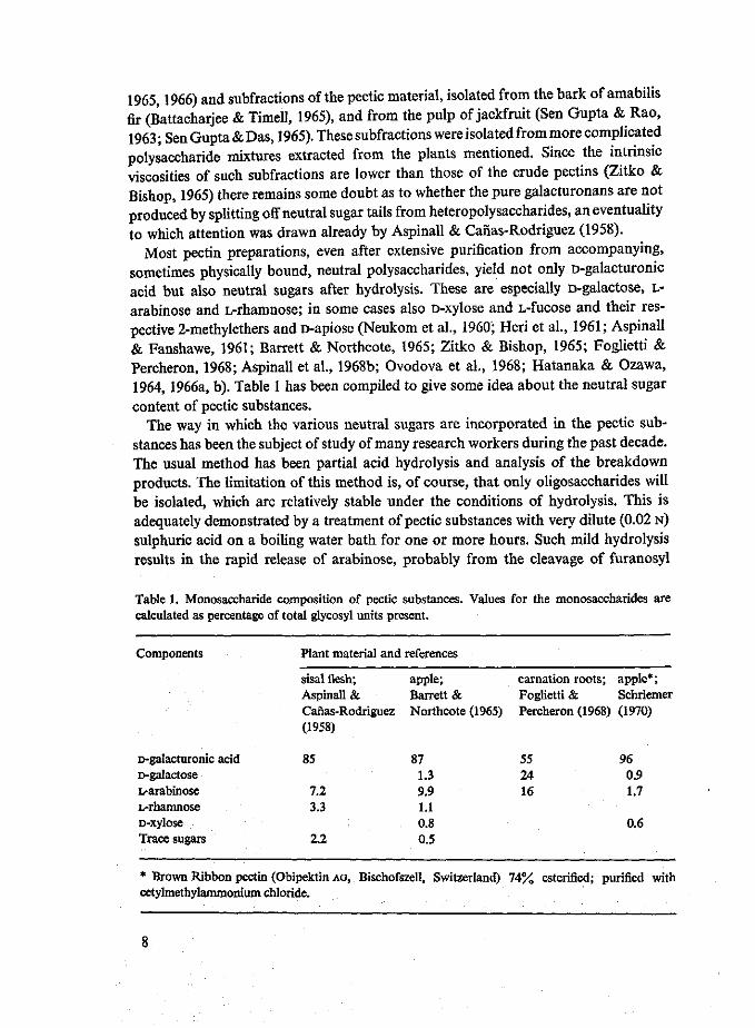
Most pectin preparations, even after extensive purification from accompanying, sometimes physically bound, neutral polysaccharides, yield not only D-galacturonic acid but also neutral sugars after hydrolysis. These are especially D-galactose, L-arabinose and L-rhamnose; in some cases also D-xylose and L-fucose and their respective 2-methylethers and D-apiose (Neukom et al., I960'; Heri et al., 1961; Aspinall & Fanshawe, 1961; Barrett & Northcote, 1965; Zitko & Bishop, 1965; Foglietti & Percheron, 1968; Aspinall et al., 1968b; Ovodova et al., 1968; Hatanaka & Ozawa, 1964, 1966a, b). Table 1 has been compiled to give some idea about the neutral sugar content of pectic substances.
The way in which the various neutral sugars are incorporated in the pectic substances has been the subject of study of many research workers during the past decade. The usual method has been partial acid hydrolysis and analysis of the breakdown products. The limitation of this method is, of course, that only oligosaccharides will be isolated, which are relatively stable under the conditions of hydrolysis. This is adequately demonstrated by a treatment of pectic substances with very dilute (0.02 N) sulphuric acid on a boiling water bath for one or more hours. Such mild hydrolysis results in the rapid release of arabinose, probably from the cleavage of furanosyl
Table 1. Monosaccharide composition of pectic substances. Values for the monosaccharides are calculated as percentage of total glycosyl units present.
Components
D-galacturonic acid D-galactose L-arabinose L-rhamnose D-xylose Trace sugars
Plant material and references
sisal flesh; Aspinall & Canas-Rodriguez (1958)
85
7.2 3.3
2.2
apple; Barrett & Northcote (1965)
87 1.3 9.9 1.1 0.8 0.5
carnation roots; apple*; Foglietti & Schriemer Percheron (1968) (1970)
55 96 24 0.9 16 1.7
0.6
* Brown Ribbon pectin (Obipektin AG, Bischofszell, Switzerland) 74% esterified; purified with cetylmethylammonium chloride.

linkages, followed by fucose, but no oligosaccharides are detected (Aspinall et al., 1967b). In addition to acid hydrolysis, acetolysis (i.e. breakdown of acetylated polysaccharides in glacial acetic acid-sulphuric acid) and enzymolysis have been used successfully to produce structurally relevant oligosaccharides (Bouveng, 1965; Aspinall et al., 1967b, c, 1968a, b). Table 2 lists all the different oligosaccharides isolated from pectic substances up till now. Moreover the table shows the oligosaccharides grouped according to the method of hydrolysis used and to the origin of the oligosaccharides: either from the main chain of the pectic molecule or from side chains. The number of references given with the different oligosaccharides indicates the frequency with which they are found in different plants.
Table 2 shows that the acid hydrolysis is extremely useful for the production of aldobiuronic acids (i.e. disaccharides, consisting of an uronic acid unit glycosidically linked to a neutral sugar unit). The glycosidic linkages of these acids are known to be stable during acid hydrolysis (BeMiller, 1967). It is interesting to note that acetolysis gives additional information to that obtained by acid hydrolysis because acetolysis favours the retention of rhamnopyranosyl-rhamnose linkages. So far enzymes have been used only incidentally and only in the form of commercial 'pectinase' preparations which are really mixtures of various enzymes (Bouveng, 1965; Aspinall et al., 1967b, 1968a, b). Yet enzymatic hydrolysis has already provided valuable information about the molecular structure of pectic substances by allowing the isolation of some unique oligosaccharides, especially some pseudoaldobiuronic acids (disaccharides consisting of a neutral sugar unit glycosidically linked to an uronic acid unit).
Some of the oligosaccharides isolated are not yet fully characterized. This is particularly so with D-GalA-Fuc, D-GalA-FucMe, D-GalA-L-Ara, D-GalA-XylMe (Fog-lietti & Percheron, 1968); GalA-Xyl (Stoddart et al., 1967) and D-GalA-D-Gal (Stod-dart et al., 1967; Foglietti & Percheron, 1968). It is likely that the hexuronic acid constituent of some of these aldobiuronic acids is not galacturonic but glucuronic acid. In fact, some aldobiuronic acids (GalA-Fuc, GalA-Gal) isolated by Aspinall & Fanshawe (1961) later proved to be GpA-Fuc and GpA-Gal (Aspinall, 1968b). Some of these aldobiuronic acids may be pseudoaldobiuronic acids. The neutral sugar constituent of these pseudoaldobiuronic acids will have been (part of) a side chain and not part of the main chain (not linked to Cx of galacturonic acid). An alternative is that galacturonic acid itself may also be present in the side chains.
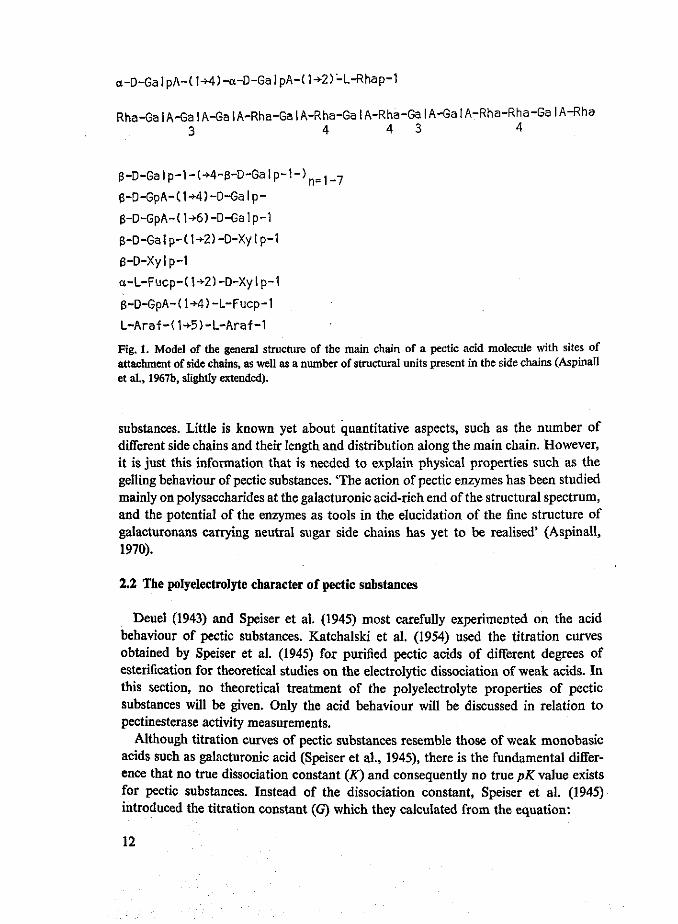
Apart from these complications, Table 2 contains much information which may be used for the construction of a pectate molecule model, showing the structural details that generally occur (see Figure 1). The main chain is built up of galacturonic acid and rhamnose only. Although rhamnose is a minor component in pectic substances (see Table 1), oligosaccharides rich in rhamnose and even bearing rhamnosyl-rhamnose linkages (Table 2) can be isolated. According to Aspinall et al. (1968a, b) the rhamnose residues appear to be unevenly distributed in the galacturonorhamnan chains, so that there are regions of uninterrupted galacturonic acid residues and regions in which the rhamnose residues are concentrated.
In the manufacture of pectins from citrus as well as from apple pomace, the

'pickling' method for the solubilization of the insoluble protopectin is generally used (Pilnik & Zwiker, 1970; Eshuis, 1970). This is a hydrochloric acid treatment (pH 0.5-0.7) at 42 °C for 22 or 46 h. It is very likely that under these conditions the weak rhamnosyl-rhamnose bonds are preferentially hydrolysed, so that an unknown portion of the pectin molecules may terminate with a rhamnose unit at the reducing as well as at the non-reducing end. The terminal position of a rhamnose unit is of paramount importance when such substrates are used for the study of exo enzymes such as the
Table 2. Oligosaccharides isolated from pectic substances after partial hydrolysis by different methods. *
1. Oligosaccharides which are fragments of the main chain and which have been isolated after hydrolysis with 1 N sulphuric acid for 3 to 7 h at 100 "C
a-D-GalpA-(l-*2)-L-Rha ' 1 ,2 ,4 ,5 ,6 ,7 ,8 , 9 jo 11 12**
GalpA-(l-4)-GalpA-(l-*2)-Rha 5 ' 8 ' '
GalpA-(l ->2)-Rhap-(l ->4)-GalpA-(l -*2)-Rha 4] 5> 7> 8
^Oligosaccharide, which is a fragment of the main chain and which has been isolated after acetolysis
GalpA-(l -»2)-Rhap-(l -2)-Rha 4> 5> ? g
^ S S ^ T WH ? 7 (frTentS °° S l d e Cha iDS a n d W h i c h h a v e b e e n i s o l a t e d * « hydrolysis witn 1 N sulphuric acid for 3 to 7 h at 100 °C
5 t r P A n l D ; G a l 4 ,5 ,7 ,8 ,10,11,12 /?-D-GpA-(1^4)-L-Fucp 4 ' ' ' ' ' /S-D-GpA-(l -4)-D-GalP J J 7' 8 ' 10'U' 12
/?-D-GpA-(l->2)-D-Man ' Galp-(l-*4)-GaIp GaIp-H-4<Wp. l . ) n_ 1 _ 4 . G a I p '] D-GalpA-D-Gal ' 5
D-GalA-Fuc q' D-GalA-FucMe . D-GalA-L-Ara D-GalA-XylMe GalA-Xyl
6 4 Oligosaccharides which are (fragments of) side chains and which have been isolated after acetolysis
a-D-Fucp-(l-i-2)-D-Xyl . /?-D-Galp-(l-*2)-D-Xyl J *
h y X ^ ^-D-Xylp-d^-D-GalA , a-D-GalpA-(l -*4)-D-GalA ' ' 7 ' 8
^-D-Xylp-(1^3)J 3 a-D-GalpA-(l -*4)-D-GalpA-(l -*»)-D-GalA
/?-D-Xylp-(3^4)J 3 Araf-(1^3)-GalA
10

exo pectate lyase of Clostridium multifermentans which acts from the reducing end of the substrate chain (Macmillan & Vaughn, 1964). This position may even partly account for the differences in pattern of action ascribed to different pectinesterases, studied with the aid of exo pectate lyase of Clostridium multifermentans (Lee et al., 1970).
Side chains are attached to the galacturonorhamnan chain at C3 of some galactu-ronic acid residues and at C4 of some rhamnose residues. Attachment of side chains at C4 of rhamnose is not evident from the oligosaccharides in Table 2, but Aspinall et al. (1967a) obtained evidence for attachment at C4 by methylation studies. The structure of most of the side chain fragments as given in Figure 1 is evident from the oligosaccharides listed in Table 2. The very unstable arabinofuranosyl-arabinose linkage is known to be present in arabinogalactan of soya beans (Aspinall et al., 1967a) and presumed to be present in pectic substances too (Aspinall et al., 1967b). The actual way of linkage of these side chain fragments to the main chain is known only for those linked via xylose and arabinose, due to the isolation of the pseudo-aldobiuronic acids Xyl-(l->3)-GalA and Ara-(l-»3)-GalA after enzymatic hydrolysis (Bouveng, 1965; Aspinall, 1967b, 1968a, b).
At present, therefore, much is known about the qualitative aspects of the hetero-polysaccharide structure of pectic substances such as the different sugars involved and how they may be linked to one another and to the galacturonic acid of the main chain of the molecule. Further experimental work will have to show in more detail how side chains are linked to the main chain, for example by studying the pseudo-aldobiuronic acids and oligosaccharides formed by enzymatic hydrolysis of pectic
Footnote Table 2.
* Symbols e.g.: a-D-GalpA = a-D-galactopyranosyluronic acid L-Rhap = L-rhamnopyranose /?-D-GpA = /S-D-glucopyranosyluronic acid FucMe = fucose-2-methylether L-Araf = L-arabinofuranose ** Plant material and references 1. 2. 3. 4. 5. 6. 7. 8. 9.
10. 11. 12.
Lucerne leaves and stems Apple fruit Mountain pine pollen Soyabean cotyledons Soyabean hulls Sycamore callus Lemon peel Lucerne leaves and stems Carnation roots Sycamore cambial cells Lemon peel Lemon peel
Aspinall & Fanshawe, 1961 Barrett & Northcote, 1965 Bouveng, 1965 Aspinall et al., 1967b Aspinall et al., 1967c Stoddart et al., 1967 Aspinall et al., 1968a Aspinall et al., 1968b Foglietti & Percheron, 1968 Aspinall et al., 1969 Aspinall & Cottrell, 1970 Aspinall et al., 1970.
11
a-D-Ga!pA-(1->4)-a-D-GaIpA-(1+2)-L-Rhap-1
Rha_Ga | A_Ga | A_Ga I A-Rha-Ga I A-Rha-Ga I A-Rha-Ga I A-Ga I A-Rha-Rha-Ga I A-Rha 3 4 4 3 4
e-D-Galp-1-(-*-4-B-D-Galp-1-)n=1_7
p-D-GpA-C1+4)-D-Galp-
g-D-GpA-(1-*6)-D-GaIp-1
g-D-Ga I p-( 1 ->2) -D-Xy I p-1
g-D-Xylp-1
a-L-Fucp-(W)-D-Xylp-1
3-D-GpA-(l-*-4)-L-Fucp-1
L-Araf-(1->5)-L-Araf-1
Fig. 1. Model of the general structure of the main chain of a pectic acid molecule with sites of attachment of side chains, as well as a number of structural units present in the side chains (Aspinall et al., 1967b, slightly extended).
substances. Little is known yet about quantitative aspects, such as the number of different side chains and their length and distribution along the main chain. However, it is just this information that is needed to explain physical properties such as the gelling behaviour of pectic substances. 'The action of pectic enzymes has been studied mainly on polysaccharides at the galacturonic acid-rich end of the structural spectrum, and the potential of the enzymes as tools in the elucidation of the fine structure of galacturonans carrying neutral sugar side chains has yet to be realised' (Aspinall, 1970).
2.2 The polyelectrolyte character of pectic substances
Deuel (1943) and Speiser et al. (1945) most carefully experimented on the acid behaviour of pectic substances. Katchalski et al. (1954) used the titration curves obtained by Speiser et al. (1945) for purified pectic acids of different degrees of esterification for theoretical studies on the electrolytic dissociation of weak acids. In this section, no theoretical treatment of the polyelectrolyte properties of pectic substances will be given. Only the acid behaviour will be discussed in relation to pectinesterase activity measurements.
Although titration curves of pectic substances resemble those of weak monobasic acids such as galacturonic acid (Speiser et al., 1945), there is the fundamental difference that no true dissociation constant (K) and consequently no true pK value exists for pectic substances. Instead of the dissociation constant, Speiser et al. (1945) introduced the titration constant (G) which they calculated from the equation:
12

G _ [ H + ] a _ [ H + ] [ C O Q - ]
1 - a N - [COO -]
in which a = the degree of dissociation, calculated as the ratio of [COO -] to N N = the total concentration of carboxyl groups in equivalents per litre [COO -] = the concentration of dissociated carboxyl groups, calculated from the relation [COO -] = [B+] + [H+] - [OH -] , in which [B+] represents the concentration of base in equivalents per litre.
Unlike a dissociation constant for a weak monobasic acid, which remains constant, this titration constant was shown by Speiser et al. (1945) and also by Deuel (1943) to increase (and pG to decrease) with increasing concentration of the pectin, increasing degree of esterification and decreasing degree of neutralization of the pectin.
The change ofpG (= — log G) with changing degree of esterification and changing concentration of pectin is illustrated in Table 3. Although Speiser et al. (1945) found no effect of the method of saponification (enzymatic or by acid) on the pG value, it may be assumed that the distribution of the free carboxyls along the pectin chain also influences pG. Indeed Schultz et al. (1945) found enzymatically saponified pectins to be weaker acids and to resemble pectic acid more closely than pectins prepared by alkali.
The acid behaviour of pectins complicates accurate titrimetric activity measurements of pectinesterases, especially of those of fungal origin with an optimum pH near 4.0. Not all carboxyl groups, liberated by pectinesterase action are titrated by continuous titration at a fixed pH and the portion which is titrated is smaller when the chosen pH is lower. Furthermore, in continuous titration experiments the degree of esterification of the substrate as well as its concentration are continuously lowered. Consequently the degree of dissociation also decreases continuously and therefore no straight lines can be expected as titrigrams (alkali consumption versus time curves), when the pectinesterase activity remains the same throughout the experiment. Even, in quite
Table 3. pG values of pectins and pK value of galacturonic acid at 27°C (from Speiser et al., 1945).
Mode of saponification
Acid Acid Acid Acid
Enzyme Enzyme
Galacturonic acid
Degree of esterification
44% 10% 10% 10%
33% 14%
Concentration (g/litre)
2 1 2 4
2 2
pG value at a = 0.5
3.9 4.3 4.1 3.9
3.9 4.1
pK value 3.42
13

recent work these complications have been generally disregarded (Schubert, 1952; Endo, 1964e; Roller, 1966) and one has to go back in literature as far as 1947 (McCol-loch & Kertesz) and 1945 (Fish & Dustman) to find correct titrimetric pectinesterase activity measurements. These authors kept the pH of the reaction mixtures constant for a time during which not more than 30 % demethylation occurred, and thereafter rapidly titrated the mixture to pH 7.0. Blanks were obtained for each enzyme by titrating to pH 7.0 identical mixtures in which the added enzyme had been previously boiled for five minutes.
As pointed out before, the use of this correct method becomes all the more important when the pH of pectinesterase activity measurement is lower. In fact at a pH of 3.0, which is quite common in fruit juice technology, a system in which a pectinesterase is active will only require a negligible amount of alkali for its pH to be maintained at 3.0. Likewise the use of the correct method is of great importance for the validity of optimum pH curves of fungal pectinesterases.
2.3 Summary
In this chapter reference is made to the literature on two aspects of pectic substances which are important for the study of pectic enzymes: the heteropolysaccharide and the polyelectrolyte character of pectic substances.
Only a few true galacturonans i.e. polysaccharides, entirely built up of 1,4-linked D-galacturonic acid residues, have been found. Usually pectins, when completely hydrolysed yield not only galacturonic acid but also a number of neutral sugars. The pattern in which the various neutral sugars are incorporated in the pectic substances has been successfully studied by partial hydrolysis of pectic substances and analysis of the oligomeric breakdown products. All oligosaccharides so far isolated from pectic substances after partial hydrolysis by different methods are given in one table. Models of the general structure of the main chain and side chains are presented. Although much is known about qualitative aspects of the heteropolysaccharide structure of pectic substances, there is little information on the quantitative aspects such as number and length of side chains and distribution along the main chain.
The polyelectrolyte properties of pectic substances are discussed in relation to pectinesterase activity measurements. It is emphasized that pectic substances have no true dissociation constant, and that the titration constant, which has therefore been introduced depends on the concentration, the degree of esterification and the degree of neutralization of the pectin. It is shown that pectinesterase activity measurements near pH 4 therefore cannot accurately be carried out by continuous titration.
14

3 Classification and properties of pectin depolymerases
Pectic substances are degraded by two main groups of enzymes, the depolymerizing pectic enzymes and the saponifying enzymes or pectinesterases. This chapter deals only with the first group. In 1963, Neukom presented a classification scheme for the depolymerizing enzymes, which is still used (Table 4). He applied the following three classification criteria: hydrolytic or transeliminative splitting of the glycosidic bonds (Figure 2), exo or endo mechanism of the splitting reaction and preference for pectic acid or pectin as substrate. He thereby arrived at the eight groups of Table 4. However enzymes belonging to the exo polymethylgalacturonase and exo pectin lyase groups have never been described. The enzyme groups of Table 4 for which the high polymer pectic substances are the normal substrates, degrade these polymers to various mixtures of oligogalacturonides. These enzymes are secreted into the culture medium by micro-organisms. The galacturonide oligomers can be further degraded by oligo-merases, a special group of microbial intracellular enzymes which has been discovered only recently (Hasegawa & Nagel, 1967, 1968; Moran et al., 1968b; Hatanaka & Ozawa, 1970). Oligomerases can be distinguished from depolymerases by their property of degrading their substrate at a rate which is inversely proportional to its chain length.
The pectin depolymerases, for which the high polymer pectic substances are the normal substrates, also show activity on oligomers. In fact their activity and action patterns on oligomers yield further classification criteria. In a recent communication
Table 4. Classification of depolymerising pectic enzymes (Neukom, 1963; Roller, 1966).
Pectic enzymes acting mainly on pectin Pectic enzymes acting mainly on pectic acid
polymethylgalacturonases pectin lyases* polygalacturonases pectate lyases* (PMG) (PL) (PG) (pAL>
1. endo PMG (3.2.1.41)** 3. endo PL (4.2.2.3) 5. endo PG (3.2.1.15) 7. endo PAL (4.2.2.1) 2. exo PMG 4. exo PL 6. exo PG (3.2.1.40) 8. exo PAL (4.2.2.2)
* The term 'lyase' is preferred to 'transeliminase' by the International Union of Biochemistry
(Florkin & Stotz, 1965). . , T . ** Numbers, based on the recommendations on enzyme nomenclature of the International Union of Biochemistry as assigned to the enzymes by Roller (1966).
15

COOH COOH COOH COOH
Jh—° Jr—°\ A ° \ °HJ—°*
polygalacturonase
COOH
pectate lyase
Fig. 2. Hydrolytic and transeliminative splitting of a pectic acid chain by polygalacturonase and pectate lyase, respectively. ,
(Voragen & Pilnik, 1970a) special attention was paid to the breakdown of oligo-galacturonides by polygalacturonases and pectate lyases.
The literature on pectin depolymerases has recently been selectively reviewed by Rombouts & Pilnik (1972). This review, covering over 140 titles, deals mainly with the literature which has appeared since the discovery of pectin lyases by Albersheim et al. in 1960. Only reports of investigations with purified enzyme preparations and rather well defined substrates were included. According to their action on high polymer pectic substances, the enzymes were placed in one of the six groups of Neu-kom. Mutual relationships of some of the groups were discussed. It was further emphasized that the choice of substrates and reaction conditions, as well as that of methods of analysis, to a large extent influence the value found for a number of enzyme properties.
This chapter is a general description of these six enzyme groups (endo and exo polygalacturonase, endo and exo pectate lyase, endo polymethylgalacturonase and endo pectin lyase). This description covers their occurrence and production as well as their general properties, e.g. substrate preference, optimum pH, requirement for cations, percentage breakdown at 50% viscosity reduction of the substrate, degradation limit of their substrate, end-products formed etc.
3.1 Endo polygalacturonases
This group of enzymes hydrolyses the a-l,4-galacturonide links of pectic acid to different degrees of randomness to produce a series of oligogalacturonides.
16

Occurrence and formation Endo polygalacturonases are the most widely distributed and most frequently occurring pectin depolymerases in nature. They occur in fruits, stems and leaves of many higher plants (Pilnik & Voragen, 1970). They are produced by a great number of plant pathogenic and saprophytic fungi e.g. Aspergillus, Peni-cillium, Monilia, Geotrichum, Rhizopus, Sclerotinia and Coniothyrium (Rombouts & Pilnik, 1972). It is the only pectin depolymerase known to be produced by yeasts (Roelofsen, 1953; Phaff, 1966). Erwinia carotovora and Pseudomonas marginalis are two of the few bacteria which seem to produce this enzyme (Nasuno & Starr, 1966a, b). The enzyme may be produced constitutively as is so with Saccharomyces fragilis (Phaff, 1966), but most fungi produce it adaptively together with other pectic enzymes such as pectinesterase, exo polygalacturonase and pectin lyase.
Substrates Pectic acid, also called polygalacturonic acid or polypectate is the best substrate for endo polygalacturonases. Pectins are attacked, but hydrolysed at a lower rate and to a lower hydrolysis limit. In fact, hydrolysis limits for pectin preparations with different degrees of esterification, obtained by chemical saponification of completely esterified pectin, are found to decrease with increasing degree of esterification and to become zero at 75% esterification (Koller & Neukom, 1969; Jansen & MacDonnell, 1945). Acetylation of the secondary alcohol groups (at C2
and C3) up to 70% has little effect on endo polygalacturonase activity of Aspergillus niger (Koller & Neukom, 1969).
Optimum pH With pectic acid as the substrate the optimum pH of the enzymes of this group ranges from 3.5 to 5.6 (Rombouts & Pilnik, 1972). The range of optimum pH of enzymes from different sources is so uniform, that this is no useful criterion to distinguish them according to origin, perhaps with the exception of one endo polygalacturonase with an unusual low optimum pH of 2.5, which was purified from Corticium rolfsii (Kaji & Okada, 1969). The optimum pH on oligomers has often been found to be lower than that on pectic acid (Patel & Phaff, 1960a, b; Phaff, 1966; Barash, 1968; Barash & Eyal, 1970).
Percentage hydrolysis at 50% viscosity reduction This criterion is often used to prove the endo mode of attack of pectin depolymerases. However, data obtained have only a limited quantitative value since the degree of polymerization (the molecular weight) of the substrate used is usually not known, whereas the percentage of hydrolysis of the substrate at 50% viscosity reduction is inversely proportional to the degree of polymerization of the substrate (Chapter 9). Moreover, to measure percentage hydrolysis of the substrate by polygalacturonase, reducing end-groups are generally measured by one of the standard procedures for reducing end-group analysis. These methods have been shown by Voragen et al. (1971a) to be of limited value for the measurement of reducing groups of pectic substances, because under the alkaline reaction conditions of these analytical procedures the pectic substances are decomposed, so that un-realistically high percentages of free end-groups are found. Thus it is not surprising
17
that the percentage of hydrolysis of many sorts of pectates found at 50 % specific viscosity reduction varies from 0.5 % to over 10 % for different endo polygalacturonases described in literature. Some authors, studying more than one endo polygalacturonase and using the same substrate and the same end-group method, found different values for different enzymes (Endo, 1964a, b, c; Koller, 1966). These observed differences in the 'endo mode of attack' may perhaps be explained with the concept of 'multiple attack' of the enzyme on the substrate (Chapter 10).
Final degree of hydrolysis of the substrate Endo polygalacturonases hydrolyse pectate with a rapid initial linear phase up to 25 to 50% hydrolysis. Then the hydrolysis rate slows down and finally 50% to over 70% of the glycosidic bonds may be hydrolysed (Phaff, 1966; Endo, 1964a, b, c; Koller, 1966). Commercial high methoxyl pectin with about 70% esterification is only hydrolysed to a limited extent: for 6 to 17% (Uchino et al., 1966; Yamasaki et al., 1966; Endo, 1964a, b, c).
Oligomer end-products The type of end-product formed from pectate is connected with the degree of hydrolysis. Mono- and digalacturonic acids are always the major end-products, but some trigalacturonic acid or even higher oligomers may remain present in the reaction mixture as well (Koller, 1966; Barash, 1968; Barash & Khazzam, 1970; Nasuno & Starr, 1966a).
Stability Endo polygalacturonases are most stable in the pH range of 4 to 6.5 (Koller, 1966; Yamasaki et al., 1966; Endo, 1964a, b, c). The optimum temperature for activity is reported to be 40 to 45 °C (Nasuno & Starr, 1966a; Slezarik & Rexova, 1967).
3.2 Exo polygalacturonases
This group of enzymes hydrolyses the <x-l,4-galacturonide links of pectic acid by a terminal attack on the substrate.
Occurrence and formation Exo polygalacturonases, although not so frequently encountered as endo polygalacturonases, are also widely distributed in nature. They are known to be present in carrots (Daucus carota), as the only depolymerase (Hatanaka & Ozawa, 1964, 1966c). They also occur in many fungi, such as Aspergillus niger (Mill, 1966a, b), Coniothyrium diplodiella (Endo, 1964d; Hatanaka & Ozawa, 1966d) and Rhizopus tritici (McClendon & Kreisher, 1963). They have been isolated from Erwinia aroideae (Hatanaka & Ozawa, 1969) and were even found in an insect Pyrrocoris apterus (Courtois et al., 1968). These enzymes which are produced by induction may be excreted into the culture medium (Endo, 1964d) or may remain associated with the mycelium or the cells (Mill, 1966a, b; Hatanaka & Ozawa, 1969).
18

Substrates Pectic acid and normal, unesterified galacturonide oligomers are readily attacked, but pectin is not a substrate (Mill, 1966a, b) or only little attacked (Endo, 1964d). Unsaturated pectic acid, with a double bond in the galacturonide unit at the non-reducing end may (Hatanaka & Ozawa, 1969) or may not (Hatanaka & Ozawa, 1964, 1966c) be attacked. The insect exo polygalacturonase is inactive on reduced pectic acid (Courtois et al., 1968).
Optimum pH Enzymes from different sources all have an optimum pH in the range of 4.0 to 5.6, except for that of Erwinia aroideae, which has its optimum at pH 7.5.
Percentage hydrolysis at 50 % viscosity reduction Endo (1964d) reported that the exo polygalacturonase of Coniothyrium diplodiella had hydrolysed pectate for 40% to galacturonic acid when the specific viscosity had dropped to 50 %.
Final degree of hydrolysis of the substrate Exo polygalacturonases may hydrolyse pectate with an initial linear phase up to 60 to 80% (Endo, 1964d; Hatanaka & Ozawa, 1966d) and finally degrade this substrate for 80 to 100% to galacturonic acid (Mill, 1966a; Endo, 1964d; Hatanaka & Ozawa, 1966d). However, this is not a general rule, since the exo polygalacturonase of carrots only hydrolyses pectic acid for 47% to galacturonic acid (Hatanaka & Ozawa, 1964, 1966c) and an exo polygalacturonase of Aspergillus niger even stops at 28 % hydrolysis of pectic acid to galacturonic acid (Mill, 1966b). Occurrence of neutral sugars (Chapter 2) or of residual methylester groups in the substrate may be the reasons for the failure of the enzyme to complete the hydrolysis of its substrate. The presence of unsaturated pectic acid molecules may also cause an incomplete hydrolysis (Hatanaka & Ozawa, 1964,1966c).
End-products With one exception all known polygalacturonases produce galacturonic acid only from pectic acid and from galacturonide oligomers including the dimer. The enzyme of Erwinia aroideae splits off digalacturonic acid (Hatanaka & Ozawa, 1969).
Action from (non-)reducing chain end Since the exo polygalacturonases of carrots (Hatanaka & Ozawa, 1964, 1966c) and of Coniothyrium diplodiella (Hatanaka & Ozawa, 1966d) are inactive on unsaturated pectic acid, action of these enzymes from the non-reducing chain end is indicated. The enzyme of Erwinia aroideae hydrolysed unsaturated oligogalacturonides and polygalacturonides with the formation of unsaturated digalacturonic acid, and therefore this enzyme is also thought to attack the non-reducing end (Hatanaka & Ozawa, 1969). Reduced pectic acid is not attacked at all by the insect exo polygalacturonase. Attack of pectic acid from the reducing chain end is therefore postulated (Courtois et al., 1968).
Stability The pH and heat stability of exo polygalacturonases are probably comparable with those of endo polygalacturonases (Endo, 1964d).
19

3.3 Endo pectate lyases
This group of enzymes splits the a-l,4-galacturonide links of pectic acid to different degrees of randomness, by a transelimination mechanism so that a series of C4-C5
unsaturated oligogalacturonides (Figure 2) is produced.
Occurrence and formation This group of enzymes is commonly produced by pectoly tic bacteria. In fact it is the major bacterial pectin depolymerase. In addition it is produced by certain Fusarium species (Hancock, 1968) and by the rumen ciliate Ophryos-colexpurkynei (Mah & Hungate, 1965). The extracellular enzyme is generally produced by induction (Nagel & Vaughn, 1961a, b; Fuchs, 1965; Preiss & Ashwell, 1963a). Nasuno & Starr (1966a) reported Pseudomonas marginalis, to produce extracellular endo pectate lyase either adaptively or constitutively, depending on the strain. Hsu & Vaughn (1969) found the production of the constitutive endo pectate lyase of Aero-monas liquefaciens to be subject to catabolite repression, through the unsaturated oligomeric breakdown products of pectate. The regulation of pectate lyase synthesis in Erwinia carotovora was studied by Zucker & Hankin (1970) and in Erwinia caro-tovora and Erwinia aroideae by Moran & Starr (1969). Constitutive enzyme is produced, but Zucker & Hankin could not reveal any catabolite repression, whereas Moran & Starr found the enzymes of their strains to be under catabolic repression.
Substrates Pectate is most generally used for endo pectate lyase studies. Sometimes enzyme activity on pectate is compared with and found to be higher than that on commercial high methoxyl pectin (about 70 % esterified). Completely esterified pectin is hardly attacked by Fusarium pectate lyase (Hancock, 1968). In Chapter 10 results of more detailed studies of the influence of the degree of esterification on the activity of endo pectate lyase are reported. It was found that pectate lyases from Arthrobacter and Bacillus polymyxa have maximum values for Vmax and l/Km on 21 to 44% esterified pectins. Moreover the degradation limit obtained with Arthrobacter enzymes is maximum for pectins of 21 to 33 % esterification.
An interesting observation was made by McNicol & Baker (1970) who studied the enzymatic degradation of Vi antigen, a bacterial surface polysaccharide containing oc-l,4-linked 2-N-acetyl-3-0-acetyl-D-galacturonic acid. They found that the Vi antigen degrading enzyme produced by Bacillus sphaericus and the pectate lyase of Bacillus polymyxa both degrade Vi antigen, O-deacetylated Vi antigen, pectin and poly-galacturonic acid. It may therefore be concluded that pectate lyases, just like polygalacturonases, are active on C2 and C3 derivatives of their normal substrate.
Requirement for divalent cations All endo pectate lyases have an absolute requirement for some divalent cations, of which calcium and magnesium ions are the most effective. The calcium ions concentration for maximum lyase activity on pectate varies between 0.0002 and 0.001 M (Nagel & Vaughn, 1961a, b; Nagel & Wilson, 1970; Preiss & Ashwell, 1963a; Starr & Moran, 1962; Chapter 10). Consequently the endo
20

pectate lyases may be inactivated by the addition of sequestering agents such as EDTA (Starr & Moran, 1962; Chapter 10).
Optimum pH Endo pectate lyases have an optimum pH ranging from 8.0 to 9.8 (Nagel & Wilson, 1970; Garibaldi & Bateman, 1971; Chapter 10). The actual optimum pH found depends on minor differences in the procedure. Buffers, calcium ions and substrate should be mixed immediately before the measurements (Nagel & Wilson, 1970).
Percentage breakdown at 50 % viscosity reduction Contrary to polygalacturonases, no reducing end-groups need to be measured. As shown in Figure 2, every split in the substrate chain caused by a pure lyase preparation, results in the formation of a C4-C5 double bond in the galacturonide residue at the newly formed, non-reducing end of the chain. This double bond is conjugated with the carboxyl group, and gives an absorption peak in the ultraviolet region at 232-235 nm. Therefore enzyme activities are easily and accurately measured in a recording spectrophotometer. For calculation of pectate lyase activity, a molar extinction coefficient of 4 800 can best be used (Macmillan & Vaughn, 1964).
The percentage of broken bonds at 50 % viscosity reduction of pectate varies from 0.5 to 3.0% for a number of well purified endo pectate lyases (Hasegawa & Nagel, 1966; Nagel & Wilson, 1970; Mount et al., 1970; Nasuno & Starr, 1967; Hancock, 1968). In this respect the Arthrobacter endo pectate lyase, described in Chapter 10, is an exception as it acts less randomly than pectate lyases of Bacillus polymyxa and of Pseudomonas.
Final degree of degradation of the substrate Endo pectate lyase of Arthrobacter degrades 21 to 26% esterified pectin up to 33% and 7% esterified pectin up to 25% (Chapter 10).
Oligomer end-products Unsaturated digalacturonic acid is the major end-product for most endo pectate lyases (Nagel & Wilson, 1970; Moran et al., 1968a; Nasuno & Starr, 1966a, 1967). Unsaturated trigalacturonic acid, which is also always present as a final degradation product, is the major end-product from pectate degraded by endo pectate lyase of Bacillus pumilus (Dave & Vaughn, 1971). As a result of the transeliminative splitting of lower unsaturated oligomers, some unsaturated monomer may be formed (Preiss & Ashwell, 1963a, b; Hasegawa & Nagel, 1966; Hsu & Vaughn, 1969; Chapter 7). This compound which is not very stable (Preiss & Ashwell, 1963a, b) is only detected on paper chromatograms when a special thiobarbituric acid reagent is used.
Small amounts of saturated monogalacturonic and digalacturonic acids which may be produced, originate from the non-reducing ends of the pectate molecules (Nagel & Wilson, 1970).
21

Iso-enzymes As the endo pectate lyase preparation of Bacillus polymyxa was successfully separated on CM-cellulose into four endo pectate lyases, which show small differences in a number of properties such as optimum pH, percentage of bond breakage at 50% viscosity reduction and action pattern on galacturonide oligomers (Nagel & Wilson, 1970). Garibaldi and Bateman (1971) found one strain of Erwinia chrysanthemi that produces two endo pectin lyases and another strain that produces even four. The iso electric points of these six enzymes range from 9.4 to 4.6. Their molecular weights, determined by gel filtration on Sephadex G-75 and by sucrose density gradient centrifuging, range from 30 000 to 36 000. As with the Bacillus polymyxa enzymes there are only small differences in optimum pH and percentage of bond breakage at 50 % viscosity reduction.
Stability Optimum temperatures for activity in the range of 45 to 50 °C are found (Nagel & Vaughn, 1961a; Moran et al., 1968a). When initial reaction velocities are measured the optimum temperature of Arthrobacter pectate lyase is 53 °C (Chapter 10). Bacillus polymyxa pectate lyases are most stable in the pH range of 5 to 8. The pH of maximum stability of Arthrobacter pectate lyase is 7.0 (Chapter 10).
3.4 Exo pectate lyases
This group of enzymes splits the a-l,4-galacturonide links of pectic acid trans-eliminatively by a terminal attack on the substrate.
Occurrence and formation Only two exo pectate lyases, both from bacterial origin, have been described. The adaptive exo pectate lyase of Clostridium multifermentans is produced extracellularly as the organism's only pectin depolymerase (Macmillan & Vaughn, 1964). The pectate lyase of Erwinia aroideae, another adaptive enzyme, is associated with the cells and is contaminated with a transeliminative oligomerase, which can be selectively inactivated by heating for 10 min at 45 °C and pH 9.0 (Oka-moto et al., 1963, 1964b, c).
Substrates Exo pectate lyase of Clostridium multifermentans is specific for pectate and oligogalacturonides, except for digalacturonic acid, which is not attacked. Poly-methylpolygalacturonic acid methyl glycoside is not a substrate, whereas pectins with variable methyl ester content are partially degraded (Macmillan & Phaff, 1966). The exo pectate lyase of Erwinia aroideae is also very active on pectate but hardly on (about 70 % esterified) citrus pectin.
Requirement for divalent cations The enzyme of Clostridium multifermentans requires divalent cations of which calcium ions stimulate the greatest activity. A concentration of 0.0005 M of calcium ions is most favourable for the complete breakdown of pectate (Macmillan & Vaughn, 1964; Macmillan & Phaff, 1966). The enzyme of Erwinia aroideae is scarcely affected by calcium ions (Okamoto et al., 1963).
22

Optimum pH The enzyme of Clostridium multifermentans has an optimum pH of 8.5 and that of Erwinia aroideae has an optimum pH in the range 8.0 to 8.3 (Macmillan & Vaughn, 1964; Okamoto et al., 1963).
Percentage breakdown at 50 % viscosity reduction The exo pectate lyase of Clostridium multifermentans is reported to convert 22.5 % of pectic acid to unsaturated digalac-turonic acid, at 50 % viscosity drop of the substrate. This amount corresponds with over 11 % broken glycosidic bonds (Macmillan & Vaughn, 1964).
End-product and final degree of degradation of the substrate Both exo pectate lyases split off unsaturated digalacturonic acid only from pectic acid. Therefore as a theoretical maximum only 50 % of the glycosidic bonds can be broken. Indeed Macmillan & Vaughn (1964) reported polygalacturonic acid to be degraded to its theoretical maximum. However, in an attempt to use the clostridial enzyme for quantitative analysis of pectate content, we never succeeded in converting more than 60 to 70 % of the pectate into unsaturated digalacturonic acid. Since we observed no product inhibition there must always be enough structural obstacles in pectates (Chapter 2) to prevent complete enzymatic degradation.
Action from reducing chain end Since during pectate degradation the only unsaturated product is the dimer, and all the higher polymer breakdown products are saturated, the enzymes apparently attack the substrate from the reducing end (Macmillan & Vaughn, 1964; Okamoto et al., 1963). However, there is not much difference in activity of the exo pectate lyase of Erwinia aroideae on normal, oxidized and reduced pectic acid (Okamoto et al., 1963). It is evident that a completely degraded poly-galacturonate chain molecule with an even number of anhydrogalacturonic acid units yields unsaturated digalacturonic acid plus one molecule of the saturated dimer originating from the non-reducing end of the chain. Similarly, a substrate molecule with an odd number of units yields unsaturated dimer plus one molecule of galac-turonic acid upon complete degradation (Macmillan & Phaff, 1966).
Pectate lyase-pectinesterase complex Clostridium multifermentans produces pectin-esterase in addition to exo pectate lyase (Macmillan & Vaughn, 1964). There is evidence that these two enzymes are complexed (Miller & Macmillan, 1970). A molecular weight of 400 000 was found by Sephadex G-200 filtration. Lyase activity can be freed of esterase activity by heating for 30 min at 38 °C in dilute calcium chloride solution with pH 7.0.
Stability Clostridial pectate lyase is most stable at pH 6 to 7 (Miller & Macmillan, 1970); that of Erwinia is stable in the pH range 5.0 to 9.0 and has an optimum temperature of 35 °C (Okamoto et al., 1963, 1964a).
23
3.5 Endo polymethylgalacturonases
This group of enzymes is supposed to hydrolyse specifically and randomly the a-1,4 links of galacturonide chains which are substantially or completely esterified with methanol.
Since the discovery of pectin lyase in 1960 by Albersheim et a l , remarkably few polymethylgalacturonases have been found. Koller (1966) described two endo PMGs partly purified from Pektinex, an enzyme preparation from Aspergillus niger. Rexova-Benkova & Slezarik (1966) isolated and described a PMG from a surface culture of Aspergillus niger. It is clear, that many of the polymethylgalacturonases described before 1960 were in fact pectin lyases. The proof of existence of the two PMGs described by Koller is doubtful, as these were not fully purified from accompanying pectinesterase, polygalacturonase and pectin lyase. Moreover, when turbid, optically dense pectin solutions are used, an increase in absorbance at 232 nm may not easily be found so that one erroneously concludes for PMG activity where, in fact, pectin lyase is active. In our laboratory, we keep trying to isolate a PMG from commercial pectolytic enzyme preparations, but have found no evidence yet for the presence of such an enzyme. Thus we have doubts about whether polymethylgalactu-ronase really exists.
3.6 Endo pectin lyases
By a transelimination reaction this group of enzymes splits specifically and randomly the a-1,4 links of galacturonide chains which are substantially or fully methylated.
Occurrence and formation The pectin lyases which have been described in considerable detail are all of fungal origin. Some have been purified from commercial fungal pectinase preparations which are rather complicated mixtures of pectolytic and other enzymes (Albersheim et al., 1960b; Albersheim, 1966; Koller, 1966; Amado, 1970). Pectin lyases are produced extracellularly and have to be induced by pectin or pectate in the culture medium (Edstrom & Phaff, 1964a; Sherwood, 1966; Bateman, 1966; Bush & Codner, 1968, 1970).
Substrates This group of enzymes splits highly and completely esterified pectins but pectate is not usually attacked (Albersheim et al., 1960b; Edstrom & Phaff, 1964a, b; Amado, 1970; Bush & Codner, 1968, 1970). The pectin lyase of Rhizoctonia solani and that of Fusarium solani do attack pectate, but are more active on pectin (Sherwood, 1966; Bateman, 1966). The pectin lyase of Aspergillus fonsecaeus attacks fully esterified oligomers with decreasing reaction rates down to tetramethyl tetragalac-turonate (Edstrom & Phaff, 1964a, b).
Role of calcium ions Unlike pectate lyases, pectin lyases have generally no absolute requirement for calcium ions although calcium may stimulate pectin lyase activity.
24

The stimulatory effect of calcium chloride on pectin lyase activity depends on the pH of the reaction mixture and the degree of esterification of the substrate (Voragen et al., 1971b). The pectin lyase of Fusarium solani is an exception as it requires calcium ions (Bateman, 1966).
Optimum pH Pectin lyases have generally their optimum pH in the range 5.1 to 6.3 (Albersheim, 1963; Edstrom & Phaff, 1964a, b; Voragen et al, 1971b; Amado, 1970; Bush & Codner, 1968, 1970). With calcium ions, a new increased optimum pH may be found at pH 8 to 8.5 (Edstrom & Phaff, 1964a, b; Voragen et al., 1971b). The pectin lyases of Rhizoctonia solani and Fusarium solani have their optimum pH values at 8.2 and 8.6, respectively (Sherwood, 1966; Bateman, 1966).
Percentage breakdown at 50 % viscosity reduction As with pectate lyases, the activity of pectin lyases is best measured with a recording spectrophotometer. The best value for the molecular extinction coefficient is 5 500 (Edstrom & Phaff, 1964a). The pectin lyase of Fusarium solani splits 1 to 2 % of the bonds of a high polymer 65 % esterified pectin to achieve a 50 % viscosity drop. Other enzymes split 3 to 5 % of the bonds of fully (95-97 %) esterified pectins of a lower degree of polymerization (Edstrom & Phaff, 1964a; Amado, 1970).
Final degree of degradation of the substrate Aspergillus fonsecaeus pectin lyase splits 68 % esterified pectin and 95 % esterified pectin to a limit of 22 % and 30 % respectively of the glycosidic bonds (Edstrom & Phaff, 1964a). That of Aspergillus niger splits even 47% of the glycosidic bonds of 97% esterified pectin (Amado, 1970).
End-products Fully (95 %) esterified pectins are finally degraded to a series of unsaturated methylated oligogalacturonides, of which the tri-, tetra- and pentamers are the major ones (Edstrom & Phaff, 1964a; Amado, 1970). Albersheim (1963) showed his enzyme to be inhibited by its unsaturated products.
Stability The pectin lyase of Aspergillus fonsecaeus looses 70% of its activity after 1 h at pH 7.5 and 30 °C (Edstrom & Phaff, 1964a).
Relationship with endo pectate lyases Although more active on 70 % esterified pectin than on pectate, the pectin lyases of e.g. Fusarium solani and Rhizoctonia solani have much in common with pectate lyases: their optimum pH, their activity on pectate and the calcium requirement for the enzyme of Fusarium. These enzymes, as well as the pectate lyase of Arthrobacter 370 which is most active on 44 % esterified pectin (Chapter 10) therefore do not fit very well into the classification scheme proposed by Neukom. For this reason it is recommended that the classification scheme be revised.
25

3.7 Summary
This literature report on depolymerizing pectic enzymes has Neukom's classification scheme as a starting point. Consequently six different groups of enzymes are distinguished: endo and exo polygalacturonases, endo and exo pectate lyases, endo poly-methylgalacturonases and endo pectin lyases. A general description of each of the six groups of enzymes is given and includes following characteristics: occurrence and production, substrate preference, eventual cation requirement, optimum pH, percentage breakdown at 50% specific viscosity reduction, breakdown limit of substrate, end-products formed, eventual action from reducing or non-reducing chain-end and enzyme stability. The values found for some of these enzyme properties are commented on. The existence of the endo polymethylgalacturonase group is questioned. It is pointed out that some enzymes have been described, which are intermediates between pectate lyases and pectin lyases, and which do not readily fit into the classification scheme. It is suggested that the scheme be revised in future.
26

4 Study of culture media for detection and counting of pectolytic micro-organisms
4.1 Introduction
At least thirty papers on media for pectolytic micro-organisms have been published since 1947, because of the interest in pectolytic micro-organisms in various fields of study, and also because of the difficulties in handling pectins.
All media are calcium gels of pectinic or pectic acid. Liquefaction of these gels is used as criterion for pectolysis. These gelled media can be divided into the Wieringa double layer type and a single layer type, already used by Starr (1947).
The Wieringa double layer medium Wieringa (1949,1953) used commercially available low methoxyl pectin in the preparation of his medium. The 1949 version of his medium was prepared as follows: to 1 litre tap-water agar or soil-extract agar was added 1 g K2HP04 , 1 g (NH4)2S04 or NH4C1 and 5 g CaCl2 • 6H20. This was used as the bottom layer in Petri dishes. After solidification an equal volume of a 2% solution of a pectinic acid was poured on top of this agar. As a result of the diffusion of calcium ions from the agar layer into the pectinic acid layer the latter solidified within half an hour. In his 1953 version, Wieringa added 0.5 g per litre Na2C03 to the agar, in order to neutralize the acid low methoxyl pectin. This important improvement of the medium was criticized by Kaiser (1961 p. 49), who warned that this method of neutralization is sufficient for Petri dishes with an absolutely flat bottom but not for poor quality dishes with a convex bottom. Kaiser and also Prunier & Kaiser (1964) neutralized the pectin phase, thereby introducing a more serious depolymerization by ^-elimination (Albersheim et al., 1960a), during sterilization of the neutral pectin solution, resulting in a softer gel. The double layer type of medium used in plates and tubes was further studied and sometimes slightly modified by Richards & Fouad (1954), Jones (1956), Dowson (1957), Graham (1958), Paton (1958, 1959), Dorey (1959), Dye (1960) and Stewart (1962). This type of medium is also recommended in Skerman (1967, p. 258-259). Paton (1959) added 1 g/litre EDTA (disodium salt) to the pectate phase. This addition prevented the unwanted gel formation of sterile stocks of pectate solution before use in Petri dishes. Prevention of gel formation appears to be due to the activity of the disodium salt as a chelating agent for the calcium ions present in the pectate. The improvement is also of importance when the inoculum is suspended in the pectate solution before it is poured as a thin layer on top of the calcium agar.
27

The single layer medium This type of medium is also a calcium pectate gel, but unlike with the double layer type the pectate is dissolved directly in a hot solution containing calcium. Starr (1947) used this type of medium, and his formula was taken over by Edwards & Ewing (1966, p. 252). Leclerc (1964) used it in a modified version. Other formulae of this type of medium were developed by Jones (1950), Sabet & Dowson (1951) and Jacobelli (1953).
Vaughn et al. (1957), King & Vaughn (1961) and Ng & Vaughn (1963) developed a series of media of this type to detect special groups of pectolytic micro-organisms in vegetables and soil. These media are recommended by Sharf (1966).
There are some inconveniences in the preparation of this type of media. To dissolve the pectate. in the solution containing calcium, a temperature of 60-70 °C of the solution is required. The pectate must be added in small increments while stirring the solution in a blendor, so that air gets occluded. Deaeration must follow in a steam cabinet or by subjecting to vacuum, in order to prevent undue frothing during sterilization. A high concentration of pectate must be used (up to 7 %) and the calcium: pectate ratio seems to be of importance. With data from Jones (1950), the ratio carboxyl groups : calcium ions can be calculated. It should be about 4 : 1 for maximum gel strength. Because of these inconveniences it was decided to use the Wieringa double layer pectin gel medium. Also, with the double layer medium I could use pectin with a degree of esterification of up to 40 %, which may be of importance for the detection of micro-organisms producing certain types of pectolytic enzymes. The use of these pectin preparations in neutral single layer media would lead to saponification and splitting during sterilization. Especially the cleavage of the polymer chain increases with increasing degree of esterification, since the /?-eliminative splitting is known to occur at the glycosidic linkages adjacent to an esterified carboxyl group only (Albersheim et al., 1960a).
Selective pectate gel media In Wieringa's 1949 medium low methoxyl pectin was the only organic compound. Later he added some asparagin (Wieringa, 1953). Kaiser (1961) and Prunier & Kaiser (1964) added potato or carrot extract to the pectin phase, which favoured the growth of more fastidious micro-organisms. The number of non pectolytic colonies counted on the media increased. Kaiser (1961) also used these media to estimate the ratio of pectolytic and non-pectolytic micro-organisms in soils. Most authors add small quantities of some vegetable extract or yeast extract and peptone to their media to meet the nutritional requirements of the various groups of micro-organisms which may be pectolytic.
Wieringa's medium is selective as only micro-organisms that can use low methoxyl pectins as sole carbon source will grow, provided the pectin is a purified preparation. Other authors looked for other ways to make their pectin or pectate medium selective for a special group of micro-organisms. Ng & Vaughn (1963) studying the pectolytic Clostridia from soil, used in a medium under anaerobic conditions, 2.5 g/litre sorbic acid, to inhibit catalase positive organisms. In addition they pasteurized their samples in a water bath at 85 °C for 5 min to eliminate the non sporeformers.
28

Vaughn et al. (1957) developed two gels, selective for Gram-negative bacteria: fuchsin sulphite (Endo) gel and eosine methylene blue (Levine) gel. King & Vaughn (1961) reported that results, obtained with these media were not always satisfactory. They developed two new media for detecting pectolytic Gram-negative bacteria: brillant green bile polypectate gel and crystal violet polypectate gel. They reported a good selectivity for Gram-negative bacteria in both media. The selectivity for Gram-negative bacteria proved to be best with crystal violet polypectate gel. The concentration of the crystal violet dye (0.02 g/litre) is very high compared with that of Olson's (1961) medium for Gram-negative bacteria (0.001 g/litre) or that of Gyllenberg's (1960) lactate crystal violet medium (0.002 g/litre). Stewart (1962) devised a selective diagnostic pectate medium for the isolation of Erwinia spp. This medium is of the double layer type, the basal layer consisting of MacConkey agar and calcium chloride. After 48 h, incubation at 25 °C, Erwinia, fermenting lactose, develops as red colonies in shallow pits formed from the liquefaction of the pectate.
Recently a solid pectate agar medium was described (Jayasankar & Graham, 1970; Hankin et al., 1971). Pectolysis must be detected with this medium by flooding the Petri dishes with a quaternary ammonium compound.
4.2 Materials and methods
Media The pectin gel medium after Wieringa (1949, 1953) was used in the following modified composition: 1 g yeast extract, 1 g Bacto peptone, 3 g CaCl2 • 2H20, 2.5 g powdered CaC03, 15 g Bacto agar and 1 litre distilled water. The medium was dissolved by heating to boiling. The medium was distributed in 10 ml quantities in culture tubes, whereby care was taken to evenly distribute the chalk. The medium was sterilized 15 min at 120 °C. The chalk was resuspended and the medium was poured out, one tube per standard Petri dish of 90-100 mm in diameter.
The pectin used was Red Ribbon pectin an apple pectin with 30-35 % esterification, produce of Obipektin AG, Bischofszell, Switzerland. An amount of 20 g pectin was moistened with ethanol 96%. Then a solution of 1 g EDTA, disodium salt in 1 litre distilled water was poured at once on top of the soaked pectin. The suspension was heated to boiling to dissolve the pectin. The acid solution was distributed in 10 ml quantities in culture tubes and sterilized exactly for 10 min at 110°C. The tubes with or without inoculum, were poured out on top of the agar layer, one tube per Petri dish. Half an hour later the pectin layer was gelled and the plates could be incubated, either in an upright position or upside down. The plates were stored overnight at room temperature for maximum gel strength, when used as streak plates. The final pH of the medium was 6.5.
A pectin medium for yeasts and molds was prepared by omitting the chalk in the agar phase of the pectin gel medium after Wieringa. The final pH of the double layer medium was then 4.5.
Pectin gel crystal violet media were prepared by adding to both the agar phase and the EDTA solution before sterilization per litre: 4 ml 0.05% (0.002 g/litre) or 2.5 or
29

5 ml 0.2% (0.005 g/litre or 0.01 g/litre) ethanolic crystal violet solution. MacConkey pectin medium after Stewart (1962) was prepared in a slightly modified
version by adding both calcium salts of the pectin medium after Wieringa in the same quantities to Difco MacConkey agar. The medium was sterilized in 15 ml quantities in culture tubes, 10 min at 115°C. It was poured out in Petri dishes, one tube per dish. The EDTA pectin solution was sterilized in 5 ml portions in culture tubes. The solution was poured out on top of the agar, one tube in a Petri dish and the pectin was quickly divided evenly over the agar layer before the pectin gellified.
Strains The strains used in the comparative study of the media were: Arthrobacter strains No. 222,370 and 547, Bacillus polymyxa, Bacillus subtilis, Erwinia carotovora and Pseudomonas fluorescens (from the Department of Microbiology of the Agricultural University of Wageningen), Erwinia atroseptica SRI, Erwinia aroideae 140V (from Dr D. C. Graham, Agricultural Scientific Services, Edinburgh) and Pseudomonas S3, Pseudomonas S7, Xanthomonas GK4 and Flavobacterium SI the last four being strains isolated during these studies.
Methods The inoculum was taken from 24 to 48 h cultures in a liquid medium pH 7.0 containing 5 g sodium pectate 7-10% esterified (from Obipektin AG, Bischofs-zell, Switzerland), 5 g yeast extract, 3 g Na2HP04 • 2H20, 2 g KH2P04 , 0.15 g CaCl2 • 2H20, and 1 litre distilled water.
The various media were inoculated by dipping the tip of a straight inoculation needle into the culture and by stabbing into the various solid media. Usually three organisms were inoculated on one plate. After one, two, four and six days of incubation at room temperature (19 to 23 °C) the plates were inspected for growth and pectolysis.
In another experiment the cells in the inoculum were microscopically counted (Thoma microscopic counting chamber) and plates were surface inoculated with appropriate decimal dilutions of the inoculum (0.1 ml portions of the dilution containing 106 to 102 cells per ml). In this case, in addition to the plating media, Difco standard plate-count agar was included as a non-selective counting medium.
4.3 Results and discussion
Observations on growth and pectolysis of the strains used are presented in Table 5. Growth and pectolysis could usually be observed after one to two days, except for the Arthrobacter strains, which were clearly pectolytic only after four days of incubation. The selectivity for Gram-negative bacteria of the Wieringa crystal violet plate proved to be insufficient for Bacillus polymyxa. On Stewart, a plate selective for coliforms, Erwinia as well as Pseudomonas and Flavobacterium grew and produced liquefaction of the pectin gel. However the medium was elective for Erwinia, which produced red colonies due to acid production from lactose.
From this preliminary experiment standard strains could be selected for a more detailed comparative study of the inhibitory properties of the pectin media. During
30

Table 5. Growth and pectolysis of standard strains on different pectin media.
Strain
Arthrobacter 222 Arthrobacter 370 Arthrobacter 547
Bacillus polymyxa 4 Bacillus subtilis Marburg
Erwinia atroseptica SRI Erwinia aroideae 140V Erwinia carotovora
Pseudomonas fluorescens 49-1 Pseudomonas S3 Pseudomonas S7
Xanthomonas GK4
Flavobacterium SI
Wieringa pH 6.5
GP GP GP
GP GP
GP GP GP
GP GP GP
GP
GP
Wieringa crystal violet 0.002 g/litre
GP
GP GP GP
GP GP GP
GP
GP
Stewart Wieringa pH 4.5
GP* GP* GP*
GP GP GP
G = growth, P = pectolysis, blank = no growth, no pectolysis. * red colonies, due to acid production from lactose.
this preliminary experiment it had become evident that in Wieringa crystal violet medium the crystal violet concentration had to be increased. In addition to 0.002 g/litre concentrations of 0.005 g/litre and 0.01 g/litre were selected. In Table 6 the results of this study are given. The Gram-positive Arthrobacter was very effectively inhibited on the crystal violet media and on Stewart.
At a concentration of 0.01 g/litre crystal violet, Bacillus polymyxa, as well as the Gram-negative Erwinia atroseptica and to a lesser extent Pseudomonas were inhibited. It is clear that no concentration of crystal violet can be selected, that is adequately inhibitive for Bacillus polymyxa and does not effect Erwinia and Pseudomonas. A crystal violet concentration of 0.005 g/litre in the Wieringa crystal violet medium is indicated, when Gram-negative bacteria have to be counted. It then remains necessary to check for Bacillus polymyxa.
King & Vaughn (1961) used Bacillus subtilis to test their crystal violet pectate medium, but Bacillus polymyxa is definitely more resistant to crystal violet. Their medium proved useful in the study of a spoilage called 'sloughing' of olives (Vaughn et al., 1969).
Stewart is inhibitive even to Erwinia atroseptica. It is therefore not suitable as a counting medium, but it may well be used as a selective diagnostic medium for the isolation of pectinolytic Enterobacteriaceae, for which purpose the medium was devised (Stewart, 1962).
31

Table 6. Inhibitory effect of some pectin media towards standard strains. Cell suspensions used were standardized by microscopical counting and appropriate dilution to contain 100 000 cells in the most dense inoculum.
Standard strains Plate-count Wieringa Wieringa with crystal violet Stewart agar pH 6.5
0.002 g/litre 0.005 g/litre 0.01 g/litre
Arthrobacter 547 37 000 32 000 0 0 0 0
Bacillus 42 000 41000 34 000 0 polymyxaA 35 200 29 200 30 000 6 000
Erwinia 29 700 15 600 15 200 7 000 atroseptica SRI 24 600 23 600 20 000 2 500
Pseudomonas 7100 5 400 5 600 5 000
fluoresces 49-1 29 600 23 400 27 600 13 200
PseudomonasS3 8 000 8 000 3 000
Flavobacterium SI 92 000 72 000 72 000 0
A higher concentration of crystal violet (0.005 g/litre), is used than in Olson's medium (0.001 g/litre) or Gyllenberg's medium (0.002 g/litre) because unlike in the last two media, the dye must be added to the medium before sterilization.
4.4 Summary and conclusions
The literature on culture media for detection and counting of pectolytic microorganisms has been reviewed. From the literature and also from experience I know that it is easier to prepare Wieringa's double layer type of medium than that of the single layer type.
The inhibitory properties of some pectin gel media towards standard strains have been studied. Growth and pectolysis of some Gram-positive as well as some Gram-negative bacteria were compared on four media: Wieringa pH 6.5, Wieringa crystal violet (0.002 g/litre), Stewart and Wieringa pH 4.5. Then, comparative counts were made of a number of strains on plate-count agar, Wieringa pH 6.5, Wieringa with 0.002,0.005 and 0.01 g/litre crystal violet added, and Stewart. Wieringa pH 6.5 gave counts comparable to, but generally slightly lower than plate-count agar. To inhibit Gram-positive bacteria as much as possible without inhibiting the Gram negatives, 0.005 g/htre crystal violet should be added to Wieringa's pectin gel medium. At that concentration, only Bacillus polymyxa will not be inhibited at all. Stewart's pectate medium, devised as a diagnostic medium, indeed is too inhibitive to be used as a counting medium.
32
5 Isolation and identification of pectolytic micro-organisms from vegetable material
5.1 Introduction
To obtain a variety of pectolytic bacteria, isolation from natural environments, as well as screening of strains from collections were carried out.
Pectolytic micro-organisms are known to be widely distributed in nature. Particularly in soils many different groups of pectolytic micro-organisms occur in large numbers (Wieringa, 1949; Kaiser, 1961). They are also known to be present on leaves (Wieringa, 1953, 1954). Whereas fruits are generally spoiled by pectolytic fungi and yeasts, some types of spoilage of vegetables and related materials are due to pectolytic action of bacteria. Softening of cucumbers (Olthof, 1955; King & Vaughn, 1961) and sloughing (softening) of olives (Vaughn et al., 1969) are examples. In the spoilage of stored peeled potatoes, treated with sulphite, pectolytic bacteria play an important role too (Lund, 1968). For a long time it has been known that certain types of plant diseases, especially the soft rots, are caused by pectolytic bacteria (Prunier & Kaiser, 1964; Echandi et al., 1957; Smith, 1958). For the isolation of pectolytic bacteria fresh vegetables were used arbitrarily.
5.2 Materials and methods
Fresh vegetables used were endive, leek, spinach, green cabbage and chicory, bought from a greengrocer during April and May. Portions of 5 g material were weighed in 100 ml mixing beakers, and homogenized with 50 ml Stokes dilution liquid (1 g Difco peptone, 8.5 g NaCl, 1 litre distilled water) with an MSE 'Nelcomix' mixer, for 2 min at a speed of 8 000 rev/min. Decimal dilutions were made in the same dilution liquid. 0.1 ml Portions of the dilutions 10"1 to 10"5 were pipetted in duplicate and spread out on Wieringa's pectin plates pH 6.5 and on Wieringa's pectin plates with 0.002 g/litre crystal violet (Chapter 4). Plates were incubated at 20 °C and examined for colonies of pectin liquefiers on three subsequent days.
Different types of colonies were isolated and purified on Difco plate-count agar and Difco nutrient agar. Pure cultures were checked for pectolytic properties on Wieringa's pectin gel, and the non-pectolytic cultures which had also been isolated from mixed cultures, were discarded. Pure cultures were tested for the following characteristics:
33
- Gram stain (Skerman, 1967); - Motility, determined by microscopical observation; - Oxidase reaction (Kovacs, 1956); - Cytochrome oxidase reaction, determined with PathoTec CO test papers, General Diagnostics Division, Warner Chillcott, Morris Plains, N.J., USA; - Catalase reaction (Mossel et al., 1962); - Attack on glucose (Hugh & Leifson, 1953); - Fluorescence on mannitol agar (Mossel & Tamminga, 1968); - Yellow pigments, observed on Difco plate-count agar or Difco nutrient agar; - Urease reaction, on Difco Christensen's ureum agar; - Utilization of citrate, on Difco Simmons citrate agar; - Growth at 25 °C and at 40 °C, observed in Difco nutrient broth; - Growth on Olson's crystal violet medium (Olson, 1961); - Growth on Gyllenberg's lactate crystal violet medium (Gyllenberg et al., 1960); - Utilization of galacturonic acid on a medium containing 0.2 g MgS04, 1 g NH4H2P04 , 1 g K2HP04, 5 g galacturonic acid (Fluka AG, Buchs, Switzerland), 5 g NaCl, 15 g Agar, 0.08 g bromothymol blue and 1 litre distilled water, pH 6.8. Slant cultures were observed for 7 days, for growth and change of medium colour from deep green to deep blue. A medium containing 1 g/litre yeast extract in addition to the compounds mentioned was used simultaneously.
Incubation was at room temperature (19-23 °C), except when other temperatures are given. Attack on glucose, urease reaction and citrate utilization were followed until no further changes occurred; usually for 7 to 10 days.
The strains not being pseudomonads were studied in more detail. Except where other media are mentioned, these isolates were grown on yeast-extract soytone broth or agar, made up as follows: 5 g Difco yeast extract, 5 g Difco soytone, 5 g glucose, if necessary. 15 g Difco agar, 1 litre distilled water, pH adjusted to 7.0-7.2. The enzymatic hydrolysate of soya bean favoured growth and lengthened life-time of cultures of these isolates.
Additional characteristics studied included:
- Type of flagellation: young cells grown on freshly prepared moist slants were coloured with Difco Bacto flagella stain according to the directions in Difco Supplementary Literature (1962) and studied microscopically at 1 500 times magnification; - Nitrate reduction to nitrite, using PathoTec nitrate test papers (General Diagnostics Division, Warner Chillcott, Morris Plains, N.J., USA; - Behaviour on Difco's Kligler iron agar (Buttiaux et al., 1962); - Indole reaction (Buttiaux et al., 1962) with nitric-nitrous acid and iso-amyl alcohol; - Methyl red and Voges-Proskauer reaction (Buttiaux et al., 1962) with Difco MR-VP medium; - Motility of stab cultures in Difco motility test medium and semi-solid motility agar composed of 5 g yeast extract, 5 g trypticase BBL, 5 g glucose, 5 g agar, 1 litre
34

distilled water, pH adjusted to 7.0-7.2; - Spreading growth, studied on plates containing yeast-extract soytone agar and semi-solid motility agar; - Phenylalanine deaminase (Buttiaux et al., 1962) using Difco's phenylalanine agar; - Lysine decarboxylase (Buttiaux et al., 1962) using Difco's lysine decarboxylase broth; - Malonate utilization (Buttiaux et al., 1962) using Difco's malonate broth; - Carboxymethyl cellulose (CM-cellulose) depolymerization in a medium composed of 5 g CM-cellulose (AKU NV, Arnhem, the Netherlands), 5 g Difco yeast extract, 2 g KH2P04 , 3 g Na2HP04 • 2H20, 1 litre distilled water, pH 7.0-7.1. 100 ml Erlenmeyer flasks, containing 25 ml medium were inoculated and incubated on a rotary shaker, at 30 °C and 100 rev/min. CM-cellulose depolymerization during growth was tested by adding 2 ml ethanol to 1 ml culture filtrate and looking for absence of the gel-like precipitate, which was present when a non-inoculated medium was tested.
5.3 Results and discussion
The purpose of this work was to collect a variety of pectolytic bacteria. That is why no systematic study was made of the numbers of pectolytic bacteria present on sound fresh vegetables, and why changes in numbers and groups of pectolytic microorganisms during a season were not even considered. The numbers of pectolytic bacteria counted on spinach and the inner sound tissue of green cabbage were of the order of 10 000 per g. The leek and endive samples contained some 100 000 pectolytic bacteria per g. Table 7 shows a number of characteristics of 53 isolates. Table 8 summarizes the origin of these 53 strains. In Table 7 the isolates are listed according to their characteristics in the following groups: Pseudomonas, Xanthomonas, Flavo-bacterium, Enterobacteriaceae and Achromobacter. One isolate, GK1 did not fit into these groups or any other group and remains unclassified.
The great majority of the isolates were Pseudomonas spp.; 33 fluorescent strains and 11 non-fluorescent strains. All 44 isolates grew on Olson's (1961) crystal violet medium, producing colonies of 1 to 3 mm diameter after 48 h. Growth on Gyllen-berg's (1960) medium was absent (28 strains) or very poor, resulting in colonies of 1 mm maximum diameter after only 6 days. Obviously these pseudomonads did not usually utilize lactate, the only carbon source present in this type of crystal violet medium, which has been devised for counting pseudomonads causing 'psychrophilic deterioration' in market milk (Gyllenberg et al., 1960). Recently Sands et al. (1970) found the majority of phytopathogenic fluorescent pseudomonads to be DL-lactate negative. Galacturonic acid was utilized by all 44 isolates, on both galacturonate media (with and without yeast extract). The presence of yeast extract in the medium caused the test to turn positive more quickly.
The three isolates placed in the genus Xanthomonas were highly motile, Gram-negative, yellow pigmented rods. In yeast-extract soytone broth, the slightly pointed rods, 0.8 to 1.5 um in diameter and 4 to 8 um long, occurred singly or in short chains.
35
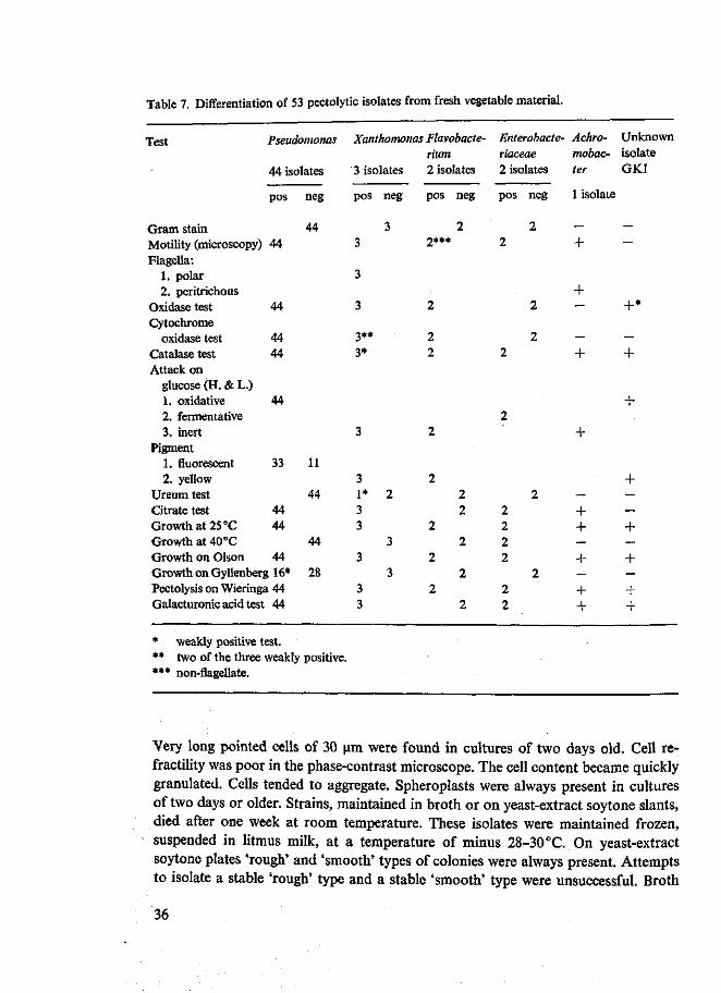
Table 7. Differentiation of 53 pectolytic isolates from fresh vegetable material.
Test Pseudomonas Xanthomonas Flavobacte- Enterobacte- Achro- Unknown Hum riaceae mobac- isolate
44 isolates 3 isolates 2 isolates 2 isolates ter GK1
Gram stain Motility (microscopy) Flagella:
1. polar 2. peritrichous
Oxidase test Cytochrome
oxidase test Catalase test Attack on
glucose (H. & L.) 1. oxidative 2. fermentative 3. inert
Pigment 1. fluorescent 2. yellow
Ureum test Citrate test Growth at 25 °C Growth at 40 °C Growth on Olson
pos
44
44
44 44
44
33
44 44
44 Growth on Gyllenberg 16* Pectolysis on Wieringa 44 Galacturonic acid test 44
* weakly positive test.
neg
44
11
44
44
28
** two of the three weakly positive. *** non-flagellate.
pos
3
3
3
3** 3*
3
3 1* 3 3
3
3 3
neg
3
2
3
3
pos
2***
2
2 2
2
2
2
2
2
neg
2
2 2
2
2
2
pos
2
2
2
2 2 2 2
2 2
neg
2
2
2
2
2
1 isolate
—
+
+ —
— +
+
— + + _ + — + +
— —
+*
— +
+
+ — — + — + — + +
Very long pointed cells of 30 urn were found in cultures of two days old. Cell re-fractility was poor in the phase-contrast microscope. The cell content became quickly granulated. Cells tended to aggregate, Spheroplasts were always present in cultures of two days or older. Strains, maintained in broth or on yeast-extract soytone slants, died after one week at room temperature. These isolates were maintained frozen, suspended in litmus milk, at a temperature of minus 28-30°C. On yeast-extract soytone plates 'rough' and 'smooth' types of colonies were always present. Attempts to isolate a stable 'rough' type and a stable 'smooth' type were unsuccessful. Broth
36
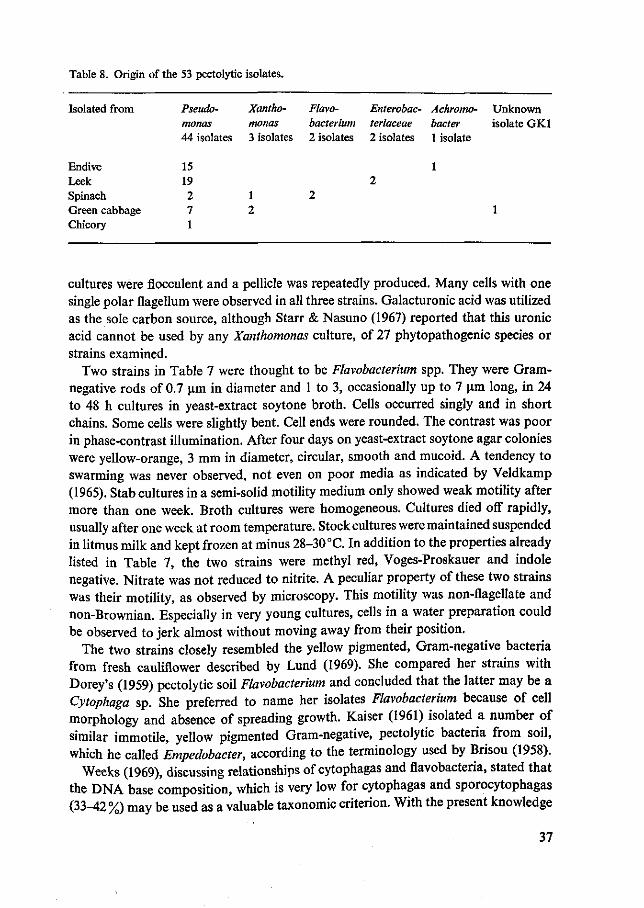
Table 8. Origin of the 53 pectolytic isolates.
Isolated from
Endive Leek Spinach Green cabbage Chicory
Pseudo-monas 44 isolates
15 19 2 7 1
Xantho-monas 3 isolates
1 2
Flavo-bacterium 2 isolates
2
Enterobac-teriaceae 2 isolates
2
Achromo-bacter 1 isolate
1
Unknown isolate GK1
1
cultures were fiocculent and a pellicle was repeatedly produced. Many cells with one single polar flagellum were observed in all three strains. Galacturonic acid was utilized as the sole carbon source, although Starr & Nasuno (1967) reported that this uronic acid cannot be used by any Xanthomonas culture, of 27 phytopathogenic species or strains examined.
Two strains in Table 7 were thought to be Flavobacterium spp. They were Gram-negative rods of 0.7 um in diameter and 1 to 3, occasionally up to 7 um long, in 24 to 48 h cultures in yeast-extract soytone broth. Cells occurred singly and in short chains. Some cells were slightly bent. Cell ends were rounded. The contrast was poor in phase-contrast illumination. After four days on yeast-extract soytone agar colonies were yellow-orange, 3 mm in diameter, circular, smooth and mucoid. A tendency to swarming was never observed, not even on poor media as indicated by Veldkamp (1965). Stab cultures in a semi-solid motility medium only showed weak motility after more than one week. Broth cultures were homogeneous. Cultures died off rapidly, usually after one week at room temperature. Stock cultures were maintained suspended in litmus milk and kept frozen at minus 28-30 °C. In addition to the properties already listed in Table 7, the two strains were methyl red, Voges-Proskauer and indole negative. Nitrate was not reduced to nitrite. A peculiar property of these two strains was their motility, as observed by microscopy. This motility was non-flagellate and non-Brownian. Especially in very young cultures, cells in a water preparation could be observed to jerk almost without moving away from their position.
The two strains closely resembled the yellow pigmented, Gram-negative bacteria from fresh cauliflower described by Lund (1969). She compared her strains with Dorey's (1959) pectolytic soil Flavobacterium and concluded that the latter may be a Cytophaga sp. She preferred to name her isolates Flavobacterium because of cell morphology and absence of spreading growth. Kaiser (1961) isolated a number of similar immotile, yellow pigmented Gram-negative, pectolytic bacteria from soil, which he called Empedobacter, according to the terminology used by Brisou (1958).
Weeks (1969), discussing relationships of cytophagas and flavobacteria, stated that the DNA base composition, which is very low for cytophagas and sporocytophagas (33-42 %) may be used as a valuable taxonomic criterion. With the present knowledge
37
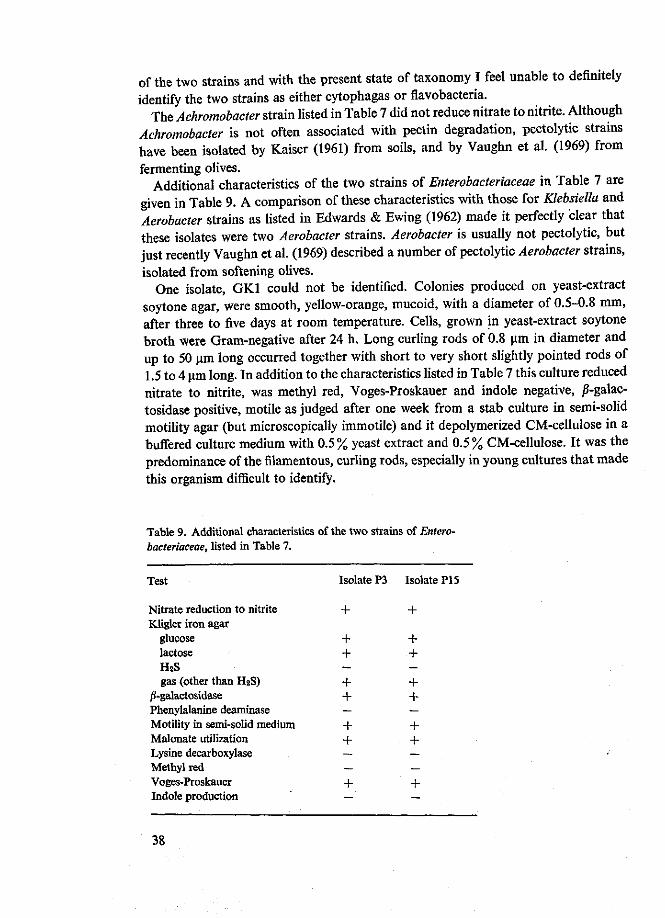
of the two strains and with the present state of taxonomy I feel unable to definitely identify the two strains as either cytophagas or flavobacteria.
The Achromobacter strain listed in Table 7 did not reduce nitrate to nitrite. Although Achromobacter is not often associated with pectin degradation, pectolytic strains have been isolated by Kaiser (1961) from soils, and by Vaughn et al. (1969) from fermenting olives.
Additional characteristics of the two strains of Enterobacteriaceae in Table 7 are given in Table 9. A comparison of these characteristics with those for Klebsiella and Aerobacter strains as listed in Edwards & Ewing (1962) made it perfectly clear that these isolates were two Aerobacter strains. Aerobacter is usually not pectolytic, but just recently Vaughn et al. (1969) described a number of pectolytic Aerobacter strains, isolated from softening olives.
One isolate, GK1 could not be identified. Colonies produced on yeast-extract soytone agar, were smooth, yellow-orange, mucoid, with a diameter of 0.5-0.8 mm, after three to five days at room temperature. Cells, grown in yeast-extract soytone broth were Gram-negative after 24 h. Long curling rods of 0.8 um in diameter and up to 50 um long occurred together with short to very short slightly pointed rods of 1.5 to 4 um long. In addition to the characteristics listed in Table 7 this culture reduced nitrate to nitrite, was methyl red, Voges-Proskauer and indole negative, ^-galac-tosidase positive, motile as judged after one week from a stab culture in semi-solid motility agar (but microscopically immotile) and it depolymerized CM-cellulose in a buffered culture medium with 0.5% yeast extract and 0.5% CM-cellulose. It was the predominance of the filamentous, curling rods, especially in young cultures that made this organism difficult to identify.
Table 9. Additional characteristics of the two strains of Enterobacteriaceae, listed in Table 7.
Test
Nitrate reduction to nitrite Kligler iron agar
glucose lactose H2S gas (other than H2S)
^-galactosidase Phenylalanine deaminase Motility in semi-solid medium Malonate utilization Lysine decarboxylase Methyl red Voges-Proskauer Indole production
Isolate P3
+
+ + —
+ + —
+ + — —
+ —
Isolate PI 5
+
+ + —
+ + —
+ + — —
+ —
38

Apart from some morphological characteristics there was much similarity between this organism and Dorey's (1959) Cytophaga which both utilized galacturonic acid as the sole carbon source.
5.4 Summary and conclusions
Pectolytic bacteria from such natural environments as fresh endive, leek, spinach, green cabbage and chicory were isolated on Wieringa's pectin medium with and without crystal violet. 53 Of the strains were identified to the generic level. The majority, 44 strains proved to belong to the genus Pseudomonas, 33 fluorescent and 11 non-fluorescent. Although good growth of these pseudomonads was observed on Olson's (1961) medium, the response to Gyllenberg's (1960) lactate crystal violet medium, sometimes used in microbiology for the detection and counting of pseudomonads, was negative or very poor. Many of these pseudomonads obviously cannot utilize lactate as a carbon source. Three isolates were identified as Xanthomonas. These three strains utilized galacturonic acid as the sole carbon source. Two strains have been called Flavobacterium. These two strains resembled closely those isolated from cauliflower by Lund (1969), which she also preferred to call Flavobacterium. It is at present not clear whether these strains should be regarded as Cytophaga or Flavobacterium. One strain was found to belong to Achromobacter. Two isolates could clearly be identified as Aerobacter. These isolates are exceptional because the genus Aerobacter is very usually not pectolytic (Edwards & Ewing, 1962). One strain remains unidentified. Its peculiar cell morphology in young stage (filamentous, curling rods) was a conspicuous phenomenon. Microscopically immotile, the isolate clearly showed motility in a stab culture in semi-solid motility agar.
42 Of the strains studied were isolated from Wieringa's pectin medium, and only 11 from Wieringa's pectin medium, containing crystal violet. Since not a single Gram-positive pectolytic strain was found among the 53 isolates studied it is justified to state that the pectolytic bacterial flora on the fresh vegetable material studied consisted predominantly of Gram-negative rods, of which most were Pseudomonas.
39
6 Screening of Arthrobacter strains for pectolytic properties
6.1 Introduction
The presence of a unique collection of Arthrobacter and Brevibacterium strains at the Department of Microbiology of the Agricultural University of Wageningen made it possible to study the presence and eventually the characteristics of pectolytic enzymes in these genera. Up till now the presence of pectolytic enzymes in Arthrobacter has only been mentioned by Bhat et al. (1968). An eliminative degradation of alginic acid by Arthrobacter, similar to that of pectic substances for example by Pseudomonas (Fuchs, 1965) has been reported by Lynn (1967) and Lynn et al. (1968).
6.2 Materials and methods
The 298 strains of Arthrobacter and Brevibacterium were supplied by Prof E. G. Mulder and J. Antheunisse of the Department of Microbiology of the Agricultural University of Wageningen. These strains originate from soils, cheese rind, cheese (brevibacteria), activated sludge from dairy sewage, milk, poultry litter, sea water, fish and fish boxes, salt pans and type culture collections. Many morphological and physiological characteristics of these strains have been extensively described by Mulder & Antheunisse (1963) and Mulder et al. (1966).
Culture media used were Wieringa's pectin plate (Chapter 4) and a buffered pectate medium of the following composition: 10 g sodium pectate 7 to 10% esterified (Obipektin AG, Bischofszell, Switzerland), 5 g Difco yeast extract, 0.15 g (0.001 M) CaCl2 • 2H20, 3 g Na2HP04 • 2H20, 2 g KH2P04 , 1 litre distilled water. All ingredients, except sodium pectate were dissolved in distilled water. The sodium pectate was separately moistened with 96% ethanol and then the solution of the other ingredients was poured on top of the sodium pectate. The pH was adjusted to 7.0 and the pectate dissolved by stirring and warming up. The medium was suspended in 5 ml portions in culture tubes and sterilized for 15 min at 115°C.
Young slant cultures on yeast glucose agar were used as inoculum. Wieringa's pectin plates were stabbed with a straight inoculation needle; three cultures on one plate. The tubes with liquid buffered pectate medium were also inoculated directly from the young slant cultures. The plates and tubes were incubated at 25 °C. Each day for one week the plates were examined for pectolytic strains, which were easily detected through the pits, formed in the medium at the inoculation point. The tubes were checked for pectolysis three times in the first week of incubation and a fourth
40

time at the end of the second week by an ethanol test. A sample of 1 ml of culture liquid was pipetted into another culture tube and mixed with 2 ml 96% ethanol. After gently shaking the tubes, they were examined for a gel-like precipitate which is formed if the pectin has not been degraded.
6.3 Results and discussion
A total of 32 of the 298 strains proved to be pectolytic as they liquefied Wieringa's pectin plate and generally depolymerized the pectate in the liquid buffered pectate medium. Five strains which liquefied the plate medium, did not depolymerize the pectate in the liquid medium, enough to be seen in the ethanol test. For this reason and also because of the longer incubation time needed with the liquid medium to detect pectolysis, it can be concluded, that for screening the pectin plate is more sensitive than the liquid pectate medium. Two factors may contribute to this difference in sensitivity. The plate medium contained 35 % esterified pectin, a different substrate from the almost completely saponified pectate in the liquid medium. There was therefore still a chance of detecting a pectin lyase producing strain on the plate, but not in the liquid medium of the composition used here. Another factor was, that, in the liquid culture the substrate had to be degraded to a certain limit through the whole medium and not just in some places as in the solid medium, before pectolysis could be detected.
In Table 10 the numbers and percentages of positive strains are differentiated according to the origin of the strains. The pectolytic strains are only found amongst the arthrobacters from soil, activated sludge from dairy sewage and sea water. None of the brevibacteria were pectolytic, nor any of the arthrobacters from cheese rind
Table 10. Pectolytic properties of 298 Arthrobacter and Brevibacterium strains, differentiated after the origin of the strains.
Origin
Soils Cheese rind Cheese (brevibacteria) Activated sludge from dairy sewage Milk Poultry litter Sea water Fish and fish boxes Salt pans Type cultures
Total
Number of strains
tested
117 47 58 23 1 3 3
32 9 5
298
positive
21 0 0 9 0 0 2 0 0 0
32
% positives
18 0 0
39 0 0
67 0 0 0
11
41

and fish and fish boxes. To test the salt-requiring arthrobacters from salt pans for pectolytic properties 4% salt was added to the liquid pectate medium. This prevented the pectate from being dissolved. After three weeks of incubation the pectate coagulum was not visibly affected.
Light induced pigmentation is known to be an important characteristic in the classification of coryneform bacteria (Mulder et al, 1966). For that reason I checked the colours of the pectolytic strains and found that they were all grey-white, pinkly shaded or yellow. No orange pigmented strains were found amongst the pectolytic strains.
Mulder & Antheunisse (1963) found great numbers of arthrobacters in different soils (60 to 90% of the bacterial colonies on casein agar plates), and I found 18% of a rather big group of these arthrobacters to be pectolytic. It is surprising that Kaiser (1961), who especially studied the pectolytic bacteria from soils, did not mention any pectolytic arthrobacters or coryneform bacteria, although he also used Wieringa's (1949,1953) pectin plate. This fact is not easy to understand but underlines the usefulness of culture collections for screening purposes.
Lynn (1967) and Lynn et al. (1968) studied the eliminative degradation of alginic acid by Arthrobacter and some other micro-organisms. Criteria for alginate utilization were growth and production of reducing substances in a medium containing mineral salts, 0.01 % yeast extract and 1 % alginic acid. They studied seven Arthrobacter strains, and found that these strains were all capable of alginate utilization. Cell-free extracts produced a series of unsaturated oligomers from alginic acid, indicating a random degradation of the substrate. They supposed that alginate utilization may be a general characteristic of Arthrobacter, making it a useful aid in the identification of the genus. In analogy to the screening procedure for pectolytic arthrobacters, 15 pectolytic and 15 non-pectolytic Arthrobacter strains of different origin were tested for alginolysis. For this purpose the pectin in Wieringa's medium was replaced by alginic acid (Kelco Company, Clark, N.J., USA) and Lynn's liquid medium with yeast extract and alginic acid was used instead of the liquid pectate medium. None of the strains were capable of liquefying the calcium alginate gel or of depolymerizing the alginate enough to be seen in the ethanol test. These results had not been expected. The test for alginolysis will have to be studied in more detail.
6.4 Summary and conclusions
A collection of 298 strains of Arthrobacter and Brevibacterium were screened for pectolytic properties on Wieringa's pectin gel medium (Chapter 4) and in a buffered liquid pectate medium. Of 32 strains that liquefied the gel, 27 also degraded pectate in the liquid medium enough to be seen in an ethanol test. The positive strains originated from soils, activated sludge from dairy sewage and sea water. None of the brevi-bacteria from cheese or of the arthrobacters from cheese rind, or fish and fish boxes were pectolytic.
42
7 Typing of pectolytic enzymes produced by a variety of bacterial strains
7.1 Introduction
To obtain a general picture of the types of pectolytic enzymes produced by a variety of bacterial strains, crude enzyme preparations of these strains were tested. The detailed testing scheme was based on up-to-date knowledge of pectolytic enzymes, especially those from bacteria. Strains of the genera or species of which one or more enzymes had already been studied extensively were included as reference material (Nagel & Vaughn, 1961a; Preiss & Ashwell, 1963a; Macmillan & Vaughn, 1964; Macmillan et al., 1964; Okamoto et al, 1964a, b, c; Fuchs, 1965; Nagel & Anderson, 1965; Hasegawa & Nagel, 1966; Nasuno & Starr, 1966a, b; Nasuno & Starr, 1967; Nagel & Hasegawa, 1967). Several strains of one genus or one species were usually included to see whether the results differed between strains or applied rather to the genus or species. This cannot be seen from literature since the published detailed studies of bacterial pectolytic enzymes were all carried out on single strains. At the same time it was hoped that, during the comparative study of the crude enzymes of a number of different bacteria, some strains might emerge whose pectolytic activities would warrant a deeper study.
Apart from a not very convincing report on enzymes of Streptomyces viridochromo-genes which attack pectin rather than pectate (Agate et al., 1962), no bacterial enzymes have been found to degrade preferentially highly esterified pectins. For this reason the testing scheme was focussed mainly on pectate splitting enzymes, produced in a pectate containing medium. However the production of pectinesterase and pectin chain splitting enzymes in a medium containing highly esterified pectin was given some attention.
7.2 Materials and methods
Strains The following strains, described in Chapter 5, were used: Flavobacterium S2, Pseudomonas S3, S7 and GK5, Xanthomonas GK6 and the unidentified strain GK1. The Arthrobacters, found to be pectolytic (Chapter 6), were represented by strains 215, 222, 370 and 547. Bacillus polymyxa 4, Bacillus subtilis and Erwinia carotovora were kindly provided by J. Antheunisse of the Department of Microbiology of the Agricultural University of Wageningen who also supplied the arthrobacters. From Dr D. C. Graham, Agricultural Scientific Services, Edinburgh, 12,1 received a number of erwinias of which Erwinia atroseptica SRI, Erwinia carotovora G117 and Erwinia
43

aroideae 140V were selected for this study. Included were also three Bacillus strains, namely CUB, CIC and CAIIA, selected on Wieringa's pectin gel medium (see Chapter 4) from dry, powdered coriander by Ir A. G. J. Voragen of the Department of Food Science of the Agricultural University, Wageningen.
Enzyme production At this stage of the study I looked for a culture medium which could be used generally for the production of pectolytic enzymes by different groups of bacteria. However, I realized that such a medium would not satisfy the nutritional requirements of the different groups of bacteria for maximum enzyme production. The choice of culture medium was based on data from the literature.
Since most pectolytic enzymes from bacteria are adaptive, pectate and sometimes pectin are used as the main carbon sources. With concentrations of 0.5 to 30 g/litre pectate has been successfully used for the production of enzymes by Flavobacterium pectinovorum (Dorey, 1959), Bacillus polymyxa (Nagel & Vaughn, 1961a), Pseudo-monas (Preiss & Ashwell, 1963a), Clostridium multifermentans (Macmillan & Vaughn, 1964) and Xanthomonas (Starr & Nasuno, 1967). The production of depolymerizing enzymes by Erwinia carotovora (Dorey, 1959) was stimulated more by pectin than by pectate. Although the production of constitutive enzymes is generally not influenced by pectate or pectin in the medium, catabolite repressed constitutive pectate lyase has been reported from Aeromonas liquefaciens (Hsu & Vaughn, 1969). Here the production of enzyme was stimulated when growth was restricted by substrate (pectate) restriction or by limiting enzymatic catabolite formation by restriction of the divalent cations in the culture medium. Most media for the production of enzymes contained in addition to the pectate or the pectin either yeast extract or peptone or casamino acids. In the media of Nagel & Vaughn (1961a) for Bacillus polymyxa and Preiss & Ashwell (1963a) for Pseudomonas pectin was the only carbon source, ammonium nitrate or ammonium phosphate being used as the nitrogen source. All media always contained 0.001 M or more of a calcium or magnesium salt, apparently to meet the divalent cation requirements of the pectate lyases. The media were buffered by phosphates at pH values ranging from 6.7 to 7.6.
One of the media adopted and called 'buffered pectate medium' had the following composition: 5 g sodium pectate, 7 to 10% esterified (Obipektin AG, Bischofszell, Switzerland) 5 g Difco yeast extract, 0.15 g (0.001 M) CaCl2 • 2H20, 3 g Na2HP04 • 2H20, 2 g KH2P04, 1 litre distilled water. All ingredients except sodium pectate were dissolved in distilled water. The sodium pectate was first moistened with 96% ethanol and the solution of the other ingredients was then poured on it. The pH was adjusted with 1 N HC1 to 7.0 and the pectate dissolved by stirring and warming. The medium was sterilized in 10 ml portions in culture tubes and in 75 ml portions in 300 ml Erlenmeyer flasks for 15 min at 115°C.
A 'buffered pectin medium' was also used. This medium was identical to the buffered pectate medium except for the pectate which was replaced by 'Brown Ribbon pectin', an apple pectin of about 75% esterification (Obipektin AG, Bischofszell, Switzerland). In order to prevent saponification and j3-eliminative degradation of this
44

substrate during sterilization the medium had to be prepared and sterilized without Na 2HP0 4-2H 20 and with 900 ml distilled water. A solution containing 3% Na2HP04 • 2H20 and a predetermined concentration (0.05 N) of NaOH was sterilized separately and 10% v/v of this solution was added aseptically to the pectin solution so that the pH of the final medium was about 7.
One or two days old cultures grown in tubes containing buffered pectate or pectin medium were used as inoculation material. Erlenmeyer flasks were inoculated with 0.75 to 1.5 ml of the precultures and incubated in a Gallenkamp (London, England) orbital incubator at 300 strokes per min (stroke width 32 mm) at 25 to 27 °C. A sub-optimum temperature of incubation proved to be favourable for enzyme production, e.g. with Clostridium multifermentans (Macmillan & Vaughn, 1964), Aeromonas liquefaciens (Hsu & Vaughn, 1969) and Erwinia aroideae (Okamoto et al., 1964a). The incubation was normally stopped when growth, as measured with an EEL-nephelometer (Evans Electroselenium Ltd, Halstead, England), reached its stationary maximum, or, in the case of Bacillus polymyxa, following Nagel & Vaughn (1961a), when spores could readily be seen under the microscope. Simultaneous with the turbidity measurements, an ethanol test (Chapter 6) was carried out to check pectate degradation in the culture medium.
At the end of incubation, the Erlenmeyer flasks were cooled in a refrigerator and subsequently the cells were removed by centrifuging in a Christ Junior II centrifuge at 4 500 x g. The culture liquid was Seitz filtered and stored as crude enzyme solution in sterile infusion flasks in the refrigerator with 0.02% thiomersal added. One crude enzyme preparation that of Cellulomonas fimi was stored for three years under these conditions without any demonstrable loss in viscosimetric activity.
Typing of the enzymes The crude enzyme preparations were dialysed overnight against running tap water to discard salts and other low molecular material from the original culture medium as well as low molecular degradation products of the pectate in the medium. These enzyme preparations were then used for the typing without further treatment.
Whereas the activity on pectin was only studied by a titrimetric pectinesterase test and a viscosity test, the enzymatic degradation of pectic acid was studied in more detail, beginning with a viscosimetric test. Samples were withdrawn at an early and a late stage related to the viscosity curve, then inactivated and used for further study which included analysis of the formed oligogalacturonides by UV absorption measurement, by a periodate-thiobarbituric acid test and by paper chromatography.
These tests are useful for the differentiation of the four possible pectate depolymer-ases, namely exo and endo pectate lyase (pectic acid transeliminase) and exo and endo polygalacturonase. With the sample taken at an early stage related to the viscosity curve the exo enzymes can be detected. Macmillan et al. (1964) found that the exo pectic acid transeliminase of Clostridium multifermentans had degraded 22.5% of polygalacturonic acid to unsaturated digalacturonic acid whilst the specific viscosity of the reaction mixture had dropped to 50%. The degree of degradation at a 50%
45
drop of specific viscosity is much smaller for endo enzymes. Thus Nagel & Vaughn (1961a) found 2% degradation for the endo enzyme of Bacillus polymyxa and Hase-gawa & Nagel (1966) 1.6% for the endo pectic acid transeliminase of yet another Bacillus sp. These results imply that the paper chromatogram of an early sample of a reaction mixture of substrate and endo enzyme will hardly differ, if at all, from the chromatogram of substrate with inactivated enzyme, because the chromatogram only shows spots of oligomers up to tetragalacturonic acid. With an exo enzyme, however, the chromatogram of the early sample may differ considerably from that of a blank sample, whereas the chromatogram of the late sample can be quite similar to that of the early sample. As pectolytic enzymes often occur as mixtures many intermediary situations are possible.
Pectic acid was used as the substrate. It was prepared from a commercial apple pectin (Pink Ribbon, 26% esterified, Obipektin AG, Bischofszell, Switzerland) by saponification and purification according to the procedure of Altermatt (1954) and Derungs (1958). The preparation was 0-1 % esterified and had a polygalacturonide content of 77%. To determine the degree of polymerization of this substrate, reducing end-groups were measured with the sodium chlorite reducing end-group method of Launer & Tomimatsu (1954, 1959a, b) as adapted for pectic substances by Voragen et al. (1971a). The degree of polymerization, calculated as the -COOH/-CHO coefficient was 45. More details on the preparation of this pectic acid are to be given in Chapter 9. The substrate was used in a concentration of 0.25%, with 0.00025 M CaCl2 • 2H20 added, pH 7.0, adjusted with NaOH. Calcium was added because pectate lyases have an absolute requirement for certain divalent cations (Chapter 3). A pH of 7.0 was selected being near to the pH of Wieringa's pectin gel medium (Chapter 4) and a value between the optimum pH values of the bacterial pectate hydrolases and pectate lyases (Chapter 3). Since the pH did not change to any appreciable extent during the experiments, buffers that may affect enzyme activity were omitted.
Ubbelohde glass capillary viscosimeters suspended in a water bath at 30 °C were used. The water value of the viscosimeters was 30 to 35 seconds. Into each viscosi-meter, 8 ml substrate was pipetted and allowed to equilibrate. The dialysed enzyme adjusted to PH 7.0 was brought to temperature and of this, 4 ml or less was pipetted in. Distilled water was added when necessary to bring the total content of the viscometer o 12 ml. The amount of enzyme was selected to give a 50% drop in specific viscosity m about 30 min. The time it takes to reach a 50% drop in specific viscosity was termed viscosimetric half-value time'. The reaction mixture was homogenized
i ™ ° H T ? ZUg ^ v i s c o s i m e t e r a n d t h e first viscosity reading was then recorded immediately. This value was taken as initial viscosity of the reaction mixture. Readings
S r , ?rod:?aliy untii the specific v i sc°s i tv had *™»*& * ^ n h ^ of ^ fiX^ ' t h
+ T a C t l 0 n m i x t u r e w a s l e f t a t 30°C in the water bath. Two samples, I t Z t i ' T K ' ^ WhCn t hC r e a C t i 0 n h a d P r o c e e d e d f o r a time period two Lef th t T V 1 S C °T t r f C h a l f 'V a l u e t i m e <earlv s a m ^ )> ^ other after 10 to 20
times that time period (late sample). The early and late samples were immediately
46

heat inactivated (5 min in boiling water). A blank sample of 6 ml of substrate and heat inactivated enzyme was prepared. Then two volumes (12 ml) of 96% ethanol were added to all three samples and after mixing the gel was filtered off under suction, usingaBuchner funnel with a gel filter (Schleicher & Schiill, No. 520b). It was found that under these conditions mono to at least penta galacturonic acids were able to pass the filter. The samples were subsequently evaporated to dryness in a film evaporator and taken up in 1 ml distilled water. To obtain the oligomeric breakdown products in the acid form, 1 g of Dowex W-X8 50/100 mesh, moisture content 50% w/w in the H form was added to all concentrated samples. This was important for paper chromatography (Young & Corden, 1964) and for the preservation of the samples which were stored in the refrigerator.
UV test The UV test differentiates between the lyases (transeliminases) producing C4-C5 double bonds at the split glycosidic linkages and the hydrolases. The unsaturated uronides resulting from transeliminase action cause absorption in the ultraviolet region (Linker et al., 1956; Albersheim et al., 1960b; Hasegawa & Nagel, 1962). In principle an adequate dilution of any sample can be measured directly in the spectrophotometer and when the spectrum is measured between 220 and 260 nm a peak will be found at or near 235 nm. Macmillan & Vaughn (1964) showed that the pH of the measured solution cannot be ignored. When measuring a solution of unsaturated digalacturonic acid, at pH 9.5, 3.7 and 1.5 they observed the absorption peak shifting to higher wave lengths and higher absorption values. They calculated a molar extinction coefficient for unsaturated digalacturonic acid at pH 3.7 of 4 800 M^cm"1. Since then slightly different molar extinction coefficients, both higher and lower have been reported: 4 600 for unsaturated trimer and tetramer at 232 nm and pH 6.7 and 4 500 for unsaturated dimer (Nagel & Anderson, 1965) and 5 200 for unsaturated digalacturonic acid at pH 3.7 (Nasuno & Starr, 1967). Because of these differences the constants are being redetermined by Voragen (1972).
Nagel & Anderson (1965) found no absorption peak in the UV region for unsaturated monogalacturonic acid so that they concluded that this compound does not occur in the ring form, but as 5-keto-4-deoxygalacturonic acid. Earlier, Preiss & Ashwell (1963a), studying this compound as an intermediate in the polygalacturonic acid metabolism of Pseudomonas, had come to the same formula.
The UV test was carried out as follows. In a 10 mm, 4 ml quartz cuvette was pipetted successively 0.1 ml of the (diluted) sample, 2.5 ml distilled water and 1 ml 0.36 N HC1. The absorbance was measured against a standard of distilled water at 220-225-230-232-235-237-240-245-250-255-260 nm. A Zeiss PMQ-U spectrophotometer was used.
Periodate-TBA test The periodate-thiobarbituric acid test is a new, very sensitive colorimetric method in carbohydrate analysis. The method was first described by Waravdekar & Saslaw (1959) for the determination of 2-deoxysugars. They oxidized the free 2-deoxysugars with periodate whereby malondialdehyde was formed. The malondialdehyde when heated with thiobarbituric acid yields a red chromogen with
47

an absorption maximum at 532 nm. This condensation product has been crystallized and its chemical constitution described by Schmidt (1959). The molar extinction coefficient for malondialdehyde, calculated by me from a calibration curve in the paper of Waravdekar & Saslaw (1959) was 150 000; Schmidt (1959) himself, reported it to be 156 000 M^cm - 1 .
The method was modified by Weissbach & Hurwitz (1959) and Shrinivasan & Sprinson (1959) for the detection and estimation of 2-keto-3-deoxy heptonic acids. Here the periodate treatment yields jS-formylpyruvic acid which reacts with thio-barbituric acid to give a red condensation product with an absorption peak at 545 to 550 nm. The molar extinction coefficient for the heptonic acid was 44 000 M_1cm_1
under the test conditions of Weissbach & Hurwitz, namely 20 min periodate oxidation at room temperature and 10 min boiling with thiobarbituric acid. But this constant was higher, namely 72 000 under the test conditions of Shrinivasan & Sprinson (1959) who oxidized with periodate for 45 min at room temperature and boiled 5 min with thiobarbituric acid. The reaction equation of j8-formylpyruvic acid with thiobarbituric acid, as given by Schmidt (1959) is presented in Figure 3. Shrinivasan & Sprinson (1959), as well as Schmidt (1959) observed that glyoxal (CHO-CHO), when heated with thiobarbituric acid yielded the same chromogen, apparently through condensation to /S-formylpyruvic acid.
To the group of compounds, which yield /?-formylpyruvic acid upon periodate treatment, belong the 'unsaturated monogalacturonic acid' (4-deoxy-L-threo-4-hexoseulose uronic acid) and the unsaturated uronic acid constituent of the oligo-galacturonic acids (Preiss & Ashwell, 1963a), as well as the comparable unsaturated
SH
N*^N.
+ H O
C—COOH II CH I CH II O +
-2H 2 0
I SH
Fig. 3. Reaction of 0-formylpyruvic acid with thiobarbituric acid, after Schmidt (1959).
48

degradation products of alginic acid, for example 'unsaturated mannuronic acid' (4-deoxy-L-ery?/zro-5-hexoseulose uronic acid) and the unsaturated uronic acid constituent of the oligomannuronic acids (Preiss & Ashwell, 1962; Preiss, 1966). Preiss & Ashwell (1962) compared the rates of periodate oxidation of unsaturated oligomeric degradation products and the unsaturated monomeric degradation product of alginic acid and found that the rate of /?-formylpyruvic acid formation from the unsaturated oligomers was quicker. They referred to Waravdekar & Saslaw (1959) who similarly found the formation of malondialdehyde from galactal to be faster than from 2-deoxyribose (Figure 4). Preiss & Ashwell pointed out that galactal may be considered as the enol form of a 2-deoxy sugar just as the unsaturated oligouronide may be regarded as the enol form of a 2-keto-3-deoxycarboxylic acid.
There is one more factor that influences the rate of periodate oxidation. The hydroxyl groups at C2 and C3 of mannuronic acid are in cis position, whereas the
.OR
H5IQ6
H O — C — H I
H O — C — H
H - C
II c o I COOH
unsaturated oligomannuronic
acid
CHO ' I H - C
I C - O H I
COOH
• CHO H5IQ6
H - C - H
I c=o I
COOH
p - fo rmy l pyruvic
acid
CHO
I H O - C — H
I H O - C — H
I H - C - H
I c=o I
COOH
4-deoxy 5-keto mannuronic
acid
H I
C — -II
H - C I
H O - C - H
I H O - C - H
I H - C
I CH2OH
galactal
H5I06
CHOH CHO II — I
H-C-H - I
CHO
H—C I
CHO
H5I06
malon dialdehyde
CHO
I H - C - H
I H - C - O H
I H - C - O H
CH2OH
D-deoxyribose
Fig. 4. Action of periodate on unsaturated oligouronides, deoxyketouronic acid, galactal and D-
deoxyribose (Preiss & Ashwell, 1962).
49
corresponding hydroxyl groups of galacturonic acid are in trans position. It has been shown by Waravdekar & Saslaw (1959) that oxidation for 20 min with periodate is optimum for sugars like 2-deoxyribose, with a cw-diol configuration at the C-atoms 3 and 4, whereas 40 min is needed for sugars like 2-deoxyxylose with a trans configuration. Hence in the periodate-thiobarbituric acid test as applied to unsaturated breakdown products of pectic substances, the periodate oxidation should last 40 min.
In one respect the work of Preiss & Ashwell (1962, 1963a) may easily be misunderstood. It may be thought that the actual yield of /Mbrmylpyruvic acid from unsaturated breakdown products of alginic and polygalacturonic acids under the right conditions of oxidation with periodate is equal to the theoretical yield. They do not mention the simultaneous disappearance of /?-formylpyruvic acid during its formation which must certainly be considerable. This can be concluded from their own statement that '0.01 umole of j?-formylpyruvic gave an optical density reading of 0.290 at 549 nm' (Preiss & Ashwell, 1962, 1963a; Preiss, 1966). Since the total reaction mixture of 2.95 ml, was measured in a 1 cm cuvette, the molar extinction coefficient of jS-formyl-pyruvate could easily be calculated to be 83 000 M-1cm_1. The molar extinction coefficient of unsaturated digalacturonic acid at 235 nm, however, was 4 800 M~ m" \ according to Macmillan & Vaughn (1964). These values mean that the periodate-TBA test would be 18 times as sensitive as the UV test, if the /?-formylpyruvic acid yield was indeed equal to the theoretical yield. My determination of the molar extinction coefficient in the periodate-TBA test for unsaturated digalacturonic acid gave results of the order of 3 000 M_1cm_1. The difference between this value and that for ^-formylpyruvic acid is obviously caused by the oxidation of jS-formylpyruvic acid by periodate. As the periodate treatment becomes more intensive to liberate fi-formylpyruvic acid from the test substance the loss of )3-formylpyruvic acid increases (Shrinivasan & Sprinson, 1959). Okamoto et al. (1965) were well aware of the simultaneous formation and destruction of j3-formylpyruvic acid. They selected the periodate oxidation conditions for maximum net yield of 0-formylpyruvic acid from unsaturated digalacturonic acid and found 15 min at 80 °C to be the most favourable time-temperature combination.
Albersheim et al. (1960a, b), Fuchs (1965), Nagel & Anderson (1965) and Nagel & Hasegawa (1967) applied a TBA test without periodate step to the chemical and enzymatic degradation products of pectic substances. In this test the peak of maximum absorption was also at 548-550 nm. It is supposed that 0-formylpyruvic acid is an intermediate in this test too (Albersheim et al., 1960b). This test can also be used for the detection of monogalacturonic acid which shows an absorption maximum at 510-515 nm (Neukom, 1960). This specificity for monogalacturonic acid may be an advantage of this test which, however was clearly demonstrated by Okamoto et al. (1965), to be less sensitive for unsaturated degradation products of pectic substances, than the periodate-TBA method.
The periodate-TBA test is of great value for the study of unsaturated degradation products of pectic substances because of its high specificity and its high sensitivity and because it may also be used for 4-deoxy-5-keto-uronic acid which cannot be
50
measured with the UV test. Although the test is not stoechiometric it may be used quantitatively, provided the test conditions are well standardized.
The test used in this study is that of Weissbach & Hurwitz (1959) with a prolonged periodate oxidation step, according to Waravdekar & Saslaw (1959).
The following reagents were prepared:
- 0.025 M Periodic acid in 0.125 N H2S04 : 0.5700 g of H5IOe (Merck, p.a.) is dissolved in 100 ml 0.125 N H2S04. - 2% Sodium arsenite in 0.5 N HC1: I g of NaAs02 (BDH, Lab. reagent) is dissolved in 50 ml 0.5 N HC1. - 0.3 % Thiobarbituric acid, pH 2.0: 0.300 g of thiobarbituric acid (BDH, minimum acidimetric assay 98.5%, M.W. 144.15) was dissolved in 100 ml of distilled water by stirring and gently warming up.
The test was carried out by pipetting 0.20 ml of the sample or a total of 0.20 ml sample and distilled water into a test tube. Then 0.25 ml 0.025 M periodic acid in 0.125 N H2S04 was added. After 40 min reaction at room temperature, 0.50 ml 2% sodium arsenite in 0.5 N HC1 was added. The mixture was gently shaken and allowed to stand for 2 min, during which time the brown iodine colour appeared and disappeared again. Then 2 ml of 0.3 % thiobarbituric acid was pipetted in. After shaking, the tubes were provided with tear drop condensors or glass marbles and heated for 10 min in a boiling water bath, along with a blank tube, containing 1 ml of distilled water and 2 ml of thiobarbituric acid. The tubes were then cooled in tap water. The content was measured against the blank without delay in 1 cm cuvettes in a (Zeiss PMQ-II) spectrophotometer normally at 550 nm only or sometimes at 500-510-515-520-530-540-545-550-555-560 and 570 nm. When assaying mixtures of higher oligomers or undegraded pectic substances a precipitate may be observed, which is centrifuged off.
Paper chromatography Many solvents have been used for paper chromatographic separation of breakdown products of pectic acid. However, there is only one which separates normal and unsaturated oligogalacturonic acids. This is ethyl acetate-pyridine-water-acetic acid ( 5 : 5 : 3 : 1 ) , the solvent which was first used for this purpose by Hasegawa & Nagel (1962) and Nagel & Anderson (1965). The Rgal values (the migration distances of oligogalacturonic acids relative to that of galacturonic acid) obtained with this solvent by Nagel & Anderson (1965) and by me, are given in Table II A. Unfortunately the solvent gives low Rf values. This disadvantage can partly be overcome by using a quick paper, Whatman No. 4, and by having the solvent dripping off the paper for a certain time, in descending chromatography. Other solvents used are w-butanol-acetic acid-water (50 : 12 : 25) (Preiss & Ashwell, 1963a; Fuchs, 1965) and the epiphase of ethyl acetate-acetic acid-water ( 3 : 1 : 3) (Preiss & Ashwell, 1963a).
The sprays used were aniline phthalate, thiobarbituric acid spray and acridine
51
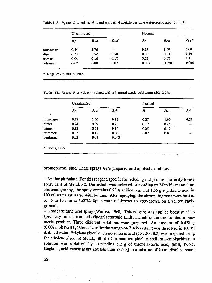
Table 11A. Rf and Rgai values obtained with ethyl acetate-pyridine-water-acetic acid (5:5:3:1).
monomer dimer trimer tetramer
Unsaturated
Rf
0.44 0.13 0.04 0.02
Rgal
1.76 0.52 0.16 0.08
Rgal*
— 0.50 0.18 0.07
Normal
Rf
0.25 0.06 0.02 0.007
Rgal
1.00 0.24 0.08 0.028
Rgal*
1.00 0.30 0.11 0.004
* Nagel& Anderson, 1965.
Table 11B. Rf and Rgai values obtained with w-butanol-acetic acid-water (50:12:25).
monomer dimer trimer tetramer pentamer
* Fuchs, 1965.
Unsaturated
Rf
0.38 0.24 0.12 0.05 0.02
Rgal
1.40 0.89 0.44 0.19 0.07
Rf*
0.35 0.23 0.14 0.08 0.043
Normal
Rf
0.27 0.12 0.05 0.02
Rgal
1.00 0.44 0.19 0.07
Rf*
0.26 — — —
bromophenol blue. These sprays were prepared and applied as follows:
- Aniline phthalate. For this reagent, specific for reducing end-groups, the ready-to-use spray cans of Merck AG, Darmstadt were selected. According to Merck's manual on chromatography, the spray contains 0.93 g aniline p.a. and 1.66 g o-phthalic acid in 100 ml water saturated with butanol. After spraying, the chromatograms were heated for 5 to 10 min at 105 °C. Spots were red-brown to grey-brown on a yellow background. - Thiobarbituric acid spray (Warren, 1960). This reagent was applied because of its specificity for unsaturated oligogalacturonic acids, including the unsaturated mono-meric product. Three different solutions were prepared. An amount of 0.428 g (0.002 mol)NaI04 (Merck 'zur Bestimmung von Zuckerarten') was dissolved in 100 ml distilled water. Ethylene glycol-acetone-sulfuric acid (50 : 50 : 0.3) was prepared using the ethylene glycol of Merck, 'fiir die Chromatographic'. A sodium 2-thiobarbiturate solution was obtained by suspending 5.2 g of thiobarbituric acid, (BDH, Poole, England, acidimetric assay not less than 98.5 %) in a mixture of 70 ml distilled water
52
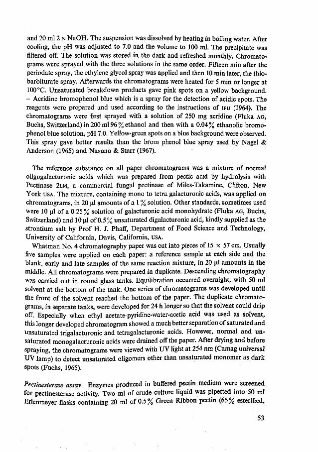
and 20 ml 2 N NaOH. The suspension was dissolved by heating in boiling water. After cooling, the pH was adjusted to 7.0 and the volume to 100 ml. The precipitate was filtered off. The solution was stored in the dark and refreshed monthly. Chromato-grams were sprayed with the three solutions in the same order. Fifteen min after the periodate spray, the ethylene glycol spray was applied and then 10 min later, the thio-barbiturate spray. Afterwards the chromatograms were heated for 5 min or longer at 100 °C. Unsaturated breakdown products gave pink spots on a yellow background. - Acridine bromophenol blue which is a spray for the detection of acidic spots. The reagents were prepared and used according to the instructions of IFU (1964). The chromatograms were first sprayed with a solution of 250 mg acridine (Fluka AG, Buchs, Switzerland) in 200 ml 96 % ethanol and then with a 0.04% ethanolic bromophenol blue solution, pH 7.0. Yellow-green spots on a blue background were observed. This spray gave better results than the brom phenol blue spray used by Nagel & Anderson (1965) and Nasuno & Starr (1967).
The reference substance on all paper chromatograms was a mixture of normal oligogalacturonic acids which was prepared from pectic acid by hydrolysis with Pectinase 2LM, a commercial fungal pectinase of Miles-Takamine, Clifton, New York USA. The mixture, containing mono to tetra galacturonic acids, was applied on chromatograms, in 20 ul amounts of a 1 % solution. Other standards, sometimes used were 10 ul of a 0.25% solution of galacturonic acid monohydrate (Fluka AG, Buchs, Switzerland) and 10 ul of 0.5 % unsaturated digalacturonic acid, kindly supplied as the strontium salt by Prof H. J. Phaff, Department of Food Science and Technology, University of California, Davis, California, USA.
Whatman No. 4 chromatography paper was cut into pieces of 15 x 57 cm. Usually five samples were applied on each paper: a reference sample at each side and the blank, early and late samples of the same reaction mixture, in 20 ul amounts in the middle. All chromatograms were prepared in duplicate. Descending chromatography was carried out in round glass tanks. Equilibration occurred overnight, with 50 ml solvent at the bottom of the tank. One series of chromatograms was developed until the front of the solvent reached the bottom of the paper. The duplicate chromatograms, in separate tanks, were developed for 24 h longer so that the solvent could drip off. Especially when ethyl acetate-pyridine-water-acetic acid was used as solvent, this longer developed chromatogram showed a much better separation of saturated and unsaturated trigalacturonic and tetragalacturonic acids. However, normal and unsaturated monogalacturonic acids were drained off the paper. After drying and before spraying, the chromatograms were viewed with UV light at 254 nm (Camag universal UV lamp) to detect unsaturated oligomers other than unsaturated monomer as dark spots (Fuchs, 1965).
Pectinesterase assay Enzymes produced in buffered pectin medium were screened for pectinesterase activity. Two ml of crude culture liquid was pipetted into 50 ml Erlenmeyer flasks containing 20 ml of 0.5% Green Ribbon pectin (65% esterified,
53

Obipektin AG) pH 7.0. The reaction mixtures were adjusted to pH 7.0 with 0.01 N NaOH and preserved by addition of 0.2 ml of a 1 % thiomersal solution (BDH, Poole, England). After one and four days at 30 °C the pH was measured and readjusted to 7.0 with 0.01 N NaOH. Blanks containing substrate and heat inactivated enzyme were included in the series. Some of the most active preparations were also tested in identical reaction mixtures with a Metrohm Combi titrator (Metrohm, Herisau, Switzerland) with the pH maintained at 7.0 with 0.01 N NaOH.
Viscosimetric assay on high and low methoxyl pectin Enzymes produced in buffered pectin medium were also tested for viscosimetric activity on high methoxyl pectin compared with that on low methoxyl pectin. As a high methoxyl pectin I used a Brown Ribbon pectin (Obipektin AG), further esterifled to 88 % with methanol-HCl (Heri et al, 1961). The low methoxyl pectin was Pink Ribbon (Obipektin AG), an apple pectin with 25 % esterification. Of both pectins a 0.25 % solution in 0.25 mM CaCl2 • 2H20, pH 7.0 was prepared. All reaction mixtures were made up directly in viscosimeters and contained 8 ml substrate, 2 ml distilled water and 2 ml of the same enzyme solution as used in the PE test. During incubation at 30 °C, the viscosity was read periodically.
7.3 Results and discussion
All bacteria tested grew well in both buffered pectate and pectin media. The beginning of the stationary growth phase was attained after 14 to 24 h sometimes after
Absorbance
1.0-
0.5
Absorbance
1.0i
0.5
220 235 260 wavelength (nm)
500 550 570 wavelength (nm)
Fig. 5. UV test (Fig. 5a) and periodate-thiobarbituric acid test (Fig. 5b) of products of pectic acid degraded by the enzyme preparation of Pseudomonas S3, B, blank; E, early; L, late sample. All curves apply to 0.05 ml aliquots of the samples.
54
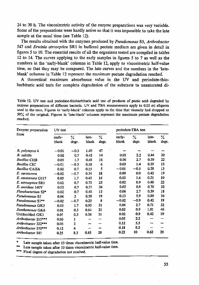
24 to 30 h. The viscosimetric activity of the enzyme preparations was very variable. Some of the preparations were hardly active so that it was impossible to take the late sample at the usual time (see Table 12).
The results obtained with the enzymes produced by Pseudomonas S3, Arthrobacter 547 and Erwinia atroseptica SRI in buffered pectate medium are given in detail in figures 5 to 10. The essential results of all the organisms tested are compiled in tables 12 to 14. The curves applying to the early samples in figures 5 to 7 as well as the numbers in the 'early-blank' columns in Table 12, apply to viscosimetric half-value time, so that they may be compared. The late curves and the numbers in the 'late-blank' columns in Table 12 represent the maximum pectate degradation reached.
A theoretical maximum absorbance value in the UV and periodate-thio-barbituric acid tests for complete degradation of the substrate to unsaturated di-
Table 12. UV test and periodate-thiobarbituric acid test of products of pectic acid degraded by enzyme preparations of different bacteria. UV and TBA measurements apply to 0.05 ml aliquots used in the tests. Figures in 'early-blank' columns apply to the time that viscosity had dropped to 50% of the original. Figures in 'late-blank' columns represent the maximum pectate degradation reached.
Enzyme preparation from
UV test periodate-TBA test
early-blank
% degr.
late- % blank degr.
early-blank
% degr.
late-blank
% degr.
B.polymyxaA -0.01 -0 .3 1.40 47 - - - -B.subtilis 0.02 0.7 0.42 14 0.05 2.2 0.44 20 Bacillus CUB 0.05 1.7 0.48 16 0.06 2.7 0.50 22 Bacillus CIC -0.01 -0 .3 0.18 6 0.03 1.4 0.29 13 Bacillus CAIIA 0.02 0.7 0.15 5 -0.01 -0 .5 0.28 13 E. carotovora -0 .02 -0 .7 0.54 18 0.00 0.0 0.42 19 E. carotovora G117 0.05 1.7 0.43 14 0.03 1.4 0.21 10 E. atroseptica SRI 0.02 0.7 0.70 23 0.02 0.9 0.48 22 E. aroideae MOV 0.02 0.7 0.71 24 0.02 0.9 0.70 32 Flavobacterium S2* 0.02 0.7 0.40 13 0.06 2.7 0.39 18 Pseudomonas S3 0.06 2 0.58 19 0.13 5.9 0.80 36 Pseudomonas SI** -0 .02 -0 .7 0.23 8 -0.02 -0 .9 0.42 19 Pseudomonas GK5 0.05 1.7 0.93 31 0.06 2.7 0.71 32 Xanthomonas GK6 0.01 0.3 0.64 21 0.02 0.9 1.01 46 Unidentified GK1 0.01 0.3 0.34 11 0.02 0.9 0.42 19 Arthrobacter 215*** 0.03 1 - - °-05 2-2 ~ ~ Arthrobacter 222*** 0.06 2 — - 0-12 5.5 - — Arthrobacter 370*** 0.12 4 — — ° 1 8 8-2 ~ —
Arthrobacter 547 0.25 8.3 0.85 28 0.22 10 0.62 28
* Late sample taken after 13 times viscosimetric half-value time. ** Late sample taken after 10 times viscosimetric half-value time. *** Final degree of degradation not reached.
55
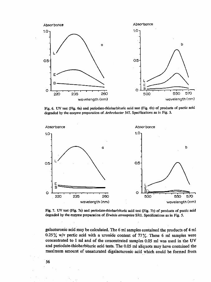
Absorbance
1.0-
0.5
Absorbance
1.0 n
ci
0.5
— i 1 1 1 1 1 r
220 235 260
wavelength (nm)
5 00 5 5 0 570
wavelength (nm)
Fig. 6. UV test (Fig. 6a) and periodate-thiobarbituric acid test (Fig. 6b) of products of pectic acid degraded by the enzyme preparation of Arthrobacter 547. Specifications as in Fig. 5.
Absorbance
1.0-
0.5
Absorbance
1.0-t
0.5-
- i 1 1 r- 0 220 260
wavelength (nm)
— i — i 1 — i — i — i — i — i — i
500 550 570 wavelength (nm)
Fig. 7. UV test (Fig. 7a) and periodate-thiobarbituric acid test (Fig. 7b) of products of pectic acid degraded by the enzyme preparation of Erwinia atroseptica SRI. Specifications as in Fig. 5.
galacturonic acid may be calculated. The 6 ml samples contained the products of 4 ml 0.25% w/v pectic acid with a uronide content of 77%. These 6 ml samples were concentrated to 1 ml and of the concentrated samples 0.05 ml was used in the UV and periodate-thiobarbituric acid tests. The 0.05 ml aliquots may have contained the maximum amount of unsaturated digalacturonic acid which could be formed from
56
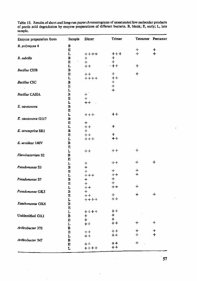
Table 13. Results of short and long-run paper chromatograms of unsaturated low molecular products of pectic acid degradation by enzyme preparations of different bacteria. B, blank; E, early; L, late sample.
Enzyme preparation from
B. polymyxa 4
B. subtilis
Bacillus CIIB
Bacillus CIC
Bacillus CAIIA
E. carotovora
E. carotovora Gil7
E. atroseptica SRI
E. aroideae 140V
Flavobacterium S2
Pseudomonas S3
Pseudomonas S7
Pseudomonas GK5
Xanthomonas GK6
Unidentified GK1
Arthrobacter 370
Arthrobacter 547
Sample
B E L B E L B E L B E L B E L B E L B E L B E L B E L B E L B E L B E L B E L B E L B E L B E L B E L
Dimer
+ + + + + + + +
+ + + + + +
+ + + +
+ + +
+ + + + + + + + +
+ +
+ + + + + + + + + + + + + + + + +
+ + + + + + + +
+ + + +
+ + + + + +
Trimer
+ + + + +
•+ +
+ + + + + +
+ +
+
+ + +
+ +
+ +
+ + + + + + +
+ + +
+ + + + + +
+ + + +
+ + + +
Tetramer
+ +
+
+
+
+
+ +
+
+
+
+ +
+
Pentamer
+ +
+
+
+
+ +
57

3.85 x 10"4 g pectic acid 100% pure. The molecular weight of unsaturated digalac-turonic acid is 342, and since this is just double that of the anhydrogalacturonic acid unit, 3.85 x 10 - 4 g pectic acid may have been converted to 3.85 x 10_4/342 = 1.1 x 10"6mol unsaturated digalacturonic acid. In the UV test the 1.1 x 10_6mol unsaturated digalacturonic acid was contained in a volume of 3.6 ml in a 1 cm cuvette, in the periodate-thiobarbituric acid test, in a volume of 2.95 ml, so that the concentrations were 3.1 x 10~4 M and 3.7 x 10~4 M, respectively. Since the molar extinction coefficients are 4 800 M~x cm - * at 235 nm (Macmillan & Vaughn, 1964) and 3 000 M - 1 cm - 1 at 550 nm the theoretical maximum absorbance values were 1.50 at 232 nm and 1.10 at 550 nm for the two tests. For certain reasons these theoretical values were not obtained in the late samples (see also Table 12). Firstly with the organisms studied the pectic acid was not broken down to unsaturated digalacturonic acid only (Tables 13 and 14), as it was for example with Clostridium multifermentans (Macmillan & Vaughn, 1964; Macmillan et al., 1964) but higher unsaturated oligomers and sometimes saturated products were present in the final reaction mixtures. In fact, the ultimate degree of degradation was a function of the enzyme, especially of its sensibility to product inhibition and its ability to split oligogalacturonides; but when the enzyme preparation was not very active it was also influenced by enzyme in-activation during the necessary prolonged incubation period. Secondly, as will be shown, an unsaturated monomeric compound may have been formed and this did not
Table 14. Results of short-run and long-run paper chromatograms of saturated low molecular products of pectic acid degradation by enzyme preparations of different bacteria. B, blank; E, early; L, late sample. (The strains listed in Table 13 and not in Table 14 do not produce saturated products).
Enzyme preparation from
B. subtilis
Bacillus CIC
Bacillus CAIIA
E. carotovora
E. carotovora G112
E. atroseptica SRI
E. aroideae 140V
Sample
B E L B E L B E L B E L B E L B E L B E L
Monomer
+ + + + + + + + +
+ + +
Dimer
+ + + + + + +
+
+ + + + + +
+
Trimer
+ + +
+ + + + +
+
Tetramer
+
58

contribute to the absorbance value in the UV test (Nagel & Anderson, 1965), although it did in the periodate-thiobarbituric acid test. Thirdly, the standardized pretreatment of the samples also resulted in small losses of products, probably mainly through occlusion in the gel during ethanol treatment.
From the 50% transeliminative degradation values and from the UV and TBA measurements in blank, early and late samples the degradation values, due to transeliminative enzyme action, could be calculated from early and late samples. These degradation values are recorded in Table 12, together with the UV and TBA values. These values are most realistic for the late samples, but less for the early samples, because unsaturated oligogalacturonides with a degree of polymerization of higher than five were removed from the samples by ethanol precipitation, and hence did not contribute to the UV and TBA values. The final degree of degradation as calculated from TBA measurements was very often considerably higher than that calculated from UV measurements. This was because of the presence of an unsaturated monomeric product in the late sample. Since the presence of an unsaturated monomeric product on paper chromatograms could not be detected with aniline phthalate, the spray most generally used, no unsaturated monomeric product was listed in Table 13. However a number of chromatograms were sprayed with thiobarbiturate and the un-
Fig. 8. Short-run and long-run paper chromatograms of low molecular products of pectic acid degradation by enzymes of Pseudomonas S3.
Solvent, ethyl acetate-pyri-dine-water-acetic acid (5:5:3:1). Spray, aniline phthalate. B, blank; E, early; L, late sample. Reference sample of mono to tetra galacturonic acids at the sides. M, monomer; D, dimer; D\ unsaturated di-mer; T, trimer; T\ unsaturated trimer; Te, tetramer; Te\ unsaturated tetramer.
59

saturated monomer was shown to be present on several of these. Its presence was particularly clearly shown when «-butanol-acetic acid-water (50 : 12 :25) was used as the solvent. An unsaturated monomer was shown to be present in the late, but not in the blank and early samples of B. polymyxa 4, Pseudomonas S7, unidentified strain GK1, Arthrobacter 370 and 547. It was not present in any of the samples of E. caroto-vora G117 or E. atroseptica SRI. The blank, early and late samples of one more organism studied: Flavobacterium S2 all showed unsaturated product, although the enzyme had been dialysed as usual. The undialysed enzyme contained an interesting amount of unsaturated monomeric product and this accumulation phenomenon, as well as the product are being studied in more detail. Another reason for the higher percentage degradation measured with the periodate-TBA test was that both unsaturated trigalacturonic acid and tetragalacturonic acid contribute more to the colour formation in the test than unsaturated digalacturonic acid. This became clear during the study of pure unsaturated oligomers (Voragen, 1972) and during the study of the enzymes of Arthrobacter. Therefore percentage degradation, measured in the late sample, lies between that found by UV measurements and that by TBA measurements, but closer to the first.
Figures 8 to 10 show replicas of short-run and long-run paper chromatograms of
Te
Te1
T
T1
D
0 0
0
long run
B E L
„ 0 0 Q
00
0
Fig. 9. Short-run and long-run paper chromatograms of low molecular products of pectic acid degradation by enzymes of Arthrobacter 547. Solvent, spray and symbols as in Fig. 8.
60

Fig. 10. Short-run and long-run paper chromatograms of low molecular products of pectic acid degradation by enzymes of Erwinia atro-septica SRI. Solvent, spray and symbols as in Fig. 8, but the short-run chromato-gram was sprayed with thiobarbiturate spray.
samples of reaction mixtures with enzymes of some of the organisms. Moreover the chromatographic results of all the micro-organisms tested are listed in tables 13 (unsaturated degradation products) and 14 (saturated products). In these tables, spot intensities and spot surfaces are expressed by the number of plusses. The early samples have been taken at a time point after two to three times viscosimetric half-value time, the late samples after 20 to 30 times viscosimetric half-value time. Rf and Rgal values were measured in a number of chromatograms developed with two different sprays, and the results obtained are compared with those from the literature in tables 11A and 1 IB. In a preliminary experiment it was found that the sugars, known to be linked to pectic substances, namely glucose, galactose, mannose, arabinose, xylose and rhamnose had Rgal values of well over two with ethyl acetate-pyridine-water-acetic acid (5 : 5 : 3 :1) as the solvent.
The combined results of the UV and TBA measurement (Table 12) and the chromatograms (tables 13 and 14) give the best picture of what pectic acid degrading enzymes may be produced in pectate containing medium. In Figure 11 and Table 15 pectin-esterase and pectin depolymerizing activities, respectively are listed for enzyme preparations produced in pectin containing medium. The typing results are listed in Table 16 and briefly discussed overleaf.
61
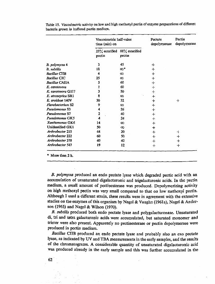
Table 15. Viscosimetric activity on low and high methoxyl pectin of enzyme preparations of different bacteria grown in buffered pectin medium.
B. polymyxa 4 B. subtilis Bacillus CUB Bacillus CIC Bacillus CAIIA E. carotovora E. carotovora G117 E. atroseptica SRI E. aroideae 140V Flavobacterium S2 Pseudomonas S3 Pseudomonas S7 Pseudomonas GK5 Xanthomonas GK6 Unidentified GK1 Arthrobacter 215 Arthrobacter 222 Arthrobacter 370 Arthrobacter 547
Viscosimetric half-value time (min) on
25% esterified pectin
3 18 4
25 5 1 3 8
30 9 4 2 4
14 50 44 60 40 19
88% esterified pectin
45 00*
CO
CO
60 60 50 CO
32 00
26 40 24 CO
CO
20 50 40 12
Pectate depolymerase
4-
+ 4-4-+ 4-4-+ + 4-+ 4-4-4-4-4-4-4-4-
Pectin depolymerase
4-
4-4-4-4-
* More than 2 h.
B. polymyxa produced an endo pectate lyase which degraded pectic acid with an accumulation of unsaturated digalacturonic and trigalacturonic acids. In the pectin medium, a small amount of pectinesterase was produced. Depolymerizing activity on high methoxyl pectin was very small compared to that on low methoxyl pectin. Although I used a different strain, these results were in agreement with the extensive studies on the enzymes of this organism by Nagel & Vaughn (1961a), Nagel & Anderson (1965) and Nagel & Wilson (1970).
B. subtilis produced both endo pectate lyase and polygalacturonase. Unsaturated di, tri and tetra galacturonic acids were accumulated, but saturated monomer and trimer were also present. Apparently no pectinesterase or pectin depolymerase were produced in pectin medium.
Bacillus CUB produced an endo pectate lyase and probably also an exo pectate lyase, as indicated by UV and TBA measurements in the early samples, and the results of the chromatograms. A considerable quantity of unsaturated digalacturonic acid was produced already in the early sample and this was further accumulated in the
62
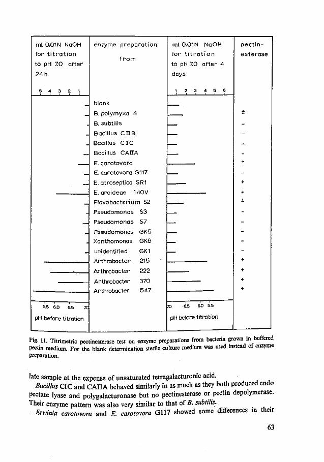
ml. 0.01 N NaOH
for t i t r a t i o n
to pH 70 after
24 h.
5 4 3 2 1
.
-
.
—
_
_
.
_
5-5 6.0 6.5 70
pH before titration
enzyme preparation
f r o m
blank
B. polymyxa 4
B. subtil is
Bacillus CDB
Bacillus C IC
Bacillus CAIIA
E. carotovora G117
E. atroseptica SR1
E.aroideae MOV
Pseudomonas S3
Pseudomonas 57
Pseudomonas GK5
Xanthomonas GK6
unidentified GK1
Arthrobacter 215
Arthrobacter 222
Arthrobacter 370
Arthrobacter 547
ml. 0.01 N NaOH
for t i t r a t i o n
to pH 70 after 4
days.
1 2 3 4 5 6
___
_ _
^__
—
—
—
—
70 6 5 6 0 5.5
pH before titration
pect in-
esterase
±
-
-
-
-+
-
+
+
±
-
-
-
-
-+
+
+
+
Fig. 11. Titrimetric pectinesterase test on enzyme preparations from bacteria grown in buffered pectin medium. For the blank determination sterile culture medium was used instead of enzyme preparation.
late sample at the expense of unsaturated tetragalacturonic acid. Bacillus CIC and CAIIA behaved similarly in as much as they both produced endo
pectate lyase and polygalacturonase but no pectinesterase or pectin depolymerase. Their enzyme pattern was also very similar to that of B. subtilis.
Erwinia carotovora and E. carotovora G117 showed some differences in their
63
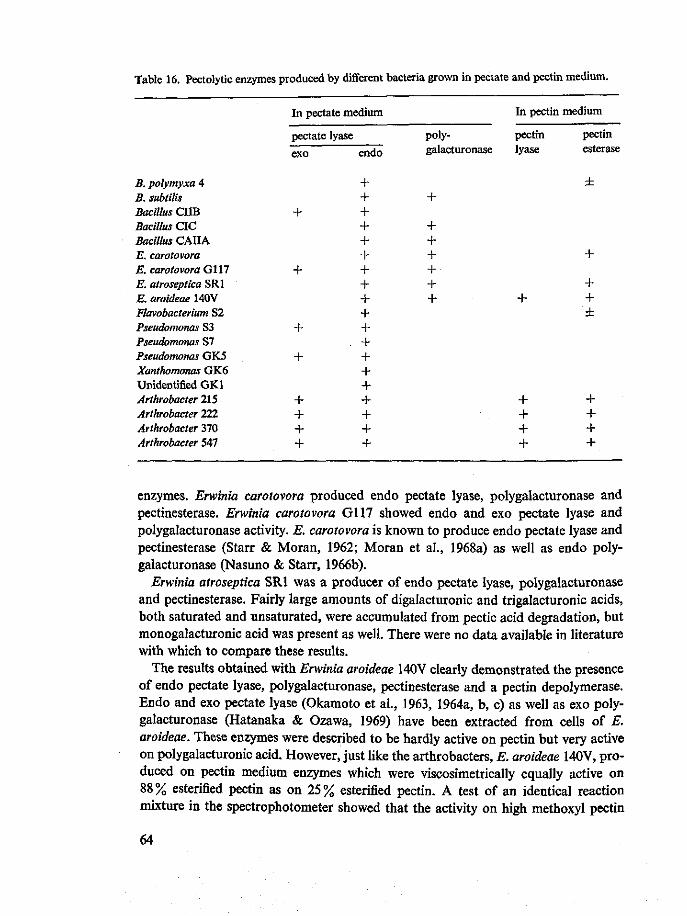
Table 16. Pectolytic enzymes produced by different bacteria grown in pectate and pectin medium.
B. polymyxa 4 B. subtilis Bacillus CUB Bacillus CIC Bacillus CAIIA E. carotovora E. carotovora G117 E. atroseptica SRI E. aroideae MOV Flavobacterium S2 Pseudomonas S3 Pseudomonas S7 Pseudomonas GK5 Xanthomonas GK6 Unidentified GK1 Arthrobacter 215 Arthrobacter 222 Arthrobacter 370 Arthrobacter 547
In pectate medium
pectate lyase
exo
+
+
+
+
+ + + +
endo
+ + + + + + + + + + + + + + + + + + +
polygalacturonase
+
+ + + + + +
In pectin medium
pectin lyase
+
+ + + +
pectin esterase
±
+
+ + ±
+ + + +
enzymes. Erwinia carotovora produced endo pectate lyase, polygalacturonase and pectinesterase. Erwinia carotovora G117 showed endo and exo pectate lyase and polygalacturonase activity. E. carotovora is known to produce endo pectate lyase and pectinesterase (Starr & Moran, 1962; Moran et al., 1968a) as well as endo polygalacturonase (Nasuno & Starr, 1966b).
Erwinia atroseptica SRI was a producer of endo pectate lyase, polygalacturonase and pectinesterase. Fairly large amounts of digalacturonic and trigalacturonic acids, both saturated and unsaturated, were accumulated from pectic acid degradation, but monogalacturonic acid was present as well. There were no data available in literature with which to compare these results.
The results obtained with Erwinia aroideae 140V clearly demonstrated the presence of endo pectate lyase, polygalacturonase, pectinesterase and a pectin depolymerase. Endo and exo pectate lyase (Okamoto et al., 1963, 1964a, b, c) as well as exo polygalacturonase (Hatanaka & Ozawa, 1969) have been extracted from cells of E. aroideae. These enzymes were described to be hardly active on pectin but very active on polygalacturonic acid. However, just like the arthrobacters, E. aroideae MOV, produced on pectin medium enzymes which were viscosimetrically equally active on 88% esterified pectin as on 25% esterified pectin. A test of an identical reaction mixture in the spectrophotometer showed that the activity on high methoxyl pectin
64

was a transeliminase. Among the well studied bacterial enzymes, both hydrolases and eliminases, there is not one with a preference for high methoxyl pectin. It is therefore very important to study this pectin lyase in more detail.
Flavobacterium S2 produced endo pectate lyase and pectinesterase. As mentioned before, the blank, early and late samples of pectate degradation all showed an unsaturated monomer spot on the chromatograms, sprayed with thiobarbiturate spray. It was found that this compound was present in relatively high concentration in the crude enzyme preparation, as a metabolic end-product. It was not accumulated during pectate degradation with the extracellular enzyme preparation of Flavobacterium.
Pseudomonas S3, S7 and GK5 all produced endo pectate lyase. Moreover S3 and GK5 showed exo pectate lyase activity. No polygalacturonase or pectinesterase was found. These results were in good agreement with those obtained by Fuchs (1965) in a study of pectate degradation by Pseudomonas fluorescens. Nasuno & Starr (1966a) however, found Pseudomonas marginalis to produce endo polygalacturonase as well. Preiss & Ashwell (1963a) also suggested the presence of both endo pectate lyase and endo polygalacturonase in a Pseudomonas strain, but since they used cell-free extracts it is possible that this polygalacturonase was really an oligogalacturonide hydrolase, an enzyme, which splits oligogalacturonides preferentially. Such an enzyme was found for example in the cells of Bacillus polymyxa by Hasegawa & Nagel (1967).
Xanthomonas GK6 showed endo pectate lyase activity only. Unsaturated digalac-turonic acid was accumulated as the major end-product of pectate degradation. These results were in agreement with those obtained by Starr & Nasuno (1967) and Nasuno & Starr (1967) for Xanthomonas campestris.
The unidentified strain GK1 was also a producer of endo pectate lyases. The enzyme gave a series of unsaturated mono to penta galacturonic acids from pectic acid. Unsaturated dimers and trimers were apparently accumulated.
The Arthrobacter strains 215, 222, 370 and 547 showed the most unconventional picture. From the UV and TBA measurements it can be concluded that these strains produced both exo and endo pectate lyase; the exo enzyme being produced most abundantly by 370 and 547. The paper chromatograms of these two strains did not show saturated degradation products. The chromatograms of the early and late samples were similar and showed a series of unsaturated mono to penta galacturonic acids. Although it was difficult with the other exo and endo pectate lyase mixtures to state what oligogalacturonide was produced by the exo enzyme, it was quite impossible for Arthrobacter strains 370 and 547. That is why an enzyme with some exo-endo intermediate character might also be present. Quite some pectinesterase was produced in a pectin medium. With a Metrohm automatic trtrator the pectinesterase of Arthrobacter 547 was measured to produce 1.7 x 10 meq of acid j e r ml of enzyme solution at 25 °C. Most interesting was the pectin depolymerase which was present in the enzyme preparation produced in pectin medium and which caused high methoxyl pectin to be depolymerized more quickly than 25% estenfied pectin. It could be calculated that pectinesterase hardly interacted because of the insignificant small decrease of degree of esterification during the reaction. The two ml of enzyme
65

solution of Arthrobacter 547, present in the viscosimetric reaction mixture may have produced some 3.4 x 10"4 meq/min free carboxyl groups. Lowering the degree of esterification of the substrate by 1 % meant saponification of 9 x 10" 4 meq of ester groups, since a total of 8 ml 0.25 % substrate, 88 % esterified, uronide content about 75 % was present in the reaction mixture. In the 12min (Table 15) viscosimetric half-value time, the degree of esterification of the substrate decreased only by some 4.5 % from 88 to 83.5%, when the pectinesterase activity was not too much affected by degree of esterification and concentration of the substrate.
Since in the spectrophotometer the absorbance increased more quickly with 88 % esterified pectin than with 25 % esterified pectin as the substrate, the pectin depoly-merase may be a pectin lyase, an enzyme, thus far not found among the well described pectolytic enzymes of bacteria. A detailed study of its properties therefore is important.
7.4 Summary and conclusions
A typing method for enzyme preparations of bacteria has been developed. Enzymes were produced on two different media: a pectate and a pectin containing medium. The enzymes produced in the pectate medium were subjected to a detailed testing scheme, beginning with a viscosimetric study of the degradation of pectic acid. At an early and a late stage related to the viscosity curve, samples were withdrawn and subjected to spectrophotometric measurements at 235 nm, a periodate-thiobarbituric acid test and paper chromatography. The UV test, periodate-thiobarbituric acid test and the paper chromatographic techniques were carefully selected from a survey of the literature. The enzyme preparations from the pectin medium were tested for pectinesterase activity and pectin depolymerizing activity only; the latter in comparison with pectate depolymerizing activity. Nineteen strains of different bacterial genera and species were subjected to the testing scheme. In addition to a confirmation of many data from literature some very interesting observations were made. It appeared that all strains tested produced endo pectate lyase, but only a few strains produced it as the only pectolytic enzyme: Pseudomonas S7, Xanthomonas GK6 and unidentified strain GK1. Polygalacturonase was produced by strains of the genera Bacillus and Erwinia only. Pectinesterase was found with Bacillus polymyxa, most of the strains of Erwinia and all of Arthrobacter. A pectin lyase, thus far not found among the well described pectolytic enzymes of bacteria was produced by a strain of Erwinia aroideae and by all strains of Arthrobacter. The arthrobacters also showed a typical pattern of pectate degradation, which either was achieved by exo and endo pectate lyase or by an intermediate type of pectate lyase. Flavobacterium S2 accumulated unsaturated monogalacturonic acid as a metabolic end-product in the culture medium.
66

8 Typing of commercially available fungal pectolytic enzyme preparations
8.1 Introduction
Pectolytic enzymes produced by fungi are known to be rather different from those of bacteria (Chapter 3). Although this study mainly concerns bacterial pectolytic enzymes, it was thought valuable to apply the typing procedures to a number of commercially available pectolytic enzyme preparations, which are all of fungal origin. Firstly, it was of interest to know which commercial preparations would be good sources for the isolation of certain types of enzyme. Secondly, a better knowledge of the composition of such enzyme preparations would be advantageous in understanding their industrial performance. It was mainly the second consideration which made me decide to extend the investigations to industrial enzymes, although these preparations are known to be rather complex mixtures of pectolytic and other enzymes (McClendon & Kreisher, 1963; Endo, 1963a, b; Roller & Neukom, 1967; Amado, 1970).
8.2 Materials and methods
Enzyme preparations Five enzyme preparations all produced by Aspergillus strains, were studied in detail: Rohament P (Rohm GmbH, Darmstadt), Pektolase CL 100 (Grindstedvaerket, Aarhus), Clarizyme L 5 (Societe Rapidase, Seclin, France), Wtrazym 20 ('69) (Dr Schubert AG, Laufen, Switzerland) and Pectinase Spark L (Miles Chem. Co., Elkhart, Ind., USA). TWO of these preparations were powders (Rohament P and Pektolase CL 100), one (Ultrazym 20 ('69)) was dried on plant material and two (Pectinase Spark L and Clarizyme L 5) were liquid concentrates. One gram of the solid preparations was extracted for 1 h with 20 ml distilled water and filtered over filter paper by suction to remove insoluble material. The filtrates as well as the liquid enzymes were dialysed overnight in cellophane tubing against running tap water. The dialysates were quick-frozen in small tubes and kept frozen at -28°C until used.
Orange pectinesterase was prepared from pulp obtained by cutting and pressing 5 kg of whole oranges. The pulp was homogenized in 5 litre buffer, containing per litre: 42.5 g borax (Na2B407 • 10H2O), 27.5 g boric acid and 40.0 g sodium acetate (pH 8.2). After 2 h of stirring, the mixture was pressed. The liquid was filtered. In the 5.5 litre filtrate, 2 750 g ammonium sulphate was dissolved and the solution was stored overnight in the cold room. The precipitate was then collected by centn-
67

fuging for 15 min at 10 000 x g at 2°C. The precipitate was dissolved in 400 ml distilled water and stored overnight at 4°C, and then centrifuged to remove insoluble material. The solution was dialysed for three days against running tap water. This crude enzyme solution, 500 ml, containing 140 units of pectinesterase per ml was stored at 4 °C.
Substrates In addition to the pectic acid preparation to be described in Chapter 9 as pectic acid preparation A, four more substrates were used for activity determination. 'Brown Ribbon' pectin, an apple pectin of 74 % esterification (Obipektin AG, Bischofszell, Switzerland) was used as high methoxyl pectin. The (methyl)galacturonide content was 71 % (0.38 meq free and esterified carboxyl groups per 100 mg). A second even more highly esterified pectin (95% esterification) was prepared from Brown Ribbon pectin by repeated methanol-HCl treatment according to Heri et al. (1961) as to be described in Chapter 9 (pectin B). The (methyl)galacturonide content of this preparation was 79% (0.42 meq free and esterified carboxyl groups per 100 mg). The remaining two substrates were glycolesters of pectic acid with 74 and 95 % esterification, respectively. The glycolesters were prepared according to the procedures of Deuel (1947) and Vajda (1957). Pektinsaure (Obipektin AG, Bischofszell, Switzerland) was further saponified under cooling and desalting in a heterogeneous system. The esterification was also carried out in a heterogeneous cooled system, using ethylene oxide (Merck, purissimum). To arrive at 95 % esterification the esterification had to be repeated with fresh ethylene oxide. The 74 % preparation had a (glycol)galacturonide content of 73% (0.35 meq free and esterified carboxyl groups per 100 mg). The 95 % esterified preparation had a (glycol)galacturonide content of 78 % (0.36 meq free and esterified carboxyl groups per 100 mg). Unless recorded otherwise, 0.25 % solutions in 0.25 mM CaCl2 • 2H20 were used in enzyme activity determinations. For pectinesterase activity measurements, Green Ribbon pectin, an apple pectin of 65 % esterification (Obipektin AG) was used, in a concentration of 0.625 %.
Procedure The characterization procedure adopted for these enzyme preparations was principally the same as that used for bacterial enzymes, described in Chapter 7. However, some extensions and changes proved to be necessary.
A viscosimetric study of the degradation of the five substrates was carried out at pH 4.0. Reaction mixtures consisted of 8 ml 0.25 % substrate in 0.25 mM CaCl2 • 2H20, 4 ml distilled water and 0.1 ml enzyme dialysate. Because the samples were to be analysed by paper chromatography no buffers were added, leaving the substrate itself as the only buffering compound in the reaction mixture. However, at the end of the reactions, pH values were recorded. Blank, early (at 3 times viscosimetric half-value time) and late (at 20 times viscosimetric half-value time) samples were collected and treated as in the procedure for bacterial enzymes. Samples were analysed in the same way as for reaction mixtures with bacterial enzymes (UV test, periodate-thio-barbituric acid test and paper chromatography).
The presence and activity of lyases was also studied by spectrophotometric activity
68

measurements. Two ml 0.25% substrate pH 4.0 (either pectic acid, Brown Ribbon pectin or 95 % esterified pectin) in 0.25 mM CaCl2 • 2H20 was pipetted into a 1 cm quartz cuvette, followed by 1 ml distilled water and 0.025 ml enzyme dialysate. The increase in absorbance, at 232 nm as measured at 30 °C in a Zeiss PMQ-II spectrophotometer was recorded over a time period of 15 to 60 min. In view of the optimum pH of pectin lyases of 5.1 to 6.0 (Albersheim et al., 1960b; Edstrom & Phaff, 1964a; Roller, 1966; Voragen, 1972) pectin lyase was also assayed spectrophotometrically at pH 6.0 and at 30 °C in the following reaction mixture: 0.25 ml pectin, 95 % esterified (1% solution, pH 6.0), 2.4 ml citrate-phosphate buffer (0.1 M trisodium citrate, acidified to pH 6.0 with concentrated o-phosphoric acid), 0.25 ml distilled water and 0.1 ml enzyme dialysate.
Pectinesterase was measured titrimetrically at pH 4.0 and 30 °C with the Combi titrator (Metrohm AG, Herisau, Switzerland). A mixture of 25 ml, containing 20 ml 0.625 % Green Ribbon pectin, 65 % esterified, 1 to 5 ml enzyme dialysate and 4 to 0 ml distilled water was maintained continually at pH 4.0 using 0.01 N NaOH. The effect of the salt concentration was checked by making the pectin solution 0; 0.05 and 0.1 M in NaCl.
The optimum pH of Spark L pectinesterase was measured. At lower pH values the titration values obtained could not be used directly for calculating pectinesterase activity, because not all carboxyl groups, liberated by the saponification action of pectinesterase, were measured by continuous titration. The following procedure was applied (Fish & Dustman, 1945; McColloch & Kertesz, 1947, modified).
In the above specified reaction mixture, pectinesterase was allowed to react for a certain time (during which approximately 1 and 5% demethylation occurred) at different pH values, whereby the pH was kept constant titrimetrically and the amount of 0.01 N NaOH with time was recorded. Then the pH was quickly raised to near 6 by the addition of 0.1 N NaOH from a microburette. Finally the reaction mixture was titrated to pH 7.0 with 0.01 N NaOH from the titrator. The final volume was 30 to 35 ml. The amount of 0.01 N NaOH, used during the reaction to maintain the pH at the desired value, as well as the reaction time were taken from the titrigram. The total amount of NaOH, diminished with the amount of NaOH used in a blank determination with heat inactivated enzyme was equivalent to the carboxyl groups formed by pectinesterase action.
8-3 Results and discussion
The specific viscosity (rjs) versus time curves obtained from degradation of pectic acid with the bacterial enzymes always showed a regular curvilinear pattern, and the reciprocal specific viscosity (1/i/J versus time curves were always straight lines (see Chapter 9). In contrast the t,s versus time curves obtained from the five substrates (Pectic acid, pectins of 74 and 95 % esterification and glycolesters of pectic acid with ?4 and 95 % esterification) during degradation with the commercial preparations were very difficult to interpret and the 1/ij, versus time curves were not straight lines. The
69

non-linearity of l/f/s versus time of Brown Ribbon pectin (74% esterified) during degradation with Pektolase was already observed by Vas et al. (1967). Although the direct plotting of viscosity-time curves (Figure 12) was not very informative, some important conclusions could be derived when the reciprocal specific viscosities were plotted against time (figures 13-17). The glycolesters were included to provide a substrate which was not attacked by pectinesterase (McDonnell et al., 1950). It was hoped to get a good picture of the activity of pectin lyase and polymethylgalacturonase by excluding pectinesterase activity in this way. However, figures 13 to 17 show that neither pectin lyase nor polymethylgalacturonase seem to degrade these glycolesters. The initial loss of viscosity should entirely be ascribed to polygalacturonase. I was able to show by spectrophotometry that pectate lyase from Bacillus polymyxa, is also active on these glycolesters and Voragen (1972) demonstrated that a purified pectin lyase indeed was inactive on these substrates. A comparison of the various enzyme preparations showed striking differences between the polygalacturonases of e.g. Rohament P, which have a relatively quick action on the glycolesters and the polygalacturonases of Pektolase CL 100, which are relatively less active on these substrates. These differences among the enzymes can be seen very clearly from Table 17
80 100 t ime Cmin)
Fig. 12. Specific viscosity curves of pectic acid, 74 and 95% esterified pectins and 74 and 95% esterified glycolesters of pectic acid, during degradation by Ultrazym 20 ('69). Reaction mixtures contained 8 ml 0.25% substrate in 0.25 mM CaCl2, pH 4.0; 4 ml distilled water and 0.1 ml enzyme dialysate.
70

80 100 t ime (min)
Fig. 13. Reciprocal specific viscosity curves of substrates as in Fig. 12 degraded by Rohament P. Reaction mixtures as in Fig. 12.
Table 17. Times (sec) required for five enzyme preparations to achieve a fixed increase in reciprocal specific viscosity of the five substrates. Data derived from figures 13 to 17.
Enzyme
Rohament P Pektolase CL 100 Clarizyme L 5 Ultrazym 20 ('69) Spark L
Pectic acid
lfat 5.25-10.5
100 260 290
1040 1260
Pectin DEI'4
Vn> 0.70->7.0
6000 2400
420 2 400 3 100
Pectin £ £ 9 5
\h. 6.25—12.50
>6000 4 200
500 1750 1 150
Glycolester DE14
1/iJ. 1.25-6.25
50 3 700
240 2 800 2 800
Glycolester DE 95
V* 2.9-3.9
60 >6000
220 1900
>6000
where the times are given which the five enzymes require to achieve a given drop in viscosity (increase in reciprocal specific viscosity) of the five substrates.
The action of Rohament P on pectins was similar to that on glycolesters. This is in agreement with the manufacturers' description (Rohm & Haas, brochure) of this
71
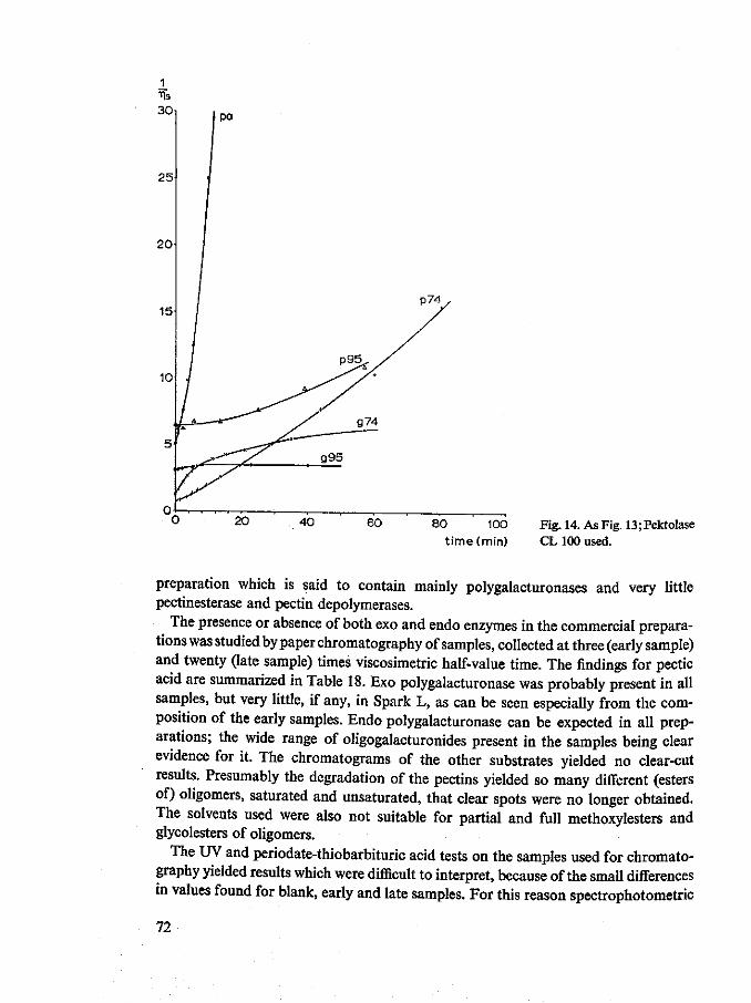
80 100 time(min)
Fig. 14. As Fig. 13; Pektolase CL 100 used.
preparation which is said to contain mainly polygalacturonases and very little pectinesterase and pectin depolymerases.
The presence or absence of both exo and endo enzymes in the commercial preparations was studied by paper chromatography of samples, collected at three (early sample) and twenty (late sample) times viscosimetric half-value time. The findings for pectic acid are summarized in Table 18. Exo polygalacturonase was probably present in all samples, but very little, if any, in Spark L, as can be seen especially from the composition of the early samples. Endo polygalacturonase can be expected in all preparations; the wide range of oligogalacturonides present in the samples being clear evidence for it. The chromatograms of the other substrates yielded no clear-cut results. Presumably the degradation of the pectins yielded so many different (esters of) oligomers, saturated and unsaturated, that clear spots were no longer obtained. The solvents used were also not suitable for partial and full methoxylesters and glycolesters of oligomers.
The UV and periodate-thiobarbituric acid tests on the samples used for chromatography yielded results which were difficult to interpret, because of the small differences in values found for blank, early and late samples. For this reason spectrophotometric
72
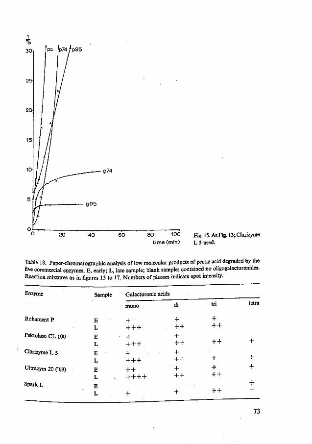
3 0 l J Pa |p74/p95
, — g74
g95
20 40 60 80 100 Fig.l5.AsFig.l3;Clarizyme t ime(min) L 5 used.
Table 18. Paper-chromatographic analysis of low molecular products of pectic acid degraded by the five commercial enzymes. E, early; L, late sample; blank samples contained no oligogalacturonides. Reaction mixtures as in figures 13 to 17. Numbers of plusses indicate spot intensity.
Enzyme
Rohament P
Pektolase CL 100
Clarizyme L 5
Ultrazym 20 ('69)
Spark L
Sample
E L
E L
E L
E L
E L
Galacturonic acids
mono
+ + + +
• + + + + + + + + + + + + +.+
+
di
+ + + + + + + + + + + +
+
tri
+ + +
+ +
+ + + +
+ +
tetra
+
+ +
+ +
73

80 100
t ime (min) Fig. 16. As Fig. 13; Ultrazym 20 ('69) used.
reactions were run parallel to the viscosimetric studies on substrates identical to those for viscosimetry, and the absorbance (A) was recorded for 1 h on 74 and 95% estenfied pectins, and for 20 to 40 min on pectic acid. From the results in Figure 18 it may be concluded that all preparations contained pectin lyase, which at the pH value used (about 4) showed an appreciable activity in Clarizyme L 5 and Spark L only, and even more on 95% esterified than on 74% esterified pectin. At pH 6.0 the pectin lyase activity was much more pronounced. At this pH it could easily be shown that pectin lyase was present in all 20 commercial pectinases and cellulases tested.
During measurements at pH 4.0, the absorbance increased linearly or degressively with time. With Spark L an absorbance of 0.95 and 0.42 was measured on 95 and 74% estenfied pectin, respectively after 1 h of reaction; with Clarizyme on 95% esterified pectin after 1 h this value was 0.75, which shows that these curves also become bent with time.
Looking again at the curves in figures 13 to 17 it is obvious that the described lyase activity cannot be responsible for the 1/,, values to increase progressively with time. The increase in A at 232 nm is not great enough to explain the viscosity change, as will be shown in the following calculation.
74
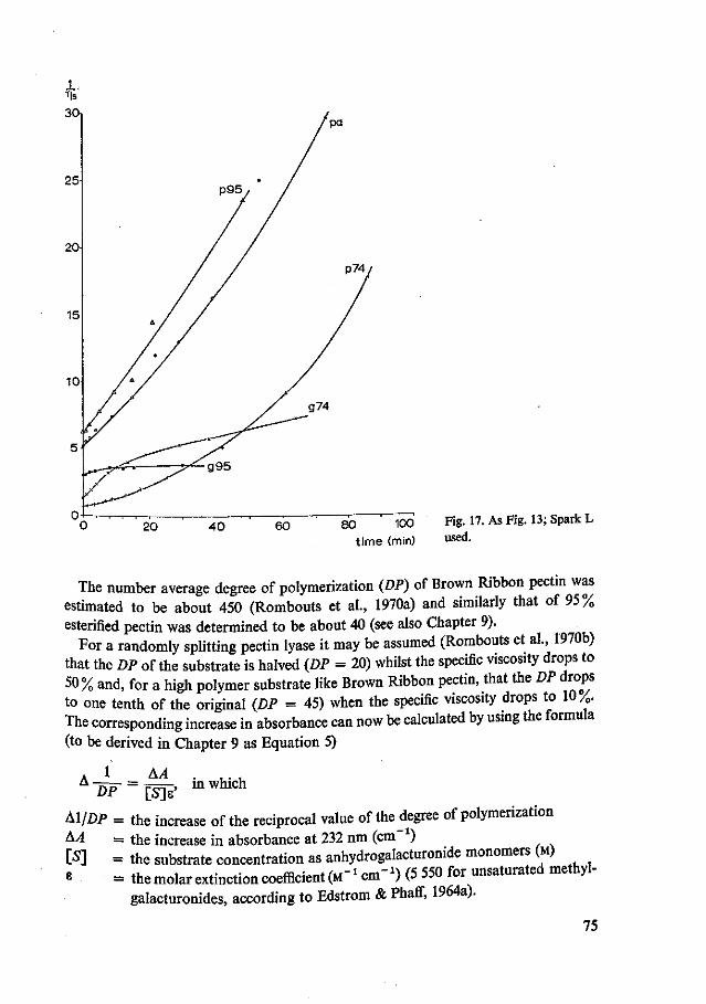
80 100 Fig. 17. As Fig. 13; Spark L
time (min) u s e d -
The number average degree of polymerization (DP) of Brown Ribbon pectin was estimated to be about 450 (Rombouts et al., 1970a) and similarly that of 95% esterified pectin was determined to be about 40 (see also Chapter 9).
For a randomly splitting pectin lyase it may be assumed (Rombouts et al., 1970b) that the DP of the substrate is halved (DP = 20) whilst the specific viscosity drops to 50% and, for a high polymer substrate like Brown Ribbon pectin, that the DP drops to one tenth of the original (DP = 45) when the specific viscosity drops to 10%. The corresponding increase in absorbance can now be calculated by using the formula (to be derived in Chapter 9 as Equation 5)
AA in which
DP [S]e'
Al/DP = the increase of the reciprocal value of the degree of polymerization &A = the increase in absorbance at 232 nm (cm ) [S] = the substrate concentration as anhydrogalacturonide monomers (M)
= themolarextinctioncoefficient^1 cm"1) (5 550 for unsaturated methyl-galacturonides, according to Edstrom & Phaff, 1964a).
75

Absorbance at 232 nm
P95 Clarizyme L5
Absorbance at 232 nm
Ultrazym 20C69) 1
0.4
0.2H
60 t ime (min)
li^L^T^T™ aCliVhy °f t h e fiw c o m m e r c i a l enzyme preparations on three substrates. c o r n n S l n ** A f f " ^ ^ ^ P 95 ' 9 5 % e s t e r i f i e d P ** 1 - R e c t i ° * ™*ures of the same composition as in figures 12 to 17.
Smce Brown Ribbon pectin was used in a concentration of 2/3 x 2.5 g per litre, and the uromde^content of the preparation was 0.38 meq p e r 100 mg, [S] was 2/3 x 2.5 x 0.38/100 - 0.0065 M. For Brown Ribbon pectin I calculated the ab-
Z^IZ? r ! m ! e a S e d aS f ° l l 0 W S W h e n t h e d e S r e e of Polymerization was lowered from 450 to 45 by the action of endo pectin lyase:
76

_1 1 _ A^ A^ = 0 72 45 4 50 " 0.0065 x 5 550'
Likewise I calculated an increase in absorbance for 95 % esterified pectin, when the degree of polymerization was lowered from 40 to 20 by the action of endo pectin lyase, and the substrate concentration was 2/3 x 2.5 x 0.42/100 = 0.0070 M:
1 1 - AA AA = 097 20 40 0.0070 x 5 550'
From figures 13 to 18 the measured increase in absorbance for Brown Ribbon pectin when the reciprocal specific viscosity had become tenfold and for 95% esterified pectin, when the reciprocal specific viscosity had doubled, can be read to be negative and 0.1 resp. for Rohament P negative and 0.1 resp. for Pektolase CL 100 0.06 and 0.15 resp. for Clarizyme L 5 0.05 and 0.10 resp. for Ultrazym 20 ('69) 0.35 and 0.36 resp. for Spark L.
Although the above calculations were only approximate it may be concluded that the viscosity reduction power of the five commercial enzymes on 74 and 95% esterified pectins at pH 4 cannot be attributed to their pectin lyase content, except for Spark L, where this enzyme indeed appears to be important.
The spectrophotometric activity of Clarizyme and Rohament on pectic acid was investigated further. At pH 4.0 with 0.001 M EDTA in the reaction mixture no increase in absorbance was recorded. Neither was this so at pH 5.0, 6.5 and 8.0 in 0.1 M tris - 0.1 M succinate buffer. Transeliminative action at pH 4.0 could not clearly be confirmed in the TBA test. Thus pectate lyase, if present at all, has an unusual optimum pH.
It also seems unlikely that under the conditions of reaction a polymethylgalacturo-nase should be responsible for the larger part of the viscosity reduction of the 74 and 95% esterified pectins. Here convex l/r,s versus time curves could be expected because of the competing action of pectinesterase. The small drop in pH from 4 to 3.5 during the reaction, would be a disadvantage for the activity of the po lyga lac tu ronases which are supposed to have an optimum pH of 4 and 7 (Roller, 1966; Amadd, 1970) and 6.5 to 7 (Rexova-Benkova & Slezarik, 1966), and thereby even enhance a degression in the lfo, versus time curves. The action of polymethylgalacturonase becomes all the more unlikely, since Voragen (1972) tried without success to isolate and thereby found no evidence for the presence of polymethylgalacturonase in Pektolase.
Hence it becomes important to study whether the joined action of pectinesterase and Polygalacturonases might be responsible for the increase of \MS of the pectins, tor this reason pectinesterase activity measurements were made. Pectinesterase causes a drop in PH of the reaction mixtures (Table 19) with 74 and 95% estmfiec1 pectins ^ i ch can be used as a measure of pectinesterase activity. Although the pH of the taction mixtures with 95 % esterified pectin generally drops to a lower value than that
77
Table 19. pH of reaction mixtures used for viscosimetry, measured at the end of the viscosimetric studv. study,
Enzyme
Rohament P Pektolase CL 100 Clarizyme L 5 Ultrazym 20 ('69) Spark L
Pectic acid
3.6 3.5
3.8
Pectin DE 74
4.1 3.8 3.35 3.7 3.8
Pectin DE 95
4.2 3.75 3.2 3.5 3.6
Glycolester #£•74
4.15 4.1
4.15
Glycolester DE 95
4.45 4.35
4.35
of the reaction mixtures with 74% esterified pectin, it is premature to conclude that pectinesterase is more active on the more highly esterified pectin because the buffering capacity of the more highly esterified pectin is much lower than that of the less esterified pectin. The drop in pH in itself may have its influence on the course of the l/t]s curves. However, by comparison with relative viscosity versus pH diagrams of Schultz et al. (1945), this effect may be estimated to be rather small for 74 and 95 % esterified pectin.
I measured in collaboration with Heemskerk (1971) pectinesterase activities of the five commercial preparations by continuous titration at pH 4.0, on Green Ribbon pectin, a 65 % esterified apple pectin (Figure 19). Since NaCl, added in concentrations of 0.05 and 0.1 M, had very little effect on pectinesterase activity of these commercial preparations, it was further omitted. Even when C0 2 was eliminated by using C02-free substrate and sodium hydroxide solutions and by carrying out the continuous titration under nitrogen, no straight titration curves could be obtained. It was thought that this was due to the simultaneous chain splitting action, especially of the polygalacturonases, which in this way may produce a rest-substrate with a very high degree of esterification and therefore less suitable for pectinesterase action. However, when orange pectinesterase was used in the continuous titration test, the titrigram was also clearly curvilinear at pH 4.8 (Figure 19) but gave nearly a straight line at pH 7.0. Since polygalacturonase is absent in orange (Mannheim & Siv, 1969) I concluded that polygalacturonase action, if it is at all, is not the only reason for the curvi-lmeanty of the titrigrams. Another reason is probably the changing dissociation constant of the substrate during enzymatic saponification (see Chapter 2).
The activity of pectinesterase of Spark L as affected by pH was also determined by the same continuous titration method as well as by the method derived from that of Fish & Dustman (1945). In addition to the integral commercial preparation a pectinesterase fraction purified from Spark L in our laboratory by Heemskerk (1971) was used. The results are given in Figure 20. Two interesting observations could be made trom this figure. Firstly, with the commercial preparation at pH 4,'the titrimetric method indicated an even higher pectinesterase activity than Fish& Dustman's (1945) method. The simultaneous action of polygalacturonase probably accounted
78

ml. 0.01 N NaOH
Spark 5 ml.
Pektol. 5ml.
Orange PE pH4.8
20
t i m e ( m i n )
Fig. 19. Activity of pectinesterase of the 5 preparations measured by continuous titration at pH 4.0. Composition of reaction mixtures: 20 ml 0.625 Green Ribbon pectin, 4.0 or 0 ml distilled water and 1.0 or 5.0 ml enzyme dialysate.
for it, but not by making the substrate less suitable for pectinesterase action. By splitting the chain between neighbouring free carboxyl groups this enzyme probably caused the degree of dissociation to increase at the constant pH. It thus compensated for the decrease of the degree of dissociation which resulted from the decrease ot degree of esterification and concentration of the substrate during enzymatic saponification with continuous titration, at a constant pH. Secondly, with the commercm preparation the true pectinesterase activity as measured by Fish & Dustman s uswj method remained constant, but the activity measured titrimetncally was seen to decrease before the degree of esterification of the substrate had dropped nve per cent. With the purified pectinesterase preparation both, true and titnmetri c.pectinesterase activity could be seen to decrease before this drop in degree of estenncation had occurred. t
From the above observations and discussions, it becomes evident that at present it is impossible to predict quantitatively the results of the s^™™**™" polygalacturonase and pectinesterase. With the following calculations the sapomtying action of the pectinesterases during the viscosity studies is roughly estima eo. r Figure 19 the relative pectinesterase activities can be read to have been 1, *• , , 20 and 21 for the dialysates of Rohament, Pektolase, Clarizyme Ultrazym and Spark respectively. Denning the pectinesterase unit as the amount of ^ ^ ^ ^ the reaction conditions produces 1 ueq of acid per min, 5 ml enzyme dialysate con
79
ueq. NaOH min
ueq. NaOH m in
1.2
1.0
0.8
0.6-
0.4-
0.2-
2.0 30 4.0 5.0
PH
1.81
1.5
1.2
0.9
0.6
0.3'
2.0 3.0 4.0 5 0
pH
Fig. 20. Optimum pH of Spark L pectinesterase. For Fig. 20A the integral commercial preparation was used; for Fig. 20B, a purified pectinesterase, isolated from Spark L. Composition of reaction mixtures: 20 ml 0.625% Green Ribbon pectin; 4.0 ml distilled water; 1.0 ml commercial enzyme dialysate or 1.0 ml of a 1:20 dilution of purified pectinesterase. • continuous titration; after a 1% drop of DE • continuous titration; after a 5% drop of DE + method of Fish & Dustman; after a 1 and a 5% drop of DE (independent of the drop of DE) O method of Fish & Dustman; after a 1% drop of DE a method of Fish & Dustman; after a 5% drop of DE
tained 0.27,2.5, 24, 5.4 and 5.7 pectinesterase units, respectively. Assuming the same order of activity of the pectinesterase in the viscosity experiments with 0.25 % of 74 % esterified pectin, where only 0.1 ml of enzyme had been used, the pectinesterase would in 50 min have produced 0.27, 2.5, 24, 5.4 and 5.7 ueq of acid. Since the viscosity reaction mixtures contained 8 ml 0.25 % pectin with a uronide content of 380 ueq per 100 mg, the amount of acid produced when the degree of esterification was lowered by one per cent, was
8 x 0.25 x 10 x 380 100 x 100
= 0.76 ueq
80
Consequently, during 50 min of reaction in the viscosity experiments, the pectin-esterases of the five preparations could have lowered the degree of esterification of 74% esterified pectin to 73.7, 71,42, 67 and 66.5, respectively. The decrease in degree of esterification will even have been smaller for 95% esterified pectin, since it is not so readily attacked by pectinesterase. If, in addition, the mode of action of pectfn-esterase is thought to resemble that of a zipper (Schultz et al., 1945; Solms & Deuel, 1955; Lee et al., 1970) it becomes evident, that the drop in viscosity of the pectin solutions in the viscosity tests indeed cannot be predicted so simply on the basis of activity measurements of single enzymes present in the complex commercial enzyme preparations.
8.4 Summary and conclusions
Five commercial pectolytic enzyme preparations: Rohament P, Pektolase CL 100, Clarizyme L 5, Ultrazym 20 ('69) and Pectinase Spark L were characterized, by a comparative study of their depolymerizing activity at PH 4 on five substrates: pec ic acid, 74 and 95% esterified pectins and 74 and 95% esterified glycolesters of pectic acid. Tests to follow the depolymerization of these substrates included viscosimetry, paper chromatography and spectrophotometry. Moreover pectinesterase activity was measured titrimetrically at pH 4.0 on 65% esterified pectin. In addition to me usual titrimetric method, which in the pH region of 4 and below does not measure he true pectinesterase activity because of the changing degree of dissociation o1 the substrate, an improved titrimetric method similar to that of Fish & D u s t m a i ! ^ j was used. The optimum pH curve of pectinesterase as present in one preparation as a purified enzyme was determined with both methods. nAatinn ;n
By plotting viscosity measurements obtained during substrate Jpada t ionm reciprocal specific viscosity (1/^) versus time graphs, it was shown thatjta> gtf» esters were only initially depolymerized, apparently by polygalactu ™ ^ ™ *J£ under the reaction conditions chosen all preparations except R ° ^ 1 j * j d
1/1. of the pectins to increase rapidly and progressively. It was sh owntht. tbs i jp and progressive increase of l/„. cannot be caused by pectin " J J ^ ^ ^ d galacturonase. The joined action of pectinesterase and P 0 1 ^ ™ ™ t b e
to be so complex that at present its effect on the viscosity of *« P ^ " £ £ £ quantified. It seems that in all preparations pectinesterase limits the activity po y galacturonase under the reaction conditions <*osen- oo lYgalacturonase, pectin-
All preparations were found to contain exo and endo Pff^ esterase and pectin lyase but there were large proportional ditterenc .
81
9 The use of lyases for the estimation of the number average degree of polymerization of pectic substances
9.1 Introduction
During the study of pectate lyases it became clear that the endo type of lyase during its action on pectate caused both the reciprocal specific viscosity (1/>;S) and the absorbance to increase linearly with time, and that these linear changes extended over quite a long period of degradation during which the absorbance increased with 1.0 (Rombouts et al., 1970a). By using a pectic acid with a known degree of polymerization the change in absorbance during the reaction could be used to calculate the corresponding change in DP. Plotting 1/T]S against l/DP yielded a straight line, which upon extrapolation bisected the coordinates at the origin. These experimental results made it possible to construct a mathematical relationship between the initial reciprocal specific viscosity (l/^s(0), the change of reciprocal specific viscosity with changing reciprocal degree of polymerization (Al/t]JAl/DP) and the initial degree of polymerization (1/Z»P(0); (Rombouts et al., 1970b). By simple and reliable measurement o f V^CO and Al/riJAl/DP, it is possible to calculate l/DP(t0).
9.2 Materials and methods
Substrates Pectic acid preparation A was prepared from a commercial apple pectin preparation (Pink Ribbon, 25% esterified, Obipektin AG, Bischofszell, Switzerland), by the procedure of Altermatt (1954) and Derungs (1958). This procedure includes cold alkaline saponification of the crude preparation, bleaching with sodium chlorite, filtration of the dissolved preparation through a celite-fibra flow or a solka floe BW-200 (Grefco Inc., Los Angeles, USA) filter bed in a Biichner funnel, precipitation with acidified ethanol and desalting by three washings with acidified ethanol 70% v/v then by eight washings with ethanol 70% v/v then once by ethanol 96% v/v and once by acetone. The galacturonide content and the degree of esterification were simultaneously determined by a titrimetric method (Doesburg, 1965). The air dried preparation was 0-1% esterified. It had 440 ueq carboxyl groups per 100 mg which corresponds to a galacturonide content of 77%. The degree of polymerization of this preparation was determined by the sodium chlorite end-group method of Launer and Tomimatsu (1954, 1959a, b). A reaction time of 6 h at 50°C, a phosphate buffer
f n ™ P H 3*2 m t hC r e a c t l o n m i x t u r e a n d a concentration of the chlorite reagent ol 0.009 M were used. Galacturonic acid monohydrate (Fluka AG, Buchs, Switzerland)
82

was used as standard. Under these conditions the DP of this pectic acid preparation (calculated as the ratio -COOH : -CHO was 45).
Pectin preparation B was prepared from 75 % esterified apple pectin (Brown Ribbon, Obipektin AG, Bischofszell, Switzerland). To 20 litre of 0.5% pectin, 1.6 litre of 10% aluminium sulphate was added. Then 550 ml 10% sodium carbonate was added under continuous stirring. The precipitate was filtered off with a cheese cloth and desalted by three washings with acidified ethanol 70% v/v, then by eight washings with ethanol 70% v/v, then with ethanol 96% v/v and finally with acetone. The preparation (52 g) was dried over CaCl2 at 60°C. It was esterified according to Heri et al. (1961) by suspending it in 2 litre cold acidified (2 N H 2 S 0 4 ) absolute methanol p.a. It was stored for 14 days in the cold. The mixture was filtered over a Buchner funnel, washed with methanol and the residue was once more treated with acidified methanol for 14 days. The preparation was washed eight times with ethanol 70% v/v, then once with ethanol 96% v/v, finally with acetone and air dried. The preparation was depolymerized during these treatments, its degree of esterification was 95.5% and its (methyl)galacturonide content, calculated as percentage free plus esterified anhydrogalacturonide was 79% (0.42 meq. free plus esterified carboxyl groups per 100 mg).
Pectin preparation C was prepared from pectic acid preparation A by the same esterification procedure applied to pectin B. The preparation was dried over calcium chloride. Its degree of esterification was 95% and its (methyl)galacturonide content was 94.6%.
Pectin preparations D, E and F were three apple pectins prepared in our laboratory by Eshuis (1970). Their degrees of esterification and (methyl)galacturonide contents were 58% and 71% for preparation D, 59.5% and 72% for preparation E and 51.5% and 77% for preparation F, respectively.
The following commercial pectin preparations were used without further purification: Pink Ribbon pectin (Obipektin AG, Bischofszell, Switzerland), 25/„ <*tenflea, (methyDgalacturonide content 72.5%; Green Ribbon pectin (Obipektin AG, Biscnois-zell, Switzerland), 63 % esterified, (methyl)galacturonide content 76.8 /„.
Enzymes Pectate lyase of Bacillus polymyxa, as well as that of i W * — ' G K * Flavobacterium S2 and Arthrobacter 547, were produced in the BPC medium described in Chapter 7. The crude preparations were dialysed overnight ^junrnngtap water and freeze-dried. They were used without further purification Three_ ditterent fractions of the crude enzyme of B. polymyxa were obtained by coin m i * f ™ * £ graphy according to the procedure of Nagel & Wilson (19 0). Inste d of CM cellulose I used CM-Sephadex C-25 (Pharmacia Fine Chemicals, Uppsala Sweden) but their linear buffer gradient of 0.001 to 0.1 M sodium phospha*: pH 5.2 wa maintained. I obtained only three pectate lyase peaks from the crude enzyme, whereas Nagel & Wilson got four. Fractions of individual peaks were VoM^ti* overnight against running tap water and stored in small tubes irozen ai • optimum PH values of these three fractions were determined spectrophotometries
83

at 30 °C with pectic acid A as the substrate in a concentration of 0.015% in the cuvette. With higher concentrations substrate inhibition was observed. The reaction mixture in the cuvette was 0.25 mM in CaCl2, 0.05 M in tris and 0.05 M in glycine. I used the same buffer as Nagel & Wilson (1970) and also mixed the substrate, calcium containing buffer and enzyme just before measurement.
Pectin lyase was obtained from A. G. J. Voragen from the Department of Food Science of the Agricultural University of Wageningen, who purified this from a commercial pectolytic enzyme preparation of fungal origin (Voragen, 1972).
Pectinesterase was prepared from orange fruits as described in Chapter 8.
Viscosimeters Ubbelohde glass capillary viscosimeters with a calibration factor (c) of about 0.03 (water value about 30 seconds) were used. In these viscosimeters, placed in a water bath at 30 °C, flow times of a volume of 5 ml of the solution through a capillary of 10 cm length and about 0.4 mm diameter were measured. Specific viscosities were calculated as flow times (t seconds) of the substrate solution, diminished by the flow time of the buffer solution (tb) and divided by the flow time of the buffer solution: if, = (f - tb)/tb.
Spectrophotometer A Zeiss PMQ-II spectrophotometer was used. A recorder of the change of absorbance with time was attached. The cuvette housing was thermostatically maintained at 30 °C by the circulating water of a water bath. During the measurement the slit width was kept at a constant value of 0.1-0.5 mm (depending on the preparation used) and the sensitivity selector was adjusted when necessary.
Standard procedure for pectic acids with pectate lyase A tris-HCl buffer solution of the following composition was prepared: 0.1 M tris, 0.25 mM CaCl2 and HC1 to adjust the pH at 8.5. The addition of calcium chloride was necessary because the enzyme has an absolute requirement for calcium ions. 0.125 g (0.25%) Pectic acid was moistened with ethanol in a 100 ml glass beaker. Forty ml of buffer was added and the substrate was dissolved by stirring on a magnetic stirrer. The pH was re-adjusted to 8.5 and the solution was brought to volume with buffer in a 50 ml volumetric flask. It was filtered through a medium fine sintered glass filter (G-3, Fa. Schott & Gen., Mainz).
A portion of 30 ml of the solution in a 50 ml glass beaker and the remaining 20 ml in another 30 ml beaker as well as three clean, dry Ubbelohde viscosimeters were placed in a water bath at 30 °C. After temperature equilibration, a predetermined amount of enzyme was well mixed with the 30 ml substrate and a stop clock was started (the amount of enzyme should give an increase in absorbance of the solution of 0.5 to 1.0 in 30 min). With a syringe without a needle, 12 ml of the reaction mixture was pipetted into one of the viscosimeters and the viscosity was read using a second stop clock. The time of measurement, read from the first stop clock, and the flow time of the solution through the capillary of the viscosimeter were noted.
Next, two cuvettes were filled with 3 ml of the solution and placed in the temperature equilibrated cuvette housing of the spectrophotometer. The change of absorbance
84

in one of the cuvettes was recorded continuously, and that of the other cuvette was
only checked at intervals. The second viscosimeter was then filled with the remaining 12 ml reaction mixture.
With time intervals of 2 or 3 min over the period of 30 min the viscosity was read from both viscosimeters and both flow times and the time points of the measurements were noted.
The 20 ml of blank substrate were diluted with an amount of water equal to the amount of enzyme used. The third viscosimeter was filled with this solution and the flow time was measured for the calculation of the initial specific viscosity. A sample of this blank substrate solution may be used for a colorimetric determination of the anhydrogalacturonide content for which the sensitive carbazole test of Rouse & Atkins (1955) was used.
Standard procedure for highly esterified pectins with pectin lyase This procedure was exactly the same as that for pectic acids and pectate lyase, except for the butter bince pectin lyases have their optimum pH at, or just above 6, a tris-succinate buffer, pH 6.0, 0.1 M in succinate was used. No calcium ions need to be added. A citrate-phosphate buffer (e.g. a Mcllvaine buffer) may be used as well.
Procedure for pectins, using pectinesterase and pectate lyase The pectin was first saponified enzymatically. Twenty ml 0.625% pectin in distilled water was pipetted into a 50 ml beaker and titrated to pH 7.0 with 0.1 N NaOH wi h -"automate titrator (Chapter 8). Enough citrus pectinesterase was then added to lower the degree of esterification to a DE of about 20% in half an hour, maintaining the pH a t : m The amount of consumed alkali was recorded by the titrator. Then tef™™* slightly to about 8 to have the f ^ f f i * * * £ £ » ^ Z I lower. Ten ml of a solution, which was 0.5 M in tns HCI pn v n l l I i n e t r i c
CaCl2, were added and the solution was brought to volume * * * * £ % ^ flask with distilled water. The standard procedure as described for pectic acids pectate lyase was then applied. . . , , , h v cjtrus
Highly verified pectins (90% or more) are * » J ^ J ^ j S pectinesterase and need to be presaponified chemically .<° ° * ™ ^ * ^ « L This was done by adding an amount of 0 , * ^ £ * E £ ^ £ g r o u p s ^ , Both alkali and *™ ^ J ™ ™ ^ ^ v ^ and the mixture was stored overnight at 2 C. 1 he enzyiuai was started subsequently. ,
.c ^ „„ / ;« 0 75 g Pectin substrate was Standard chemical procedure for highly esterified pec tns »• J t r i s.succinate dissolved in 150 ml PH 5.5 or 6.0 buffer ^ o n ^ ^ d i T & i Q ) o r PlPES-NaOH (0.1 M in succinate), sodium citrate-phosphoric acid u ^ distributed (0.1 M in piperazine-NN'-bis-2-ethane-sulphonic acid), in ^ fa & ^
in 15 ml portions in 10 screw-capped tubes Nine, o ^ $ to
bath of 90 °C when pH 6.0 was used and of 100 C wnen pn
85

10 min one tube was taken out of the water bath and cooled in melting ice. With unpurified apple pectins a fluffy, dark brown sediment (condensed polyphenols) was sometimes formed during heating. This was removed by filtration through a G-3 sintered glass filter. Twelve ml of all ten tubes were used to measure the viscosity and the 3 ml that remained after dilution with buffer served for absorbance measurements.
Osmometric procedure of DP measurement The Knauer electronic membrane osmometer (Dr Ing Herbert Knauer & Co, GmbH, Wissenschaftlicher Geratebau, Berlin, Zehlendorf, Western Germany) was used with Sartorius membranes 'aller-feinst', type SM 115-39, permeability for water: 0.25 to 0.12 x 10 - 2 ml min - 1 cm - 2 at a pressure difference of 700 mm Hg, mean pore diameter < 5 nm (Sartorius Membran-filter GmbH, Gottingen, Western Germany). A solution of 0.5 or 1.0% of the pectin preparation was made in 0.1 M NaCl and dialysed overnight against 10 or more volumes of 0.1 M NaCl. The NaCl solution was refreshed three or more times and the last portion was kept for the measurements and for making dilutions of the preparation. Before the measurement the dialysed pectin solution was centrifuged for 30 min at 30 000 x g and at 2°C. Osmotic pressures (n in cm water column) of a series of dilutions of the preparation (e.g. 0.5, 0.4, 0.3, 0.2, 0.1 %) were read at 27 °C. The anhydrogalacturonide content (c in g per 100 ml) of the centrifuged preparation was determined by the carbazole test of Rouse & Atkins (1955). n/c of the different concentrations was plotted against c in % and by extrapolation the value of [7t/c]c->0
was found. The number average molecular weight M„ was then calculated with
T-, 848 T M„ [>/c]c-,0
in which 848 = the gas constant (kgm kmol -1 degree-1) T = absolute temperature = 300 K. The number average degree of polymerization was ultimately found by dividing M„ by 176 for pectic acid, or by 176 + x/100 x 14 for a preparation withx % esterifica-tion.
9.3 Results and discussion
In figures 21 and 22 the results of experiments I and 2 which were run separately on the same pectic acid preparation A are shown. The experimental conditions were identical except for a difference in concentration of enzyme. The enzyme was the dialysed culture liquid of Bacillus polymyxa, concentrated by ammonium sulphate precipitation. It can be seen that both I/?/, and A increase linearly during the time period of measurement, a period longer than was necessary for the specific viscosity to reach half of its original value.
To calculate the changes in DP from the increase of absorbance at 232 nm, due to the C4-C5 double bonds at the split glycosidic linkages of pectic acid by pectate
86

h
y /
/ . / + /
v Absorbance
experiment 1
time(min) 40 60
Figs 21 and 22. Linear increase of reciprocal specific viscosity (l/»?») and absorbance (A) during enzymatic degradation of pectic acid preparation A with pectate lyase in experiments 1 and 2, respectively.
lyase, I used a molar extinction coefficient e = 4 800 M^cm -1, as reported by Mac-millan & Vaughn (1964) for unsaturated digalacturonic acid. Different molar extinction coefficients, both higher and lower have been reported (Nagel & Anderson, 1965; Nasuno & Starr, 1967; Wilson, 1968). For calculations on pectins, degraded chemically or by pectin lyase, I used 5 500, the molar extinction coefficient for unsaturated dimethyl digalacturonate determined by Edstrom & Phaff (1964a). However, it remains to be seen whether chemical transeliminative splitting of pectin molecules is not followed by a chemical saponification of the methylgalacturonide unit in which the double bond is formed, or, in other words whether the saponification rate of terminally unsaturated methylated galacturonide units is not far greater than that of methylated galacturonide units in the chain. Heim & Neukom (1962), Kiss (1970) and Kiss & Noack (1971) studied the elimination reaction of methylated model substances in methanolic sodium methoxide solutions and thereby observed that the methylester group activated the splitting reaction but otherwise did not enter into the reaction so that methylated unsaturated products were formed.
In experiments 1 and 2, reaction mixtures consisted of 22 ml 0.25% w/v pectic
87
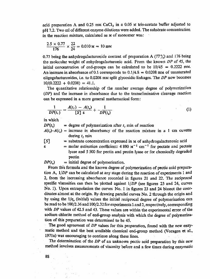
acid preparation A and 0.25 mM CaCl2 in a 0.05 M tris-acetate buffer adjusted to pH 7.2. Two ml of different enzyme dilutions were added. The substrate concentration in the reaction mixture, calculated as M of monomer was:
2.5x0.77 22 . . . . in
__ x —- = 0.010 M = 10 mM 176 24
0.77 being the anhydrogalacturonide content of preparation A (77%) and 176 being the molecular weight of anhydrogalacturonic acid. From the known DP of 45, the initial concentration of end-groups can be calculated to be 10/45 = 0.2222 mM. An increase in absorbance of 0.1 corresponds to 0.1/4.8 = 0.0208 mM of unsaturated oligogalacturonides, i.e. to 0.0208 mM split glycosidic linkages. The DP now becomes 10/(0.2222 + 0.0208) = 41.1.
The quantitative relationship of the number average degree of polymerization (DP) and the increase in absorbance due to the transeliminative cleavage reaction can be expressed in a more general mathematical form:
1 _ A(t,) - A(Q + 1 ( 1 )
DP(t,) [S] e DP(t„)
in which DP(tt) = degree of polymerization after /, min of reaction A(tf)-A(t^ = increase in absorbancy of the reaction mixture in a 1 cm cuvette
during t, min [S] = substrate concentration expressed in M of anhydrogalacturonic acid E = molar extinction coefficient: 4 800 M _ 1 cm - 1 for pectate and pectate
lyase and 5 500 for pectin and pectin lyase or for chemically degraded pectin
DP(t0) = initial degree of polymerization. From this formula and the known degree of polymerization of pectic acid prepara
tion A, 11 DP can be calculated at any stage during the reaction of experiments 1 and 2, from the increasing absorbance recorded in figures 21 and 22. The reciprocal specific viscosities can then be plotted against 11 DP (see figures 23 and 24, curves No. 1). Upon extrapolation the curves No. 1 in figures 23 and 24 bissect the coordinates almost at the origin. By drawing parallel curves No. 2 through the origin and by using the l/^s (initial) values the initial reciprocal degree of polymerization can be read to be 100/2.36 and 100/2.32 for experiments 1 and 2, respectively, corresponding with DP values of 42.5 and 43. These values are within the experimental error of the sodium chlorite method of end-group analysis with which the degree of polymerization of this preparation was determined to be 45.
The good agreement of DP values for this preparation, found with the new enzymatic method and the best available chemical end-group method (Voragen et al., 1971a) was encouraging to continue along these lines.
The determination of the DP of an unknown pectic acid preparation by this new method involves measurements of viscosity before and a few times during enzymatic
88

Is
0 , J
1"DP(to> ^DP = °/o f r e S e n d 9 r o u P s IQO^j IQQ. o/o f r e e endgroups
Figs 23 and 24. Results obtained in experiments 1 and 2, respectively in 1/J;, against l/DP plots. From curves 2 and values of 1/^(initial), l/DP(initial) can be read to be 100/2.36 and 100/2.32, corresponding to DP values 42.5 and 43, respectively.
breakdown by a randomly splitting pectate lyase, with a simultaneous recording of increase in absorbance at 232 nm. The results can then be plotted in diagrams of IM, and A against time as shown in figures 21 and 22. To be able to calculate the DP more directly from such plots, Equation 1 has to be adepted, as follows. Curves No. 2 in figures 23 and 24 can be described by the equation:
Consequently, at a time (^) during the degradation that l/ijs is double the original value, l/DP has also doubled, i.e.
DP(t0)
From equations 1 and 2 the following formula can be derived:
1 A(tj) - A(tB) DP{Q [S]e
(2)
(3)
in which A^y-AiQ = increase in absorbance of the reaction mixture measured in a 1 cm cuvette during the time that 1/q, has become double the original value, or t], has decreased to half its original value.
During my experiments I found that high polymer pectin preparations with a low value of the initial reciprocal specific viscosity showed linear increases in absorbance and reciprocal specific viscosity during a much longer period than necessary for the specific viscosity to be halved. For certain low polymer preparations, the time of linear relationships did not extend to viscosimetric half-value time. For this reason I
89
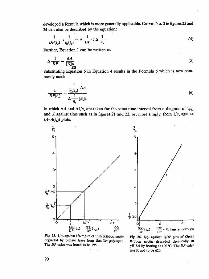
developed a formula which is more generally applicable. Curves No. 2 in figures 23 and 24 can also be described by the equation:
1 = A l
DP • ns (4)
DP(t0) 'qJLQ
Further, Equation 1 can be written as
1 _ AA DP [S]e
m Substituting Equation 5 in Equation 4 results in the Formula 6 which is now commonly used:
(5)
1 >/s(0 AA
^ A J - [ S ] B (6)
in which AA and Al/f/s are taken for the same time interval from a diagram of ljr\s and A against time such as in figures 21 and 22, or, more simply, from Ifa, against (A-A(t0)) plots.
5n
i ( t*}
£«o>
10v
100 DP ( t 0 ) 100,+ ,
20 100 DP
Ts(to)
100 f t \ D P ( t o )
2 4
100 _ 0/o f r e e endqroups DP
Fig. 25. 1/77, against l/DP plot of Pink Ribbon pectin Fig. 26. l/»y, against 1/DP plot of Green degraded by pectate lyase from Bacillus polymyxa. Ribbon pectin degraded chemically at The DP value was found to be 195. P H 5.5 by heating at 100 °C. The DP value
was found to be 620.
90
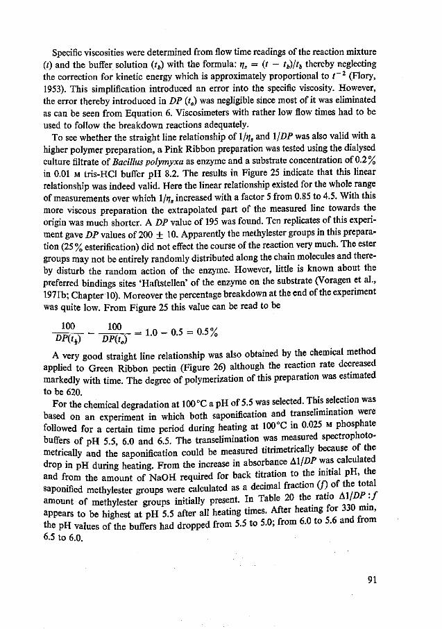
Specific viscosities were determined from flow time readings of the reaction mixture (0 and the buffer solution (tb) with the formula: rjs = (t — tb)/tb thereby neglecting the correction for kinetic energy which is approximately proportional to t~2 (Flory, 1953). This simplification introduced an error into the specific viscosity. However, the error thereby introduced in DP (t0) was negligible since most of it was eliminated as can be seen from Equation 6. Viscosimeters with rather low flow times had to be used to follow the breakdown reactions adequately.
To see whether the straight line relationship of 1/j/, and 1 /DP was also valid with a higher polymer preparation, a Pink Ribbon preparation was tested using the dialysed culture filtrate of Bacillus polymyxa as enzyme and a substrate concentration of 0.2% in 0.01 M tris-HCl buffer pH 8.2. The results in Figure 25 indicate that this linear relationship was indeed valid. Here the linear relationship existed for the whole range of measurements over which l/t]s increased with a factor 5 from 0.85 to 4.5. With this more viscous preparation the extrapolated part of the measured line towards the origin was much shorter. A DP value of 195 was found. Ten replicates of this experiment gave DP values of 200 + 10. Apparently the methylester groups in this preparation (25 % esterification) did not effect the course of the reaction very much. The ester groups may not be entirely randomly distributed along the chain molecules and thereby disturb the random action of the enzyme. However, little is known about the preferred bindings sites 'Haftstellen' of the enzyme on the substrate (Voragen et al., 1971b; Chapter 10). Moreover the percentage breakdown at the end of the experiment was quite low. From Figure 25 this value can be read to be
1 0 0 1 0° = 1 . 0 - 0 . 5 = 0.5% DP(ti) DP(tB)
A very good straight line relationship was also obtained by the chemical method applied to Green Ribbon pectin (Figure 26) although the reaction rate decreased markedly with time. The degree of polymerization of this preparation was estimated to be 620.
For the chemical degradation at 100 °C a pH of 5.5 was selected. This selection was based on an experiment in which both saponification and transehmination were followed for a certain time period during heating at 100°C in 0.025 M phosphate buffers of pH 5.5, 6.0 and 6.5. The transehmination was measured spectrophoto-metrically and the saponification could be measured titrimetrically because of the drop in pH during heating. From the increase in absorbance AX/DP was calculated and from the amount of NaOH required for back titration to the initial pH the saponified methylester groups were calculated as a decimal fraction C/) of the total amount of methylester groups initially present. In Table 20 the ratio Al/DP:J appears to be highest at pH 5.5 after all heating times. After heating for 330 mm, the pH values of the buffers had dropped from 5.5 to 5.0; from 6.0 to 5.6 and from 6.5 to 6.0.
91
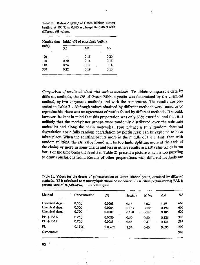
Table 20. Ratios A1/DP:/of Green Ribbon during heating at 100°C in 0.025 M phosphate buffers with different pH values.
Heating time
(min)
20 60
140 330
Initial pH of phosphate buffers
5.5
0.20 0.24 0.22
6.0
0.15 0.14 0.17 0.19
6.5
0.20 0.15 0.14 0.13
Comparison of results obtained with various methods To obtain comparable data by different methods, the DP of Green Ribbon pectin was determined by the chemical method, by two enzymatic methods and with the osmometer. The results are presented in Table 21. Although values obtained by different methods were found to be reproducible, there was no agreement of results found by different methods. It should, however, be kept in mind that this preparation was only 63 % esterified and that it is unlikely that the methylester groups were randomly distributed over the substrate molecules and along the chain molecules. Thus neither a fully random chemical degradation nor a fully random degradation by pectin lyase can be expected to have taken place. When the splitting occurs more in the middle of the chains, than with random splitting, the DP value found will be too high. Splitting more at the ends of the chains or more in some chains and less in others results in a DP value which is too low. For the time being the results in Table 21 present a picture which is too puzzling to draw conclusions from. Results of other preparations with different methods are
Table 21. Values for the degree of polymerization of Green Ribbon pectin, obtained by different methods. [S] is calculated as M (methyl)galacturonide monomer. PE is citrus pectinesterase; PAL is pectate lyase of B. polymyxa; PL is pectin lyase.
Method
Chemical degr. Chemical degr. Chemical degr.
PE + PAL PE + PAL
PL
Osmometer
Concentration
0.5% 0.5% 0.5%
0.2% 0.2%
0.17%
IS]
0.0209 0.0204 0.0209
0.0080 0.0083
0.00695
IMto)
0.16 0.185 0.180
0.50 0.43
1.34
Al/»/,
3.02 0.185 0.180
0.50 0.43
0.66
AA
3.49 0.190 0.185
0.126 0.134
0.095
DP
660 600 620
302 297
200
320
92

Table 22. Values for the degrees of polymerization of different pectins obtained by different methods. [S] is expressed as M (methyl)galacturonide monomer. PE is citrus pectinesterase, PAL is pectate lyase of B. polymyxa, PL is pectin lyase.
Preparation
Pectic acid A Pectic acid A Pectic acid A Pectic acid A Pectin B, 95% DE Pectin B, 95% DE Pectin B, 95% DE Pectin B, 95% DE Pectin B, 95% DE Pectin B, 95% DE Pectin C, 95% DE Pectin C, 95% DE Pectin C, 95% DE Pectin C, 95% DE Pectin D, 58% DE Pectin D, 58% DE Pectin E, 59.5% DE Pectin E, 59.5% DE Pectin E, 59.5% DE Pectin F, 51.5% DE Pectin F, 51.5% DE Pectin F, 51.5% DE Pink Ribbon 25% DE Pink Ribbon 25% DE
Method
PAL osmometer Sephadex G-75 sodium chlorite PE + PAL PL chemical degr. chemical degr.* osmometer Sephadex G-150 PL chemical degr. osmometer Sephadex G-150 PE + PAL chemical degr. PE + PAL PE + PAL chemical degr. PE + PAL PE + PAL chemical degr. PAL PAL
Concentration
0.25%
0.25% 0.25% 0.25% 0.25%
0.25%
0.18% 0.18% 0.18% 0.18% 0.18% 0.18% 0.18% 0.18% 0.2 % 0.17%
[S]
0.0100
0.0103 0.0104 0.0104 0.0104
0.0124
0.0081 0.0068
!/»?,(/<,) Al/i/,
2.56
2.96 2.90 2.40 2.90
2.14
0.95 1.20 0.95 1.15 1.30 2.15 2.70 2.70 0.85 1.18
3.64
0.65 1.65 1.80 2.10
0.44
0.85 1.18
AA
1.50
0.24 0.60 1.00 1.00
0.54
0.100 0.160
DP
43
63 34 45 45 51 44 41 86 93 38 26 66 34
130 135 127 126 129 75 71 52
195 200
* After cold alkaline saponification to 80% rest esterification.
presented in Table 22. Snoeren (1971) studied the molecular weight distribution of some of the preparations by filtration on Sephadex G-75 and G-150 columns (Pharmacia Fine Chemicals, Uppsala, Sweden) and found the preparations to consist.oi molecules with quite a broad range of molecular weights. In his experiments dextran types of different molecular weight (Pharmacia Fine ChemicaIs Uppsala Sweden were used as references. The fractionation according to molecular weight made it Possible to estimate the number average molecular weight of the preparat,ons^and these values are also included in Table 22. In this table too there is " ^ ™ * ™ in the results. The results obtained by the three independent reference meth«h. osmometer, Sephadex and sodium chlorite end-group method did not agree so that
it is difficult to evaluate the new methods. ,,•«•„..„„• A»ar»A* The degree of polymerization of pectin B was estimated by four different degrada-
93
tion methods: degradation by pectate lyase following chemical and enzymatic saponification, by pectin lyase and by chemical degradation with and without previous cold alkaline saponification to 80 % rest esterification. The results with these four methods were in quite good agreement for this preparation. The initial specific viscosity of the preparation did not change during the chemical and enzymatic saponification. Since it is known that in 0.1 M salt or buffer solutions the viscosity of pectin preparations is almost independent of the degree of esterification (Smit & Bryant, 1969; Owens et al., 1946) it may be concluded that no substantial degradation of pectin B occurred during saponification.
Heating of the almost fully esterified pectin preparation B at 90 °C and at pH 6.0 is most likely to give a fully random degradation of the preparation. A non-random distribution of methoxyl groups can be excluded. The only reason for a non-statistical degradation in this case could be the presence of 'weak bonds'. Such weak bonds could be due to the presence of neutral sugars in the main galacturonide chain (e.g. rhamnose) or in side chains (e.g. galactose, arabinose or xylose; see Chapter 2). However, Bryce & Greenwood (1957) and Banks & Greenwood (1968) showed that there was no evidence for weak bonds in linear polymers such as cellulose and amylose as long as, on degradation, the reciprocal intrinsic viscosity or the reciprocal degree of polymerization increased linearly with time, i.e. as long as there was no decrease in the degradation rate. In the new method, linear increases of reciprocal specific viscosity and absorbance were, of course, prerequisites.
If pectin B is indeed randomly split when chemically degraded, pectin lyase should also degrade the preparation in almost a fully random manner as shown by the good agreement of the DP values found with the two methods, 44 and 51, respectively. Voragen (1972) studying this pectin lyase on pectin B, found the same DP value for a pH range of 5.8 to 7.3. He observed from the action on methylated galacturonide oligomers that the first two or three glycosidic linkages on either side of the substrate molecules were relatively slowly attacked by the enzyme. This phenomenon may very well be the reason for the higher DP of pectin B found with the enzyme. A similar difference of results obtained with pectin lyase and with the chemical degradation method was observed with pectin preparation C.
The DP values measured for pectins D, E and F by the chemical degradation method and by the pectate lyase method, were in rather good agreement, but probably too low. With these low degrees of esterification the chemical degradation was very likely not random. The DP values of Green Ribbon pectin (Table 21) obtained with B. polymyxa pectate lyase were much lower than those obtained by the chemical method. Thus it would seem that by lowering the degree of esterification the results of both methods come closer to one another and that in pectin preparation F (51.5 % esterified) the pectate lyase method gives higher values than the chemical method.
These experimental results raise the question as to whether my B. polymyxa pectate lyase exhibits a pure random pattern of action on pectic acids. Recently Nagel & Wilson (1970), by CM-cellulose column chromatography, succeeded in separating four enzymes from the extracellular preparation of their strain of Bacillus polymyxa.
94
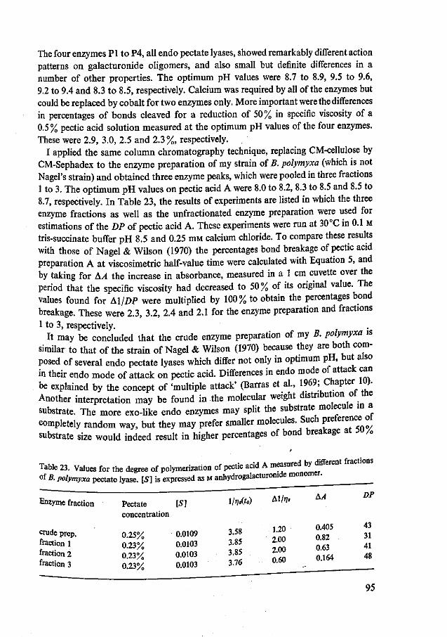
The four enzymes PI to P4, all endo pectate lyases, showed remarkably different action patterns on galacturonide oligomers, and also small but definite differences in a number of other properties. The optimum pH values were 8.7 to 8.9, 9.5 to 9.6, 9.2 to 9.4 and 8.3 to 8.5, respectively. Calcium was required by all of the enzymes but could be replaced by cobalt for two enzymes only. More important were the differences in percentages of bonds cleaved for a reduction of 50% in specific viscosity of a 0.5% pectic acid solution measured at the optimum pH values of the four enzymes. These were 2.9, 3.0, 2.5 and 2.3 % respectively.
I applied the same column chromatography technique, replacing CM-cellulose by CM-Sephadex to the enzyme preparation of my strain of B. polymyxa (which is not Nagel's strain) and obtained three enzyme peaks, which were pooled in three fractions 1 to 3. The optimum pH values on pectic acid A were 8.0 to 8.2, 8.3 to 8.5 and 8.5 to 8.7, respectively. In Table 23, the results of experiments are listed in which the three enzyme fractions as well as the unfractionated enzyme preparation were used for estimations of the DP of pectic acid A. These experiments were run at 30 °C in 0.1 M tris-succinate buffer pH 8.5 and 0.25 mM calcium chloride. To compare these results with those of Nagel & Wilson (1970) the percentages bond breakage of pectic acid preparation A at viscosimetric half-value time were calculated with Equation 5, and by taking for AA the increase in absorbance, measured in a 1 cm cuvette over the period that the specific viscosity had decreased to 50% of its original value. The values found for All DP were multiplied by 100% to obtain the percentages bond breakage. These were 2.3, 3.2, 2.4 and 2.1 for the enzyme preparation and fractions 1 to 3, respectively.
It may be concluded that the crude enzyme preparation of my B. polymyxa is similar to that of the strain of Nagel & Wilson (1970) because they are both composed of several endo pectate lyases which differ not only in optimum pH, but also in their endo mode of attack on pectic acid. Differences in endo mode of attack can be explained by the concept of 'multiple attack' (Barras et al., 1969; Chapter 10). Another interpretation may be found in the molecular weight distribution ot the substrate. The more exo-like endo enzymes may split the substrate molecule in a completely random way, but they may prefer smaller molecules. Such preference! oi substrate size would indeed result in higher percentages of bond breakage at DU/0
Table 23. Values for the degree of polymerization of pectic acid A measured by different fractions of B. polymyxa pectate lyase. [5] is expressed as M anhydrogalacturonide monomer.
Enzyme fraction Pectate [S] 1W») A 1 / , / ' concentration
i ?n 0405 43 "udeprep. 0.25% 0.0109 3.58 ^ ^ 3 1
fraction 1 0.23% 0.0103 3.85 2.00 ^ fraction 2 0.23% 0.0103 3 85 2.00 4 g
fraction 3 0.23% 0.0103 3.76 U-DU . ^ _ _
95

viscosity reduction. This explanation should be checked by kinetic studies of such enzymes on different sizes of substrates. Nevertheless, it is clear that the observed differences in endo mode of attack of the substrate by different enzyme fractions make this enzyme source unsuitable for the enzymatic method of molecular weight estimation for which a truly randomly splitting enzyme is necessary.
Looking for enzymes which would split pectic acids truly at random, I tested pectate lyases from four strains of the collection. The results are presented in Table 24. It can be seen that two enzyme preparations namely that of Flavobacterium S2 and that of Arthrobacter 547 act even more in an exo-like fashion than that of Bacillus polymyxa, but that the enzyme of Pseudomonas GK5 probably acts more endo-like than the preparation of B. polymyxa. The DP value found for pectic acid A in this case is in agreement with that found with the osmometer. Future experiments may show whether this enzyme is the truly randomly acting enzyme which at first I thought I had found with the B. polymyxa enzyme preparation.
Since the method that is primarily based on the linear increase of both l/t]s and 11 DP during enzymatic or chemical degradation is a rather unusual approach to the viscosity-molecular weight relationship of pectins, the theory of the method cannot be easily reconciled with the experimental relationships of viscosity and molecular weight as described in literature for macromolecules (Rafikov et al., 1964; Flory, 1953). For molecularly heterogeneous polymers Flory (1953) gives the formula
[IJ] = K' Ml ( 7 )
in which [IJ] = intrinsic viscosity = (t]Jc)c^0 = [fa, - l)/c]c_0
K' = a material constant, to be determined experimentally M „ = the viscosity average molecular weight, which depends on the molecular weight
distribution and is somewhere between M„ (number average molecular weight) and Mw (weight average molecular weight)
a = a constant determined by the heterogeneity of the preparation.
Table 24. Values for the degree of polymerization of pectic acid A measured by pectate lyases of different bacterial sources. [5] is expressed as M anhydrogalacturonide monomer.
Pectate lyase from
B. polymyxa Pseudomonas GK5 Flavobacterium S2 Arthrobacter 547 (osmometer) (sodium chlorite) (Sephadex G-75)
Pectate concentration
0.33% 0.33% 0.33% 0.33%
IS]
0.0146 0.0146 0.0146 0.0146
l/»7.(fo)
2.03 2.28 2.20 2.18
A1/J?8
2.03 2.28 0.85 0.645
AA
1.53 1.09 0.90 1.70
DP
46 65 30 12
(63) (45) (34)
96

Owens et al. (1946) obtained the following approximate equation for pectins and pectates by comparing intrinsic viscosities of their own preparations with viscosities and weight average molecular weights of citrus pectins determined by Saverborn (1945).
[IJ] = 1.4 x 10~6 MWU34- (8)
They also found that there was little relationship between intrinsic viscosities and number average molecular weights (M„), except that pectins from the same source gave values for these properties which followed the same trend. Theyjhought that the reason for the lack of a more direct relationship between [>] and M„, regardless of the source of the pectin probably lay in the shapes of the molecular weight distribution curves. Moreover, working in appropriate electrolyte solutions to suppress electroviscous effects (Owens etal., 1944; Malsch, 1941) they found that removal of methoxyl groups caused little change in surface characteristics. The flexibility of the galacturonide chain is probably not affected by this removal of methoxyl groups so that viscosity equations can be applied with equal reliability for pectmic acids of various methoxyl contents (Owens et al., 1946).
A linear curve which goes through the origin in an \\r\s against l/DP plot and which describes the random enzymatic or chemical degradation, can be described by the following equation
— (9) r\s = k DP = k' M„
in which k and k' are constants of the preparation under the experimental conditions
applied. . . , Equations 8 and 9 should be compared against the background ofthe f P e r i m ^ t a
procedures followed in both cases. The numerical constants in Equation 8 are thougn to apply to all pectin preparations, regardless of molecular weight distortion or degree of esterification (Owens et al., 1946), whereas the k and k in E ^ l o n J a r e
constants which only apply to one preparation and one set of ^ ^ ^ ^ ^ If equations 8 and 9 are thought to describe one and the same enzymatic or chem a
degradation experiment, they can be in agreement with each other iboth^ ratio M : , . and Mj^.Mn change in « ^ f l ^ S — gradation or if both remain constant. Indeed Malsch (194) fou n d j ^ ^ ^ for many pectins over the narrow „. range of 0.025KU• • i m e n t a l
eventuality is probably valid, since the second is ruled out oy * relationship </[„] a j , , (Equa tion ,0), - « * < * - £ ? * & £ £ £ « valid for the many pectates and pectins they tested over a mucn concentrations:
1. (10>
in which c = concentration of pectin in g/100 ml (measured over a concentration
range of 0.05 to 0.3) and k' = a constant.
97

I also found log qjc of Green Ribbon pectin to increase linearly with pectin concentrations of 0.1 g/100 ml and higher but with the pectic acid preparation A, rjjc remained almost constant over the concentration range of 0.1 to 0.8 g/100 ml. Both ratios \t\] : t]s and Afw
1-34 : M„ probably change during a degradation experiment. It is, however, at present impossible to discuss these changes quantitatively.
From the results obtained so far it may be concluded that the described enzymatic and chemical methods of molecular weight estimation of pectic substances represent a new and interesting approach. The methods may also give more insight into the 'endo' or 'random' mode of attack of certain enzymes.
It is also clear that more work has to be done, to compare molecular weight estimations obtained from different enzymes with results obtained by other independent methods, especially by osmometry and by Sephadex gel filtration. Determination of intrinsic viscosities at certain stages during the degradation experiments will make it possible to calculate changes in the ratio Mw : Mn. The method requires no highly technical equipment and the measurement is rather simple. Since degradation and all measurements are carried out on one and the same reaction mixture, inaccuracies due to pipetting, weighing etc. are eliminated. Care should be taken to have the same temperature + 0.1 °C in the viscosimeter and in the cuvette. For calculations of molecular weights no material constants, except for the molar extinction coefficients, need to be used. These material constants should but perhaps do not apply to all pectin preparations.
9.4 Summary and conclusions
A new method for the estimation of the number average degree of polymerization of pectic substances has been worked out. The method was based on the experimentally observed linear increases of the reciprocal specific viscosity and the reciprocal degree of polymerization during enzymatic or chemical transeliminative degradation of pectates and pectins. Differences in results obtained for a number of preparations with several variations of the method as well as with other methods initiated a further study of the enzyme used: the endo pectate lyase of Bacillus polymyxa. The enzyme appeared to consist of at least three fractions which were different not only in their optimum pH values but also in their endo character. The differences in endo character of various endo pectate lyases were interpreted. Since a fully random degradation of the substrate was required the enzyme seemed less suitable for use in this method. As a more promising enzyme the endo pectate lyase of a Pseudomonas strain could be selected. I tried to reconcile the theory of this method, which involves an unusual approach to the viscosity - molecular weight relationship of pectins, with the experimental relationships of viscosity and molecular weight described in literature.
It would be interesting to study changes in the ratio of weight average to number average molecular weights at certain stages during degradation experiments, e.g. by measuring intrinsic viscosities. Future work should also involve studies of the molecular weight distribution of pectic substances e.g. by Sephadex gel filtration to
98
interpret the observed differences in results obtained with enzymatic and chemical degradation methods and other methods. In this chapter pectic substances with pectic enzymes and pectic enzymes with pectic substances were studied at the same time with this new method.
99

10 Pectate lyase of Arthrobacter
10.1 Introduction
The screening for pectolysis of 240 Arthrobacter strains showed 10 % to be pectolytic (Chapter 6; Rombouts & Pilnik, 1971a). The extracellular enzymes of some of the pectolytic strains were typed (Chapter 7). It was found that Arthrobacter strain 547 possibly produced both exo and endo pectate lyases, but only a small pectinesterase activity could be detected. The enzyme preparation of another strain, Arthrobacter 370, showed very good viscosimetric activity on both high esterified and low esterified apple pectins. It was concluded that this preparation probably contained both pectate and pectin lyases. This preparation also contained a small amount of pectinesterase. Since no bacterial pectin lyase, except that from Streptomyces viridochromogenes described briefly by Agate et al. (1962), has been found yet and since no data are available on the production and properties of the pectolytic enzymes of Arthrobacter it was decided to study these enzymes in more detail. Arthrobacter is a widespread group of bacteria, proliferating on foods, in waste water and soil. The genus contributes much to the biodegradation of organic waste material (Mulder & Antheunisse, 1963; Mulder et al., 1971).
10.2 Materials and methods
The growth and enzyme production of Arthrobacter strain 547 was compared in two media: the buffered pectate calcium (BPC) medium, described in Chapter 7 and a minerals pectate (MPC) medium with biotin as the only growth factor. The MPC medium was that described by Veldkamp et al. (1966) for Arthrobacter except for the glucose which was replaced by pectate and further a more concentrated buffer was used. The medium was prepared as follows: Solution I (NH4)2S04, 5 g; CaCl2 • 2H20,0.15 g; Na2HP04 • 2H20, 3 g; KH2P04, 2 g; MgS04 • 7H20, 0.5 g; FeCl3 • 6H20, 0.0025 g; distilled water, 990 ml. Solution II MnCl2,100 mg; CuS04 • 5H20, 10 mg; ZnS04 • 7H20, 10 mg; H3B03, 10 mg; CoCl2 • 6H20, 1 mg; NaMo04 • 2H20, 1 mg; distilled water, 1000 ml.
oTT/1 B l ° t m ' 1 0 m g ; d i S t i l l e d W a t e r ' 1 0 0° m L T e n m l ^ Solution II was added to 990 ml of Solution I. Five g of sodium pectate (Obipektin AG, Bischofszell, Switzerland) was moistened with ethanol and the 1000 ml mixture of solution I and II was quickly poured on top of the pectate. The pectate was dissolved by stirring and the PH was adjusted to 7.0. The medium was distributed in 200 ml portions in 1 litre Erlenmeyer
100
flasks and sterilized for 15 min at 115°C. Solution III was separately sterilized and 0.2 ml of it was added to 200 ml medium to give a final biotin concentration of 0.01 mg/litre. The Erlenmeyer flasks were inoculated from a culture in the corresponding medium and incubated in a Gallenkamp (London, England) orbital incubator at 150 strokes per min (stroke width 32 mm) at 28 to 30 °C.
Growth was measured with a nephelometer (Chapter 7). Degradation of the pectate in the culture medium was checked with the ethanol test (Chapter 6). Galacturonide residues in the culture medium were measured with the carbazole test of Rouse & Atkins (1955). Pectate lyase activity was measured in the dialysed culture medium with a recording spectrophotometer (Chapter 9).
Definition of the unit of enzyme The unit of enzyme (u) is that amount of enzyme which will catalyse the transeliminative splitting of 1 micro-equivalent of glycosidic linkages under standard reaction conditions (Florkin & Stotz, 1965). The units were measured at 30 °C, in 3 ml reaction mixtures, pH 8.3, consisting of tris-HCl buffer (0.1 M tris), 0.25 mM calcium chloride and 0.1 % pectic acid A (Chapter 9). If a molar extinction coefficient of 4 800 M _ 1 cm - 1 (Chapter 9) is used, one unit of enzyme gives an increase in absorbance at 232 nm of 1.6 per min, when the above reaction mixture is measured in a 1 cm cuvette.
Calcium phosphate gel treatment At the beginning of the stationary growth phase, the cells were removed by centrifuging at 2 °C and at 6 000 x g and the culture liquid was dialysed overnight against running tap water. The dialysed crude enzyme preparation was frozen at — 80 °C in the shell-freezer of a freeze-dryer type B71 (New Brunswick Scientific Co., Inc. New Brunswick, New Jersey, USA) and kept frozen at -30°C until required. For purification by calcium phosphate gel, the crude enzyme solution was diluted to give an ultraviolet absorption at 280 nm of 0.17 in a 1 cm cuvette. Calcium phosphate gel (BDH, Poole, England, 3.0 percent w/v suspension in water) was added to this solution in an amount of 280 ml of gel to 4 litre of enzyme solution. The mixture was stirred for 10 min at room temperature and centrifuged at 3000 x g for 10 min. The supernatant was discarded and 140 ml of 0.04 M potassium phosphate buffer, pH 8.0 was added. After 10 min of stirring, the gel was again centrifuged off, and the supernatant was dialysed for 24 h at 2°C against 3 or 4 changes of a buffer of pH 7.5 made by mixing 0.1 M of tris and 0.1 M of succinic acid. The enzyme was eventually stored in the frozen state.
Chromatography on DEAE-Sephadex About 25 g of DEAE-Sephadex A-25 (Pharmacia Fine Chemicals Uppsala, Sweden) was first treated with 1 M of sodium succinate to exchange the chloride ions for succinate ions. Then the ion exchanger was equilibrated with tris-succinate buffer, 0.1 M tris adjusted to pH 7.5 with 0.1 M succinic acid. A column of 1.5 by 25 cm was packed and equilibrated with the tris-succinate buffer pH 7.5. A sample of 265 ml of enzyme, dialysed against the tris-succinate buffer, pH 7.5 was applied to the column at a rate of 66 ml per hour by a Perpex
101

peristaltic pump, type 10 200 (LKB-Produkter AB, Stockholm-Bromma, Sweden). Elution was performed at a rate of 20 ml per hour. First 16 fractions of about 17 ml and then fractions of about 6 ml were collected in an Ultrorac fraction collector, type 7 000 (LKB-Produkter AB, Stockholm-Bromma, Sweden).
Elution was started by a continuous linear ionic strength gradient. Therefore two communicating vessels one of which was provided with a stirrer, were filled with 250 ml each tris-succinate buffer of pH 7 and a strength of 0.1 M in the stirring vessel and 0.3 M in the other. The buffer was pumped from the stirring vessel onto the column by the peristaltic pump. After elution with the gradient, the column was eluted with 500 ml of 0.3 M tris-succinate buffer and finally with 400 ml of 1 M tris-succinate pH 7.5. Every second or third fraction was analysed for activity and for protein by an absorbance measurement at 280 nm. Pools of fractions were assayed for activity and for protein with Folin-Ciocalteu reagent (Layne, 1957).
Disc electrophoresis Lyophilized samples of dialysed culture liquid and dialysed fractions of DEAE-Sephadex chromatography were used. The gels and other solutions were prepared as described by Davis (1964), but a different electrophoresis buffer of 0.0125 M tris, adjusted to pH 9.5 by 0.125 M glycine was taken. A disc electrophoresis apparatus for eight tubes (Shandon, England) was used and electrophoresis was carried out at a constant current of 2 mA per tube. Gels were coloured with amido black or segmented in 20 segments of 0.25 cm for enzyme assay. The segments were extracted overnight in the refrigerator in tubes containing 0.5 ml of tris-HCl buffer (0.1 M in tris) pH 7.0. Aliquots of 0.3 ml of the buffer were assayed spectrophoto-metrically for lyase activity.
Optimum pH The same buffers as those of Nagel & Wilson (1970) were used. Stock solutions of buffers with calcium chloride and a stock solution of substrate (pectic acid preparation A, Chapter 9) were prepared. The proper amounts of the solutions were mixed just before assay. The reaction mixtures contained 0.05 M each of tris and glycine 0.25 mM calcium chloride and 0.1 % pectic acid. The reaction rate was measured for 4 min in the spectrophotometer at 232 nm.
pH Stability Portions of chromatographed enzyme were added to tris-succinate buffers of pH 5, 6, 7 and 8 (0.1 M succinic acid brought to pH by the addition of 0.1 M tris). At 10 min intervals samples were withdrawn from the buffers, maintained at 38 °C and assayed for rest activity on pectic acid A, at the pH of the treatment.
Effect of cations Different cations and EDTA were assayed for their effect on enzyme activity. Solutions of pectic acid preparation A (0.1 %) in buffers (0.1 M tris adjusted to pH 8.3 by 0.1 M succinic acid), containing 0.5 mM magnesium, calcium, strontium, barium or manganese ions, or 0.5 mM EDTA (disodium salt) were prepared. Reaction rates of the enzyme in these solutions were measured in the spectrophotometer.
The optimum calcium concentration was determined for two different pectic acid
102

concentrations. Two series of reaction mixtures (pH 8.6) were made up to contain either 0.25% or 0.025% pectic acid, and also tris-HCl buffer (0.11 M in tris), 0.02 to 16 M calcium chloride and 0.033 units of enzyme in a total volume of 3 ml. Stock solutions of substrate, buffer with calcium and enzyme were mixed just before the spectrophotometric measurement at 30 °C.
Activation energy Initial reaction velocities were measured with 9 different substrate concentrations at 6 different temperatures obtained by adjusting the thermostat of the spectrophotometer. Reaction mixtures (pH 8.3 with tris-HCl) of a total volume of 3.0 ml were made up to be 0.1 M in tris, 0.25 mM in calcium chloride and 11.7, 8.8, 5.8, 4.4, 2.9, 2.2, 1.8, 1.46 and 1.17 mM in pectic acid A (calculated as anhydro-galacturonide monomers). Portions of 0.4 ml of purified diluted enzyme were used. The temperatures in the cuvettes were measured with a thermocouple. They were 17.5, 21.6, 25.5, 30.8, 35.0 and 41.9°C. Km and Vmax values were read from Line-weaver-Burk plots.
Paper chromatography of products A reaction mixture (25 ml) of 0.5 % pectic acid A, 0.25 mM calcium ions, 0.055 lyase units per ml, pH maintained at 8.7 with NaOH, was incubated at 30 °C, at which temperature its viscosimetric half-value time had been determined to be 30 min. Samples of 2 ml were withdrawn after 0.25, 0.5, 0.75, 1, 2, 3, 4, 8 and 15 times viscosimetric half-value time. The samples, together with a blank sample of the same composition but with inactivated enzyme, were immediately treated in a batch by shaking with 0.5 g wet Dowex 50W-X8 cation exchanger in the H form, to inactivate the enzyme and to remove cations (Chapter 7). The liquid was collected, freeze-dried and subsequently redissolved in 0.5 ml distilled water. Samples of 20 ul were applied to Whatman No. 4. Pectic acid A, fully degraded by Bacillus polymyxa pectate lyase was used as the chromatography standard. The chromato-grams were developed in the new solvent of Nagel & Wilson (1970), pyridine-ethyl acetate-acetic acid-water ( 3 : 5 : 2 : 3 ) . Aniline phthalate spray was used to detect the spots.
Viscosity reduction in relation to bond breakage The same procedure was used as that for the determination of the degree of polymerization of pectic acids with pectate lyase from Bacillus polymyxa, described in Chapter 9. Pectic acid preparation A (Chapter 9) was used as the substrate in a concentration of 0.25% in the reaction mixtures, which also contained tris-HCl buffer, pH 8.3 (0.1 M of tris) and were 0.25 DIM in calcium chloride. Measurements were made with crude enzyme and with purified fractions, at 30°C and at pH 8.3, but also at different pH values and temperatures. For the measurements at different pH values a 25% esterified pectin was used (Pink Ribbon, Obipektin AG, Bischofszell, Switzerland).
Influence of the degree of esterification of the substrate on the Km and Vmax values and on the degree of degradation Pectin preparation B (Chapter 9) was used to prepare
103

samples, which differed in degree of esterification but as little as possible in other properties such as degree of polymerization. For this purpose 6.25 g 95 % esterified pectin B was dissolved in 200 ml distilled water by stirring. The solution was brought to pH 7.0 with 13.1 ml 0.1 N sodium hydroxide. The solution was brought to volume in a 250 ml volumetric flask. Into 50 ml volumetric flasks, cooled in melting ice, 20 g portions were weighed and then to each of the flasks a precalculated amount of 0.1 N NaOH was added to arrive at the desired degree of esterification. The amounts of 0.1 N NaOH added from a microburette were 3.14 ml (to arrive at 80 % esterification); 7.30 ml (60%), 10.42 ml (45%), 12.50 ml (35%), 13.54 ml (30%), 14.59 ml (25%), 15.62 ml (20%), 16.68 ml (15%), 17.71 ml (10%) and 19.78 ml (0%). The flasks were brought to volume with 4°C distilled water and stored for 48 h in the refrigerator. Then the 1 % pectin solutions were centrifuged at 30 000 x g and used without further treatment. Residual methoxyl content was determined by the titrimetric method (Doesburg, 1965). The degrees of esterification measured were 83, 64, 49, 39, 35, 31, 26, 21, 16 and 7, respectively.
A preparation with 3% residual ester groups was prepared by adding another 3 ml 0.1 N NaOH to 25 ml of the 7 % esterified preparation and storing it overnight in the refrigerator.
Initial reaction velocities were measured in the recording spectrophotometer at 30°C. Reaction mixtures (total volume 3 ml) contained 1.5 ml tris-HCl buffer pH 8.3 (0.2 M in tris, 0.5 mM in calcium chloride), 0.1 ml of enzyme (0.034 units) and 1.4 ml substrate plus distilled water. The amounts of substrates used and their final concentrations (mM anhydrogalacturonic acid) in the cuvettes were 0.9 ml (13 mM), 0.6 ml (8.5 mM), 0.5 ml (7.1 mM), 0.35 ml (5.0 mM), 0.25 ml (3.6 mM), 0.2 ml (2.8 mM) and of a 1 to 4 dilution of the substrates 0.6 ml (2.1 mM), 0.4 ml (1.4 mM), 0.2 ml (0.7 mM), and 0.1 ml (0.35 mM), respectively.
Degrees of degradation of the preparations with different degree of esterification were measured in reaction mixtures (10 ml) of the same composition as above, but with a concentration of substrates of 4.0 mM and with a pH of 7.0. The absorbance was measured at time intervals of 10 to 15 h for 70 h. Thus 0.5 or 0.1 ml portions of reaction mixtures were diluted to 3 ml with buffer. The reaction mixtures were filtered daily through membrane filters (type SM 11406, Sartorius Membranfilter GmbH, Gottingen, Western Germany) to keep the bacterial load down.
10.3 Results and discussion
Growth and lyase production In figures 27 and 28 growth and lyase production of Arthrobacter 547 can be seen to increase simultaneously up to the stationary phase in both BPC and MPC media. In these figures the enzyme production is expressed in units per ml culture liquid after dialysis. After prolonged incubation in BPC medium all enzyme activity was rapidly lost. In the growth curve of the organism in MPC medium (Figure 28) a shoulder appeared when lyase activity was maximum. Microscopic checks showed that beyond this stage the rod-shaped cells became fragmented
104

1000H
500 H
/ml N
Q3 1000-
u/ml
r0.3
500H
80 100
t ime ( h )
40 5 0
t i m e ( h )
Figs 27 and 28. Growth (+) and lyase production ( O) of Arthrobacter 547 in BPC medium (Fig. 27) and in MPC medium (Fig. 28) N is nephelometer reading, u/ml is units of enzyme per ml culture medium.
into small coccoid cells. In both BPC and MPC media the depolymerization of the pectate as shown by the ethanol test, was achieved after 20 h, i.e. in the early stage of the enzyme production. However, low molecular galacturonide material remained in the culture medium throughout the whole period, as shown by the carbazole test. Even prolonged incubation of the culture medium, with the cells removed, and subsequent dialysis, did not result in a galacturonide negative crude enzyme solution.
The same BPC and MPC media were also used to study the lyase production of some other bacteria: Bacillus polymyxa, Arthrobacter 370, Pseudomonas GK5 and Flavobacterium S2. The amount of enzyme usually increased in the culture medium simultaneously with growth, but with Bacillus polymyxa the production of enzyme followed the growth with a lag. Prolongation of the incubation beyond the stationary phase of growth resulted in a loss of enzyme activity of Pseudomonas and Flavobacterium, but the enzyme of Bacillus polymyxa remained stable under both anaerobic and aerobic conditions. Some maximum enzyme production levels of these organisms in the two media are given in Table 25. Bacillus polymyxa and Pseudomonas were most productive on BPC medium, whereas for Arthrobacter the MPC medium was also suitable. These differences in enzyme productivity may also exist when the or-
105

Table 25. Enzyme productivity of some bacteria on BPC and MPC media.
Organism
Bacillus poly myxa (anaerobic) Bacillus polymyxa (aerobic) Arthrobacter 547 Arthrobacter 370 Pseudomonas GK5 Flavobacterium S2
Formation of
BPC medium
0.7 1.7 0.35 0.08 1.8 0.06
enzyme (u/ml) on
MPC medium
0.017 0.24
0.009 0.000
ganisms are grown on Wieringa's pectin gel medium (Chapter 4) and may be the reason for the different modes of attack of the pectate gel observed: Bacillus polymyxa and Pseudomonas rapidly destroyed the gel by liquefaction, whereas Arthrobacter and Flavobacterium formed distinct pits in the gel. The endo/exo character of the enzymes as well as their diffusion mobility may also effect the behaviour of the enzyme producing organism on pectate medium.
Purification of the lyase Several batches of enzyme produced by Arthrobacter 547 on pectate medium were tested for pectinesterase activity by the titrimetric test described in Chapter 7, but no activity could be detected. The calcium phosphate gel treatment, which proved most satisfactory as a concentration and partial purification step for the pectate lyase was a modification of that applied by Macmillan & Phaff(1966)to the crude pectate lyase preparation from Clostridium multifermentans. Since the culture liquid contained a galacturonide residue, which was not lost by dialysis, the purification was also followed by the carbazole test. In Table 26 the results are given of the calcium phosphate gel treatment of the dialysed culture fluid of Arthrobacter 547. Under the conditions employed, the specific activity related to
Table 26. Purification of pectate lyase from Arthrobacter 547 by calcium phosphate gel treatment.
Fraction
Crude enzyme Supernatant liquid after gel treatment Eluate from gel
Volume after dialysis (ml)
4000
4000 140
Lyase activity (u/ml)
0.0125
0.00125 0.263
Az%o
0.176
0.143 0.206
Specific activity (u/mg protein)
0.071
0.0087 1.28
Total galacturonide residue (mg)
360
154 146
Recovery
(%)
100
(10) 74
106

absorbance at 280 nm, increased with a factor 18 and a recovery of 74 % was achieved. Galacturonide residue also diminished, but only from 4.5 to 2.5 mg per unit of enzyme. By taking less calcium phosphate gel under otherwise the same conditions, more of the galacturonide residue could be removed, however, at the expense of both the recovery and the specific activity.
For the chromatography of the pectate lyase of Arthrobacter 547 on DEAE-Sephadex, shown in Figure 29 a sample of enzyme was used, which was treated twice by calcium phosphate gel. The enzyme came off the column in one sharp peak with a rather long tail. At the tail end of the peak the concentration of the elution buffer remained the same. Direct measurements of absorbance at 280 and 260 nm in the
u/ml
0.5-
0.4 H
0.3-
0.2^
0.1
^ -i 1-, 1 ^-^^f
>4v+ VU+-\
'VHV+*<t-Sv
20 50 — i —
100 X
150
l ^
application of sample in 0.1 M buffer pH 7.5
fraction nr 1
buffer gradient 0.1 to 0.3 M pH 7.5
0.3 M buffer pH 7.5
Fig. 29. Chromatography of pectate lyase of Arthrobacter 547 on DEAE-Sephadex.
107
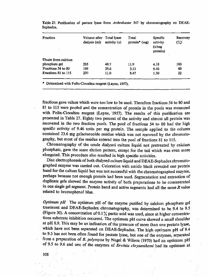
Table 27. Purification of pectate lyase from Arthrobacter 547 by chromatography on DEAE-Sephadex.
Fraction
Eluate from calcium phosphate gel Fractions 54 to 80 Fractions 81 to 115
Volume after dialysis (ml)
265 169 220
Total lyase activity (u)
49.7 29.6 11.0
Total protein*
11.9 3.13 8.47
(mg) Specific activity (u/mg protein)
4.18 9.46 1.30
Recovery
(%)
100 60 22
* Determined with Folin-Ciocalteu reagent (Layne, 1957).
fractions gave values which were too low to be used. Therefore fractions 54 to 80 and 81 to 115 were pooled and the concentration of protein in the pools was measured with Folin-Ciocalteu reagent (Layne, 1957). The results of this purification are presented in Table 27. Eighty two percent of the activity and almost all protein was recovered in the two fraction pools. The pool of fractions 54 to 80 had the high specific activity of 9.46 units per mg protein. The sample applied to the column contained 23.6 mg galacturonide residue which was not removed by the chromatography, but most of the residue entered into the pool of fractions 81 to 115.
Chromatography of the crude dialysed culture liquid not pretreated by calcium phosphate, gave the same elution pattern, except for the tail which was even more elongated. This procedure also resulted in high specific activities.
Disc electrophoresis of both dialysed culture liquid and DEAE-Sephadex chromato-graphed enzyme was carried out. Coloration with amido black revealed one protein band for the culture liquid but was not successful with the chromatographed enzyme, perhaps because not enough protein had been used. Segmentation and extraction of duplicate gels showed the enzyme activity of both preparations to be concentrated m one single gel segment. Protein band and active segments had all the same R value related to bromophenol blue.
Optimum pH The optimum pH of the enzyme purified by calcium phosphate gel treatment and DEAE-Sephadex chromatography, was determined to be 9.4 to 9.5 (Figure 30). A concentration of 0.1 % pectic acid was used, since at higher concentrations substrate inhibition occurred. The optimum pH curve showed a small shoulder at pH 8.9. This may be an indication of the presence of more than one pectate lyase, which have not been separated on DEAE-Sephadex. The high optimum pH of 9.4 to 9.5 has not been often found for pectate lyase, but one of the enzymes, separated from a preparation of B. polymyxa by Nagel & Wilson (1970) had an optimum pH oi y.i to 9.6 and one of the enzymes of Erwinia chrysanthemi had its optimum at
108
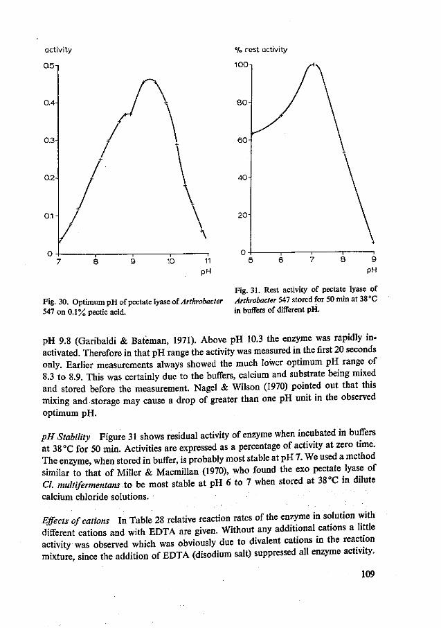
% rest activity
100
Fig. 31. Rest activity of pectate lyase of Fig. 30. Optimum pH of pectate lyase of Arthrobacter Arthrobacter 547 stored for 50 min at 38 °C 547 on 0.1% pectic acid. in buffers of different pH.
pH 9.8 (Garibaldi & Bateman, 1971). Above pH 10.3 the enzyme was rapidly inactivated. Therefore in that pH range the activity was measured in the first 20 seconds only. Earlier measurements always showed the much lower optimum pH range of 8.3 to 8.9. This was certainly due to the buffers, calcium and substrate being mixed and stored before the measurement. Nagel & Wilson (1970) pointed out that this mixing and storage may cause a drop of greater than one pH unit in the observed optimum pH.
pH Stability Figure 31 shows residual activity of enzyme when incubated in buffers at 38 °C for 50 min. Activities are expressed as a percentage of activity at zero time. The enzyme, when stored in buffer, is probably most stable at PH 7. We used a method similar to that of Miller & Macmillan (1970), who found the exo pectate lyase of CI multifermentans to be most stable at pH 6 to 7 when stored at 38 °C in dilute calcium chloride solutions.
Effects of cations In Table 28 relative reaction rates of the enzyme in solution with different cations and with EDTA are given. Without any additional cations a little activity was observed which was obviously due to divalent cations in the reaction mixture, since the addition of EDTA (disodium salt) suppressed all enzyme activity.
109
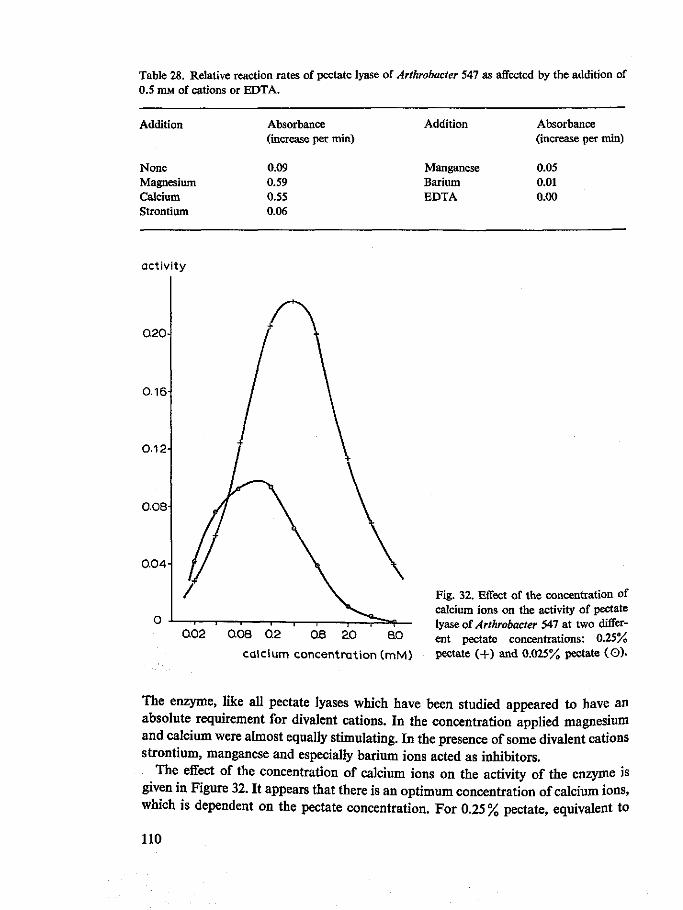
Table 28. Relative reaction rates of pectate lyase of Arthrobacter 547 as affected by the addition of 0.5 mM of cations or EDTA.
Addition
None Magnesium Calcium Strontium
Absorbance (increase per
0.09 0.59 0.55 0.06
min) Addition
Manganese Barium EDTA
Absorbance (increase per min)
0.05 0.01 0.00
activity
0.20-1
0.16-1
0.12
0.08
0.04 H
0.02 0.08 0.2 0.8 2.0 B0
calcium concentration (mM)
Fig. 32. Effect of the concentration of calcium ions on the activity of pectate lyase of Arthrobacter 547 at two different pectate concentrations: 0.25% pectate (+) and 0.025% pectate (O).
The enzyme, like all pectate lyases which have been studied appeared to have an absolute requirement for divalent cations. In the concentration applied magnesium and calcium were almost equally stimulating. In the presence of some divalent cations strontium, manganese and especially barium ions acted as inhibitors.
The effect of the concentration of calcium ions on the activity of the enzyme is given in Figure 32. It appears that there is an optimum concentration of calcium ions, which is dependent on the pectate concentration. For 0.25% pectate, equivalent to
110

11 mM of anhydrogalacturonide monomers, the optimum concentration of calcium was 0.4 mM; for 0.025 % (1.1 mM) this optimum was 0.133 mM. Nagel & Wilson (1970) obtained maximum activation on trigalacturonic acid, when calcium and substrate were present on an equivalence basis of 1.0. This was not so in my experiments with pectic acid. Gelling phenomena, which became visible at a calcium concentration of 4 mM, may be responsible for the decrease of activity above a certain concentration of calcium. I have adopted a calcium concentration of 0.25 mM in the reaction mixtures for all my activity measurements.
Activation energy Vmax values found for different temperatures are plotted in an Arrhenius plot in Figure 33. The activation energy was calculated from this plot with the Arrhenius equation:
EA= - 2.303 R A l 0 g k
AT - 1
EA = - 2.303 x 1.98 x U 0 ~ ° ' 8 0 4 , = 6.800 cal/mol (3.25 - 3.45)10"3
No data are present in the literature, except for the value of 12 200 found for pectate lyase of Bacillus pumilus by Dave & Vaughn (1971). They worked with one single and rather high (0.46%) pectate concentration, which would have inhibited my enzyme (substrate inhibition). Initial reaction velocities with three different substrate
2 + I o g V n
1.15
1.05
0.95
0.85
3.15 a25 a35 345 Pig- 33. Temperature dependence of Vmax of VT X 103 pectate lyase of Arthrobacter S47 on pectate.
I l l
concentrations were also measured at higher temperature and under these conditions an optimum temperature of 53 °C at pH 8.3 could be measured at a pectic acid concentration of 0.5 mg/ml (2.2 mM galacturonide monomers). The optimum temperature was slightly raised at higher substrate concentrations.
Paper chromatography of products Products found in samples taken at time intervals during the enzymatic degradation of pectic acid, as well as their relative amounts judged from aniline phthalate spot intensity are recorded in Table 29. Rgal values of the spots agreed with those reported by Wilson (1968). The first spots to appear were those of unsaturated trigalacturonic and tetragalacturonic acids which were already obvious in the sample taken after a quarter of the viscosimetric half-value time. In the next sample unsaturated dimer could also be shown to be present. In a rather late stage a faint spot of galacturonic acid appeared, and its intensity slowly increased in later samples. During the whole reaction the amount of unsaturated dimer increased, and this product was the major end-product. The spot of unsaturated trimer also increased during the whole reaction, though less than that of the unsaturated dimer. Unsaturated tetramer was apparently formed in the early stages of the reaction, reached its maximum concentration after one to three times viscosimetric half-value time and then disappeared slowly. The aniline phthalate spray showed that some material in all samples remained at the starting point. The pattern of products, obtained after 15 times viscosimetric half-value time was identical to the reference sample, a mixture of products obtained by exhaustive degradation of pectic acid A with a preparation of Bacillus polymyxa pectate lyases. From the pattern of breakdown products of the purified Arthrobacter pectate lyase it can be concluded that the enzyme consists of one or more endo pectate lyases. However the pattern gives no
Table 29. Paper chromatography of products at different times* during pectic acid breakdown by purified pectate lyase of Arthrobacter 547.
blank 0.25 0.5 0.75 1 2 3 4 8
15
* Time expressed
Monomer
(+) (+) (+) + +
in multiples
Unsat. dimer
+ +
+ + + +
+ + + + + + + + + + +
+ + + + +
of viscosimetric half-value time.
Unsat. trimer
+ + +
+ + + + + +
+ + + + + + + + +
Unsat. tetramer
+ + +
+ + + + + +
+ +
(+)
112

evidence for the presence of exo pectate lyase. The exo pectate lyases found up till now are those of Clostridium multifermentans (Macmillan & Vaughn, 1964) and of Erwinia aroideae (Okamoto et al., 1963) which both split offunsaturated digalacturonic acid only from pectic acid. Activity of exo pectate lyase in the Arthrobacter preparation should have resulted in an early appearance of pronounced amounts of one of the oligomers, most likely that of unsaturated dimer, and should have caused the mixture of final degradation products to be composed of almost one single product, e.g. unsaturated dimer.
Viscosity reduction in relation to bond breakage As pointed out in Chapter 9 the Arthrobacter pectate lyase preparation caused a much bigger rate of increase in absorbance with increasing reciprocal specific viscosity than the crude preparation of Bacillus polymyxa and hence gave a flatter curve in a l/ns against AA plot. In fact the absorbance increased so quickly during pectate degradation, that with the normal 1 cm cuvettes the increase could only be recorded in the spectrophotometer during the period in which the reciprocal specific viscosity increased by 10 to 30%. For this reason percentages of bond breakage given here apply to the stage that the reciprocal specific viscosity (fluidity) has increased by 10%. The percentages of broken bonds were calculated from absorbance increases as described in Chapter 9. Measurements were made with crude dialysed culture liquid as well as with three enzyme fractions from Arthrobacter 547. Crude preparations of Bacillus polymyxa and Pseudomonas GK5 were used for comparison. The three enzyme fractions I, II and III were pools from parts of one peak as shown in Figure 29; namely from the sharp peak, the middle part and the end part of the tail, respectively.
Table 30 shows that the crude Arthrobacter enzyme preparation acts far less randomly' than that of Bacillus polymyxa, and that raising the temperature makes the preparation act 'more randomly'. The three fractions from one enzyme peak which came off a DEAE-Sephadex column also show different properties; ,n this respect.
The PH also affected this action pattern, as was measured with Pink Ribbon pectin When the relative amount of bond breakage by the crude Arthrobacter enzyme was 1 at pH 7.0, this value increased with increasing pH to 1.6 at pH 7.7, 1.8 at pH *.>, 2.0 at pH 8.9 and 2.2 at pH 9.1. „ „„.„n,«tinfi
The most reasonable explanation of this behaviour is that ^enzyme pnparat™ consists of several pectate lyases that are not separated on the D E ^ S e p t a t o column. These are probably endo pectate lyases, since ™°\ch"™^l£i™ products showed no evidence for the presence of exo pectate lyase. Th b e h a v ^ L a r d s temperature and PH could then indicate that the enzymes » PH and activation energy such that the 'more randomly acting <nzyme s) have a iower optimum PH and the activation energy of the splitting ^ " ^ * them is higher. However, there may also be an '^^^^J^^^ degree of Ldomness of the action pattern of enzymes Barras et * ^ ™ £ £ £ the significance of 'more random' and 'less random' ^ ^ 7 ^ ^ ^ with cellulases. They explained that these differences must he in the nature of events
113

Table 30. Percentage of broken bonds of pectic acid at the stage when the reciprocal specific viscosity has increased 10%, and degrees of multiple attack on pectic acid.
Enzyme
Arthrobacter crude enzyme
Arthrobacter fraction I fraction II
fraction III
Bacillus polymyxa crude enzyme
Pseudomonas GK5 crude enzyme
Temperature ( °Q
20 30 40
30 15 23 32 30
30
30
% Breakdown
0.92 0.71 0.61
0.64 1.78 1.63 1.31 0.59
0.22
0.15
Degree of multiple attack
6 4.7 4
4.3 12 11 9 4
1.5
1
occurring at the active sites of the enzymes. They gave two possibilities: after the initial hydrolytic event at an internal linkage, the residual portions of the polysaccharide may diffuse away, to be later replaced by another substrate molecule. Alternatively one of the fragments of the polymer substrate could be retained at the surface of the enzyme and become involved in another enzyme-substrate association, which could lead to a further splitting. By repetition of this process, 'multiple attack' (French & Robyt, 1967) on fragments derived from the one molecule could occur. The second explanation may be true for Arthrobacter pectate lyase(s). One may assume that the delicate phenomena that occur with a multiple attack on the substrate could very well be influenced by pH and temperature. French & Robyt (1967) showed for porcine pancreatic amylase, that the degree of multiple attack (the number of repetitive attacks on one substrate molecule) was lowered at a pH unfavourable for the reaction. They calculated the degree of multiple attack for pig pancreatic amylase as eight and concluded that after the initial random attack on an amylose molecule, the following points of attack were mainly the second and third glycosidic bonds from the reducing end of the amylose molecule fragments which remained attached to the enzyme.
If the Pseudomonas enzyme preparation produced one splitting per substrate encounter, then the average number of splittings per encounter (the degree of multiple attack) for instance for fraction II can be calculated to be 12,11 and 9 at 15 °C, 23 °C and 32 °C, respectively (Table 29). The production of unsaturated tetramers, trimers and dimers at an early stage of the reaction indicated that the bonds mainly attacked after the first random attack of a substrate molecule was the fourth, third or second glycoside bond from either the reducing or the non-reducing end of one of the substrate fragments which remained attached to the enzyme.
114

Influence of the degree of esterification of the substrate on Km and Vmax values and on the degree of degradation The substrates most generally used for the study of pectic enzymes are 100% esterified Link pectin (Morell et al., 1934), commercial high methoxyl pectin (ca 70 % esterified) and pectate (0 % esterified). From results obtained with these substrates it is generally accepted that the activity of pectin depolymerases increases, and that of pectate depolymerases decreases with increasing degree of esterification of the substrate. However, in my experiments in which I used a series of substrates with varying degrees of esterification and minimum differences in other properties, I came to different conclusions.
Initial reaction velocities measured on different substrate concentrations were plotted in Lineweaver-Burk plots. The results obtained with pectate lyase of Arthro-bacter 547 are shown in Figure 34. Vmax and \\Km values taken from these graphs are plotted against degree of esterification of the substrates in Figure 35. Both Vmax and l/Km can be seen to be maximum on 21 % esterified pectin. This figure could be ex-
JT -f-—L 2' / / / /
-1.0 O 1-0 iUJ . 3 -°
[?] ( m M " 1 )
Fig. 34. Lineweaver-Burk plots of pectate lyase of Arthrobacter 547 on pectins with different degrees of esterification. Reaction velocity, v is expressed as increase in absorbance per min, in a 1 cm cuvette.
115
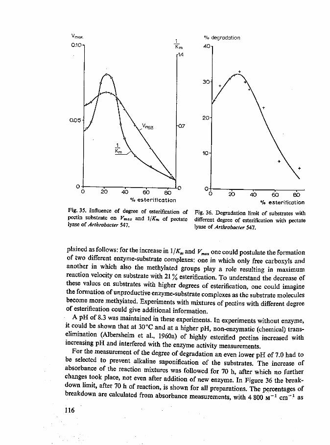
°A> degradation
40
0.05
40 60 80 °/o esterification
20 40 60 80 % esterification
Fig. 35. Influence of degree of esterification of Fig. 36. Degradation limit of substrates with pectin substrate on Vmax and 1/Km of pectate different degree of esterification with pectate lyase of Arthrobacter 547. lyase of Arthrobacter 547.
plained as follows: for the increase in l/Km and V^ one could postulate the formation of two different enzyme-substrate complexes: one in which only free carboxyls and another in which also the methylated groups play a role resulting in maximum reaction velocity on substrate with 21 % esterification. To understand the decrease of these values on substrates with higher degrees of esterification, one could imagine the formation of unproductive enzyme-substrate complexes as the substrate molecules become more methylated. Experiments with mixtures of pectins with different degree of esterification could give additional information.
A pH of 8.3 was maintained in these experiments. In experiments without enzyme, it could be shown that at 30°C and at a higher pH, non-enzymatic (chemical) trans-ehmmation (Albersheim et al., 1960a) of highly esterified pectins increased with increasing pH and interfered with the enzyme activity measurements.
For the measurement of the degree of degradation an even lower pH of 7.0 had to be selected to prevent alkaline saponification of the substrates. The increase of absorbance of the reaction mixtures was followed for 70 h, after which no further changes took place, not even after addition of new enzyme. In Figure 36 the breakdown limit, after 70 h of reaction, is shown for all preparations. The percentages of breakdown are calculated from absorbance measurements, with 4 800 M" 1 cm"1 as
116

the molar extinction coefficient. The degradation limit was also maximum for substrates with 21 to 26% esterification. The average degree of polymerization of the breakdown products of these preparations (21 to 26% esterified), calculated from the degradation limit, was three. As the absorbance measurements at 232 nm do not account for the unsaturated monomer, which is formed (Chapter 7), the actual degree of degradation may be somewhat higher for all preparations.
Hydrolysis limits for pectin preparations with different degrees of esterification were measured for polygalacturonase from Aspergillus niger by Roller & Neukom (1969) and for another polygalacturonase from a commercial pectinase preparation by Jansen & MacDonnell (1945). Both groups found that for these enzymes the hydrolysis limit decreased with increasing degree of esterification of preparations prepared by partial alkaline saponification of about 95 % esterified pectin. Therefore the pectate lyase of Arthrobacter 547 behaves differently in this respect from polygalacturonases.
To study the activity of other pectate lyases on substrates with different degrees of esterification I selected the enzyme of Arthrobacter 370 since the crude enzyme of this organism showed remarkably high activity on highly esterified pectin (Chapter 7). The enzyme was produced in a BPC medium which contained 75 % esterified pectin (Brown Ribbon, Obipektin AG) instead of pectate. The enzyme was purified by calcium phosphate gel treatment and DEAE-Sephadex chromatography. It contained some pectinesterase. Under conditions, such as maintained for lyase activity measurements, the pectinesterase activity was zero at pH 7.0 but catalysed the saponification reaction at pH 8.0 to such extent that its velocity became about twice that of the chemical saponification reaction alone. The enzyme was tested on a similar series of substrates as used for pectate lyase of Arthrobacter 547 at pH 8.3. During the short time of the assay no significant deesterification occurred. The V„„ and reciprocal Km values, plotted in Figure 37, show a very interesting picture. The l/Km curve has a very high maximum on 44% esterified pectin. This maximum l/Km value is 6 times higher than that of Arthrobacter 547 pectate lyase on 21 % esterified pectin, whereas for very low and very high esterified substrates the two enzymes show comparable \IKn values. Vmax shows a very broad maximum and is clearly little influenced by degree of esterification over a wide range of substrates (10 to 80% esterified). Degradation limits of substrates, obtained with Arthrobacter 370 enzyme, showed the same trend as those obtained with Arthrobacter 547 lyase: the maximum degradation limit was found with substrates between 21 and 33 % esterification.
From these results it is easily understood that the enzyme of Arthrobacter 370 may show higher viscosimetric and spectrophotometric activities on commercial high methoxyl pectin (DE = 75 %) than on commercial pectates. In this respect the enzyme is almost a perfect intermediate between pectin depolymerases and pectate depoly-merases. It remains to be seen whether the Arthrobacter 370 enzyme, which is a pectate lyase can split pectin to produce substrate fragments with a methylated carboxyl group at the newly formed non-reducing end as the fungal pectin lyases do. The kinetics of one of the lyase peaks (fraction II) obtained from a Baallus polymyxa
117

0.03
0.02
0.01-
20 40 60 80 % esterification
40 60 °/o esterif ication
Fig. 37. Influence of degree of esterification of pectin Fig. 38. Influence of degree of esterifica-substrate on Vmax and l/Km of pectate lyase of tion of pectin substrate on Vmax and Arthrobacter 370. 1/Km of a pectate lyase of Bacilluspolymyxa.
enzyme preparation, by chromatography on CM-Sephadex (Chapter 9) were also studied on differently esterified pectins. Experimental conditions were identical to those used for Arthrobacter enzymes. The curves obtained for Kmax and l/Km (Figure 38) were similar to those obtained with the Arthrobacter pectate lyases. Here the values of Vmax and l/Km were obtained from Hanes plots (Dixon & Webb, 1964). The [S]/v against [S1] plots were calculated by the linear regression technique with a computer (700 A/B Electronic Calculator, Wang Labs, Inc. Tewksbury Mass. USA). Correlation coefficients were all over 0.95 and most were over 0.98.
Vmax appeared to climb to a maximum on 26% esterified pectin, whereas the l/Km
curve showed a broad maximum over the substrate range of 21 to 31 % esterification. In contrast to 1/Km values of the two Arthrobacter pectate lyases, the l/Km value of this enzyme was not much higher on the substrates with an optimum degree of esterification than on very low esterified pectate.
The phenomenon of an optimum degree of esterification for the substrate is not restricted to Arthrobacter lyases alone but also occurs with a Bacillus polymyxa lyase and perhaps is a common characteristic of many or all pectate lyases. It remains to be seen how the action pattern may be influenced by pH and buffer systems. The role of highly esterified pectin on pectate breakdown should be studied to show its presumed
118
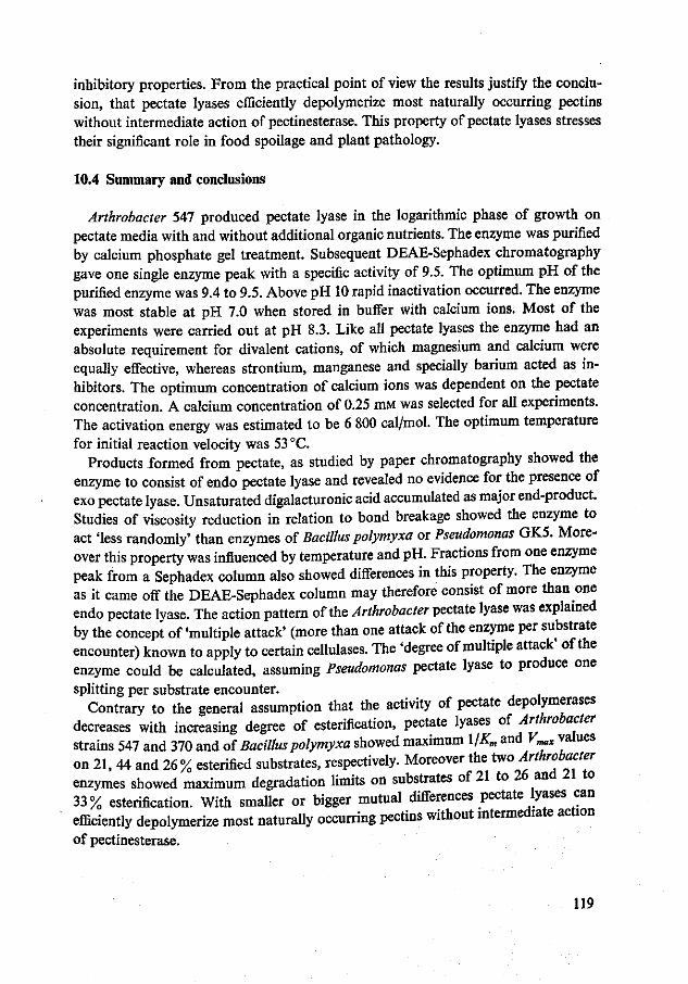
inhibitory properties. From the practical point of view the results justify the conclusion, that pectate lyases efficiently depolymerize most naturally occurring pectins without intermediate action of pectinesterase. This property of pectate lyases stresses their significant role in food spoilage and plant pathology.
10.4 Summary and conclusions
Arthrobacter 547 produced pectate lyase in the logarithmic phase of growth on pectate media with and without additional organic nutrients. The enzyme was purified by calcium phosphate gel treatment. Subsequent DEAE-Sephadex chromatography gave one single enzyme peak with a specific activity of 9.5. The optimum pH of the purified enzyme was 9.4 to 9.5. Above pH 10 rapid inactivation occurred. The enzyme was most stable at pH 7.0 when stored in buffer with calcium ions. Most of the experiments were carried out at pH 8.3. Like all pectate lyases the enzyme had an absolute requirement for divalent cations, of which magnesium and calcium were equally effective, whereas strontium, manganese and specially barium acted as inhibitors. The optimum concentration of calcium ions was dependent on the pectate concentration. A calcium concentration of 0.25 mM was selected for all experiments. The activation energy was estimated to be 6 800 cal/mol. The optimum temperature for initial reaction velocity was 53 °C.
Products formed from pectate, as studied by paper chromatography showed the enzyme to consist of endo pectate lyase and revealed no evidence for the presence of exo pectate lyase. Unsaturated digalacturonic acid accumulated as major end-product. Studies of viscosity reduction in relation to bond breakage showed the enzyme to act 'less randomly' than enzymes of Bacillus polymyxa or Pseudomonas GK5. Moreover this property was influenced by temperature and pH. Fractions from one enzyme peak from a Sephadex column also showed differences in this property. The enzyme as it came off the DEAE-Sephadex column may therefore consist of more than one endo pectate lyase. The action pattern of the Arthrobacter pectate lyase was explained by the concept of 'multiple attack' (more than one attack of the enzyme per substrate encounter) known to apply to certain cellulases. The 'degree of multiple attack' of the enzyme could be calculated, assuming Pseudomonas pectate lyase to produce one splitting per substrate encounter.
Contrary to the general assumption that the activity of pectate depolymerases decreases with increasing degree of esterification, pectate lyases of Arthrobacter strains 547 and 370 and of Bacillus polymyxa showed maximum l/Km and Vmax values on 21, 44 and 26% esterified substrates, respectively. Moreover the too Arthrobacter enzymes showed maximum degradation limits on substrates of 21 to 26 and 21 to 33% esterification. With smaller or bigger mutual differences pectate lyases can efficiently depolymerize most naturally occurring pectins without intermediate action of pectinesterase.
119

Summary
This study was set up to obtain, by isolation and screening of bacteria, a few strains that specifically produced pectate lyases, and to investigate the properties of a few of these enzymes, especially their mode of action on high polymer pectic substances. Afterwards these pure well characterized enzymes would -be available for further studies on vegetable spoilage, on technological processes involving pectolysis and on the structure of pectic substances.
Chapters 2 and 3 are literature reports. In Chapter 2, reference was made to pectic substances, particularly to their heteropolysaccharide and polyelectrolyte character. Depolymerizing pectic enzymes were reviewed in Chapter 3. Six different groups of enzymes were distinguished, according to Neukom: endo and exo polygalacturonases, endo and exo pectate lyases, endo polymethylgalacturonases and endo pectin lyases. The existence of endo polymethylgalacturonases was questioned. It was pointed out, that some enzymes have been described in literature which must be considered intermediates between pectate lyases and pectin lyases. It was therefore suggested that the scheme be revised in future.
Chapter 4 deals with culture media for detection and counting of pectolytic microorganisms. The suitability of Wieringa's pectin gel medium for total count of pectolytic bacteria was studied with pure cultures. A pectin gel medium with crystal violet, selective for Gram-negative pectolytic bacteria was developed. A counting medium for pectolytic yeasts and molds and a diagnostic medium for pectolytic Entero-bacteriaceae were described.
Fifty three strains of pectolytic bacteria, isolated from leafy vegetables, with Wieringa's pectin gel medium, were identified to the generic level in Chapter 5. Forty four strains, both fluorescent and non-fluorescent belonged to the genus Pseudomonas. Other genera represented were Xanthomonas, Flavobacterium, Achromobacter and Aerobacter. It was concluded that the pectolytic bacterial flora of leafy vegetables consists predominantly of Gram-negative rods, especially Pseudomonas.
The occurrence of pectolysis within the genus Arthrobacter was demonstrated in Chapter 6. Out of 240 strains, 32 were pectolytic. The pectolytic Arthrobacter strains originated from soils, activated sludge from dairy sewage and sea water. None of 58 strains of Brevibacterium from cheese were pectolytic.
A typing method for enzyme preparations of pectolytic bacteria was developed and described in Chapter 7. The enzymes of 19 strains of different bacterial genera and species were subjected to this typing scheme. All strains tested produced endo pectate lyase. Polygalacturonase was produced by strains of Bacillus and Erwinia only.
120
Pectinesterase was found with Bacillus polymyxa, most of the strains of Erwinia and all of Arthrobacter. The typing results also pointed towards the formation of pectin lyase by Erwinia aroideae and all strains of Arthrobacter. The unusual pattern of pectate degradation by Arthrobacter enzymes which was observed, could be caused by a mixture of exo and endo pectate lyase or by an intermediate type of pectate lyase.
In Chapter 8 the application of a typing procedure similar to that used in Chapter 7 to five commercial fungal 'pectinase' preparation was described. The substrates used were 0-1 % esterified pectate, 74 and 95 % esterified pectin (methylester) and 74 and 95 % esterified glycolester of pectate. Although all preparations could be shown to contain endo and exo polygalacturonase, pectin lyase and pectinesterase, large quantitative differences in the enzyme composition of the 'pectinases' were observed. The glycolesters were only partly depolymerized by all preparations apparently by their polygalacturonases only. The pectate and the pectins were rapidly degraded, but it was impossible to find out to what extent the joint action of pectinesterase and polygalacturonase and to what extent the competing action of pectin lyase caused de-polymerization of pectins. A corrected method for titrimetric pectinesterase activity measurements at low pH was given. The influence of pH on the activity of pectinesterase, purified from a commercial 'pectinase' preparation was measured with this titrimetric method.
A new method for the determination of the number average degree of polymerization of pectic substances was introduced in Chapter 9. The method was based on the experimentally observed linear increases of the reciprocal specific viscosity and the reciprocal degree of polymerization of pectates and pectins during enzymatic or chemical transeliminative degradation. A pectate lyase preparation of Bacillus polymyxa was used in the enzymatic method. It was found, however, that degrees of polymerization obtained with this enzyme were rather low when compared with values found with a membrane osmometer. By CM-Sephadex chromatography it appeared that the enzyme preparation consisted of at least three pectate lyases, with different endo characters. Since a fully random degradation of the substrate is a prerequisite for the determination of degrees of polymerization by this new method, the enzyme preparation of Bacillus polymyxa appeared to be less suitable. It could be replaced by the endo pectate lyase of a Pseudomonas strain.
A study of Arthrobacter pectate lyase is presented in Chapter 10. The enzyme of Arthrobacter 547 was purified by calcium phosphate gel treatment and DEAE-Sephadex chromatography. The purified enzyme had an optimum pH of 9.4 to 9.5, and was most stable at pH 7.0. The enzyme had an absolute requirement for divalent cations of which magnesium and calcium were most effective. The optimum concentration of calcium ions, which was slightly dependent on the pectate concentration, was approximately 0.25 mM. The activation energy was estimated to be 6 800 cal/moi. Products formed from pectate showed the enzyme to be an endo pectate lyase Studies of viscosity reduction in relation to bond breakage showed the enzyme to act 'less randomly' than those of Bacillus polymyxa and Pseudomonas. This behaviour was explained with the concept of 'multiple attack' of an enzyme molecule on a
121
substrate molecule. The 'degree of multiple attack' could be calculated, assuming Pseudomonas pectate lyase to produce one splitting per substrate encounter. Contrary to the general assumption that pectates are the best substrates for pectate lyases, enzymes of Arthrobacter strains 547 and 370 and of Bacillus polymyxa showed maximum l/Km and Vmax values on 21, 44 and 26% esterified pectins, respectively. The enzymes of Arthrobacter 547 and 370 also showed maximum degradation of these optimum substrates. It appeared that pectate lyases can efficiently depolymerize most naturally occurring pectins without intermediate action of pectinesterase.
122

References
Agate, A. D., M. H. Bilimoria & J. V. Bhat, 1962. Pectin transeliminase activity in Streptomyces
viridochromogenes. Curr. Sci. 31: 462-463. Albersheim, P., 1963. Auxin induced product inhibition of pectin transeliminase as shown by ozono-
lysis. Plant Physiol. 38: 426-429. Albersheim, P., 1966. Pectin lyase from fungi. Meth. Enzym. 8: 628-631. Albersheim, P., H. Neukom & H. Deuel, 1960a. Splitting of pectin chain molecules in neutral
solutions. Arch. Biochem. Biophys. 90: 46-51. Albersheim, P., H. Neukom & H. Deuel, 1960b. Uber die Bildung von ungesattigten Abbauprodukten
durch ein pektinabbauendes Enzym. Helv. chim. Acta 43: 1422-1426. Altermatt, H., 1954. Darstellung von Oligogalakturonsauren. Diss, ETH, Zurich. Amad6, R., 1970. Isolierung und Charakterisierung einer Pektin-Lyase von Aspergillus mger. Diss.
ETH, Ziirich, Nr 4536. Aspinall, G. O., 1970. Polysaccharides. Pergamon Press, Oxford, 228 pp. Aspinall, G. O., R. Begbie, A. Hamilton & J. N. C. Whyte, 1967a. Polysaccharides of soy-beans.
Part III. Extraction and fractionation of polysaccharides from cotyledon meal. J. chem. hoc. it,;,
1065-1070. Aspinall, G. O. & A. Canas-Rodriguez, 1958. Sisal pectic acid. J. chem. Soc., 4020-402/. Aspinall, G. O. & I. W. Cottrell, 1970. Lemon-peel pectin. II. Isolation of homogeneous pectins and
examination of some associated polysaccharides. Can. J. Chem. 48:1283 1289. _ Aspinall, G. O., I. W. Cottrell, S. V. Egan, I. M. Morrison & J. N. C. Whyte, 1967b. " * » * « £ »
of soy-beans. Part IV. Partial hydrolysis of the acidic polysaccharide complex from cotyledon
meal. J. chem. Soc. (Q, 1071-1080. umrtinnation Aspinall, G. O., I. W. Cottrell, J. A. Molloy & M. Uddin, 1970. Lemon-peel pectm III. Fract.onat.on
of pectic acids from lemon-peel and lucerne. Can. J. Chem. 48:1290-1295. . Aspinall, G. O., J. W. T. Craig & J. L. Whyte, 1968a. Lemon-peel pectin. Part I. Fractionation and
partial hydrolysis of water soluble pectin. Carbohyd. Res. 7: 442-452. Aspinall, G. O. & R. S. Fanshawe, 1961. Pectic substances from lucerne (Medusa sama). Part I.
Pectic acid. J. chem. Soc., 4215-4225. t,„,„_„»o fVnm lucerne Aspinall, G. O., B. Gestetner, J. A. Molloy & M. Uddin, 1968b Pectic sub stances ^ »
(Medicago sativa). Part II. Acidic oligosaccharides from partial hydrolysis of leaf and stem pectic acids. J. chem. Soc. (Q, 2554-2559. . p rt y Acidic
Aspinall, G. O., K. Hunt & I. M. Morrison, 1967c. Polysaccharides of soy-beans. Part V. Ac. polysaccharides from the hulls. J. chem. Soc. (Q, 1080-1086 . . ,,„ f r o m SUSnension-
Aspinall, G. O., J. A. Molloy & J. W. T. Craig, 1969. Extracellular polysaccharides from suspens cultured sycamore cells. Can. J. Biochem. 47: 1063-1070. nnmher-average degree
Banks, W. & C. T. Greenwood, 1968. A novel method of determining the number^average of polymerization of linear maltodextrins and amylose. C a r b ° ^ . . Geotrichum candidum.
Barash, I., 1968. Liberation of polygalacturonase dunng spore germination by Metric
Phytopathology 58: 1364-1371. . , aentrichum candidum. Barash, I. & Z. Eyal, 1970. Properties of a polygalacturonase produced by Geotrichum
Phytopathology 60: 27-30.
123
Barash, I. & S. Khazzam, 1970. The relationship and properties of pectic glycosidases produced by host and pathogen during anthracnose disease of avocado. Phytochemistry 9: 1189-1197.
Barras, D. R., A. E. Moore & B. A. Stone, 1969. Enzyme-substrate relationships among /S-glucan hydrolases. Adv. Chem. Ser. 95: 105-138.
Barrett, A. J. & D. H. Northcote, 1965. Apple fruit pectic substances. Biochem. J. 94: 617-627. Bateman, D. F., 1966. Hydrolytic and trans-eliminative degradation of pectic substances by extra
cellular enzymes of Fusarium solani f. phaseoli. Phytopathology 56: 238-244. Bateman, D. F. & R. L. Millar, 1966. Pectic enzymes in tissue degradation. Ann. Rev. Phytopath. 4:
119-146. BeMiller, J. N., 1967. Acid-catalyzed hydrolysis of glycosides. Adv. Carbohyd. Chem. 22: 25-108. Beyerinck, M. W. & A. van Delden, 1904. Over de bacterien welke bij het roten van vlas werkzaam
zijn. Versl. Kon. Akad. Wetensch., Wis.- Natuurk. Afd. 12: 673-693. Bhat, J. V., N. P. Jayasankar, A. D. Agate & M. H. Bilimoria, 1968. Microbial degradation of
pectic substances. J. sci. ind. Res. 27: 196-203. Bhattacharjee, S. S. & T. E. Timell, 1965. A study of the pectin present in the bark of amabilis fir
(Abies amabilis). Can. J. Chem. 43: 758-765. Bishop, C. T., 1955. Carbohydrates of sunflower heads. Can. J. Chem. 33: 1521-1529. Bouveng, H. D., 1965. Polysaccharides in pollen. II. The xylogalacturonan from mountain pine
(Pinus mugo Turra) pollen. Acta chem. Scand. 19: 953-963. Brisou, J., 1958. Etude de quelques Pseudomonadaceae. These de Sciences, Paris. Bryce, W. A. J. & C. T. Greenwood, 1957. The degradation of high polymers. J. Polym. Sci. 25:
480-483. Bush, D. A. & R. C. Codner, 1968. The nature of macerating factor of Penicillium digitatum Saccardo.
Phytochemistry 7: 863-869. Bush, D. A. & R. C. Codner, 1970. Comparison of the properties of the pectin transeliminases of
Penicillium digitatum and Penicillium italicum. Phytochemistry 9: 87-97. Buttiaux, R., H. Beerens & A. Tacquet, 1962. Manuel de Techniques bacteriologiques. Editions
Medicates Flammarion, Paris. Byrde, R. J. W. & A. H. Fielding, 1968. Pectin methyl-trans-eliminase as the maceration factor of
Sclerotinia fructigena and its significance in brown rot of apple. J. gen. Microbiol. 52: 287-297. Charley, V. L. S., 1969. Some advances in food processing using pectic and other enzymes. Chem. &
Ind., 635-641. Courtois, J. E., F. Percheron & M. J. Foglietti, 1968. Isolement et etude du mode d'action d'une
polygalacturonase d'insecte (Pyrrhocoris apterus). C.r.hebd. Seanc. Acad. Sci., Paris, Serie D, 266: 164-166.
Dave, B. A. & R. H. Vaughn, 1971. Purification and properties of a polygalacturonic acid trans-eliminase produced by Bacillus pumilus. J. Bact. 108: 166-174.
Davis, B. J., 1964. Disc electrophoresis. II. Method and application to human serum proteins. Ann. N.Y. Acad. Sci. 121: 404-427.
Derungs, R., 1958. Trennung von Oligogalakturonsauren an Anionenaustauschern. Diss, ETH, Zurich Nr. 2792.
Deuel, H., 1943. Pektin als hochmolekularer Elektrolyt. Mitt. Geb. Lebensmittelunters.u.Hyg. 34: 41-51.
Deuel, H., 1947. Ober Glycolester der Pektinsaure. Helv. chim. Acta 30: 1523-1534. Difco Supplementary Literature, 1962. Page 115. Difco Laboratories, Detroit. Dixon, M. & E. C. Webb, 1964. Enzymes. Academic Press, London. Doesburg, J. J., 1965. Pectic substances in fresh and preserved fruits and vegetables. iBVT-Communica-
tion No. 25. Institute for Research on Storage and Processing of Horticultural Produce, Wage-ningen, the Netherlands.
Dorey, M. J., 1959. Some properties of a pectolytic soil Flavobacterium. J. gen. Microbiol. 20:91-104. Dowson, W. J., 1957. Isolation of soft rot bacteria on Wieringa's pectate gel. Nature 179: 682.
124

Dye, D. W., 1960. Pectolytic activity in Xanthomonas. N.Z.J1 Sci. 3: 61-69. Echandi, E., S. D. Van Gundy & J. C. Walker, 1957. Pectolytic enzymes secreted by soft-rot bacteria.
Phytopathology 47: 549-552. Edstrom, R. D. & H. J. Phaff, 1964a. Purification and certain properties of pectin trans-eliminase.
J. biol. Chem. 239: 2403-2407. Edstrom, R. D. & H. J. Phaff, 1964b. Eliminative cleavage of pectin and of oligogalacturonide
methyl esters by pectin trans-eliminase. J. biol. Chem. 239: 2409-2415. Edwards, P. R. & W. H. Ewing, 1966. Identification of Enterobacteriaceae. Burgess Publ. Co.,
Minneapolis, p. 252. Endo, A., 1963a. Studies on pectolytic enzymes of molds. Part V. The fractionation of pectolytic
enzymes of Coniothyrium diplodiella (1). Agric. biol. Chem. 27: 741-750. Endo, A., 1963b. Studies on the pectolytic enzymes of molds. Part VI. The fractionation of pectolytic
enzymes of Coniothyrium diplodiella (2). Agric. biol. Chem. 27: 751-757. Endo, A., 1964a. Studies on pectolytic enzymes of molds. Part VIII. Purification and properties of
endo-polygalacturonase I. Agric. biol. Chem. 28: 535-542. Endo, A., 1964b. Studies on pectolytic enzymes of molds. Part IX. Purification and properties of
endo-polygalacturonase II. Agric. biol. Chem. 28: 543-550. Endo, A., 1964c. Studies on pectolytic enzymes of molds. Part X. Purification and properties of
endo-polygalacturonase III. Agric. biol. Chem. 28: 551-558. Endo, A., 1964d. Studies on pectolytic enzymes of molds. Part XL Purification and properties of
exo-polygalacturonase. Agric. biol. Chem. 28: 639-645. Endo, A., 1964e. Studies on pectolytic enzymes of molds. Part XII. Purification and properties of
pectinesterase. Agric. biol. Chem. 28: 757-764. Endo, A., 1965. Studies on pectolytic enzymes of molds. Part XVI. Mechanism of enzymatic clarifi
cation of apple juice. Agric. biol. Chem. 29: 229-238. Eshuis, A. F., 1970. Eigenschappen van pectinen en hun afhankelijkheid van de gevolgde extractie-
methode. M.Sc.-Thesis, Agricultural University, Dept Food Science, Wageningen, The Netherlands. Fish, V. B. & R. B. Dustman, 1945. The enzyme activities of Pectinol A on pectin and other sub
stances. J. Am. chem. Soc. 67: 1155-1157. . „ • • . • • Florkin, M. & E. H. Stotz (editors), 1965. Enzyme nomenclature. Comprehensrve Biochemistry,
Vol. 13, Elseviers Publ. Comp., Amsterdam. Flory, P. J., 1953. Principles of Polymer Chemistry. Cornell University Press, Ithaca, New York
Foglietti,PM. J. & F. Percheron, 1968. Etude chimique de la pectine de racines d'cdllet (Dianthus
caryophyllus L.). Carbohyd. Res. 7: 146-155. . . French, D. & J. F. Robyt, 1967. Multiple attack by alpha amylase. Int. Congr. Biochem. 7. Abstr.
nr F-188, p. 795. Frey-Wyssling, A., 1959. Die pflanzliche Zellwand. Springer Verlag, Berlin. Fuchs, A., 1965. The trans-eliminative breakdown of Na-polygalacturonate by Pseudomonas fluores
cens. Antonie van Leeuwenhoek 31: 323-340. „„„,«,„„,,• anH their Garibaldi, A. & D. F. Bateman, 1971. Pectic enzymes produced by Erwuua chrysanthem, and their
effectsonplanttissue.Physiol.Pl. Path. 1:25-40. foraees Gradel, C J [ . & B . A. Dehority, 1972. Fermentation of isolated pectin and pectin from intact forages
by pure cultures of rumen bacteria. Appl. Microbiol. 23: 332 340. Graham, D. C , 1958. Occurrence of soft rot bacteria in S o r t * | ^ £ ^ 2 i £ ^ e r i o r a t i o n o f
Gyllenberg, H., E. Eklund, M. Antila & U. Vartiovaara, 1960. C o n t ~ t , 0 ° ™ . ^ ^ 1 0 :
market milk. II. Significance of pseudomonads as deteriorating bactena. Acta agric Scand.
H £ £ . J. G., 1968. Degradation of pectic substances during pathogenesis by Fusarium solani
f.sp. cucurbitae. Phytopathology 58: 62-69. detection and enumera-Hankin, L., M. Zucker & D. C. Sands, 1971. Improved solid medium for the detection an
tion of pectolytic bacteria. Appl. Microbiol. 22: 205-209. 125

Hasegawa, S. & C. W. Nagel, 1962. The characterization of an ^-unsaturated digalacturonic acid. J. biol. Chem. 237: 619-621.
Hasegawa, S. & C. W. Nagel, 1966. A new pectic acid transeliminase produced exocellularly by a Bacillus. J. Fd Sci. 31: 838-845.
Hasegawa, S. & C. W. Nagel, 1967. Separation of a hydrolase from a pectic acid transeliminase in cell extracts of a Bacillus. Nature 213: 207-208.
Hasegawa, S. & C. W. Nagel, 1968. Isolation of an oligogalacturonate hydrolase from a Bacillus specie. Arch. Biochem. Biophys. 124: 513-520.
Hatanaka, C. & J. Ozawa, 1964. Enzymic degradation of pectic acid. Part I. Limited hydrolysis of pectic acids by carrot exopolygalacturonase. Agric. biol. Chem. 28: 627-632.
Hatanaka, C. & J. Ozawa, 1966a. Enzymic degradation of pectic acid. II. Chromatography of pectic substances on DEAE-cellulose columns. Ber. Ohara Inst, landw. Biol. 13: 89-102.
Hatanaka, C. & J. Ozawa, 1966b. Enzymic degradation of pectic acid. III. Sugar constituents of pectic acids. Ber. Ohara Inst, landw. Biol. 13: 103-109.
Hatanaka, C. & Ozawa, J., 1966c. Enzymic degradation of pectic acid. IV. Action of carrot exopolygalacturonase on the pectic acids prepared by saponification of pectin with alkali and with pectin esterase. Ber. Ohara Inst, landw. Biol. 13: 161-174.
Hatanaka, C. & J. Ozawa, 1966d. Enzymic degradation of pectic acid. V. Mode of action of fungal saccharifying polygalacturonase. Ber. Ohara Inst, landw. Biol. 13: 175-183.
Hatanaka, C. & J. Ozawa, 1969. Isolation of a new exopolygalacturonase producing digalacturonic acid from pectic acid. Agric. biol. Chem. 33: 116-118.
Hatanaka, C. & J. Ozawa, 1970. An oligogalacturonate transeliminase from Erwinia aroideae. Agric. biol. Chem. 34: 1618-1624.
Heemskerk, P. A. M., 1971. Schimmel pektine-esterase. Aktiviteitsmeting, produktie en zuivering van schimmel pektine-esterase, alsmede chemische en fysische eigenschappen van pektine in ver-band met verzeping. M.Sc.-Thesis, Agricultural University, Dept. Food Science, Wageningen, the Netherlands.
Heim, P. & H. Neukom, 1962. Alkalische Spaltung der Glycosidbindung in Methyl-digalacturonosid-dimethylester. Helv. chim. Acta 45: 1737-1738.
Heri, W., H. Neukom & H. Deuel, 1961. Chromatographische Fraktionierung von Pektinstoffen an Diathylaminoafhyl-Cellulose. Helv. chim. Acta 44: 1939-1944.
Hsu, E. J. & R. H. Vaughn, 1969. Production and catabolite repression of the constitutive poly-galacturonic acid frans-eliminase of Aeromonas liquefaciens. J. Bact. 98: 172-181.
Hugh, R. & E. Leifson, 1953. The taxonomic significance of fermentative versus oxidative metabolism of carbohydrates by various Gram negative bacteria. J. Bact. 66: 24-26.
IFU - Internationale Fruchtsaft-Union, 1964. Analysen Nr. 23, 6 pp. Jacobelli, G., 1953. Utilisation de milieux de culture gelifies par des substances pectiques. C. r. hebd.
Seanc. Acad. Sci., Paris, 236: 228-230. Jansen, E. F. & L. R. MacDonnell, 1945. Influence of methoxyl content of pectic substances on the
action of polygalacturonase. Arch. Biochem. 8: 97-112. Jayasankar, N. P. & P. H. Graham, 1970. An agar plate method for screening and enumerating
pectinolytic microorganism. Can. J. Microbiol. 16: 1023. Jones, G. E., 1950. A note on the preparation of a pectate gel medium. Proc. Soc. appl. Bact. 13:
170-173. Jones, G. E., 1956. A study of the pectolytic bacterial population in some farm water supplies. J.
appl. Bact. 19: 231-236. Jones, J. K. N. & W. W. Reid, 1954. The structure of the oligosaccharides produced by the enzymic
breakdown of pectic acid. Part I. J. chem. Soc. 1361-1365. Jones, J. K. N. & W. W. Reid, 1955. The structure of the oligosaccharides produced by the enzymic
breakdown of pectic acid. Part II. J. chem. Soc, 1890-1891. Joslyn, M. A., 1962. The chemistry of protopectin: A critical review of historical data and recent
developments. Adv. Fd Res. 11: 1-107.
126
Kaiser, P., 1961. Etude de l'activite pectinolytique du sol et d'autres substrats naturels. These de Sciences, Paris.
Kaji, A. & T. Okada, 1969. Purification and properties of an unusually acid-stable endo-polygalactu-ronase produced by Corticium rolfsii. Arch. Biochem. Biophys. 131: 203-209.
Katchalsky, A., N. Shavit & H. Eisenberg, 1954. Dissociation of weak polymeric acids and bases. J. Polym. Sci. 13: 69-84.
King, A. D. & R. H. Vaughn, 1961. Media for detecting pectolytic Gram-negative bacteria, associated with the softening of cucumbers, olives, and other plant tissues. J. Fd Sci. 26: 635-643.
Kiss, J., 1970. Neighboring group effect in the ^-elimination-reaction of some glycopyranosyl-(l ->4)-hexopyranosiduronates. Tetrahedron Lett. 23: 1983-1986.
Kiss, J. & K. Noack, 1971. ^-elimination in pentafuranuronates to give enol acetals. Carbohyd. Res. 16: 245-247.
Roller, A., 1966. Fraktionierung und Charakterisierung der pektolytischen Enzyme von Aspergillus niger. Diss, ETH, Zurich Nr. 3774.
Koller, A. & H. Neukom, 1967. Untersuchungen tiber die pektolytischen Enzyme von Aspergillus niger. Mitt. Geb. Lebensmittelunters.u. Hyg. 58: 512-517.
Koller, A. & H. Neukom, 1969. Untersuchungen tiber den Abbaumechanismus einer gereinigten Polygalakturonase aus Aspergillus niger. Eur. J. Biochem. 7: 485-489.
Kovacs, N., 1956. Identification of Pseudomonaspyocyanea by the oxidase reaction. Nature 178: 703. Launer, H. F. & Y. Tomimatsu, 1954. Reaction between chlorous acid and glucose; quantitative
stoichiometry and evaluation of reagent decomposition. Anal. Chem. 26: 382-386. Launer, H. F. & Y. Tomimatsu, 1959a. Stoichiometry of chlorite-aldehyde reactions; analytical
procedures. Anal. Chem. 31: 1385-1390. Launer, H. F. & Y. Tomimatsu, 1959b. Reaction of sodium chlorite with various polysaccharides;
rate studies and aldehyde group determinations. Anal. Chem. 31: 1569-1574.
Layne, E., 1957. Spectrophotometric and turbidimetric methods for measuring proteins. Meth.
Enzym. 3: 447-454. ... _ ,. Leclerc, H., 1964. Recherche de la pectine glucosidase chez quelques bacilles a Gram negatit. am.
Ass. Dipl. Microbiol., Pharm., Nancy, 93: 15-20. Lee, M., L. Miller & J. D. Macmillan, 1970. Similarities in the action patternof exopolygalac-
turonate lyase and pectinesterase from CI. multifermentans. J. Bact. 103: 595-600. Linker, A., K. Meyer & P. Hoffman, 1956. The production of unsaturated uron.des by bacterial
hyaluronidases. J. biol. Chem. 219:13-25. , _ Lund, B. M., 1968. A bacteriological study of stored, sulphite treated, peeled potatoes. J. appl. Bact.
Lund, B. M., 1969. Properties of some pectolytic yellow pigmented Gram negative bacteria, isolated
from fresh cauliflowers. J. appl. Bact. 32: 60-67. . . . q_~Q
Lund, B. M., 1971. Bacterial spoilage of vegetables and certain fruits. J. appl. Bact. n. y • Lynn, H. A., 1967. Alginic acid and mannuronic acid catabolism of an Arthrobacter soil .solate.
Ph. D.-Thesis, University of Georgia, USA. . J»*:„« n( alrinic Lynn, H. A., M. L. Webb, J. J. Farmer III & R. G. Eagon, 1968. Eliminate degradation of alginic
acid by microorganisms. Bull. Ga Acad. Sci. 26: 33-38. „,.uiUrmmtans Macmillan, J. D. & H. J. Phaff, 1966. Exopolygalacturonate lyase from Clostridium multifermentans.
Meth. Enzym. 8:632-635. . . O„„„„i,,<ra1acturonic Macmillan, J. D., H. J. Phaff & R. H. Vaughn, 1964. The pattern of action of an exopolygalacturonic
acid-^-eliminase from Clostridium multifermentans. Biochemistry 3: 57TO/»-Macmillan, J. D. & R. H. Vaughn, 1964. Purification and propert.es of a polygalacturonic
/m/w-eliminase produced by Clostridium multifermentans. Biochemistry 3. 3M- .. Mah, R. A. & R. E. Hungate, 1965. Physiological studies on the rumen abate. OphryoscolexpurKy
Stein. J. Protozool. 12: 131-136. rj iiVrirraft Biochem. Z. Malsch, L., 1941. Die Viskositat von Pektinlosungen und ihre Beziehung zur Gelierkraft. B.oc
309: 283-296. 127
Mannheim, C. H. & S. Siv, 1969. Occurrence of polygalacturonase in citrus fruit. Fruchtsaftindustrie 14: 98-103.
McClendon, J. H. & J. H. Kreisher, 1963. Chromatography on cellulose phosphate of polysaccharide hydrolases from fungi. Anal. Biochem. 5: 295-312.
McColloch, R. J. & Z. I. Kertesz, 1947. Pectic enzymes. VIII. A comparison of fungal pectin-methyl-esterase with that of higher plants, especially tomatoes. Arch. Biochem. 13: 217-229.
McDonnell, L. R., R. Jang, E. F. Jansen & H. Lineweaver, 1950. The specificity of pectinesterases from several sources with some notes on purification of orange pectinesterase. Arch. Biochem. 28: 260-273.
McNicol, L. A. & E. E. Baker, 1970. Pectate lyase and polygalacturonate lyase activity in a Vi antigen-degrading enzyme preparation. Biochemistry 9: 1017-1023.
Merck, E. (AG). Chromatographic - unter besonderer Beriicksichtigung der Papierchromatographie (Zweite Aufiage) E. Merck, Darmstadt.
Mill, P. J., 1966a. The pectic enzymes of Aspergillus niger. A mercury activated exopolygalacturonase. Biochem. J. 99: 557-561.
Mill, P. J., 1966b. The pectic enzymes of Aspergillus niger. A second exopolygalacturonase. Biochem. J. 99: 562-565.
Miller, L. & J. D. Macmillan, 1970. Mode of action of pectic enzymes. II. Further purification of exopolygalacturonate lyase and pectinesterase from Clostridium multifermentans. J. Bact. 102: 72-78.
Moran, F., S. Nasuno & M. P. Starr, 1968a. Extracellular and intracellular polygalacturonic acid trans-eliminases of Erwinia carotovora. Arch. Biochem. Biophys. 123: 298-306.
Moran, F., S. Nasuno & M. P. Starr, 1968b. Oligogalacturonide trans-eliminase of Erwinia carotovora. Arch. Biochem. Biophys. 125: 734-741.
Moran, F. & M. P. Starr, 1969. Metabolic regulation of polygalacturonic acid trans-eliminase in Erwinia. Eur. J. Biochem. 11: 291-295.
Morell, S., L. Baur & K. P. Link, 1934. The methylglycosides of the naturally occurring hexuronic acids. III. Polygalacturonic acid-methylglycosides derived from pectin. J. biol. Chem. 105: 1-13.
Mossel, D. A. A., J. Bechet & R. Lambion, 1962. La prevention des infections et des toxi-infections alimentaires. Cooperative d'£dition pour les Industries Alimentaires (CEPIA), Bruxelles (279 pp).
Mossel, D. A. A. & S. K. Tamminga, 1968. Methoden voor het microbiologisch onderzoek van ge-conserveerde en andere levensmiddelen. Conserva 17: 73.
Mount, M. S., D. F. Bateman & H. Grant Basham, 1970. Induction of electrolyte loss, tissue maceration and cellular death of potato tissue by an endopolygalacturonate trans-eliminase. Phytopathology 60: 924-931.
Mulder, E. G., A. D. Adamse, J. Antheunisse, M. H. Deinema, J. W. Woldendorp & L. P. T. M. Zevenhuizen, 1966. The relationship between Brevibacterium linens and bacteria of the genus Arthrobacter. J. appl. Bact. 29: 44-71.
Mulder, E. G. & J. Antheunisse, 1963. Morphologie, physiologie et ecologie des Arthrobacter. Ann. Inst. Pasteur 105: 46-74.
Mulder, E. G., J. Antheunisse & W. H. J. Crombach, 1971. Microbial aspects of pollution in the food and dairy industries. In: Sykes, G. & F. A. Skinner (editors): Microbial aspects of pollution, Academic Press, London, pp. 71-89.
Nagel, C. W. & M. M. Anderson, 1965. Action of a bacterial transeliminase on normal and unsaturated oligogalacturonic acids. Arch. Biochem. Biophys. 112: 322-330.
Nagel, C. W. & S. Hasegawa, 1967. Kinetics and site of attack of oligogalacturonic acids by an endo-pectic acid transeliminase. Arch. Biochem. Biophys. 118: 590-595.
Nagel, C. W. & R. H. Vaughn, 1961a. The characteristics of a polygalacturonase produced by Bacillus polymyxa. Arch. Biochem. Biophys. 93: 344-352.
Nagel, C. W. & R. H. Vaughn, 1961b. The degradation of oligogalacturonides by the polygalacturonase of Bacillus polymyxa. Arch. Biochem. Biophys. 94: 328-332.
128

Nagel, C. W. & T. M. Wilson, 1970. Pectic acid lyases of Bacillus polymyxa. Appl. Microbiol. 20:
374-383. Nasuno, S. & M. P. Starr, 1966a. Pectic enzymes of Pseudomonas marginalis. Phytopathology 56:
1414-1415. Nasuno, S. & M. P. Starr, 1966b. Polygalacturonase of Erwinia carotovora. J. biol. Chem. 241:
5298-5306. Nasuno, S. & M. P. Starr, 1967. Polygalacturonic acid franj-eliminase of Xanthomonas campestns.
Biochem. J. 104: 178-185. Neukom, H., 1960. Ober Farbreaktionen von Uronsauren mit Thiobarbitursaure. Chimia 14:
165-167. Neukom, H., 1963. Ober den Abbau von Pektinstoffen. Schweiz. landw. Forsch. 2: 112-122. Neukom, H., 1967. Pectic substances. Kirk-Othmer Encycl. Chem. Technol. 14: 636-651. Neukom, H., H. Deuel, W. J. Heri & W. Kiindig, 1960. Chromatographische Fraktionierung von
Polysacchariden an Cellulose-Anionenaustauschern. Helv. chim. Acta 43: 64-71. Ng, H. & R. H. Vaughn, 1963. Clostridium rubrum sp.n. and other pectinolytic Clostridia from soil.
J. Bact. 85: 1104-1113. . . Okamoto, K., C. Hatanaka & J. Ozawa, 1963. On the saccharifying pectolytic enzymes of Erwmia
aroideae. Agric. biol. Chem. 27: 596-597. ' . , Okamoto, K., C. Hatanaka & J. Ozawa, 1964a. A saccharifying pectate /nm-ehminase of Erwmia
aroideae. Agric. biol. Chem. 28: 331-336. Okamoto, K., C. Hatanaka & J. Ozawa, 1964b. Some properties of the saccharifying pectate trans-
eliminase of Erwinia aroideae. Ber. Ohara Inst, landw. Biol. 12: 107-114. Okamoto, K., C. Hatanaka & J. Ozawa, 1964c. The mechanism of action of the saccharifying pectate
transeliminase. Ber. Ohara Inst, landw. Biol. 12: 115-119. Okamoto, K., C. Hatanaka & J. Ozawa, 1965. The thiobarbituric acid test of 4,5-unsaturated di-
galacturonic acid. Ber. Ohara Inst, landw. Biol. 13: 7-12. Olson, H. C , 1961. Selective media for detecting spoilage organisms in dairy products. J. Dairy bci.
44:970-971. Olthof, E., 1955. Waarom ook zure augurken pasteuriseren? Conserva 4: 145-lM- . Ovodova, R. G. & Yu. S. Ovodov, 1969. The pectic substances of Zosteraceae. Part II. The structural
features of the pectic acid. Carbohyd. Res. 10: 387-390. 7„.t„aceae Ovodova, R. G., V. E. Vaskovsky & Yu. S. Ovodov, 1968. The pectic substances of Zosteraceae.
Carbohyd. Res. 6: 328-332. . . * n \ „ t ; n n * J Am Owens, a s . , H. Lotzkar, R. C. Merrill & M. Peterson, 1944. Viscos.t.es of pectin solut.ons. J. Am.
chem. Soc. 66: 1178-1182. . . , „--finic acid Owens, H. S., H. Lotzkar, T. H. Schultz & W. D. Maclay, 1946. S h a p e r t £ £***»* "*
molecules deduced from viscometric measurements. J. Am. chem. boc. b • p . R
Patel, D. S. & H. J. Phaff, 1960a. Studies on the purification of tomato polygalacturonase.
P a S D.16& H. J. Phaff, 1960b. Properties of purified tomato polygalacturonase FcI Res. 25:47-57. Paton, A. M., 1958. Pectin-decomposing strains of Pseudomonas. Nature 81. M-o•• Paton, A. M., 1959. An improved method for preparing pectate gels. N a t u r 1 8 8 2 81 Pee, W. van & J. M. Castelein, 1972. Study of the pectinolytic microflora^particularly
bacteriaceae, from fermenting coffee in the Congo. J. Fd Sci. . c } f r o m Saccha. Phaff, H. J., 1966. a-l,4-Polygalacturonide glycanohydrolase (endo-polygalacturo
romyc^/m^/w. Meth.Enzym. 8:636-641. p n z v men bei der Fruchtverarbeitung. Pilnik, W. & F. M. Rombouts,1972. Die Bedeutung von Enzymen
Dechema-Monographien 70:187-202. „,„nides In- Hulme, A. C. (editor): Pilnik, W. & A. G. J. Voragen, 1970. Pectic substances a n d ^ ^ S ^ ^ ' d o n .
The biochemistry of fruits and the* products. Vo l 1 . ^ 3 02_305> 343.346. Pilnik, W. & P. Zwiker, 1970. Pektine. Gordian 70. 20Z-ZU4, at.
129

Preiss, J., 1966. Bacterial alginate lyase. Meth. Enzym. 8: 641-644. Preiss, J. & G. Ashwell, 1962. Alginic acid metabolism in bacteria. I. Enzymatic formation of un
saturated oligosaccharides and 4-deoxy-L-er>'M/-o-3-hexoseulose uronic acid. J. biol. Chem. 237: 309-316.
Preiss, J. & G. Ashwell, 1963a. Polygalacturonic acid metabolism in bacteria. I. Enzymatic formation of 4-deoxy-L-rtreo-5-hexoseulose uronic acid. J. biol. Chem. 238: 1571-1576.
Preiss, J. & G. Ashwell, 1963b. Polygalacturonic acid metabolism in bacteria. II. Formation and metabolism of S-deoxy-d-^/ycero^.S-hexodiulosonic acid. J. biol. Chem. 238: 1577-1583.
Prunier, J. P. & P. Kaiser, 1964. Etude de l'activite pectinolytique chez des bacteries phytopathogenes et saprophytes des plantes. I. Recherche des enzymes pectinolytiques. Annls Epiphyt. 15: 205-219.
Put, H. M. C. & J. Th. Kruiswijk, 1964. Disintegration and organoleptic deterioration of processed strawberries caused by the mould Byssochlamys nivea. J. appl. Bact. 27: 53-58.
Rafikov, S. R., S. A. Pavlova & 1.1. Tverdokhlebova, 1964. Determination of molecular weights and polydispersity of high polymers. Israel Program for Scientific Translations Ltd., Jerusalem.
Rees, D. A., 1969. Structure, conformation and mechanism in the formation of polysaccharide gels and networks. Adv. Carbohyd. Chem. Biochem. 24: 267-332.
Rexova-Benkova, L. & A. Slezarik, 1966. Isolation of extracellular pectolytic enzymes produced by Aspergillus niger. Colin. Czech, chem. Commun. (Engl. Edn) 31: 122-129.
Richards, T. & M. T. A. Fouad, 1954. The use of glucose-inorganic salts media in the classification of the coli-aerogenes bacteria. II. The indole test and pectate gel liquefaction. J. appl. Bact. 17: P19-P20.
Roelofsen, P. A., 1953. Polygalacturonase activity in yeast, Neurospora and tomato extract. Biochim. biophys. Acta 10: 410-413.
Rohm & Haas GmbH, Darmstadt. Rohament P (Pectinglycosidase), Brochure, 8 pp. Rombouts, F. M., W. Norde, H. Oortwijn & W. Pilnik, 1970a. A method for the determination of
the number-average degree of polymerization in pectic substances, IUPAC, International Symposium on Macromolecules, Leiden, Abstract No. 11-41.
Rombouts, F. M., W. Norde & W. Pilnik, 1970b. A simple method for the determination of the number-average degree of polymerization in pectic substances. Lebensmittelwiss.u.Technol. 3: 94-97.
Rombouts, F. M. & W. Pilnik, 1971a. The occurrence of pectolysis within the genus Arthrobacter. Antonie van Leeuwenhoek 37: 247-250.
Rombouts, F. M. & W. Pilnik, 1971b. Pektolytische Enzyme in der Fruchtsaftindustrie. Fliissiges Obst 38: 93-98.
Rombouts, F. M. & W. Pilnik, 1972. Research on pectin depolymerases in the sixties; a literature review. Chem. Rubb. Comp. Crit. Rev. Fd Technol. 3: 1-26.
Rouse, A. H. & C. D. Atkins, 1955. Pectinesterase and pectin in commercial citrus juices as determined by methods used at the citrus experiment station. Techn. Bull, agric. Exp. Stns Univ. Fla 570.
Sabet, K. A. & W. J. Dowson, 1951. Action of phytopathogenic bacteria on pectate gel. Nature 168: 605.
Sands, D. C , M. N. Schroth & D. C. Hildebrand, 1970. Taxonomy of phytopathogenic pseudomonads. J. Bact. 101: 9-23.
Saverborn, S., 1945. A contribution to the knowledge of the acid polyuronides. Dissertation, Uppsala. Schmidt, H., 1959. Uber Thiobarbitursaure-Methin-Farbstoffe. Fette Seifen AnstrMittel 61: 881-886. Schriemer, W. H., 1970. Onderzoek naar methodieken voor de bepaling van ballaststoffen in pectine.
Report, Agricultural University, Dept. Food Science, Wageningen, The Netherlands. Schubert, E., 1952. Uber die Wirkung von Pektinase auf Apfelsaft und Pektinlosung. Schweiz.
Brau.-Rdsch. 63: 95-101. Schultz, T. H., H. Lotzkar, H. S. Owens & W. D. Maclay, 1945. Influence of method of deesterifica-
tion on the viscosity and acid behavior of pectinic acid solutions. J. phys. Chem. 49: 554-563. Sen Gupta, U. K. & C. V. N. Rao, 1963. The pectic acid from the pulp of jackfruit (Arthrocarpus
integrifolia). I. Methylation studies. Bull. chem. Soc. Japan 36: 1683-1688.
130
Sen Gupta, U. K. & A. Das, 1965. The pectic acid from the pulp of jackfruit (Arthrocarpus integrifolia). II. Bull. chem. Soc. Japan 38: 1074-1077.
Sharf, J. M. (ed.), 1966. Recommended methods for the microbiological examination of foods (p. 179). American Public Health Association Inc., New York.
Sherwood, R. T., 1966. Pectin lyase and polygalacturonase production by Rhizoctonia solani and other fungi. Phytopathology 56: 279-286.
Shrinivasan, R. R. & D. B. Sprinson, 1959. 2-keto-deoxy-D-arafo-heptonic acid 7-phosphate-synthe-tase. J. biol. Chem. 234: 716-722.
Skerman, V. D. B., 1967. A guide to the identification of the genera of bacteria (p. 258). The Williams & Wilkins Co., Baltimore.
Slezarik, A. & L. Rexova, 1967. Characterization of extracellular pectolytic enzymes of Monilia laxa. Biologia (Bratislava) 22: 407-413.
Smit, C. J. B. & E. F. Bryant, 1969. Changes in molecular weight of pectin during methylation with diazomethane. J. Fd Sci. 34: 191-193.
Smith, W. K., 1958. A survey of the production of pectic enzymes by plant pathogenic and other bacteria. J. gen. Microbiol. 18: 33-41.
Snoeren, Th. H. M., 1971. An investigation of the milk reactivity of pectin. M.Sc.-Thesis, Netherlands Institute of Dairy Science, Ede, and Agricultural University, Dept. Food Science, Wageningen, The Netherlands.
Solms, J. & H. Deuel, 1955. Uber den Mechanismus der enzymatischen Verseifung von Pektinstoffen. Helv. chim. Acta 38: 321-329.
Soloveva, T. F., L. V. Arsenyuk & Yu. S. Ovodov, 1969. Some structural features of Panax ginseng C. A. Mey pectin. Carbohyd. Res. 10: 13-18.
Speiser, R., C. H. Hills & C. R. Eddy, 1945. The acid behavior of pectinic acids. J. phys. Chem. 49:
328-343. Starr, M. P., 1947. The causal agent of bacterial root and stem disease of guayule. Phytopathology
37: 291-300. Starr, M. P. & F. Moran, 1962. Eliminative split of pectic substances by phytopathogenic soft-rot
bacteria. Science 135: 920-921. Starr, M. P. & S. Nasuno, 1967. Pectolytic activity of phytopathogenic Xanthomonads. J. gen. Micro
biol. 46: 425-433. Stewart, D. J., 1962. A selective-diagnostic medium for the isolation of pectinolytic organisms in the
Enterobacteriaceae. Nature 195: 1023. Stoddart, R. W., A. J. Barrett & R. W. Northcote, 1967. Pectic polysaccharides of growing plant
tissues. Biochem. J. 102: 194-204. . Szejtly, J., 1967. Allgemeine Gesetzmassigkeiten der Hydrolyse glykosidischer B.ndungen. btarKe
19" 145-152 Uchino, F., Y. Kurono & S. Doi, 1966. Crystallization of an endo-polygalacturonase from Aero-
cylindrium sp. Agric. biol. Chem. 30: 1066-1068. _ Vajda, A., 1957. Gewinnung und Eigenschaften von Oxypropylestern der Pektinsaure. D.ss. ETH,
Zurich Nr 2076. . . . . . .• .-„„* ™ th* Vas, K., M. Nedbalek, H. Scheffer & G. Kovacs-Proszt, 1967. Methodological invesfgations on the
determination of some pectic enzymes. Fruchtsaftindustne 12: 164-184; AMr^itm nf Vaughn, R. H., G. D. Balatsouras, G. K. York II & C. W. Nagel, 1957. Med.afor kbr tonoT
pectinolytic microorganisms associated with softening of cucumbers, olives and other plant
tissues. Fd Res. 22: 597-603. . ^ », n „ s, n K- Vnrk IT Vaughn, R. H., A. D. King, C. W. Nagel, H. Ng, R. E. I^vin, J D. MacmiUan & a K. Y « * n .
1969. Gram negative bacteria associated with sloughing, a softening of California npe olives.
J. Fd Sci. 34: 224-227. , , H r P a r ) f 1 9 7 2 Fermenting yeasts associated with Vaughn, R. H., K. E. Stevenson, B. A. Dave & H. C. Park, 19//. ™ ™ * '
softening and gas-pocket formation in olives. Appl. Microbiol. 23: 3lb-jzu.
131
Veldkamp, H., 1965. Isolation of Cytophaga and Sporocytophaga. Zentbl. Bakt. ParasitKde (le Abt.) Suppl. 1, 81-90.
Veldkamp, H., P. A. A. Venema, W. Harder & W. N. Konings, 1966. Production of riboflavin by Arthrobacter globiformis. J. appl. Bact. 29: 107-113.
Voragen, A. G. J., 1972. Characterization of pectin lyases on pectins and methyl oligogalacturonates. Doctoral Thesis, Agricultural University, Wageningen, The Netherlands.
Voragen, A. G. J. & W. Pilnik, 1970a. Pektindepolymerasen. I. Mitteilung. Einteilung der Pektin-sauredepolymerasen nach ihrer Wirkung auf oligomere Galakturonsauren. Z. Lebensmittelunters. u.Forsch. 142: 346-359.
Voragen, A. G. J. & W. Pilnik, 1970b. Pectin research in the sixties. Dt. Lebensmitt. Rdsch. 66: 325-329.
Voragen, A. G. J., F. M. Rombouts, M. Hooydonk & W. Pilnik, 1971a. Comparison of endgroup methods for the measurement of pectin degradation. Lebensmittelwiss.u. Technol. 4: 7-11.
Voragen, A. G. J., F. M. Rombouts & W. Pilnik, 1971b. The influence of the degree of esterification on the activity of pectin and pectate lyases. Lebensmittelwiss.u.Technol. 4: 126-128.
Waravdekar, V. S. & L. D. Saslaw, 1959. A sensitive colorimetric method for the estimation of 2-deoxy sugars with the use of the malonaldehyde-thiobarbituric acid reaction. J. biol. Chem. 234: 1945-1950.
Warren, L., 1960. Thiobarbituric acid spray reagent for deoxy sugars and sialic acids. Nature 186: 237. Weeks, O. B., 1969. Problems concerning the relationships of cytophagas and flavobacteria. J. appl.
Bact. 32: 13-18. Weissbach, A. & J. Hurwitz, 1959. The formation of 2-keto-3-deoxyheptonic acid in extracts of
Escherichia coli B. J. biol. Chem. 234: 705-709. Wilson, T. M., 1968. The purification and kinetics of the transeliminases of Bacillus polymyxa.
Ph.D.-Thesis, Washington State University, USA. Wieringa, K. T., 1949. A method for isolating and counting pectolytic microbes. Int. Congr. Microbiol.
4 (Copenhagen, 1947): 482. Wieringa, K. T., 1953. On the dissimilation of pectic substances in nature. Int. Congr. Microbiol. 6
(Italy, 1953): 281-284. Wieringa, K. T., 1954. De verspreiding van pectine-ontledende micro-organismen in de natuur.
Landbouwk. Tijdschr. 66: 292-301. Wieringa, K. T., 1956. The microorganisms decomposing pectic substances in the dew retting process
of flax. Neth. J. agric. Sci. 4: 204-209. Yamasaki, M., A. Kato, S.-Y. Chu & K. Arima, 1967. Pectic enzymes in the clarification of apple
juice. Part II. The mechanism of clarification. Agric. biol. Chem. 31: 552-560. Yamasaki, M., T. Yasui & K. Arima, 1964. Pectic enzymes in the clarification of apple juice. Part I.
Study on the clarification in a simplified model. Agric. biol. Chem. 28: 779-787. Yamasaki, M., T. Yasui & K. Arima, 1966. Studies on pectic enzymes of microorganisms. Part III.
Endo-polygalacturonase of Aspergillus saito. Agric. biol. Chem. 30: 1119-1128. Young, R. J. & M. E. Corden, 1964. Paper chromatography of galacturonic acids to determine poly
galacturonase activity. Biochim. biophys. Acta 83: 124-126. Zitko, V. & C. T. Bishop, 1965. Fractionation of pectins from sunflowers, sugar beets, apples and
citrus fruits. Can. J. Chem. 43: 3206-3214. Zitko, V. & C. T. Bishop, 1966. Structure of a galacturonan from sunflower pectic acid. Can. J.
Chem. 44: 1275-1282. Zucker, M. & L. Hankin, 1970. Regulation of pectate lyase synthesis in Pseudomonas fluorescens and
Erwinia carotovora. J. Bact. 104: 13-18.
132