Occurrence of microplastic particles in marine mammals from ...
192
University of Veterinary Medicine Hannover Occurrence of microplastic particles in marine mammals from German waters and improvement of sample storage Mikroplastikpartikel in marinen Säugetieren aus deutschen Gewässern und Etablierung einer geeigneten Probenaufbewahrung INAUGURAL – DISSERTATION in fulfilment of the requirements for the degree of - Doctor rerum naturalium - (Dr. rer. nat.) submitted by Carolin Philipp Bremerhaven Büsum 2021
-
Upload
khangminh22 -
Category
Documents
-
view
2 -
download
0
Transcript of Occurrence of microplastic particles in marine mammals from ...
University of Veterinary Medicine Hannover
Occurrence of microplastic particles
in marine mammals from German waters
and improvement of sample storage
Mikroplastikpartikel in marinen Säugetieren aus deutschen Gewässern und
Etablierung einer geeigneten Probenaufbewahrung
INAUGURAL – DISSERTATION
in fulfilment of the requirements for the degree of
- Doctor rerum naturalium -
(Dr. rer. nat.)
submitted by Carolin Philipp Bremerhaven
Büsum 2021

Scientific supervisor: Prof. Prof. h. c. Dr. Ursula Siebert
Institute for Terrestrial and Aquatic Wildlife Research,
University of Veterinary Medicine Hannover, Foundation.
Prof. Dr. rer. nat. Dieter Steinhagen
Institute of Parasitology, Fish Disease Research Unit,
University of Veterinary Medicine Hannover, Foundation.
First supervisors: Prof. Prof. h. c. Dr. Ursula Siebert
Institute for Terrestrial and Aquatic Wildlife Research,
University of Veterinary Medicine Hannover, Foundation.
Prof. Dr. rer. nat. Dieter Steinhagen
Institute of Parasitology, Fish Disease Research Unit,
University of Osnabrück.
Second examinor: Assoc. Prof. Dr. Marcus Schulz
Department of Mathematics / Informatics
University of Veterinary Medicine Hannover, Foundation.
Date of oral examination: 14.05.2021

Parts of this study were published in a peer reviewed journal:
Philipp, C., Unger, B., Fischer, E. K., Schnitzler, J. G., and Siebert, U. (2020). Handle with
Care—Microplastic Particles in Intestine Samples of Seals from German Waters.
Sustainability, 12, 10424. doi:10.3390/su122410424
Philipp, C., Unger, B., Ehlers, S.M., Koop, J.H.E., Siebert, U. (2021). First Evidence of
Retrospective Findings of Microplastics in Harbour Porpoises (Phocoena phocoena) From
German Waters. Frontiers in Marine Science, 8, Article 682532.
doi: 10.3389/fmars.2021.682532
Results, or parts of the results of this study were published in reports:
Philipp, C., Unger, B., Siebert, U. (2021). Impacts of marine litter on biota – Marine
Mammals. in Assessment and implementation of long-term monitoring of pollution of diverse
marine compartments and biota with marine litter, final report on behalf of the German
Environment Agency, FKZ 3717 25 225 0
Groß, S., Philipp., C., Siebert, U. (2020). Untersuchungen zum Massensterben von
Trottellummen (Uria aalge) und Tordalken (Alca torda) Anfang 2019. Report to the
Ministry of Energy, Agriculture, the Environment, Nature and Digitalization and the Agency
for Coastal Defence, National Park and Marine Conservation Schleswig-Holstein.

Results of this study were presented on the following conferences:
Philipp, C., Unger, B., Fischer, E., Siebert, U. (2020). Proof of the invisible ones –
Microplastic burden in marine mammals from the German North and Baltic Seas. In: talk,
MICRO2020, Lanzarote and Beyond, virtually conference, 23rd to 27th of November, 2020,
abstract book p. 345. https://www.micro.infini.fr/IMG/pdf/micro2020proceedingsbook.pdf
Philipp, C., Unger, B., Fischer, E., Siebert, U. (2019). Microplastics in marine top predators
– A pilot study from German waters. In: poster, World Marine Mammal Conference,
Barcelona, Spain 9th to 12th of December, 2019, abstract book p. 557.
https://www.wmmconference.org/wp-content/uploads/2020/02/WMMC-Book-of-Abstracts-
3.pdf
Philipp, C., Unger, B., Hillmann, M., Siebert, U. (2018). Pilot study on possible micro plastic
occurrence in marine mammals from German waters. In: poster, MICRO 2018, Lanzarote,
Spain, 19th to 23rd of November, 2018, abstract book, p. 348.
https://micro2018.sciencesconf.org/data/pages/micro2018_proceedings_book_1.pdf

For my Parents with Love & Gratitude


“Cherish the natural world,
because you’re a part of it and you depend on it.”
Sir David Attenborough


Table of Contents
List of Abbreviations ................................................................................................................ 9
Introduction ............................................................................................................................ 17
I. History of Plastics ........................................................................................................ 17
II. Production of Plastics ................................................................................................. 18
III. Facing the Mankind Problem .................................................................................... 19
IV. From Macro- to Nanoplastics ................................................................................... 21
V. Marine Litter .............................................................................................................. 22
V.I. Impacts of Marine Litter to the Marine Environment .......................................... 25
V.II. Microplastics and marine mammals .................................................................... 26
VI. Identifying Microplastic Particles ............................................................................ 28
VI.I. Collection techniques .......................................................................................... 29
VI.II. Removing Biogenic Matter ................................................................................ 30
VI.III. Identification of Microplastics .......................................................................... 31
VII. Target species in the German North and Baltic Seas .............................................. 32
VIII. Aims of the Thesis ................................................................................................. 35
Chapter I: Handle with Care – Microplastic Particles in Intestine Samples of Seals from
German Waters ...................................................................................................................... 41
Abstract ........................................................................................................................... 41
Introduction ..................................................................................................................... 42
Materials and Methods .................................................................................................... 43
Results ............................................................................................................................. 50

Discussion ....................................................................................................................... 56
Conclusion ...................................................................................................................... 60
Acknowledgements ......................................................................................................... 61
Supplementary Material .................................................................................................. 62
1. Preparation .................................................................................................................. 62
1.1 Pre-trials ................................................................................................................. 62
1.2 Working in an Acrylic Hood .................................................................................. 64
2. Intestinal and Faecal Samples ..................................................................................... 64
2.1 New design of the washing bags ............................................................................ 64
2.2 Prevention of overcounting .................................................................................... 65
3. Identification Catalogue .............................................................................................. 66
3.1 Categorization of parameters ................................................................................. 73
4. Results ......................................................................................................................... 75
Chapter II: First Evidence of Retrospective Findings of Microplastics in Harbour
Porpoises (Phocoena phocoena) From German Waters ..................................................... 83
Abstract ........................................................................................................................... 83
Introduction ..................................................................................................................... 84
Materials and Methods .................................................................................................... 85
Results ............................................................................................................................. 88
Discussion ....................................................................................................................... 98
Conclusion .................................................................................................................... 104
Acknowledgements ....................................................................................................... 106
Supplementary Material ................................................................................................ 107
Overall Discussion ................................................................................................................ 111
Overall Conclusion ............................................................................................................... 131
Outlook .................................................................................................................................. 135
Summary ............................................................................................................................... 137
Zusammenfassung ................................................................................................................ 139
Bibliography ......................................................................................................................... 141
List of Figures ....................................................................................................................... 183
List of Supplementary Figures ............................................................................................ 185
List of Supplementary Tables ............................................................................................. 186
Acknowledgements ............................................................................................................... 187
Curriculum Vitae ................................................................................................................. 191


List of Abbreviations
List of Abbreviations In addition to the commonly used abbreviations following short forms were used:
ALDFG Abandoned, lost or otherwise discarded fishing gear
ATR Attenuated total reflectance
BS Baltic Sea
CTD Conductivity, temperature and pressure of seawater (depth) measurement device
DDT Dichlorodiphenyltrichloroethane
ECHA European Chemicals Agency
EEA European Environment Agency
EU European Union
FTIR Fourier-transform infrared spectroscopy
GC-MS Gas chromatograph mass spectrometry
GES Good Environmental Status
GIT Gastrointestinal tract
GPGP Great Pacific Garbage Patch
H2O2 Hydrogen peroxide
HDPE High density polyethylene
HELCOM Helsinki Commission
Hg Grey seal (Halichoerus grypus)
HNO3 Nitric acid
HOC Hydrophobic organic contaminants
ICES International Council for the Exploration of the Sea
IMO International Maritime Organization
ITAW Institute for Terrestrial and Aquatic Wildlife Research
KOH Potassium hydroxide
LDC London Dumping Convention
LDPE Low density polyethylene
LLDPE Linear low density polyethylene
MARPOL International Convention for the Prevention of Pollution from Ships (Marine
Pollution)

List of Abbreviations
MSFD Marine Strategy Framework Directive
MP Microplastic
MPA Marine Protected Areas
MPW Mismanaged plastic waste
NaCl Sodium chloride
NS North Sea
OSPAR The convention for the Protection of the Marine Environment of the North-East
Atlantic, which was concluded in Oslo (1972) and updated in Paris (1992) is
managed by the OSPAR Commission.
PA Polyamide
PCB Polychlorinated biphenyls
PE Polyethylene
PEST Polyester
PET Polyethylene terephthalate
PFAS Per- and polyfluoroalkyl substances
POP Persistent organic pollutants
PP Polypropylene
PS Polystyrene
Pv Harbour seal (Phoca vitulina)
PVC Polyvinyl chloride
SAC Special Area of Conservation
SCOS Special Committee on Seals
SH Schleswig-Holstein
TiHo Stiftung Tierärztliche Hochschule Hannover (University of Veterinary
Medicine Hannover, Foundation)
TNR True negative rate
TPR True positive rate
UBA Federal Environment Agency (Umweltbundesamt)
UN United Nations
UNEP United Nations Environmental Programme
WWTP Wastewater treatment plants
Introduction

Introduction
17
Introduction
I. History of Plastics
The history of synthetic polymers reaches back to the early 19th century, when Bakelite was
invented in 1909 (Baekeland, 1909). This discovery could be assumed as the inception of the
‘Plastic Age’ (Yarsley and Couzens, 1945; Thompson et al., 2009), and as a benchmark of the
so-called Anthropocene (Lewis and Maslin, 2015). Firstly invented for the military, the
industrialised fabrication of synthetic polymers conquered the everyday life and businesses of
the western countries in the 1950’s, based on its miscellaneous qualities (Life Magazine, 1955;
Andrady and Neal, 2009; Sperling, 2011; Geyer et al., 2017). Synthetic polymers are
characterised by plasticity, dye ability, lightness, heat-, cold- and weather-resistance. In
addition, they are characterised by low production costs and thus meet the needs in almost all
bygone and recent industries until today (Andrady and Neal, 2009). Since this time, plastic has
become an indispensable part of the everyday life resulting in a global production of almost
400 million tonnes in 2019 (PlasticsEurope, 2020).
As a consequence, the rising plastic production cause a global litter issue (Geyer et al., 2017),
based on high consume and low decomposition properties (Barnes et al., 2009; Andrady, 2015).
Geyer et al. (2017) estimated the weight of all ever produced plastic resin until 2015 of 7,800
tonnes, whereof 60% are not in use anymore and are located in landfills. However, the plastic
production increased steeply since the late 2000’s, thus 12,000 tonnes of plastic waste will be
combusted and additionally 12,000 tonnes are supposed to end in landfills by 2050 (Geyer et
al., 2017).
Back in the early 20th century, synthesised materials were produced and appreciated for their
long durability. Nowadays those qualities are taken for granted, but the consequences of this
durability is poorly understood (Galgani et al., 2000; Andrady, 2015). To overcome the still
increasing utilisation of single-used plastic items, the European Commission enacted a
legislation (Environment and Public Health and Food Safety, 2019). This regulation will
specifically ban single-used items and force the recycling and reusing of all produced packaging
material by 2030. Furthermore, the European Chemicals Agency (ECHA) proposed some
microplastic reduction and restriction approaches to decrease the intentional utilisation (ECHA,
2019a). This proposal was submitted to the European Commission, and numerous EU Member

Introduction
18
States already apply some of the required restrictions (ECHA, 2019b). Nevertheless, especially
in the medical industry, synthesised materials are needed for single-use objects like syringes or
gloves, and are thus indispensable (Lebreton and Andrady, 2019; Patrício Silva et al., 2020).
II. Production of Plastics
Polyethylene (PE; incl. LDPE, LLDPE and HDPE) and polypropylene (PP) have the highest
demand in Europe (PlasticsEurope, 2020). These thermoplastics dominate the plastic use (PE:
29.8%; PP: 19.4%), besides polyethylene terephthalate (PET; 7.9 %), polystyrene (PS; 6.2 %)
and polyvinyl chloride (PVC; 10%) (PlasticsEurope, 2020). In addition, the global production
of PE and PP is steadily growing and is mostly used as packaging material (Gregory and
Andrady, 2004; PlasticsEurope, 2020). Thus, it is not surprisingly that packaging items are the
most frequently found items in landfilling, beach litter or marine litter surveys, due to the fact
that it is used only for a short duration (OSPAR Commission, 2000, 2020; Widmer and
Hennemann, 2010; Schulz et al., 2013; Lebreton and Andrady, 2019; PlasticsEurope, 2020). In
42% of all international produced packaging items, the manufacturing was done by the
utilisation of synthesised microplastic resin (Geyer et al., 2017).
Industrially synthesised small-sized plastic particles are produced for a variety of industries or
for the production of plastic items, melted and moulded on site of application (Galgani et al.,
2015; Karlsson et al., 2018). This so-called primary plastic, plastic resin or further described as
virgin pellets is categorised by its shape as nurdles, pellets or beads, which gives further
information on the application (Karlsson et al., 2018). The usage of beads in facial cleaners and
other cosmetics facilitating the foam or peeling qualities increased since the 1990’s (Zitko and
Hanlon, 1991; Gregory, 1996; Fendall and Sewell, 2009). In addition, these microplastics are
also utilised for grinding purposes (Browne et al., 2011), e.g. in the shipyard industry replacing
sand in sandblasters for removing old paintings or biofouling growth. Nurdles or beads are often
used for manufacturing of new plastic objects, whilst melting down and shaped into a larger
plastic item, whereof PE and PP are also the dominating polymers (PlasticsEurope, 2020).
The transportation via containers and the manufacturing and utilisation on site of virgin pellets
is easier and cheaper based on their small size and low weight (Hopewell et al., 2009; Karlsson
Introduction
19
et al., 2018; Ackerman et al., 2019). Based on a high demand and the global transport ways to
the production sites, occurring spills have to be taken into account as point source for
microplastics (Karlsson et al., 2018).
III. Facing the Mankind Problem
The increased requirements result in an ascending plastic production. As consequence mankind
faces the problem of an increased waste disposal, enhanced by a growing world population
(Jambeck et al., 2015; Lebreton et al., 2017). Especially the removal of synthesised
microplastics in personal care products and lost fibres originating from washed cloths are
difficult to remove in wastewater treatment plants (WWTP) (Bretas Alvim et al., 2020). There
is still no unified, efficient and practicable approach to remove all small-sized particles out of
wastewater from WWTP (Prata, 2018; Sun et al., 2019), although this issue is also known since
the 1990s (Zitko and Hanlon, 1991; Gregory, 1996). Thus, microplastics are still present in the
released water or the sewage sludge (Dubaish and Liebezeit, 2013; Dris et al., 2015; Weithmann
et al., 2018; Corradini et al., 2019). Further, the sludge is often used as field fertilisers,
introducing the microplastics into the environment (Nizzetto et al., 2016b). Several studies
already identified the microplastic burden in crop fields and agricultural soils (Weithmann et
al., 2018; Corradini et al., 2019; Crossman et al., 2020).
The reprocessing of older and unused plastic items is still complicated. Different polymer
networks, additives, toxic substances, and impureness by e.g. household waste, inhibit the
recycling process (Hopewell et al., 2009). Moreover, the only effective and progressive
approach of the removal of already used and manufactured plastics are a disruptive thermal
treatment like incineration (Geyer et al., 2017). Moreover, combustion processes generate
energy and a chemical feedstock, thus saving fossil fuels (Al-Salem et al., 2017; Anuar
Sharuddin et al., 2017). To further successfully decrease the plastic input into the environment
the primary production has to be reduced, which can be decreased by the development of
feasible recycling methods (Geyer et al., 2016). However, recycled plastic polymers for
example PE or PP have the same or similar traits as the virgin ones (Achilias et al., 2007).

Introduction
20
It has to be taken into account that all plastic items ever produced are everlasting thus remain
in the environment, either fractured or in one piece (Thompson et al., 2005). Nevertheless, the
recovering of plastic waste into a recycling loop, ensures a variety of economic benefits like
the conserving of emissions and fossil fuels, next to the protection of the environment
(Hopewell et al., 2009).
Areas with a highly anthropogenic frequency are stressed by intense plastic production,
consume and litter properties (Jambeck et al., 2015). The intentional or the unintentional
littering is supported by the missing of professional dumpsites in mostly developing countries,
which especially in open landfills causes spreading of litter items by wind and rain (Ryan et al.,
2009; Jambeck et al., 2015; Sharma et al., 2019; Nanda and Berruti, 2020). The mishandling
and mismanagement of plastic waste (MPW) is further increasing, unless the prevention of
waste or the recycle processes are improving (Lebreton and Andrady, 2019). Thus, the global
MPW results in approximately 80 million tonnes in 2015, that is roughly half of the produced
amount (47%) (Lebreton and Andrady, 2019). In addition, a recent study of Borelle et al. (2020)
estimated a global influx of 19 to 23 million tonnes of plastic (11% of produced plastic waste)
reaching the marine and limnic environment in 2016. It furthermore cannot be ruled out that
this is an underestimation, since primary microplastics and abandoned fishing gear were not
considered.
The diverse circumstances of landfilling, affect even uninhabited or remote areas, like the Alps,
both Polar regions, distant islands or the deep sea, and even the Marianas Trench is burdened
by marine litter (Slip and Burton, 1991; Barnes, 2005; Eriksen et al., 2014; Tekman et al., 2017;
Chiba et al., 2018; Bergmann et al., 2019; Parolini et al., 2021).Thus, it is not surprising that
litter was shown to impact wildlife. The harmful characteristics were already confirmed in
several studies for different species in the aquatic environment in the last decade (Fossi et al.,
2020; Kühn and van Franeker, 2020), whereas scientific information on terrestrial species is
still lacking (Lu et al., 2019; Kolenda et al., 2021). Although the harmful risk of persistent
plastics was already addressed in the late 1980’s (Bean, 1987).
Introduction
21
IV. From Macro- to Nanoplastics
Plastic items can be categorised by their size. The term macro-litter describes items which are
between 20 mm and 15 cm in size (Gregory and Andrady, 2004), whereas meso-litter is used
for particles in a size range of 5 to 10 mm (Gregory and Andrady, 2003). Gregory and Andrady
(2003) further relate to micro-litter, when a particle measures a size between 67 and 500 µm.
Most studies refer to micro-litter or microplastics when investigating particles smaller than 5
mm (Moore, 2008; Arthur et al., 2009; Fendall and Sewell, 2009). For clarifying, this
dissertation follows the term microplastic describing particles smaller than 5 mm. Until today,
there is no formal term for nano-litter or nanoplastics (Koelmans, 2019). Thus, for classifying
those particles, the attributes and qualities of nanomaterials are used (Klaine et al., 2012).
Further definitions for narrowing down nanoplastic are given by Wagner et al. (2014)
describing particles smaller than 20 µm, and Klaine et al. (2012) categorise nanoplastics if the
particle size is less than 100 nm.
The definitions often refer to already given size ranges in plankton quantification systems.
Particles between 5 mm and 333 µm in size were initially called microplastics, based on the
common mesh sizes of used plankton nets (Arthur et al., 2009). This is similar to nanoplastics,
were the size limit of nanoplankton is set (Sieburth et al., 1978).
Next to primary plastics as described in the former chapter, secondary microplastics is another
form of small-sized particles. Those ones emerges if larger plastic items crack into smaller
pieces due to different physical forces. Especially, weathering processes promote the brittleness
of plastic items. Several laboratory and field studies investigate the degradation of plastic under
different circumstances. In particular UV-radiation, the physical abrasion by wind and waves,
or chemical reactions like oxidation, are confirmed to force the decaying process (Andrady,
1990, 2011). Whereas seawater and biofouling inhibit the ageing process by protecting the
plastic for overheating, UV-radiation and physical abrasion (Andrady, 1990).
Thus, it is not surprising, that microplastics seem to be present and accumulate in the world
oceans since more than 50 years (Carpenter et al., 1972; Thompson, 2004; Geyer et al., 2017).
Based on the concerns of microplastics, the research increased steeply in the last decade
(Cowger et al., 2020).Whereas, knowledge on the presence and health effects of nanoplastic is
still lacking (Paul et al., 2020). Certainly, based on the physical characteristics, nanoplastic

Introduction
22
particles aggregate in water or air to larger clusters, adsorbing pollutants and biogenic material
(Ward and Kach, 2009; Andrady, 2011; Song et al., 2014; Yu et al., 2019). In addition, these
small-sized particles are known to be cell-permeable, thus open up a new concern in
accumulation of pollutants and its health effects in organisms (Jani et al., 1989; LeFevre et al.,
1989; Lehner et al., 2019; Li et al., 2020b; Sarasamma et al., 2020).
V. Marine Litter
When focussing on the marine environment, found garbage is often called marine litter. The
United Nations Environmental Programme (UNEP) is giving the following definition of those
items: “[…] marine litter is defined as any persistent, manufactured or processed solid material
discarded, disposed of or abandoned in the marine and coastal environment (UNEP, 2009).
Pasternak et al. (2017) further proposed: “Marine debris is a consequence of poor or inadequate
solid waste management practices […]”. The entering of debris into the seas is aided by the
transport of rivers, drainage or sewage systems or the wind as proposed by the Marine Strategy
Framework Directive (MSFD) (Galgani et al., 2010). In addition, ecological calamites like
flooding events e.g. tsunamis or river discharges have to be taken into account as pathways of
marine litter (Murray et al., 2018; van Emmerik et al., 2019). It is often cited that 80% of marine
litter in the ocean originates from land-based sources (Jambeck et al., 2015; LI et al., 2016).
Even though the presence, potential entries and pathways of marine litter were already
recognised in the 1970’s (National Research Council, 1975), the global influx of waste into the
oceans is still increasing (Jambeck et al., 2015). This results in a plastic burden in all seven
oceans (Eriksen et al., 2014).
The regulations of the international maritime organisation (IMO) prohibits the discharge of on
board generated waste into the sea in accordance to the MARPOL (Annex V) convention 73/78
(MARPOL, 2017; IMO, 2021). Nevertheless, container ships and fishing vessels, ferries and
cruise liners are considered to be one of the major sources of ocean based litter input (Pruter,
1987), next to generated garbage on oil- and gas platforms or offshore wind turbines (Galgani
et al., 2013).

Introduction
23
The consensus of the issue of marine litter was already addressed in 1972 by several member
states of the United Nations (UN), resulting in the London Dumping Convention (LDC) that
adapt the legislations several times in previous years (IMO, 2002; Butt, 2007). Nevertheless,
the MARPOL (Annex V) convention 73/78 passed legislations to manage the avoidance of
discarded waste and pollutants into the oceans until today (Lentz, 1987; MARPOL, 2017).
Focussing on the study area of this thesis, the Helsinki Convention already took part in
environmental protection for the Baltic Sea in 1974 (HELCOM, 1993), whereas regulations on
dumping waste into the North Sea became effective in 1972 under the Oslo Convention (Lentz,
1987). Furthermore, the North and Baltic Seas are considered as MARPOL Special Areas
(Annex V) since the 1990’s. Within these areas, the disposal of waste (incl. fishing gear, plastic
and garbage) is banned (OSPAR Commission, 2000). In addition, the EU declared several areas
in the North and Baltic Seas as so-called Marine Protected Areas (MPA) since 2008 based on
the MSFD, protecting the marine environment and its biodiversity from several anthropogenic
impacts with special legislations (EEA, 2015). Around 18% of the North Sea are MPAs,
presenting the highest protection status in European seas (EEA, 2015).
Despite these regulations, several publications are still documenting higher abundances of
marine litter along shipping routes and anchor places (Katsanevakis and Katsarou, 2004;
Grøsvik et al., 2018). Furthermore, it is assumed that only 27% of the global ship waste is
disposed as required. The remaining is illegally dumped or burned (Sheavly and Register, 2007;
Øhlenschlaeger et al., 2013).
In addition, a variety of studies confirms a high influx of marine litter from land into the oceans
(Galgani et al., 2015; Jambeck et al., 2015; Lebreton et al., 2017). Estimations demonstrate a
global inflow of plastic waste between 1.15 and 2.41 million tonnes annually into the open sea,
whereof 86% are mainly transported by rivers located in Asia (Lebreton et al., 2017). This is
not surprising since Jambeck et al. (2015) already indicated the missing waste management in
Asia, which is most likely the reason for a high litter input into the marine environment. This
is also enhanced by the import of plastic waste from European countries (Liang et al., 2021).
The remaining 14% of plastic waste is conveyed by rivers of Africa, America, Europe and
Australia (Lebreton et al., 2017). The Yangtze River for example showed the largest amount
with approximately 0.33 million tonnes per year, whereas the estimated annually quantity of

Introduction
24
plastic input by European rivers ranges between 2,310 – 9,320 tonnes (Lebreton et al., 2017).
Regarding the world’s oceans, the study of Eriksen et al. (2014) assumed that an amount of
5.25 trillion drifting plastic particles including all size classes, are located at the surface waters
and sum up to a total weight of 268,940 tons. Those pathways of plastic items are depending
on their density attributes i.e., is the object either buoyant or does it sink to the sediment at once.
Currently, there are several studies investigating the plastic burden in their local study areas,
confirming the seafloor as potential sink for plastics (Matsuguma et al., 2017; Lorenz et al.,
2019). Furthermore, Brandon et al. (2019) confirmed an increased amount of plastic pollution
over several decades in sediment cores from the Santa Barbara Basin (USA, California). To
estimate or define trends for the burden in ocean sediments further research is needed (Eriksen
et al., 2014), since investigating studies in large scales for the sea floor are lacking in recent
years (Galgani et al., 2000).
However, the density of approximately 60% of all produced plastics is lower than seawater,
thus floating on the marine water surface (Andrady, 2011; Lebreton et al., 2018). Furthermore,
biofouling by different invertebrate or botanical species is confirmed to additionally change the
density and stability characteristics of plastics (Andrady, 1990; Muthukumar et al., 2011; Fazey
and Ryan, 2016; Van Melkebeke et al., 2020).
Ocean currents, eddies and winds causing accumulation of marine litter in five subtropical
gyres, the so-called garbage patches (Eriksen et al., 2014; van Sebille, 2015). The Great Pacific
Garbage Patch (GPGP) is the largest and most distinct one. Although still growing, it is already
confirmed as an area with high quantity of plastics (Lebreton et al., 2018). It is important to
note, that the accumulated litter of all size-classes, is distributed in the entire water column,
based on the density characteristics of the items (Andrady, 2011). They are rather floating and
aggregating than building concrete litter islands (Lebreton et al., 2018). Faunal and floral
species in this area are thus affected, no matter if they are living near the surface, in the water
column or near the seabed (Boerger et al., 2010). Sightings of different cetacean species,
including mother-calf pairs were recorded in the GPGP in 2016 (Gibbs et al., 2019), which
highlights the rather loose aggregation of the garbage patch.

Introduction
25
V.I. Impacts of Marine Litter to the Marine Environment
Marine litter is mostly known to influence the marine environment in three different ways: i)
cause harm by entanglement, ii) lead to severe health impacts after ingestion, and iii) increase
the distribution of invasive species. The following chapter gives a more detailed overview on
those impacts.
Floating marine litter are assumed to function as transport devices for alien species and invasive
species, introducing them to new marine habitats (Miralles et al., 2018). Especially adherent
invertebrates like oysters or barnacles are assumed to drift on marine debris over the sea. This
was observed in the Mediterranean Sea (Rech et al., 2018), the Atlantic region (Holmes et al.,
2015) and the Southern Ocean (Barnes and Fraser, 2003). These studies highlight the mobility
and ubiquitous presence of marine litter.
The risk of entanglement in or the ingestion of litter items is of concern for a variety of species,
mainly affecting larger specimens like top predators. One of the first incidents was reported for
a shark trapped in a car tyre in 1931 (Gudger and Hoffmann, 1931). This report describes an
entanglement in anthropogenic manufactured debris of an individual long way before the plastic
production rose commercially. Nowadays, several studies confirm the exposure for different
marine specimens (Laist, 1997; Galgani et al., 2018; Gibbs et al., 2019) and the risk of
abandoned, lost or otherwise discarded fishing gear (ALDFG) (Macfadyen et al., 2009), which
was already noted as risk potential in the beginning of the 1990’s (Andrady, 1990).
Different studies reveal impacts in moving and feeding behaviour for marine mammals,
seabirds, sea turtles and other species around the world (Laist, 1997). When focussing on
marine mammals in the North Atlantic area, especially right whales (Eubalaena glacialis) are
highly affected by ALDFG, where entanglement often ends fatal (Sharp et al., 2019). Other
cetaceans like humpback whales (Megaptera novaeangliae) or harbour porpoises (Phocoena
phocoena) are also reported to be affected (Unger et al., 2017; Basran et al., 2019). Furthermore,
cases of severe and lethal entanglements are also described for the two most occurring seal
species in the North Atlantic region, the harbour seal (Phoca vitulina) and the grey seal
(Halichoerus grypus) (Allen et al., 2012; Unger et al., 2017).
Beside the risk of an entanglement, the ingestion of marine litter is also assumed to have an
impact on marine species of different trophic levels in all seven seas (van Franeker et al., 2011;
Introduction
26
Foekema et al., 2013; Forrest and Hindell, 2018; Kühn and van Franeker, 2020). Top predators
in particular, are exposed to an intentional or unintentional uptake and thus accumulate litter
and ALDFG in the gastrointestinal tract (GIT) (Kühn et al., 2015). Based on the high
accumulation rate of plastic debris in GITs of northern fulmars (Fulmarus glacialis) in the
North Atlantic region (van Franeker et al., 2011), OSPAR appointed this species as indicator
for the environmental quality in 2008 (OSPAR, 2008). In addition, records of litter items in
GITs of different marine mammal species are already recorded for the North and Baltic Seas
(Unger et al., 2016, 2017; van Franeker et al., 2018). In former studies, it is described that
especially ALDFG and sharp-edged indigestible items result in severe lesions, inflammations,
and septicaemia in the whole GIT – from the jaw up to the anus and mostly ends fatal.
V.II. Microplastics and marine mammals
Based on their small size (>5 mm), microplastic particles are particularly proposed to
accumulate within the food web (Browne et al., 2008; Farrell and Nelson, 2013). Whereas the
bioavailability depends on the attributes of the particles like colour, shape and size (Wright et
al., 2013). The size coincides with several phyto- and zooplankton species. Thus, particles are
unintentionally up taken by filtration specimens of all trophic levels from Krill species (Dawson
et al., 2018) as well as top predators like humpback whales (Megaptera novaeangliae)
(Besseling et al., 2015). The microplastic ingestion and resulting health effects are determined
in several laboratory studies with molluscs, crustaceans and fish species (Cole et al., 2013;
Farrell and Nelson, 2013; Lei et al., 2018; Naidoo and Glassom, 2019). Thus, the smaller the
particles are, the higher the bioavailability for several species (Lebreton and Andrady, 2019).
In contrast, knowledge on the impacts of microplastic on larger species, like marine mammals
is widely unknown. Furthermore, the presence of microplastics in wild-ranging invertebrate
specimens is confirmed in all seven seas (Davison and Asch, 2011; Wieczorek et al., 2018;
Arias et al., 2019; Bakir et al., 2020; Daniel et al., 2020; Sfriso et al., 2020). Due to ethical
reasons, laboratory studies investigating microplastic ingestion and resulting effects in marine
mammals is lacking. However, studies confirming the presence of microplastics in wild-ranging
marine mammals increased in the last decade (Zantis et al., 2021). Those studies analysed GITs
of stranded or bycaught individuals. The microplastic presence was already observed in
Introduction
27
different cetaceans in the North Atlantic, e.g. a stranded humpback whale in the North Sea
(Besseling et al., 2015), different toothed whale species like the harbour porpoise or short-
beaked common dolphin (Delphinus delphis) around the British coast (Nelms et al., 2019).
Furthermore, investigations in microplastic presence in GIT samples were conducted in
different areas of the North Atlantic for Phocidae species (Phoca vitulina and Halichoerus
grypus) (Hernandez-Milian et al., 2019; Nelms et al., 2019). The analysis of microplastics
ingested in pinnipeds could also be examined by investigating faecal samples of living
individuals, as it was performed in Otariids from the North Pacific (Callorhinus ursinus)
(Donohue et al., 2019), the South Pacific and the South Atlantic Ocean (Arctocephalus
philippii, Otaria bvronia and Arctocephalus australis) (Perez-Venegas et al., 2020) or in
Phocidae species from the western North Atlantic (Phoca vitulina and Halichoerus grypus)
(Hudak and Sette, 2019).
Certainly, these small-sized particles are assumed to cause irritations and inflammations in the
tissue of the GIT, while settling down in the lumen of the stomach or the intestine, as it is
confirmed in other mammal species like mice (Li et al., 2020a). Those punctual inflammations
or irritations are assumed to cause diseases like gastritis or enteritis, since it is already proven
that polystyrene particles cause pro-inflammatory responses in human gastric epithelial cells
(Forte et al., 2016). Nevertheless, those effects also depend on the size and diameter, and the
digestion rate of an individual. Moreover, Jâms et al. (2020) investigated the correlation
between the body size of various faunal species of different trophic levels and the impact of the
sizes of ingested plastic objects. Thus, the risk of inflammations is depending on the quantity
and size of the ingested plastics, and the dimensions of the GIT from the ingesting individual.
Several health effects triggered by nano- and microplastics are already proven in smaller
species, e.g. zebrafish (Danio rerio) (Lei et al., 2018; Naidoo and Glassom, 2019).In addition,
oxidative stress for several cells, heart complaints, reproduction failure, and behavioural
disorders are confirmed in laboratory studies in mice and rats (Hou et al., 2020; Li et al., 2020c;
An et al., 2021; da Costa Araújo and Malafaia, 2021).
Another risk of microplastics are the sorbing and accumulating abilities for pollutants (e.g.
polychlorinated biphenyls (PCB)) due to their lipophilic attributes and the enlarged surface
(Endo et al., 2005; Song et al., 2014). They serve as vector for chemicals (e.g.
Introduction
28
dichlorodiphenyltrichloroethane (DDT)), bacteria and viruses and end up in ingesting
individuals (Teuten et al., 2009; Viršek et al., 2017; Razanajatovo et al., 2018; Curren and
Leong, 2019). This accumulated bioavailability is especially harmful for marine predators at
the top of the food web. Particularly persistent organic pollutants (POPs), like DDT and PCB
were confirmed to accumulate in marine mammals (Kleivane et al., 1995; Aguilar and Borrell,
2005; Ross, 2006; Dorneles et al., 2013; Jepson et al., 2016), and were also identified on
microplastics (Endo et al., 2005; Bakir et al., 2012). Marine top predators accumulate these
toxic compounds in their fatty tissue and organs, which can cause severe health impacts
(Schmidt et al., 2020b, 2020a; Sonne et al., 2020). Furthermore, Ryan et al. (1988) already
confirmed a linkage between POP-contaminated plastics and the accumulation in the fatty
tissue of seabirds. In addition, the transfer of POPs while gestation and lactation was already
identified in marine mammals and in humans (Wolkers et al., 2004; Desforges et al., 2012;
Aerts et al., 2019; Witczak et al., 2021).
Besides PCB, further hydrophobic organic contaminants (HOC) like per- and polyfluoroalkyl
substances (PFAS) were recently investigated. PFAS are a highly discussed issue in pollutant
research, but only few indices of interactions with microplastics are given (Guo et al., 2012;
Atugoda et al., 2020). There are already reports of PFAS accumulations in marine mammals
(Gebbink et al., 2016; Spaan et al., 2020). Nevertheless, a linkage between microplastics and
PFAS is not confirmed to date (Pramanik et al., 2020).
VI. Identifying Microplastic Particles
In recent years, several techniques were developed to isolate microplastic particles from
biogenic or other organic matter originating from marine or limnic environments. This results
in a high amount of publications and a still increasing interest in this topic (Thompson, 2015).
Thus, the reproducibility between studies is often hampered due to different terminologies, used
size limits or techniques (Löder and Gerdts, 2015; Cowger et al., 2020). Furthermore, with
decreasing particle size, the risk of loss or misidentification is increasing (Shim et al., 2017).
Nevertheless, some protocols are considered as already established in marine litter analysis, and
especially in microplastic research. Some of them are presented in the following paragraph.

Introduction
29
VI.I. Collection techniques
Water samples are commonly collected with plankton nets, e.g. manta trawls or bulk samples
(Eriksen et al., 2014; Fischer et al., 2016; Mani et al., 2016; Tamminga et al., 2019). Those bulk
samples can be collected with simple containers, attached pumps (Desforges et al., 2014) or
CTD-devices (conductivity, temperature and depth measure devices) with affiliated hydrocasts
(Pieper et al., 2020). Samples of the beach or of the tidal flat are often simply collected with a
spoon (Stolte et al., 2015; Hengstmann et al., 2018). Whereas, samples of sediments need
specialised pump systems or other specialised collection systems (Van Cauwenberghe et al.,
2013; Mani et al., 2019; Zhang et al., 2020).
Small specimens of fish or invertebrates like crustaceans are easy to catch, collect or to held in
aquariums (Browne et al., 2008; Farrell and Nelson, 2013; Rummel et al., 2016). In the case of
larger species, such as vertebrates, samples are collected in the course of necropsies of carcasses
resulting from fished, hunted, bycaught, stranded or mercy-killed individuals (Lusher et al.,
2013, 2017b; Fossi et al., 2014; Nelms et al., 2018; Siebert et al., 2020). In addition, faecal
samples are another opportunity (Nelms et al., 2018; Donohue et al., 2019; Hudak and Sette,
2019; Perez-Venegas et al., 2020).
Samples of air however, are collected with positioned containers or petri dishes (Dris et al.,
2016). Furthermore, those procedures are also commonly used for identifying the potential
contamination in the working environment (Torre et al., 2016). In addition, further procedural
control blanks need to be taken into account for avoiding an overestimation of found
microplastics (Norén, 2007; Hidalgo-Ruz et al., 2012; Nuelle et al., 2014; Vandermeersch et
al., 2015).
The storage of those samples depends on the volume and the medium. In several studies, the
samples were stored in plastic bags or other plastic containers (Hidalgo-Ruz and Thiel, 2013;
Lusher et al., 2013; Donohue et al., 2019), whereas other studies recommend the usage of glass
containers (Mani et al., 2016; Hengstmann et al., 2018; Lusher and Hernandez-Milian, 2018;
Tamminga et al., 2018) or a wrapping in aluminium foil (Perez-Venegas et al., 2018). Studies
comparing benefits or disadvantages concerning the contamination risk are still lacking,
although the risk is considered by the ones previously mentioned.

Introduction
30
VI.II. Removing Biogenic Matter
The organic or biogenic material of both faunal and floral origin, has to be taken into account
in all research topics when investigating microplastics in environmental samples (Thompson,
2004; Hidalgo-Ruz et al., 2012; Nuelle et al., 2014). Thus, cleaning procedures for subsequently
isolating microplastic particles from unwanted material have to be conducted to facilitate the
polymer identification (Löder and Gerdts, 2015).
Prior to all cleaning procedures, sieving the sample with different mesh sizes is recommended
in almost all studies to narrow down the number of unwanted material (Hidalgo-Ruz et al.,
2012; Van Cauwenberghe et al., 2013; Mani et al., 2016; Hernandez-Milian et al., 2019). A
density separation is commonly used and highly recommended for sediment or beach samples
(Hidalgo-Ruz et al., 2012; Vermeiren et al., 2020). Especially density separations in saturated
sodium chloride (NaCl, ~ 1.2 g/cm3) solutions are preferred (Hidalgo-Ruz et al., 2012; Nuelle
et al., 2014; Stolte et al., 2015). In addition, an attached air- or liquid circulation system is an
upgrade in conducting density separations (Claessens et al., 2013; Nuelle et al., 2014; Stolte et
al., 2015).
Chemical digestion techniques are used to remove the biogenic matter. For example, hydrogen
peroxide (H2O2), potassium hydroxide (KOH) or nitric acid (HNO3) are common detergents for
eliminating the unwanted material (Claessens et al., 2013; Dehaut et al., 2016; Kühn et al.,
2017; Naidoo et al., 2017; Lusher and Hernandez-Milian, 2018; Tamminga et al., 2018).
Furthermore, faunal samples originating from higher trophic levels have a higher biogenic
content in comparison to low trophic levels. Thus, also enzymatic approaches are proposed for
removing biogenic material (Cole et al., 2014; Mani et al., 2016; Bretas Alvim et al., 2020)
Furthermore, the protocol for investigating vertebrate samples and their microplastic burden
established by Lusher and Hernandez-Milian (2018) describes the basic regulations for
microplastic research. This protocol refers to samples of the GIT and its content. Besides
filtering processes with steel mesh sieves in different sizes (e.g. 1,000 µm, 500 µm and 250
µm), a KOH digestion of the biogenic material is suggested. Based on the digesting volume,
the procedure lasts from days to weeks (Lusher and Hernandez-Milian, 2018).

Introduction
31
The used mesh size in the beginning of collecting and cleaning procedures and the pore size of
the used filters limit the sizes of the investigated parts. This is still one of the major issues in
comparing different studies (Cowger et al., 2020).
VI.III. Identification of Microplastics
Once the samples are cleaned, the identification process is not highly differing. In this
paragraph, the most common identification techniques are presented.
Characterisation takes place by the naked eye, when the samples have to be sorted by e.g. colour
or other attributes, which could be damaged in further processing steps (Lusher et al., 2014).
Especially for larger microplastics (1 – 10 mm) this procedure is applicable (Hidalgo-Ruz and
Thiel, 2013; Hernandez-Milian et al., 2019). Furthermore, this step could be supported by
binoculars or different types of microscopes to enable the identification of smaller particles,
e.g. by stereo microscopy (Norén, 2007; Song et al., 2015; Lusher and Hernandez-Milian,
2018). Certainly, recent studies do not recommend this technique, due to high over- or
underestimation of plastic particles (Löder and Gerdts, 2015). To pre-select particles for further
polymer identification, a staining is advisable. Particularly Nile Red staining is currently one
of the most used procedures to differentiate biogenic materials from synthetic ones (Desforges
et al., 2014; Tamminga et al., 2017; Vermeiren et al., 2020).
As recommended by the MSFD, at least 10% of all found particles needs to be spectroscopically
analysed to reduce miss-identifications (Lusher and Hernandez-Milian, 2018). A case study
examining sediment samples revealed that only 1.4% of all potential microplastic particles
which were identified by visual investigations, could be confirmed as true microplastics by
Fourier-transform infrared (FTIR) spectroscopy (Löder and Gerdts, 2015). Furthermore, plastic
particles found in biogenic material are exposed to several environmental factors leading to
changes in the polymer presence and bonding. Nevertheless, a FTIR spectroscopic analysis
enables to identify those weathered particles despite these changes (Turner and Holmes, 2011).
Next to FTIR analysis, Raman spectroscopy is one of the most common techniques for polymer
identification (Löder and Gerdts, 2015). Raman spectroscopy assesses the attributes of the
particle surface measuring the frequency, intensity and polarisation of back scattered lights
induced by a laser beam (Löder and Gerdts, 2015). In contrast, FTIR spectroscopy energises

Introduction
32
different molecular levels compared to Raman, thus both techniques could support each other
in polymer identification (Löder and Gerdts, 2015). In order to investigate smaller microplastic
particles, µRaman or µFTIR measures are used (Tamminga et al., 2019; Pereira et al., 2020)
Besides those two non-destructive methods, where the particles stay intact for further
processing or archiving, the destructive method of using pyrolysis-gaschromatography linked
with mass spectrometry (GC-MS) are among the three frequently used analysing techniques
(Löder and Gerdts, 2015). This destructive method is a thermal measurement that identifies
occurring incineration products of present polymer bonds (Nuelle et al., 2014; Löder and
Gerdts, 2015).
Thus, a subsequently analysis of the identification and thus confirmation seems to be inevitable
and is already established in microplastic research (Lusher et al., 2013; Lenz et al., 2015; Löder
and Gerdts, 2015). Furthermore, a combination of different identification tools lead to most
reliable results (Löder and Gerdts, 2015; Dehaut et al., 2016; Shim et al., 2017).
VII. Target species in the German North and Baltic Seas
This thesis focusses on three marine mammal species inhabiting the German North and Baltic
Seas. The harbour seal (Phoca vitulina) and the grey seal (Halichoerus grypus) are the two
native pinniped species in German waters (Reijnders and Lankester, 1990). The harbour
porpoise (Phocoena phocoena) is the only regularly occurring and reproducing cetacean in the
German North and Baltic Seas (Hammond et al., 2017; Bjørge and Tolley, 2018).
The harbour seals inhabiting the North and Baltic Seas belonging to the subspecies of the North-
East Atlantic: Phoca vitulina vitulina (Teilmann and Galatius, 2018). When focussing on the
Wadden Sea and the island Helgoland, the trilateral surveys which were conducted in Dutch,
German and Danish waters estimated an abundance of 41,700 individuals in 2020 (Galatius et
al., 2020a). In the Baltic Sea, the harbour seal is mainly inhabiting the southern area and
approximately 2,000 individuals are estimated (Teilmann and Galatius, 2018; ICES, 2019). The
breeding season is followed by the moulting time in summertime (May – September) (Teilmann
and Galatius, 2018). Mating season begins directly after closure of lactation time, followed by
a diapause in females. New-born harbour seals are able to swim from the first day and are
Introduction
33
lactated for three to four weeks (Teilmann and Galatius, 2018). After weaning, the mother
animal is leaving the pup, thus it starts feeding on crustaceans and small fish itself (Hall and
Thompson, 2009; SCOS, 2014). In contrast, the pupping season of grey seals starts in
November and lasts up to January in the Wadden Sea (Brasseur et al., 2020). Whereas the
pupping season in the Baltic Sea lasts from February to March (Galatius et al., 2020b). Grey
seal pups are dependent on lactation for up to three weeks, and are born with lanugo fur which
is pervious for water (Hall and Thompson, 2009; SCOS, 2014). Thus, grey seal pups are unable
to swim and prey on fish until the lanugo is replaced by pelage after the weaning period
(Grajewska et al., 2020).
It has to be noted, that the grey seal is repopulating the North Sea since the 20th century, as
several anthropogenic impacts lead to a decline in the population size (Reijnders et al., 1995).
Since the last decade, the population of grey seals is still increasing in the southern North Sea
7,649 individuals were counted in the Wadden Sea area of the North Sea in the winter season
2019-2020 (Brasseur et al., 2020). A population size of more than 30,000 individuals is
estimated for grey seals in the whole Baltic Sea (ICES, 2019). Furthermore, genetic analyses
of grey seal individuals from the Baltic Sea result in the assumption of different subspecies with
limited gene exchange (Graves et al., 2009). Thus, individuals in the North Atlantic area,
including the North Sea are part of the subspecies Halichoerus grypus atlantica and individuals
of the Baltic Sea form a second subspecies Halichoerus grypus grypus (Olsen et al., 2016;
Galatius et al., 2020b).
Both seal species are typically feeding on demersal or benthic prey species like sandeels, flatfish
and gadoids, with seasonal differences, but also on pelagic species like herring, whiting and
scad (Pierce and Santos, 2003; Wilson and Hammond, 2019). Certainly, both seal species are
assumed to feed in offshore and in coastal areas (Hall and Thompson, 2009; Wilson and
Hammond, 2019).
Harbour porpoises are listed in the EU Habitats Directive in the Annexes II and IV and is subject
to a high conservation status in Europe (EU, 1992). This toothed whale is feeding on benthic
prey species like sandeels, gadoids, gobies, cods and flatfishes, even so on pelagic schooling
fishes like clupeids (Gilles, 2008; Leopold, 2015; Andreasen et al., 2017; Gross et al., 2020).
Feeding grounds of harbour porpoises in the North Sea are identified at “Borkum Reef Ground”

Introduction
34
and the “Dogger Bank tail” (Gilles et al., 2011). Those areas are declared German protection
sites (Special Area of Conservation, SAC) under the EU Natura 2000 Habitats Directive, next
to the important breeding ground at “Sylt Outer Reef” (Gilles et al., 2009, 2011; Federal Agency
for Nature Conservation, 2010; Nachtsheim et al., 2021). The “Fehmarn Belt”, the “Kadet
Trench” and the “Pomeranian Bay with Odra Bank” are identified as important residence
habitats of the harbour porpoise and are additional declared SACs in the Baltic Sea (Federal
Agency for Nature Conservation, 2008, 2020; Viquerat et al., 2014).
Based on different genetic analyses, up to thirteen subpopulations of the harbour porpoise are
expected for the North Atlantic Area (Bjørge and Tolley, 2018). Focussing on the North Sea,
no differences could be identified between the northern and the southern North Sea, whereas
variabilities in gene expression between individuals from the North Sea, compared to the Baltic
Sea are determined (Andersen, 2003; Wiemann et al., 2010; Lah et al., 2016). Furthermore,
genetic analyses of harbour porpoises originating from the Baltic Sea, revealed genetic varieties
and thus confirmed the hypothesis of different sub-populations in the following areas:
Skagerrak, Kattegat, Øresund, Belts and the Baltic Sea Proper (Andersen, 2003; Wiemann et
al., 2010; Lah et al., 2016). An abundance of 345,373 individuals were estimated within the
North Sea, whereas approximately 40,000 - 42,000 individuals are estimated for the western
Baltic Sea (incl. Kattegat, Belt Sea, Western Baltic) (Viquerat et al., 2014; Hammond et al.,
2017). Moreover, a decline of 1.79% of the abundance of harbour porpoises in the German
North Sea was determined by Nachtsheim et al. (2021).
The risk of exposure to macro- and meso-litter for those three marine mammals was already
determined in the North and Baltic Seas (Unger et al., 2017). Some knowledge is gained for the
occurrence of microplastics larger than 1 mm in harbour porpoises and harbour seals from the
southern North Sea (Bravo Rebolledo et al., 2013; van Franeker et al., 2018). However,
information on the occurrence of smaller microplastics are still poorly understood and studies
investigating specimen of the Baltic Sea are scarce.
During regularly conducted necropsies at ITAW of stranded, bycaught or mercy-killed marine
mammals of both seas (Siebert et al., 2001, 2007, 2020), samples of the GIT are collected since
2014. Those samples are analysed in the course of this thesis for the very first time.

Introduction
35
Furthermore, faecal samples of seals were collected at a well-known haul-out site in the North
Sea.
VIII. Aims of the Thesis
This thesis aims at firstly establishing a new protocol for avoiding secondary contamination
while processing samples of the gastrointestinal tract collected from marine mammals.
Secondly, this protocol is implemented for identifying the quantity and quality of microplastic
particles found in marine mammals from German waters (harbour porpoises, harbour seals and
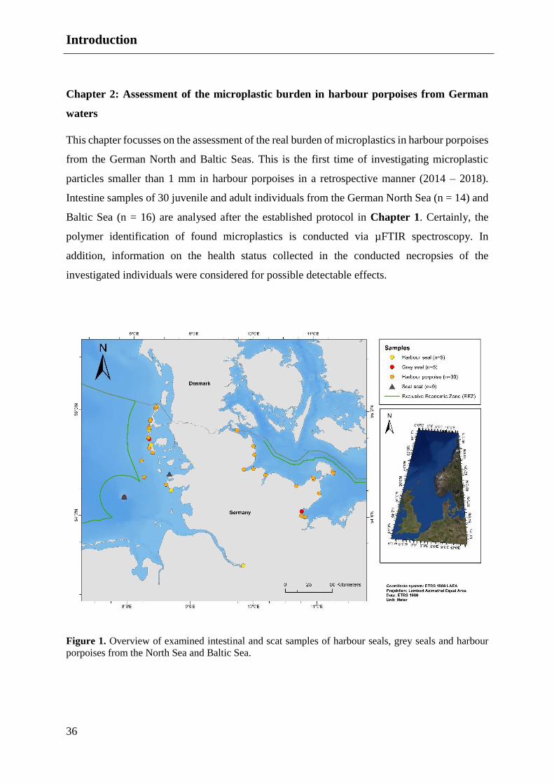
grey seals, see Figure 1).
This thesis consists of the following two chapters:
Chapter 1: Improvement of sample storage and sample handling with the focus on
reducing secondary pollution
The first chapter focuses on a combination and improvement of already established procedures
to decrease the risk of secondary contamination. Therefore, protecting measures already start
during opening the carcasses and end in the stage of staining the filtered samples to avoid an
overestimation of microplastic particles. To ensure a low contamination risk, the samples were
collected in washed and disinfected glass jars since it could not be excluded that the usage of
plastic bags might contaminate the sample. Furthermore, the whole treatment of the intestine
and faecal samples is conducted in a closed acrylic box wearing white cotton gloves above
nitrile ones and a cotton laboratory coat. All used instruments were rinsed several times under
Milli-Q water (Millipore) to ensure that rinsing water can be excluded as a contamination
source. Procedural blanks are used to determine the ambient contamination within the working
environment and narrow down the actual burden of microplastics in marine mammals
inhabiting German waters.
For the isolation of microplastics (>100 µm) in the biogenic material, the samples are washed
in self-sewed washing bags in a conventional washing machine, as already conducted in diet
analysis research. Furthermore, the identification of potential microplastic particles is
conducted using fluorescence microscopy and µRaman spectroscopy.
Introduction
36
Chapter 2: Assessment of the microplastic burden in harbour porpoises from German
waters
This chapter focusses on the assessment of the real burden of microplastics in harbour porpoises
from the German North and Baltic Seas. This is the first time of investigating microplastic
particles smaller than 1 mm in harbour porpoises in a retrospective manner (2014 – 2018).
Intestine samples of 30 juvenile and adult individuals from the German North Sea (n = 14) and
Baltic Sea (n = 16) are analysed after the established protocol in Chapter 1. Certainly, the
polymer identification of found microplastics is conducted via µFTIR spectroscopy. In
addition, information on the health status collected in the conducted necropsies of the
investigated individuals were considered for possible detectable effects.
Figure 1. Overview of examined intestinal and scat samples of harbour seals, grey seals and harbour
porpoises from the North Sea and Baltic Sea.

Chapter I: Handle with Care – Microplastic
Particles in Intestine Samples of
Seals from German Waters


Published:
Philipp, C., Unger, B., Fischer, E.K., Schnitzler, J.G., Siebert, U. (2020).
Handle with Care – Microplastic Particles in Intestine Samples of Seals
from German Waters.
Sustainability.
DOI: 10.3390/su122410424


Chapter I
41
Chapter I: Handle with Care – Microplastic Particles in Intestine Samples of
Seals from German Waters
C. Philipp a, B. Unger a, E.K. Fischer b, J.G. Schnitzler a, U. Siebert a
a Institute for Terrestrial and Aquatic Wildlife Research, University of Veterinary Medicine
Hannover, Foundation, Werftstraße 6, 25761 Büsum, Germany
b Center for Earth System Research and Sustainability (CEN), University of Hamburg,
Bundesstraße 55, 20146 Hamburg, Germany
Keywords: microplastic; plastic isolation; plastic ingestion; gastrointestinal tract; marine
mammals
Abstract
The Marine Strategy Framework Directive (MSFD) aims to reduce the marine debris burden in
the marine environment by 2020. This requires an assessment of the actual situation, which
includes the occurrence as well as the caused impacts. Information on both is scarce when it
comes to top predators like marine mammals and the burden of microplastic. This is hampered
by the limited access to free ranging marine mammals for collecting samples, as well as sample
handling. The present study investigated gastrointestinal tracts and faecal samples of harbour
seals (Phoca vitulina) and grey seals (Halichoerus grypus) regularly occurring in the German
North Sea and Baltic Sea with the aim of gaining information on the occurrence of
microplastics. In total, 255 particles ≥100 µm (70 fibres, 185 fragments) were found in
exemplary ten intestine and nine faecal samples. The findings ranged from no fibres and six
fragments, up to 35 fibres and 55 fragments per sample. This study established a protocol for
sample handling, microplastic isolation (≥100 µm) and quantification of gastrointestinal tracts
and faecal samples of marine mammals with a low share of contamination. This approach helps
to quantify the presence of microplastics in free-ranging marine mammals and is therefore
applicable to assess the real burden of microplastic presence in the marine environment.
Introduction
42
Introduction
The challenging nature of marine debris pollution is well known (Galgani et al., 2015). Due to
its widespread use, its specific characteristics and discard virtue, synthetic polymers, so-called
“plastics” make up a high share of marine debris in our oceans (Thompson, 2004; Ryan et al.,
2009; Thiel et al., 2013). Microplastic (MP) includes synthetic polymers in the form of particles
smaller than 5 mm (Arthur et al., 2009). These particles originate either from large plastic items
cracking down into smaller fragments due to various forces (secondary MP); or are intentionally
produced in those small sizes (primary MP) (Gregory and Andrady, 2003; Browne et al., 2007;
Cole et al., 2011; Andrady, 2015). The awareness of microplastics started back in the 1970s
(Carpenter et al., 1972; Morris and Hamilton, 1974) but has only recently become more into
the focus of environmental research (Hermsen et al., 2018; Zhang et al., 2019).
MPs were revealed to be ingested by organisms of lower trophic levels, and even a trophic
transfer was detected (Farrell and Nelson, 2013; Setälä et al., 2014; Rummel et al., 2016). A
variety of international studies confirmed the presence of different sized plastic particles in the
gastrointestinal tract (GIT) of various marine mammalian species (Bravo Rebolledo et al., 2013;
Unger et al., 2016, 2017; Donohue et al., 2019; Nelms et al., 2019).
Dealing with faunal samples implies the initial elimination of organic matter enclosing the
particles; their detection otherwise would be obscured (Cole et al., 2014; Löder and Gerdts,
2015). Therefore, the biogenic compounds have to be removed prior to further investigations
in order to obtain convincing and reliable results (Cole et al., 2014; Löder and Gerdts, 2015).
Furthermore, it needs to be ensured that the risk of secondary contamination during sampling,
storing and treating is kept as low as possible. Contamination of samples might occur during
their handling and origin, e.g. from storage containers, laboratory cloths, gloves and the
working environment in general (Roux et al., 2001; Hidalgo-Ruz et al., 2012; Foekema et al.,
2013; Woodall et al., 2014; Vandermeersch et al., 2015).
Previous studies already presented various digestion protocols of biota samples in recent years
(Bravo Rebolledo et al., 2013; Lusher and Hernandez-Milian, 2018; Nelms et al., 2019).
Enzymes were predominantly used to remove the organic matter (Cole et al., 2014; Catarino et

Chapter I
43
al., 2017), in some cases acids or alkaline solutions were applied to digest organic remains
(Claessens et al., 2013; Foekema et al., 2013; Lusher and Hernandez-Milian, 2018).
Subsequently, MP identification techniques such as the staining and fluorescence microscopic
approach (Tamminga et al., 2017; Fischer, 2019) and spectroscopic analyses like Fourier
transform infrared (FT-IR) (Mintenig et al., 2017; Primpke et al., 2017; Jung et al., 2018) or
Raman spectroscopy are commonly used (Lenz et al., 2015; Fischer, 2019; Karbalaei et al.,
2019). A combination of both methods is considered to be a reliable option regarding the
identification of polymer compositions (Shim et al., 2017).
However, information on the presence of MP in and impacts on marine top predator species is
scarce. Therefore, the present study focusses on an establishing a protocol to isolate particles,
evaluate potential contamination and propose a method to discriminate between MP and other
particles in faecal and intestinal samples of seals in order to investigate the presence of MP in
marine mammals in further research. Samples were taken from harbour seals (Phoca vitulina)
and grey seals (Halichoerus grypus), which regularly occur in the North and Baltic Seas. This
study was realised within the framework of the project ‘Assessment and implementation for
long-term monitoring of pollution of diverse marine compartments and biota with marine litter’
(Federal Environment Agency Germany), the aims of which was to optimise sample handling
and to reduce the risk of secondary contamination for detecting microplastic particles in
intestinal samples of marine mammals
Materials and Methods
Sample Collection
The Institute for Terrestrial and Aquatic Wildlife Research (ITAW) at the University of
Veterinary Medicine Hannover (Foundation, Germany) regularly conducts necropsies of
stranded, bycaught or euthanised marine mammals found along the coastline of the Federal
State of Schleswig-Holstein, Germany (Siebert et al., 2001, 2007; Unger et al., 2017). These
are predominantly the three species regularly occurring in German waters; harbour porpoise
(Phocoena phocoena), harbour seal and grey seal.

Materials and Methods
44
Necropsy Sampling
Concerning microplastic analyses in the GIT of marine mammals, the rectum of harbour seals
and grey seals has been sampled since 2014. Out of these, ten pinniped samples were used for
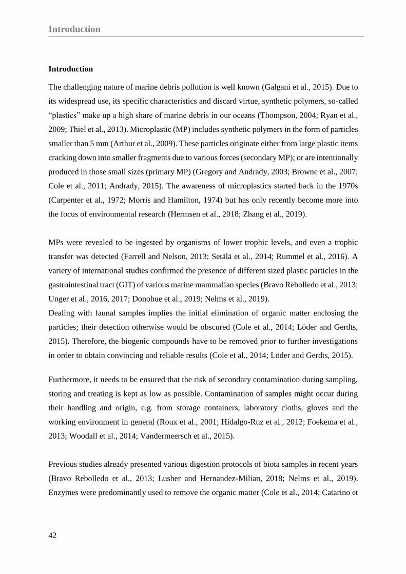
this study. The caudal part of the rectum was tied off with a drawstring (Figure 2A).
Subsequently, in a cranial direction, an 8 - 10 cm section was tied off. Both ends were cut off
behind the drawstrings to prevent the loss of faeces (Figure 2B). With the help of metallic
tweezers, the samples were transferred to cleaned and disinfected glass jars. Afterwards, the
glass jars were stored at - 20°C until further processing (Figure 2C and D).
Sandbank Sampling
In the course of a regularly conducted health monitoring of live animals at the Lorenzenplate
(Hasselmeier et al., 2008), a tide-dependent submerged sandbank in the North Sea, further
faecal samples of free-ranging harbour seals and grey seals have been collected since 2012. Out
of these, nine faecal samples were analysed. No genetic analyses have been performed to
distinguish between harbour seal and grey seal faeces. Hence, this study consecutively refers to
them as seal faecal samples.
Chapter I
45
Figure 2. Sampling of intestinal samples: (A) The caudal part of the rectum is tied off; (B) An 8-10 cm
section of the rectum is measured in cranial direction and tied off a second time; (C) The intestinal
sample is cut with a scalpel and placed with metal tweezers in a glass jar (D).
The following steps were conducted in a closed acrylic box (see supplementary information)
and are applicable for all further investigations on intestinal and faecal samples. The box was
equipped with two holes, which formed the only openings within the box, these being necessary
for processing the samples. During the whole procedure, the samples were solely processed
with glass or metallic instruments. Furthermore, cotton gloves were worn over nitrile ones
during all processing steps.
Materials and Methods
46
Preparation
After defrosting the samples in the glass jars, the closed intestinal parts were rinsed with MilliQ
water (Millipore), measured and opened inside the washing sachets on a glass cutting board to
prevent the loss of faeces and potential MP.
For separating the MP from the biogenic matter, the intestine and faeces samples were washed
in self-sewed double layer washing sachets in a commercial washing machine (OK., OWM
15012 A1). Each sample was placed in an inner bag (mesh size 300 µm) which was then placed
in the outer nylon bag (mesh size 100 µm). The sachets were made of nylon cloth and were
sewn together in the acrylic box with a conventional sewing machine (Singer, Tradition TM
2282) using black cotton yarn. Furthermore, each nylon sachet was used only once to prevent
a cross-contamination.
Washing Procedure
The washing procedure of the samples was based on the protocol established by Bravo
Rebolledo et al. (2013). To remove biogenic organic matter, enzyme-based washing powder
(Biotex® stain removing powder, biological detergent, bleach free; 35 g) was added in the pre
wash cycle. Subsequently, conventional detergent (35 g, Gut & Guenstig Classic,Edeka
Zentrale AG & Co. KG) was used in the main wash cycle, assisting the cleaning procedure. The
samples were washed in a delicate wash cycle without spinning at 60°C. A delicate wash cycle
without spinning was chosen in order to prevent particles from escaping from the sachets and
thus not being available for further analysis. For a valid evaluation, all samples were weighed
before and after the washing procedure, for quantify the loss of biogenic compounds (e.g. blood,
faeces, soft tissue).
Prior to each wash cycle, an empty wash cycle was run using disinfectant (Impresan, Brauns-
Heitmann GmbH & Co.KG) at 90°C. This step cleaned the washing machine and additionally
reduced the likelihood of cross-contamination.

Chapter I
47
Isolation
After the washing procedure, the washing sachets containing the sample residues were covered
in aluminium foil and stored in a half-closed box under a fume hood for drying overnight. The
outside of the sachets were cleaned with MilliQ water prior to opening. The residue was rinsed
with a filtered, saturated sodium chloride (NaCl) solution (350 g of table salt dissolved in 1 l of
MilliQ water) in glass beakers. The solution was left overnight for density separation.
Approximately 20 to 30 mL of the surface of the supernatant was pipetted onto a cellulose filter
(Rotilabo®, Typ11A, Ø 55 mm, retention 12 – 15 µm) using a Buechner funnel (Rotilabo®,
porcelain, volume: 70 mL; Ø 55 mm) attached to a vacuum pump. The remaining solution and
the sediment were decanted onto a second filter. Both surface and bottom sample suspensions
were investigated separately in order to avoid an MP underestimation. The filtration steps
resulted in a total of four filters for each sample (inner bag: surface water and water from the
beaker bottom; outer bag: surface water and water from the beaker bottom).
The filters were stored in glass petri dishes (STERIPLAN®, Ø 60 mm) and dried in a heating
cabinet for three hours at 50°C. Until further processing, the closed petri dishes including the
sample filter were stored in a closed and dry environment.
Pre-Trials to Verify the Procedure
The methodical efficiency was evaluated, focussing on potential losses of particles and
secondary contaminations. The use of washing sachets was assessed for potential loss of
particles and loss of nylon fibres and thus the recovery rate. To determine the loss of potential
MP, polyethylene microbeads (fragment size: 150 µm - 800 µm) were placed in the nylon
sachets and processed in the same manner as the intestine and scat samples.
Prior to sample processing, five sachets were solely washed without any detergent and sample
(at 60°C, delicate cycle, no spinning cycle as mentioned above). A Cora ball®, which traps
loose fibres, was added. After the washing procedure using a Cora ball® the inspection showed
no attached fibres.

Materials and Methods
48
Polymer Identification
For identifying possible MP, the filters were stained with Nile Red diluted in chloroform (1 mg
/ mL in chloroform) in accordance to Tamminga et al. (2017). The stained filters remained
covered for drying for at least 24 hours, before being analysed under a plan fluorescence
microscope (Kern OBN- 148 and Zeiss AX10) using a TRITC HC filter set (F36-503, AHF
Analysetechnik, Tuebingen, Germany) in accordance with Tamminga et al. (2019).
Potential MP particles were photographed (Kern ODC 832 and Canon EOS 80D) under the
fluorescence microscope. Subsequently, the photos were visually evaluated and the suspected
MP were measured and counted by using Adobe Photoshop (Version 21.0.3). After visual
identification, single particles were isolated with tweezers and pins, and placed on microscopic
slides for further identification by µRaman spectroscopy (ThermoFisher Scientific GmbH,
DXR2xi Raman Imaging Microscope).
In general, direct comparisons among different studies are hampered due to varying sample
treatment protocols and mesh sizes being used. To address this problem, the present study
followed the sample processing conducted by Bravo Rebolledo et al. (2013) and focussed on
particle sizes ≥100 µm. In this study, the term MP included synthetic fragments and fibres. The
classification of fibres was used, if a fragment has an elongated cylindrical shape and a
thickness of up to 23 µm.
All particles larger than 100 µm were taken into account for valid results due to the used mesh
size (100 µm) of the outer washing sachet to avoid an overestimation. Two researchers
independently investigated the photos taken of particles to identify the potential MP. The
following parameters were used for discrimination: fluorescence intensity and appearance of
the particle (surface, isolation, completeness, etc.). For additional information, see the
supplement.
Based on the aforementioned discrimination parameters, we used recursive partitioning to
explore decision rules for predicting whether the particle was plastic or not. The decision tree
was created in RStudio (Version 1.1.447) with the package ‘rpart’ R package rpart.plot
(Therneau et al., 2019) with 1185 characterised particles (biogenic or synthetic). This number
of particles results of all available samples including pinnipeds and cetaceans, which build the
basis for the used parameters in the decision tree. The original dataset (n=1185) was randomly
Chapter I
49
split into training (80%) and test (20%) subsets. The training subset was used for model tuning
and training and the performance of the final model was compared using the Test subset.
Evaluation of the Methodical Efficiency
In the present study, the entire working space was located in an acrylic box situated under a
fume hood to decrease the potential secondary contamination of samples and used items. All
equipment and instruments were rinsed several times with MilliQ water prior to usage. The box
was washed with MilliQ water and wiped between each step with cotton cloths, which were
exchanged regularly.
For monitoring secondary contamination, procedural blanks of the applied chemicals and
detergents were used in accordance with Hengstmann et al. (2018) and Nuelle et al. (2014).
Procedural blanks helped to estimate the number of particles being introduced during the
processing; particles on the procedural blank filters were separately quantified, measured and
qualified. Contamination of cloths or instruments was deducted after the quantitative analysis
as performed in former studies.
To prove the possible contamination of plastic bags, five faecal samples frozen which had been
frozen in low density polyethylene (LDPE) bags since 2012. Nine faecal samples were
additionally included in this study. These samples were divided: half of each sample was stored
in LDPE-bags, the other half in glass jars for three, six and 12 months. The plastic bags in which
the samples are stored are not disinfected or cleaned prior usage. The comparison of the found
number of particles in glass and plastic samples was performed using a Wilcoxon signed-rank
test, because of the relation between the samples and the assumed no normal distribution. To
test if the findings of suspected MP increased over time, the plastic samples taken from different
periods were analysed by a non-parametric ANOVA using a Kruskal-Wallis test.
Results
50
Results
Evaluation of the Methodical Efficiency
Procedural Efficiency of Washing Sachets
The pre-trials showed a loss of 0.358 g (2.99%) was determined for those sachets, which were
closed with a hook-and-loop fastener. Due to this small percentage loss, the usage of nylon
cloth was maintained, but the hook-and-loop fastener was discarded and replaced by a seam.
For further details, refer to the supplementary information.
By comparing the dry weights of the samples (before and after the washing procedure), the
average reduction in biogenic matter in intestinal samples differed between 9.97 g and 0.22 g
(mean = 1.57 g). In the faecal samples, the average reduction varied between 5.07 and 48.55 g
(mean = 13.33 g).
Efficiency of Contamination Control Measures
Procedural blanks analysed for the background contamination of the working space showed a
share ranging from zero to two particles per sample (M±SD = 0.9±0.99). Only one fibre larger
than 100 µm was found on the working environment controls (n = 12). One to two fragments
were found within the NaCl blanks (n = 5), which also included a potential contamination by
the used plastic laboratory bottles (M±SD = 1.4±0.99). Furthermore, five control washing
sachets showed a contamination of two to at most eight particles (M±SD = 4.6±2.7). Therefore,
a mean number of seven particles ≥100 µm (six fragments and one fibre) per sample was
considered as contamination. Figure 3 shows the share of suspected MP particles found in the
procedural blanks.

Chapter I
51
Figure 3. Suspected MP from procedural blank controls (≥100 µm). Working environment: 12 control
filter; Washing sachets: Five blank samples; NaCl Solution: Five control samples.
Comparison of suitable storage methods for avoiding contamination
Each sample archived in a glass jar showed a lower amount of suspected MP compared with
those stored in plastic bags (Figure 4). The Wilcoxon test revealed significant differences,
showing that plastic stored samples included eight particles (≥100 µm) more compared with
those stored in glass jars (particles ≥100 µm: p = 0.012; particles ≤100 µm: p = 0.0104).
The direct comparison of samples stored for different periods in plastic bags (three, six and 12
months) revealed no significant increase in microplastic particles over time (Figure 4). This
was due to a high variability in each period group. However, a comparison with samples, which
had been stored in plastic for eight years, showed no significant differences to the present
samples (Figure 5).

Results
52
Figure 4. Differential values in the quantity of suspected MPs (≥100 µm) in samples stored in plastic
bags, compared to ones stored in glass jars from different storage times.

Chapter I
53
Figure 5. Suspected microplastics in faecal samples stored in glass jars (n = 9) and plastic bags (n = 9)
combined from all storing periods (three, six and 12 months; particles ≥100 µm). In addition, five
samples of seal faeces, which had been stored in LDPE bags for eight years, were presented for
comparison purposes.
Evaluation of the Methodical Efficiency
The results of the used decision tree (Figure 6) revealed the following parameters to be
decisive: 1) If a particle could be associated with a biogenic shape or structure (e.g. crustacean
limbs or algae). If yes, it was classified as no MP (No-Plastic). If not, the fluorescent colour of
the particle (orange including red and yellow or white) defined the next classification (2). If the
fluorescent colour was white, the particle was classified as suspected MP. If the colour was
yellow or orange, the next category was the appearance: (3) blurred, melted or defined
appearance. If it was defined, the particle was classified as plastic; if not it was identified as
No-Plastic. If, the fluorescent colour the second category was white, the particle was
categorised as plastic. A catalogue of examples and categories is presented in the supplement.

Results
54
Potential microplastic particles identified by using this decision tree are than analysed using
µRaman spectroscopy.
The calculated sensitivity (true positive rate, TPR) of 84%, that measures the proportion of
particles that were identified as possible MP and the specificity (true negative rate, TNR) of
69%, which measures the proportion of particles that were identified as being non-plastic,
revealed a good performance of the decision tree.
Figure 6. Example of a decision tree of the test set-up of 510 particles showing the decisive parameters.
Isolation of Potential MP in Intestinal and Faecal Samples
In total, 653 potential plastic particles were determined in ten intestinal samples of free-ranging
harbour and grey seals, and nine seal faecal samples where the described procedure (sample
collection, isolation and identification) was used. Due to the used mesh sizes of the washing
sachets (100 µm and 300 µm), all particles smaller than 100 µm were excluded from the study,
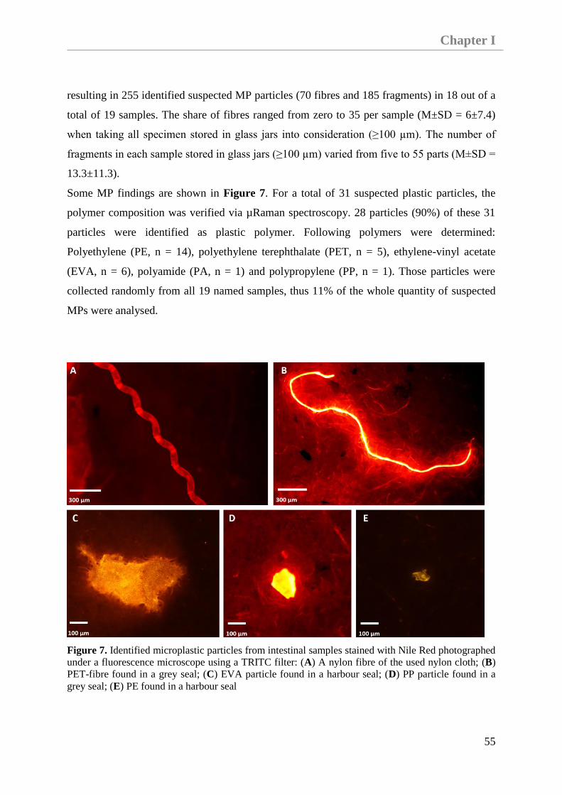
Chapter I
55
resulting in 255 identified suspected MP particles (70 fibres and 185 fragments) in 18 out of a
total of 19 samples. The share of fibres ranged from zero to 35 per sample (M±SD = 6±7.4)
when taking all specimen stored in glass jars into consideration (≥100 µm). The number of
fragments in each sample stored in glass jars (≥100 µm) varied from five to 55 parts (M±SD =
13.3±11.3).
Some MP findings are shown in Figure 7. For a total of 31 suspected plastic particles, the
polymer composition was verified via µRaman spectroscopy. 28 particles (90%) of these 31
particles were identified as plastic polymer. Following polymers were determined:
Polyethylene (PE, n = 14), polyethylene terephthalate (PET, n = 5), ethylene-vinyl acetate
(EVA, n = 6), polyamide (PA, n = 1) and polypropylene (PP, n = 1). Those particles were
collected randomly from all 19 named samples, thus 11% of the whole quantity of suspected
MPs were analysed.
Figure 7. Identified microplastic particles from intestinal samples stained with Nile Red photographed
under a fluorescence microscope using a TRITC filter: (A) A nylon fibre of the used nylon cloth; (B)
PET-fibre found in a grey seal; (C) EVA particle found in a harbour seal; (D) PP particle found in a
grey seal; (E) PE found in a harbour seal

Discussion
56
Figure 8 shows the higher quantify of particles found in intestinal samples of grey and harbour
seals compared with the collected faecal samples where the species is unknown. It is significant
that the quantity of suspected MP exceeded the maximal assumed secondary contamination of
seven particles per sample (see dotted line in Figure 8).
Figure 8. Suspected microplastics in intestinal samples of harbour (n = 5) and grey seals (n = 5), in
comparison to faecal samples from seals collected on the sandbank (Lorenzenplate, North Sea) (n = 9,
no determined species per sample: faeces originated from either a grey or a harbour seal). All samples
were stored in glass jars. The dotted line show the maximal presumption of secondary contamination of
seven particles per sample.
Discussion
An initial and essential step for studying the effects of MP particles in GITs of marine mammals
is the detection and evaluation of their current presence and distribution. Apart from assessing
the presence of microplastics in marine mammals, the main focus of this study was to improve
the avoidance of secondary contamination during sample handling. This problematic nature of
secondary contamination, which was also shown in previous studies (Bravo Rebolledo et al.,
Chapter I
57
2013; Foekema et al., 2013; Nuelle et al., 2014; Hengstmann et al., 2018; Lusher and
Hernandez-Milian, 2018), needs to be focused on to achieve genuine results. The presented
protocol combines established protocols for isolating MP from biota samples (Bravo Rebolledo
et al., 2013; Lusher and Hernandez-Milian, 2018) new approaches for specifically avoiding
secondary contamination.
The first step for avoiding contamination was to store the samples in glass jars, since they are
prone to contamination when stored in conventional plastic bags. The present study presented
evidences of this aforementioned fact. The results of the direct comparison between archived
samples in plastic bags and in glass jars showed that a contamination by plastic bags is likely.
The amount of plastic particles did not significantly increase over time, but the basis of
contamination from external sources seems to be higher when archiving samples in plastic bags
instead of storing them in glass jars. It has to be taken into account that the used samples already
show a basic microplastic contamination. This differs from sample to sample, resulting in the
fact that the plastic burden of the samples also varies between the time periods. A conceivable
parameter besides the polymer structure of plastic bags is the cleaning aspect. Glass jars were
cleaned and disinfected prior to usage; plastic bags were used in their original state. Based on
the results of this study, it is recommended to use glass jars instead of plastic bags or containers.
A closed working environment was created to improve the measures undertaken in previous
studies to prevent secondary contamination. The procedural blanks analysed for the background
contamination of the working space showed a very low number of particles (0-2 per sample).
Furthermore, the share of fibres was significantly lower in this study compared to similar
approaches in marine mammals (Lusher et al., 2018; Nelms et al., 2018, 2019; Donohue et al.,
2019) or other marine species (Lusher et al., 2013; Steer et al., 2017; Fischer, 2019).
Additionally, it has been previously highlighted, that fibres in particular are an indicator of
secondary contamination, originating from the laboratory clothing and working environment
(Hidalgo-Ruz et al., 2012; Nuelle et al., 2014; Torre et al., 2016; Lusher et al., 2017b;
Hengstmann et al., 2018; Lusher and Hernandez-Milian, 2018). Therefore, the acrylic box
safeguarded the samples from direct contact with the laboratory environment and personnel.
The presented study revealed that closed sealing, like the washing sachet, reduces the loss of
particles. To minimise the risk of losing particles as well as reducing the contamination of the
samples, it is advisable to place the samples into a nylon cloth, which is then sewn together.

Discussion
58
Pre-trials revealed the application of hook-and-loop fasteners to be unsatisfactory (see
supplementary information); similar results are noted for rubber bands (Orr et al., 2003).
The usage of nylon cloths was inevitable due to the favourable mesh sizes and the fact that no
solid material can be used in the washing machine, although polyamide (nylon) is one of the
most abundant polymers (Andrady, 2011; ter Halle et al., 2017). Indeed, fibres of the nylon
washing sachets can be easily identified showing a distinct fibre pattern and were therefore not
included in our study. Thus, there was no need to exclude polyamides from this study, if these
polymers were found. To validate this assumption, a found fibre showing a distinct structure
and fluorescence intensity compared with the fibres of the used nylon cloths was identified as
polyamide (nylon) by µRaman spectroscopy. Therefore, the polymer identification via µRaman
spectroscopy was used to manifest the presence of microplastics, and is an already established
and reliable application in microplastic analysis (Lang et al., 1986; Andrady, 2011; Lenz et al.,
2015; Cabernard et al., 2018). Thus, the benefits of this applied cleaning procedure (1. time and
cost effective, 2. valuable results) outweigh the disadvantages of the used nylon cloths. The
usage of a conventional washing machine and detergent was demonstrated as a successful
application to speed up the purification procedure resulting in clean residues. Removing
biogenic substances with the help of the washing procedure preserves the original particles, and
it is less time- (approx. 90 min) and budget-consuming compared with the application of
enzymatic or chemical solutions for the digestion of biogenic material. Thus, the reduction of
biogenic compounds (e.g. faeces, blood, and soft remains of prey species) was effective to
decrease the sample to the remaining hard parts (e.g. sand grains, fish bones and lenses, beaks,
and microplastics), as also seen by the compared dry weights. These results are also shown by
the comparison of dry weights before and after the washing procedure. In the intestinal samples,
the intestinal tissue and the hard parts remained. A mean difference of 1.75 g was determined.
In comparison to the faecal samples where a mean difference of 13.33 g was ascertained. The
weights were mostly dependent on the quantity of hard parts (e.g. sand grains, fish otoliths, and
lenses, bones or squid beaks) within the respective sample. Furthermore, the weight of pure
faeces was higher in the sandbank faecal samples (mean = 19.52 g) compared to faeces included
in the intestinal tissue (mean = 15.06 g). Firstly, these differences can be explained by the
intestinal physiology of phocid species, with a short length of the large intestine where the
chymus is dehydrated and stored as faeces until excretion (Helm, 1983; Pabst et al., 1999).
Chapter I
59
Secondly, passage times of marine mammals have to be taken into account if an assessment of
the microplastic burden in marine mammals is required (Panti et al., 2019). The passage times
of harbour seals and grey seals were estimated to be longer (6.5 – 30 h) (Prime and Hammond,
1987; Harvey, 1989). Thus, the sandbank samples may include the faeces of a full egestion of
the rectum. In contrast, the intestinal samples represent only a part of the actual excretion, which
is here further analysed. Furthermore, a reduction in size and loss of microplastics is
conceivable like it is confirmed for otoliths (Prime and Hammond, 1990). Combining collected
faecal and intestinal samples is advisable for further investigations as this will display the
burden of microplastics in an extended way.
Besides the method, there are more side parameters which have to be considered while
collecting samples from wild ranging animals. This study only investigated a part of the
intestinal system instead of examining the whole GIT or the stomach as performed in recent
studies (Lusher et al., 2015a, 2018; Nelms et al., 2019). This step was chosen to enable a
potential contamination-free sample although a complete examination of the residual GIT and
the carcass can be conducted. The reason for this is the usage of one organ for only one
assessment is applicable in the fewest of cases during necropsies. Another benefit of this
strategy is the unhindered necropsy of the whole individual, which enables an entire overview
of the health status and possibly provides more background information.
Furthermore, it is already known that the stomach of marine mammals accumulates a high
quantity of ingested hard parts (Nelms et al., 2019).
The amount of positive samples (suspected MP ≥ 100 µm, in glass jars) is higher than in
similar studies of faecal samples of seal species from the northern and southern Pacific Ocean
(Perez-Venegas et al., 2018; Donohue et al., 2019) despite it beings conformable with studies
investigating marine mammals of the north-eastern North Atlantic region (Lusher et al., 2018;
Hernandez-Milian et al., 2019; Nelms et al., 2019). As a consequence of the results of the used
statistical applications (precision of 94% and sensitivity of 84%), in combination with the 90%
chance of a correct classification as MP, this study is assumed to be a valuable approach in MP
monitoring in marine mammals.
The establishment of a decision tree helps to categorize different variables, which are helpful
for untrained researchers to gain results that are comparable between research groups. This is
essential to compare the burden in different marine environments and biota. Furthermore, the

Conclusion
60
decision tree helps to narrow down suspected microplastic particles out of a bunch of a variety
of particles in a sample, which are then used for further analysis. This is cost- and time-effective
and a useful tool for encouraging decision makers to support necessary monitoring
programmes.
In addition, the applied methods comprise a combination of interdisciplinary analysis:
identifying microplastics and their consumed prey (diet analysis of hard parts). This is based
on the conciliatory washing procedure; the retained hard parts can be used for prey species
identification (Orr et al., 2003; Robinson et al., 2018). Since it is proven that microplastics are
transported across the food web (Farrell and Nelson, 2013; Setälä et al., 2014; Ory et al., 2018),
it is assumed that in particular the contained contaminants accumulate due to absorption and
leaching (Rani et al., 2015) in top predator species as it is already evidenced for example for
organochlorides (Ross et al., 2000; Siebert et al., 2012). This allows new insights into the
correlation between microplastic occurrence in marine top predator species and their prey,
which serve as a vector.
Conclusion
The sample protocol applied within this study is applicable for isolating and identifying
microplastics from samples of gastrointestinal tracts of marine mammals and is comparable
with other approaches like diet analysis. Low numbers of detected fibres led to the conclusion
that secondary contamination is low. This seems to be most likely an effect of using a closed
environment, the acrylic box and the usage of glass jars as storage container. Furthermore, the
used isolation procedure is less cost-intensive, more gentle and faster in comparison to an
enzyme or chemical digestion. However, every working environment and samples of different
species and different sizes pose different challenges for microplastic analysis.
It can be assumed that the current approach provides reliable results, reflecting the actual
microplastic burden in the analysed species. Thus, this study presents a possible application for
microplastic monitoring in marine mammal samples, which has to be extended to a higher
amount of samples to achieve convincing findings in different species and water bodies.
Nevertheless, this method is also transferable to other mammalian species.
Chapter I
61
Author Contributions
Conceptualization, B.U. and C.P.; Methodology, B.U. and C.P.; Investigation: C.P.; Formal
analysis and Validation: J.G.S. and C.P.; Supervision: U.S. and E.K.F.; Resources: E.K.F. and
U.S.; Writing—original draft: C.P.; Writing—review and editing: B.U., E.K.F., J.G.S., U.S.
and C.P. All authors have read and agreed to the published version of the manuscript.
Funding
The stranding network and the health monitoring is funded by the Schleswig-Holstein Agency
for Coastal Defence, National Park and Marine Conservation, Germany. The pathological
investigations of the marine mammals are partly funded by the Ministry of Energy, Agriculture,
Environment, Nature and Digitalization of Schleswig-Holstein, Germany. The Federal
Environment Agency (UBA) partly funded the analysis of a number of samples within the
project “Assessment and implementation for long-term monitoring of pollution of diverse
marine compartments and biota with marine litter” (FKZ 3717252250). This publication was
supported by the German Research Funding Organisation (Deutsche Forschungsgemeinschaft)
and the University of Veterinary Medicine Hannover, Foundation (Germany), within the
funding programme Open Access Publishing.
Acknowledgements
We would like to thank all seal rangers in Schleswig-Holstein (SH), Germany for reporting and
collecting carcasses as well as our colleagues in the ITAW necropsy team for collecting and
dissecting carcasses continuously throughout the year. Special thanks go to E. Ludes-
Wehrmeister and S. Gross for advising us regarding tissue sampling and storing. We would like
to thank the “Microplastic Research at CEN – MRC” working group at the University of
Hamburg (Germany), for their assistance and hospitality. Special thanks in particular go to M.
Tamminga for sharing his expertise.
Conflicts of Interest
The authors declare no conflict of interest. The funders had no role in the design of the study;
in the collection, analyses, or interpretation of data; in the writing of the manuscript, or in the
decision to publish the results.

Supplementary Material
62
Supplementary Material
1. Preparation
1.1 Pre-trials
Pre-trials for testing the efficiency of the procedure have been carried out beforehand. To
determine the loss of particles in the nylon washing bags, aluminium silicate (200 µm – 500
µm) and polyethylene microbeads (150 µm - 800 µm) were used. Due to the mesh size, the
inner washing bag retains particles ≥300 µm (Supplementary Figure 1A), the outer washing
bag particles ≥100 µm (Supplementary Figure 1B). Therefore, particles smaller than 100 µm
were excluded from the analysis. Using three washing bags five times, each time freshly filled
with aluminium silicate tested the loss of particles. These pre-trial bags were weight before and
after the washing procedure. An average loss of 6.29 g (14.79%) was determined. This high
loss is assumed to result from the sharp form of the aluminium silicate fractions, which damages
the nylon fabrics (Supplementary Figure 1D).
Supplementary Figure 1. The washing bag design used for the pre-trials using pre-sewed bags closed
with stitched hook and loop fastener (A & B). The size fragmentation based on the nylon cloths operates
successful during the washing procedure, what approved the method of different cloths (C). These pre-
trials were conducted with aluminium silicate (200 µm – 500 µm).
A C
B D
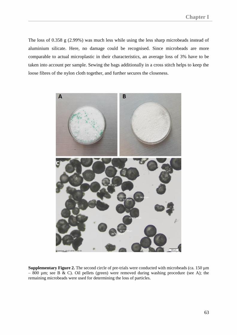
Chapter I
63
The loss of 0.358 g (2.99%) was much less while using the less sharp microbeads instead of
aluminium silicate. Here, no damage could be recognised. Since microbeads are more
comparable to actual microplastic in their characteristics, an average loss of 3% have to be
taken into account per sample. Sewing the bags additionally in a cross stitch helps to keep the
loose fibres of the nylon cloth together, and further secures the closeness.
Supplementary Figure 2. The second circle of pre-trials were conducted with microbeads (ca. 150 µm
– 800 µm; see B & C). Oil pellets (green) were removed during washing procedure (see A); the
remaining microbeads were used for determining the loss of particles.
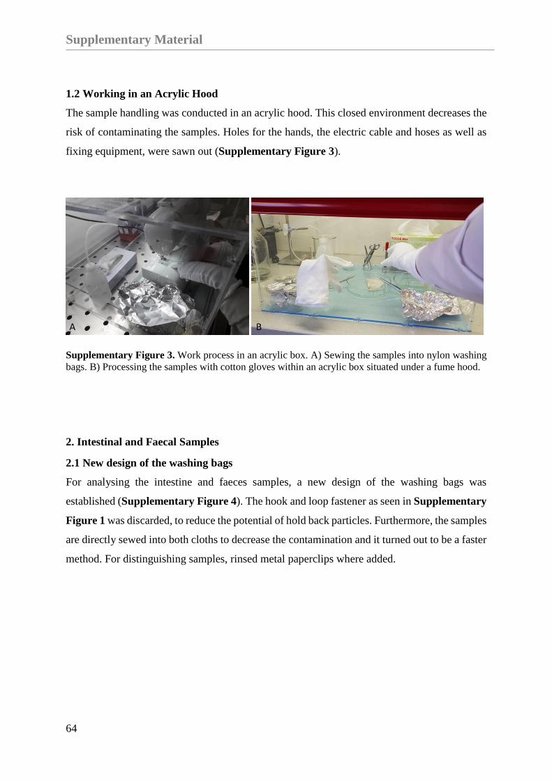
Supplementary Material
64
1.2 Working in an Acrylic Hood
The sample handling was conducted in an acrylic hood. This closed environment decreases the
risk of contaminating the samples. Holes for the hands, the electric cable and hoses as well as
fixing equipment, were sawn out (Supplementary Figure 3).
Supplementary Figure 3. Work process in an acrylic box. A) Sewing the samples into nylon washing
bags. B) Processing the samples with cotton gloves within an acrylic box situated under a fume hood.
2. Intestinal and Faecal Samples
2.1 New design of the washing bags
For analysing the intestine and faeces samples, a new design of the washing bags was
established (Supplementary Figure 4). The hook and loop fastener as seen in Supplementary
Figure 1 was discarded, to reduce the potential of hold back particles. Furthermore, the samples
are directly sewed into both cloths to decrease the contamination and it turned out to be a faster
method. For distinguishing samples, rinsed metal paperclips where added.
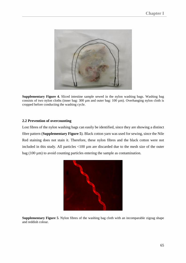
Chapter I
65
Supplementary Figure 4. Sliced intestine sample sewed in the nylon washing bags. Washing bag
consists of two nylon cloths (inner bag: 300 µm and outer bag: 100 µm). Overhanging nylon cloth is
cropped before conducting the washing cycle.
2.2 Prevention of overcounting
Lost fibres of the nylon washing bags can easily be identified, since they are showing a distinct
fibre pattern (Supplementary Figure 5). Black cotton yarn was used for sewing, since the Nile
Red staining does not stain it. Therefore, these nylon fibres and the black cotton were not
included in this study. All particles <100 µm are discarded due to the mesh size of the outer
bag (100 µm) to avoid counting particles entering the sample as contamination.
Supplementary Figure 5. Nylon fibres of the washing bag cloth with an incomparable zigzag shape
and reddish colour.
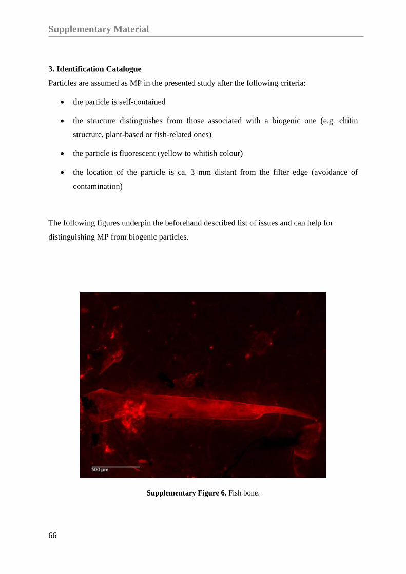
Supplementary Material
66
3. Identification Catalogue
Particles are assumed as MP in the presented study after the following criteria:
the particle is self-contained
the structure distinguishes from those associated with a biogenic one (e.g. chitin
structure, plant-based or fish-related ones)
the particle is fluorescent (yellow to whitish colour)
the location of the particle is ca. 3 mm distant from the filter edge (avoidance of
contamination)
The following figures underpin the beforehand described list of issues and can help for
distinguishing MP from biogenic particles.
Supplementary Figure 6. Fish bone.

Chapter I
67
Supplementary Figure 7. Fish bones and plant-based fragments.
Supplementary Figure 8. Fish vertebrae.

Supplementary Material
68
Supplementary Figure 9. Fishbone.
Supplementary Figure 10. Biogenic fragment (e.g. plant-based seed or a big fish lens).
Certainly, no microplastic bead.

Chapter I
69
Supplementary Figure 11. Biogenic fragment.
Supplementary Figure 12. Plant-based fragment.

Supplementary Material
70
Supplementary Figure 13. Biogenic fragment.
Supplementary Figure 14. Cellulose fibre.
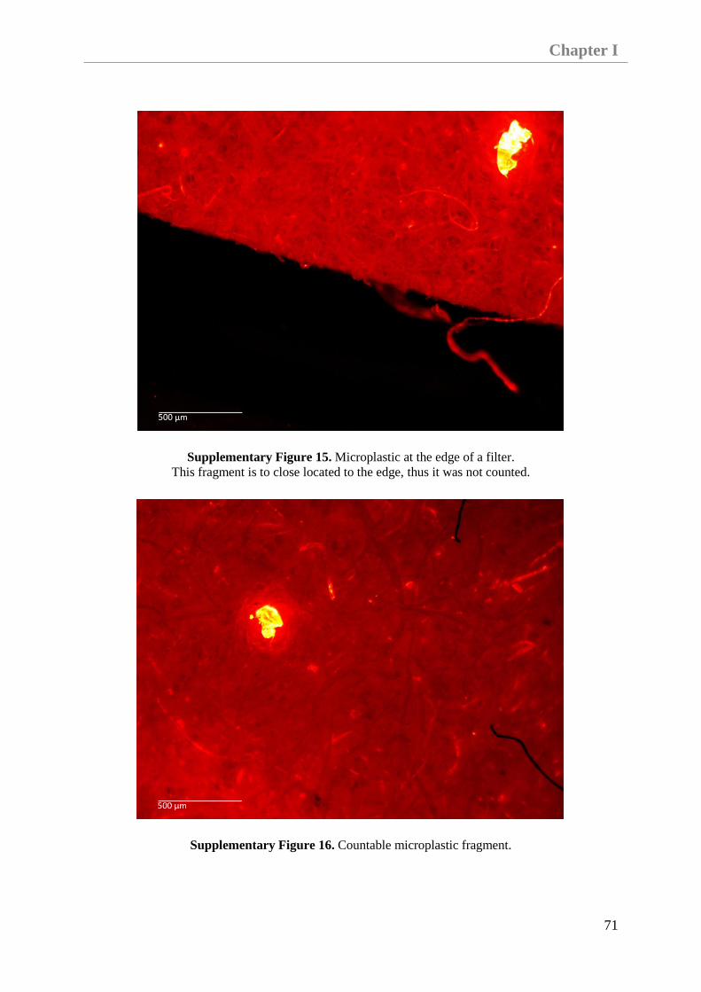
Chapter I
71
Supplementary Figure 15. Microplastic at the edge of a filter.
This fragment is to close located to the edge, thus it was not counted.
Supplementary Figure 16. Countable microplastic fragment.

Supplementary Material
72
Supplementary Figure 17. Countable microplastic fragment surrounded by fish bones.
Supplementary Figure 18. Countable microplastic fragment surrounded by a cellulose fibre.

Chapter I
73
3.1 Categorization of parameters
Supplementary Figure 19. Fluorescence intensity.
Supplementary Figure 20. Surface.
Supplementary Figure 21. Appearence.

Supplementary Material
74
Supplementary Figure 22. Completeness.
Supplementary Figure 23. Surroundings.
Supplementary Figure 24. Organic matter.

Chapter I
75
4. Results
All microplastic particles (≥100 μm) found in intestinal and faecal samples of seals from
German waters are shown in Supplementary Table 1. Labelling is made up of species initials
(Pv = Phoca vitulina; Hg = Halichoerus grypus) and sampling location (e.g. Intestine). All
samples are archived in glass jars.
Faecal samples archived in plastic bags since 2012, are named as “LDPE_Bag_2012”. Faecal
samples of seals (no information on the species) were halved and stored in glass jars or plastic
bags for different time periods, which is also stated in the label (“Glass_3_months” or
“Plastic_3_months”, etc.). The column “identified polymer structures” gives information on the
identified polymers regardless the number of particles analysed. NA (no information available)
refers to samples in which no particles could be further identified by μRaman spectroscopy.
The findings of fragments and fibres, in addition to the identified polymer structure should only
give information on the usefulness of the applied and presented protocol.
Supplementary Table 1. Results of microplastic analysis.

Supplementary Material
76

Chapter I
77


Chapter II: First Evidence of Retrospective Findings
of Microplastics in Harbour Porpoises
(Phocoena phocoena) From German Waters

Published:
Philipp, C., Unger, B., Ehlers, S.M., Koop, J.H.E., Siebert, U.
First Evidence of Retrospective Findings of Microplastics
in Harbour Porpoises (Phocoena phocoena) From German Waters.
Frontiers in Marine Science.
DOI: 10.3389/fmars.2021.682532


Chapter II
83
Chapter II: First Evidence of Retrospective Findings of Microplastics in
Harbour Porpoises (Phocoena phocoena) From German Waters
Carolin Philipp a, Bianca Unger a, Sonja M. Ehlersb,c, Jochen H. E. Koopb,c, Ursula Siebert a
a Institute for Terrestrial and Aquatic Wildlife Research, University of Veterinary Medicine
Hannover, Foundation. Werftstr. 6, 25761 Büsum, Germany
b Department of Animal Ecology, Federal Institute of Hydrology, Am Mainzer Tor 1, 56068
Koblenz, Germany
c Institute for Integrated Natural Sciences, University of Koblenz-Landau, 56070, Koblenz,
Germany
Keywords: microplastic burden; FTIR; marine mammals; cetacean; North Sea; Baltic Sea;
nutritional status; health
Abstract
Microplastic ingestion by lower trophic level organisms is well known, whereas information
on microplastic ingestion, egestion and accumulation by top predators such as cetaceans is still
lacking. This study investigates microplastics in intestinal samples from harbour porpoises
(Phocoena phocoena) found along the coastline of Schleswig- Holstein (Germany) between
2014 and 2018. Out of 30 individuals found along the North Sea (NS) and the Baltic Sea (BS)
coast, 28 specimens contained microplastic. This study found a relationship between the
nutritional status of cetaceans and the amount of found microplastics. Harbour porpoises with
a good or moderate nutritional status contained a higher number of microplastics, when
compared with specimens in a poor nutritional status. In addition, when individuals died
accidently due to suspected bycatch in gillnets, where a feeding event is highly assumed or a
pharyngeal entrapment happened, the microplastic burden was higher. In total, 401
microplastics (≥100 µm), including 202 fibres and 199 fragments were found. Intestines of the

Introduction
84
specimens of the BS contained more microplastics than the ones from the NS. Differences in
the share of fibres could be revealed: for BS fibres constituted 51.44% and for NS, fibres
constituted 47.97%. The polymers polyester, polyethylene, polypropylene, polyamide, acrylic
(with nitrile component) and an acrylic/alkyd paint chip (with styrene and kaolin components)
were identified. This is the first study investigating the occurrence of microplastics in harbour
porpoises from German waters and will, thus, provide valuable information on the actual burden
of microplastics in cetaceans from the North and Baltic Seas.
Introduction
The ubiquitous presence of marine litter, and especially the occurrence of small particles called
microplastics (<5 mm) (Arthur et al., 2009) is already confirmed in different marine habitats
and organisms (Fossi et al., 2014; Lusher et al., 2015b; Pereira et al., 2020). A trophic transfer
of microplastic particles between species of different trophic levels can be assumed as this has
previously been determined by other studies (Farrell and Nelson, 2013; Setälä et al., 2014;
Nelms et al., 2018). In addition, the ingestion and presence of microplastics has been highly
studied throughout the food web in recent years (Miller et al., 2020). Besides the unintentional
uptake of microplastics by prey species, an intentional uptake by organisms caused by a
burdened environment or due to accidental prey resemblance has already been shown (Ory et
al., 2017; Roch et al., 2020).
When focusing on the study area, a microplastic burden in the North-East Atlantic area and its
organisms occupying different trophic levels has also been verified Furthermore, it is assumed
that microplastics and pollutants accumulate in marine top predator species (Fossi et al., 2014;
Jepson et al., 2016; Garcia-Cegarra et al., 2021). The ingestion of marine litter is already
confirmed by previous studies in the North Sea (NS) and Baltic Sea (BS) (Unger et al., 2017;
van Franeker et al., 2018). This is not surprising, since the NS and adjacent waters are assumed
to be highly affected by anthropogenic exploitations (Halpern et al., 2008). Nevertheless, the
knowledge on the burden of microplastics in top predators, particularly in cetaceans of the
eastern North Atlantic region, including the BS, is still lacking. Both seas are different in
topography, salinity, hydrology (Frid et al., 2003; Sjöqvist et al., 2015), and the occurring ship
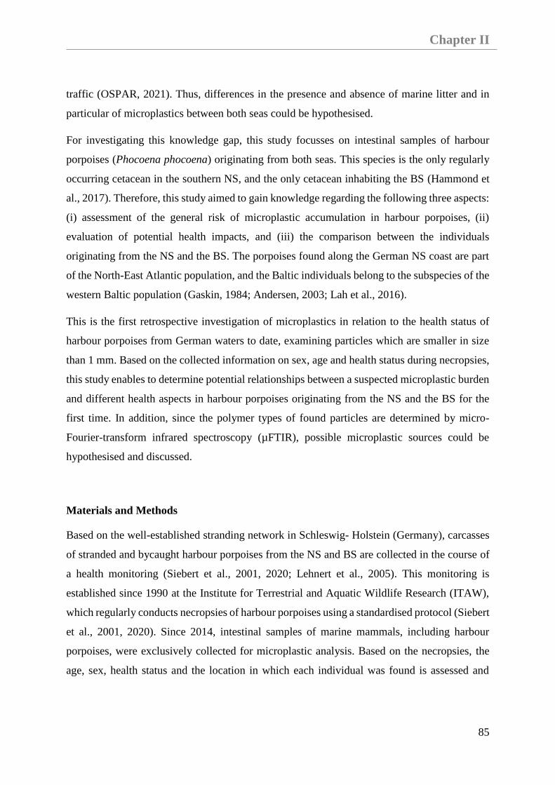
Chapter II
85
traffic (OSPAR, 2021). Thus, differences in the presence and absence of marine litter and in
particular of microplastics between both seas could be hypothesised.
For investigating this knowledge gap, this study focusses on intestinal samples of harbour
porpoises (Phocoena phocoena) originating from both seas. This species is the only regularly
occurring cetacean in the southern NS, and the only cetacean inhabiting the BS (Hammond et
al., 2017). Therefore, this study aimed to gain knowledge regarding the following three aspects:
(i) assessment of the general risk of microplastic accumulation in harbour porpoises, (ii)
evaluation of potential health impacts, and (iii) the comparison between the individuals
originating from the NS and the BS. The porpoises found along the German NS coast are part
of the North-East Atlantic population, and the Baltic individuals belong to the subspecies of the
western Baltic population (Gaskin, 1984; Andersen, 2003; Lah et al., 2016).
This is the first retrospective investigation of microplastics in relation to the health status of
harbour porpoises from German waters to date, examining particles which are smaller in size
than 1 mm. Based on the collected information on sex, age and health status during necropsies,
this study enables to determine potential relationships between a suspected microplastic burden
and different health aspects in harbour porpoises originating from the NS and the BS for the
first time. In addition, since the polymer types of found particles are determined by micro-
Fourier-transform infrared spectroscopy (µFTIR), possible microplastic sources could be
hypothesised and discussed.
Materials and Methods
Based on the well-established stranding network in Schleswig- Holstein (Germany), carcasses
of stranded and bycaught harbour porpoises from the NS and BS are collected in the course of
a health monitoring (Siebert et al., 2001, 2020; Lehnert et al., 2005). This monitoring is
established since 1990 at the Institute for Terrestrial and Aquatic Wildlife Research (ITAW),
which regularly conducts necropsies of harbour porpoises using a standardised protocol (Siebert
et al., 2001, 2020). Since 2014, intestinal samples of marine mammals, including harbour
porpoises, were exclusively collected for microplastic analysis. Based on the necropsies, the
age, sex, health status and the location in which each individual was found is assessed and

Materials and Methods
86
recorded. Thus, this information is available for the investigated intestinal samples from
harbour porpoises found between 2014 and 2018.
The following criteria were applied for choosing the most suitable samples: (i) the
gastrointestinal tract (GIT) had to be intact, (ii) faeces were present, and (iii) the individual was
already weaned. 30 individuals were chosen for analysis: 14 individuals from the NS and 16
individuals from the BS (Figure 9).
Figure 9. Locations where investigated harbour porpoises were found (n = 30).
The intestinal samples were stored in pre-cleaned glass jars at −20°C until further processing.
Then, each defrosted and opened intestinal sample was placed into a double-layered washing
sachet made of nylon cloths. The inner bag of the washing sachet has a mesh size of 300 µm
and the outer bag has a mesh size of 100 µm. Both cloths, including the sample, were sewn
together with the help of a conventional sewing machine, resulting in a so-called washing
sachet. These washing sachets were washed in a conventional washing machine at 60◦C without

Chapter II
87
spinning cycle. For the removal of biogenic matter, an enzyme based detergent and a
conventional detergent were added for facilitating the washing procedure. Subsequently, a
density separation, a vacuum filtration onto cellulose filters (Rotilabo®, Typ 11A, Ø 55 mm,
retention 12–15 µm) and fluorescence microscopy enabled by Nile Red (diluted with
chloroform) staining were conducted for microplastic isolation and identification.
Subsequently, all potential microplastics found on the cellulose filters were photographed,
counted, and measured in size. All steps of sample processing were conducted in a closed
acrylic box to avoid airborne contamination. The whole implementation of sample handling
and processing is described in detail in Philipp et al. (2020).
For polymer identification, selected microplastic particles were manually collected. In addition,
a disinfectant step was conducted to exclude a passing on of bacterial or parasitical zoonosis.
For this purpose, the cellulose filters containing the stained particles were sprayed with ethanol
(70%). After evaporation, the particles showed the same fluorescence qualities as before. Thus,
the potential microplastics were selected and manually collected with tweezers or needle pins
and placed into a droplet of ethanol (70%) onto an aluminium oxide membrane filter (Anodisc,
Ø47 mm, 0.2 µmpore size, Whatman, Freiburg, Germany). The filter was kept still until the
droplet was evaporated and the particles had attached to the filter. Since the transfer of particles
was done manually, a loss of particles needs to be taken into account.
The polymer composition of 77 potential microplastics (incl. fragments and fibres) from
intestinal sample were analysed by using a µFTIR spectroscope (Hyperion 2000, Bruker,
Ettlingen, Germany). All measurements were conducted in transmission mode with 32 co-
added scans (sometimes 100 scans for very thin fibres) and a spectral resolution of4 cm−1 in a
wavenumber range of 4,000–1,250 cm−1 as aluminium oxide membrane filters are infrared
inactive between 3,800 and 1,250 cm−1. For background measurements, the blank aluminium
oxide membrane filter was used. For thick particles, for which transmission mode was not
suitable, the measurements were conducted in attenuated total reflectance (µATR) mode. Those
µATR measurements were conducted between 4,000 and 600 cm−1.
Procedural blanks of the used detergents and materials, e.g., nylon sachets (n = 3), and the
working environment (n = 10) were taken into account for avoiding an overestimation caused
by secondary contamination. The analysed blank filters of the working environment

Results
88
accompanied the samples from time of collection until the staining procedure was finished. On
average, one fibre and seven particles were found in those procedural blanks and were finally
subtracted from the microplastic counts in each parallel sample. Four of those potential
microplastics could be collected, manually placed on the aluminium oxide membrane filter and
were considered for µFTIR analysis. Moreover, the polymer composition of different
equipment materials like the nitrile gloves and shavings of the used acrylic box were
additionally determined by FTIR in ATR mode (Vertex 70; Bruker, Ettlingen, Germany) or by
µFTIR to avoid an overestimation. For the Vertex measurements, ATR measurements were
performed in a wavenumber range of 4,000– 370 cm−1 with 8 co-added scans and a spectral
resolution of 4 cm−1.
The quantity of found microplastics in comparable groups is given in mean ± standard deviation
(M ± SD) to enable a comparison between findings. Moreover, the results were statistically
analysed by determining the Cohen’s d and applying a paired t-test using the package “pwr” in
the R software Version 4.0.2 (Champely et al., 2020; R Core Team, 2020). Thus, results were
described as significant if p <0.05. In addition, the Figures Figure 10, Figure 13Figure
14Figure 15 were visualised using the package ‘ggplot2’ (Wickham, 2016).
Results
Quantity and Size
In total, 30 intestinal samples were available for analyses. An amount of 611 potential
microplastics (incl. fragments and fibres, >100 µm) were found. A secondary contamination of
one fibre and seven fragments were considered and subtracted from each sample. Thus, 401
microplastics were finally determined. This amount of microplastics was found in 28 intestinal
samples, in the remaining an absence of microplastics was noticed. When categorising into
particle type, 202 fibres and 199 fragments were found. Hence, only two intestines were free
from microplastics. Most of the found fibres had a length between 100 and 2,000 µm (Figure
10).
Four additional fibres longer than 5,000 µm, thus defined as mesoplastics (Gregory and
Andrady, 2003), were found. Three of them occurred in a sample of an adult male harbour

Chapter II
89
porpoise found in 2017 (lengths: 8,450, 6,964, and 8,029 µm). The fourth fibre (7,365 µm in
length) was found in a juvenile male stranded in 2014. Both carcasses were found in the BS.
Based on the size, those four fibres were excluded from the results.
Figure 10. Width-length distribution of all suspected microplastic particles in intestine samples of 30
harbour porpoise in the size range of 100 µm up to 5,000 µm (n = 611). Secondary contamination was
not considered.
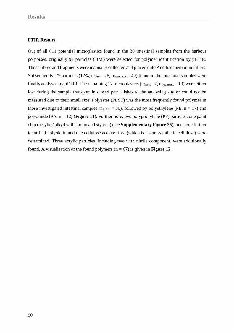
Results
90
FTIR Results
Out of all 611 potential microplastics found in the 30 intestinal samples from the harbour
porpoises, originally 94 particles (16%) were selected for polymer identification by µFTIR.
Those fibres and fragments were manually collected and placed onto Anodisc membrane filters.
Subsequently, 77 particles (12%, nfibres= 28, nfragments = 49) found in the intestinal samples were
finally analysed by µFTIR. The remaining 17 microplastics (nfibres= 7, nfragments = 10) were either
lost during the sample transport in closed petri dishes to the analysing site or could not be
measured due to their small size. Polyester (PEST) was the most frequently found polymer in
those investigated intestinal samples (nPEST = 30), followed by polyethylene (PE, n = 17) and
polyamide (PA, n = 12) (Figure 11). Furthermore, two polypropylene (PP) particles, one paint
chip (acrylic / alkyd with kaolin and styrene) (see Supplementary Figure 25), one none further
identified polyolefin and one cellulose acetate fibre (which is a semi-synthetic cellulose) were
determined. Three acrylic particles, including two with nitrile component, were additionally
found. A visualisation of the found polymers (n = 67) is given in Figure 12.

Chapter II
91
Figure 11. Microplastic particles stained with Nile Red (A-D) and the corresponding measured µFTIR
spectra of found polymers (in red; Polyamide: PA; Polyethylene: PE; Polypropylene: PP and Polyester:
PEST). Reference spectra from the OPUS software are blue (Bruker, Ettlingen, Germany).
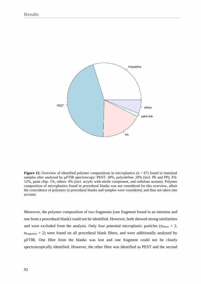
Results
92
Figure 12. Overview of identified polymer compositions in microplastics (n = 67) found in intestinal
samples after analysed by µFTIR spectroscopy: PEST: 30%, polyolefine: 20% (incl. PE and PP), PA:
12%, paint chip: 1%, others: 4% (incl. acrylic with nitrile component, and cellulose acetate). Polymer
composition of microplastics found in procedural blanks was not considered for this overview, albeit
the coincidence of polymers in procedural blanks and samples were considered, and thus not taken into
account.
Moreover, the polymer composition of two fragments (one fragment found in an intestine and
one from a procedural blank) could not be identified. However, both showed strong similarities
and were excluded from the analysis. Only four potential microplastic particles (nfibres = 2,
nfragments = 2) were found on all procedural blank filters, and were additionally analysed by
µFTIR. One fibre from the blanks was lost and one fragment could not be clearly
spectroscopically identified. However, the other fibre was identified as PEST and the second

Chapter II
93
fragment was determined as varnish with kaolin, styrene and calcium carbonate. Furthermore,
two fragments from the intestinal samples had spectra which were highly similar to the varnish
which was found in a procedural blank. Hence, those particles were excluded from the analysis.
In five cases of potential microplastic particles, biogenic matter was identified and a sixth
particle was clearly different from plastic. In addition, one particle could not be identified due
to its small size, since it was broken during the manual collection and placement on the
aluminium oxide filter.
Differences between Seas
Comparing the samples based on their origin (NS or BS), there was a significantly higher
amount of microplastics in individuals from the BS if compared to the NS (nBS = 278;
M±SD=18.27±14.54; nNS = 123; M±SD = 8.2±7.89; p-value = 0.03). Furthermore, the highest
number of 48 microplastic particles was found in an adult female from the BS. When comparing
the share of fibres in both seas, significant differences could be revealed (BS: 51.44%; NS:
47.97%; p-value = 0.02). The share of fragments, however, was similiar across locations (BS:
48.56%; NS: 52.03%; p-value = 0.1).
Differences per Year
The annual mean values for each sea revealed a higher number of microplastic particles in
harbour porpoises from the BS. Furthermore, the range of microplastics found in individuals
from the NS was mostly between zero and up to 10 particles per individual in 2015, 2016, and
2018. Only in 2014 and 2017, more than 20 particles were found in the intestinal samples from
the NS (2014: 29 and 2017: 21). However, in the BS samples more than 30 particles were found
in 2015, 2017, and 2018. The years 2015 and 2018 were the ones with the highest number of
findings per individual (44 particles in 2015 and 48 particles in 2018). In two cases from the
NS, microplastics were not present (2014 and 2016). In comparison, in the BS microplastic was
found in all samples. All this information is presented in Figure 13. However, no significant
differences could be determined between the two sample sites (NS and BS; p-value = 0.21),
mainly because of the low power of the statistical analysis, which resulted from the low sample

Results
94
size within each year and sea. Following the power analysis, a sample size of at least 12
individuals per year for each sea would be necessary for a reliable trend interpretation (power
80%, p-value = 0.05).
Figure 13. Quantity of suspected microplastics from each year, divided by sea (BS = Baltic Sea; NS =
North Sea; n = 401).
Differences in Age and Sex
This study investigates intestinal samples of 13 female and 17 male harbour porpoises (Figure
14). The microplastic burden in females is slightly higher (M ± SD = 13.38 ± 15.41), when
compared to the amount of microplastics in males (M ± SD = 13.35 ± 10.56). Certainly, no
significant difference in microplastic load could be revealed between sexes (p- value = 0.99).
Moreover, no significant differences between adult harbour porpoises (n = 21; M ± SD = 13.82

Chapter II
95
± 13.25) and juvenile ones (n = 9; M ± SD = 12.18 ± 12.31) were confirmed (p-value = 0.82),
although the microplastic amount in adult ones seemed higher. The two unburdened samples
from the NS originated from a juvenile male and an adult female. In both age classes the highest
amount of microplastics was found in females (adult: 48 particles; juvenile: 44 particles; see
Figure 14).
Figure 14. Quantity of suspected microplastic particles (n = 401; secondary contamination was
considered and subtracted) separated by sex and age of the investigated harbour porpoises (n = 30) from
German waters (NS and BS).
Results
96
Health Status
The evaluation of the whole GIT revealed an absence of parasite specimens in all investigated
intestines. A mild enteritis was found for seven individuals. In detail, in one of those harbour
porpoises a mild diffuse eosinophilic enteritis was determined. Furthermore, in two cases
(juveniles) a mild diffuse eosinophilic enteritis and a focal mural one were determined. A most
likely parasitic etiology was observed in those two harbour porpoises. A fourth individual was
affected by a diffuse mild lymphocytic-plasmacellular and eosinophilic infiltration of the
lamina propria in combination with a moderate hyperplasia of the Peyer’s plaques. Three further
porpoises showed evidence of gastritis (mild and high grade) and enteritis. Whereof two
individuals suffered from a mild non-suppurative enteritis, and the third one was also affected
by a diffuse moderate lymphocytic- plasmacellular and eosinophilic infiltration ofthe lamina
propria. In total, parasite infestations of e.g. Pholeter gastrophilus and Anisakis simplex in the
multi-chambered stomach was confirmed in 12 harbour porpoises.
Harbour porpoises investigated in this study, which were either accidentally bycaught (Siebert
et al., 2020) or affected by a pharyngeal entrapment (Gross et al., 2020), showed a microplastic
burden of 19.8 particles (SD = 12.77; n = 11) per individual. Compared to the remaining ones
(n = 19), where no accidental death could be diagnosed (incl. the three pregnant females), a
lower mean value of10 (SD = 11.59) microplastics per porpoise was identified. Furthermore, if
the individual was in a good (n = 9) or moderate (n = 14) nutritional status, the mean number
of particles was significantly higher (Meangood = 14.11; Meanmoderate = 16.07) in contrast to a
bad nutritional status (n = 7; Meanbad = 7) (Figure 15). This is also confirmed by the statistical
analysis (p-value = 0.04).

Chapter II
97
Figure 15. Quantity of suspected microplastics (n = 401) in combination with the nutritional status and
further information about the carcasses (bycaught: the harbour porpoise was (suspected to be) bycaught
by a fishing boat or in a gillnet; flatfish: the harbour porpoise was affected by a pharyngeal entrapment;
na: no extraordinary finding could be revealed; pregnant: pregnancy in female was noticed). The
nutritional status is coded as follows: 1: bad; 2: moderate and 3: good.
Discussion
98
Discussion
This study is the first to evaluate the microplastic burden in marine mammals inhabiting
German waters focussing on particles smaller than 1 mm, in marine mammals inhabiting
German waters. Furthermore, intestinal samples of harbour porpoises originating from the BS
were investigated in microplastic research for the very first time. In total, 93% of all
investigated samples from the NS and the BS show a burden of microplastic particles. Minor
differences in the range of detected fibres, and no differences in the quantity of found fragments
were revealed for both seas. Based on the loss of only 3% of hard parts during sample
processing, and the considered secondary pollution, revealed by the preceding publication, the
results are reliable and not overrated (Philipp et al., 2020).
Evaluation of the Method and Results
The Nile Red staining is a well-established method in microplastic pre-identification to
preselect particles for further investigation (Erni-Cassola et al., 2017). Since some polymer
types are melted or dissolved when stained with Nile Red diluted in chloroform (Tamminga et
al., 2017), a loss of, e.g. polystyrene or cellulose acetate particles is highly likely. Hence, melted
particles were excluded from pre-selection and further polymer identification based on their
deficit in quality and the fragile consistency. Furthermore, based on the manual transferring of
single particles onto the Anodisc filter, a loss of promising polymer particles has to be taken
into account. For future analyses it is advisable to use the Anodisc filter straight away for
filtering the samples, instead of transferring it manually after the filtration process.
Based on the fact that many potential microplastics (91%, 73 out of 80, incl. 77 analysed
particles found in intestines and 3 analysed particles found on procedural blanks) could be
identified as microplastics (incl. nintestinal= 77 and nproceduralsamples= 3) by µFTIR analysis, it is
highly likely that most particles counted in our study were microplastic. Taken this evaluation
and the validation of Philipp et al. (2020) into account (90%), a reliable number of 361
microplastics out of the 401 suspected particles is assumed. Thus, the results show the
applicability of the protocol introduced by Philipp et al. (2020) for intestinal samples of
cetaceans and determine the actual burden in a reliable way.
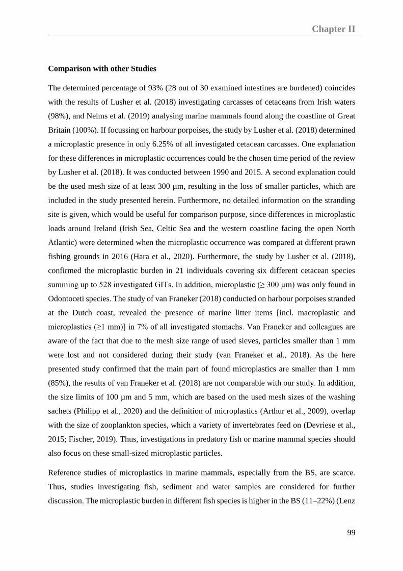
Chapter II
99
Comparison with other Studies
The determined percentage of 93% (28 out of 30 examined intestines are burdened) coincides
with the results of Lusher et al. (2018) investigating carcasses of cetaceans from Irish waters
(98%), and Nelms et al. (2019) analysing marine mammals found along the coastline of Great
Britain (100%). If focussing on harbour porpoises, the study by Lusher et al. (2018) determined
a microplastic presence in only 6.25% of all investigated cetacean carcasses. One explanation
for these differences in microplastic occurrences could be the chosen time period of the review
by Lusher et al. (2018). It was conducted between 1990 and 2015. A second explanation could
be the used mesh size of at least 300 µm, resulting in the loss of smaller particles, which are
included in the study presented herein. Furthermore, no detailed information on the stranding
site is given, which would be useful for comparison purpose, since differences in microplastic
loads around Ireland (Irish Sea, Celtic Sea and the western coastline facing the open North
Atlantic) were determined when the microplastic occurrence was compared at different prawn
fishing grounds in 2016 (Hara et al., 2020). Furthermore, the study by Lusher et al. (2018),
confirmed the microplastic burden in 21 individuals covering six different cetacean species
summing up to 528 investigated GITs. In addition, microplastic (≥ 300 µm) was only found in
Odontoceti species. The study of van Franeker (2018) conducted on harbour porpoises stranded
at the Dutch coast, revealed the presence of marine litter items [incl. macroplastic and
microplastics (≥1 mm)] in 7% of all investigated stomachs. Van Franeker and colleagues are
aware of the fact that due to the mesh size range of used sieves, particles smaller than 1 mm
were lost and not considered during their study (van Franeker et al., 2018). As the here
presented study confirmed that the main part of found microplastics are smaller than 1 mm
(85%), the results of van Franeker et al. (2018) are not comparable with our study. In addition,
the size limits of 100 µm and 5 mm, which are based on the used mesh sizes of the washing
sachets (Philipp et al., 2020) and the definition of microplastics (Arthur et al., 2009), overlap
with the size of zooplankton species, which a variety of invertebrates feed on (Devriese et al.,
2015; Fischer, 2019). Thus, investigations in predatory fish or marine mammal species should
also focus on these small-sized microplastic particles.
Reference studies of microplastics in marine mammals, especially from the BS, are scarce.
Thus, studies investigating fish, sediment and water samples are considered for further
discussion. The microplastic burden in different fish species is higher in the BS (11–22%) (Lenz

Discussion
100
et al., 2016; Beer et al., 2018) when compared to the southern NS (5.4%) (Foekema et al., 2013).
In addition, studies investigating fish species inhabiting waters between Norway and Denmark
determined a low risk of microplastic occurrence in fish (1.2%) (Foekema et al., 2013). In
contrast, the microplastic concentrations in surface waters and sediment samples show higher
concentration in the southern NS (Karlsson et al., 2017; Lorenz et al., 2019), compared to
findings of the BS (Graca et al., 2017; Tamminga et al., 2018). Whereas, a model on the global
fibre distribution in surface waters estimated a higher accumulation in the BS
(∼1,760±4,500m−3), compared to the North Atlantic region (∼1,800 ± 1,720 m−3) (Lima et
al., 2021). Nevertheless, an ubiquitous distribution of synthetic particles along the German BS
coast is assumed (Stolte et al., 2015). Furthermore, the fact that marine mammals of the BS
might be more exposed to marine litter than in the NS was already confirmed (Unger et al.,
2017). Thus, a higher risk of microplastic burden in the BS could be hypothesised. The results
obtained in this study underline the findings of marine litter and support the following
hypothesis: Investigated harbour porpoises of the BS show a higher number of microplastic
particles (incl. fibres and fragments), in contrast to individuals from the NS in each year. In
particular, two females from the BS show a high amount of microplastics (44 and 48 particles).
For avoiding an overassessment in future research, higher sample sizes per sea are highly
recommended.
It will be worth to strive for a reference study investigating the area of the Baltic Proper, since
this area is assumed to accumulate pollutants from the whole BS (Stolte et al., 2015), and the
here occurring harbour porpoise subspecies is critically endangered (Carlén et al., 2018).
Nevertheless, the time span of the available sample collection and the quantity of samples is
still too low for identifying a trend in both seas. After statistical assessment with paired t-test
and Cohen’s d, the samples size has to be increased if reliable comparisons in microplastic
burden of individuals of both seas should be evaluated. Thus, a continuation of the herein
presented approach is advisable and is intended.
Polymer Findings
In this study, the most frequently found polymer was PEST. Based on the fact that six fragments
and 24 fibres were found in the intestinal samples and only one PEST fibre was identified in

Chapter II
101
one of the procedural blanks, PEST microplastics were still taken into account and were not
excluded from this study. Additionally, the procedural blanks show a low amount of fibres
(Øfibres = 1 and Øfragments = 7 per procedural blank) and were already subtracted from the results
presented here. To control for microplastic contamination, only cotton gloves and lab coats
were worn while processing the samples (Philipp et al., 2020). Other studies investigating GIT
samples of marine mammals from the North Atlantic found PEST particles, even though high
protective measurements were used (Lusher et al., 2015a; Nelms et al., 2019). In addition, a
high amount of synthetic fibres like PEST fibres were determined in the Northeast Atlantic
region (Thompson, 2004; Lusher et al., 2014), and in inhabiting fish species (Lusher et al.,
2013; McGoran et al., 2017; Pereira et al., 2020). The twelve found polyamide particles were
not excluded from the analysis, since fibres of the used nylon cloth (PA) are obviously
identifiable due to their unique fibre pattern (Philipp et al., 2020), and are clearly different from
the PA particles found in the intestinal samples. Thus, those fibres were immediately excluded
while pre-selecting, counting and collecting particles for microplastic analyses. Furthermore,
not only PA fibres but also PA fragments were found. Previously, high numbers of PA
microplastics were identified in GITs of marine mammals (Nelms et al., 2019) and in fish from
the North-East Atlantic region (Lusher et al., 2013; McGoran et al., 2017).
Both, the NS and the BS are enclosed by highly populated countries (Halpern et al., 2008,
2015). The system of waste management is relatively sophisticated in European countries, but
there is still a high amount of produced waste, thus the likelihood of litter input into the marine
environment is increasing (Andreasi Bassi et al., 2017). Another source of microplastic
particles, and in particular of fibres which originate from washed clothes are waste water
treatment plants (WWTP) (Browne et al., 2011; Bretas Alvim et al., 2020). Moreover, it is well
known that WWTP are not able to hold back microplastic fibres and particles (Dubaish and
Liebezeit, 2013; Mani et al., 2016). Thus, it could be assumed that the found PA and PEST
microplastics originated from waste water, which is transported by rivers and ends up in the
marine environment (Browne et al., 2011; Dubaish and Liebezeit, 2013; Mani et al., 2016).
Besides land-based sources, which are responsible for 80% of marine litter inputs (Jambeck et
al., 2015), sea-based sources like lost fishing gear or waste generated on ships or offshore
installations need to be taken into account (Galgani et al., 2013). Furthermore, both seas are
highly impacted by anthropogenic exploitations, such as fishing activities (Catchpole et al.,
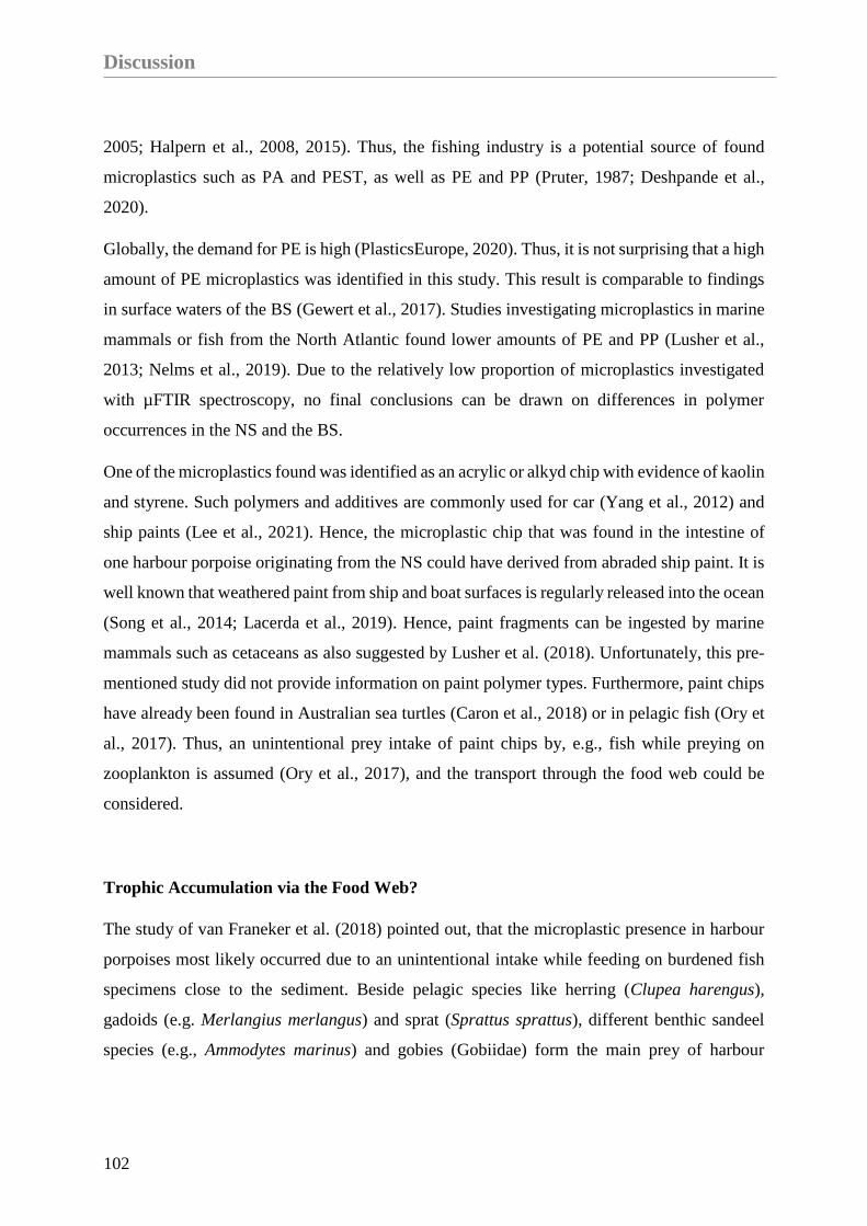
Discussion
102
2005; Halpern et al., 2008, 2015). Thus, the fishing industry is a potential source of found
microplastics such as PA and PEST, as well as PE and PP (Pruter, 1987; Deshpande et al.,
2020).
Globally, the demand for PE is high (PlasticsEurope, 2020). Thus, it is not surprising that a high
amount of PE microplastics was identified in this study. This result is comparable to findings
in surface waters of the BS (Gewert et al., 2017). Studies investigating microplastics in marine
mammals or fish from the North Atlantic found lower amounts of PE and PP (Lusher et al.,
2013; Nelms et al., 2019). Due to the relatively low proportion of microplastics investigated
with µFTIR spectroscopy, no final conclusions can be drawn on differences in polymer
occurrences in the NS and the BS.
One of the microplastics found was identified as an acrylic or alkyd chip with evidence of kaolin
and styrene. Such polymers and additives are commonly used for car (Yang et al., 2012) and
ship paints (Lee et al., 2021). Hence, the microplastic chip that was found in the intestine of
one harbour porpoise originating from the NS could have derived from abraded ship paint. It is
well known that weathered paint from ship and boat surfaces is regularly released into the ocean
(Song et al., 2014; Lacerda et al., 2019). Hence, paint fragments can be ingested by marine
mammals such as cetaceans as also suggested by Lusher et al. (2018). Unfortunately, this pre-
mentioned study did not provide information on paint polymer types. Furthermore, paint chips
have already been found in Australian sea turtles (Caron et al., 2018) or in pelagic fish (Ory et
al., 2017). Thus, an unintentional prey intake of paint chips by, e.g., fish while preying on
zooplankton is assumed (Ory et al., 2017), and the transport through the food web could be
considered.
Trophic Accumulation via the Food Web?
The study of van Franeker et al. (2018) pointed out, that the microplastic presence in harbour
porpoises most likely occurred due to an unintentional intake while feeding on burdened fish
specimens close to the sediment. Beside pelagic species like herring (Clupea harengus),
gadoids (e.g. Merlangius merlangus) and sprat (Sprattus sprattus), different benthic sandeel
species (e.g., Ammodytes marinus) and gobies (Gobiidae) form the main prey of harbour

Chapter II
103
porpoises (Leopold, 2015). The linkage between burdened prey species and top predators was
previously confirmed for grey seals preying on gadoids (Hernandez-Milian et al., 2019).
Moreover, three of the here analysed harbour porpoises asphyxiated by pleuronectiforms (e.g.,
Solea solea) and two further individuals were bycaught in gillnets for benthic and demersal fish
species. Those harbour porpoises showed a high mean microplastic amount per individual (M
± SD = 17.7 ± 12.42). Thus, a relationship between preying on benthic fish and the presence of
microplastics in predator species is highly assumed. Furthermore, the bycaught or asphyxiated
porpoises were in a better nutritional status in comparison to the remaining ones. However, a
good nutritional status is not synonymous with a good health of a harbour porpoise (Siebert et
al., 2020). Based on the results presented herein, it can be assumed that a harbour porpoise,
which is not physically hampered in feeding or hunting, will accumulate more microplastics in
the GIT than a diseased one. The results of this study are supported by the assumptions of
Leopold (2015) and Rummel et al. (2016) who claimed that 5,000 individual gobies per day are
needed for maintaining a harbour porpoise’s good physical condition, and the mean burden of
0.03 particles in each benthic fish may result in a daily intake of almost 150 microplastic
particles by one adult harbour porpoise. Kastelein et al. (1998) detected a passage time of 143
- 196 min in captive harbour porpoises. If the assumption is that a full digestion takes ~3 h, a
harbour porpoise will approximately defaecate eight times a day. Thus, the potential
accumulation of 150 microplastics has to be divided by eight, resulting in 18.75 particles per
egestion. Certainly, this is just a rough estimate and many factors influence egestion and
ingestion rates, but this calculation coincides with the findings of microplastics in samples from
the BS (M±SD = 18.27±14.54).
Impact on the Health Status
Across all 30 individuals no intralesional parasites could be identified in the intestines.
However, seven individuals showed an enteritis, whereof two are attributed to parasites and
another one accompanied by a hyperplasia of Peyer’s plaques. These accumulations of follicles
are known to adsorb different kinds of particles (biogenic and synthetic), and to transfer it into
the lymph system when the particle is adequately small in size(Ensign et al., 2012). In total,
only two juvenile harbour porpoises originating from the BS showed changes in the Peyer’s
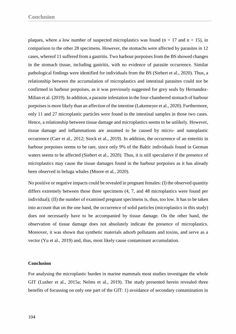
Conclusion
104
plaques, where a low number of suspected microplastics was found (n = 17 and n = 15), in
comparison to the other 28 specimens. However, the stomachs were affected by parasites in 12
cases, whereof 11 suffered from a gastritis. Two harbour porpoises from the BS showed changes
in the stomach tissue, including gastritis, with no evidence of parasite occurrence. Similar
pathological findings were identified for individuals from the BS (Siebert et al., 2020). Thus, a
relationship between the accumulation of microplastics and intestinal parasites could not be
confirmed in harbour porpoises, as it was previously suggested for grey seals by Hernandez-
Milian et al. (2019). In addition, a parasite infestation in the four-chambered stomach of harbour
porpoises is more likely than an affection of the intestine (Lakemeyer et al., 2020). Furthermore,
only 11 and 27 microplastic particles were found in the intestinal samples in those two cases.
Hence, a relationship between tissue damage and microplastics seems to be unlikely. However,
tissue damage and inflammations are assumed to be caused by micro- and nanoplastic
occurrence (Carr et al., 2012; Stock et al., 2019). In addition, the occurrence of an enteritis in
harbour porpoises seems to be rare, since only 9% of the Baltic individuals found in German
waters seems to be affected (Siebert et al., 2020). Thus, it is still speculative if the presence of
microplastics may cause the tissue damages found in the harbour porpoises as it has already
been observed in beluga whales (Moore et al., 2020).
No positive or negative impacts could be revealed in pregnant females: (I) the observed quantity
differs extremely between those three specimens (4, 7, and 48 microplastics were found per
individual); (II) the number of examined pregnant specimens is, thus, too low. It has to be taken
into account that on the one hand, the occurrence of solid particles (microplastics in this study)
does not necessarily have to be accompanied by tissue damage. On the other hand, the
observation of tissue damage does not absolutely indicate the presence of microplastics.
Moreover, it was shown that synthetic materials adsorb pollutants and toxins, and serve as a
vector (Yu et al., 2019) and, thus, most likely cause contaminant accumulation.
Conclusion
For analysing the microplastic burden in marine mammals most studies investigate the whole
GIT (Lusher et al., 2015a; Nelms et al., 2019). The study presented herein revealed three
benefits of focussing on only one part of the GIT: 1) avoidance of secondary contamination in

Chapter II
105
smaller samples is easier, 2) the remaining carcass and GIT can be entirely evaluated for a
health monitoring, and 3) the findings in the rectum and faeces confirm the egestion of
microplastic particles. For evaluating the intake and egestion rate, further research is needed.
Nevertheless, microplastic investigations and experiments in mammals, and especially in free-
ranging marine mammals, are complicated based by field conditions. Furthermore, ethical
concerns arise as indicated by Nelms et al. (2018). Thus, samples of carcasses and faecal
samples of alive individuals are the most feasible approach to assess the microplastic burden in
marine mammals.
This is the first study investigating harbour porpoises from different subpopulations for
microplastics, and revealed differences in microplastic presence in the NS and the BS. A higher
risk of exposure to microplastics was revealed for the western Baltic population, if compared
to the North-East Atlantic population. Thus, a higher microplastic burden in the BS is assumed.
Furthermore, evidence for the continuous accumulation of microplastics via the food web was
given, but could not significantly be confirmed in adult individuals, compared to juvenile ones.
Additionally, there is no significant difference in the quantity of synthetic particles in male or
female harbour porpoises. To gain further knowledge on differences in sex or age, the quantity
of samples has to be increased in future research.
An important relationship between a good or moderate nutritional status and the occurrence of
microplastics is demonstrated in this study. Moreover, the egestion and thus, a discharge of
microplastic particles out of the organism could be confirmed. No relationship between
parasites, tissue damage and microplastic presence could be identified. Therefore, a histological
investigation of cell damage or tissue damage localisation with the help of biomarkers would
be advisable in future research. Further investigations are needed for evaluating the rate of
accumulation and burden in harbour porpoises in the different seas. Indeed, this study outlines
first evidence in retrospective microplastic burden. Nevertheless, a higher sample size, as well
as a larger temporal coverage is needed to reliably estimate trends in the microplastic burden in
harbour porpoises. Furthermore, this study supports the need for a comprehensive marine litter
monitoring in predatory species to gain knowledge on accumulation processes and health
assessment in apex species of the marine food web.

Acknowledgements
106
Author Contributions
CP, BU, and US conceptualized the study and acquired funding. CP conducted the laboratory
analysis of processing the samples and isolate microplastics, assisted by BU. CP did the
statistical analysis. JHEK provided the µFTIR and FTIR for polymer type analysis. SME
conducted the polymer identification by µFTIR and FTIR. CP and SME generated the figures.
The manuscript was prepared by CP, and contributed editing with perspectives and arguments
was done by BU, SME, JHEK, and US. All authors approved the submitted version.
Funding
The stranding network and the health monitoring are partly funded by the Schleswig-Holstein
Agency for Coastal Defence, National Park and Marine Conservation, Germany. The
pathological investigations of the marine mammals are partly funded by the Ministry of Energy,
Agriculture, Environment, Nature and Digitalization of Schleswig-Holstein, Germany. The
Federal Environment Agency (UBA) partly funded the analysis of samples within the project
“Assessment and implementation for long-term monitoring of pollution of diverse marine
compartments and biota with marine litter” (FKZ 3717252250).
Acknowledgements
We would like to thank all seal rangers in Schleswig-Holstein, Germany for reporting and
collecting carcasses. Further thanks go to our colleagues in the ITAW necropsy team for
collecting and dissecting carcasses, as well as collecting samples for our purpose continuously
throughout the year. Special thanks go to Dieter Steinhagen for improving this study with his
valuable comments. Besides, we would like to thank the reviewers for their valuable comments
and advice, which helped to improve the manuscript.

Chapter II
107
Supplementary Material
Supplementary Table 2. Information on all 30 investigated harbour porpoises from German waters
(2014 – 2018). Every individual was examined after Siebert et al. (2001, 2020) and microplastic
investigation was conducted after Philipp et al. (2020).
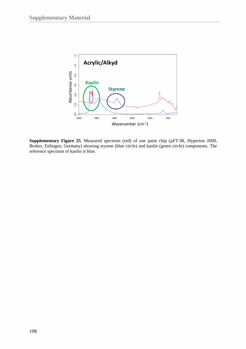
Supplementary Material
108
Supplementary Figure 25. Measured spectrum (red) of one paint chip (µFT-IR, Hyperion 2000,
Bruker, Ettlingen, Germany) showing styrene (blue circle) and kaolin (green circle) components. The
reference spectrum of kaolin is blue.

Overall Discussion

Overall Discussion
111
Overall Discussion
The number of studies investigating microplastic particles in the marine environment increased
during recent years (Thompson, 2015). Moreover, in- and egestion rates of microplastics and
associated health effects in invertebrates are well-known, but still a cause of concern. Most
findings are based on laboratory studies with invertebrates or rodents like mice and rats, which
revealed new insights into pathways of microplastic particles within cells and organisms (Carr
et al., 2012; Cole et al., 2013; Setälä et al., 2016; Bhagat et al., 2020; da Costa Araújo and
Malafaia, 2021). However, detailed knowledge of the presence in free-ranging top predators,
like marine mammals, is still lacking. The mentioned knowledge gaps exist due to two aspects:
i) the access to these species is difficult because of their aquatic lifestyle. For cetaceans, the
easiest opportunity is to collect samples from stranded or bycaught specimen (Lusher et al.,
2015a, 2018; Hernandez-Milian et al., 2019; Nelms et al., 2019). The advantage of focussing
on pinnipeds is the possibility to collect scat samples of live individuals on known haul-out
sites, to assess the microplastic burden of a certain population (Donohue et al., 2019; Perez-
Venegas et al., 2020). ii) Laboratory studies, pre-trials or similar investigations are not feasible
due to ethical concerns.
Focussing on the study sites the North and Baltic Seas, investigations in microplastic particles,
in particular in the size range between 300 µm and 100 µm is lacking for all three species
(harbour porpoise, harbour seal and grey seal). Thus, the present study will complement existing
investigations on microplastic occurrence in marine mammals from the North Sea. Since no
data is currently available from the Baltic Sea, so this thesis will provide the first baseline
information on microplastic burden in Baltic marine mammal species.
Besides these listed problems, only limited standardisation is found in the high number of
arising protocols in sample handling, sample processing and microplastic identification in the
literature, which reduces the comparability (Löder and Gerdts, 2015). Different sample
collection, cleaning, and isolation procedures for purification of the synthesised particles for
further polymer identification (loss of biogenic material) are suggested. In addition, to enable a
valuable examination of microplastic presence in different media, an afterwards or so-called
secondary contamination needs to be considered (Norén, 2007; Hidalgo-Ruz et al., 2012; Nuelle
et al., 2014; Vandermeersch et al., 2015; Torre et al., 2016).

Overall Discussion
112
Thus, the samples of interest need to be treated with protective care to avoid an overestimation
of particles, which is initiated by airborne contamination or impure detergents and instruments
(Norén, 2007; Hidalgo-Ruz et al., 2012; Nuelle et al., 2014; Vandermeersch et al., 2015; Torre
et al., 2016).
To overcome these problems in sample handling, the first aim of this doctoral thesis was the
establishment of a protocol which is effective in cost and time, and is thus applicable for
different working groups, particularly focussing on gastrointestinal tract (GIT) and scat
samples. To verify the protocol and its outcomes, the six following key questions were
considered in Chapter I:
1) Is it possible to lower the risk of secondary contamination by using selected protective
measures during sample collection and preparation?
2) What are the benefits and disadvantages of using a washing machine for sample
purification in microplastic research?
3) Does the type of sample (intestine or scat) require different sample handling
procedures?
4) How is it possible to avoid an over- or underestimation in microplastic findings?
5) Does the quantity of microplastic particles differ in intestine and scat samples?
1) Is it possible to lower the risk of secondary contamination by using selected protective
measures during sample collection and preparation?
The establishment of an efficient protocol of sample handling and processing, which meets the
standards necessary to prevent secondary contamination and to purify the samples, was
developed by examining intestinal and scat samples of seals from the North and Baltic Seas.
In order to reduce the risk of contamination to the lowest possible level, the sample processing
begins at the stage of sample collection. While some studies give advice to handle samples with
metal instruments and to store them in glass jars (Lusher and Hernandez-Milian, 2018; Nelms
et al., 2019), other studies recommend the usage of plastic containers or bags (Lusher et al.,

Overall Discussion
113
2013; Donohue et al., 2019). Moreover, several researches investigated the change of physical
plastics properties under various environmental conditions such as UV radiation, low and high
temperatures or the exposure to sea water (Andrady, 1990, 2011). To assess the potential effects
of different storage types on the samples, this study is the first one to report the comparison
between the use of glass jars and plastic bags in affecting samples. The results presented here
confirm a lower contamination of samples stored in glass jars compared to samples stored in
plastic bags, for different time periods (3, 6, 12 and 96 months) at -20°C. Further, since no
particles were found in the samples matching the characteristics of the polymer of the used
plastic bags, it can be assumed that the contamination is the result of the bags and further due
to the fact, that the plastic bags were not cleaned prior to usage, as it was carried out for the
glass jars.
Accordingly, secondary contamination can play a vital role in overestimating the real burden
and several studies investigating microplastics give advice to process the samples within a
laminar flow or in a clean laboratory (Nelms et al., 2018; Bergmann et al., 2019). Due to the
fact that this is not always feasible, other studies already introduced low-budget alternatives
like the usage of procedural blanks (Nuelle et al., 2014; Hengstmann et al., 2018; Nelms et al.,
2018), or totally exclude fibres from the analysis (Kühn et al., 2020). Since secondary
contamination is often indicated by a high proportion of fibres, we could show that new
protective measures, such as wearing cotton gloves, next to cotton lab coats and processing the
samples within a closed acrylic box, reduce the proportion of fibres: The low share of fibres in
the analysed procedural blanks (one fibre was found in 12 blanks) and in investigated samples
(M±SD = 6±7.4 fibres) demonstrates the success in largely avoiding secondary contamination
(Nuelle et al., 2014; Donohue et al., 2019; Kühn et al., 2020) by the measures applied.
Moreover, the proportion of detected fibres in this study are lower compared to other studies
investigating GIT samples of marine mammals (Lusher et al., 2018; Donohue et al., 2019;
Nelms et al., 2019), which further supports the established protocol.
Furthermore, all used detergents and materials were analysed, and results were corrected
respectively in the presented method. Those were the first steps to decrease the risk of secondary
contamination. In that respect, we advise to process only parts of the GIT, since experience has
taught, that the use of smaller samples (in size, volume, and weight) further reduces the risk of

Overall Discussion
114
secondary contamination, in as much as smaller samples are simpler to handle, and thus easier
to protect.
2) What are the benefits and disadvantages of using a washing machine for sample
purification in microplastic research?
The choice of a purification method within this doctoral thesis fell on the use of a washing
machine, an already established technique in diet analysis for gaining clean hard parts like
otoliths and fish bones for prey identification (Orr et al., 2003; Leopold et al., 2015).
Investigating GIT samples for diet analysis is thus easily connectable with microplastic
research. Moreover, this benefits microplastic research since the analysis of prey species in
combination with the accumulation of microplastics in the prey and the predator species can
reveal possible relations. Moreover, the utilisation of a washing machine saved time in the
cleaning process (1.5 hours), when compared to other approaches e.g. enzymatic or chemical
digestion, which can last for days to weeks (Foekema et al., 2013; Cole et al., 2014; Lusher and
Hernandez-Milian, 2018). Sewing the washing bags around the sample directly (referred to as
“washing sachet”) will avoid a loss of sample, as it is shown in this study (3%) and is thus
highly recommended for further analyses, since no adequate information on the sealing was
given in former studies dealing with stomach or intestine contents (Orr et al., 2003; Bravo
Rebolledo et al., 2013; Leopold et al., 2015).
Approaching the real burden of microplastics includes the consideration of different size
classes. Since tools for the isolation and identification of nanoplastics (≤20 µm) from marine
mammal tissue is lacking (Wagner et al., 2014; Ferreira et al., 2019; Prüst et al., 2020) and are
not within the scope of this thesis. Even the focus on particles in a size range of 100 µm to 5
mm in this thesis is uncommon in microplastic research, as the majority of microplastic
investigations in marine mammals focussed on particle sizes over 300 µm (Besseling et al.,
2015; Lusher et al., 2018; van Franeker et al., 2018; Donohue et al., 2019; Hudak and Sette,
2019; Perez-Venegas et al., 2020), it complements the existing knowledge in microplastic
presence.
Overall Discussion
115
The fact of size limitations to 300 µm is based on the beginning in microplastic analysis, since
first findings of synthetic particles were found in manta trawls (333 µm) (Arthur et al., 2009)
and sediment samples (Thompson, 2004), both focussing on plankton species and in virtue of
the limitations in the applied techniques for isolation and identification (Foekema et al., 2013;
van Franeker et al., 2018; Donohue et al., 2019; Hudak and Sette, 2019). Yet, this study
demonstrated, that when using the presented methods, a detection of particles smaller than 300
µm is well possible without the use of highly advanced techniques.
3) Does the type of sample (intestine or scat) require different sample handling
procedures?
In order to highlight the applicability for different sample types, intestinal tissue (including
faeces) and scat samples were examined. The results show, the used method was suitable for
both sample types. Nevertheless, both types showed different benefits for further research. The
examination of intestinal samples for microplastic burden analysis revealed the following three
advantages: i) the intestinal tissue, which is potentially carrying microplastics within the lumen
is included, ii) the possibility of collecting sub-samples of the intestinal tissue prior to
processing, enabled further investigations, e.g. histological examinations, and iii) samples were
collected during necropsies, which allows to note valuable information on sex, age and health
status of each individual, and thus facilitates further studies in microplastic research related to
health issues. Despite these advantages of intestinal samples, the analysis of scat samples has
also benefit, as the microplastic burden and its suspected effects in live animals can be assessed
and estimated. Since seals are assumed to be philopatric (Pomeroy et al., 2000; Dietz et al.,
2012), information on the used habitat can be incorporated and sample collection repeated over
time. In addition, correlations between the volume of scats and the microplastic burden is
assessable, since the actual load could be determined, which gives new insights in microplastic
egestion. Furthermore, it has to be taken into account that necropsy samples represent only a
small part of the population,, since an unknown share of individuals is dying offshore (Williams
et al., 2011; Peltier et al., 2012), thus bearing the risk of an underestimation, which could be
likely somehow compensated for by examining scat samples.
Overall Discussion
116
4) How is it possible to avoid an over- or underestimation in microplastic findings?
By using the washing machine and self-sewed washing sachets out of fabrics in different mesh
sizes (100 µm and 300 µm), the inclusion of small synthesised particles (≥100 µm) was feasible.
Thus, this study was able to fill the knowledge gaps in occurrence of those smaller particles in
marine mammals. Here it was shown, that the exclusion of these smaller particles results in a
considerable difference: only 37% of suspected microplastics would be considered in 9 out of
10 examined seal intestinal samples (instead 10/10 if no secondary contamination would be
considered; Chapter I), and only 44% of potential microplastics in 24 out of 30 examined
harbour porpoise samples (instead 28/30 if no secondary contamination would be considered;
Chapter II). Particles, which are suspected to originate from secondary contamination were
not subtracted in this part, since in our opinion, the size of a particle plays a subordinate role
compared to the polymer composition or the appearance. Moreover, the size of each particle
(originating from contamination or not) can vary, thus an exclusion of microplastics by size
was not considered in the whole study. Accordingly, applying the suggested protocol has
therefore the potential to considerably reduce the risk of underestimating the true microplastic
burden.
After the inclusion of different size classes, the identification of synthesised particles out of the
biogenic ones plays a major role in avoiding an overestimation. Correspondingly, an approach
to pre-select suspected microplastics out of the total number of particles was additionally
established within this thesis. To pre-select potential microplastics for further polymer
determination, the samples were stained with Nile Red and visually examined under a
fluorescence microscope, which is an already established tool in microplastic research (Erni-
Cassola et al., 2017; Shim et al., 2017; Tamminga et al., 2017). Moreover, it is recognised that
only 1.4% out of particles which were visually inspected by the naked eye or under a traditional
microscope, could be confirmed as synthetic ones after polymer identification spectroscopy
(Löder and Gerdts, 2015).
In order to differentiate whether stained particles are of biogenic or synthetic origin they were
categorised following three different characteristics: i) associations with biogenic material, ii)
fluorescence intensity/colour, and iii) the appearance of the particle. Based on those categories,
1,185 particles (found in GIT samples of different marine mammal species) were examined and

Overall Discussion
117
a decision tree was established. Accordingly, the utilisation of this protocol, catalogue, and the
decision tree results in a reliable determination of microplastics, which was confirmed by
µRaman spectroscopy (match rate of 90%). This decision tree is applicable for untrained
researchers, next to an emerged catalogue of different characteristics, for identifying suspected
microplastics out of biogenic ones. Furthermore, narrowing down the number of potential
microplastic particles prior to polymer identification by e.g. µRaman spectroscopy, decreases
the risk of misidentification, thus saves time and budget. For instance, polyethylene,
polyethylene terephthalate, polypropylene and polyamide are some of the identified polymers
found in the intestinal and scat samples of seals.
5) Does the quantity of microplastic particles differ in intestine and scat samples?
To put the results of this study in relation to other works dealing with intestinal samples of
marine mammals it is advisable to consider investigations from the same study area (North
Atlantic region). Since investigations from the Baltic Sea are lacking, comparisons between our
results (90%, 0 to 83 microplastics per individual) and microplastic findings in different marine
mammals from the North Sea were considered. Accordingly, findings of microplastics in
marine mammals stranded around the British coastline (100%, 1 to 12 microplastics larger than
35 µm per individual) (Nelms et al., 2019) and around Ireland (100%, 1 to 88 microplastics
larger than 300 µm per individual) (Lusher et al., 2018) overlap with the here presented results.
Similar to the results of a study examining scat samples of fur seals (fragments: 68%, fibres:
32%) (Donohue et al., 2019) a higher proportion of fragments (70%) was found, when
compared to the quantity of fibres (30%) within this thesis. Nevertheless, the percentage of
samples containing microplastics in general in this study is higher (100%, when only the scat
samples are considered), compared to the previously mentioned study on fur seals (41%)
(Donohue et al., 2019) or as in studies examined scat samples of seals found in South America
(68%) (Perez-Venegas et al., 2020) or from the south-east coastline of the USA (6%) (Hudak
and Sette, 2019). The main reasons for this could be the investigated sizes of microplastics. The
focussed particles in the aforementioned studies had at least a size of 250 µm or 500 µm, thus
the smaller fractions were lost whereas those microplastics are included in our study (100 µm
– 5 mm). Thus, an underestimation in the other studies is possible (Donohue et al., 2019). In

Overall Discussion
118
addition, differences between seas and species need to be considered, since also our study
confirmed variations in harbour porpoises, harbour seals and grey seals (if Chapter I and II
are considered here).
To further overcome the knowledge gaps in microplastic presence in excretions of seals, a weak
correlation between the weight of scats and the number of detected microplastics could be
determined (Spearmen p = 0.23). In addition, about 1 microplastic particle (100 µm – 5 mm)
could be identified within 2 g of scat (wet weight; Mparticles±SD = 0.5±0.2 per gram). A
comparison between the intestine samples with faeces content and the microplastic load was
not feasible due to differences in size and thickness of the intestinal tissue, thus resulting in
inappropriate quantities. Moreover, intestine samples and scat samples are not comparable with
each other since scat samples are characterised by a higher volume and weight, and scat samples
from a sandbank represent an accumulation of faeces over a longer time period before egestion.
Further a secondary contamination of the scat samples originating from the sandbank during
defecation cannot be fully ruled out and need to be considered. To enable a comparison between
those sample types, the acquisition of dry weights of the faeces should be considered, and a
subsequent correlation with microplastic findings has to be taken into account in future
research.
Moreover, the inclusion of different GIT parts needs to be considered, since studies
investigating stomach samples of marine mammals confirmed the retaining characteristics of
this hollow organ in comparison to intestinal or scat samples (Nelms et al., 2019). This fact
leads to higher numbers of microplastic particles in stomachs, when compared to intestinal or
scat samples (Nelms et al., 2019), what could be also revealed in unpublished data of comparing
stomach contents with the intestinal samples of the same animal. The findings support and
underline the hypothesis as the determined quantity of microplastics is twice as high in the
stomachs compared to the intestines (Philipp et al., unpublished data).
Thus, the outcome of this study complements existing knowledge and reveals new insights in
microplastic occurrence in seals from German waters.

Overall Discussion
119
Chapter II
To enhance the awareness of microplastic presence in marine top predator species, intestinal
samples of harbour porpoises from the North and Baltic Seas were examined and compared in
the course of the second chapter. For processing the samples and isolating potential
microplastics, the established protocol, presented in Chapter I, was used. The aim here was to
gain further knowledge on the microplastic burden in relation to health and life history of the
harbour porpoises from German waters. For accomplish the main objectives of this chapter, the
following key questions will lead through the following discussion parts.
1) Are harbour porpoises of the North and Baltic Seas are exposed to microplastic
particles?
2) Is the microplastic burden linked to life history parameters or the health status?
3) Which polymers could be identified by µFTIR spectroscopy?
1) Are harbour porpoises of the North and Baltic Seas exposed to microplastic particles?
The results of this analysis confirmed the presence of microplastic particles in intestinal samples
from harbour porpoises originating from German waters (28/30). Intestinal samples from 30
individuals from the North Sea (NS) and the Baltic Sea (BS) show differences between the
origin sea and the collection year (2014 – 2018). Only two samples were free of microplastics
and both originated from individuals found in the NS. Furthermore, in each year the mean
quantity of microplastics found were significantly higher in the samples from the BS (M±SD =
18.27±14.54), compared to ones from the NS (M±SD = 8.2±7.89). Based on the low sample
size from each year originating from the NS or BS, no trend analysis could be conducted, since
the power analysis revealed a power of 20%. Thus, analyses to estimate trends would result in
over- or underestimations. Nevertheless, the aim of this study was to gain first evidence of
microplastic burden in relation to the health status of harbour porpoises from German waters.
This aim was accomplished and results are further discussed in the following paragraphs, albeit
trend estimations with an adapted and enlarged sample size should be considered in future
research.

Overall Discussion
120
In total, 401 microplastics (incl. 202 fibres and 199 fragments) were determined, besides four
meso-fibres which were longer than 5 mm. Three of those fibres were found in an adult male,
and the fourth fibre was identified in a juvenile male harbour porpoise. Both individuals were
found in the Baltic Sea. Certainly, no significant differences between the share of fibres or
fragments could be determined, when both seas were taken into account. As already shown in
Chapter I, the share of fibres is in this study again lower (50%), if compared to other studies
dealing with marine mammals close to the study area (83 - 85%) (Lusher et al., 2018;
Hernandez-Milian et al., 2019; Nelms et al., 2019).
Comparable studies investigating marine mammals from the BS are missing. Thus, studies
focussing on sea water (Tamminga et al., 2018), sediment (Graca et al., 2017; Näkki et al.,
2019) and fish samples (Lenz et al., 2016; Beer et al., 2018) collected from the BS were
considered for comparison purpose. The microplastic burden in different fish species seems to
be higher in the BS compared to the NS (Lenz et al., 2016; Beer et al., 2018). However,
sediment and water samples of the BS show a lower amount of microplastic particles (Graca et
al., 2017; Tamminga et al., 2018), when compared to samples originating from the NS
(Karlsson et al., 2017; Lorenz et al., 2019). The missing knowledge on microplastic burden in
marine mammals inhabiting the BS underlines the importance of this study. Moreover, this
study provides one of the missing pieces in the puzzle of microplastic distribution, occurrence
and accumulation within the ecosystem of the Baltic Sea.
As already mentioned in Chapter I, investigations in microplastic particles smaller than 300
µm are lacking for the southern NS and for the entire BS, thus comparisons are only possible
to a limited extent. There is only one study focussing on stomach contents of harbour porpoises
and investigated particles larger than 1 mm (van Franeker et al., 2018). Nevertheless, this
research revealed a microplastic burden in 9% of the examined stomachs (van Franeker et al.,
2018). This low percentage is not surprising, since the loss of smaller particles is tremendous if
compared with other studies. Moreover, an underestimation in microplastic occurrence is
assumed by us, and the authors itself (van Franeker et al., 2018). Thus, this study seems to be
negligible in the context of microplastic presence and in further discussion, even though it is
the only one focussing on harbour porpoises inhabiting the southern North Sea.
Overall Discussion
121
Two other studies focussing on marine mammals stranded around Great Britain (≥35 µm) and
Ireland (≥300 µm), revealed both microplastic occurrence in each examined individual (Lusher
et al., 2018; Nelms et al., 2019). Those values are within the range of microplastic burden of all
harbour porpoises examined here. Focussing on smaller particles, the here presented study
revealed that 85% of the detected microplastics range between 100 µm and 1 mm. This
percentage is only comparable with the findings around Great Britain, since the majority of that
study lays within the size classes ranging between 35 µm and 1 mm, giving no accurate
percentage (Nelms et al., 2019). In cetacean carcasses found around Ireland, only 17% of all
detected microplastics are in the size range of 300 µm to 1 mm (Lusher et al., 2018). To
overcome those deficiencies in underestimations and comparability between studies, the
inclusion of particles in size of 100 µm to 1 mm needs to be considered in future studies. In
order to be able to compare microplastic findings, future studies should also pay attention on
the same particle size and agree on uniform size classes.
Moreover, those small-sized particles coincide with the size of various zoo- and phytoplankton
species or their prey species, hence an unintentional uptake by organisms of lower trophic levels
and the potential accumulation in top predator species thereafter can be assumed (Thompson,
2004; Farrell and Nelson, 2013; Cole et al., 2014; Setälä et al., 2014; Dawson et al., 2018). For
instance, one study investigating fish species of the South Pacific Ocean revealed the accidental
uptake of paint chips, instead of feeding on blue-coloured copepods (Ory et al., 2017). This
discovery coincides with the findings of paint chips in intestinal samples of harbour porpoises
originating from the North Sea from this study and additionally emphasises the importance of
focussing on small-sized particles. The accumulation of synthetic particles in different size
classes within the last 50 years in the Northeast Atlantic is evident (Thompson, 2004), yet the
degradation of those particles into smaller ones by different physical forces needs to be
considered (Andrady, 1990, 2011), when investigating microplastics. Thus, further studies need
to focus on small-sized particles (e.g. 100 µm to 1 mm) to give a comprehensive overview in
microplastic burden.
Overall Discussion
122
2) Is the microplastic burden linked to life history parameters or the health status?
The study described in this chapter is one of the very first ones, investigating microplastic
burden in relation to life history parameters like age and sex of examined harbour porpoises.
Information on the age and sex of the individuals compared with the amount of microplastics,
could not reveal significant differences. The first hypothesis of accumulating microplastics in
the GIT over the time, resulting in a higher burden in older individuals could not be confirmed.
Since, no correlation between age and number of microplastics was found, the egestion of
particles needs to be considered and is likely. Moreover, the results of the scat samples analysed
in Chapter I support this assumption. Thus, an accumulation of microplastics in the intestinal
lumen, as it is assumed for nanoparticles (Domenech et al., 2020) in particular in the area of
Peyer’s plaques, could likely be excluded for the sizes considered in our study (Jani et al.,
1992a, 1992b; Florence et al., 2000; Stock et al., 2019). In particular, it can be expected that
some smaller particles get stuck in straitened parts of the GIT or that they are held back by
mucosa or microvilli in the lumen of the intestine (Jani et al., 1992b; Florence and Hussain,
2001; Stock et al., 2019). By including the intestinal tissue in the microplastic analysis, all
potential particles attached to the tissue were considered for this investigation. Moreover, a
transport of nanoparticles by the lymph and blood system, and thus the passing on of pollutants
and plastic particles is already confirmed during gestation and lactation for mammals and
humans (Wolkers et al., 2004; Desforges et al., 2012; Aerts et al., 2019; Ragusa et al., 2021;
Witczak et al., 2021). Even though nanoparticles were not considered here, an assessment of
the microplastic presence in females (incl. three pregnant ones) and males was evaluated.
However, no significant differences could be revealed by the statistical analysis when
comparing both sexes. It needs to be highlighted though, that the sample size is too low for a
comprehensive understanding and the confirming of incontrovertible facts. Thus, an increased
sample size and the focus of small-sized particles like nanoparticles is advisable for further
investigations.
Besides the information on the life history, data on the health status and the potential cause of
death could be evaluated and considered in microplastic research. Thus, a linkage between the
health status and microplastic occurrence is expected to enhance the knowledge in microplastic
burden. In particular, the results unveiled a relationship between the nutritional status of a
harbour porpoise and its microplastic burden. Individuals having a good or moderate nutritional

Overall Discussion
123
status showed a higher number of microplastics, when compared with porpoises in a poor
nutritional status. However, a good or moderate nutritional status is not necessarily
accompanied by good health of an individual (Siebert et al., 2020). The fact that harbour
porpoises with a good constitutional status are highly active in feeding and thus the constant
uptake of microplastics need to be considered, since those cetaceans are well-known to feed
and hunt on benthic fish species (Gilles, 2008; Leopold, 2015; Andreasen et al., 2017; van
Franeker et al., 2018; Gross et al., 2020). Hence, due to their feeding strategy the sediment is
stirred up and microplastics are released into the marine environment and can be accidentally
consumed. In general, the burden of microplastic in benthic and pelagic fish species like herring
(Clupea harengus), cod (Gadus morhua) and a variety of pleuronectiforms (e.g. common sole
(Solea solea) and European flounder (Platichthys flesus)) originating from the NS and BS are
already determined and the load does not significantly differ from each other (Foekema et al.,
2013; Lenz et al., 2016; Rummel et al., 2016; Beer et al., 2018; Fischer, 2019).
Several studies already investigated sediments and their load of marine litter, and ascertain that
sediments serve as a sink in particular for microplastics (Woodall et al., 2014; Kanhai et al.,
2019; Näkki et al., 2019). Beside the study of Hernandez-Milian et al. (2019), investigating
GIT samples of grey seals, benthic prey species and its microplastic load, our study gives also
a significant evidence for underlining the hypothesis of an uptake of microplastics in top
predators (such as marine mammals) by feeding on benthic prey species (such as fish).
In order to gain a better understanding of the relationship between nutritional status and
microplastic burden it needs to be highlighted, that in individuals where a pre-mortem feeding
event is assumed (accidental bycaught individuals in gillnets for benthic fish species or
individuals died by a pharyngeal entrapment by benthic flatfish, n = 11) and which were further
considered to be in a good nutritional status, a higher microplastic burden was found (M±SD =
19.18±12.77) in comparison to the remaining ones (n = 19, M±SD = 10±11.59).
Correspondingly, those results reveal a relationship between individuals, which could be likely
considered as healthy, and are thus expected to hunt and feed on prey in a natural and effective
way, and the number of microplastic particles within their GIT in contrast to individuals for
which recent feeding events are considered unlikely. These results show that during a constant
uptake of prey species, the amount of plastic particles is kept high while for individuals not
having fed for a certain period of time due to e.g. sickness, the burden is reduced due to the

Overall Discussion
124
likely constant excretions of particles. This raises the questions whether actual healthy
individuals are exposed to an increased risk of ingesting microplastics and whether pelagic vs.
benthic feeding strategies influence this risk. Moreover, the selection of prey species and their
exposure to microplastics needs also to be further addressed in future studies.
Furthermore, it was already shown for krill species, that they digest microplastics and thus serve
as a source of nanoplastics for other organisms or enhance the uptake of those particles when
ingested by predator species (Dawson et al., 2018). Concerning this matter, the isolation and
detection of nanoplastics by e.g. histological analysis (Ahrendt et al., 2020; Batel et al., 2020)
or the utilisation of biomarkers (Revel et al., 2019; Sarasamma et al., 2020), need to be
considered for future research, since current research is mainly focussing on cell damage
triggered by small-sized particles in organisms of low trophic levels, e.g. specimens of mussels
(Gaspar et al., 2018; Li et al., 2020b; Sendra et al., 2020). In contrast, knowledge in mammals
is based on in vitro experiments, and the available knowledge is thus still scarce (Prata et al.,
2020; Banerjee and Shelver, 2021). Moreover, evidence for lesions or inflammations in the GIT
as the result of microplastic ingestion were found in mice (particle size: 10 – 150 µm) and
zebrafish (particle size: ~70 µm) (Lei et al., 2018; Li et al., 2020a). In addition, the impact of
nanoparticles (44 nm and 100 nm) is assumed to be more serious than microparticles as it is
confirmed by in vitro experiments in human gastric cells (Forte et al., 2016). Furthermore, an
in vivo study using mice detected no evidence of cytotoxicity or lesions in intestinal tissue
(particle size: 1 µm, 4 µm and 10 µm) (Stock et al., 2019). Thus, there has been less evidence
for lesions or inflammations in the GIT triggered by microplastic particles, when compared to
nanoparticles. Moreover, a linkage between information on the health status and the
microplastic occurrence within the organism needs to be correlated with the particle size, and
the body size of the investigated organism.
Around this issue, the comparison between the investigated harbour porpoises diagnosed with
gastritis or/and enteritis and individuals which showed no similar lesions, revealed no
significant evidence of a relation between microplastic presence (≥100 µm) and respective
effects on the health e.g. lesions like ulcers and inflammations. In addition, no correlation in
parasite infestations and microplastic occurrence was found, as it is assumed in grey seals
(Hernandez-Milian et al., 2019).

Overall Discussion
125
3) Which polymers could be identified by µFTIR spectroscopy?
In virtue of the first chapter, where the polymer identification was conducted by µRaman
spectroscopy, a comparison between this technique and the usage of micro-Fourier-transform
infrared (µFTIR) spectroscopy was aimed in Chapter II. Specifically, based on the time saving
aspect, the µFTIR technique seems more practical, although the costs for the specific filter
(aluminium oxide) are high. Nevertheless, they are needed to avoid a scattering by the filter and
thus enable a reliable and fast polymer identification of the particles. In contrast, the used
identification procedure by µRaman was conducted with particles placed on glass object slides,
resulting in a cheap but time intense analysis. Despite the differences in costs and time, both
approaches revealed a similar match rate in determining synthetic particles out of the pre-
selection (µRaman: 90% and µFTIR: 93%). Thus, a combination of both seems to be the most
reliable method to detect and identify different polymer compounds and associated bonds
within one sample due to their different abilities (Turner and Holmes, 2011; Löder and Gerdts,
2015; Shim et al., 2017), as described in detail in the introduction paragraph.
The determination of the specific polymer compounds in microplastics found in intestines of
harbour porpoises by µFTIR spectroscopy enables an allocation to different potential sources.
Besides the paint chip, which presumably originates from ship or car varnish (Yang et al., 2012;
Lee et al., 2021), polyamide, polyester, polyethylene and polypropylene were found. It is highly
likely that those four last-mentioned polymers are originating from abandoned, lost or discarded
fishing gear (ALDFG) (Pruter, 1987; Macfadyen et al., 2009; UNEP, 2016). This further
coincides with some of the bycaught harbour porpoises, found in gillnets (Siebert et al., 2020).
Gillnets, one of the most common types of fishing gear in the Northeast Atlantic, are often made
of polyamide fibres (Brown and Macfadyen, 2007; Macfadyen et al., 2009; Gilam et al., 2016;
Deshpande et al., 2020). Furthermore, gillnets are often reported to end as ALDFG (Deshpande
et al., 2020), thus increasing the risk of entanglement and bycatch of marine mammals. A
potential approach to prevent this threat and to decrease the loss of synthetic fibres, is the
development and usage of biodegradable fibres for gillnets (Grimaldo et al., 2020). The
replacement of biodegradable polymers is also considered but rather discouraged in
aquaculture, since it is another source for microplastics as the time needed for degradation is
still long (Lusher et al., 2017a; Schoof and DeNike, 2017).
Overall Discussion
126
The two thermoplastics polyethylene and polypropylene are the most frequently used synthetic
polymers in particular for packaging items (Gregory and Andrady, 2004; PlasticsEurope, 2020),
next to the usage in fishing gear (Pruter, 1987; Deshpande et al., 2020). Moreover, those kinds
of polymers are the most common synthetics found in beach litter and marine litter surveys
(OSPAR Commission, 2000; Widmer and Hennemann, 2010; Schulz et al., 2013). Hence, it is
not surprising that those polymers were also found in the investigated intestines of harbour
porpoises (Chapter II) and in seals (Chapter I).
Polyester was the dominating polymer in all harbour porpoise samples. One potential
explanation is the loss of fibres during washing procedures in e.g. washing machines (Browne
et al., 2011) or drying procedures in cloth dryers (Kapp and Miller, 2020). Thus, a possible
source of this synthetic kind are wastewaters and the resulting sludge in wastewater treatment
plants (WWTPs) (Dubaish and Liebezeit, 2013; Dris et al., 2015; Weithmann et al., 2018;
Corradini et al., 2019; Masiá et al., 2020). To date, no reliable technique to effectively remove
microplastics from wastewater is available for WWTPs (Prata, 2018; Sun et al., 2019).
Moreover, heavy rain and flooding events are spreading the microplastics after the yielding of
sludge and transfer it to the limnic and ultimately to the marine environment (Galgani et al.,
2010; Jambeck et al., 2015; Nizzetto et al., 2016a).
The next step will be to conduct a toxicological risk assessment to investigate the correlation
between microplastic presence and the burden of toxic pollutants within an individual. This
approach would be reasonable, since microplastics are known to sorb additives, pollutants, toxic
compounds and heavy metals on their large surface (surface area to volume ratio), next to
bacteria and viruses (Teuten et al., 2009; Viršek et al., 2017; Razanajatovo et al., 2018; Curren
and Leong, 2019). Thus, those small particles can act as a vector and provide a toxic
conglomerate after the intake by an individual. Hence, the risk of accumulation of pollutants
and other toxic substances in top predator species is assumed to be higher, if they are exposed
to microplastics (Fossi et al., 2012; Baini et al., 2017; Germanov et al., 2018). Within this
context, the relationship between benthic and pelagic feeding and the exposure to microplastics
and thus toxic compounds needs to be further investigated. Accordingly, the health impacts
seem to be exacerbated if the contamination of the marine environment by microplastics will
Overall Discussion
127
not decrease within the next years. Broadly speaking, this problem can only be addressed if the
utilisation of recycling and reusing techniques is fully developed or improved within the next
years, the use of single-use plastic items is banned and the production of primary microplastics
and resins is reduced in the future.
Therefore, the herein presented results are highly valuable for assessing the microplastic burden
in the three regularly occurring free-ranging marine mammal species in German waters, which
are known to be under frequent anthropogenic pressure (Halpern et al., 2008, 2015). The results
lay the basis for further studies on the impact on marine mammals in relation to microplastic
occurrence. Moreover, investigating marine mammal species as organisms of higher trophic
levels will help to assess the environmental status e.g. of the North and Baltic Seas.
Accordingly, marine mammals should be considered as sentinel species since they represent
exposures of different abiotic and biotic factors, and in particular the pollutant accumulation in
the whole food web within the habitat (Fossi and Panti, 2017; Nelms et al., 2019; Sonne et al.,
2020). Thus, the outcomes of this doctoral thesis are urgently needed to provide a baseline on
habitat condition and marine mammal burden concerning microplastics as requested by
Descriptor 10 in the Marine Strategy Framework Directive (MSFD) of the European Union
(European Union, 2008). Moreover, the conservation and protection of the marine environment
and its sustainable utilisation (Sustainable Development Goal 14) are a major aim of the United
Nations and should be reached by 2030 (United Nations, 2016).


Overall Conclusion


Overall Conclusion
131
Overall Conclusion
The aim of this study was to establish and implement a standardised and scientific sound
protocol focusing on time and budget constraints as well as to reduce the risk of secondary
contamination in order to provide first evidence of the microplastic burden in marine mammals
inhabiting German waters.
The four major outcomes during the establishment of the adapted protocol are i) the use of a
protective hood to reduce contamination, as it is essential and an effective and easy approach
to implement, ii) that the storage in plastic bags does not necessarily lead to a contamination by
the bag itself but rather due to plastic residues still present in the bags following production, iii)
the development of a decision tree based on the characteristics of stained microplastic particles
facilitates the pre-selection prior to polymer identification even for untrained personnel, and iv)
the protocol is applicable for analysing smaller microplastic particles (<1 mm). The results of
this study revealed the importance of those small particles (~80% out of all found ones). Since
these particles coincide with sizes of zoo- and phytoplankton, an accumulation within the food
web can be assumed and should further be considered in future research. Furthermore, an
exclusion of small-sized microplastics (100 – 299 µm) will lead to a drastic underestimation
which will skew the risk assessment (e.g. only 44% out of the identified microplastics identified
here in harbour porpoises would be considered). In addition, the analysis of different parts of
the gastrointestinal tract (stomach, intestine and scat) confirmed the transport of microplastics
through the gastrointestinal passage in marine mammals, resulting in an egestion. Thus,
microplastics are reintroduced into the environment and are once again bioavailable for
organisms.
By applying the established procedure in the analysis of samples originating from marine
mammals from German waters, this study could show that i) the microplastic burden is higher
in marine mammals originating from the Baltic Sea, ii) the nutritional status, thus the fitness of
an individual seems to be linked to the amount of ingested microplastic particles, and iii) the
burden in individuals feeding on flatfish is higher which leads to the assumption that the uptake
rate by feeding on benthic species is increased.
This thesis gives first indices of microplastic exposure and its uptake in marine mammals
inhabiting German waters. Nevertheless, to acquire more information in microplastic burden

Overall Conclusion
132
and its resulting health effects, the sample size of investigated individuals needs to be increased.
Moreover, techniques to analyse lesions in tissues and organs, next to assessments of pollutant
and nanoplastic accumulation in the organism should be considered in future research. Hence,
the gained contributions out of this thesis have wide applicability in the field of microplastic
research, both, in terms of sample handling as well as in risk assessments in top predator
species.

Outlook


Outlook
135
Outlook
This study gives first evidence on the microplastic burden in marine mammals from the North
and Baltic Seas. Furthermore, the results are applicable as a baseline for future monitoring on
the microplastic burden in both seas. Since marine mammals are top predator species in the
aquatic food web, they are sentinels and thus represent the habitat’s microplastic exposure.
Correspondingly, the results of this study are essential for serving as baseline for the definition
of “Good Environmental Status” as required in the course of the implementation of the Marine
Strategy Framework Directive.
For ensuring a reliable assessment in future research, a higher sample size per sea and species
is essential to close existing knowledge gaps in terms of microplastic occurrence and its
potential health impacts on the organisms. In addition, an increased sample size enables the
performance of trend analyses in future research and a retrospective manner. These approaches
help to outline the dimensions of microplastic presence in the selected habitats for the past and
the future.
The here used techniques allow for an analysation of particles of at least 100 µm and larger in
size, and thus cover the sizes of various zoo- and phytoplankton species that present the lowest
trophic levels of the food web. Those size classes were neglected in recent years. However, this
study underlines the importance of these sizes and emphasises the need of focusing on predator-
prey relations and feeding behaviours in future research. Correspondingly, the developed
approaches in microplastic isolation and quantification in this study are applicable as a
methodological roadmap for future studies since they are feasible for different sample types
and lab conditions.
Besides the ingestion, the egestion of microplastic particles in marine mammals is now
confirmed. Nevertheless, information on the excretion rate and the potential harm risk is still
lacking in top predator species. Thus, further research is needed concerning microplastics’
performance within the gastrointestinal tract: their sorbing charcteristics, capabilities of causing
lesions and inflammations, and cell motility especially for even smaller particles, such as
nanoplastics.

Summary
137
Summary
Carolin Philipp - Occurrence of microplastic particles in marine mammals from German
waters and improvement of samples storage
This work aimed to combine interdisciplinary approaches from microplastic research in order
to determine a reliable estimation on the potential exposure of microplastics in marine
mammals. For so far, little is known about the presence and conceivable health effects of
microplastic particles in the organism of top predators.
In order to obtain valuable results, the following two aspects were pursued: i) processing of the
samples need to be affordable and time efficient, ii) the risk of secondary contamination has to
be kept as low as possible. Thus, a protocol of sampled processing and handling was established
(Chapter I) and utilised to assess the microplastic load in gastrointestinal tract and scat samples
of marine mammals inhabiting the German North and Baltic Seas (Chapter I & II).
The developed protocol combines already established procedures in the field of diet analysis of
marine mammals, and approaches to isolate and identify microplastic particles out of different
biogenic media. The cleaning procedure based on a washing cycle and different detergents
result in purified remains within 1.5 hour, which are narrowed down to biogenic hard parts and
synthesised particles. Moreover, a low share of fibres was detected in procedural blank filters,
which underlines a low risk of secondary contamination during sample handling, thus, an
overestimation could be ruled out. To identify particles that are supposed to be of synthetic
origin, a decision tree was established, which can subsequently be applied to a subset to evaluate
the likelihood of a synthetic origin of a particle. Since, polymer identification measurements
like µRaman or µFTIR spectroscopy are cost and time intensive, a pre-selection of suspected
particles is advisable and turns out as effective tool as it is confirmed in Chapter I (chance of
correct classification by 90%). Protective measures like wearing cotton lab coats and gloves,
and processing the samples within a closed acrylic box, seems to be accountable for those
results. Moreover, two additional outcomes were gained within this study: i) there is a low risk
of contamination by using plastic bags for storing samples and ii) the analysis of particles in a
size of at least 100 µm and larger is feasible if dealing with intestinal or scat samples of marine
mammals. Until today, information on these two topics was scarce.

Summary
138
The established protocol was utilised to give first evidence in microplastic burden in harbour
porpoises, harbour seals and grey seals inhabiting the North and Baltic Seas. Microplastic
particles have been detected in the scat samples as well as in the GIT samples of all three
species. In particular, the analysis of the harbour porpoise samples showed a higher microplastic
burden in individuals from the Baltic Sea. Using µRaman and (Chapter I) and µFTIR
spectroscopy (Chapter II), various polymer types such as polyester, polyethylene and
polypropylene and also a paint chip (car or ship paint) could be identified. Accordingly, the
egestion of microplastics out of the mammalian organism is confirmed, but also implies the
reintroduction of microplastics back to environment.
This is the very first time that samples of marine mammals from the Baltic Sea have been
investigated for potential microplastic burden. In addition, the study on harbour porpoises is
characterised by a retrospective analysis (2014 – 2018) including information on age, sex and
the health status of the examined individuals. A correlation between diseases of the GIT
(gastritis and enteritis) or parasite infestation on a macroscopic level and microplastic
occurrence could not be determined. However, the increased intake of microplastic particles
seems far more likely in individuals with good or moderate nutritional and health status, as they
are most likely to be able to feed unhindered and continuously on prey (compare to senile or
diseased individuals) and thus an increased unintentional uptake of microplastics is assumable.
Nevertheless, no correlations could be found between age, sex or the respective medical
conditions.
In conclusion, the results of this thesis emphasise the need of an enhanced monitoring of marine
mammals and its exposure to microplastics. It serves as a proposal for the needed baseline in
microplastic monitoring in marine mammals in the North and Baltic Seas. This is a valuable
approach to take measures for achieving Good Environmental Status (GES) within the
European Marine Strategy Framework Directive (MSFD), if this state has not been reached.
Zusammenfassung
139
Zusammenfassung
Carolin Philipp - Mikroplastikpartikel in marinen Säugetieren aus deutschen Gewässern und
Etablierung einer geeigneten Probenaufbewahrung
Ziel dieser Dissertation war es, interdisziplinäre Ansätze aus der Mikroplastik-Forschung zu
kombinieren, um eine verlässliche Abschätzung über die potenzielle Exposition von
Mikroplastik in marinen Säugetieren zu ermitteln. Denn bisher ist nur wenig über die Präsenz
und die denkbaren gesundheitlichen Auswirkungen von Mikroplastikpartikeln im Organismus
von Topprädatoren bekannt. Um aussagekräftige Ergebnisse zu erhalten, wurden die folgenden
zwei Aspekte verfolgt: i) die Verarbeitung der Proben muss kostengünstig und zeiteffizient sein
und ii) das Risiko einer Sekundärkontamination muss so gering wie möglich gehalten werden.
Daher wurde ein Protokoll zur Probenverarbeitung und -handhabung erstellt (Kapitel I). Dies
wurde genutzt, um die Mikroplastikbelastung in Kotproben, sowie im Gastrointestinal Trakt
(GIT) mariner Säugetiere aus der deutschen Nord- und Ostsee, zu erfassen (Kapitel I & II).
Das entwickelte Protokoll verbindet bereits etablierte Verfahren im Bereich der
Nahrungsanalyse von Meeressäugern und Ansätze zur Isolierung und Identifizierung von
Mikroplastikpartikeln aus verschiedenen biogenen Medien. Das auf einem Waschgang
basierende Reinigungsverfahren führt innerhalb von 1,5 Stunden zu gereinigten biogenen
Hartteilen, sowie synthetischen Partikeln (Mikroplastikfasern und –fragmente). Darüber hinaus
wurde ein geringer Anteil an Fasern in prozessbegleitenden Blindfiltern (engl.: procedural
blanks) nachgewiesen, was das geringe Risiko einer Sekundärkontamination bei der
Probenhandhabung unterstreicht, so dass eine Überschätzung ausgeschlossen werden konnte.
Um Partikel zu identifizieren, die vermutlich synthetischen Ursprungs sind, wurde ein
Entscheidungsbaum (engl.: decision tree) erstellt, dieser ist im Nachgang für eine Teilmenge
(engl.: subset) anwendbar, um die Wahrscheinlichkeit eines synthetischen Ursprungs eines
Partikels zu bewerten. Eine Polymeranalyse mittels µRaman- oder µFTIR-Spektroskopie ist
kosten- und zeitintensiv, daher ist eine Vorselektion verdächtiger Partikel ratsam und erweist
sich als effektives Werkzeug, wie in Kapitel I bestätigt wird (Chance auf korrekte
Klassifizierung bei 90%). Darüber hinaus wurden in dieser Studie zwei weitere Resultate
gewonnen: i) Es besteht ein (geringes) Kontaminationsrisiko durch die Verwendung von

Zusammenfassung
140
Plastikbeuteln für die Lagerung von Proben und ii) die Analyse von Partikeln mit einer
Mindestgröße von 100 µm ist möglich, wenn es sich um Darm- oder Kotproben von
Meeressäugern handelt. Beide Informationen fehlten bis heute.
Das etablierte Protokoll wurde genutzt, um erste Hinweise auf die Mikroplastikbelastung von
Seehunden, Kegelrobben und Schweinswalen aus Nordsee und Ostsee zu geben. Sowohl in den
Kotproben, als auch in den GIT Proben aller drei Arten sind Mikroplastikpartikel nachgewiesen
worden. Besonders die Analyse der Schweinswalproben zeigte eine höhere Belastung in
Individuen aus der Ostsee, im Gegensatz zu untersuchten Individuen aus der Nordsee, auf.
Mittels µFT-Spektroskopie (Kapitel I) und µFT-IR (Kapitel II) konnten diverse Polymere wie
z.B. Polyester, Polyethylen und Polypropylen und auch ein Farbchip (Auto- oder Schiffslack)
identifiziert werden. Dementsprechend ist die Ausscheidung von Mikroplastik aus dem
Säugetierorganismus bestätigt, impliziert jedoch eine Wiedereinführung von Mikroplastik in
die Umwelt.
Dies ist die erste Studie, in der marine Säugertiere aus der Ostsee auf eine potentielle
Mikroplastikbelastung hin untersucht worden sind. Zusätzlich zeichnet sich die Untersuchung
an den Schweinswalen, durch eine retrospektive Analyse (2014 – 2018) unter Einbeziehung
von Informationen zu Alter, Geschlecht und Gesundheitszustand der untersuchten Tiere aus.
Eine Korrelation zwischen Erkrankungen des GIT (Gastritis und Enteritis), sowie des Befalls
von Parasiten konnte auf makroskopischer Ebene nicht festgestellt werden. Allerdings scheint
die erhöhte Aufnahme von Mikroplastikpartikeln in Individuen mit gutem oder moderatem
Ernährungs- und Gesundheitszustand weitaus wahrscheinlicher, da sie höchstwahrscheinlich
ungehindert und kontinuierlich Nahrung und damit Mikroplastik zu sich nehmen können (im
Vergleich zu altersschwachen oder kranken Individuen). Dennoch konnten keine Korrelationen
zwischen dem Alter, dem Geschlecht oder der jeweiligen Krankheitsbilder festgestellt werden.
Zusammenfassend unterstreichen die Ergebnisse dieser Arbeit die Notwendigkeit eines
verstärkten Monitorings von Meeressäugern und ihrer Exposition gegenüber Mikroplastik. Im
Rahmen der Europäischen Meeresstrategie Rahmenrichtlinie kann sie als Basislinie für die
Nord- und Ostsee genutzt werden, um Maßnahmen zur Einhaltung oder Erreichung eines guten
Umweltzustandes (engl.: Good Environmental Status, GES), falls dieser noch nicht vorliegt.
Bibliography
141
Bibliography
Achilias, D. S., Roupakias, C., Megalokonomos, P., Lappas, A. A., and Antonakou, Ε. V.
(2007). Chemical recycling of plastic wastes made from polyethylene (LDPE and HDPE)
and polypropylene (PP). J. Hazard. Mater. 149, 536–542.
doi:10.1016/j.jhazmat.2007.06.076.
Ackerman, J., McRobert, D., Sears, M., and Levin, D. (2019). Challenges for a circular
economy for plastics : policy advice to the CCME. doi:10.13140/RG.2.2.28454.47688.
Aerts, R., Van Overmeire, I., Colles, A., Andjelković, M., Malarvannan, G., Poma, G., et al.
(2019). Determinants of persistent organic pollutant (POP) concentrations in human breast
milk of a cross-sectional sample of primiparous mothers in Belgium. Environ. Int. 131,
104979. doi:10.1016/j.envint.2019.104979.
Aguilar, A., and Borrell, A. (2005). DDT and PCB reduction in the western Mediterranean from
1987 to 2002, as shown by levels in striped dolphins (Stenella coeruleoalba). Mar.
Environ. Res. 59, 391–404. doi:10.1016/j.marenvres.2004.06.004.
Ahrendt, C., Perez-Venegas, D. J., Urbina, M., Gonzalez, C., Echeveste, P., Aldana, M., et al.
(2020). Microplastic ingestion cause intestinal lesions in the intertidal fish Girella
laevifrons. Mar. Pollut. Bull. 151, 110795. doi:10.1016/j.marpolbul.2019.110795.
Al-Salem, S. M., Antelava, A., Constantinou, A., Manos, G., and Dutta, A. (2017). A review
on thermal and catalytic pyrolysis of plastic solid waste (PSW). J. Environ. Manage. 197,
177–198. doi:10.1016/j.jenvman.2017.03.084.
Allen, R., Jarvis, D., Sayer, S., and Mills, C. (2012). Entanglement of grey seals Halichoerus
grypus at a haul out site in Cornwall, UK. Mar. Pollut. Bull. 64, 2815–2819.
doi:10.1016/j.marpolbul.2012.09.005.
An, R., Wang, X., Yang, L., Zhang, J., Wang, N., Xu, F., et al. (2021). Polystyrene microplastics
cause granulosa cells apoptosis and fibrosis in ovary through oxidative stress in rats.
Toxicology 449, 152665. doi:10.1016/j.tox.2020.152665.
Andersen, L. W. (2003). Harbour porpoises (Phocoena phocoena) in the North Atlantic:
Distribution and genetic population structure. NAMMCO Sci. Publ. 5, 11–29.
Bibliography
142
Andrady, A. L. (1990). Environmental degradation of plastics under land and marine exposure
conditions. in Second International Conference on Marine Debris (Honolulu, Hawaii: US
Department of Commerce, NOAA Technical Memorandum NMFS, NOAA4-SWFC-
154), 848–869.
Andrady, A. L. (2011). Microplastics in the marine environment. Mar. Pollut. Bull. 62, 1596–
1605. doi:10.1016/j.marpolbul.2011.05.030.
Andrady, A. L. (2015). “Persistence of Plastic Litter in the Oceans,” in Marine Anthropogenic
Litter, eds. M. Bergmann, L. Gutow, and M. Klages (Cham: Springer International
Publishing), 57–72. doi:10.1007/978-3-319-16510-3.
Andrady, A. L., and Neal, M. A. (2009). Applications and societal benefits of plastics. Philos.
Trans. R. Soc. B Biol. Sci. 364, 1977–1984. doi:10.1098/rstb.2008.0304.
Andreasen, H., Ross, S. D., Siebert, U., Andersen, N. G., Ronnenberg, K., and Gilles, A. (2017).
Diet composition and food consumption rate of harbor porpoises (Phocoena phocoena) in
the western Baltic Sea. Mar. Mammal Sci. 33, 1053–1079. doi:10.1111/mms.12421.
Andreasi Bassi, S., Christensen, T. H., and Damgaard, A. (2017). Environmental performance
of household waste management in Europe - An example of 7 countries. Waste Manag.
69, 545–557. doi:10.1016/j.wasman.2017.07.042.
Anuar Sharuddin, S. D., Abnisa, F., Wan Daud, W. M. A., and Aroua, M. K. (2017). Energy
recovery from pyrolysis of plastic waste: Study on non-recycled plastics (NRP) data as the
real measure of plastic waste. Energy Convers. Manag. 148, 925–934.
doi:10.1016/j.enconman.2017.06.046.
Arias, A. H., Ronda, A. C., Oliva, A. L., and Marcovecchio, J. E. (2019). Evidence of
Microplastic Ingestion by Fish from the Bahía Blanca Estuary in Argentina, South
America. Bull. Environ. Contam. Toxicol. 102, 750–756. doi:10.1007/s00128-019-02604-
2.
Arthur, C., Baker, J., and Bamford, H. (2009). Proceedings of the International Research
Workshop on the Occurrence , Effects , and Fate of Microplastic Marine Debris. Sept 9-
11 2008.

Bibliography
143
Atugoda, T., Wijesekara, H., Werellagama, D. R. I. B., Jinadasa, K. B. S. N., Bolan, N. S., and
Vithanage, M. (2020). Adsorptive interaction of antibiotic ciprofloxacin on polyethylene
microplastics: Implications for vector transport in water. Environ. Technol. Innov. 19,
100971. doi:10.1016/j.eti.2020.100971.
Baekeland, L. H. (1909). The Synthesis, Constitution, and Uses of Bakelite. J. Ind. Eng. Chem.
1, 149–161. doi:10.1021/ie50003a004.
Baini, M., Martellini, T., Cincinelli, A., Campani, T., Minutoli, R., Panti, C., et al. (2017). First
detection of seven phthalate esters (PAEs) as plastic tracers in superficial
neustonic/planktonic samples and cetacean blubber. Anal. Methods 9, 1512–1520.
doi:10.1039/C6AY02674E.
Bakir, A., Desender, M., Wilkinson, T., Van Hoytema, N., Amos, R., Airahui, S., et al. (2020).
Occurrence and abundance of meso and microplastics in sediment, surface waters, and
marine biota from the South Pacific region. Mar. Pollut. Bull. 160, 111572.
doi:10.1016/j.marpolbul.2020.111572.
Bakir, A., Rowland, S. J., and Thompson, R. C. (2012). Competitive sorption of persistent
organic pollutants onto microplastics in the marine environment. Mar. Pollut. Bull. 64,
2782–2789. doi:10.1016/j.marpolbul.2012.09.010.
Banerjee, A., and Shelver, W. L. (2021). Micro- and nanoplastic induced cellular toxicity in
mammals: A review. Sci. Total Environ. 755, 142518.
doi:10.1016/j.scitotenv.2020.142518.
Barnes, D., and Fraser, K. (2003). Rafting by five phyla on man-made flotsam in the Southern
Ocean. Mar. Ecol. Prog. Ser. 262, 289–291. doi:10.3354/meps262289.
Barnes, D. K. A. (2005). Remote Islands Reveal Rapid Rise of Southern Hemisphere Sea
Debris. Sci. World J. 5, 915–921. doi:10.1100/tsw.2005.120.
Barnes, D. K. A., Galgani, F., Thompson, R. C., and Barlaz, M. (2009). Accumulation and
fragmentation of plastic debris in global environments. Philos. Trans. R. Soc. B Biol. Sci.
364, 1985–1998. doi:10.1098/rstb.2008.0205.
Basran, C., Bertulli, C., Cecchetti, A., Rasmussen, M., Whittaker, M., and Robbins, J. (2019).

Bibliography
144
First estimates of entanglement rate of humpback whales Megaptera novaeangliae
observed in coastal Icelandic waters. Endanger. Species Res. 38, 67–77.
doi:10.3354/esr00936.
Batel, A., Baumann, L., Carteny, C. C., Cormier, B., Keiter, S. H., and Braunbeck, T. (2020).
Histological, enzymatic and chemical analyses of the potential effects of differently sized
microplastic particles upon long-term ingestion in zebrafish (Danio rerio). Mar. Pollut.
Bull. 153, 111022. doi:10.1016/j.marpolbul.2020.111022.
Bean, M. J. (1987). Legal strategies for reducing persistent plastics in the marine environment.
Mar. Pollut. Bull. 18, 357–360. doi:10.1016/S0025-326X(87)80026-7.
Beer, S., Garm, A., Huwer, B., Dierking, J., and Nielsen, T. G. (2018). No increase in marine
microplastic concentration over the last three decades – A case study from the Baltic Sea.
Sci. Total Environ. 621, 1272–1279. doi:10.1016/j.scitotenv.2017.10.101.
Bergmann, M., Mützel, S., Primpke, S., Tekman, M. B., Trachsel, J., and Gerdts, G. (2019).
White and wonderful? Microplastics prevail in snow from the Alps to the Arctic. Sci. Adv.
5, eaax1157. doi:10.1126/sciadv.aax1157.
Besseling, E., Foekema, E. M., Van Franeker, J. A., Leopold, M. F., Kühn, S., Bravo Rebolledo,
E. L., et al. (2015). Microplastic in a macro filter feeder: Humpback whale Megaptera
novaeangliae. Mar. Pollut. Bull. 95, 248–252. doi:10.1016/j.marpolbul.2015.04.007.
Bhagat, J., Zang, L., Nishimura, N., and Shimada, Y. (2020). Zebrafish: An emerging model to
study microplastic and nanoplastic toxicity. Sci. Total Environ. 728, 138707.
doi:10.1016/j.scitotenv.2020.138707.
Bjørge, A., and Tolley, K. A. (2018). “Harbor Porpoise: Phocoena phocoena,” in Encyclopedia
of Marine Mammals, eds. W. F. Perrin, B. Würsig, and J. G. M. Thewissen (Amsterdam:
Elsevier), 448–451. doi:10.1016/B978-0-12-804327-1.00144-8.
Boerger, C. M., Lattin, G. L., Moore, S. L., and Moore, C. J. (2010). Plastic ingestion by
planktivorous fishes in the North Pacific Central Gyre. Mar. Pollut. Bull. 60, 2275–2278.
doi:10.1016/j.marpolbul.2010.08.007.
Borrelle, S. B., Ringma, J., Law, K. L., Monnahan, C. C., Lebreton, L., McGivern, A., et al.

Bibliography
145
(2020). Predicted growth in plastic waste exceeds efforts to mitigate plastic pollution.
Science. 369, 1515–1518. doi:10.1126/science.aba3656.
Brandon, J. A., Jones, W., and Ohman, M. D. (2019). Multidecadal increase in plastic particles
in coastal ocean sediments. Sci. Adv. 5, eaax0587. doi:10.1126/sciadv.aax0587.
Brasseur, S., Carius, F., Diederichs, B., Galatius, A., Jeß, A., Körber, P., et al. (2020). EG-Seals
grey seal surveys in the Wadden Sea and Helgoland in 2019-2020. Wilhelmshaven,
Germany: Common Wadden Sea Secretariat (CWSS) Available at:
https://www.waddensea-worldheritage.org/sites/default/files/2020_Greysealreport 2019-
2020_0.pdf.
Bravo Rebolledo, E. L., Van Franeker, J. A., Jansen, O. E., and Brasseur, S. M. J. M. (2013).
Plastic ingestion by harbour seals (Phoca vitulina) in The Netherlands. Mar. Pollut. Bull.
67, 200–202. doi:10.1016/j.marpolbul.2012.11.035.
Bretas Alvim, C., Mendoza-Roca, J. A., and Bes-Piá, A. (2020). Wastewater treatment plant as
microplastics release source – Quantification and identification techniques. J. Environ.
Manage. 255, 109739. doi:10.1016/j.jenvman.2019.109739.
Brown, J., and Macfadyen, G. (2007). Ghost fishing in European waters: Impacts and
management responses. Mar. Policy 31, 488–504. doi:10.1016/j.marpol.2006.10.007.
Browne, M. A., Crump, P., Niven, S. J., Teuten, E., Tonkin, A., Galloway, T., et al. (2011).
Accumulation of Microplastic on Shorelines Woldwide: Sources and Sinks. Environ. Sci.
Technol. 45, 9175–9179. doi:10.1021/es201811s.
Browne, M. A., Dissanayake, A., Galloway, T. S., Lowe, D. M., and Thompson, R. C. (2008).
Ingested Microscopic Plastic Translocates to the Circulatory System of the Mussel,
Mytilus edulis (L.). Environ. Sci. Technol. 42, 5026–5031. doi:10.1021/es800249a.
Browne, M. a, Galloway, T., and Thompson, R. (2007). Microplastic-an emerging contaminant
of potential concern? Integr. Environ. Assess. Manag. 3, 559–561.
doi:10.1002/ieam.5630030412.
Butt, N. (2007). The impact of cruise ship generated waste on home ports and ports of call: A
study of Southampton. Mar. Policy 31, 591–598. doi:10.1016/j.marpol.2007.03.002.

Bibliography
146
Cabernard, L., Roscher, L., Lorenz, C., Gerdts, G., and Primpke, S. (2018). Comparison of
Raman and Fourier Transform Infrared Spectroscopy for the Quantification of
Microplastics in the Aquatic Environment. Environ. Sci. Technol. 52, 13279–13288.
doi:10.1021/acs.est.8b03438.
Carlén, I., Thomas, L., Carlström, J., Amundin, M., Teilmann, J., Tregenza, N., et al. (2018).
Basin-scale distribution of harbour porpoises in the Baltic Sea provides basis for effective
conservation actions. Biol. Conserv. 226, 42–53. doi:10.1016/j.biocon.2018.06.031.
Caron, A. G. M., Thomas, C. R., Berry, K. L. E., Motti, C. A., Ariel, E., and Brodie, J. E.
(2018). Ingestion of microplastic debris by green sea turtles (Chelonia mydas) in the Great
Barrier Reef: Validation of a sequential extraction protocol. Mar. Pollut. Bull. 127, 743–
751. doi:10.1016/j.marpolbul.2017.12.062.
Carpenter, E. J., Anderson, S. J., Harvey, G. R., Miklas, H. P., and Peck, B. B. (1972).
Polystyrene Spherules in Coastal Waters. Science. 178, 749–750.
doi:10.1126/science.178.4062.749.
Carr, K. E., Smyth, S. H., McCullough, M. T., Morris, J. F., and Moyes, S. M. (2012).
Morphological aspects of interactions between microparticles and mammalian cells:
intestinal uptake and onward movement. Prog. Histochem. Cytochem. 46, 185–252.
doi:10.1016/j.proghi.2011.11.001.
Catarino, A. I., Thompson, R., Sanderson, W., and Henry, T. B. (2017). Development and
optimization of a standard method for extraction of microplastics in mussels by enzyme
digestion of soft tissues. Environ. Toxicol. Chem. 36, 947–951. doi:10.1002/etc.3608.
Catchpole, T. L., Frid, C. L. J., and Gray, T. S. (2005). Discards in North Sea fisheries: causes,
consequences and solutions. Mar. Policy 29, 421–430. doi:10.1016/j.marpol.2004.07.001.
Champely, S., Ekstrom, C., Dalgaard, P., Gill, J., Weibelzahl, S., Anandkumar, A., et al. (2020).
Package “pwr.” Available at: https://github.com/heliosdrm/pwr.
Chiba, S., Saito, H., Fletcher, R., Yogi, T., Kayo, M., Miyagi, S., et al. (2018). Human footprint
in the abyss: 30 year records of deep-sea plastic debris. Mar. Policy 96, 204–212.
doi:10.1016/j.marpol.2018.03.022.
Bibliography
147
Claessens, M., Van Cauwenberghe, L., Vandegehuchte, M. B., and Janssen, C. R. (2013). New
techniques for the detection of microplastics in sediments and field collected organisms.
Mar. Pollut. Bull. 70, 227–233. doi:10.1016/j.marpolbul.2013.03.009.
Cole, M., Lindeque, P., Fileman, E., Halsband, C., Goodhead, R., Moger, J., et al. (2013).
Microplastic Ingestion by Zooplankton. Environ. Sci. Technol. 47, 6646–6655.
doi:10.1021/es400663f.
Cole, M., Lindeque, P., Halsband, C., and Galloway, T. S. (2011). Microplastics as
contaminants in the marine environment: A review. Mar. Pollut. Bull. 62, 2588–2597.
doi:10.1016/j.marpolbul.2011.09.025.
Cole, M., Webb, H., Lindeque, P. K., Fileman, E. S., Halsband, C., and Galloway, T. S. (2014).
Isolation of microplastics in biota-rich seawater samples and marine organisms. Sci. Rep.
4, 4528. doi:10.1038/srep04528.
Corradini, F., Meza, P., Eguiluz, R., Casado, F., Huerta-Lwanga, E., and Geissen, V. (2019).
Evidence of microplastic accumulation in agricultural soils from sewage sludge disposal.
Sci. Total Environ. 671, 411–420. doi:10.1016/j.scitotenv.2019.03.368.
Cowger, W., Booth, A. M., Hamilton, B. M., Thaysen, C., Primpke, S., Munno, K., et al. (2020).
Reporting Guidelines to Increase the Reproducibility and Comparability of Research on
Microplastics. Appl. Spectrosc. 74, 1066–1077. doi:10.1177/0003702820930292.
Crossman, J., Hurley, R. R., Futter, M., and Nizzetto, L. (2020). Transfer and transport of
microplastics from biosolids to agricultural soils and the wider environment. Sci. Total
Environ. 724, 138334. doi:10.1016/j.scitotenv.2020.138334.
Curren, E., and Leong, S. C. Y. (2019). Profiles of bacterial assemblages from microplastics of
tropical coastal environments. Sci. Total Environ. 655, 313–320.
doi:10.1016/j.scitotenv.2018.11.250.
da Costa Araújo, A. P., and Malafaia, G. (2021). Microplastic ingestion induces behavioral
disorders in mice: A preliminary study on the trophic transfer effects via tadpoles and fish.
J. Hazard. Mater. 401, 123263. doi:10.1016/j.jhazmat.2020.123263.
Daniel, D. B., Ashraf, P. M., and Thomas, S. N. (2020). Microplastics in the edible and inedible
Bibliography
148
tissues of pelagic fishes sold for human consumption in Kerala, India. Environ. Pollut.
266, 115365. doi:10.1016/j.envpol.2020.115365.
Davison, P., and Asch, R. (2011). Plastic ingestion by mesopelagic fishes in the North Pacific
Subtropical Gyre. Mar. Ecol. Prog. Ser. 432, 173–180. doi:10.3354/meps09142.
Dawson, A. L., Kawaguchi, S., King, C. K., Townsend, K. A., King, R., Huston, W. M., et al.
(2018). Turning microplastics into nanoplastics through digestive fragmentation by
Antarctic krill. Nat. Commun. 9, 1001. doi:10.1038/s41467-018-03465-9.
Dehaut, A., Cassone, A.-L., Frère, L., Hermabessiere, L., Himber, C., Rinnert, E., et al. (2016).
Microplastics in seafood: Benchmark protocol for their extraction and characterization.
Environ. Pollut. 215, 223–233. doi:10.1016/j.envpol.2016.05.018.
Desforges, J.-P. W., Galbraith, M., Dangerfield, N., and Ross, P. S. (2014). Widespread
distribution of microplastics in subsurface seawater in the NE Pacific Ocean. Mar. Pollut.
Bull. 79, 94–99. doi:10.1016/j.marpolbul.2013.12.035.
Desforges, J.-P. W., Ross, P. S., and Loseto, L. L. (2012). Transplacental transfer of
polychlorinated biphenyls and polybrominated diphenyl ethers in arctic beluga whales
(Delphinapterus leucas). Environ. Toxicol. Chem. 31, 296–300. doi:10.1002/etc.750.
Deshpande, P. C., Philis, G., Brattebø, H., and Fet, A. M. (2020). Using Material Flow Analysis
(MFA) to generate the evidence on plastic waste management from commercial fishing
gears in Norway. Resour. Conserv. Recycl. X 5, 100024. doi:10.1016/j.rcrx.2019.100024.
Devriese, L. I., van der Meulen, M. D., Maes, T., Bekaert, K., Paul-Pont, I., Frère, L., et al.
(2015). Microplastic contamination in brown shrimp (Crangon crangon, Linnaeus 1758)
from coastal waters of the Southern North Sea and Channel area. Mar. Pollut. Bull. 98,
179–187. doi:10.1016/j.marpolbul.2015.06.051.
Dietz, R., Teilmann, J., Andersen, S. M., Rigét, F., and Olsen, M. T. (2012). Movements and
site fidelity of harbour seals (Phoca vitulina) in Kattegat, Denmark, with implications for
the epidemiology of the phocine distemper virus. ICES J. Mar. Sci. 70, 186–195.
doi:10.1093/icesjms/fss144.
Domenech, J., Hernández, A., Rubio, L., Marcos, R., and Cortés, C. (2020). Interactions of
Bibliography
149
polystyrene nanoplastics with in vitro models of the human intestinal barrier. Arch.
Toxicol. 94, 2997–3012. doi:10.1007/s00204-020-02805-3.
Donohue, M. J., Masura, J., Gelatt, T., Ream, R., Baker, J. D., Faulhaber, K., et al. (2019).
Evaluating exposure of northern fur seals, Callorhinus ursinus, to microplastic pollution
through fecal analysis. Mar. Pollut. Bull. 138, 213–221.
doi:10.1016/j.marpolbul.2018.11.036.
Dorneles, P. R., Sanz, P., Eppe, G., Azevedo, A. F., Bertozzi, C. P., Martínez, M. A., et al.
(2013). High accumulation of PCDD, PCDF, and PCB congeners in marine mammals
from Brazil: A serious PCB problem. Sci. Total Environ. 463–464, 309–318.
doi:10.1016/j.scitotenv.2013.06.015.
Dris, R., Gasperi, J., Rocher, V., Saad, M., Renault, N., and Tassin, B. (2015). Microplastic
contamination in an urban area: a case study in Greater Paris. Environ. Chem. 12, 592.
doi:10.1071/EN14167.
Dris, R., Gasperi, J., Saad, M., Mirande, C., and Tassin, B. (2016). Synthetic fibers in
atmospheric fallout: A source of microplastics in the environment? Mar. Pollut. Bull. 104,
290–293. doi:10.1016/j.marpolbul.2016.01.006.
Dubaish, F., and Liebezeit, G. (2013). Suspended Microplastics and Black Carbon Particles in
the Jade System, Southern North Sea. Water, Air, Soil Pollut. 224, 1352.
doi:10.1007/s11270-012-1352-9.
ECHA (2019a). Annex XV Restriction Report 2019. Proposal for a restriction. Available at:
https://echa.europa.eu/documents/10162/05bd96e3-b969-0a7c-c6d0-441182893720
[Accessed March 22, 2021].
ECHA (2019b). ECHA proposes to restrict intentionally added microplastics. Available at:
https://echa.europa.eu/de/-/echa-proposes-to-restrict-intentionally-added-
microplastics?_cldee=aHVuZGhhdXNlbi5tb3JpdHpAZGloay5kZQ%3D%3D&recipient
id=lead-aa1352ad0c0ce81180fb005056952b31-
f57c5285b0a641508317769a0000038f&esid=cf706a7a-7824-e911-8109-0050569
[Accessed March 23, 2021].
Bibliography
150
EEA (2015). Marine protected areas in Europe’s seas . An overview and perspectives for the
future. Available at: http://www.eea.europa.eu/publications/marine-protected-areas-in-
europes/#parent-fieldname-title.
Endo, S., Takizawa, R., Okuda, K., Takada, H., Chiba, K., Kanehiro, H., et al. (2005).
Concentration of polychlorinated biphenyls (PCBs) in beached resin pellets: Variability
among individual particles and regional differences. Mar. Pollut. Bull. 50, 1103–1114.
doi:10.1016/j.marpolbul.2005.04.030.
Ensign, L. M., Cone, R., and Hanes, J. (2012). Oral drug delivery with polymeric nanoparticles:
The gastrointestinal mucus barriers. Adv. Drug Deliv. Rev. 64, 557–570.
doi:10.1016/j.addr.2011.12.009.
Environment, and Public Health and Food Safety (2019). Reduction of the impact of certain
plastic products on the environment. Available at:
https://ec.europa.eu/commission/presscorner/detail/en/STATEMENT_19_1873.
Eriksen, M., Lebreton, L. C. M., Carson, H. S., Thiel, M., Moore, C. J., Borerro, J. C., et al.
(2014). Plastic Pollution in the World’s Oceans: More than 5 Trillion Plastic Pieces
Weighing over 250,000 Tons Afloat at Sea. PLoS One 9, e111913.
doi:10.1371/journal.pone.0111913.
Erni-Cassola, G., Gibson, M. I., Thompson, R. C., and Christie-Oleza, J. A. (2017). Lost, but
Found with Nile Red: A Novel Method for Detecting and Quantifying Small Microplastics
(1 mm to 20 μm) in Environmental Samples. Environ. Sci. Technol. 51, 13641–13648.
doi:10.1021/acs.est.7b04512.
EU (1992). Council Directive 92/43/EEC on the Conservation of natural habitats and of wild
fauna and flora. Off. J. Eur. Union 206, 7–50. doi:10.1017/cbo9780511610851.039.
European Union (2008). Directive 2008/56/EC of the European Parliament and of the Council
of 17 June 2008, establishing a framework for community action in the field of marine
environmental policy (Marine Strategy Framework Directive). Off. J. Eur. Union, 19–40.
Farrell, P., and Nelson, K. (2013). Trophic level transfer of microplastic: Mytilus edulis (L.) to
Carcinus maenas (L.). Environ. Pollut. 177, 1–3. doi:10.1016/j.envpol.2013.01.046.
Bibliography
151
Fazey, F. M. C., and Ryan, P. G. (2016). Biofouling on buoyant marine plastics: An
experimental study into the effect of size on surface longevity. Environ. Pollut. 210, 354–
360. doi:10.1016/j.envpol.2016.01.026.
Federal Agency for Nature Conservation (2008). Erhaltungsziele für das FFH-Gebiet in der
deutschen AWZ der Ostsee Bundesamt für Naturschutz , Stand Januar 2008.
Federal Agency for Nature Conservation (2010). Conservation objectives for the Dogger Bank
SCI (DE 1003-301) in the German North Sea EEZ. Available at:
https://www.bfn.de/fileadmin/BfN/meeresundkuestenschutz/Dokumente/Erhaltungsziele/
Conservation-objectives-Dogger-Bank-Ground-SCI-accessible.pdf [Accessed March 23,
2020].
Federal Agency for Nature Conservation (2020). Die Meeresschutzgebiete in der deutschen
ausschließlichen Wirtschaftszone der Ostsee. Available at:
https://www.bfn.de/fileadmin/BfN/service/Dokumente/skripten/Skript553.pdf.
Fendall, L. S., and Sewell, M. A. (2009). Contributing to marine pollution by washing your
face: Microplastics in facial cleansers. Mar. Pollut. Bull. 58, 1225–1228.
doi:10.1016/j.marpolbul.2009.04.025.
Ferreira, I., Venâncio, C., Lopes, I., and Oliveira, M. (2019). Nanoplastics and marine
organisms: What has been studied? Environ. Toxicol. Pharmacol. 67, 1–7.
doi:10.1016/j.etap.2019.01.006.
Fischer, E. (2019). Distribution of microplastics in marine species of the Wadden Sea along the
coastline of Schleswig-Holstein, Germany. Hamburg, Germany: Hamburg University
Available at:
https://www.researchgate.net/profile/Elke_Fischer/publication/330712669_Distribution_
of_microplastics_in_marine_species_of_the_Wadden_Sea_along_the_coastline_of_Schl
eswig-Holstein_Germany/links/5c6292d845851582c3e1b362/Distribution-of-
microplastics-in-mar.
Fischer, E. K., Paglialonga, L., Czech, E., and Tamminga, M. (2016). Microplastic pollution in
lakes and lake shoreline sediments – A case study on Lake Bolsena and Lake Chiusi
(central Italy). Environ. Pollut. 213, 648–657. doi:10.1016/j.envpol.2016.03.012.

Bibliography
152
Florence, A. ., Sakthivel, T., and Toth, I. (2000). Oral uptake and translocation of a polylysine
dendrimer with a lipid surface. J. Control. Release 65, 253–259. doi:10.1016/S0168-
3659(99)00237-0.
Florence, A. T., and Hussain, N. (2001). Transcytosis of nanoparticle and dendrimer delivery
systems: evolving vistas. Adv. Drug Deliv. Rev. 50, S69–S89. doi:10.1016/S0169-
409X(01)00184-3.
Foekema, E. M., De Gruijter, C., Mergia, M. T., van Franeker, J. A., Murk, A. J., and Koelmans,
A. a (2013). Plastic in North Sea Fish. Environ. Sci. Technol. 47, 8818–8824.
doi:10.1021/es400931b.
Forrest, A. K., and Hindell, M. (2018). Ingestion of plastic by fish destined for human
consumption in remote South Pacific Islands. Aust. J. Marit. Ocean Aff. 10, 81–97.
doi:10.1080/18366503.2018.1460945.
Forte, M., Iachetta, G., Tussellino, M., Carotenuto, R., Prisco, M., De Falco, M., et al. (2016).
Polystyrene nanoparticles internalization in human gastric adenocarcinoma cells. Toxicol.
Vitr. 31, 126–136. doi:10.1016/j.tiv.2015.11.006.
Fossi, M. C., Baini, M., and Simmonds, M. P. (2020). Cetaceans as Ocean Health Indicators of
Marine Litter Impact at Global Scale. Front. Environ. Sci. 8, 1–7.
doi:10.3389/fenvs.2020.586627.
Fossi, M. C., Coppola, D., Baini, M., Giannetti, M., Guerranti, C., Marsili, L., et al. (2014).
Large filter feeding marine organisms as indicators of microplastic in the pelagic
environment: The case studies of the Mediterranean basking shark (Cetorhinus maximus)
and fin whale (Balaenoptera physalus). Mar. Environ. Res. 100, 17–24.
doi:10.1016/j.marenvres.2014.02.002.
Fossi, M. C., and Panti, C. (2017). “Sentinel Species of Marine Ecosystems,” in Oxford
Research Encyclopedia of Environmental Science
doi:10.1093/acrefore/9780199389414.013.110.
Fossi, M. C., Panti, C., Guerranti, C., Coppola, D., Giannetti, M., Marsili, L., et al. (2012). Are
baleen whales exposed to the threat of microplastics? A case study of the Mediterranean

Bibliography
153
fin whale (Balaenoptera physalus). Mar. Pollut. Bull. 64, 2374–2379.
doi:10.1016/j.marpolbul.2012.08.013.
Frid, C., Hammer, C., Law, R., Loeng, H., Pawlak, J. F., Reid, P. C., et al. (2003).
Environmental Status of the European Seas. Available at:
https://archimer.ifremer.fr/doc/00040/15135/12473.pdf.
Galatius, A., Brackmann, J., Brasseur, S., Diederichs, B., Jeß, A., Klöpper, S., et al. (2020a).
Trilateral surveys of Harbour Seals in the Wadden Sea and Helgoland in 2020.
Wilhelmshaven, Germany: Common Wadden Sea Secretariat (CWSS) Available at:
https://www.waddensea-worldheritage.org/sites/default/files/2020 Harbour seal
report_0.pdf.
Galatius, A., Teilmann, J., Dähne, M., Ahola, M., Westphal, L., Kyhn, L. A., et al. (2020b).
Grey seal Halichoerus grypus recolonisation of the southern Baltic Sea, Danish Straits and
Kattegat. Wildlife Biol. doi:10.2981/wlb.00711.
Galgani, F., Fleet, D., van Franeker, J., Katsanevakis, S., Maes, T., Oosterbaan, L., et al. (2010).
Marine strategy framework directive, Task group 10 Report, Marine litter.
doi:10.2788/86941.
Galgani, F., Hanke, G., and Maes, T. (2015). “Global Distribution, Composition and
Abundance of Marine Litter,” in Marine Anthropogenic Litter, eds. M. Bergmann, L.
Gutow, and M. Klages (Cham: Springer International Publishing), 29–56.
doi:10.1007/978-3-319-16510-3_2.
Galgani, F., Hanke, G., Werner, S., and De Vrees, L. (2013). Marine litter within the European
Marine Strategy Framework Directive. ICES J. Mar. Sci. 70, 1055–1064.
doi:10.1093/icesjms/fst122.
Galgani, F., Leaute, J. ., Moguedet, P., Souplet, A., Verin, Y., Carpentier, A., et al. (2000).
Litter on the Sea Floor Along European Coasts. Mar. Pollut. Bull. 40, 516–527.
doi:10.1016/S0025-326X(99)00234-9.
Galgani, F., Pham, C. K., Claro, F., and Consoli, P. (2018). Marine animal forests as useful
indicators of entanglement by marine litter. Mar. Pollut. Bull. 135, 735–738.

Bibliography
154
doi:10.1016/j.marpolbul.2018.08.004.
Garcia-Cegarra, A. M., Jung, J.-L., Orrego, R., Padilha, J. de A., Malm, O., Ferreira-Braz, B.,
et al. (2021). Persistence, bioaccumulation and vertical transfer of pollutants in long-
finned pilot whales stranded in Chilean Patagonia. Sci. Total Environ. 770, 145259.
doi:10.1016/j.scitotenv.2021.145259.
Gaskin, D. E. (1984). The harbour porpoise Phocoena phocoena (L.) regional populations,
status, and infromation on direct and indirect catches. Reports Int. Whal. Comm.
SC/35/SM24, 18.
Gaspar, T. R., Chi, R. J., Parrow, M. W., and Ringwood, A. H. (2018). Cellular Bioreactivity
of Micro- and Nano-Plastic Particles in Oysters. Front. Mar. Sci. 5, 1–8.
doi:10.3389/fmars.2018.00345.
Gebbink, W. A., Bossi, R., Rigét, F. F., Rosing-Asvid, A., Sonne, C., and Dietz, R. (2016).
Observation of emerging per- and polyfluoroalkyl substances (PFASs) in Greenland
marine mammals. Chemosphere 144, 2384–2391.
doi:10.1016/j.chemosphere.2015.10.116.
Germanov, E. S., Marshall, A. D., Bejder, L., Fossi, M. C., and Loneragan, N. R. (2018).
Microplastics: No Small Problem for Filter-Feeding Megafauna. Trends Ecol. Evol. 33,
227–232. doi:10.1016/j.tree.2018.01.005.
Gewert, B., Ogonowski, M., Barth, A., and MacLeod, M. (2017). Abundance and composition
of near surface microplastics and plastic debris in the Stockholm Archipelago, Baltic Sea.
Mar. Pollut. Bull. 120, 292–302. doi:10.1016/j.marpolbul.2017.04.062.
Geyer, R., Jambeck, J. R., and Law, K. L. (2017). Production, use, and fate of all plastics ever
made. Sci. Adv. 3, e1700782. doi:10.1126/sciadv.1700782.
Geyer, R., Kuczenski, B., Zink, T., and Henderson, A. (2016). Common Misconceptions about
Recycling. J. Ind. Ecol. 20, 1010–1017. doi:10.1111/jiec.12355.
Gibbs, S. E., Salgado Kent, C. P., Slat, B., Morales, D., Fouda, L., and Reisser, J. (2019).
Cetacean sightings within the Great Pacific Garbage Patch. Mar. Biodivers. 49, 2021–
2027. doi:10.1007/s12526-019-00952-0.

Bibliography
155
Gilam, E., Chopin, F., Suuronen, P., and Kuemlangan, B. (2016). Abandoned, lost and
discarded gillnets and trammel nets: methods to estimate ghost fishing mortality, and the
status of regional monitoring and management. Rome, Italy.
Gilles, A. (2008). Characterisation of harbour porpoise (Phocoena phocoena) habitat in
German waters. Available at: http://eldiss.uni-
kiel.de/macau/receive/dissertation_diss_00003429.
Gilles, A., Adler, S., Kaschner, K., Scheidat, M., and Siebert, U. (2011). Modelling harbour
porpoise seasonal density as a function of the German Bight environment: Implications
for management. Endanger. Species Res. 14, 157–169. doi:10.3354/esr00344.
Gilles, A., Scheidat, M., and Siebert, U. (2009). Seasonal distribution of harbour porpoises and
possible interference of offshore wind farms in the German North Sea. Mar. Ecol. Prog.
Ser. 383, 295–307. doi:10.3354/meps08020.
Graca, B., Szewc, K., Zakrzewska, D., Dołęga, A., and Szczerbowska-Boruchowska, M.
(2017). Sources and fate of microplastics in marine and beach sediments of the Southern
Baltic Sea—a preliminary study. Environ. Sci. Pollut. Res. 24, 7650–7661.
doi:10.1007/s11356-017-8419-5.
Grajewska, A., Falkowska, L., Saniewska, D., and Pawliczka, I. (2020). Fur and faeces – Routes
of mercury elimination in the Baltic grey seal (Halichoerus grypus grypus). Sci. Total
Environ. 717, 137050. doi:10.1016/j.scitotenv.2020.137050.
Graves, J. A., Helyar, A., Biuw, M., Jüssi, M., Jüssi, I., and Karlsson, O. (2009). Microsatellite
and mtDNA analysis of the population structure of grey seals (Halichoerus grypus) from
three breeding areas in the Baltic Sea. Conserv. Genet. 10, 59–68. doi:10.1007/s10592-
008-9517-1.
Gregory, M. R. (1996). Plastic ‘scrubbers’ in hand cleansers: a further (and minor) source for
marine pollution identified. Mar. Pollut. Bull. 32, 867–871. doi:10.1016/S0025-
326X(96)00047-1.
Gregory, M. R., and Andrady, A. L. (2003). “Plastics in the marine environment,” in Plastics
and the Environment, ed. A. L. Andrady (New Jersey: John Wiley & Sons, Inc.), 389–90.

Bibliography
156
Available at: http://linkinghub.elsevier.com/retrieve/pii/S0969806X97002569.
Gregory, M. R., and Andrady, A. L. (2004). “Plastics in the Marine Environment,” in Plastics
and the Environment (Hoboken, NJ, USA: John Wiley & Sons, Inc.), 379–401.
doi:10.1002/0471721557.ch10.
Grimaldo, E., Herrmann, B., Jacques, N., Kubowicz, S., Cerbule, K., Su, B., et al. (2020). The
effect of long-term use on the catch efficiency of biodegradable gillnets. Mar. Pollut. Bull.
161, 111823. doi:10.1016/j.marpolbul.2020.111823.
Gross, S., Roller, M., Haslob, H., Grilo, M., Lakemeyer, J., Reckendorf, A., et al. (2020).
Spatiotemporal accumulation of fatal pharyngeal entrapment of flatfish in harbour
porpoises (Phocoena phocoena) in the German North Sea. PeerJ 8, e10160.
doi:10.7717/peerj.10160.
Grøsvik, B. E., Prokhorova, T., Eriksen, E., Krivosheya, P., Horneland, P. A., and
Prozorkevich, D. (2018). Assessment of Marine Litter in the Barents Sea, a Part of the
Joint Norwegian–Russian Ecosystem Survey. Front. Mar. Sci. 5, 1–11.
doi:10.3389/fmars.2018.00072.
Gudger, E. W., and Hoffmann, W. H. (1931). A Shark Encircled with a Rubber Automobile
Tyre. Sci. Mon. 33, 275–277. Available at: http://links.jstor.org/sici?sici=0096-
3771%28193109%2933%3A3%3C275%3AASEWAR%3E2.0.CO%3B2-9.
Guo, X., Wang, X., Zhou, X., Kong, X., Tao, S., and Xing, B. (2012). Sorption of Four
Hydrophobic Organic Compounds by Three Chemically Distinct Polymers: Role of
Chemical and Physical Composition. Environ. Sci. Technol. 46, 7252–7259.
doi:10.1021/es301386z.
Hall, A., and Thompson, D. (2009). “Gray Seal,” in Encyclopedia of Marine Mammals, eds. B.
Würsig, J. G. M. Thewissen, and K. Kovacs (Elsevier), 500–503. doi:10.1016/B978-0-12-
373553-9.00118-8.
Halpern, B. S., Frazier, M., Potapenko, J., Casey, K. S., Koenig, K., Longo, C., et al. (2015).
Spatial and temporal changes in cumulative human impacts on the world’s ocean. Nat.
Commun. 6, 7615. doi:10.1038/ncomms8615.
Bibliography
157
Halpern, B. S., Walbridge, S., Selkoe, K. a, Kappel, C. V, Micheli, F., D’Agrosa, C., et al.
(2008). A Global Map of Human Impact on Marine Ecosystems. Science. 319, 948–952.
doi:10.1126/science.1149345.
Hammond, P. S., Lacey, C., Gilles, A., Viquerat, S., Börjesson, P., Herr, H., et al. (2017).
Estimates of cetacean abundance in European Atlantic waters in summer 2016 from the
SCANS-III aerial and shipboard surveys. Available at: https://synergy.st-
andrews.ac.uk/scans3/files/2017/04/SCANS-III-design-based-estimates-2017-04-28-
final.pdf.
Hara, J., Frias, J., and Nash, R. (2020). Quantification of microplastic ingestion by the decapod
crustacean Nephrops norvegicus from Irish waters. Mar. Pollut. Bull. 152, 110905.
doi:10.1016/j.marpolbul.2020.110905.
Harvey, J. T. (1989). Assessment of errors associated with harbour seal (Phoca vitulina) faecal
sampling. J. Zool. 219, 101–111. doi:10.1111/j.1469-7998.1989.tb02569.x.
Hasselmeier, I., Fonfara, S., Driver, J., and Siebert, U. (2008). Differential Hematology Profiles
of Free-Ranging, Rehabilitated, and Captive Harbor Seals (Phoca vitulina) of the German
North Sea. Aquat. Mamm. 34, 149–156. doi:10.1578/AM.34.2.2008.149.
HELCOM (1993). Convention on the protection of the marine environment of the Baltic Sea,
1974 (Helsinki Convention). doi:10.1016/0025-326X(93)90092-X.
Helm, R. C. (1983). Intestinal length of three California pinniped species. J. Zool. 199, 297–
304. Available at: http://doi.wiley.com/10.1111/j.1469-7998.1983.tb02098.x.
Hengstmann, E., Tamminga, M., vom Bruch, C., and Fischer, E. K. (2018). Microplastic in
beach sediments of the Isle of Rügen (Baltic Sea) - Implementing a novel glass elutriation
column. Mar. Pollut. Bull. 126, 263–274. doi:10.1016/j.marpolbul.2017.11.010.
Hermsen, E., Mintenig, S. M., Besseling, E., and Koelmans, A. A. (2018). Quality Criteria for
the Analysis of Microplastic in Biota Samples: A Critical Review. Environ. Sci. Technol.
52, 10230–10240. doi:10.1021/acs.est.8b01611.
Hernandez-Milian, G., Lusher, A., MacGabban, S., and Rogan, E. (2019). Microplastics in grey
seal (Halichoerus grypus) intestines: Are they associated with parasite aggregations? Mar.

Bibliography
158
Pollut. Bull. 146, 349–354. doi:10.1016/j.marpolbul.2019.06.014.
Hidalgo-Ruz, V., Gutow, L., Thompson, R. C., and Thiel, M. (2012). Microplastics in the
Marine Environment: A Review of the Methods Used for Identification and
Quantification. Environ. Sci. Technol. 46, 3060–3075. doi:10.1021/es2031505.
Hidalgo-Ruz, V., and Thiel, M. (2013). Distribution and abundance of small plastic debris on
beaches in the SE Pacific (Chile): A study supported by a citizen science project. Mar.
Environ. Res. 87–88, 12–18. doi:10.1016/j.marenvres.2013.02.015.
Holmes, A. M., Oliver, P. G., Trewhella, S., Hill, R., and Quigley, D. T. G. (2015). Trans-
atlantic rafting of inshore Mollusca on Macro-Litter: American molluscs on British and
Irish shores, new records. J. Conchol. 42, 41–49.
Hopewell, J., Dvorak, R., and Kosior, E. (2009). Plastics recycling: challenges and
opportunities. Philos. Trans. R. Soc. B Biol. Sci. 364, 2115–2126.
doi:10.1098/rstb.2008.0311.
Hou, B., Wang, F., Liu, T., and Wang, Z. (2020). Reproductive toxicity of polystyrene
microplastics: In vivo experimental study on testicular toxicity in mice. J. Hazard. Mater.,
124028. doi:10.1016/j.jhazmat.2020.124028.
Hudak, C. A., and Sette, L. (2019). Opportunistic detection of anthropogenic micro debris in
harbor seal (Phoca vitulina vitulina) and gray seal (Halichoerus grypus atlantica) fecal
samples from haul-outs in southeastern Massachusetts, USA. Mar. Pollut. Bull. 145, 390–
395. doi:10.1016/j.marpolbul.2019.06.020.
ICES (2019). “Baltic Sea Ecoregion – Ecosystem overview,” in ICES Ecosystem Overviews
doi:10.17895/ices.advice.5752.
IMO (2002). Report of the second and final session of the IMO/LC Working Group. Available
at: https://linkinghub.elsevier.com/retrieve/pii/S0308597X07000218.
IMO (2021). Prevention of Pollution by Garbage from Ships. Available at:
https://www.imo.org/en/OurWork/Environment/Pages/Garbage-Default.aspx [Accessed
February 17, 2021].

Bibliography
159
Jambeck, J. R., Geyer, R., Wilcox, C., Siegler, T. R., Perryman, M., Andrady, A., et al. (2015).
Plastic waste inputs from land into the ocean. Science. 347, 768–771.
doi:10.1126/science.1260352.
Jâms, I. B., Windsor, F. M., Poudevigne-Durance, T., Ormerod, S. J., and Durance, I. (2020).
Estimating the size distribution of plastics ingested by animals. Nat. Commun. 11, 1–7.
doi:10.1038/s41467-020-15406-6.
Jani, P., Halbert, G. W., Langridge, J., and Florence, A. T. (1989). The Uptake and
Translocation of Latex Nanospheres and Microspheres after Oral Administration to Rats.
J. Pharm. Pharmacol. 41, 809–812. doi:10.1111/j.2042-7158.1989.tb06377.x.
Jani, P. U., Florence, A. T., and McCarthy, D. E. (1992a). Further histological evidence of the
gastrointestinal absorption of polystyrene nanospheres in the rat. Int. J. Pharm. 84, 245–
252. doi:10.1016/0378-5173(92)90162-U.
Jani, P. U., McCarthy, D. E., and Florence, A. T. (1992b). Nanosphere and microsphere uptake
via Peyer’s patches: observation of the rate of uptake in the rat after a single oral dose. Int.
J. Pharm. 86, 239–246. doi:10.1016/0378-5173(92)90202-D.
Jepson, P. D., Deaville, R., Barber, J. L., Aguilar, À., Borrell, A., Murphy, S., et al. (2016).
PCB pollution continues to impact populations of orcas and other dolphins in European
waters. Sci. Rep. 6, 18573. doi:10.1038/srep18573.
Jung, M. R., Horgen, F. D., Orski, S. V., Rodriguez C., V., Beers, K. L., Balazs, G. H., et al.
(2018). Validation of ATR FT-IR to identify polymers of plastic marine debris, including
those ingested by marine organisms. Mar. Pollut. Bull. 127, 704–716.
doi:10.1016/j.marpolbul.2017.12.061.
Kanhai, L. D. K., Johansson, C., Frias, J. P. G. L., Gardfeldt, K., Thompson, R. C., and
O’Connor, I. (2019). Deep sea sediments of the Arctic Central Basin: A potential sink for
microplastics. Deep Sea Res. Part I Oceanogr. Res. Pap. 145, 137–142.
doi:10.1016/j.dsr.2019.03.003.
Kapp, K. J., and Miller, R. Z. (2020). Electric clothes dryers: An underestimated source of
microfiber pollution. PLoS One 15, e0239165. doi:10.1371/journal.pone.0239165.

Bibliography
160
Karbalaei, S., Golieskardi, A., Hamzah, H. B., Abdulwahid, S., Hanachi, P., Walker, T. R., et
al. (2019). Abundance and characteristics of microplastics in commercial marine fish from
Malaysia. Mar. Pollut. Bull. 148, 5–15. doi:10.1016/j.marpolbul.2019.07.072.
Karlsson, T. M., Arneborg, L., Broström, G., Almroth, B. C., Gipperth, L., and Hassellöv, M.
(2018). The unaccountability case of plastic pellet pollution. Mar. Pollut. Bull. 129, 52–
60. doi:10.1016/j.marpolbul.2018.01.041.
Karlsson, T. M., Vethaak, A. D., Almroth, B. C., Ariese, F., van Velzen, M., Hassellöv, M., et
al. (2017). Screening for microplastics in sediment, water, marine invertebrates and fish:
Method development and microplastic accumulation. Mar. Pollut. Bull. 122, 403–408.
doi:10.1016/j.marpolbul.2017.06.081.
Kastelein, R. A., Nieuwstraten, S. H., and Verstegen, M. W. A. (1998). “Passage time of
carmine red dye through the digestive tract of Harbour Porpoises (Phocoena phocoena),”
in Food consumption and growth of marine mammals, 235–245.
Katsanevakis, S., and Katsarou, A. (2004). Influences on the distribution of marine debris on
the seafloor of shallow coastal areas in Greece (Eastern Mediterranean). Water Air Soil
Pollut. 159, 325–337. Available at:
https://link.springer.com/article/10.1023/B:WATE.0000049183.17150.df.
Klaine, S. J., Koelmans, A. A., Horne, N., Carley, S., Handy, R. D., Kapustka, L., et al. (2012).
Paradigms to assess the environmental impact of manufactured nanomaterials. Environ.
Toxicol. Chem. 31, 3–14. doi:10.1002/etc.733.
Kleivane, L., Skaare, J. U., Bjørge, A., de Ruiter, E., and Reijnders, P. J. H. (1995).
Organochlorine pesticide residue and PCBs in harbour porpoise (Phocoena phocoena)
incidentally caught in Scandinavian waters. Environ. Pollut. 89, 137–146. Available at:
http://www.ncbi.nlm.nih.gov/pubmed/15091526.
Koelmans, A. A. (2019). Proxies for nanoplastic. Nat. Nanotechnol. 14, 307–308.
doi:10.1038/s41565-019-0416-z.
Kolenda, K., Pawlik, M., Kuśmierek, N., Smolis, A., and Kadej, M. (2021). Online media
reveals a global problem of discarded containers as deadly traps for animals. Sci. Rep. 11,

Bibliography
161
267. doi:10.1038/s41598-020-79549-8.
Kühn, S., Bravo Rebolledo, E. L., and van Franeker, J. A. (2015). “Deleterious Effects of Litter
on Marine Life,” in Marine Anthropogenic Litter, eds. M. Bergmann, L. Gutow, and M.
Klages (Cham: Springer International Publishing), 75–116. doi:10.1007/978-3-319-
16510-3_4.
Kühn, S., and van Franeker, J. A. (2020). Quantitative overview of marine debris ingested by
marine megafauna. Mar. Pollut. Bull. 151, 110858. doi:10.1016/j.marpolbul.2019.110858.
Kühn, S., van Franeker, J. A., O’Donoghue, A. M., Swiers, A., Starkenburg, M., van Werven,
B., et al. (2020). Details of plastic ingestion and fibre contamination in North Sea fishes.
Environ. Pollut. 257, 113569. doi:10.1016/j.envpol.2019.113569.
Kühn, S., van Werven, B., van Oyen, A., Meijboom, A., Bravo Rebolledo, E. L., and van
Franeker, J. A. (2017). The use of potassium hydroxide (KOH) solution as a suitable
approach to isolate plastics ingested by marine organisms. Mar. Pollut. Bull. 115, 86–90.
doi:10.1016/j.marpolbul.2016.11.034.
Lacerda, A. L. d. F., Rodrigues, L. dos S., van Sebille, E., Rodrigues, F. L., Ribeiro, L., Secchi,
E. R., et al. (2019). Plastics in sea surface waters around the Antarctic Peninsula. Sci. Rep.
9, 3977. doi:10.1038/s41598-019-40311-4.
Lah, L., Trense, D., Benke, H., Berggren, P., Gunnlaugsson, Þ., Lockyer, C., et al. (2016).
Spatially Explicit Analysis of Genome-Wide SNPs Detects Subtle Population Structure in
a Mobile Marine Mammal, the Harbor Porpoise. PLoS One 11, e0162792.
doi:10.1371/journal.pone.0162792.
Laist, D. W. (1997). “Impacts of Marine Debris: Entanglement of Marine Life in Marine Debris
Including a Comprehensive List of Species with Entanglement and Ingestion Records,” in,
99–139. doi:10.1007/978-1-4613-8486-1_10.
Lakemeyer, J., Siebert, U., Abdulmawjood, A., Ryeng, K. A., IJsseldijk, L. L., and Lehnert, K.
(2020). Anisakid nematode species identification in harbour porpoises (Phocoena
phocoena) from the North Sea, Baltic Sea and North Atlantic using RFLP analysis. Int. J.
Parasitol. Parasites Wildl. 12, 93–98. doi:10.1016/j.ijppaw.2020.05.004.
Bibliography
162
Lang, P. L., Katon, J. E., O’Keefe, J. F., and Schiering, D. W. (1986). The identification of
fibers by infrared and Raman microspectroscopy. Microchem. J. 34, 319–331.
doi:10.1016/0026-265X(86)90127-X.
Lebreton, L., and Andrady, A. (2019). Future scenarios of global plastic waste generation and
disposal. Palgrave Commun. 5, 6. doi:10.1057/s41599-018-0212-7.
Lebreton, L. C. M., van der Zwet, J., Damsteeg, J.-W., Slat, B., Andrady, A., and Reisser, J.
(2017). River plastic emissions to the world’s oceans. Nat. Commun. 8, 15611.
doi:10.1038/ncomms15611.
Lebreton, L., Slat, B., Ferrari, F., Sainte-Rose, B., Aitken, J., Marthouse, R., et al. (2018).
Evidence that the Great Pacific Garbage Patch is rapidly accumulating plastic. Sci. Rep. 8,
4666. doi:10.1038/s41598-018-22939-w.
Lee, H., Lee, D., and Seo, J. M. (2021). Analysis of paint traces to determine the ship
responsible for a collision. Sci. Rep. 11, 134. doi:10.1038/s41598-020-80088-5.
LeFevre, M. E., Boccio, A. M., and Joel, D. D. (1989). Intestinal Uptake of Fluorescent
Microspheres in Young and Aged Mice. Ann. N. Y. Acad 246, 164–171.
doi:10.3181/00379727-190-42825.
Lehner, R., Weder, C., Petri-Fink, A., and Rothen-Rutishauser, B. (2019). Emergence of
Nanoplastic in the Environment and Possible Impact on Human Health. Environ. Sci.
Technol. 53, 1748–1765. doi:10.1021/acs.est.8b05512.
Lehnert, K., Raga, J., and Siebert, U. (2005). Macroparasites in stranded and bycaught harbour
porpoises from German and Norwegian waters. Dis. Aquat. Organ. 64, 265–269.
doi:10.3354/dao064265.
Lei, L., Wu, S., Lu, S., Liu, M., Song, Y., Fu, Z., et al. (2018). Microplastic particles cause
intestinal damage and other adverse effects in zebrafish Danio rerio and nematode
Caenorhabditis elegans. Sci. Total Environ. 619–620, 1–8.
doi:10.1016/j.scitotenv.2017.11.103.
Lentz, S. A. (1987). Plastics in the marine environment: Legal approaches for international
action. Mar. Pollut. Bull. 18, 361–365. doi:10.1016/S0025-326X(87)80027-9.
Bibliography
163
Lenz, R., Enders, K., Beer, S., Sørensen, T. K., Stedmon, C. A., and Reeh, L. (2016). Analysis
of microplastic in the stomachs of herring and cod from the North Sea and Baltic Sea.
doi:10.13140/RG.2.1.1625.1769.
Lenz, R., Enders, K., Stedmon, C. A., Mackenzie, D. M. A., and Nielsen, T. G. (2015). A critical
assessment of visual identification of marine microplastic using Raman spectroscopy for
analysis improvement. Mar. Pollut. Bull. 100, 82–91.
doi:10.1016/j.marpolbul.2015.09.026.
Leopold, M. F. (2015). Eat and be eaten - Porpoise diet studies. Available at:
https://www.wur.nl/upload_mm/6/a/a/8ba64cfe-3209-4f3f-a6dc-08d2647b11c4_Leopold
2015 PhD thesis Eat and be eaten-Porpoise diet studies.pdf.
Leopold, M. F., Begeman, L., Heße, E., van der Hiele, J., Hiemstra, S., Keijl, G., et al. (2015).
Porpoises: From predators to prey. J. Sea Res. 97, 14–23.
doi:10.1016/j.seares.2014.12.005.
Lewis, S. L., and Maslin, M. A. (2015). Defining the Anthropocene. Nature 519, 171–180.
doi:10.1038/nature14258.
Li, B., Ding, Y., Cheng, X., Sheng, D., Xu, Z., Rong, Q., et al. (2020a). Polyethylene
microplastics affect the distribution of gut microbiota and inflammation development in
mice. Chemosphere 244, 125492. doi:10.1016/j.chemosphere.2019.125492.
LI, W. C., TSE, H. F., and FOK, L. (2016). Plastic waste in the marine environment: A review
of sources, occurrence and effects. Sci. Total Environ. 566–567, 333–349.
doi:10.1016/j.scitotenv.2016.05.084.
Li, Z., Feng, C., Wu, Y., and Guo, X. (2020b). Impacts of nanoplastics on bivalve: Fluorescence
tracing of organ accumulation, oxidative stress and damage. J. Hazard. Mater. 392,
122418. doi:10.1016/j.jhazmat.2020.122418.
Li, Z., Zhu, S., Liu, Q., Wei, J., Jin, Y., Wang, X., et al. (2020c). Polystyrene microplastics
cause cardiac fibrosis by activating Wnt/β-catenin signaling pathway and promoting
cardiomyocyte apoptosis in rats. Environ. Pollut. 265, 115025.
doi:10.1016/j.envpol.2020.115025.
Bibliography
164
Liang, Y., Tan, Q., Song, Q., and Li, J. (2021). An analysis of the plastic waste trade and
management in Asia. Waste Manag. 119, 242–253. doi:10.1016/j.wasman.2020.09.049.
Life Magazine (1955). Throwaway living: Disposable items cut down household chores. Life,
43–44.
Lima, A. R. A., Ferreira, G. V. B., Barrows, A. P. W., Christiansen, K. S., Treinish, G., and
Toshack, M. C. (2021). Global patterns for the spatial distribution of floating microfibers:
Arctic Ocean as a potential accumulation zone. J. Hazard. Mater. 403.
doi:10.1016/j.jhazmat.2020.123796.
Löder, M. G. J., and Gerdts, G. (2015). “Methodology Used for the Detection and Identification
of Microplastics—A Critical Appraisal,” in Marine Anthropogenic Litter, eds. M.
Bergmann, L. Gutow, and M. Klages (Cham: Springer International Publishing), 201–227.
doi:10.1007/978-3-319-16510-3_8.
Lorenz, C., Roscher, L., Meyer, M. S., Hildebrandt, L., Prume, J., Löder, M. G. J., et al. (2019).
Spatial distribution of microplastics in sediments and surface waters of the southern North
Sea. Environ. Pollut. 252, 1719–1729. doi:10.1016/j.envpol.2019.06.093.
Lu, L., Luo, T., Zhao, Y., Cai, C., Fu, Z., and Jin, Y. (2019). Interaction between microplastics
and microorganism as well as gut microbiota: A consideration on environmental animal
and human health. Sci. Total Environ. 667, 94–100. doi:10.1016/j.scitotenv.2019.02.380.
Lusher, A., and Hernandez-Milian, G. (2018). Microplastic Extraction from Marine Vertebrate
Digestive Tracts, Regurgitates and Scats: A Protocol for Researchers from All Experience
Levels. BIO-PROTOCOL 8. doi:10.21769/BioProtoc.3087.
Lusher, A., Hollman, P., and Mandoza-Hill, J. (2017a). Microplastics in fisheries and
aquaculture: status of knowledge on their occurrence and implications for aquatic
organisms and food safety. Rome, Italy Available at: http://www.fao.org/3/a-i7677e.pdf.
Lusher, A. L., Burke, A., O’Connor, I., and Officer, R. (2014). Microplastic pollution in the
Northeast Atlantic Ocean: Validated and opportunistic sampling. Mar. Pollut. Bull. 88,
325–333. doi:10.1016/j.marpolbul.2014.08.023.
Lusher, A. L., Hernandez-Milian, G., Berrow, S., Rogan, E., and O’Connor, I. (2018). Incidence

Bibliography
165
of marine debris in cetaceans stranded and bycaught in Ireland: Recent findings and a
review of historical knowledge. Environ. Pollut. 232, 467–476.
doi:10.1016/j.envpol.2017.09.070.
Lusher, A. L., Hernandez-Milian, G., O’Brien, J., Berrow, S., O’Connor, I., and Officer, R.
(2015a). Microplastic and macroplastic ingestion by a deep diving, oceanic cetacean: The
True’s beaked whale Mesoplodon mirus. Environ. Pollut. 199, 185–191.
doi:10.1016/j.envpol.2015.01.023.
Lusher, A. L., McHugh, M., and Thompson, R. C. (2013). Occurence of microplastics in the
gastrointestinal tract of pelagic and demersal fish from the English Channel. Mar. Pollut.
Bull. 67, 94–99. doi:10.1016/j.marpolbul.2012.11.028.
Lusher, A. L., Tirelli, V., O’Connor, I., and Officer, R. (2015b). Microplastics in Arctic polar
waters: the first reported values of particles in surface and sub-surface samples. Sci. Rep.
5, 14947. doi:10.1038/srep14947.
Lusher, A. L., Welden, N. A., Sobral, P., and Cole, M. (2017b). Sampling, isolating and
identifying microplastics ingested by fish and invertebrates. Anal. Methods 9, 1346–1360.
doi:10.1039/C6AY02415G.
Macfadyen, G., Huntington, T., and Cappell, R. (2009). Abandoned, lost or otherwise discarded
fishing gear. Rome Available at:
http://www.unep.org/regionalseas/marinelitter/publications/default.asp.
Mani, T., Hauk, A., Walter, U., and Burkhardt-Holm, P. (2016). Microplastics profile along the
Rhine River. Sci. Rep. 5, 17988. doi:10.1038/srep17988.
Mani, T., Primpke, S., Lorenz, C., Gerdts, G., and Burkhardt-Holm, P. (2019). Microplastic
Pollution in Benthic Midstream Sediments of the Rhine River. Environ. Sci. Technol. 53,
6053–6062. doi:10.1021/acs.est.9b01363.
MARPOL (2017). Guidelines for the Implementation of Marpol Annex V. Available at:
http://www.imo.org/en/OurWork/Environment/PollutionPrevention/Garbage/Documents
/MEPC.295(71).pdf.
Masiá, P., Sol, D., Ardura, A., Laca, A., Borrell, Y. J., Dopico, E., et al. (2020). Bioremediation
Bibliography
166
as a promising strategy for microplastics removal in wastewater treatment plants. Mar.
Pollut. Bull. 156, 111252. doi:10.1016/j.marpolbul.2020.111252.
Matsuguma, Y., Takada, H., Kumata, H., Kanke, H., Sakurai, S., Suzuki, T., et al. (2017).
Microplastics in Sediment Cores from Asia and Africa as Indicators of Temporal Trends
in Plastic Pollution. Arch. Environ. Contam. Toxicol. 73, 230–239. doi:10.1007/s00244-
017-0414-9.
McGoran, A. R., Clark, P. F., and Morritt, D. (2017). Presence of microplastic in the digestive
tracts of European flounder, Platichthys flesus , and European smelt, Osmerus eperlanus ,
from the River Thames. Environ. Pollut. 220, 744–751.
doi:10.1016/j.envpol.2016.09.078.
Miller, M. E., Hamann, M., and Kroon, F. J. (2020). Bioaccumulation and biomagnification of
microplastics in marine organisms: A review and meta-analysis of current data. PLoS One
15, e0240792. doi:10.1371/journal.pone.0240792.
Mintenig, S. M., Int-Veen, I., Löder, M. G. J., Primpke, S., and Gerdts, G. (2017). Identification
of microplastic in effluents of waste water treatment plants using focal plane array-based
micro-Fourier-transform infrared imaging. Water Res. 108, 365–372.
doi:10.1016/j.watres.2016.11.015.
Miralles, L., Gomez-Agenjo, M., Rayon-Viña, F., Gyraitė, G., and Garcia-Vazquez, E. (2018).
Alert calling in port areas: Marine litter as possible secondary dispersal vector for
hitchhiking invasive species. J. Nat. Conserv. 42, 12–18. doi:10.1016/j.jnc.2018.01.005.
Moore, C. J. (2008). Synthetic polymers in the marine environment: A rapidly increasing, long-
term threat. Environ. Res. 108, 131–139. doi:10.1016/j.envres.2008.07.025.
Moore, R. C., Loseto, L., Noel, M., Etemadifar, A., Brewster, J. D., MacPhee, S., et al. (2020).
Microplastics in beluga whales (Delphinapterus leucas) from the Eastern Beaufort Sea.
Mar. Pollut. Bull. 150, 110723. doi:10.1016/j.marpolbul.2019.110723.
Morris, A. W., and Hamilton, E. I. (1974). Polystyrene spherules in the Bristol Channel. Mar.
Pollut. Bull. 5, 26–27. doi:10.1016/0025-326X(74)90311-7.
Murray, C. C., Maximenko, N., and Lippiatt, S. (2018). The influx of marine debris from the

Bibliography
167
Great Japan Tsunami of 2011 to North American shorelines. Mar. Pollut. Bull. 132, 26–
32. doi:10.1016/j.marpolbul.2018.01.004.
Muthukumar, T., Aravinthan, A., Lakshmi, K., Venkatesan, R., Vedaprakash, L., and Doble,
M. (2011). Fouling and stability of polymers and composites in marine environment. Int.
Biodeterior. Biodegrad. 65, 276–284. doi:10.1016/j.ibiod.2010.11.012.
Nachtsheim, D. A., Viquerat, S., Ramírez-Martínez, N. C., Unger, B., Siebert, U., and Gilles,
A. (2021). Small Cetacean in a Human High-Use Area: Trends in Harbor Porpoise
Abundance in the North Sea Over Two Decades. Front. Mar. Sci. 7, 606609.
doi:10.3389/fmars.2020.606609.
Naidoo, T., and Glassom, D. (2019). Decreased growth and survival in small juvenile fish, after
chronic exposure to environmentally relevant concentrations of microplastic. Mar. Pollut.
Bull. 145, 254–259. doi:10.1016/j.marpolbul.2019.02.037.
Naidoo, T., Goordiyal, K., and Glassom, D. (2017). Are Nitric Acid (HNO3) Digestions
Efficient in Isolating Microplastics from Juvenile Fish? Water, Air, Soil Pollut. 228, 470.
doi:10.1007/s11270-017-3654-4.
Näkki, P., Setälä, O., and Lehtiniemi, M. (2019). Seafloor sediments as microplastic sinks in
the northern Baltic Sea – Negligible upward transport of buried microplastics by
bioturbation. Environ. Pollut. 249, 74–81. doi:10.1016/j.envpol.2019.02.099.
Nanda, S., and Berruti, F. (2020). Municipal solid waste management and landfilling
technologies: a review. Environ. Chem. Lett. doi:10.1007/s10311-020-01100-y.
National Research Council (1975). Assessing Potential Ocean Pollutants: A Report of the Study
Panel on Assessing Potential Ocean Pollutants to the Ocean Affairs Board, Commission
on Natural Resources, National Research Council. National Academies.
Nelms, S. E., Barnett, J., Brownlow, A., Davison, N. J., Deaville, R., Galloway, T. S., et al.
(2019). Microplastics in marine mammals stranded around the British coast: ubiquitous
but transitory? Sci. Rep. 9, 1075. doi:10.1038/s41598-018-37428-3.
Nelms, S. E., Galloway, T. S., Godley, B. J., Jarvis, D. S., and Lindeque, P. K. (2018).
Investigating microplastic trophic transfer in marine top predators. Environ. Pollut., 1–9.

Bibliography
168
doi:10.1016/j.envpol.2018.02.016.
Nizzetto, L., Bussi, G., Futter, M. N., Butterfield, D., and Whitehead, P. G. (2016a). A
theoretical assessment of microplastic transport in river catchments and their retention by
soils and river sediments. Environ. Sci. Process. Impacts 18, 1050–1059.
doi:10.1039/C6EM00206D.
Nizzetto, L., Futter, M., and Langaas, S. (2016b). Are Agricultural Soils Dumps for
Microplastics of Urban Origin? Environ. Sci. Technol. 50, 10777–10779.
doi:10.1021/acs.est.6b04140.
Norén, F. (2007). Small plastic particles in Coastal Swedish waters. Available at:
http://journals.sagepub.com/doi/10.1111/j.1471-6402.2009.01516.x.
Nuelle, M.-T., Dekiff, J. H., Remy, D., and Fries, E. (2014). A new analytical approach for
monitoring microplastics in marine sediments. Environ. Pollut. 184, 161–169.
doi:10.1016/j.envpol.2013.07.027.
Øhlenschlaeger, J. P., Newman, S., and Farmer, A. (2013). Reducing ship generated marine
litter – recommendations to improve the EU port reception facilities directive. Brussels
Available at: https://seas-at-
risk.org/images/pdf/archive/2013/FINAL_IEEP_2013_PRF_Directive_1.pdf.
Olsen, M. T., Galatius, A., Biard, V., Gregersen, K., and Kinze, C. C. (2016). The forgotten
type specimen of the grey seal [Halichoerus grypus (Fabricius, 1791)] from the island of
Amager, Denmark. Zool. J. Linn. Soc. 178, 713–720. doi:10.1111/zoj.12426.
Orr, A. J., Laake, J. L., Dhruv, M. I., Banks, A. S., DeLong, R. L., and Huber, H. R. (2003).
Comparison of processing pinniped scat samples using a washing machine and nested
sieves. Wildl. Soc. Bull. 31, 253–257. Available at:
https://www.jstor.org/stable/3784381?seq=1#page_scan_tab_contents.
Ory, N. C., Gallardo, C., Lenz, M., and Thiel, M. (2018). Capture, swallowing, and egestion of
microplastics by a planktivorous juvenile fish. Environ. Pollut. 240, 566–573.
doi:10.1016/j.envpol.2018.04.093.
Ory, N. C., Sobral, P., Ferreira, J. L., and Thiel, M. (2017). Amberstripe scad Decapterus
Bibliography
169
muroadsi (Carangidae) fish ingest blue microplastics resembling their copepod prey along
the coast of Rapa Nui (Easter Island) in the South Pacific subtropical gyre. Sci. Total
Environ. 586, 430–437. doi:10.1016/j.scitotenv.2017.01.175.
OSPAR (2008). Background Document for the EcoQO on plastic particles in stomachs of
seabirds. OSPAR Comm. Biodivers. Ser. 355, 1–18. Available at:
https://www.ospar.org/ospar-data/p00355_ecoqo plastics in seabird stomachs.pdf.
OSPAR (2021). Shipping and Ports. Available at: https://oap.ospar.org/en/versions/transport-
shipping-and-ports-en-0-1-0/ [Accessed February 18, 2021].
OSPAR Commission (2000). Quality Status Report 2000 - Region II Greater North Sea.
London Available at:
https://www.google.de/url?sa=t&rct=j&q=&esrc=s&source=web&cd=2&cad=rja&ved=
0CDYQFjAB&url=http%3A%2F%2Fqsr2010.ospar.org%2Fmedia%2Fassessments%2F
QSR_2000_Region_II.pdf&ei=aiLhUt_EDcurhQfimICoBQ&usg=AFQjCNEcsk0E-
SxMSS8iUrPjQXJ1QnQjSw&sig2=HI9CFMeTOoQT_LRz5g.
OSPAR Commission (2020). Annual Report 2019-2020. Available at:
https://www.ospar.org/documents?v=43089.
Pabst, D. A., Rommel, S. A., and Mclellan, W. A. (1999). “The Functional Morphology of
Marine Mammals,” in Biology of Marine Mammals, eds. J. E. Reynolds and S. A. Rommel
(Smithsonian Institution Press), 15–72.
Panti, C., Baini, M., Lusher, A., Hernandez-Milan, G., Bravo Rebolledo, E. L., Unger, B., et al.
(2019). Marine litter: One of the major threats for marine mammals. Outcomes from the
European Cetacean Society workshop. Environ. Pollut. 247, 72–79.
doi:10.1016/j.envpol.2019.01.029.
Parolini, M., Antonioli, D., Borgogno, F., Gibellino, M. C., Fresta, J., Albonico, C., et al.
(2021). Microplastic Contamination in Snow from Western Italian Alps. Int. J. Environ.
Res. Public Health 18, 768. doi:10.3390/ijerph18020768.
Pasternak, G., Zviely, D., Ribic, C. A., Ariel, A., and Spanier, E. (2017). Sources, composition
and spatial distribution of marine debris along the Mediterranean coast of Israel. Mar.

Bibliography
170
Pollut. Bull. 114, 1036–1045. doi:10.1016/j.marpolbul.2016.11.023.
Patrício Silva, A. L., Prata, J. C., Walker, T. R., Campos, D., Duarte, A. C., Soares, A. M. V.
M., et al. (2020). Rethinking and optimising plastic waste management under COVID-19
pandemic: Policy solutions based on redesign and reduction of single-use plastics and
personal protective equipment. Sci. Total Environ. 742, 140565.
doi:10.1016/j.scitotenv.2020.140565.
Paul, M. B., Stock, V., Cara-Carmona, J., Lisicki, E., Shopova, S., Fessard, V., et al. (2020).
Micro- and nanoplastics – current state of knowledge with the focus on oral uptake and
toxicity. Nanoscale Adv. 2, 4350–4367. doi:10.1039/D0NA00539H.
Peltier, H., Dabin, W., Daniel, P., Van Canneyt, O., Dorémus, G., Huon, M., et al. (2012). The
significance of stranding data as indicators of cetacean populations at sea: Modelling the
drift of cetacean carcasses. Ecol. Indic. 18, 278–290. doi:10.1016/j.ecolind.2011.11.014.
Pereira, J. M., Rodríguez, Y., Blasco-Monleon, S., Porter, A., Lewis, C., and Pham, C. K.
(2020). Microplastic in the stomachs of open-ocean and deep-sea fishes of the North-East
Atlantic. Environ. Pollut. 265, 115060. doi:10.1016/j.envpol.2020.115060.
Perez-Venegas, D. J., Seguel, M., Pavés, H., Pulgar, J., Urbina, M., Ahrendt, C., et al. (2018).
First detection of plastic microfibers in a wild population of South American fur seals
(Arctocephalus australis) in the Chilean Northern Patagonia. Mar. Pollut. Bull. 136, 50–
54. doi:10.1016/j.marpolbul.2018.08.065.
Perez-Venegas, D. J., Toro-Valdivieso, C., Ayala, F., Brito, B., Iturra, L., Arriagada, M., et al.
(2020). Monitoring the occurrence of microplastic ingestion in Otariids along the Peruvian
and Chilean coasts. Mar. Pollut. Bull. 153, 110966.
doi:10.1016/j.marpolbul.2020.110966.
Philipp, C., Unger, B., Fischer, E. K., Schnitzler, J. G., and Siebert, U. (2020). Handle with
Care—Microplastic Particles in Intestine Samples of Seals from German Waters.
Sustainability 12, 10424. doi:10.3390/su122410424.
Pieper, C., Martins, A., Zettler, E., Loureiro, C. M., Onink, V., Heikkilä, A., et al. (2020).
“INTO THE MED: Searching for Microplastics from Space to Deep-Sea,” in Proceedings
Bibliography
171
of the International Conference on Microplastic Pollution in the Mediterranean Sea, 129–
138. doi:10.1007/978-3-030-45909-3_21.
Pierce, G. J., and Santos, M. B. (2003). Diet of harbour seals ( Phoca vitulina ) in Mull and
Skye (Inner Hebrides, western Scotland). J. Mar. Biol. Assoc. United Kingdom 83, 647–
650. doi:10.1017/S0025315403007604h.
PlasticsEurope (2020). Plastics – the Facts 2020. Available at:
https://www.plasticseurope.org/en/resources/publications/4312-plastics-facts-2020.
Pomeroy, P. P., Twiss, S. D., and Redman, P. (2000). Philopatry, site fidelity and local kin
associations within grey seal breeding colonies. Ethology 106, 899–919.
doi:10.1046/j.1439-0310.2000.00610.x.
Pramanik, B. K., Roychand, R., Monira, S., Bhuiyan, M., and Jegatheesan, V. (2020). Fate of
road-dust associated microplastics and per- and polyfluorinated substances in stormwater.
Process Saf. Environ. Prot. 144, 236–241. doi:10.1016/j.psep.2020.07.020.
Prata, J. C. (2018). Microplastics in wastewater: State of the knowledge on sources, fate and
solutions. Mar. Pollut. Bull. 129, 262–265. doi:10.1016/j.marpolbul.2018.02.046.
Prata, J. C., da Costa, J. P., Lopes, I., Duarte, A. C., and Rocha-Santos, T. (2020).
Environmental exposure to microplastics: An overview on possible human health effects.
Sci. Total Environ. 702, 134455. doi:10.1016/j.scitotenv.2019.134455.
Prime, J. H., and Hammond, P. S. (1987). “Quantitative assessment of gray seal diet from fecal
analysis,” in Approaches in marine mammal energetics (The Society for Marine
Mammalogy Special Publication), 165–188.
Prime, J. H., and Hammond, P. S. (1990). The Diet of Grey Seals from the South-Western North
Sea Assessed from Analyses of Hard Parts Found in Faeces. J. Appl. Ecol. 27, 435–447.
doi:10.2307/2404292.
Primpke, S., Lorenz, C., Rascher-Friesenhausen, R., and Gerdts, G. (2017). An automated
approach for microplastics analysis using focal plane array (FPA) FTIR microscopy and
image analysis. Anal. Methods 9, 1499–1511. doi:10.1039/C6AY02476A.

Bibliography
172
Prüst, M., Meijer, J., and Westerink, R. H. S. (2020). The plastic brain: neurotoxicity of micro-
and nanoplastics. Part. Fibre Toxicol. 17, 24. doi:10.1186/s12989-020-00358-y.
Pruter, A. T. (1987). Sources, quantities and distribution of persistent plastics in the marine
environment. Mar. Pollut. Bull. 18, 305–310. doi:10.1016/S0025-326X(87)80016-4.
R Core Team (2020). R: a language and environment for statistical computing, Vienna, Austria.
Available at: https://cran.r-project.org/src/base/R-4/.
Ragusa, A., Svelato, A., Santacroce, C., Catalano, P., Notarstefano, V., Carnevali, O., et al.
(2021). Plasticenta: First evidence of microplastics in human placenta. Environ. Int. 146,
106274. doi:10.1016/j.envint.2020.106274.
Rani, M., Shim, W. J. oo., Han, G. M. yun., Jang, M., Al-Odaini, N. A. hme., Song, Y. K. yon.,
et al. (2015). Qualitative Analysis of Additives in Plastic Marine Debris and Its New
Products. Arch. Environ. Contam. Toxicol. 69, 352–366. doi:10.1007/s00244-015-0224-
x.
Razanajatovo, R. M., Ding, J., Zhang, S., Jiang, H., and Zou, H. (2018). Sorption and desorption
of selected pharmaceuticals by polyethylene microplastics. Mar. Pollut. Bull. 136, 516–
523. doi:10.1016/j.marpolbul.2018.09.048.
Rech, S., Salmina, S., Borrell Pichs, Y. J., and García-Vazquez, E. (2018). Dispersal of alien
invasive species on anthropogenic litter from European mariculture areas. Mar. Pollut.
Bull. 131, 10–16. doi:10.1016/j.marpolbul.2018.03.038.
Reijnders, P. J. H., and Lankester, K. (1990). Status of marine mammals in the North Sea.
Netherlands J. Sea Res. 26, 427–435. Available at:
http://www.sciencedirect.com/science/article/pii/0077757990900982.
Reijnders, P. J. H., van Dijk, J., and Kuiper, D. (1995). Recolonization of the Dutch Wadden
Sea by the grey seal Halichoerus grypus. Biol. Conserv. 71, 231–235. doi:10.1016/0006-
3207(94)00032-L.
Revel, M., Lagarde, F., Perrein-Ettajani, H., Bruneau, M., Akcha, F., Sussarellu, R., et al.
(2019). Tissue-Specific Biomarker Responses in the Blue Mussel Mytilus spp. Exposed to
a Mixture of Microplastics at Environmentally Relevant Concentrations. Front. Environ.

Bibliography
173
Sci. 7, 1–14. doi:10.3389/fenvs.2019.00033.
Robinson, H., Thayer, J., Sydeman, W. J., and Weise, M. (2018). Changes in California sea
lion diet during a period of substantial climate variability. Mar. Biol. 165, 169.
doi:10.1007/s00227-018-3424-x.
Roch, S., Friedrich, C., and Brinker, A. (2020). Uptake routes of microplastics in fishes:
practical and theoretical approaches to test existing theories. Sci. Rep. 10, 3896.
doi:10.1038/s41598-020-60630-1.
Ross, P. ., Ellis, G. ., Ikonomou, M. ., Barrett-Lennard, L. ., and Addison, R. . (2000). High
PCB Concentrations in Free-Ranging Pacific Killer Whales, Orcinus orca: Effects of Age,
Sex and Dietary Preference. Mar. Pollut. Bull. 40, 504–515. doi:10.1016/S0025-
326X(99)00233-7.
Ross, P. S. (2006). Fireproof killer whales ( Orcinus orca ): flame- retardant chemicals and the
conservation. Can. J. Fish. Aquat. Sci. 234, 224–234. doi:10.1139/F05-244.
Roux, C., Huttunen, J., Rampling, K., and Robertson, J. (2001). Factors affecting the potential
for fibre contamination in purpose-designed forensic search rooms. Sci. Justice 41, 135–
144. doi:10.1016/S1355-0306(01)71878-3.
Rummel, C. D., Löder, M. G. J., Fricke, N. F., Lang, T., Griebeler, E.-M., Janke, M., et al.
(2016). Plastic ingestion by pelagic and demersal fish from the North Sea and Baltic Sea.
Mar. Pollut. Bull. 102, 134–141. doi:10.1016/j.marpolbul.2015.11.043.
Ryan, P. G., Connell, A. D., and Gardner, B. D. (1988). Plastic ingestion and PCBs in seabirds:
Is there a relationship? Mar. Pollut. Bull. 19, 174–176. doi:10.1016/0025-326X(88)90674-
1.
Ryan, P. G., Moore, C. J., van Franeker, J. A., and Moloney, C. L. (2009). Monitoring the
abundance of plastic debris in the marine environment. Philos. Trans. R. Soc. B Biol. Sci.
364, 1999–2012. doi:10.1098/rstb.2008.0207.
Sarasamma, S., Audira, G., Siregar, P., Malhotra, N., Lai, Y.-H., Liang, S.-T., et al. (2020).
Nanoplastics Cause Neurobehavioral Impairments, Reproductive and Oxidative Damages,
and Biomarker Responses in Zebrafish: Throwing up Alarms of Wide Spread Health Risk

Bibliography
174
of Exposure. Int. J. Mol. Sci. 21, 1410. doi:10.3390/ijms21041410.
Schmidt, B., Sonne, C., Nachtsheim, D., Dietz, R., Oheim, R., Rolvien, T., et al. (2020a).
Variation in skull bone mineral density of ringed seals (Phoca hispida) from the Gulf of
Bothnia and West Greenland between 1829 and 2019. Environ. Int. 143, 105968.
doi:10.1016/j.envint.2020.105968.
Schmidt, B., Sonne, C., Nachtsheim, D., Wohlsein, P., Persson, S., Dietz, R., et al. (2020b).
Liver histopathology of Baltic grey seals (Halichoerus grypus) over three decades.
Environ. Int. 145, 106110. doi:10.1016/j.envint.2020.106110.
Schoof, R. A., and DeNike, J. (2017). Microplastics in the context of regulation of commercial
shellfish aquaculture operations. Integr. Environ. Assess. Manag. 13, 522–527.
doi:10.1002/ieam.1905.
Schulz, M., Neumann, D., Fleet, D. M., and Matthies, M. (2013). A multi-criteria evaluation
system for marine litter pollution based on statistical analyses of OSPAR beach litter
monitoring time series. Mar. Environ. Res. 92, 61–70.
doi:10.1016/j.marenvres.2013.08.013.
Sendra, M., Saco, A., Yeste, M. P., Romero, A., Novoa, B., and Figueras, A. (2020).
Nanoplastics: From tissue accumulation to cell translocation into Mytilus galloprovincialis
hemocytes. resilience of immune cells exposed to nanoplastics and nanoplastics plus
Vibrio splendidus combination. J. Hazard. Mater. 388, 121788.
doi:10.1016/j.jhazmat.2019.121788.
Setälä, O., Fleming-Lehtinen, V., and Lehtiniemi, M. (2014). Ingestion and transfer of
microplastics in the planktonic food web. Environ. Pollut. 185, 77–83.
doi:10.1016/j.envpol.2013.10.013.
Setälä, O., Norkko, J., and Lehtiniemi, M. (2016). Feeding type affects microplastic ingestion
in a coastal invertebrate community. Mar. Pollut. Bull. 102, 95–101.
doi:10.1016/j.marpolbul.2015.11.053.
Sfriso, A. A., Tomio, Y., Rosso, B., Gambaro, A., Sfriso, A., Corami, F., et al. (2020).
Microplastic accumulation in benthic invertebrates in Terra Nova Bay (Ross Sea,

Bibliography
175
Antarctica). Environ. Int. 137, 105587. doi:10.1016/j.envint.2020.105587.
Sharma, A., Aloysius, V., and Visvanathan, C. (2019). Recovery of plastics from dumpsites
and landfills to prevent marine plastic pollution in Thailand. Waste Dispos. Sustain.
Energy 1, 237–249. doi:10.1007/s42768-019-00027-7.
Sharp, S., McLellan, W., Rotstein, D., Costidis, A., Barco, S., Durham, K., et al. (2019). Gross
and histopathologic diagnoses from North Atlantic right whale Eubalaena glacialis
mortalities between 2003 and 2018. Dis. Aquat. Organ. 135, 1–31. doi:10.3354/dao03376.
Sheavly, S. B., and Register, K. M. (2007). Marine Debris & Plastics: Environmental Concerns,
Sources, Impacts and Solutions. J. Polym. Environ. 15, 301–305. doi:10.1007/s10924-
007-0074-3.
Shim, W. J., Hong, S. H., and Eo, S. E. (2017). Identification methods in microplastic analysis:
a review. Anal. Methods 9, 1384–1391. doi:10.1039/C6AY02558G.
Siebert, U., Heidmann, A., Friedhoff, N., Kruse, H., Rigét, F., Adler, S., et al. (2012).
Organochlorine Burdens in Harbour Seals from the German Wadden Sea Collected During
Two Phocine Distemper Epizootics and Ringed Seals from West Greenland Waters. J.
Environ. Anal. Toxicol. 2, 126. doi:10.4172/2161-0525.1000126.
Siebert, U., Pawliczka, I., Benke, H., von Vietinghoff, V., Wolf, P., Pilāts, V., et al. (2020).
Health assessment of harbour porpoises (PHOCOENA PHOCOENA) from Baltic area of
Denmark, Germany, Poland and Latvia. Environ. Int. 143, 105904.
doi:10.1016/j.envint.2020.105904.
Siebert, U., Wohlsein, P., Lehnert, K., and Baumgärtner, W. (2007). Pathological Findings in
Harbour Seals (Phoca vitulina): 1996–2005. J. Comp. Pathol. 137, 47–58.
doi:10.1016/j.jcpa.2007.04.018.
Siebert, U., Wünschmann, A., Weiss, R., Frank, H., Benke, H., and Frese, K. (2001). Post-
mortem Findings in Harbour Porpoises (Phocoena phocoena) from the German North and
Baltic Seas. J. Comp. Pathol. 124, 102–114. doi:10.1053/jcpa.2000.0436.
Sieburth, J. M., Smetacek, V., and Lenz, J. (1978). Pelagic ecosystem structure: Heterotrophic
compartments of the plankton and their relationship to plankton size fractions. Limnol.
Bibliography
176
Oceanogr. 23, 1256–1263. doi:10.4319/lo.1978.23.6.1256.
Sjöqvist, C., Godhe, A., Jonsson, P. R., Sundqvist, L., and Kremp, A. (2015). Local adaptation
and oceanographic connectivity patterns explain genetic differentiation of a marine diatom
across the North Sea–Baltic Sea salinity gradient. Mol. Ecol. 24, 2871–2885.
doi:10.1111/mec.13208.
Slip, D. J., and Burton, H. R. (1991). Accumulation of Fishing Debris , Plastic Litter , and Other
Artefacts , on Heard and Macquarie Islands in the Southern Ocean. Environ. Conserv. 18,
249–254.
Song, Y. K., Hong, S. H., Jang, M., Han, G. M., Rani, M., Lee, J., et al. (2015). A comparison
of microscopic and spectroscopic identification methods for analysis of microplastics in
environmental samples. Mar. Pollut. Bull. 93, 202–209.
doi:10.1016/j.marpolbul.2015.01.015.
Song, Y. K., Hong, S. H., Jang, M., Kang, J.-H., Kwon, O. Y., Han, G. M., et al. (2014). Large
Accumulation of Micro-sized Synthetic Polymer Particles in the Sea Surface Microlayer.
Environ. Sci. Technol. 48, 9014–9021. doi:10.1021/es501757s.
Sonne, C., Siebert, U., Gonnsen, K., Desforges, J.-P., Eulaers, I., Persson, S., et al. (2020).
Health effects from contaminant exposure in Baltic Sea birds and marine mammals: A
review. Environ. Int. 139, 105725. doi:10.1016/j.envint.2020.105725.
Spaan, K. M., Van Noordenburg, C., Plassmann, M. M., Schultes, L., Shaw, S., Berger, M., et
al. (2020). Fluorine Mass Balance and Suspect Screening in Marine Mammals from the
Northern Hemisphere. Environ. Sci. Technol. 54, 4046–4058.
doi:10.1021/acs.est.9b06773.
Special Committee on Seals (SCOS) (2014). Scientific advice on matters related to the
management of seal populations: 2014. St Andrews, Scotland: Natural Environment
Research Council Available at: http://www.smru.st-andrews.ac.uk/files/2016/08/SCOS-
2014.pdf.
Sperling, L. H. (2011). “History of Interpenetrating Polymer Networks Starting with Bakelite-
Based Compositions,” in ACS Symposium Series, 69–82. doi:10.1021/bk-2011-

Bibliography
177
1080.ch005.
Steer, M., Cole, M., Thompson, R. C., and Lindeque, P. K. (2017). Microplastic ingestion in
fish larvae in the western English Channel. Environ. Pollut. 226, 250–259.
doi:10.1016/j.envpol.2017.03.062.
Stock, V., Böhmert, L., Lisicki, E., Block, R., Cara-Carmona, J., Pack, L. K., et al. (2019).
Uptake and effects of orally ingested polystyrene microplastic particles in vitro and in
vivo. Arch. Toxicol. 93, 1817–1833. doi:10.1007/s00204-019-02478-7.
Stolte, A., Forster, S., Gerdts, G., and Schubert, H. (2015). Microplastic concentrations in beach
sediments along the German Baltic coast. Mar. Pollut. Bull. 99, 216–229.
doi:10.1016/j.marpolbul.2015.07.022.
Sun, J., Dai, X., Wang, Q., van Loosdrecht, M. C. M., and Ni, B.-J. (2019). Microplastics in
wastewater treatment plants: Detection, occurrence and removal. Water Res. 152, 21–37.
doi:10.1016/j.watres.2018.12.050.
Tamminga, M., Hengstmann, E., and Fischer, E. K. (2017). Nile Red Staining as a Subsidiary
Method for Microplastic Quantification: A Comparison of Three Solvents and Factors
Influencing Application Reliability. SDRP J. Earth Sci. Environ. Stud. 2.
doi:10.15436/JESES.2.2.1.
Tamminga, M., Hengstmann, E., and Fischer, E. K. (2018). Microplastic analysis in the South
Funen Archipelago, Baltic Sea, implementing manta trawling and bulk sampling. Mar.
Pollut. Bull. 128, 601–608. doi:10.1016/j.marpolbul.2018.01.066.
Tamminga, M., Stoewer, S.-C., and Fischer, E. K. (2019). On the representativeness of pump
water samples versus manta sampling in microplastic analysis. Environ. Pollut. 254,
112970. doi:10.1016/j.envpol.2019.112970.
Teilmann, J., and Galatius, A. (2018). “Harbor Seal: Phoca vitulina,” in Encyclopedia of
Marine Mammals, eds. B. Würsig, J. G. M. Thewissen, and K. Kovacs (Academic Press),
451–455. doi:10.1016/b978-0-12-804327-1.00145-x.
Tekman, M. B., Krumpen, T., and Bergmann, M. (2017). Marine litter on deep Arctic seafloor
continues to increase and spreads to the North at the HAUSGARTEN observatory. Deep

Bibliography
178
Sea Res. Part I Oceanogr. Res. Pap. 120, 88–99. doi:10.1016/j.dsr.2016.12.011.
ter Halle, A., Ladirat, L., Martignac, M., Mingotaud, A. F., Boyron, O., and Perez, E. (2017).
To what extent are microplastics from the open ocean weathered? Environ. Pollut. 227,
167–174. doi:10.1016/j.envpol.2017.04.051.
Teuten, E. L., Saquing, J. M., Knappe, D. R. U., Barlaz, M. A., Jonsson, S., Björn, A., et al.
(2009). Transport and release of chemicals from plastics to the environment and to
wildlife. Philos. Trans. R. Soc. B Biol. Sci. 364, 2027–2045. doi:10.1098/rstb.2008.0284.
Therneau, T., Atkinson, B., and Ripley, B. (2019). rpart: Recursive Partitioning and Regression
Trees. R packageversion 4.1-15. Available at: https://cran.r-project.org/package=rpart.
Thiel, M., Hinojosa, I. A., Miranda, L., Pantoja, J. F., Rivadeneira, M. M., and Vásquez, N.
(2013). Anthropogenic marine debris in the coastal environment: A multi-year comparison
between coastal waters and local shores. Mar. Pollut. Bull. 71, 307–316.
doi:10.1016/j.marpolbul.2013.01.005.
Thompson, R. C. (2004). Lost at Sea: Where Is All the Plastic? Science. 304, 838–838.
doi:10.1126/science.1094559.
Thompson, R. C. (2015). “Microplastics in the Marine Environment: Sources, Consequences
and Solutions,” in Marine Anthropogenic Litter (Cham: Springer International
Publishing), 185–200. doi:10.1007/978-3-319-16510-3_7.
Thompson, R. C., Swan, S. H., Moore, C. J., and vom Saal, F. S. (2009). Our plastic age. Philos.
Trans. R. Soc. B Biol. Sci. 364, 1973–1976. doi:10.1098/rstb.2009.0054.
Thompson, R., Moore, C., Andrady, A., Gregory, M., and Takada, H. (2005). New Directions
in Plastic Debris. Science. 310, 1117. doi:10.1126/science.310.5751.1117b.
Torre, M., Digka, N., Anastasopoulou, A., Tsangaris, C., and Mytilineou, C. (2016).
Anthropogenic microfibres pollution in marine biota. A new and simple methodology to
minimize airborne contamination. Mar. Pollut. Bull. 113, 55–61.
doi:10.1016/j.marpolbul.2016.07.050.
Turner, A., and Holmes, L. (2011). Occurrence, distribution and characteristics of beached
Bibliography
179
plastic production pellets on the island of Malta (central Mediterranean). Mar. Pollut. Bull.
62, 377–381. doi:10.1016/j.marpolbul.2010.09.027.
UNEP (2009). Marine Litter : A Global Challenge Marine Litter : A Global Challenge. Nairobi.
UNEP (2016). Marine plastic debris & microplastics - Global lessons and research to inspire
action and guide policy change. Nairobi.
Unger, B., Herr, H., Benke, H., Böhmert, M., Burkhardt-Holm, P., Dähne, M., et al. (2017).
Marine debris in harbour porpoises and seals from German waters. Mar. Environ. Res.
130, 77–84. doi:10.1016/j.marenvres.2017.07.009.
Unger, B., Rebolledo, E. L. B., Deaville, R., Gröne, A., IJsseldijk, L. L., Leopold, M. F., et al.
(2016). Large amounts of marine debris found in sperm whales stranded along the North
Sea coast in early 2016. Mar. Pollut. Bull. 112, 134–141.
doi:10.1016/j.marpolbul.2016.08.027.
United Nations (2016). Transforming Our World: The 2030 Agenda for Sustainable
Development A/RES/70/1. Available at: sustainabledevelopment.un.org.
Van Cauwenberghe, L., Vanreusel, A., Mees, J., and Janssen, C. R. (2013). Microplastic
pollution in deep-sea sediments. Environ. Pollut. 182, 495–499.
doi:10.1016/j.envpol.2013.08.013.
van Emmerik, T., Tramoy, R., van Calcar, C., Alligant, S., Treilles, R., Tassin, B., et al. (2019).
Seine Plastic Debris Transport Tenfolded During Increased River Discharge. Front. Mar.
Sci. 6, 1–7. doi:10.3389/fmars.2019.00642.
van Franeker, J. A., Blaize, C., Danielsen, J., Fairclough, K., Gollan, J., Guse, N., et al. (2011).
Monitoring plastic ingestion by the northern fulmar Fulmarus glacialis in the North Sea.
Environ. Pollut. 159, 2609–2615. doi:10.1016/j.envpol.2011.06.008.
van Franeker, J. A., Bravo Rebolledo, E. L., Hesse, E., IJsseldijk, L. L., Kühn, S., Leopold, M.,
et al. (2018). Plastic ingestion by harbour porpoises Phocoena phocoena in the
Netherlands: Establishing a standardised method. Ambio 47, 387–397.
doi:10.1007/s13280-017-1002-y.
Bibliography
180
Van Melkebeke, M., Janssen, C., and De Meester, S. (2020). Characteristics and Sinking
Behavior of Typical Microplastics Including the Potential Effect of Biofouling:
Implications for Remediation. Environ. Sci. Technol. 54, 8668–8680.
doi:10.1021/acs.est.9b07378.
van Sebille, E. (2015). The oceans’ accumulating plastic garbage. Phys. Today 68, 60–61.
doi:10.1063/PT.3.2697.
Vandermeersch, G., Van Cauwenberghe, L., Janssen, C. R., Marques, A., Granby, K., Fait, G.,
et al. (2015). A critical view on microplastic quantification in aquatic organisms. Environ.
Res. 143, 46–55. doi:10.1016/j.envres.2015.07.016.
Vermeiren, P., Muñoz, C., and Ikejima, K. (2020). Microplastic identification and
quantification from organic rich sediments: A validated laboratory protocol. Environ.
Pollut. 262, 114298. doi:10.1016/j.envpol.2020.114298.
Viquerat, S., Herr, H., Gilles, A., Peschko, V., Siebert, U., Sveegaard, S., et al. (2014).
Abundance of harbour porpoises (Phocoena phocoena) in the western Baltic, Belt Seas
and Kattegat. Mar. Biol. 161, 745–754. doi:10.1007/s00227-013-2374-6.
Viršek, M. K., Lovšin, M. N., Koren, Š., Kržan, A., and Peterlin, M. (2017). Microplastics as a
vector for the transport of the bacterial fish pathogen species Aeromonas salmonicida.
Mar. Pollut. Bull. 125, 301–309. doi:10.1016/j.marpolbul.2017.08.024.
Wagner, M., Scherer, C., Alvarez-Muñoz, D., Brennholt, N., Bourrain, X., Buchinger, S., et al.
(2014). Microplastics in freshwater ecosystems: what we know and what we need to know.
Environ. Sci. Eur. 26, 12. doi:10.1186/s12302-014-0012-7.
Ward, J. E., and Kach, D. J. (2009). Marine aggregates facilitate ingestion of nanoparticles by
suspension-feeding bivalves. Mar. Environ. Res. 68, 137–142.
doi:10.1016/j.marenvres.2009.05.002.
Weithmann, N., Möller, J. N., Löder, M. G. J., Piehl, S., Laforsch, C., and Freitag, R. (2018).
Organic fertilizer as a vehicle for the entry of microplastic into the environment. Sci. Adv.
4, eaap8060. doi:10.1126/sciadv.aap8060.
Wickham, H. (2016). ggplot2: elegant graphics for data analysis.

Bibliography
181
Widmer, W. M., and Hennemann, M. C. (2010). Marine Debris in the Island of Santa Catarina,
South Brazil: Spatial Patterns, Composition, and Biological Aspects. J. Coast. Res. 26,
993–1000. doi:10.2112/JCOASTRES-D-09-00072.1.
Wieczorek, A. M., Morrison, L., Croot, P. L., Allcock, A. L., MacLoughlin, E., Savard, O., et
al. (2018). Frequency of Microplastics in Mesopelagic Fishes from the Northwest Atlantic.
Front. Mar. Sci. 5, 1–9. doi:10.3389/fmars.2018.00039.
Wiemann, A., Andersen, L. W., Berggren, P., Siebert, U., Benke, H., Teilmann, J., et al. (2010).
Mitochondrial Control Region and microsatellite analyses on harbour porpoise (Phocoena
phocoena) unravel population differentiation in the Baltic Sea and adjacent waters.
Conserv. Genet. 11, 195–211. doi:10.1007/s10592-009-0023-x.
Williams, R., Gero, S., Bejder, L., Calambodikis, J., Kraus, S. D., Lusseau, D., et al. (2011).
Underestimating the damage: interpreting cetacean carcass recoveries in the context of the
Deepwater Horizon/BP incident. Conserv. Lett. 4, 228–233. doi:10.1111/j.1755-
263X.2011.00168.x.
Wilson, L. J., and Hammond, P. S. (2019). The diet of harbour and grey seals around Britain:
Examining the role of prey as a potential cause of harbour seal declines. Aquat. Conserv.
Mar. Freshw. Ecosyst. 29, 71–85. doi:10.1002/aqc.3131.
Witczak, A., Pohoryło, A., and Abdel-Gawad, H. (2021). Endocrine-Disrupting
Organochlorine Pesticides in Human Breast Milk: Changes during Lactation. Nutrients 13,
229. doi:10.3390/nu13010229.
Wolkers, H., Lydersen, C., and Kovacs, K. M. (2004). Accumulation and lactational transfer of
PCBs and pesticides in harbor seals (Phoca vitulina) from Svalbard, Norway. Sci. Total
Environ. 319, 137–146. doi:10.1016/S0048-9697(03)00449-2.
Woodall, L. C., Sanchez-Vidal, A., Canals, M., Paterson, G. L. J., Coppock, R., Sleight, V., et
al. (2014). The deep sea is a major sink for microplastic debris. R. Soc. Open Sci. 1,
140317. doi:10.1098/rsos.140317.
Wright, S. L., Thompson, R. C., and Galloway, T. S. (2013). The physical impacts of
microplastics on marine organisms: A review. Environ. Pollut. 178, 483–492.

Bibliography
182
doi:10.1016/j.envpol.2013.02.031.
Yang, S.-H., Shen, J. Y., Chang, M. S., and Wu, G. J. (2012). Quantification of vehicle paint
components containing polystyrene using pyrolysis-gas chromatography/mass
spectrometry. Anal. Methods 4, 1989. doi:10.1039/c2ay05809j.
Yarsley, V. E., and Couzens, E. G. (1945). Plastics. Penguin, London.
Yu, F., Yang, C., Zhu, Z., Bai, X., and Ma, J. (2019). Adsorption behavior of organic pollutants
and metals on micro/nanoplastics in the aquatic environment. Sci. Total Environ. 694,
133643. doi:10.1016/j.scitotenv.2019.133643.
Zantis, L. J., Carroll, E. L., Nelms, S. E., and Bosker, T. (2021). Marine mammals and
microplastics: A systematic review and call for standardisation. Environ. Pollut. 269,
116142. doi:10.1016/j.envpol.2020.116142.
Zhang, D., Liu, X., Huang, W., Li, J., Wang, C., Zhang, D., et al. (2020). Microplastic pollution
in deep-sea sediments and organisms of the Western Pacific Ocean. Environ. Pollut. 259,
113948. doi:10.1016/j.envpol.2020.113948.
Zhang, S., Wang, J., Liu, X., Qu, F., Wang, X., Wang, X., et al. (2019). Microplastics in the
environment: A review of analytical methods, distribution, and biological effects. TrAC
Trends Anal. Chem. 111, 62–72. doi:10.1016/j.trac.2018.12.002.
Zitko, V., and Hanlon, M. (1991). Another source of pollution by plastics: Skin cleaners with
plastic scrubbers. Mar. Pollut. Bull. 22, 41–42. doi:10.1016/0025-326X(91)90444-W.

List of Figures
183
List of Figures
Figure 1. Overview of examined intestinal and scat samples of harbour seals, grey seals and
harbour porpoises from the North Sea and Baltic Sea. ........................................... 36
Figure 2. Sampling of intestinal samples: (A) The caudal part of the rectum is tied off; (B)
An 8-10 cm section of the rectum is measured in cranial direction and tied off a
second time; (C) The intestinal sample is cut with a scalpel and placed with metal
tweezers in a glass jar (D). ...................................................................................... 45
Figure 3. Suspected MP from procedural blank controls (≥100 µm). Working environment:
12 control filter; Washing sachets: Five blank samples; NaCl Solution: Five
control samples. ...................................................................................................... 51
Figure 4. Differential values in the quantity of suspected MPs (≥100 µm) in samples stored
in plastic bags, compared to ones stored in glass jars from different storage times.
................................................................................................................................ 52
Figure 5. Suspected microplastics in faecal samples stored in glass jars (n = 9) and plastic
bags (n = 9) combined from all storing periods (three, six and 12 months; particles
≥100 µm). In addition, five samples of seal faeces, which had been stored in
LDPE bags for eight years, were presented for comparison purposes. .................. 53
Figure 6. Example of a decision tree of the test set-up of 510 particles showing the decisive
parameters. .............................................................................................................. 54
Figure 7. Identified microplastic particles from intestinal samples stained with Nile Red
photographed under a fluorescence microscope using a TRITC filter: (A) A nylon
fibre of the used nylon cloth; (B) PET-fibre found in a grey seal; (C) EVA particle
found in a harbour seal; (D) PP particle found in a grey seal; (E) PE found in a
harbour seal ............................................................................................................. 55
Figure 8. Suspected microplastics in intestinal samples of harbour (n = 5) and grey seals (n
= 5), in comparison to faecal samples from seals collected on the sandbank
(Lorenzenplate, North Sea) (n = 9, no determined species per sample: faeces
originated from either a grey or a harbour seal). All samples were stored in glass
jars. The dotted line show the maximal presumption of secondary contamination
of seven particles per sample. ................................................................................. 56
Figure 9. Locations where investigated harbour porpoises were found (n = 30). ................... 86

List of Figures
184
Figure 10. Width-length distribution of all suspected microplastic particles in intestine
samples of 30 harbour porpoise in the size range of 100 µm up to 5,000 µm (n =
611). Secondary contamination was not considered. .............................................. 89
Figure 11. Microplastic particles stained with Nile Red (A-D) and the corresponding
measured µFTIR spectra of found polymers (in red; Polyamide: PA;
Polyethylene: PE; Polypropylene: PP and Polyester: PEST). Reference spectra
from the OPUS software are blue (Bruker, Ettlingen, Germany). .......................... 91
Figure 12. Overview of identified polymer compositions in microplastics (n = 67) found in
intestinal samples after analysed by µFTIR spectroscopy: PEST: 30%,
polyolefine: 20% (incl. PE and PP), PA: 12%, paint chip: 1%, others: 4% (incl.
acrylic with nitrile component, and cellulose acetate). Polymer composition of
microplastics found in procedural blanks was not considered for this overview,
albeit the coincidence of polymers in procedural blanks and samples were
considered, and thus not taken into account. .......................................................... 92
Figure 13. Quantity of suspected microplastics from each year, divided by sea (BS = Baltic
Sea; NS = North Sea; n = 401). .............................................................................. 94
Figure 14. Quantity of suspected microplastic particles (n = 401; secondary contamination
was considered and subtracted) separated by sex and age of the investigated
harbour porpoises (n = 30) from German waters (NS and BS). ............................. 95
Figure 15. Quantity of suspected microplastics (n = 401) in combination with the nutritional
status and further information about the carcasses (bycaught: the harbour
porpoise was (suspected to be) bycaught by a fishing boat or in a gillnet; flatfish:
the harbour porpoise was affected by a pharyngeal entrapment; na: no
extraordinary finding could be revealed; pregnant: pregnancy in female was
noticed). The nutritional status is coded as follows: 1: bad; 2: moderate and 3:
good. ....................................................................................................................... 97

List of Supplementary Figures
185
List of Supplementary Figures
Supplementary Figure 1. The washing bag design used for the pre-trials using pre-sewed
bags closed with stitched hook and loop fastener (A & B). The size fragmentation
based on the nylon cloths operates successful during the washing procedure, what
approved the method of different cloths (C). These pre-trials were conducted with
aluminium silicate (200 µm – 500 µm). ................................................................. 62
Supplementary Figure 2. The second circle of pre-trials were conducted with microbeads
(ca. 150 µm – 800 µm; see B & C). Oil pellets (green) were removed during
washing procedure (see A); the remaining microbeads were used for determining
the loss of particles. ................................................................................................ 63
Supplementary Figure 3. Work process in an acrylic box. A) Sewing the samples into nylon
washing bags. B) Processing the samples with cotton gloves within an acrylic box
situated under a fume hood. .................................................................................... 64
Supplementary Figure 4. Sliced intestine sample sewed in the nylon washing bags.
Washing bag consists of two nylon cloths (inner bag: 300 µm and outer bag: 100
µm). Overhanging nylon cloth is cropped before conducting the washing cycle. . 65
Supplementary Figure 5. Nylon fibres of the washing bag cloth with an incomparable
zigzag shape and reddish colour. ............................................................................ 65
Supplementary Figure 6. Fish bone. ...................................................................................... 66
Supplementary Figure 7. Fish bones and plant-based fragments. ......................................... 67
Supplementary Figure 8. Fish vertebrae. ............................................................................... 67
Supplementary Figure 9. Fishbone. ....................................................................................... 68
Supplementary Figure 10. Biogenic fragment (e.g. plant-based seed or a big fish lens). ..... 68
Supplementary Figure 11. Biogenic fragment. ..................................................................... 69
Supplementary Figure 12. Plant-based fragment. ................................................................. 69
Supplementary Figure 13. Biogenic fragment. ..................................................................... 70
Supplementary Figure 14. Cellulose fibre. ............................................................................ 70
Supplementary Figure 15. Microplastic at the edge of a filter. ............................................. 71
Supplementary Figure 16. Countable microplastic fragment. ............................................... 71
Supplementary Figure 17. Countable microplastic fragment surrounded by fish bones. ..... 72

List of Supplementary Tables
186
Supplementary Figure 18. Countable microplastic fragment surrounded by a cellulose
fibre. ........................................................................................................................ 72
Supplementary Figure 19. Fluorescence intensity. ............................................................... 73
Supplementary Figure 20. Surface. ....................................................................................... 73
Supplementary Figure 21. Appearence. ................................................................................ 73
Supplementary Figure 22. Completeness. ............................................................................. 74
Supplementary Figure 23. Surroundings. .............................................................................. 74
Supplementary Figure 24. Organic matter. ........................................................................... 74
Supplementary Figure 25. Measured spectrum (red) of one paint chip (µFT-IR, Hyperion
2000, Bruker, Ettlingen, Germany) showing styrene (blue circle) and kaolin
(green circle) components. The reference spectrum of kaolin is blue. ................. 108
List of Supplementary Tables
Supplementary Table 1. Results of microplastic analysis. .................................................... 75
Supplementary Table 2. Information on all 30 investigated harbour porpoises from German
waters (2014 – 2018). Every individual was examined after Siebert et al. (2001,
2020) and microplastic investigation was conducted after Philipp et al. (2020). . 107

Acknowledgements
187
Acknowledgements
Now, I have to acknowledge that dreams do come true. Having started as a bachelor’s student
in biology, the strength and the will to become a “real” marine biologist just commenced when
ITAW times began. Now that I have been allowed to go through various work areas and have
had the opportunity to work on a thesis in such an exciting research field, I think that I take the
final step to see myself as a dedicated marine biologist. Certainly, this success can only be
attributed to the support of my many colleagues and friends. Therefore, I want to thank you all,
from the bottom of my heart, who accompanied me.
First of all, I would like to thank my supervisors, who gave me the opportunity to write my
thesis in this exciting and emergent research field. You both gave me the needed support to
compile and complete this thesis. Many thanks go to Prof. Prof. h. c. Dr. Ursula Siebert for
the trust and the support to work in different research fields, which enabled me to gain
knowledge in various topics and achieve the doctor’s degree. Further thanks go to Prof. Dr.
rer. nat. Dieter Steinhagen, who encouraged and supported me in writing and wording.
I am especially grateful to Abbo van Neer. You brought me here and kept me here at the ITAW.
Since you mentored me in my bachelor thesis, I could always trust in you – in your help, your
knowledge, and your support and to always to have an open ear for me. In addition,
extraordinary thanks are going to Bianca Unger. You encouraged and supported me in every
situation at work and in life since I started here at the ITAW. Thank you for your time, your
nerves and your good mood – which always helped to get through it, no matter the issue. Further
thanks go to Britta Schmidt; there is no better office mate I could wish for. I want to thank you
for always calming me down or agitating with me with your support, and our discussions, in
which we tried to find the right words. Moreover, Dominik Nachtsheim – As I often say, what
would we do without you? No matter if statistical questions had to be analysed or if I needed
advice from you - regarding whatever. Thank you for your trusting nature. Further special
thanks to Johannes Baltzer, who always helped me out if something was missing or not
comprehensible to me. You calmed me down and made me realise that the thesis is realisable
and will be good in time. Thank you all for having my back. There are so many more people
belonging to the often-cited ITAW family – including current and former staff members. I also
want to thank you all for your trust and supportive manner and especially for the great times

Acknowledgements
188
we had – regarding the work, necropsies, conferences, barbecues, pizza evenings, or after-work
beer sessions. You all took part in my doctoral thesis – THANK YOU!
Next to my colleagues, which became friends over time, I would like to thank Franziska
Wilshues de Chavez for always being there if I need you, for being my best friend ever since
we started the Bachelor’s programme together in Hannover. You accompanied me in all life
situations and further encouraged and supported me every step of the way to complete the
doctoral thesis. I am very grateful to have you in my life. Further special thanks to my
hometown crew, the Wulsdorfer, I know that I can always count on you guys, and I do not
want to miss anyone of you. Thank you for still being part of my life and for your support during
the last 15 years.
Last but not least, I would like to thank my parents: Mom & Dad, you always believed in me
and supported me, whatever I wanted to do or dreamed of. Your trust and faith in me let me
grow in life and in my career. Thank you for being on my side.


Curriculum Vitae
Personal details
Name: Carolin Philipp
Birthday: 06.09.1991
Birthplace: Bremerhaven, Germany
Nationality: German
School Education
2008 – 2011 Abitur (A-level, higher education entrance qualification)
School Schulzentrum Geschwister Scholl, Bremerhaven
2002 – 2008 Comprehensive school
School Paula-Modersohn-Gesamtschule, Bremerhaven
1998 – 2002 Primary school
School Altwulsdorfer Grundschule, Bremerhaven
University Education
since 04/ 2019 Doctoral candidate
University Institute for Terrestrial and Aquatic Wildlife Research, University
of Veterinary Medicine Hannover, Foundation
2015 – 2017 Master of Science (Marine Environmental Sciences)
University Carl von Ossietzky University Oldenburg, Germany
Thesis Common minke whales Balaenoptera a. acutorastrata in the
North Sea: Habitat use and distribution patterns

2015 – 2011 Bachelor of Science (Biology)
University Gottfried Wilhelm Leibniz University Hannover, Germany
Thesis Kegelrobben (Halichoerus grypus) und Tourismus:
Verhaltensbeobachtungen auf der Helgoländer Düne
Internships
09/2017 – 10/2017 Volunteer
Institution ORCA Foundation, Plettenberg Bay, South Africa
07/2016 – 09/2016 Intern, Section Biosciences – Functional Ecology
Institution Alfred Wegener Institute, Helmholtz Centre for Polar and Marine
Research, Bremerhaven
Work Experiences
since 01/2018 Research associate in different projects of external funds
University Institute for Terrestrial and Aquatic Wildlife Research, University
of Veterinary Medicine Hannover, Foundation
04/2017 – 09/2017 Student assistant
University Institute for Terrestrial and Aquatic Wildlife Research, University
of Veterinary Medicine Hannover, Foundation
06/2016 – 03/2017 Associate of the zoo school in the “Zoo am Meer”, Bremerhaven