Natriuretic Peptides in Clinical Practice - Semantic Scholar
Upload
independentCategory
view
2download
0
NT–probrain natriuretic peptide predicts complexityand severity of the coronary lesions in patients withnon–ST-elevation acute coronary syndromesJose Luis Navarro Estrada, MD, FACC, MSc,a Fernando Rubinstein, MD, MSc,a Maria Cecilia Bahit, MD,a
Florencia Rolandi, MD,a Diego Perez de Arenaza, MD,a Jose M. Gabay, MD,a Jose Alvarez, MD,b
Ricardo Sarmiento, MD,c Carlos Rojas Matas, MD,a Carlos Sztejfman, MD,d Alejandro Tettamanzi, MD,e
Raul de Miguel, Biochem,a and Luis Guzman, MD, FACC,f on behalf of PACS Investigators Buenos Aires,Argentina; and Jacksonville, FL
Background NT–probrain natriuretic peptide (NT-proBNP) has been associated with left ventricular (LV) dysfunctionand adverse outcome in patients with non–ST-elevation acute coronary syndromes (NSTEACS). However, the underlyingpathophysiological mechanisms responsible for this association have not been well established. We sought to explore therelation between NT-proBNP levels and extension of coronary artery disease (CAD) and the presence of more complexand severe coronary lesions.
Methods This prospective, multicenter angiographic substudy included 585 patients admitted with NSTEACS. Blindedmeasurements of NT-proBNP and troponin T were performed at a median time of 3 hours after admission and analyzedcentrally. Angiograms were read at a core laboratory by 2 independent readers blinded to patient data. Complex coronarylesion was defined as the presence of at least one of the following: thrombus (+), TIMI flow b2, or ulcerated plaque.
Results NT–probrain natriuretic peptide levels increased proportionally as LV function decreased. The levels of NT-proBNP were directly related to the extent of the CAD. This association was maintained when we analyzed patients withnormal LV function (n = 257). Patients with complex coronary lesions or those with at least one of its individual componenthad higher levels of NT-proBNP compared with those without complex coronary lesions. After adjusting for clinical andelectrocardiographic variables and other biomarkers, positive troponin (OR 2.20, 95% CI 1.50-3.22, P b .0001) andsupramedian NT-proBNP levels (OR 1.72, 95% CI 1.19-2.47, P = .003) independently contributed to the prediction ofcomplex coronary lesions.
Conclusion In this study of patients with NSTEACS, NT-proBNP levels progressively increase with the severity ofCAD and degree of LV dysfunction. Increased levels of NT-proBNP independently predict the presence of more complexcoronary lesions. (Am Heart J 2006;151:1093.e1-1093.e7.)
B-type natriuretic peptide (BNP) is a cardiac neuro-
hormone released from ventricular myocardium in
response to increased wall stress and myocardial
tension.1 The prognostic value of BNP levels in patients
with heart failure has been demonstrated in several
studies.2,3 In patients with acute myocardial infarction
(MI), elevated plasma levels of BNP were associated with
From the aHospital Italiano de Buenos Aires, Ciudad de Buenos Aires, Buenos Aires,
Argentina, bHospital Britanico, Ciudad de Buenos Aires, Argentina, cHospital Frances,
Ciudad de Buenos Aires, Argentina, dSanatorio Mitre, eCEMIC, Buenos Aires, Argentina,
and fUniversity of Florida, Jacksonville, FL.
Submitted September 9, 2005; accepted December 6, 2005.
Reprint requests: Jose Luis Navarro Estrada, MD, FACC, MSc, Hospital Italiano de
Buenos Aires, Gascon 450, 1181 Buenos Aires, Argentina.
E-mail: [email protected]
0002-8703/$ - see front matter
n 2006, Mosby Inc. All rights reserved.
doi:10.1016/j.ahj.2005.12.020
increased mortality, independently of left ventricular
(LV) function. More recently, the important prognostic
information of BNP has extended across the spectrum of
patients with non–ST-elevation acute coronary syn-
dromes (NSTEACS). In this setting, elevated levels of
BNP and its inactive form NT–probrain natriuretic
peptide (NT-proBNP) were independently associated
with adverse prognosis, including increased mortality.4 - 9
However, the underlying pathophysiological mecha-
nisms responsible for the association between BNP and
NT-proBNP and outcome in patients with NSTEACS have
not been well established. We undertook the present
study to explore the hypothesis that the prognostic
information of NT-proBNP levels in these patients could
be linked to transient ischemia and, therefore, would
be associated to the extension of CAD and the presence
of more complex and severe coronary lesions.
American Heart Journal
May 20061093.e2 Navarro Estrada et al
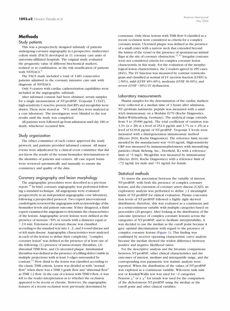
MethodsStudy patients
This was a prospectively designed substudy of patients
undergoing coronary angiography in a prospective, multicenter
cohort study (PACS) developed in 11 coronary care units of
university-affiliated hospitals. The original study evaluated
the prognostic value of different biochemical markers,
isolated or in combination, in the risk stratification of patients
with NSTEACS.9
The PACS study included a total of 1483 consecutive
patients admitted to the coronary intensive care unit with
diagnosis of NSTEACS.
Only 9 centers with cardiac catheterization capabilities were
included in the angiographic substudy.
After informed consent had been obtained, serum samples
for a single measurement of NT-proBNP, Troponin T (TnT),
high-sensitivity C-reactive protein (hsCRP) and myoglobin were
drawn. These were stored at �708C until they were analyzed at
a core laboratory. The investigators were blinded to the test
results until the study was completed.
All patients were followed up from admission until day 180 or
death, whichever occurred first.
Study organizationThe ethics committee of each center approved the study
protocol, and patients provided informed consent. All major
events were adjudicated by a clinical event committee that did
not know the results of the serum biomarker determinations or
the identities of patients and centers. All case report forms
were reviewed automatically and manually to ensure the
consistency and quality of the data.
Coronary angiography and lesion morphologyThe angiographic procedures were described in a previous
report.10 In brief, coronary angiography was performed follow-
ing a standard technique. All angiograms were evaluated
prospectively in an independent angiographic core laboratory,
following a prespecified protocol. Two expert interventional
cardiologists reviewed the angiograms with no knowledge of the
biomarker levels and patient outcome. If they disagreed, a third
expert examined the angiogram to determine the characteristics
of the lesions. Angiographic severe lesions were defined as the
presence of stenosis N50% in vessels with a diameter equal or
N 2.0 mm. Extension of coronary disease was classified
according to the standard way into 1-, 2-, and 3-vessel disease and
or left main disease. Angiographic characteristics were analyzed
in each of the lesions to define their complexity. bcomplex
coronary lesionQ was defined as the presence of at least one of
the following: (1) presence of intracoronary thrombus, (2)
abnormal TIMI flow, and (3) ulcerated plaque. Intraluminal
thrombus was defined as the presence of a filling defect visible in
multiple projections with at least 3 edges surrounded by
contrast.11 Flow distal to the lesion was classified according to
the classic TIMI criteria. Lesion was divided as with bnormal
flowQ when there was a TIMI 3 grade flow and babnormal flowQas TIMI V2 flow. In the case of a lesion with TIMI 0 flow, it was
left to the reader interpretation as to whether the occlusion
appeared to be recent or chronic. However, the angiographic
features of a recent occlusion were previously determined by
consensus. Only those lesions with TIMI flow 0 classified as a
recent occlusion were considered as criteria for a complex
coronary lesion. Ulcerated plaque was defined as the presence
of a small crater with a narrow neck that extended beyond
the lumen of the vessel or the presence of spontaneous intimal
flaps at the site of coronary obstruction.12,13 Irregular contours
were not considered criteria for complex coronary lesion
characteristic in this study. For the evaluation of the morpho-
logical lesion characteristics, the 2 readers agreed in 499 cases
(84%). The LV function was measured by contrast ventriculo-
gram and classified as normal (if LV ejection fraction [LVEF] is
z50%), mild (LVEF 40%-49%), moderate (LVEF 30-39%), and
severe (LVEF b30%) LV dysfunction.
Laboratory measurementsPlasma samples for the determination of the cardiac markers
were collected at a median time of 3 hours after admission.
NT–probrain natriuretic peptide was measured with a sand-
wich immunoassay on a Modular E170 (Roche Diagnostics,
Baden-Wurttemberg, Germany). The analytical range extends
from 5 to 35000 pg/mL. The total coefficient of variation was
3.3% (n = 28) at a level of 252.6 pg/mL and 3.7% (n = 25) at a
level of 6130.8 pg/mL of NT-proBNP. Troponin T levels were
measured with a third-generation immunoassay method
(Elecsys 2010, Roche Diagnostics). The reference limit recom-
mended by the manufacturer was b0.03 ng/mL. High-sensitivity
CRP was measured by immunonephelometry with intensifying
particles (Dade Behring, Inc., Deerfield, IL) with a reference
limit of N3 mg/L. Myoglobin was measured by immunoassay
(Elecsys 2010, Roche Diagnostics) with a reference limit of
N72 ng/mL for male and N51 ng/mL for female.
Statistical methodsTo assess the association between the variable of interest,
NT-proBNP, with both the presence of complex coronary
lesions, and the extension of coronary artery disease (CAD), an
exploratory analysis was performed to define z1 meaningful
limits of NT-proBNP for clinical evaluation. Plasma concentra-
tion levels of NT-proBNP followed a highly right skewed
distribution; therefore, this was evaluated as a continuous and
as a semicontinuous variable with multiple categories based on
percentiles (20 groups). After looking at the distribution of the
outcome (presence of complex coronary lesions) across the
categories of NT-proBNP, and to facilitate interpretability, it
was decided to use the median as the cutoff point because it
gave optimal discrimination with regard to the presence of
complex coronary lesions (Figure 1). This finding was
confirmed by receiver operating characteristic curve analysis
because the median showed the widest difference between
positive and negative likelihood ratios.
For the descriptive analysis and the bivariate comparisons
between NT-proBNP, other clinical characteristics and the
outcomes of interest, medians and interquartile range, and the
corresponding non parametric test statistic analysis were
reported. When the distribution of the values of NT-proBNP
was explored as a continuous variable, Wilcoxon rank sum
test or Kruskal-Wallis test was used for N2 categories.
Pearson m2 or a m2 for trends was used for the comparison
of the dichotomous NT-proBNP using the median as the
cutoff point and other clinical variables.

Table I. Baseline characteristics by NT-proBNP levels
NT-proBNP__bbbbV278.7 pg/mL
(n = 293)
NT-proBNPNNNN278.7 pg/mL
(n = 292) P
Age N75 y 41 (14.5) 104 (35.4) .000Male sex 223 (76.6) 205 (69.7) .60HTA 178 (61.2) 204 (69.4) .037Smoker 106 (36.4) 61 (24.8) .002Diabetes 43 (14.8) 61 (20.7) .59Hyperlipidemia 178 (61.2) 169 (57.5) .364Prior MI 51 (17.4) 78 (26,7) .007Prior CABG 23 (7.9) 41 (13.9) .019Prior PTCA 35 (12.0) 41 (13.9) .490Ischemic changes
(ECG)152 (52.2) 206 (70.1) .000
Myoglobin-positive 83 (28.5) 131 (44.6) .000hsCRP N3 mg/dL 153 (52.6) 199 (67.7) .000TnT N0.03 ng/mL 108 (36.9) 211 (72.1) .000
Data are shown as n (%) for dichotomous variables. HTA, Hypertension; CABG,coronary artery bypass graft; PTCA, percutaneous transluminal coronary angioplasty;ECG, electrocardiogram.
Figure 1
Complex coronary lesion stratified by NT-proBNP levels.
Figure 2
Left ventricular function and NT-proBNP levels. Results are expressedas median and interquartile range.
Figure 3
Coronary artery disease extension and NT-proBNP levels. Results areexpressed as median and interquartile range.
American Heart Journal
Volume 151, Number 5Navarro Estrada et al 1093.e3
To assess the independent effect of NT-proBNP level on the
risk of complex coronary lesions adjusting for potential
confounders, a stepwise interactive multiple logistic regression
analysis was performed. A model including those covariates
showing a P value of .10 on the univariate screen analysis one
at a time was built. Then, only those with a significant Wald
test or likelihood ratio test or those showing a confounding
effect on the association between NT-proBNP and the outcome
were retained in the final model. Global fit of this explanatory
model was evaluated with the Hosmer-Lemeshow test.
ResultsOf the 1483 patients from the PACS study, 1252 pa-
tients were enrolled in 9 centers that participated in the
angiographic substudy. Of those, 633 patients (50.5%),
underwent coronary angiography at a median time of
46 hours (25th to 75th percentile 20-70 hours) from
admission. Of the 633 angiograms, 48 (7.5%) were
excluded from the analysis because of technical reasons
or unacceptable quality.
A total of 585 patients had both core laboratory angio-
graphic data and NT-proBNP levels on admission. The
median (25th-75th percentile) NT-proBNP levels were
278.7 pg/mL (109.8, 758.5) pg/mL, respectively (range
12.6-35000 pg/mL).
The baseline clinical characteristics of the patients
are shown in Table I. Patients with elevated levels of
NT-proBNP were older, more commonly smokers, and
had more likely history of MI. At presentation, patients
with elevated NT-proBNP more frequently had

Figure 4
Supramedian NT-proBNP levels according to LV function and extension of CAD.
Table II. Median and 25th to 75th percentiles of NT-proBNPaccording to the presence of complex coronary lesions and theirdifferent components
Lesionmorphologyabnormality
NT-proBNP Median (25th-75th) (pg/mL)
Absent Present P
Complex coronarylesion
189 (86-523) 386 (164-874) .000
Thrombus 257 (103-759) 358 (186-790) .009Abnormal
TIMI flow235 (96-639) 443 (203-871) .000
Ulceratedplaque
309 (131-815) 331 (154-802) .686
American Heart Journal
May 20061093.e4 Navarro Estrada et al
ischemic ST-T changes and elevation of TnT, hsCRP,
and myoglobin.
Patients with baseline NT-proBNP levels N278.7 pg/mL
were at significantly higher risk of death at 180 days
(6.8% vs 2.1%, P b .0001), and the composite of death or
MI (11.9% vs 6.5%, P = .0025).
Left ventricular function, CAD extension, andNT-proBNP levels
Left ventriculograms were performed in 506 patients
(87%). Among them, 257 patients (51%) had normal
LV function; 136 (27%), 81(16%), and 32 (6%) patients
had mild, moderate, and severe LV dysfunction, respec-
tively. NT–probrain natriuretic peptide levels increased
significantly as LV function decreased (Figure 2).
Ninety-nine patients (17%) showed non significant
lesions or completely normal coronary arteries;
187 (32%), 155 (26%), and 142 (25%) had 1-vessel,
2-vessel, and 3-vessel and/or left main disease,
respectively. As seen in Figure 3, the levels of
NT-proBNP were directly related to the extent of the
CAD, and the difference between each category was
highly significant ( P = .0001).
The combination of the extension of CAD and the
severity of LV dysfunction provided the strongest
association with high NT-proBNP levels. Among patients
in every category of LV function, NT-proBNP levels
increased as the extension of CAD progressed (Figure 4).
This association between NT-proBNP levels and num-
ber of vessels was maintained in patients with normal LV
function (n = 257). The levels of NT-proBNP increased
significantly as the number of vessels affected increased)
(median [interquartile range ] in patients with normal
coronaries 110.6 pg/mL [172.3 ], 1 vessel 162.0 pg/mL
[322.5]; 2-vessels 198.7 pg/mL [261.4]; and 3-vessels or
left main disease 315.4 pg/mL [885.8] [ P b .0001].
Lesion morphology and NT-proBNP levelsComplex coronary lesions were observed in 264
(45.1%) patients with NSTEACS. Coronary thrombi were
seen in 104 (17.8%), abnormal TIMI flow in 133 (22.7%),
and ulcerated plaque in 142 (24.3%) patients. Patients
with complex coronary lesions or those with at least one
of its individual components had higher levels of NT-
proBNP compared with those without complex coro-
nary lesions. (Table II).
As seen in Figure 1, the proportion of complex
coronary lesions rose gradually as NT-proBNP centiles
increased; however, the predictive capacity of baseline
measurement of NT-proBNP showed a threshold at the

Figure 5
Stepwise logistic regression model for the prediction of complex coronary lesion. NT-proBNP 278, Supramedian NT-proBNP level.
American Heart Journal
Volume 151, Number 5Navarro Estrada et al 1093.e5
median value. This threshold was confirmed by receiver
operating characteristic curve analysis.
NT-proBNP as independent predictor of complexcoronary lesions
A multivariable logistic regression analysis was per-
formed to identify predictors of complex coronary
lesions. NT–probrain natriuretic peptide, troponin
N0.03 ng/mL, ST-segment depression z0.5 mV,
hsCRP N3 mg/L, positive myoglobin, severe angina
defined as N2 episodes of chest pain within 24 hours of
admission, and diabetes were found to be predictive in
the univariate analysis and, therefore, were introduced
in the regression model. NT–probrain natriuretic
peptide entered into the model as a dichotomous
variable with a cutoff at the median value (N278.7 vs
V278.7 pg/mL). Positive troponin and NT-proBNP
N278.7 pg/mL independently contributed to the pre-
diction of complex coronary lesions, and ST-segment
changes had only borderline significance (Figure 5).
When NT-proBNP was entered in the multivariate
analysis as a continuous variable, the predictors
remained the same (log NT-proBNP: OR 1.18, P = .018).
Goodness of fit was adequate, with a P = .785 in the
Hosmer-Lemeshow test (df = 8).
DiscussionThe results of this study demonstrate that elevated
NT-proBNP levels in patients with NSTEACS are associ-
ated with lower LV ejection fraction and more extensive
CAD. Furthermore, using a previously defined and
prospectively evaluated criteria for bunstableQ angio-
graphic lesions (complex coronary lesions), the data
from this study demonstrate that NT-proBNP level
independently predicts the presence of more complex
coronary lesions.
Several previous studies have demonstrated the
relation between elevations of BNP4,5 and NT-proBNP
levels7-9 and adverse outcomes in patients with
NSTEACS. Because BNP is released in response to
increased myocardial wall stress, it was proposed that
in patients with acute coronary syndromes with overt
heart failure or decreased LV function, elevated levels
of the biomarker reflect myocardial dysfunction and
are associated with high risk of adverse events. The
present results found a significant association between
NT-proBNP and ventricular function, confirming the
findings by Omland et al,7 which showed that supra-
median NT-proBNP values were associated with lower
LV function.
A previous work has demonstrated that patients with
elevated BNP were more likely to have multivessel
CAD.5 Furthermore, a recent study has shown that
patients with NSTEACS with elevated BNP have tighter
culprit artery diameter stenosis, a higher corrected TIMI
frame count, and a more frequently left anterior
descending involvement and proximal culprit lesion
location.14 Another study assessed NT-proBNP levels in

American Heart Journal
May 20061093.e6 Navarro Estrada et al
patients with the wide spectrum of CAD and controls
and showed that NT-proBNP concentrations increased
progressively with the increase of CAD severity.15 The
results of our study confirmed these findings; among
patients with NSTEACS, elevated NT-proBNP levels had
a very close relation with the number of vessels
involved.5 More importantly, the present study has also
shown that even in patients with normal LV function,
the level of NT-proBNP maintains the same relation with
the extension of CAD. Besides, LV ejection fraction was
measured within the first hours (median 46 hours) of
the onset of the coronary event, and the ventriculograms
were blindly analyzed in a core laboratory, giving an
accurate measurement of LVEF in the acute phase of
acute coronary syndrome. This finding suggests that a
mechanism other than LV dysfunction might be re-
sponsible for the adverse outcome in patients with
NSTEACS and high levels of NT-proBNP.
One of the more important and novel findings of the
present study was the strong and independent associa-
tion between NT-proBNP levels and the presence of
more complex coronary lesions. This association
persisted even when other well-recognized clinical
predictors like cardiac TnT and ischemic ST changes
were included in the multivariate model. The relation
between complex coronary lesions and the magnitude
of the ischemic insult and long-term prognosis is well
known.16 In contrast, other biomarkers such as
C-reactive protein does not predict either the extension
or the complexity of coronary disease in patients with
NSTEACS.10 Several observations have suggested that
BNP increases with severity and extent of ischemia.
Levels of BNP increase transiently after uncomplicated
coronary angioplasty in the absence of demonstrable
changes in LV function or LV end-diastolic pressure.17,18
Likewise, in patients with proven CAD, BNP levels
increased during dynamic exercise, compared with
patients without CAD19,20; and in another study, BNP
increased during thallium exercise testing and the
severity of myocardial perfusion defect correlated with
greater plasma levels of BNP.17,18
The pathophysiological mechanism behind the asso-
ciation between ischemia and elevation of BNP is not
well defined. However, recent reports may help to
clarify this association. Toth et al21 have found evidence
that tissue hypoxia alone trigger release of BNP in
absence of LV dysfunction. These results were con-
firmed by a recent physiological study showing that
ventricular BNP gene expression is up-regulated by
myocardial hypoxia resulting in augmented plasma
concentrations of BNP and NT-proBNP.22 Thus, the
available evidence supports a plausible relation between
severity of CAD and the presence of complex coronary
lesions that would predispose to a more severe and
extensive myocardial ischemia and, thus, provoking a
rise in NT-proBNP independently of LV function.
Study limitationsThe NT-proBNP threshold used in the present study
has not been validated before. However, given a
stepwise relation between increasing levels of
NT-proBNP, the presence of complex coronary lesions
with a robust predictive capacity, and the absence of
available data, the NT-proBNP result at the median (50th
percentile) appears to be a convenient decision limit.
In addition, these findings were confirmed when
NT-proBNP was included as a continuous variable.
ConclusionIn this large series of consecutive patients with
NSTEACS, NT-proBNP levels progressively increases
with the severity of CAD and degree of LV dysfunction.
Increased levels of NT-proBNP independently predict
the presence of more complex coronary lesions. These
findings suggest that, in addition to LV dysfunction,
greater severity and extent of myocardial ischemia may
explain, at least in part, the underlying mechanism of
the adverse clinical outcome associated with NT-proBNP
in patients with NSTEACS.
References1. Wiese S, Breyer T, Dragu A, et al. Gene expression of brain
natriuretic peptide in isolated atrial and ventricular human myo-cardium: influence of angiotensin 2 and diastolic fiber length.Circulation 2000;102:3074 -9.
2. Cowie MR, Mendex GF. BNP and congestive heart failure. ProgCardiovasc Dis 2002;44:293 -321.
3. McCullough PA, Maisel AS, BNP Multinational Study Investigators.B-type natriuretic peptide and clinical judgement in emergencydiagnosis of heart failure. Circulation 2002;106:416 -22.
4. de Lemos JA, Morrow DA, Bentley JH, et al. The prognostic value ofB-type natriuretic peptide in patients with acute coronary syndromesN Engl J Med 2001;345:1014 -21.
5. Morrow DA, de Lemos JA, Sabatine MS, et al. Evaluation ofB-type natriuretic peptide for risk assessment in unstable angina/non–ST-elevation myocardial infarction. J Am Coll Cardiol2003;41:1264 -72.
6. Jernberg T, Stridsberg M, Venge P, et al. N-terminalpro brain natriuretic peptide on admission for early riskstratification of patients with chest pain and noST-segment elevation. J Am Coll Cardiol2002;40:437 -45.
7. Omland T, Persson A, Ng L, et al. N-terminal pro–B-type natriureticpeptide and long-term mortality in acute coronary syndromes.Circulation 2002;106:2913 -8.
8. James SK, Lindahl B, Siegbahn A, et al. N–terminal pro-brainnatriuretic peptide and other risk markers for the separate predic-tion of mortality and subsequent myocardial infarction inpatients with unstable coronary artery disease. Circulation 2003;108:275 -81.
9. Bazzino O, Fuselli JJ, Botto F, et al. Relative value of N-terminalprobrain natriuretic peptide, TIMI risk score, ACC/AHAprognostic classification and other risk markers in patients withnon–ST elevation acute coronary syndromes. Eur Heart J2004;25:859 -66.

American Heart Journal
Volume 151, Number 5Navarro Estrada et al 1093.e7
10. Navarro Estrada JL, Gabay JM, Alvarez J, et al. Relation ofC-reactive protein to extent and complexity of coronarynarrowing in patients with non–ST elevation acute coronarysyndromes. A prospective cohort study. Coron Artery Dis2004;15:477 -84.
11. Alderman EL, Stadius M. The angiographic definitions of the BypassAngioplasty Revascularization Investigation study (BARI). CoronArtery Dis 1992;3:1189 -207.
12. Ellis SG, Vandormael MG, Cowley MJ. Coronary morphologic andclinical determinants of procedural outcome with angioplasty formultivessel coronary disease: implications for patients selection.(Multivessel Angioplasty Prognosis Study Group). Circulation 1990;82:1193 -202.
13. ACC/AHA guidelines for the management of patients with unstableangina and non–ST-segment elevation myocardial infarction.Braunwald y col. J Am Coll Cardiol 2000;36:970 -1062.
14. Sadanandan S, Cannon CP, Chekuri K. Association of elevatedB-type natriuretic peptide levels with angiographic findings amongpatients with unstable angina and non–ST elevation acute coronarysyndromes. JACC 2004;44:564 -8.
15. Ndrepepa G, Braun S, Mehilli J, et al. Plasma levels of N-terminalPro-BNP in patients with coronary artery disease and relation to
clinical presentation, angiographic severity, and left ventricularejection fraction. Am J Cardiol 2005;95:553 -7.
16. Ambrose JA, Israel DH. Angiography in unstable angina. Am JCardiol 1991;68:78B -84B.
17. Morrow DA, Braunwald E. Future of biomarkers in acute coronarysyndromes—moving toward a multimarker strategy. Circulation2003;108:250 -2.
18. de Lemos JA, Morrow DA. B-type natriuretic peptide measurementin acute coronary syndromes: ready for clinical application?Circulation 2002;106:2868-70.
19. Foote RS, Pearlman JD, Siegel AH, et al. Detection of exercise-induced ischemia by changes in B-type natriuretic peptides. J AmColl Cardiol 2004;44:1980-7.
20. Sabatine MS, Morrow DA, de Lemos JA, et al. Acute changes incirculating natriuretic peptide levels in relation to myocardialischemia. J Am Coll Cardiol 2004;44:1988 -95.
21. Toth M, Vuorinen KH, Vuolteenaho O, et al. Hipoxia stimulatesrelease of ANP and BNP from perfused rat ventricular myocardium.Am J Physiol 1994;266:H1572 -80.
22. Goetze JP, Christoffersen C, Perko M. Increased cardiac BNPexpression associated with myocardial ischemia. FASEB J 2003;17:1105-7.
Copyright © 2022 FDOKUMEN