Thermodynamic model development for lithium intercalation electrodes

Published: October 18, 2011
r 2011 American Chemical Society 23387 dx.doi.org/10.1021/jp206945e | J. Phys. Chem. C 2011, 115, 23387–23394
ARTICLE
pubs.acs.org/JPCC
Novel Graphene-Gold Nanoparticle Modified Electrodes for the HighSensitivity Electrochemical Spectroscopy Detection and Analysis ofCarbamazepineStela Pruneanu,*,† Florina Pogacean,† Alexandru R. Biris,† Stefania Ardelean,† Valentin Canpean,‡
Gabriela Blanita,† Enkeleda Dervishi,§ and Alexandru S. Biris*,§
†National Institute for Research and Development of Isotopic and Molecular Technologies, Donath Street, No. 65-103,RO-400293 Cluj-Napoca, Romania‡Nanobiophoton Center, Interdisciplinary Research Institute in Bio-Nano-Science, Babes-Bolyai University, Cluj Napoca 400084,Romania§Nanotechnology Center, Systems Engineering Department, University Arkansas at Little Rock, 2801 S. University Avenue, Little Rock,Arkansas 72204, United States
1. INTRODUCTION
Carbamazepine (Scheme 1) is a tricyclic compound used as ananticonvulsant drug for the treatment of epilepsy and bipolardisorder, as well as trigeminal neuralgia. It can also be adminis-tered to patients who have other illnesses including schizophrenia,neuromyotonia, attention-deficit hyperactivity disorder (ADHD),and post-traumatic stress disorder. As a result of its widespread use,carbamazepine is currently considered one of the emerging pollu-tants in ground and surface water; therefore, its accurate determi-nation by fast and reliable methods is highly desirable. Recentstudies have found that carbamazepine is persistent, and itsremoval efficiency by wastewater treatment plants (WWTPs)is below 10%.1 The employment of electrochemical techniquesfor the detection of carbamazepine is a valuable alternative to themore laborious methods generally used, like liquid chromato-graphic methods.2,3
Kalanur and Seetharamappa4 have recently reported the studyof carbamazepine oxidation at glassy carbon electrode, in PBSsolution (pH 7.4). Two oxidation peaks were noticed at
potentials of 1.183 and 1.401 V/Ag(AgCl), respectively, but nopeak was observed in the reverse scan. Hence, the oxidationprocess was an irreversible one. The proposed oxidation mechan-ism occurs by formation of two radical cations which give rise to adimer. The dimers subsequently undergo electrochemical
Scheme 1. Carbamazepine Chemical Structure
Received: July 20, 2011Revised: October 16, 2011
ABSTRACT: A novel graphene-gold nanoparticle compositedeposited on gold electrode (Au-Gr-AuNPs) was employed todetect carbamazepine (CBZ), an antiepileptic drug, used here as amodel system. The same approach can also be used to detectother additional organic compounds. The presence of gold nano-particles (size between 10 and 20 nm) encased in graphene sheetswas evidenced by TEM andHRTEM, while the AFM analysis wasused to study the morphology of the graphene-gold nanoparticlefilms used for the electrochemical studies. Various electrochemi-cal methods were employed to study carbamazepine oxidation,such as cyclic voltammetry, linear sweep voltammetry, and electro-chemical impedance spectroscopy. The results clearly showedthat the modified electrode exhibited excellent electrocatalyticeffect toward oxidation of carbamazepine. The peak current intensity significantly increased (up to 2 times) while the peak potentialshifted to lower oxidation potential (∼100mV), compared with the unmodified electrode. A detection limit (DL) of 3.03� 10�6Mwas obtained with the graphene-gold nanoparticle-modified electrode (S/N = 3). In addition, an equivalent electrical circuit wasdeveloped and used to interpret the experimental EIS data. The circuit contains the solution resistance (Rs), the charge-transferresistance (Rct), the Warburg impedance (ZWt, transmissive boundary), and the double-layer capacitance (Cdl).
23388 dx.doi.org/10.1021/jp206945e |J. Phys. Chem. C 2011, 115, 23387–23394
The Journal of Physical Chemistry C ARTICLE
oxidation to form diradical dimers. Veiga et al.5 have used amultiwalled carbon nanotube film-coated GCE for voltammetricdetermination of carbamazepine in PBS solution (pH 6.89).They reported a single oxidation peak at around +1.09 V/Ag(AgCl) with no additional postpeak in the forward scan orreduction peak in the reverse scan. The authors suggest a two-stepoxidation mechanism (electrochemical�chemical) which occurssimultaneously and therefore cannot be distinguished in thecyclic voltammogram. Moses et al.6 have studied the electro-chemical reduction of carbamazepine in aqueous solutions usingmercury electrodes. They reported a bielectronic reductionmechanism, which implies the saturation of the double bondpresent in the heterocycle of this molecule.
Given the need to detect small amounts of chemical mol-ecules, significant research has been devoted to the developmentof new high-sensitivity electrochemical sensors. Among theseresearch efforts, a new approach was to develop materials withnovel electrochemical properties that could further increase theelectrochemical sensitivity of some of the commonly usedmethods for the detection of small concentrations of variousorganic and biological molecules. Some of the most promising ofsuch methods included the use of carbonaceous nanomaterialsas components of the electrochemical sensor. Lately, graphenewith its unique properties (such as 2Dmorphology, graphitic struc-ture, excellent electrical/thermal conductivity, and ability to befunctionalized with various chemical groups) has been identifiedas one of the most suitable materials for various sensing techno-logies.7�12 Kang et al.13 have used a graphene-based electro-chemical sensor which shows excellent electrocatalytic activitytoward the reduction and oxidation of paracetamol. The methodwas applied to detect paracetamol in pharmaceutical preparationtablets (detection limit 3.2 � 10�8 M) without any interferencefrom ascorbic acid or dopamine. Li et al.14 have reported thepreparation of a nanocomposite film based on nafion and gra-phene which was subsequently used as enhanced sensing plat-form for ultrasensitive determination of cadmium. Wang et al.15
have used a graphene-chitosan modified electrode for selectivedetermination of dopamine (DA) and compared its performancewith that of a multiwalled carbon nanotube-modified electrode.The excellent performance of graphene to dopamine detectionwas attributed to π�π interaction between the phenyl structureof DA molecule and the two-dimensional hexagonal carbonstructure of graphene, which makes the electron transfer feasible.
In addition, the attachment of catalytic nanoparticles, such asgold, platinum, or silver, could further increase the electrocata-lytic activity of these graphitic sheets.16�21 Still, there is a sig-nificant challenge in controlling the uniformity of metal nano-particle decoration of the graphene surface and especially thestrength of the attachment between the organic and the inorganicnanostructure. We have developed a new method to grow in asingle step few-layer graphene structures decorated with Au nano-particles (graphene-AuNPs) by using a chemical vapor deposi-tion process. This composite nanomaterial has been depositedonto the surface of gold electrodes and subsequently used in anelectrochemical setup.
Here, we present, for the very first time to our best knowledge,the oxidation of carbamazepine (a model organic molecule),taking advantage of the electrocatalytic properties of this novelgraphene-AuNPmodified gold electrode surface (further noted asAu-GR-AuNPs). Such nanostructured surfaces used as sensingelectrode in an electrochemical setup represents an alternative toconventional analytical methods, being suitable for fast, sensitive,
and inexpensive determinations. The morphological characteristicsof graphene-AuNP composite and Au-GR-AuNP surface wereinvestigated by high resolution transmission electronmicroscopy(HRTEM) and atomic force microscopy (AFM). In addition, acombination of analytical methods including cyclic voltammetry(CV), linear sweep voltammetry (LSV), and electrochemicalimpedance spectroscopy (EIS) was used to completely char-acterize the electrocatalytic properties of Au-GR-AuNPs nano-structured surface. This approach could be the foundation forplatform technologies that could use organic�inorganic nano-structural systems to enhance the detection sensitivity of variousorganic/biological compounds in the environment. This approachis the first, to our best knowledge, that presents the novel electro-chemical properties of composite organic�inorganic nano-material composed of Au nanoclusters embedded in graphiticsheets to enhance the electrochemical sensitivity of a detectionreaction for carbamazepine. Furthermore, our findings have thepotential to provide the ability to study the low intensityinteractions between such organic molecules and biologicalmolecules in the environment, medicine, pharmaceutical indus-try, or energy storage.
2. EXPERIMENTAL DETAILS
Reagents and Solutions. All reagents used for the experi-ments were of analytical grade or better. Acetonitrile was pur-chased from Riede-deHa€en and dimethylformamide (DMF) fromSigma-Aldrich, Germany. Pure carbamazepine powder was pur-chased from Zhejiang Jiuzhou Pharmaceutical CO., Ltd., China.Tetra-butyl ammonium perchlorate (TBAP) was purchased fromFluka, Germany. A stock solution of 10�2 M carbamazepine wasprepared in acetonitrile and 0.05MTBAP and subsequently usedfor the preparationof lower concentration solutions (10�3�10�6M).Additionally, about 0.03 mg of graphene-AuNP composite wasdispersed in DMF (1 mL) and kept at room temperature.Preparation of Graphene-Gold Nanoparticles Composite
(Graphene-AuNPs). We prepared few-layer graphene-AuNPscomposite by a simple chemical vapor deposition technique byusing Au/MgO catalyst (1% wt Au) prepared by a deposition�precipitation method with urea. For the synthesis of the graphene-AuNPs structures, we used the radio frequency (1.2 MHz, 5 kW)chemical vapor deposition (RF-CCVD) method with a water-cooled reactor and methane as the carbon source (1000 �C, CH4
flow rate 80mL/min, Ar 300mL/min, synthesis time 30min).22�24
Preparation of Gold Electrode Modified with Graphene-AuNPs (Au-GR-AuNPs). Prior to modification, the gold elec-trode was polished with alumina suspension until a mirror-likesurface was obtained. Next, the electrode was ultrasonically clea-ned in ethanol and deionized water for about 3 min several times.After that, a 10 μL drop of colloidal suspension of graphene-AuNPs was deposited onto the gold substrate and dried at roomtemperature for about 10 h. In order to reduce the oxygen func-tionalities (hydroxyl, carbonyl, carboxyl, or epoxy) which are gen-erally present on graphene surface, the electrode was polarized ata potential of �1.3 V versus Ag/AgCl for 600 s, in a solution of0.1 M PBS (pH 7).17
Analytical Characterization of Graphene-AuNPs and Au-GR-AuNPs Electrode. Morphological characterization of gra-phene-AuNPs composite and Au-GR-AuNPs electrode was per-formed by transmission electron microscopy (JEOL JEM-2100F)and by atomic force microscopy (Veeco Dimension 3100 AFMandAlpha 300A instrument (Witec)). ForTEManalysis, the samples
23389 dx.doi.org/10.1021/jp206945e |J. Phys. Chem. C 2011, 115, 23387–23394
The Journal of Physical Chemistry C ARTICLE
were dispersed in 2-propanol and gently sonicated for 30 min.Several drops of the suspensions were placed on holey carbon-coated grids and further used for the analysis.Cyclic voltammetry (CV), linear sweep voltammetry (LCV),
and electrochemical impedance spectroscopy (EIS) measure-ments were performed using an Autolab Potentiostat (Nova 1.6Software, EcoChemie, Utrecht, Netherlands) connected with athree-electrode cell. The surface area of the gold electrode was0.031 cm2 (working electrode). A platinum electrode with a largesurface area (∼2 cm2) was employed as the counter electrodewhile the reference was an Ag/AgCl electrode. CVs and LCVswere recorded between 0.6 and 1.65 V/SCE at a scan rate of25mV s�1 (unless otherwise specified in the text). All impedancespectra were measured over a frequency range 0.1�105 Hzby using a small sinusoidal excitation signal (10 mV amplitude).The applied potential was +1.4 V versus Ag/AgCl. All experi-ments were carried out in quiescent solutions of acetonitrile and
0.05MTBAP. Data fitting was performed usingNova 1.6 software(EcoChemie-Netherlands). Raman spectra were collected with aJASCO NRS 3300 spectrophotometer in a backscattering geo-metry and equipped with a CCD detector (�69 �C). The exci-tation laser beam Ar-ion with a wavelength of 514 nm and apower at the sample surface of 1.5 mW had an area of around1 μm2 andwas focused by using anOlympusmicroscope coupledto a 100� objective.
3. RESULTS AND DISCUSSION
3.1. Synthesis of Graphene-Au Nanoparticles Composites.The schematic of the process presented by this paper isshown in Figure 1a. The Au/MgO catalyst was found to syn-thesize graphene-AuNPs structures composed of 2�6 sheetsand diameters of 600 ( 100 nm. An interesting observation wasthe fact that, during the growth process, the Au nanoparticles
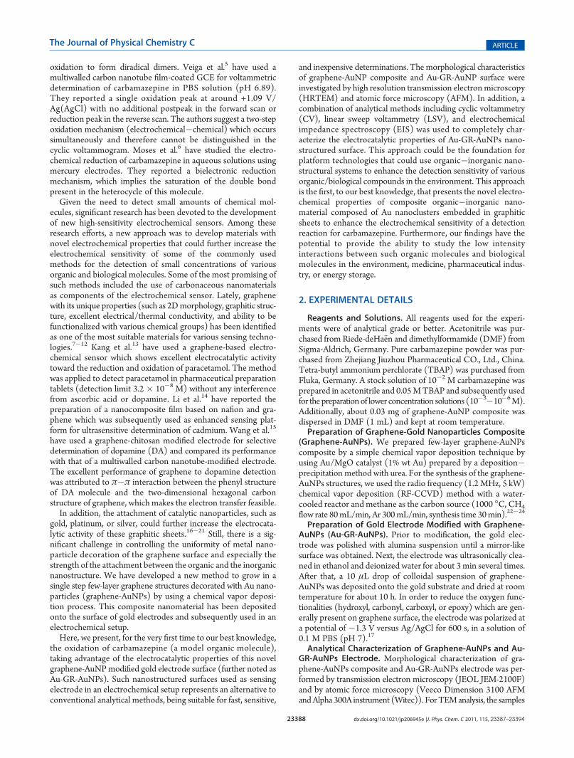
Figure 1. Schematic representation of the synthesis of graphene decorated with Au nanoparticles by RF-CCVD over an Au/MgO catalyst (a);transmission electron analysis (TEM) (80 kV) of the resulting structures (b); schematic of the process used to deposit graphene-AuNPs composite overthe top surface of a gold electrode to be further used in an electrochemical setup for the detection of carbamazepine (c); representative AFM image(tapping mode) of graphene-AuNPs composite deposited on gold substrate (d); Raman spectroscopy analysis of the graphene-AuNPs nanocompositecollected at 514 nm excitation wavelength (e).

23390 dx.doi.org/10.1021/jp206945e |J. Phys. Chem. C 2011, 115, 23387–23394
The Journal of Physical Chemistry C ARTICLE
initially supported on the MgO were lifted off by the graphenesheets during the growth process and became encased in theircrystalline structure (Figure 1b). The size of these Au nanopar-ticles was found to be relatively uniform with diameters between10 and 20 nm. The inset of Figure 1b shows the higher magni-fication of such a nanoparticle encased in the graphitic structureof the graphene sheets. The graphene-AuNPs composite was furthersolubilized and deposited onto the top surface of a gold electrodeused for electrochemical studies (Figure 1c). Atomic force micro-scopy was additionally used to directly study the graphene-AuNPs composite assembled onto the top surface of the goldsubstrate. Figure 1d provides a representative AFM image (tappingmode) of the graphene-AuNP nanostructures. The AFM analysisof the structure clearly shows the vertical stacking of the graphiticlayers. Raman analysis of the graphene-AuNPs composites indi-
cates the presence of all the graphene-specific bands (D band�1342 cm�1, G band �1589.7 cm�1, and the overtone 2D band�2678 cm�1).25
Further analysis of the graphene-AuNPs composite depositedonto gold surface reveals a clear tendency of these nanostructuresto form large agglomerates. Previous studies have confirmed thatwater molecules intercalated between the platelets form hydro-gen bonds to the epoxy or hydroxyl functionalities, a key factorin maintaining the stacked structure of the graphene-likestructures.26,27 After their deposition on the gold electrode, theclusters of graphene-AuNPs composite form agglomerationswith an average height of up to 1 μm without the single-sheetmorphology, however. In spite of this thickness value, ourelectrochemical measurements prove that the composite’selectrocatalytic properties are still preserved.3.2. Electrochemical Characterization. Since carbamazepine
has a very low solubility in water (17.7 mg L�1 at 25 �C)28 in ourstudies we have chosen acetonitrile as solvent. Figure 2 showssuccessive cyclic voltammograms (3 cycles) recorded in thesupporting electrolyte (acetonitrile +0.05 M TBAP), as well asin electrolyte containing 10�2 M carbamazepine (scan rate 25mV s�1). A two-wave oxidation peak can be seen at around +1.49V/Ag (AgCl) accompanied by a small reduction peak at +1.16 V/Ag (AgCl). The large separation between the oxidation andreduction peaks (≈330mV) suggests that carbamazepine moleculesundergo a quasireversible redox process. At a slow scan rate (between5 and 50mV s�1), the redox process is diffusion-controlled as shownby Ipeak versus υ
1/2 plot. (See inset of Figure 2.) This was furtherconfirmedby the plot of log Ipeak versus logυ, whichwas linear withinthe same scan rate range and gave a slope of 0.6 (data not shown).This value is very close to the theoretical value of 0.5 reported byLaviron for a diffusion-controlled electrode process.29
The successive cyclic voltammograms show that the electro-chemical signal of carbamazepine is almost unmodified, suggest-ing that the electrode surface is not blocked by the adsorption of
Figure 2. Successive cyclic voltammograms recorded with Au-GR-AuNPs electrode in supporting electrolyte (acetonitrile +0.05 M TBAP,black line), as well as in electrolyte solution containing 10�2 Mcarbamazepine (three cycles, scan rate 25 mVs�1, blue line). Insetshows variation of peak current intensity versus υ1/2 (diffusion-controlled process).
Scheme 2. Proposed Mechanism for Oxidation of Carbamazepine

23391 dx.doi.org/10.1021/jp206945e |J. Phys. Chem. C 2011, 115, 23387–23394
The Journal of Physical Chemistry C ARTICLE
the oxidation products. However, in order to have reproducibleresults in our analytical determinations, the data obtained fromthe first scan (either CV or LCV) were always used. Atkins et al.have recently studied the electrochemical reduction of carbama-zepine in acetonitrile (AC) and dimethylformamide (DMF),using GCE and microelectrodes.30 The reduction process tookplace at very high potential (�2.27 V/SCE) and was accom-panied by a well-defined oxidation peak at �2.2 V/SCE. A limitof detection of 3.89� 10�6 mol L�1 in AC, respectively, 3.21�10�6 mol L�1 in DMF, was reported.Wang et al.31 have used twomethods, differential pulse voltammetry (DPV) and fluorescencepolarization immunoassay (FPIA), for accurately determining thecarbamazepine level. They reported a detection limit of 1 μgmL�1
(4.2 � 10�6 M) for DPV and 0.5 μg mL�1 (2.1 � 10�6 M) forthe FPIA technique.In our case, the electrocatalytic properties of the Au-GR-
AuNPs electrode have allowed the detection of the oxidation/reduction peaks of carbamazepine at considerably lower poten-tials (+1.49 and, respectively, +1.16 V vs Ag(AgCl)). Accordingto previous studies of Kalanur and Veiga,4,5 the oxidation pro-ceeds at the nitrogen atom in the central ring of themolecule, whichgives rise to radical cations; the cations subsequently form dimersthat undergo further oxidation to dimer diradical (ECE reaction).In order to identify the oxidation products of carbamazepine,
we have performed GC/MS experiments. Our results suggest asimilar pathway for the electrochemical oxidation of carbamaze-pine, with the formation of dimers. The mechanism of carbamaze-pine oxidation is best described by an ECE type (see Scheme 2).The first step of the process is the electro-oxidation of carbamazepine
with the formation of radical cation, followed by formation ofdimeric species by a very fast-coupling chemical reaction. Theformation of dimeric species was confirmed by our mass spectraldata (identified atm/z 470). The extended conjugation of carba-mazepine allows more coupling modes.32 It cannot be estab-lished whether proton release from the cation radical occursbefore or after dimerization. The unprotonated dimeric com-pounds are further oxidized by an overall two-electron process atpotentials more positive than those required for oxidation ofcarbamazepine, and then, the resulting species are reduced in thereverse scan. Schematically, the reaction may proceed as shownin Scheme 2.The two-wave shape of the oxidation peak supports the ECE
mechanism that carbamazepine molecules undergo during oxi-dation. This was observed by CV and LCV only at high concen-trations (10�2 M); at lower concentrations, the two peaks over-lap, generating a broad oxidation wave (see Figures 2 and 3a).LCV measurements show the increase of the peak current withcarbamazepine concentration (Figure 3a). At low concentration(10�6M), the recording overlappedwith the background. A clearincrease in the peak current was obtained at higher con-centrations, which allowed the plotting of a calibration curvewithin 5 � 10�6 to 10�2 M range (see Figure 3b). A detectionlimit (DL) of 3.03� 10�6Mwas obtained in this case (S/N = 3).Our results differ from those obtained by Veiga et al.,5 who
have used a multiwalled carbon nanotube film-coated GCE forvoltammetric determination of carbamazepine in PBS solution(pH 6.89). In their case, the calibration plot was found to belinear in a very narrow range, from 1.3� 10�7 to 1.6� 10�6 M,but instead the detection limit was lower (4� 10�8M; S/N = 3).Although multiwalled carbon nanotubes and graphene areknown to have excellent conducting properties, their electro-catalytic effect may depend on a variety of parameters, suchas the morphology of the graphitic structure, the thick-ness of the nanostructured layer, or its surface absorptionproperties.In order to prove the electrocatalytic activity of the modified
gold electrode, we also recorded LCVs using a bare gold surface(Figure.4, scan rate = 25 mV s�1). A significant decrease incurrent (up to 2 times) was obtained with the bare electrode forall concentrations, along with a shift in the peak potential(≈100 mV to higher anodic potentials). For the purpose ofclarity, only two concentrations are shown here.
Figure 3. LCVs recorded with Au-GR-AuNPs electrode in electrolytecontaining various concentrations of carbamazepine (10�5 to10�2 M);scan rate 25 mVs�1 (a); variation of peak current intensity (Ipeak) withcarbamazepine concentrations within 10�5 to 10�2 M range (b).
Figure 4. LCVs recorded with Au (blue line) and Au-GR-AuNPselectrode (red line), respectively, in electrolyte containing variousconcentrations of carbamazepine (10�2 and 10�3 M), with scan rate25 mV s�1.
23392 dx.doi.org/10.1021/jp206945e |J. Phys. Chem. C 2011, 115, 23387–23394
The Journal of Physical Chemistry C ARTICLE
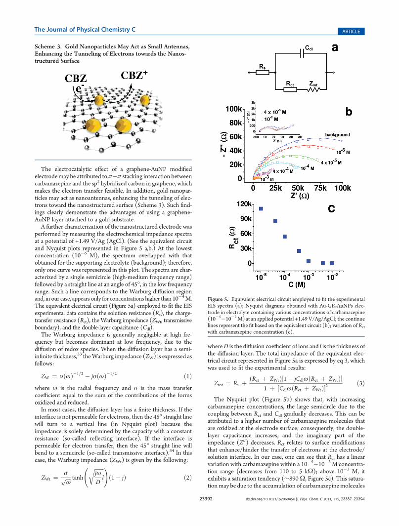
The electrocatalytic effect of a graphene-AuNP modifiedelectrodemay be attributed toπ�π stacking interaction betweencarbamazepine and the sp2 hybridized carbon in graphene, whichmakes the electron transfer feasible. In addition, gold nanopar-ticles may act as nanoantennas, enhancing the tunneling of elec-trons toward the nanostructured surface (Scheme 3). Such find-ings clearly demonstrate the advantages of using a graphene-AuNP layer attached to a gold substrate.A further characterization of the nanostructured electrode was
performed by measuring the electrochemical impedance spectraat a potential of +1.49 V/Ag (AgCl). (See the equivalent circuitand Nyquist plots represented in Figure 5 a,b.) At the lowestconcentration (10�6 M), the spectrum overlapped with thatobtained for the supporting electrolyte (background); therefore,only one curve was represented in this plot. The spectra are char-acterized by a single semicircle (high-medium frequency range)followed by a straight line at an angle of 45�, in the low frequencyrange. Such a line corresponds to the Warburg diffusion regionand, in our case, appears only for concentrations higher than 10�4M.The equivalent electrical circuit (Figure 5a) employed to fit the EISexperimental data contains the solution resistance (Rs), the charge-transfer resistance (Rct), the Warburg impedance (ZWt, transmissiveboundary), and the double-layer capacitance (Cdl).The Warburg impedance is generally negligible at high fre-
quency but becomes dominant at low frequency, due to thediffusion of redox species. When the diffusion layer has a semi-infinite thickness,33 theWarburg impedance (ZW) is expressed asfollows:
ZW ¼ σðωÞ�1=2 � jσðωÞ�1=2 ð1Þwhere ω is the radial frequency and σ is the mass transfercoefficient equal to the sum of the contributions of the formsoxidized and reduced.In most cases, the diffusion layer has a finite thickness. If the
interface is not permeable for electrons, then the 45� straight linewill turn to a vertical line (in Nyquist plot) because theimpedance is solely determined by the capacity with a constantresistance (so-called reflecting interface). If the interface ispermeable for electron transfer, then the 45� straight line willbend to a semicircle (so-called transmissive interface).34 In thiscase, the Warburg impedance (ZWt) is given by the following:
ZWt ¼ σffiffiffiffiω
p tanh
ffiffiffiffiffiffiffijωDl
r !ð1� jÞ ð2Þ
whereD is the diffusion coefficient of ions and l is the thickness ofthe diffusion layer. The total impedance of the equivalent elec-trical circuit represented in Figure 5a is expressed by eq 3, whichwas used to fit the experimental results:
Ztot ¼ Rs þ ðRct þ ZWtÞ½1� jCdlωðRct þ ZWtÞ�1 þ ½CdlωðRct þ ZWtÞ�2
ð3Þ
The Nyquist plot (Figure 5b) shows that, with increasingcarbamazepine concentrations, the large semicircle due to thecoupling between Rct and Cdl gradually decreases. This can beattributed to a higher number of carbamazepine molecules thatare oxidized at the electrode surface; consequently, the double-layer capacitance increases, and the imaginary part of theimpedance (Z00) decreases. Rct relates to surface modificationsthat enhance/hinder the transfer of electrons at the electrode/solution interface. In our case, one can see that Rct has a linearvariation with carbamazepine within a 10�5�10�3 M concentra-tion range (decreases from 110 to 5 kΩ); above 10�3 M, itexhibits a saturation tendency (∼890Ω, Figure 5c). This satura-tionmay be due to the accumulation of carbamazepinemolecules
Scheme 3. Gold Nanoparticles May Act as Small Antennas,Enhancing the Tunneling of Electrons towards the Nanos-tructured Surface
Figure 5. Equivalent electrical circuit employed to fit the experimentalEIS spectra (a); Nyquist diagrams obtained with Au-GR-AuNPs elec-trode in electrolyte containing various concentrations of carbamazepine(10�5�10�2M) at an applied potential +1.49 V/Ag/AgCl; the continuelines represent the fit based on the equivalent circuit (b); variation of Rctwith carbamazepine concentration (c).
23393 dx.doi.org/10.1021/jp206945e |J. Phys. Chem. C 2011, 115, 23387–23394
The Journal of Physical Chemistry C ARTICLE
within the graphene platelets, which in time leads to a poor elec-trical transfer between the graphene-AuNPs layer and goldsubstrate.Another representation of the EIS data is the Bode diagram
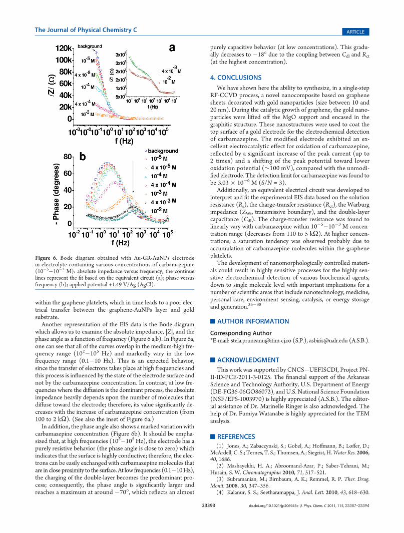
which allows us to examine the absolute impedance, |Z|, and thephase angle as a function of frequency (Figure 6 a,b). In Figure 6a,one can see that all of the curves overlap in the medium-high fre-quency range (102�105 Hz) and markedly vary in the lowfrequency range (0.1�10 Hz). This is an expected behavior,since the transfer of electrons takes place at high frequencies andthis process is influenced by the state of the electrode surface andnot by the carbamazepine concentration. In contrast, at low fre-quencies where the diffusion is the dominant process, the absoluteimpedance heavily depends upon the number of molecules thatdiffuse toward the electrode; therefore, its value significantly de-creases with the increase of carbamazepine concentration (from100 to 2 kΩ). (See also the inset of Figure 6a.)In addition, the phase angle also shows amarked variation with
carbamazepine concentration (Figure 6b). It should be empha-sized that, at high frequencies (103�105 Hz), the electrode has apurely resistive behavior (the phase angle is close to zero) whichindicates that the surface is highly conductive; therefore, the elec-trons can be easily exchanged with carbamazepine molecules thatare in close proximity to the surface. At low frequencies (0.1�10Hz),the charging of the double-layer becomes the predominant pro-cess; consequently, the phase angle is significantly larger andreaches a maximum at around �70�, which reflects an almost
purely capacitive behavior (at low concentrations). This gradu-ally decreases to �18� due to the coupling between Cdl and Rct(at the highest concentration).
4. CONCLUSIONS
We have shown here the ability to synthesize, in a single-stepRF-CCVD process, a novel nanocomposite based on graphenesheets decorated with gold nanoparticles (size between 10 and20 nm). During the catalytic growth of graphene, the gold nano-particles were lifted off the MgO support and encased in thegraphitic structure. These nanostructures were used to coat thetop surface of a gold electrode for the electrochemical detectionof carbamazepine. The modified electrode exhibited an ex-cellent electrocatalytic effect for oxidation of carbamazepine,reflected by a significant increase of the peak current (up to2 times) and a shifting of the peak potential toward loweroxidation potential (∼100 mV), compared with the unmodi-fied electrode. The detection limit for carbamazepine was found tobe 3.03 � 10�6 M (S/N = 3).
Additionally, an equivalent electrical circuit was developed tointerpret and fit the experimental EIS data based on the solutionresistance (Rs), the charge-transfer resistance (Rct), the Warburgimpedance (ZWt, transmissive boundary), and the double-layercapacitance (Cdl). The charge-transfer resistance was found tolinearly vary with carbamazepine within 10�5�10�3 M concen-tration range (decreases from 110 to 5 kΩ). At higher concen-trations, a saturation tendency was observed probably due toaccumulation of carbamazepine molecules within the grapheneplatelets.
The development of nanomorphologically controlled materi-als could result in highly sensitive processes for the highly sen-sitive electrochemical detection of various biochemical agents,down to single molecule level with important implications for anumber of scientific areas that include nanotechnology, medicine,personal care, environment sensing, catalysis, or energy storageand generation.35�38
’AUTHOR INFORMATION
Corresponding Author*E-mail: [email protected] (S.P.), [email protected] (A.S.B.).
’ACKNOWLEDGMENT
This work was supported by CNCS�UEFISCDI, Project PN-II-ID-PCE-2011-3-0125. The financial support of the ArkansasScience and Technology Authority, U.S. Department of Energy(DE-FG36-06GO86072), andU.S. National Science Foundation(NSF/EPS-1003970) is highly appreciated (A.S.B.). The editor-ial assistance of Dr. Marinelle Ringer is also acknowledged. Thehelp of Dr. Fumiya Watanabe is highly appreciated for the TEManalysis.
’REFERENCES
(1) Jones, A.; Zabaczynski, S.; Gobel, A.; Hoffmann, B.; Loffer, D.;McArdell, C. S.; Ternes, T. S.; Thomsen, A.; Siegrist, H.Water Res. 2006,40, 1686.
(2) Mashayekhi, H. A.; Abroomand-Azar, P.; Saber-Tehrani, M.;Husain, S. W. Chromatographia 2010, 71, 517–521.
(3) Subramanian, M.; Birnbaum, A. K.; Remmel, R. P. Ther. Drug.Monit. 2008, 30, 347–356.
(4) Kalanur, S. S.; Seetharamappa, J. Anal. Lett. 2010, 43, 618–630.
Figure 6. Bode diagram obtained with Au-GR-AuNPs electrodein electrolyte containing various concentrations of carbamazepine(10�5�10�2 M): absolute impedance versus frequency; the continuelines represent the fit based on the equivalent circuit (a); phase versusfrequency (b); applied potential +1.49 V/Ag (AgCl).
23394 dx.doi.org/10.1021/jp206945e |J. Phys. Chem. C 2011, 115, 23387–23394
The Journal of Physical Chemistry C ARTICLE
(5) Veiga, A.; Dordio, A.; Palace Carvalho, A. J.; Teixeira, D. M.;Teixeira, J. G. Anal. Chim. Acta 2010, 674, 182–189.(6) Moses, G. S.; Rao, K. M.; Rao, N. S.; Ramachandraiah, A.
J. Indian Chem. Soc. 1995, 72, 333–337.(7) Li, M.; Xu, S.; Tang, M.; Liu, L.; Gao, F.; Wang, Y. Electrochim.
Acta 2010, 56, 1144–1149.(8) Wang, C.; Zhang, L.; Guo, Z.; Xu, J.;Wang, H.; Zhai, K.; Zhuo, X.
Microchim. Acta 2010, 169, 1–6.(9) Huang, K.-J.; Niu, D.-J.; Sun, J.-Y.; Han, C.-H.; Wu, Z.-W.; Li,
Y.-L.; Xiong, X.-Q. Colloids Surf., B 2011, 82, 543–549.(10) Liu, K.; Zhang, J.; Yang, G.; Wang, C; Zhu, J.-J. Electrochem.
Commun. 2010, 12, 402–405.(11) Yang, X.; Zhang, X.; Liu, Z.; Ma, Y.; Huang, Y.; Chen, Y. J. Phys.
Chem. C 2008, 112, 17554–17558.(12) Dreyer, D. R.; Park, S.; Bielawski, C. W.; Ruoff, R. S. Chem. Soc.
Rev 2010, 39, 229–240.(13) Kang, X.; Wang, J.; Wua, H.; Liua, J.; Aksayc, I. A.; Lina, Y.
Talanta 2010, 81, 754–759.(14) Li, J.; Guo, S.; Zhai, Y.; Wang, E. Electrochem. Commun. 2009,
11, 1085–1088.(15) Wang, Y.; Li, Y; Tang, L.; Lu, J.; Li, J. Electrochem. Commun.
2009, 11, 889–892.(16) Liu, J.; Li, Y.; Li, Y.; Li, J.; Deng, Z. J. Mater. Chem 2010, 20,
900–906.(17) Du, M.; Yang, T.; Jiao, K. J. Mater. Chem 2010, 20, 9253–
9260.(18) Zhu, C.; Guo, S.; Zhai, Y.; Dong, S. Langmuir 2010, 26,
7614–7618.(19) Seger, B.; Kamat, P. V. J. Phys. Chem. C 2009, 113, 7990–7995.(20) Bao, Q.; Zhang, D.; Qi, P. J. Colloid Interface Sci. 2011, 360,
463–470.(21) Das, M. R.; Sarma, R. K.; Saikia, R.; Kale, V. S.; Shelke, M. V.;
Sengupta, P. Colloids Surf., B 2011, 83, 16–22.(22) Dresselhaus, M. S.; Jorio, A.; Hofmann, M.; Dresselhaus, G.;
Saito, R. Nano Lett. 2010, 10, 751–758.(23) Lupu, D.; Biris, A. R.; Jianu, A.; Bunescu, C.; Burkel, E.; Indrea,
E.; Mihailescu, G.; Pruneanu, S.; Olenic, L.; Misan, I. Carbon 2004, 42,503–507.(24) Biris, A. R.; Biris, A. S.; Lupu, D.; Trigwell, S.; Dervishi, E.;
Rahman, Z.; Marginean, P. Chem. Phys. Lett. 2006, 429, 204–208.(25) Biris, A. R.; Lupu, D.; Gr€uneis, A.; Ayala, P.; R€ummeli, M. H.;
Pichler, T.; Li, Z.; Xu, Y.; Misan, I.; Biris, A. S. Chem. Mater. 2008,20, 3466–3472.(26) Buchsteiner, A.; Lerf, A.; Pieper, J. J. Phys. Chem. B 2006,
110, 22328–22338.(27) Lerf, A.; Buchsteiner, A.; Pieper, J.; Sch€ottl, S.; Dekany, I.;
Szabo, T.; Boehm, H. P. J. Phys. Chem. Solids 2006, 67, 1106–1110.(28) SRC. PhysProp Database; 2006, http://www.syrres.com/esc/
physdemo.htm.(29) Laviron, E. J. Electroanal. Chem. 1980, 112, 11–23.(30) Atkins, S.; Sevilla, J. M.; Blazquez, M.; Pineda, T.; Gonzalez-
Rodriquez, J. Electroanalysis 2010, 22, 2961–2966.(31) Wang, H. Y.; Pan, M. L.; Su, Y. L. O.; Tsai, S. C.; Kao, C. H.;
Sun, S. S.; Lin, W. Y. J. Anal. Chem. 2011, 66, 415–420.(32) Frank, S. N.; Bard, A. J.; Ledwith, A. J. Electrochem. Soc. 1975,
122, 898–904.(33) Bard, A. J.; Faulkner, L. R. In Electrochemical Methods: Funda-
mentals and Applications; John Wiley: New York, 1980; pp 324�353.(34) Lasia, A. Electrochemical Impedance Spectroscopy and Its Applica-
tions, Modern Aspects of Electrochemistry; Conway, B. E., Bockris, J.,White, R., Eds.; Kluwer Academic/Plenum Publishers: New York, 1999;pp 143�248.(35) Pruneanu, S.; Pogacean, F.; Grosan, C.; Pica, E. M.; Bolundut,
L. C.; Biris, A. S. Chem. Phys. Lett. 2011, 504, 56–61.(36) Orza, A.; Olenic, L.; Pruneanu, S.; Pogacean, F.; Biris, A. S.
Chem. Phys. 2010, 373, 295–299.(37) Lupu, D.; Biris, A. R.; Biris, A. S.; Misan, I.; Buzatu, D. A.; Kleve,
M.; Lupsa, N. Part. Sci. Technol. 2002, 20, 225–234.
(38) Biris, A. R.; Mahmood,M.; Lazar, M. D.; Devishi, E.; Watanabe,F.; Mustafa, T.; Baciut, G.; Baciut, M.; Bran, S.; Ali, S.; Biris, A. S. J. Phys.Chem. C 2011, 115, 18967–18976.
Copyright © 2022 FDOKUMEN