Novel bladder preservation therapy for locally invasive bladder cancer: combined therapy using...
13
Abstract. We investigated the effect of balloon-occluded arterial infusion (BOAI) of anticancer agent (cisplatin/gem- citabine), used concomitantly with hemodialysis, which delivers an extremely high concentration of anticancer agent to the site of a tumor without systemic adverse effects, along with concurrent radiation (referred to as the OMC-regimen) in patients with advanced bladder cancer. One hundred and ninety-two patients were assigned to receive either the OMC- regimen (n=96) or total cystectomy (n=96). Patients in the OMC-regimen group who failed to achieve CR underwent cystectomy, or secondary BOAI with an increased amount of CDDP or gemcitabine (1600 mg). The OMC-regimen allowed >89% (69/77) of patients with locally invasive tumors to achieve CR [>70% (70/96) of all patients including those with T4 and N(+) disease]. Most (68/69) of the CR patients were still alive with no evidence of recurrence after a mean follow-up of 161 (range 12-805) weeks. The 5- and 15-year overall survival rates were 91.5 and 81.3% (vs. 59.8% and 40.1% for cystectomy, P<0.0001), respectively. No patients suffered Grade III or more severe toxicities. In contrast, at 5 and 15 years after surgery in the total cystectomy group, about 50 and 60% of patients had suffered disease progression or had died, respectively. The OMC-regimen, a new bladder- preservation strategy for patients with locally invasive bladder cancer, can be curative not only in patients for whom cystectomy is indicated, but also in patients whose condition is not amenable to curative treatment and for whom merely palliative therapy would otherwise seem the only option. Introduction Radical cystectomy has long been the standard method of treatment for patients with locally invasive bladder cancer. However, this inevitably reduces the quality of life of the patients to some degree. Moreover, more than 40% of all patients with invasive bladder cancer die within 5 years (1-8). Trimodality therapy consisting of radical transurethral resection, chemotherapy and radiation therapy has been attempted as an alternative approach for patients who require cystectomy. Data from multiple institutional and cooperative group studies have shown that this approach is safe and effective, yielding a complete response (CR) in more than 60% of cases (9-15). However, prospective protocols conducted by the Radiation Therapy Oncology Group (RTOG) demon- strated a survival rate similar to that for radical cystectomy; none of the protocols achieved a 5-year survival rate of more than 60% (9-13). A highly effective, but non- or only minimally invasive therapy that conserves the bladder is therefore needed. Accordingly, we conducted the present study to investigate the effect of combined therapy [referred to hereafter as the OMC (Osaka Medical College) regimen] involving balloon- occluded arterial infusion (BOAI) of an anticancer agent and concurrent hemodialysis (HD), which allows the anticancer agent to accumulate at a high concentration at the site of a tumor but ensures that the systemic concentration remains low after the agent has passed through the tumor, followed by radiation therapy. We found that more than 90% of patients (70/77) with locally advanced urothelial bladder cancer who were treated in this way achieved CR, of whom 97% (68/70) did not develop recurrent disease or metastasis within a mean follow-up period of 164 weeks [range, 11-805 weeks; 1st to INTERNATIONAL JOURNAL OF ONCOLOGY 37: 773-785, 2010 773 Novel bladder preservation therapy for locally invasive bladder cancer: Combined therapy using balloon-occluded arterial infusion of anticancer agent and hemodialysis with concurrent radiation HARUHITO AZUMA 1 , TERUO INAMOTO 1 , NAOKAZU IBUKI 1 , TAKANOBU UBAI 1 , YATSUGU KOTAKE 1 , KIYOSHI TAKAHARA 1 , SATOSHI KIYAMA 1 , HAYAHITO NOMI 1 , HIROSHI UEHARA 1 , KAZUMASA KOMURA 1 , KAZUHIRO YAMAMOTO 2 , YOSHIHUMI NARUMI 2 and YOJI KATSUOKA 1 Departments of 1 Urology, and 2 Radiology, Osaka Medical College, Takatsuki, Osaka 569-8686, Japan Received May 31, 2010; Accepted July 16, 2010 DOI: 10.3892/ijo_00000727 _________________________________________ Correspondence to: Dr Haruhito Azuma, Department of Urology, Osaka Medical College, Takatsuki, Osaka 569-8686, Japan E-mail: [email protected] Abbreviations: ANC, absolute neutrophil count; BOAI, balloon- occluded arterial infusion; CIS, carcinoma in situ; CTCAE, common terminology criteria for adverse events; DSA, digital subtraction angiography; ECOG, Eastern Cooperative Oncology Group; HD, hemodialysis; Qu, quartile; RTOG, radiation therapy oncology group; TURBT, transurethral resection of bladder tumor; UC, urothelial carcinoma Key words: balloon-occluded arterial infusion, hemodialysis, OMC-regimen, invasive bladder cancer
Transcript of Novel bladder preservation therapy for locally invasive bladder cancer: combined therapy using...

Abstract. We investigated the effect of balloon-occludedarterial infusion (BOAI) of anticancer agent (cisplatin/gem-citabine), used concomitantly with hemodialysis, whichdelivers an extremely high concentration of anticancer agentto the site of a tumor without systemic adverse effects, alongwith concurrent radiation (referred to as the OMC-regimen) inpatients with advanced bladder cancer. One hundred andninety-two patients were assigned to receive either the OMC-regimen (n=96) or total cystectomy (n=96). Patients in theOMC-regimen group who failed to achieve CR underwentcystectomy, or secondary BOAI with an increased amount ofCDDP or gemcitabine (1600 mg). The OMC-regimen allowed>89% (69/77) of patients with locally invasive tumors toachieve CR [>70% (70/96) of all patients including thosewith T4 and N(+) disease]. Most (68/69) of the CR patientswere still alive with no evidence of recurrence after a meanfollow-up of 161 (range 12-805) weeks. The 5- and 15-yearoverall survival rates were 91.5 and 81.3% (vs. 59.8% and40.1% for cystectomy, P<0.0001), respectively. No patientssuffered Grade III or more severe toxicities. In contrast, at5 and 15 years after surgery in the total cystectomy group,about 50 and 60% of patients had suffered disease progression
or had died, respectively. The OMC-regimen, a new bladder-preservation strategy for patients with locally invasivebladder cancer, can be curative not only in patients for whomcystectomy is indicated, but also in patients whose conditionis not amenable to curative treatment and for whom merelypalliative therapy would otherwise seem the only option.
Introduction
Radical cystectomy has long been the standard method oftreatment for patients with locally invasive bladder cancer.However, this inevitably reduces the quality of life of thepatients to some degree. Moreover, more than 40% of allpatients with invasive bladder cancer die within 5 years (1-8).Trimodality therapy consisting of radical transurethralresection, chemotherapy and radiation therapy has beenattempted as an alternative approach for patients who requirecystectomy. Data from multiple institutional and cooperativegroup studies have shown that this approach is safe andeffective, yielding a complete response (CR) in more than 60%of cases (9-15). However, prospective protocols conductedby the Radiation Therapy Oncology Group (RTOG) demon-strated a survival rate similar to that for radical cystectomy;none of the protocols achieved a 5-year survival rate ofmore than 60% (9-13). A highly effective, but non- or onlyminimally invasive therapy that conserves the bladder istherefore needed.
Accordingly, we conducted the present study to investigatethe effect of combined therapy [referred to hereafter as theOMC (Osaka Medical College) regimen] involving balloon-occluded arterial infusion (BOAI) of an anticancer agent andconcurrent hemodialysis (HD), which allows the anticanceragent to accumulate at a high concentration at the site of atumor but ensures that the systemic concentration remainslow after the agent has passed through the tumor, followedby radiation therapy. We found that more than 90% of patients(70/77) with locally advanced urothelial bladder cancer whowere treated in this way achieved CR, of whom 97% (68/70)did not develop recurrent disease or metastasis within a meanfollow-up period of 164 weeks [range, 11-805 weeks; 1st to
INTERNATIONAL JOURNAL OF ONCOLOGY 37: 773-785, 2010 773
Novel bladder preservation therapy for locally invasive bladder cancer: Combined therapy using balloon-occluded
arterial infusion of anticancer agent and hemodialysis with concurrent radiation
HARUHITO AZUMA1, TERUO INAMOTO1, NAOKAZU IBUKI1, TAKANOBU UBAI1, YATSUGU KOTAKE1,
KIYOSHI TAKAHARA1, SATOSHI KIYAMA1, HAYAHITO NOMI1, HIROSHI UEHARA1,
KAZUMASA KOMURA1, KAZUHIRO YAMAMOTO2, YOSHIHUMI NARUMI2 and YOJI KATSUOKA1
Departments of 1Urology, and 2Radiology, Osaka Medical College, Takatsuki, Osaka 569-8686, Japan
Received May 31, 2010; Accepted July 16, 2010
DOI: 10.3892/ijo_00000727
_________________________________________
Correspondence to: Dr Haruhito Azuma, Department of Urology,Osaka Medical College, Takatsuki, Osaka 569-8686, JapanE-mail: [email protected]
Abbreviations: ANC, absolute neutrophil count; BOAI, balloon-occluded arterial infusion; CIS, carcinoma in situ; CTCAE,common terminology criteria for adverse events; DSA, digitalsubtraction angiography; ECOG, Eastern Cooperative OncologyGroup; HD, hemodialysis; Qu, quartile; RTOG, radiation therapyoncology group; TURBT, transurethral resection of bladder tumor;UC, urothelial carcinoma
Key words: balloon-occluded arterial infusion, hemodialysis,OMC-regimen, invasive bladder cancer
773-785.qxd 19/8/2010 09:14 Ì ™ÂÏ›‰·773
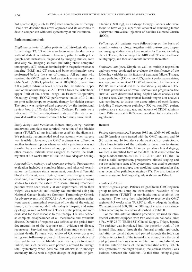
3rd quartile (Qu) = 66 to 195] after completion of therapy.Herein we describe this novel approach and its outcomes todate in comparison with total cystectomy at our institution.
Patients and methods
Eligibility criteria. Eligible patients had histologically con-firmed stage T2, T3 or T4 muscle-invasive bladder cancerwithout distant metastasis. However, patients with pelviclymph node metastasis, diagnosed by imaging studies, werealso eligible. Imaging studies, including chest computedtomography (CT) scan, abdominal/pelvic magnetic resonanceimaging (MRI) and CT scan, and bone scintigraphy wereperformed before the start of therapy. All patients whoreceived the OMC regimen had an absolute neutrophil count(ANC) of 1,500/μl, platelet count 100,000/μl, creatinine3.0 mg/dl, a bilirubin level 3 times the institutional upperlimit of the normal range, an AST level 4 times the institutionalupper limit of the normal range, an Eastern CooperativeOncology Group (ECOG) performance status of 0-2, andno prior radiotherapy or systemic therapy for bladder cancer.The study was reviewed and approved by the institutionalreview board of Osaka Medical College. Patients wereinformed of the investigational nature of the study andprovided written informed consent before study enrollment.
Study design and treatment. Before study entry, patientsunderwent complete transurethral resection of the bladdertumor (TURBT) at our institution to establish the diagnosis.We primarily recommended total cystectomy when surgerywas feasible. However, the OMC regimen was offered asanother treatment option whenever total cystectomy was notfeasible because of advanced age, performance status, orother reasons. Patients were assigned to receive the OMCregimen at 4-5 weeks after TURBT to allow adequate healing.
Assessability, toxicity, and response criteria. Pretreatmentevaluation included a complete history and physical exami-nation, performance status assessment, complete differentialblood cell count, electrolytes, blood urea nitrogen, serumcreatinine, liver function parameters, and appropriate imagingstudies to assess the extent of disease. During treatment,patients were seen weekly at our department, when theirweight was recorded and toxicity was monitored using theNational Cancer Institute's Common Terminology Criteriafor adverse events v4.0 (CTCAE). At 6 weeks, patients under-went repeat transurethral resection of the site of the originaltumor, ultrasound-guided whole-layer biopsy, and urinecytology, as well as MRI and CT scan of the pelvis, and wereevaluated for their response to this therapy. CR was definedas complete disappearance of all measurable and evaluabledisease. Duration of response was defined as the period fromdocumentation of the response until evidence of diseaserecurrence. Survival was the period from study entry untilpatient death. Patients who achieved CR were observedusing our follow-up protocol. However, any evidence ofresidual tumor in the bladder was deemed as treatmentfailure, and such patients were primarily advised to undergototal cystectomy when possible, but otherwise to undergosecondary BOAI with a higher dosage of cisplatin or gem-
citabine (1600 mg), as a salvage therapy. Patients who werefound to have only a superficial amount of remaining tumorunderwent intravesical injection of bacillus Calmette Guerin(BCG).
Follow-up. All patients were followed-up on the basis ofmonthly urine cytology, together with cystoscopy, biopsyand imaging studies, every three months for 2 years, includingchest CT scan, abdominal/pelvic MRI and CT scan, and bonescintigraphy, and then at 6-month intervals thereafter.
Statistical analyses. Simple as well as multiple regressionanalyses were conducted to evaluate the significance of thefollowing variables as risk factors of treatment failure: T-stage,tumor pathology (UC vs. non-UC), patient performance status,sex, age, and amount of CDDP administered. Differences atP<0.05 were considered to be statistically significant. Thelife table probabilities of overall survival and progression-freesurvival were determined using Kaplan-Meier analysis andlog-rank test. Cox proportional hazards regression analysiswas conducted to assess the associations of each factor,including T-stage, tumor pathology (UC vs. non-UC), patientperformance status, sex, age, and amount of CDDP adminis-tered. Differences at P<0.05 were considered to be statisticallysignificant.
Results
Patient characteristics. Between 1988 and 2009, 96 (67 malesand 29 females) were treated with the OMC regimen, and 96(80 males and 16 females) underwent radical cystectomy.The characteristics of the patients in these two treatmentgroups are shown in Table I. For preoperative clinical staging,we used a simplified form of the 2002 TNM classification tostage bladder tumors as Tis, T1, T2, T3, and T4 (16). Tomake a valid comparison, preoperative clinical staging andnot the pathologic stage after cystectomy was used to comparethe two treatment groups, thus avoiding stage migration thatmay occur after pathologic staging (17). The distribution ofclinical stage and histological grade is shown in Table I.
Treatment detailsi) OMC-regimen group. Patients assigned to the OMC-regimengroup underwent complete transurethral resection of thebladder tumor (TURBT) at our institution to establish thediagnosis. They were then scheduled to receive the OMCregimen 4-5 weeks after TURBT to allow adequate healing.We administered 100, 200, or 300 mg of cisplatin as a singlebolus according to the criteria described in Table II.
For the intra-arterial infusion procedure, we used an intra-arterial catheter equipped with two occlusion balloons (size:6 Fr., M6F-28-70-TBSB4-ST, Clinical Supply, Tokyo, Japan).The catheter was introduced into the posterior trunk of theinternal iliac artery through the femoral arterial approach,and after the distal balloon had passed through the furcationof the anterior trunk of the internal iliac artery, both the distaland proximal balloons were inflated and immobilized, sothat the anterior trunk of the internal iliac artery, whichlies upstream of the target vessels (the vesical arteries) wasisolated between the balloons. At this time, using digital
AZUMA et al: BOAI CONCURRENT WITH HD FOR LOCALLY INVASIVE BLADDER CANCER774
773-785.qxd 19/8/2010 09:14 Ì ™ÂÏ›‰·774

subtraction angiography (DSA), it was confirmed that theinjected agent did not enter the superior gluteal artery andthat there was no back-flow into the internal iliac artery,
while the tumor was markedly stained due to active flowof injected contrast medium into the urinary bladder. Fig. 1illustrates the extracorporeal circuit used in the treatment,and Fig. 2 presents DSA images of the bilateral commoniliac arteries before (Fig. 2A) and after (Fig. 2B) balloonocclusion. Various amounts of cisplatin (100, 200, or 300 mg)were locally infused through the catheter over a 1-h period(Table I). Simultaneously, HD was performed via two double-lumen catheters (size: 12 Fr., Argyle®, Tyco Healthcare, Tokyo,Japan) placed in the bilateral common iliac veins for 2 h afterthe start of arterial infusion. The catheters were connectedto a hollow-fiber dialyzer (APS150, Asahi, Tokyo, Japan)with a membrane area of 1.0-1.5 m2 according to the weightof each patient. The blood flow rate was 180-250 ml/min andthe hemodialysis-fluid flow rate was 500 ml/min.
Radiation therapy was administered to the whole pelvisusing a CT-planned three-dimensional conformal techniqueto a total of 60.4 Gy: 50.4 Gy (1.8 Gy/day x 28 days) followedby 10 Gy (2 Gy/day x 5 days) of local irradiation to thebladder. Patients were treated with the bladder empty. Theplanned target volume for the bladder included the gross targetvolume (bladder plus any extravesical tumor) with a 1-cmexpansion. At 6 weeks, patients underwent repeat transurethralresection of the site of the original tumor, ultrasound-guidedwhole-layer biopsy, and urine cytology, as well as MRI andCT scan of the pelvis, and the response to this therapy wasthen evaluated.
ii) Radical cystectomy group. Among the 96 patients in theradical cystectomy group, 32 underwent ileal conduit for-mation, 35 underwent uretero-cutaneostomy, 25 underwentcontinent urinary diversion with ileal-neobladder formation(Hartmann's method), and the remaining 4 underwent uretero-sigmoidostomy performed at the time of radical cystectomy.Standard pelvic lymphadenectomy was performed in 81patients, 5 patients underwent iliac sampling, and 10 patientswere not studied in sufficient detail to allow assessment ofthe level of lymph node dissection. As not all of the histologyreports mentioned the number of lymph nodes examined, itwas not possible to precisely evaluate the extent of dissection.There were no significant differences in cause-specific oroverall survival between the patients who underwent nodaldissection and the patients who did not. Urethrectomy wasperformed in 10 patients at the time of radical cystectomybecause of the presence of extensive carcinoma in situ ormultifocal bladder tumors.
Response to the OMC regimen. Table III summarizes thetreatment response, duration of response, and patientcharacteristics, including T stage, N stage, tumor histology(UC vs. non-UC), patient performance status, sex, age, andamount of CDDP administered. Overall, 73 of the 96 patients(76.0%, 95% CI, 66.3-84.2%) achieved a complete responseas defined by the absence of persistent disease revealed bycystoscopy, biopsy, and urine cytology after therapy (Table III).More than 95% (70/73) of patients with CR were able toretain their urinary bladder with no evidence of recurrentdisease or distant metastasis within a mean follow-up periodof 161 weeks (range, 12-805 weeks; 1st to 3rd Qu = 63-193weeks) from the completion of therapy. Most of the patients
INTERNATIONAL JOURNAL OF ONCOLOGY 37: 773-785, 2010 775
Table I. Characteristics of patients in the two groups.–––––––––––––––––––––––––––––––––––––––––––––––––Characteristic OMC Total P-value
regimen cystectomy–––––––––––––––––––––––––––––––––––––––––––––––––Age median 72 (38-98) 65 (44-79) <0.0001(range years)
SexMale (%) 67 (69.8%) 80 (83.3%) 0.0286Female (%) 29 (30.2%) 16 (16.7%)
Clinical stageT-stageCis 0 (0%) 4 (4.2%) N.S.T2 32 (33.3%) 49 (51.0%) 0.0135T3 48 (50.0%) 43 (44.8%) N.S.T4 16 (16.7%) 0 (0%)
N-stageN0 86 (89.6%) 96 (100%)N1 10 (10.4%) 0 (0%)
Tumor histologyUCG2 10 (10.4%) 32 (33.3%) 0.0002G3 78 (82.3%) 55 (57.3%) 0.0004
Others N.S.Adenocarcinoma 6 (5.2%) 1 (1.0%) N.S.Squamous cell 1 (1.05%) 8 (8.3%) N.S.carcinomaChoriocarcinoma 1 (1.05%) 0 (0%) N.S.
ECOG performancestatus0 37 (38.5%) 64 (66.7%) 0.00011 41 (42.7%) 24 (25.0%) 0.01022 18 (18.8%) 8 (8.3%) 0.0395
–––––––––––––––––––––––––––––––––––––––––––––––––
Table II. Criteria for the administration of cisplatin. –––––––––––––––––––––––––––––––––––––––––––––––––In the initially enrolled 22 patients
100 mg Renal function (sCr ≥1.3) or age ( ≥75 years)200 mg Renal function (sCr <1.3) with [age
(60-74 years) and T-stage (T2 or T3)]300 mg Renal function (sCr <1.3) with [age
(<60 years) or T-stage: T4]
In the latest 74 patients100 mg All patients
–––––––––––––––––––––––––––––––––––––––––––––––––
773-785.qxd 19/8/2010 09:14 Ì ™ÂÏ›‰·775

who achieved CR had locally invasive tumors (stage T2 or T3node-negative; 71 of 73 patients, 97.3%, 95% CI, 90.5-99.7%)and UC histologically (72 of 73 patients, 98.6%, 95% CI,
92.6-99.9%). Indeed, only 6 of 77 patients (4.4%) withlocally invasive tumors failed to achieve CR, while only 3patients with stage T4 tumors achieved CR, only 1 patient
AZUMA et al: BOAI CONCURRENT WITH HD FOR LOCALLY INVASIVE BLADDER CANCER776
Figure 1. Schema of the OMC regimen (HD-BOAI-CDDP/gemcitabine with radiation). The extracorporeal circuit allowed balloon-occluded intra-arterialinfusion of CDDP/gemcitabine concurrent with HD. Through the femoral arterial approach, an intra-arterial catheter equipped with two occlusion balloonswas introduced into the posterior trunk of the internal iliac artery on each side. Both the distal and proximal balloons were inflated and immobilized at aposition allowing the vesical arteries to be isolated between the balloons. After confirming by angiography that the catheter was in the right position, variousamounts of cisplatin (100, 200, or 300 mg), or gemcitabine (1600 mg) were infused through the side holes of the catheter between the inflated balloons overa 1-h period. Simultaneously, HD was performed via double-lumen catheters placed in the bilateral common iliac vein for 2 h after the start of arterialinfusion. The panel marked with an asterisk shows a picture of the intra-arterial catheter (M6F-28-70-TBSB4-ST, clinical supply), which is made ofpolyethylene, 6 French in size, and equipped with two occlusion balloons separated by a distance of 40 mm. It has side holes between the balloons enablinginjection of contrast medium or anticancer agent.
Figure 2. Bilateral common iliac arteriography before (A) and after (B) balloon occlusion. We ensured that contrast medium did not enter the superior glutealartery and that there was no back-flow into the internal iliac artery. We also also confirmed that the anticancer agent was delivered to the urinary bladder,especially to the tumor site.
773-785.qxd 19/8/2010 09:14 Ì ™ÂÏ›‰·776

INTERNATIONAL JOURNAL OF ONCOLOGY 37: 773-785, 2010 777T
able
III
. Res
pons
e at
3 m
onth
s af
ter
trea
tmen
t, an
d cu
rren
t out
com
e.––
––––
––––
––––
––––
––––
––––
––––
––––
––––
––––
––––
––––
––––
––––
––––
––––
––––
––––
––––
––––
––––
––––
––––
––––
––––
––––
––––
––––
––––
––––
––––
––––
––––
––––
––––
–––
CR
PRSD
PD––
––––
––––
––––
––––
––––
––––
––––
––––
––––
––––
––––
––––
––––
––––
––––
––––
––––
––––
––––
––––
––––
––––
––––
––––
––––
––––
––––
––N
o.%
95%
CI
No.
.%
95%
CI
No.
.%
95%
CI
No.
.%
95%
CI
––––
––––
––––
––––
––––
––––
––––
––––
––––
––––
––––
––––
––––
––––
––––
––––
––––
––––
––––
––––
––––
––––
––––
––––
––––
––––
––––
––––
––––
––––
––––
––––
––––
––––
––––
––––
–T
otal
no.
of
patie
nts
7376
.058
.1-8
1.8
55.
211.
71 -
11.7
88.
333.
67 -
15.8
1010
.45.
11 -
18.3
Dur
atio
n of
res
pons
e M
ean,
ran
ge15
4, 8
-802
wee
ks45
, 12-
69 w
eeks
55, 1
5-23
7 w
eeks
01s
t, 3r
d Q
U58
, 189
wee
ks27
, 68
wee
ks19
, 42
wee
ks0
Rec
urre
nce
34.
110.
86-1
1.5
00
0-52
.27
87.5
47.3
-99.
7D
eath
45.
481.
51-1
3.4
00
0-52
.25
62.5
24.5
-91.
59
90.0
55.4
-99.
7
Age
(m
ean,
ran
ge)
70, 3
8-85
yea
rs72
, 62-
78 y
ears
79, 6
8-98
yea
rs71
, 55-
81 y
ears
Sex
Mal
e53
72.6
60.9
-82.
42
40.0
5.27
-85.
35
62.5
24.5
-91.
57
70.0
34.8
-93.
3Fe
mal
e20
27.4
17.6
-39.
13
60.0
14.7
-94.
73
37.5
8.52
-75.
53
30.0
6.67
-65.
2––
––––
––––
––––
––––
––––
––––
––––
––––
––––
––––
––––
––––
––––
––––
––––
––––
––––
––––
––––
––––
––––
––––
––––
––––
––––
––––
––––
––––
––––
––––
––––
––––
––––
––––
––––
–––
No.
%95
% C
IN
o.%
95%
CI
No.
%95
% C
IN
o.%
95%
CI
––––
––––
––––
––––
––––
––––
––––
––––
––––
––––
––––
––––
––––
––––
––––
––––
––––
––––
––––
––––
––––
––––
––––
––––
––––
––––
––––
––––
––––
––––
––––
––––
––––
––––
––––
––––
–C
ateg
orie
s2
3142
.531
.0-5
4.6
10
0.50
-71.
60
00-
36.9
00
0-30
.8
T s
tage
340
53.8
42.7
-66.
53
014
.7-9
4.7
225
.03.
67-7
1.0
330
.06.
67-6
5.2
42
2.7
0.33
-9.5
51
100
0.50
-71.
66
75.0
29.0
-96.
37
70.0
34.8
-93.
3
N s
tage
N (
-)72
98.6
92.6
-100
510
047
.8-1
004
50.0
15.7
-84.
25
50.0
18.7
-81.
3N
(+
)1
1.4
0.03
-7.4
00
00-
52.2
450
.015
.7-8
4.2
550
.018
.7-8
1.3
His
tolo
gyU
C72
98.6
92.6
-100
510
047
.8-1
005
62.5
24.5
-91.
56
60.0
26.2
-87.
8N
on-U
C1
1.4
0.03
-7.4
00
00-
52.2
337
.58.
52-7
5.5
440
.012
.2-7
3.8
PS 032
43.8
32.2
-55.
91
200.
50-7
1.6
112
.50.
32-5
2.7
330
.06.
67-6
5.2
130
41.1
29.7
-53.
22
405.
27-8
5.3
675
.029
.0-9
6.3
330
.06.
67-6
5.2
211
15.1
9.80
-35.
32
405.
27-8
5.3
112
.50.
32-5
2.7
440
.012
.2-7
3.8
CD
DP
100
6589
.067
.3-9
1.8
510
047
.8-1
005
62.5
24.5
-91.
517
70.0
34.8
-93.
320
04
5.5
2.53
-21.
70
00-
52.2
112
.50.
32-5
2.7
110
.00.
25-4
4.5
300
45.
52.
53-2
1.7
00
0-52
.22
25.0
3.19
-65.
12
20.0
2.52
-55.
6
Gem
cita
bine
11.
40.
06-1
2.0
240
5.27
-85.
32
25.0
3.19
-65.
11
10.0
0.25
-44.
5––
––––
––––
––––
––––
––––
––––
––––
––––
––––
––––
––––
––––
––––
––––
––––
––––
––––
––––
––––
––––
––––
––––
––––
––––
––––
––––
––––
––––
––––
––––
––––
––––
––––
––––
––––
–––
773-785.qxd 19/8/2010 09:14 Ì ™ÂÏ›‰·777

with lymph node metastasis achieved CR, and only 1 patientwith a histologically confirmed non-UC tumor achieved CR.Fig. 3 shows horizontal, coronal and sagittal MRI imagesobtained before and after treatment.
In contrast to the high CR induction ratio in patients withlocally invasive UC tumors, most patients with lymph nodeinvolvement (9 of 10 patients; 90%, 95% CI, 55.4-99.7%),stage T4 tumors (13 of 16 patients; 26.8%, 95% CI, 14.2-
AZUMA et al: BOAI CONCURRENT WITH HD FOR LOCALLY INVASIVE BLADDER CANCER778
Figure 3. MRI before (a-c) and after (d-f) the treatment. The horizontal (a), coronal (b) and sagittal (c) MRI slices reveal that bulky tumors occupy more thanhalf of the bladder cavity, and that the tumors invade the abdominal wall, indicating stage T4. In contrast, all imaging studies (d, horizontal; e, coronal; andf, sagittal slices) confirm complete disappearance of the tumors, and that the mucosa and muscle layer are intact, at 10 weeks after completion of the therapy.
Table IV. Risk factors for treatment failure in the OMC-regimen group.–––––––––––––––––––––––––––––––––––––––––––––––––––––––––––––––––––––––––––––––––––––––––––––––––––––
Univariate Multivariate–––––––––––––––––––––– ––––––––––––––––––––––
Category Odds ratio P-value Odds ratio P-value–––––––––––––––––––––––––––––––––––––––––––––––––––––––––––––––––––––––––––––––––––––––––––––––––––––T-stage T4 vs. T2-3 55.22 <0.0001 47.310 0.0005N-stage N(+) vs. N(-) 46.285 0.0005 87.670 0.0044Histology Non-UC vs. UC 31.50 0.0018 7.565 0.0291Performance status 2 vs. 0 or 1 2.466 0.1043 0.967 0.9693Sex Male vs. female 0.587 0.2880 0.317 0.1644Age Cont. variable 1.045 0.1084 1.141 0.1348 Amount of CDDP Cont. variable 1.006 0.0896 0.994 0.5886–––––––––––––––––––––––––––––––––––––––––––––––––––––––––––––––––––––––––––––––––––––––––––––––––––––Cont. variable, continuous variable.–––––––––––––––––––––––––––––––––––––––––––––––––––––––––––––––––––––––––––––––––––––––––––––––––––––
773-785.qxd 19/8/2010 09:14 Ì ™ÂÏ›‰·778

42.9%), and/or tumors besides UC (1 patient with SCC, 1patient with choriocarcinoma, and 5 patients with adeno-carcinoma) failed to achieve CR after the treatment. Table IVshows the results of simple and multiple regression analysisof risk factors for treatment failure, the independent variablesbeing T stage, N stage, histology, age, sex, PS, and amountof CDDP. Stage T4, lymph node involvement, and histologicaltype (non-UC) were independent statistically significant risk
factors for treatment failure by simple or multiple logisticanalysis (Table IV).
Effect of salvage therapy for remaining cancer. Two patientswho were found to have only a superficial remaining tumorunderwent intravesical injection of BCG. One patient achievedCR with no evidence of recurrent disease or distant metastasisafter a follow-up period of 80 weeks. The other, however,suffered disease progression, necessitating total cystectomy.We basically recommended total cystectomy as a salvagetherapy for patients with any remaining invasive tumorwithout lymph node involvement. However, most patientswere ineligible for this option because of age, performancestatus or presence of other disease, such as myocardialinfarction and liver dysfunction. These patients receivedsecondary BOAI with gemcitabine (1600 mg), which canalso be eliminated by HD, as a salvage therapy, or requestedno further treatment. Six patients received secondary BOAIwith gemcitabine; one of them achieved CR and 2 achievedPR with no evidence of recurrent disease or distant metastasisat the 1-year follow-up point, while other 3 patients showedprogressive disease or disease recurrence after a period ofstable disease.
Postoperative clinical course after radical cystectomy. Overall,36 of the 96 patients (37.5%, 95% CI, 27.8-50.0%) werefound to have disease recurrence, of whom >90% (34/36,94%, 95% CI, 81.3-99.3%) subsequently died. Table Vshows the influence of various factors, including age, sex,PS, and chemotherapy (neoadjuvant or adjuvant) beforeand after surgery, pretreatment clinical T stage, pathologicalT stage, N stage, and histology on disease recurrence. Table VIshows the results of simple and multiple regression analysisfor each factor. Clinical stage T3 (vs. T2), pathological stagepT4 (vs. pT1-3), as well as pathological stage pT4 or pN(+)(vs. others) were independent statistically significant riskfactors for disease recurrence by simple logistic analysis.Moreover, clinical stage T3 (vs. T2) and pathological stagepT4 (vs. pT1-3) were independent statistically significant riskfactors for disease recurrence by multiple logistic analysis.
Comparison of survival between the two groupsi) Overall survival. Overall survival was significantly improvedin the OMC-regimen group, with 5- and 15-year survivalrates of 76.3 and 65.4%, respectively (vs. 59.8% and 40.1%in the cystectomy group, log-rank test, P<0.0475, Fig. 4A).Fig. 4B shows the Kaplan-Meier curves used for comparisonof overall survival among the two OMC-regimen subgroups(OMC-confined, comprising patients with organ-confineddisease, and OMC-T4-N+, comprising patients with stage T4or N+ disease) and the cystectomy group. As can be seen,the 5- and 15-year survival rates were even better, at 91.5and 81.3%, respectively (P<0.0001 vs. cystectomy group),when the results were compared under the same conditionsby matching of the clinical stage, and excluding patientswith stage T4 tumors and/or lymph node metastasis from theOMC-regimen group.
ii) Progresion-free survival. The progression-free survival ratiowas significantly better in the OMC-regimen group than in the
INTERNATIONAL JOURNAL OF ONCOLOGY 37: 773-785, 2010 779
Table V. Postoperative clinical course and risk factors fordisease recurrence in the cystectomy group.–––––––––––––––––––––––––––––––––––––––––––––––––Characteristic NED Recurrence P-value–––––––––––––––––––––––––––––––––––––––––––––––––Age median 64 (44-79) 67 (45-76) N.S.(range years)
SexMale (%) 51 (85.0%) 29 (80.6%) N.S.Female (%) 9 (15.0%) 7 (19.4%) N.S.
ECOG performance status
0 45 (75.0%) 19 (52.8%) N.S.1 12 (20.0%) 12 (33.3%) N.S. 2 3 (5.0%) 5 (13.9%) N.S.
ChemotherapyNeoadjuvant 1 (1.7%) 4 (11.1%) N.S.Adjuvant 20 (33.3%) 16 (44.4%) N.S.None 39 (65.0%) 16 (44.4%) N.S.
Clinical stage CisN0M0 4 (6.7%) 0 (0%) N.S.T2N0M0 38 (63.3%) 11 (30.6%) 0.0024T3N0M0 18 (30.0%) 25 (69.4%) 0.0003
Pathological stageT-stage
Tis 5 (8.3%) 1 (2.8%) N.S.pT1 12 (20.0%) 2 (5.6%) N.S.pT2 23 (38.3%) 9 (25.0%) N.S.pT3 19 (31.7%) 16 (44.4%) N.S.pT4 1 (1.7%) 8 (22.2%) 0.0092
N-stagepN(-) 52 (86.7%) 20 (72.2%) N.S.pN(+) 8 (13.3%) 10 (27.8%) N.S.
Tumor histologyUC
G2 24 (40.0%) 8 (22.2%) N.S.G3 30 (50.0%) 25 (69.4%) N.S.
Others N.S.Adenocarcinoma 1 (1.7%) 0 (0%) N.S.Squamous cell 5 (8.3%) 3 (8.3%) N.S.carcinoma
–––––––––––––––––––––––––––––––––––––––––––––––––
773-785.qxd 19/8/2010 09:14 Ì ™ÂÏ›‰·779

cystectomy group, the 5- and 15-year survival rates being 81.3and 69.7% vs. 52.6 and 40.4%, respectively; log-rank test,P=0.0021, Fig. 4C). After completion of the OMC regimen,almost 80% of the patients (74 of 96 patients; 77.0%, 95%CI,67.4-85.0%) survived without disease progression and werefollowed-up, showing a mean survival of 158 weeks (range12-805 weeks, 1st to 3rd Qu = 61-193 weeks). Fig. 4D showsthe Kaplan-Meier curves used for comparison of progression-free survival among the two OMC-regimen subgroups (OMC-confined and OMC-T4-N+), and the cystectomy group.The progression-free 10-, and 15-year survival rates were89.3 and 79.4% in patients with organ-confined disease,respectively, compared with 5-year survival rate of 16% inpatients with T4 tumors or lymph node metastasis.
Predictors of overall survival and progression-free survivalselected using univariate and multivariate analyses in thetwo groupsi) OMC-regimen group. We investigated the significance ofeach factor, including pretreatment T-stage, lymph nodeinvolvement, tumor pathology (UC vs. non-UC), patient perfor-mance status, sex, age, and amount of CDDP administered asa predictor of progression-free survival and overall survivalusing the Cox regression model. As shown in Table VII,univariate Cox regression analysis selected stage T4, lymphnode metastasis, and tumor pathology (non-UC) as significantfactors affecting both disease progression and overall survival.Moreover, multivariate Cox regression analysis selectedstage T4 and lymph node metastasis as significant factorsaffecting both disease progression and overall survival. Fig. 5shows the Kaplan-Meier curves for overall survival andprogression-free survival of patients at each clinical stage (Aand B) and for each histological type (C and D), respectively.Both overall survival and progression-free survival weresignificantly better for patients with organ-confined diseasethan for patients with T4 disease or lymph node metastasis.In addition, both were better for patients with histologicallyconfirmed UC tumors than for patients with non-UC tumors.
ii) Radical cystectomy group. We investigated the significanceof each factor, including pretreatment clinical T-stage,pathological T-stage (pT1-2 vs. pT3-4), pathological N stage,pathological TN stage (pT4 or lymph node involvement vs.others), tumor pathology (UC vs. non-UC), patient perfor-mance status, sex, age, and chemotherapy as a predictor ofprogression-free survival and overall survival using the Coxregression model (Table VIII). As shown in Table VIII,univariate Cox regression analyses selected pretreatmentclinical T-stage (T3 vs. T2), pathological T-stage (pT4 vs.others), pathological T-stage (pT3-4 vs. others), and lymphnode metastasis as significant factors affecting both overalland progression-free survival. Moreover, multivariate Coxregression analysis selected clinical T-stage (T3 vs. T2) as asignificant factor affecting both overall and progression-freesurvival. Fig. 6 shows the Kaplan-Meier curves for overallsurvival and progression-free survival of patients who under-went total cystectomy at each clinical stage (A and B) andpathological stage (C and D), respectively.
Survival comparison between groups at each clinical stage.Fig. 7 shows the Kaplan-Meier curves for overall survival andprogression-free survival of patients at each clinical stage(T2, A and B; T3, C and D) in both groups. For stage T3,both overall and progression-free survival were significantlybetter in the OMC-regimen group than in the cystectomygroup.
Toxicity. The most significant outcome of the OMC regimenwas that its related toxicities were markedly less severe thanthose reported for other protocols, as shown in Table IX.None of the patients suffered Grade III or more severetoxicities. Some patients experienced Grade I blood/bonemarrow toxicity [8 patients, 8.33%; 95% confidence interval(CI), 3.67-15.8%], gastrointestinal toxicity (49 patients, 35.5%;95%CI, 40.6-61.4%) or neuropathy (4 patients, 4.17%; 95%CI,1.15-10.3%). The duration of blood/bone marrow toxicity,including granulocytopenia and anemia, was relatively short:
AZUMA et al: BOAI CONCURRENT WITH HD FOR LOCALLY INVASIVE BLADDER CANCER780
Table VI. Risk factors for disease recurrence in the cystectomy group. –––––––––––––––––––––––––––––––––––––––––––––––––––––––––––––––––––––––––––––––––––––––––––––––––––––
Univariate Multivariate–––––––––––––––––––––– ––––––––––––––––––––––
Category Odds ratio P-value Odds ratio P-value–––––––––––––––––––––––––––––––––––––––––––––––––––––––––––––––––––––––––––––––––––––––––––––––––––––Age Cont. variable 1.016 0.5454 0.995 0.8971Sex Male vs. female 1.368 0.5725 1.059 0.9343Performance status 2 vs. 0-1 3.065 0.1425 5.431 0.0623Chemotherapy (+) vs. (-) 2.321 0.0507 1.651 0.3349Clinical T-stage T3 vs. T2 5.303 0.0003 3.948 0.0128Pathol T-stage pT4 vs. pT1-3 16.857 0.0092 10.798 0.0356Pathol N-stage N(+) vs. N(-) 2.500 0.0848 1.327 0.6527Pathol TN-stage T4 or N(+) vs. others 4.048 0.0047Histology Non-UC vs. UC 0.818 0.7865 0.823 0.8116–––––––––––––––––––––––––––––––––––––––––––––––––––––––––––––––––––––––––––––––––––––––––––––––––––––Cont. variable, continuous variable.–––––––––––––––––––––––––––––––––––––––––––––––––––––––––––––––––––––––––––––––––––––––––––––––––––––
773-785.qxd 19/8/2010 09:14 Ì ™ÂÏ›‰·780

INTERNATIONAL JOURNAL OF ONCOLOGY 37: 773-785, 2010 781
Figure 4. Kaplan-Meier curves for overall survival (A and B) and progression-free survival (C and D) in each group. (A) Comparison of overall survivalbetween the OMC-regimen and cystectomy groups. (B) Comparison of overall survival among the two OMC-regimen subgroups (OMC-confined and OMC-T4-N+, and the cystectomy group. The OMC-confined group comprised patients with organ-confined disease, and the OMC-T4-N+ group comprised patientswith stage T4 or N(+) disease. The 5- and 15-year overall survival rates were 91.5 and 81.3%, respectively (P<0.0001 vs. cystectomy group) the results werecompared under the same conditions by matching of the clinical stage, excluding patients with stage T4 tumors and/or lymph node metastasis from the OMC-regimen group. (C) Comparison of progression-free survival between the OMC-regimen and cystectomy groups. (D) Comparison of progression-free survivalamong the OMC-confined and OMC-T4,N+ subgroups and the cystectomy group, respectively.
Figure 5. Kaplan-Meier curves for overall survival and progression-free survival of patients treated with the OMC regimen at each clinical stage (A and B)and for each histological type (C and D), respectively. Both overall survival and progression-free survival were significantly better for patients with organ-confined disease than for patients with T4 disease or with lymph node metastasis; both were also better for patients with histologically proven UC tumors thanfor patients with non-UC tumors.
773-785.qxd 19/8/2010 09:14 Ì ™ÂÏ›‰·781

AZUMA et al: BOAI CONCURRENT WITH HD FOR LOCALLY INVASIVE BLADDER CANCER782
Table VII. Predictors of survival (PFS, OS) in the OMC-regimen group evaluated by univariate (a) and multivariate Coxregression analyses.––––––––––––––––––––––––––––––––––––––––––––––––––––––––––––––––––––––––––––––––––––––––––––––––––––a, Univariate Cox regression analysis
PFS OS––––––––––––––––––––––––– –––––––––––––––––––––––––Category Hazard ratio P-value Hazard ratio P-value
––––––––––––––––––––––––––––––––––––––––––––––––––––––––––––––––––––––––––––––––––––––––––––––––––––T-stage T4 vs. T2-3 13.33 <0.0001 14.08 <0.0001N-stage N(+) vs. N(-) 10.53 <0.0001 14.49 <0.0001Pathology Non-UC vs. UC 6.185 0.0008 7.092 <0.0001Performance status 2 vs. 0-1 1.767 0.2357 2.577 0.0624Sex Male vs. female 1.276 0.5786 1.020 0.9691Age Cont. variable 1.012 0.5985 1.038 0.1834Amount of CDDP Cont. variable 1.003 0.2661 1.004 0.1511––––––––––––––––––––––––––––––––––––––––––––––––––––––––––––––––––––––––––––––––––––––––––––––––––––b, Multivariate Cox regression analysis
PFS OS–––––––––––––––––––––––– –––––––––––––––––––––––––Category Hazard ratio P-value Hazard ratio P-value
––––––––––––––––––––––––––––––––––––––––––––––––––––––––––––––––––––––––––––––––––––––––––––––––––––T-stage T4 vs. T2-3 8.475 0.0008 6.803 0.0124N-stage N(+) vs. N(-) 4.237 0.0169 7.576 0.0052Pathology Non-UC vs. UC 1.284 0.7211 1.287 0.7576Performance status 2 vs. 0-1 1.164 0.8006 1.118 0.8879Sex Male vs. female 1.300 0.6090 1.070 0.9113 Age Cont. variable 1.023 0.3039 1.037 0.1885Amount of CDDP Cont. variable 2.088 0.1841 1.353 0.6223––––––––––––––––––––––––––––––––––––––––––––––––––––––––––––––––––––––––––––––––––––––––––––––––––––Cont. variable, continuous variable.––––––––––––––––––––––––––––––––––––––––––––––––––––––––––––––––––––––––––––––––––––––––––––––––––––
Table VIII. Predictors of survival (PFS, OS) in the total cystectomy group evaluated by univariate (a) and multivariate (b) Coxregression analyses.––––––––––––––––––––––––––––––––––––––––––––––––––––––––––––––––––––––––––––––––––––––––––––––––––––a, Univariate Cox regression analysis
PFS OS––––––––––––––––––––––––– ––––––––––––––––––––––Category Hazard ratio P-value Hazard ratio P-value
––––––––––––––––––––––––––––––––––––––––––––––––––––––––––––––––––––––––––––––––––––––––––––––––––––Clinical T-stage T3 vs. Cis or T2 4.717 0.0005 2.994 0.0004Pathol T-stage pT4 vs. others 2.404 0.0002 3.115 0.0026Pathol T-stage pT3-4 vs. others 4.505 0.0012 2.725 0.0011Pathol N-stage N(+) vs. N(-) 2.653 0.0372 2.625 0.0041Histology Non-UC vs. UC 1.335 0.6381 1.229 0.6954Performance status 0-1 vs. 2 1.222 0.7120 1.294 0.5899Sex Male vs. female 1.472 0.3210 1.535 0.2332Age Cont. variable 1.047 0.0 1.035 0.0752Chemotherapy (+) vs. (-) 1.304 0.4993 1.256 0.4546––––––––––––––––––––––––––––––––––––––––––––––––––––––––––––––––––––––––––––––––––––––––––––––––––––b, Multivariate Cox regression analysis
PFS OS–––––––––––––––––––––––– –––––––––––––––––––––––––Category Hazard ratio P-value Hazard ratio P-value
––––––––––––––––––––––––––––––––––––––––––––––––––––––––––––––––––––––––––––––––––––––––––––––––––––Clinical T-stage T3 vs. Cis or T2 2.475 0.0091 2.128 0.0437Pathol T-stage pT4 vs. others 2.500 0.0338 1.767 0.2091Pathol N-stage N(+) vs. N(-) 1.733 0.1309 1.695 0.1539Histology Non-UC vs. UC 1.019 0.9728 1.504 0.4658Performance status 2 vs. 0-1 1.038 0.9464 1.023 0.9693Sex Male vs. female 1.278 0.5625 1.279 0.5525Age Cont. Variable 1.015 0.5455 1.032 0.2180Chemotherapy (+) vs. (-) 1.162 0.6426 1.036 0.9213––––––––––––––––––––––––––––––––––––––––––––––––––––––––––––––––––––––––––––––––––––––––––––––––––––Cont. variable, continuous variable.–––––––––––––––––––––––––––––––––––––––––––––––––––––––––––––––––––––––––––––––––––––––––––––––––––––
773-785.qxd 19/8/2010 09:14 Ì ™ÂÏ›‰·782

INTERNATIONAL JOURNAL OF ONCOLOGY 37: 773-785, 2010 783
Figure 6. Kaplan-Meier curves for overall (A and C) and progression-free survival (B and D) of patients who underwent total cystectomy at each clinicalstage, as well as for each pathological stage, respectively. Both overall and progression-free survival were significantly better for patients with T2 ascompared to T3 tumors clinically; both were also better for patients with pathological stage pT2 than for patients with either pT3 or pN(+), respectively.
Figure 7. Kaplan-Meier curves for overall survival and progression-free survival of patients at each clinical stage (T2, A and B; T3, C and D) in both groups.Both overall and progression-free survival were significantly better for both stage T2 and T3 in the OMC-regimen group than in the cystectomy group.
773-785.qxd 19/8/2010 09:14 Ì ™ÂÏ›‰·783

median duration was 6 days (range 5-9 days) for granulo-cytopenia, and 5 days (range 3-10 days) for anemia. Nopatients were treated with granulocyte colony-stimulatingfactor or transfusion of red blood cells. Gastrointestinal toxicityincluded anorexia in 33 patients (34.4%; 95%CI, 25.0-44.8%),constipation in 16 (16.7%; 95%CI, 9.84-25.6%), diarrheain 19 (19.8%; 95%CI, 12.4-29.2%), nausea in 29 (29.0%;95%CI, 21.2-40.4%), and vomiting in 10 (10.4%; 95%CI,5.11-18.3%), but all symptoms disappeared within 4 daysafter intra-arterial infusion. Three patients experiencedGrade I neuropathy in the peroneal nerve area (3.13%;95%CI, 0.65-8.86%), and one had Grade II neuropathy(1.04%; 95%CI, 0.03-5.67%). With regard to renal function,four patients showed an increase of >20% in the peak level ofserum creatinine, 7 days after intra-arterial infusion, but thisreturned to the previous level after 14 days in all patients. Inother patients, however, we found no significant differencesin the level of BUN or serum creatinine before and after intra-arterial infusion. There were no other adverse reactions such asgenitourinary toxicity, radiation cystitis or life-threateningcomplications.
Discussion
BOAI allows delivery of an extremely high concentration ofanticanacer agent to the bladder and surrounding pelvic region(18-20). In addition, severe hypoxia in the target regionresulting from BOAI may play a role in the marked anti-tumor effect, as several basic studies have demonstratedthat hypoxia greatly enhances the effectiveness of cisplatin(21,22). Enhanced radiosensitivity of the cancer cells dueto the BOAI-induced high concentration of cisplatin mayalso contribute significantly to the good response achieved.Cisplatin is a well-known radiosensitizer, which facilitatescell death by inhibiting the repair of radiotherapy-induced
DNA damage, and/or may damage genes known to be relatedto radiosensitivity, e.g., BRCA2, and hMLH1, therebyenhancing radiosensitivity and eventually triggering apoptosis(21-24). These factors would likely be responsible for thebetter outcome achieved with the OMC-regimen than withcystectomy.
The predictive factors selected on the basis of the presentresults should be useful for improving the efficacy of treatmentand yielding more satisfactory outcomes. Clinical stage isone of the most important predictive factors of outcomefor this therapy. More than 90% (70/77) of patients withlocally invasive tumors achieved CR, of whom most (97%,68/70) survived with a functional bladder and no evidence ofrecurrence, while most patients with stage T4 tumors and/orlymph node metastasis showed disappointing outcomes.Our results indicate that patients with organ-confined tumorsare more promising candidates for this treatment. Thehistological type of the tumor, i.e. UC or non-UC, is anotherimportant determinant of treatment outcome. Of the 96 patientstreated using the OMC-regimen, all but one (98.6%) whoachieved CR had UC tumors, whereas the response was ratedas PD or disease recurrence in other patients with non-UCtumors.
A significant reduction of systemic side effects is anotheradvantage of the OMC-regimen. Cisplatin exerts its anti-tumoractivity via the non-protein-bound form, which decreasesrapidly after administration: its half-life is normally less than60 min, decreasing to below the detection limit 4 h afteradministration (25,26). Accordingly, removal of non-protein-bound Pt immediately after administration of cisplatin,which markedly reduces systemic side effects, may be highlyadvantageous for patients undergoing selective intra-arterialinfusion. In order to accomplish efficient drainage of cisplatinimmediately after passage through the tumor, we performedHD via the bilateral common iliac veins. As the molecular
AZUMA et al: BOAI CONCURRENT WITH HD FOR LOCALLY INVASIVE BLADDER CANCER784
Table IX. Toxicity. –––––––––––––––––––––––––––––––––––––––––––––––––––––––––––––––––––––––––––––––––––––––––––––––––––––
Grade Duration–––––––––––––––––––––––––––––––––––– –––––––––––––––––––––––––––––––––––Grade 1 Grade 2 Grades 3-4 <3 days 3-7 days >7 days
–––––––––––––––––––––––––––––––––––– –––––––––––––––––––––––––––––––––––Toxicity No. (%) No. (%) No. (%) No. No. No. –––––––––––––––––––––––––––––––––––––––––––––––––––––––––––––––––––––––––––––––––––––––––––––––––––––Blood/bone marrow
Total 8 (8.3) 0 0 0 7 (7.3) 1 (1.0)Granulocytopenia 6 (6.3) 0 0 0 5 (5.2) 1 (1.0)Anemia 8 (8.3) 0 0 0 7 (7.3) 1 (1.0)
GastrointestinalTotal 49 (51.0) 0 0 0 0 0Anorexia 33 (34.4) 0 0 21 (21.9) 12 (12.5) 0Constipation 16 (16.7) 0 0 9 (9.4) 7 (7.3) 0Diarrhea 19 (19.8) 0 0 12 (12.5) 7 (7.3) 0Nausea 29 (30.2) 0 0 23 (24.0) 6 (6.3) 0Vomiting 10 (10.4) 0 0 7 (7.3) 3 (3.1) 0
Neuropathy 3 (3.1) 1 (1.0) 0 0 0 4 (4.2)–––––––––––––––––––––––––––––––––––––––––––––––––––––––––––––––––––––––––––––––––––––––––––––––––––––
773-785.qxd 19/8/2010 09:14 Ì ™ÂÏ›‰·784
weight of protein-unbound cisplatin is approximately 300,similar to that of creatinine, HD is efficient for cisplatinelimination. Additionally, the anatomic structure and bloodsupply of the bladder may largely account for the efficientdrainage of cisplatin achieved with this approach. As theurinary bladder is situated at the base of the pelvis, therelatively close circuit formed by the internal iliac artery,bladder, and common iliac veins may contribute to efficientdrainage of the anticancer agent, thus increasing the eliminationefficiency without influencing the systemic circulation. Indeed,we found that >95% of free Pt was efficiently eliminated byHD during BOAI of cisplatin (data not shown), thus providingoptimal conditions for effective local accumulation of Pt inthe tumor, with minimal systemic toxicity.
Considering the present findings overall, it seems relevantto evaluate the significance of this therapy and its indications.The OMC-regimen can be regarded as a curative therapytargeting mainly two patient groups: those for whom totalcystectomy is indicated, or those for whom total cystectomyis not feasible because of age, performance status or otherreasons and who are considered physically incapable oftolerating the chemotherapeutic regimens that are usuallyapplied clinically. Thus it is noteworthy that this therapy willimprove the feasibility of radical cure without the need forcystectomy in patients for whom such surgery would otherwisebe necessary, and also facilitate potential cure in patientswhose condition would normally rule out this likelihood andfor whom, otherwise, merely palliative treatment would seemthe only option.
References
1. Stenzl A, Cowan NC, De Santis M, et al: Guidelines on bladdercancer muscle-invasive and metastatic. Eur Assoc Urol 3: 1-59,2008.
2. Hong S, Kwak C, Jeon H, Lee E and Lee S: Do vascular,lymphatic, and perineural invasion have prognostic implicationsfor bladder cancer after radical cystectomy? Urology 65: 697-702,2005.
3. Leissner J, Koeppen C and Wolf H: Prognostic significance ofvascular and perineural invasion in urothelial bladder cancertreated with radical cystectomy. J Urol 169: 955-960, 2003.
4. Patton S, Hall M and Ozen H: Bladder cancer. Curr Opin Oncol14: 265-272, 2002.
5. Shelley MD, Barber J and Wilt T: Surgery versus radiotherapyfor invasive bladder cancer. Cochrane Database Syst Rev:CD002079, 2002.
6. Stein JP, Lieskovsky G, Cote R, et al: Radical cystectomy in thetreatment of invasive bladder cancer: long-term results in 1,054patients. J Clin Oncol 19: 666-675, 2001.
7. Dalbagni G, Genega E, Hashibe M, et al: Cystectomy forbladder cancer: a contemporary series. J Urol 165: 1111-1116,2001.
8. Frazier H, Robertson J, Dodge R and Paulson D: The value ofpathologic factors in predicting cancer-specific survival amongpatients treated with radical cystectomy for transitional cellcarcinoma of the bladder and prostate. Cancer 71: 3993-4001,1993.
9. Shipley WU, Prout GRJ, Einstein AB, et al: Treatment ofinvasive bladder cancer by cisplatin and radiation in patientsunsuited for surgery. JAMA 258: 931-935, 1987.
10. Tester W, Caplan R, Heaney J, et al: Neoadjuvant combinedmodality program with selective organ preservation for invasivebladder cancer: results of Radiation Therapy Oncology Groupphase II trial 8802. J Clin Oncol 14: 119-126, 1996.
11. Shipley WU, Winter KA, Kaufman DS, et al: Phase III trial ofneoadjuvant chemotherapy in patients with invasive bladdercancer treated with selective bladder preservation by combinedradiation therapy and chemotherapy: initial results of RadiationTherapy Oncology Group 89-03. J Clin Oncol 16: 3576-3583,1998.
12. Kaufman DS, Winter KA, Shipley WU, et al: The initial resultsin muscle-invading bladder cancer of RTOG 95-06: phase I/IItrial of transurethral surgery plus radiation therapy with con-current cisplatin and 5-fluorouracil followed by selective bladderpreservation or cystectomy depending on the initial response.Oncologist 5: 471-476, 2000.
13. Hagan MP, Winter KA, Kaufman DS, et al: RTOG 97-06:initial report of a phase I-II trial of selective bladder conservationusing TURBT, twice-daily accelerated irradiation sensitizedwith cisplatin, and adjuvant MCV combination chemotherapy.Int J Radiat Oncol Biol Phys 57: 665-672, 2003.
14. Weiss C, Engehausen DG, Krause FS, et al: Radiochemotherapywith cisplatin and 5-fluorouracil after transurethral surgery inpatients with bladder cancer. Int J Radiat Oncol Biol Phys 68:1072-1080, 2007.
15. Gogna NK, Matthews JH, Turner SL, et al: Efficacy andtolerability of concurrent weekly low dose cisplatin duringradiation treatment of localised muscle invasive bladdertransitional cell carcinoma: a report of two sequential phase IIstudies from the Trans Tasman Radiation Oncology Group.Radiother Oncol 81: 9-17, 2006.
16. Greene FL, Page DL and Fleming ID: AJCC Cancer Stagingmanual. 6th edition. Springer Verlag, New York, pp335-340,2002.
17. Ficcara V, Dalpiaz O, Alrabi N, Novara G, Galfano A andArtibani W: Correlation between clinical and pathologicalstaging in a series of radical cystectomies for bladder carcinoma.BJU Int 95: 786-790, 2005.
18. Collins JM: Pharmacokinetic rationale for intraarterial therapy.In: Cancer Chemotherapy, Challenges for the Future. Vol. 4.Kimura K (ed). Excepta Medica, Amsterdam, 1989.
19. Mitsuzane K, Kawabata M, Terada M, Nomura S, Sato M andYamada R: Balloon-occluded arterial infusion as chemotherapyin bladder cancer-long-term results. Jpn J Cancer Chemother 17:1701-1704, 1990.
20. Cvitkovic E, Spaulding J, Bethune V, Martin J and Whitmore WF:Improvement of cis-dichlorodiammineplatinum (NSC 119875):therapeutic index in an animal model. Cancer 39: 1357-1361,1977.
21. Douple EB and Richmond RC: A review of platinum complexbiochemistry suggests a rationale for combined platinum-radiotherapy. Int J Radiat Oncol Biol Phys 8: 1335-1339, 1979.
22. Abbott DW, Freeman ML and Holt JT: Double-strand breakrepair deficiency and radiation sensitivity in BRCA2 mutantcancer cells. J Natl Cancer Inst 90: 978-985, 1998.
23. Brown JM and Wouters BG: Apoptosis, p53, and tumor cellsensitivity to anticancer agents. Cancer Res 59: 1391-1399,1999.
24. Chadwick KH, Leenhouts HP, Szumiel I and Nias AH: Ananalysis of the interaction of a platinum complex and radiationwith CHO cells using the molecular theory of cell survival. Int JRadiat Biol Relat Stud Phys Chem Med 6: 511-524, 1976.
25. Belt RJ, Himmelstein KJ, Patton TF, Bannister SJ, Sternson LAand Repta AJ: Pharmacokinetics of non-protein-bound platinumspecies following administration of cis-dichlorodiammine-platinum(II). Cancer Treat Rep 63: 1515-1521, 1979.
26. Himmelstein KJ, Patton TF, Belt RJ, Taylor S, Repta AJ andSternson LA: Clinical kinetics on intact cisplatin and somerelated species. Clin Pharmacol Ther 29: 658-664, 1981.
INTERNATIONAL JOURNAL OF ONCOLOGY 37: 773-785, 2010 785
773-785.qxd 19/8/2010 09:14 Ì ™ÂÏ›‰·785