Novel aminonaphthoquinone mannich bases derived from lawsone and their copper(II) complexes:...
30
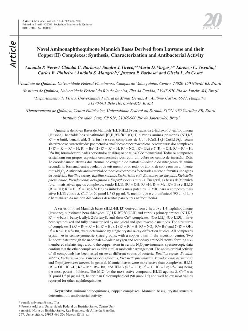
Article J. Braz. Chem. Soc., Vol. 20, No. 4, 712-727, 2009. Printed in Brazil - ©2009 Sociedade Brasileira de Química 0103 - 5053 $6.00+0.00 *e-mail: [email protected] # Present Address: Universidade Federal do Espírito Santo, Centro Uni- versitário Norte do Espírito Santo, Rua Humberto de Almeida Franklin, 257, Universitário, 29933-480 São Mateus-ES, Brazil Novel Aminonaphthoquinone Mannich Bases Derived from Lawsone and their Copper(II) Complexes: Synthesis, Characterization and Antibacterial Activity Amanda P. Neves, a Cláudia C. Barbosa, a Sandro J. Greco, a,# Maria D. Vargas, a, * Lorenzo C. Visentin, b Carlos B. Pinheiro, c Antônio S. Mangrich, d Jussara P. Barbosa e and Gisela L. da Costa e a Instituto de Química, Universidade Federal Fluminense, Campus do Valonguinho, Centro, 24020-150 Niterói-RJ, Brazil b Instituto de Química, Universidade Federal do Rio de Janeiro, Ilha do Fundão, 21945-970 Rio de Janeiro-RJ, Brazil c Departamento de Física, Universidade Federal de Minas Gerais, Av. Antônio Carlos, 6627, Pampulha, 31270-901 Belo Horizonte-MG, Brazil d Departamento de Química, Centro Politécnico, Universidade Federal do Paraná, 81531-970 Curitiba-PR, Brazil e Instituto Oswaldo Cruz, CP 926, 21045-900 Rio de Janeiro-RJ, Brazil Uma série de novas Bases de Mannich (HL1-HL13) derivadas da 2-hidroxi-1,4-naftoquinona (lausona), benzaldeídos substituídos [C 6 H 2 R 1 R 2 R 3 C(O)H] e várias aminas primárias (NH 2 R 4 , R 4 = n-butil, benzil, alil, 2-furfuril) e seus complexos de Cu 2+ , [Cu(L1) 2 ]-[Cu(L13) 2 ], foram sintetizados e caracterizados por métodos analíticos e espectroscópicos. As estruturas dos complexos 1 (R 1 = R 2 = R 3 = H; R 4 = Bu), 2 (R 1 = R 3 = H; R 2 = NO 2 ; R 4 = Bu) e 7 (R 1 = OH; R 2 = R 3 = H; R 4 = Bu) foram determinadas por estudos de difração de raios-X de monocristal. Todos os compostos cristalizam em grupos espaciais centrossimétricos, com um cobre no centro de inversão. Dois L − coordenam-se através dos átomos de oxigênio do naftalen-2-olato e do nitrogênio da amina secundária, formando anéis quelatos de seis membros ao redor do átomo de cobre em um ambiente trans-N 2 O 2 . A atividade antimicrobial de todos os compostos foi testada em sete diferentes linhagens de bactérias: Bacillus cereus, Bacillus subtilis, Escherichia coli, Enterococcus faecalis, Klebsiella pneumoniae, Pseudomonas aeruginosa e Staphylococcus aureus. Em geral, as bases de Mannich foram mais ativas que os complexos, sendo HL11 (R 1 = OH; R 2 =H; R 3 = Me; R 4 = Bn) e HL13 (R 1 = OH; R 2 = H; R 3 = Br; R 4 = Bn) os inibidores mais potentes. O MIC para o composto mais ativo HL11 contra S. Coli foi 20 µmol L -1 (8 µg mL -1 ), melhor que o cloranfenicol (90 µmol L -1 ) e bem abaixo da maioria dos valores descritos para outras naftoquinonas. A series of novel Mannich bases (HL1-HL13) derived from 2-hydroxy-1,4-naphthoquinone (lawsone), substituted benzaldehydes [C 6 H 2 R 1 R 2 R 3 C(O)H] and various primary amines (NH 2 R 4 , R 4 = n-butyl, benzyl, allyl, 2-furfuryl), and their Cu 2+ complexes, [Cu(L1) 2 ]-[Cu(L13) 2 ], have been synthesized and fully characterized by analytical and spectroscopic methods. The structures of complexes 1 (R 1 = R 2 = R 3 = H; R 4 = Bu), 2 (R 1 = R 3 = H; R 2 = NO 2 ; R 4 = Bu) and 7 (R 1 = OH; R 2 = R 3 = H; R 4 = Bu) were determined by single crystal X-ray diffraction studies. All complexes crystallize in centrosymmetric space groups, with a copper atom in the inversion centre. Two L − coordinate through the naphthalen-2-olate oxygen and secondary amine-N atoms, forming six- membered chelate rings around the copper atom in a trans-N 2 O 2 environment; spectroscopic data confirm that the other complexes exhibit similar molecular arrangement. The antimicrobial activity of all compounds has been tested on seven different strains of bacteria: Bacillus cereus, Bacillus subtilis, Escherichia coli, Enterococcus faecalis, Klebsiella pneumoniae, Pseudomonas aeruginosa and Staphylococcus aureus. In general, Mannich bases were more active than complexes, HL11 (R 1 = OH; R 2 =H; R 3 = Me; R 4 = Bn) and HL13 (R 1 = OH; R 2 = H; R 3 = Br; R 4 = Bn) being the most potent inhibitors. The MIC for the most active compound HL11 against S. Coli was 20 µmol L -1 (8 µg mL -1 ), better than Chloramphenicol (90 µmol L -1 ) and well below most values reported for other naphthoquinones. Keywords: aminonaphthoquinones, copper complexes, Mannich bases, crystal structure determination, antibacterial activity
-
Upload
independent -
Category
Documents
-
view
1 -
download
0
Transcript of Novel aminonaphthoquinone mannich bases derived from lawsone and their copper(II) complexes:...
Arti
cle
J. Braz. Chem. Soc., Vol. 20, No. 4, 712-727, 2009.Printed in Brazil - ©2009 Sociedade Brasileira de Química0103 - 5053 $6.00+0.00
*e-mail: [email protected]# Present Address: Universidade Federal do Espírito Santo, Centro Uni-versitário Norte do Espírito Santo, Rua Humberto de Almeida Franklin, 257, Universitário, 29933-480 São Mateus-ES, Brazil
Novel Aminonaphthoquinone Mannich Bases Derived from Lawsone and their Copper(II) Complexes: Synthesis, Characterization and Antibacterial Activity
Amanda P. Neves,a Cláudia C. Barbosa,a Sandro J. Greco,a,# Maria D. Vargas,a,* Lorenzo C. Visentin,b Carlos B. Pinheiro,c Antônio S. Mangrich,d Jussara P. Barbosae and Gisela L. da Costae
aInstituto de Química, Universidade Federal Fluminense, Campus do Valonguinho, Centro, 24020-150 Niterói-RJ, Brazil
bInstituto de Química, Universidade Federal do Rio de Janeiro, Ilha do Fundão, 21945-970 Rio de Janeiro-RJ, Brazil
cDepartamento de Física, Universidade Federal de Minas Gerais, Av. Antônio Carlos, 6627, Pampulha, 31270-901 Belo Horizonte-MG, Brazil
dDepartamento de Química, Centro Politécnico, Universidade Federal do Paraná, 81531-970 Curitiba-PR, Brazil
eInstituto Oswaldo Cruz, CP 926, 21045-900 Rio de Janeiro-RJ, Brazil
Uma série de novas Bases de Mannich (HL1-HL13) derivadas da 2-hidroxi-1,4-naftoquinona (lausona), benzaldeídos substituídos [C
6H
2R1R2R3C(O)H] e várias aminas primárias (NH
2R4,
R4 = n-butil, benzil, alil, 2-furfuril) e seus complexos de Cu2+, [Cu(L1)2]-[Cu(L13)
2], foram
sintetizados e caracterizados por métodos analíticos e espectroscópicos. As estruturas dos complexos 1 (R1 = R2 = R3 = H; R4 = Bu), 2 (R1 = R3 = H; R2 = NO
2; R4= Bu) e 7 (R1 = OH; R2 = R3 = H;
R4= Bu) foram determinadas por estudos de difração de raios-X de monocristal. Todos os compostos cristalizam em grupos espaciais centrossimétricos, com um cobre no centro de inversão. Dois L− coordenam-se através dos átomos de oxigênio do naftalen-2-olato e do nitrogênio da amina secundária, formando anéis quelatos de seis membros ao redor do átomo de cobre em um ambiente trans-N
2O
2. A atividade antimicrobial de todos os compostos foi testada em sete diferentes linhagens
de bactérias: Bacillus cereus, Bacillus subtilis, Escherichia coli, Enterococcus faecalis, Klebsiella pneumoniae, Pseudomonas aeruginosa e Staphylococcus aureus. Em geral, as bases de Mannich foram mais ativas que os complexos, sendo HL11 (R1 = OH; R2 =H; R3 = Me; R4= Bn) e HL13 (R1 = OH; R2 = H; R3 = Br; R4= Bn) os inibidores mais potentes. O MIC para o composto mais ativo HL11 contra S. Coli foi 20 µmol L-1 (8 µg mL-1), melhor que o cloranfenicol (90 µmol L-1) e bem abaixo da maioria dos valores descritos para outras naftoquinonas.
A series of novel Mannich bases (HL1-HL13) derived from 2-hydroxy-1,4-naphthoquinone (lawsone), substituted benzaldehydes [C
6H
2R1R2R3C(O)H] and various primary amines (NH
2R4,
R4 = n-butyl, benzyl, allyl, 2-furfuryl), and their Cu2+ complexes, [Cu(L1)2]-[Cu(L13)
2], have
been synthesized and fully characterized by analytical and spectroscopic methods. The structures of complexes 1 (R1 = R2 = R3 = H; R4 = Bu), 2 (R1 = R3 = H; R2 = NO
2; R4= Bu) and 7 (R1 = OH;
R2 = R3 = H; R4= Bu) were determined by single crystal X-ray diffraction studies. All complexes crystallize in centrosymmetric space groups, with a copper atom in the inversion centre. Two L− coordinate through the naphthalen-2-olate oxygen and secondary amine-N atoms, forming six-membered chelate rings around the copper atom in a trans-N
2O
2 environment; spectroscopic data
confirm that the other complexes exhibit similar molecular arrangement. The antimicrobial activity of all compounds has been tested on seven different strains of bacteria: Bacillus cereus, Bacillus subtilis, Escherichia coli, Enterococcus faecalis, Klebsiella pneumoniae, Pseudomonas aeruginosa and Staphylococcus aureus. In general, Mannich bases were more active than complexes, HL11 (R1 = OH; R2 =H; R3 = Me; R4= Bn) and HL13 (R1 = OH; R2 = H; R3 = Br; R4= Bn) being the most potent inhibitors. The MIC for the most active compound HL11 against S. Coli was 20 µmol L-1 (8 µg mL-1), better than Chloramphenicol (90 µmol L-1) and well below most values reported for other naphthoquinones.
Keywords: aminonaphthoquinones, copper complexes, Mannich bases, crystal structure determination, antibacterial activity
Neves et al. 713Vol. 20, No. 4, 2009
Introduction
Natural and synthetic naphthoquinones are known for a wide range of biological activities,1 amongst which anti-cancer,2,3 tripanocidal,4 molluscicidal,5 antimalarial,6
leishmaniscide,7 bacteriostatic and bactericidal.8,9 The most accepted mechanism for the antimicrobial activity of naphthoquinones is based on the generation of reactive oxygen species by two successive reduction processes to form radical anion and dianion species that are toxic to bacteria.10,11
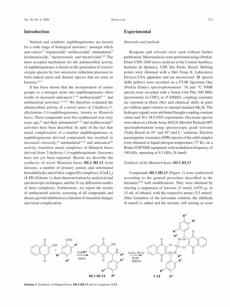
It has been shown that the incorporation of amino groups or a nitrogen atom into naphthoquinones often results in increased anticancer,12-14 molluscicidal15-17 and antibacterial activities.10,18-22 We therefore evaluated the antimicrobial activity of a novel series of 2-hydroxy-3-alkylamine-1,4-naphthoquinones, known as Mannich bases. These compounds were first synthesized over sixty years ago,23 and their antimalarial23,24 and molluscicidal25 activities have been described. In spite of the fact that metal complexation of a number naphthoquinones or naphthoquinone derived compounds has resulted in increased citoxicity,26 antimalarial27,28 and anticancer29 activity, transition metal complexes of Mannich bases derived from 2-hydroxy-1,4-naphthoquinone (lawsone) have not yet been reported. Herein we describe the synthesis of novel Mannich bases HL1-HL13 from lawsone, a number of primary amines and substituted benzaldehydes and of their copper(II) complexes, [Cu(L)
2]
(1-13) (Scheme 1), their characterization by analytical and spectroscopic techniques, and the X-ray diffraction studies of three complexes. Furthermore, we report the results of antibacterial activity screening of all compounds and discuss growth inhibition as a function of structural changes and metal complexation.
Experimental
Materials and methods
Reagents and solvents were used without further purification. Microanalyses were performed using a Perkin-Elmer CHN 2400 micro analyser at the Central Analítica, Instituto de Química, USP, São Paulo, Brazil. Melting points were obtained with a Mel-Temp II, Laboratory Devices-USA apparatus and are uncorrected. IR spectra (KBr pellets) were recorded on a FT-IR Spectrum One (Perkin Elmer) spectrophotometer. 1H and 13C NMR spectra were recorded with a Varian Unit Plus 300 MHz spectrometer in CDCl
3 or d6-DMSO; coupling constants
are reported in Hertz (Hz) and chemical shifts in parts per million (ppm) relative to internal standard Me
4Si. The
hydrogen signals were attributed thought coupling constant values and 1H × 1H-COSY experiments. Electronic spectra were taken on a Diode Array 8452A (Hewlett Packard-HP) spectrophotometer using spectroscopic grade solvents (Tedia Brazil) in 10-3 and 10-4 mol L-1 solutions. Electron paramagnetic resonance (EPR) spectra of the solid samples were obtained at liquid nitrogen temperature (77 K), on a Bruker ESP300E equipment with modulation frequency of 100 kHz, operating at 9.5 GHz (X-band).
Synthesis of the Mannich bases HL1-HL13
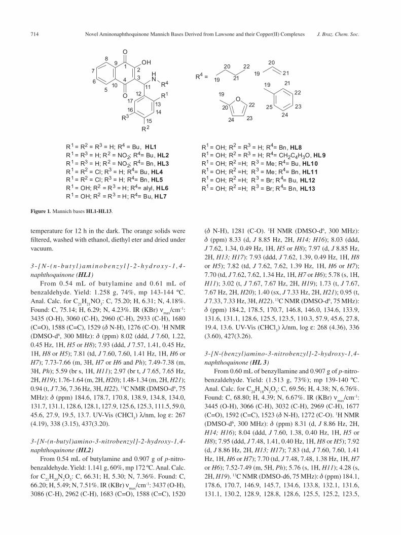
Compounds HL1-HL13 (Figure 1) were synthesised according to the general procedure described in the literature24,30 with modifications. They were obtained by reacting a suspension of lawsone (5 mmol, 0.870 g), in 15 mL of ethanol, with the respective amine (5.5 mmol). After formation of the lawsonate solution, the aldehyde (6 mmol) is added and the mixture, left stirring at room
Scheme 1. Synthesis of Mannich bases HL1-HL13 and of complexes 1-13.
Novel Aminonaphthoquinone Mannich Bases Derived from Lawsone and their Copper(II) Complexes J. Braz. Chem. Soc.714
temperature for 12 h in the dark. The orange solids were filtered, washed with ethanol, diethyl eter and dried under vacuum.
3 - [ N - ( n - bu t y l ) a m i n o b e n z y l ] - 2 - h y d ro x y - 1 , 4 -naphthoquinone (HL1)
From 0.54 mL of butylamine and 0.61 mL of benzaldehyde. Yield: 1.258 g, 74%, mp 143-144 ºC. Anal. Calc. for C
21H
21NO
3: C, 75.20; H, 6.31; N, 4.18%.
Found: C, 75.14; H, 6.29; N, 4.23%. IR (KBr) νmax
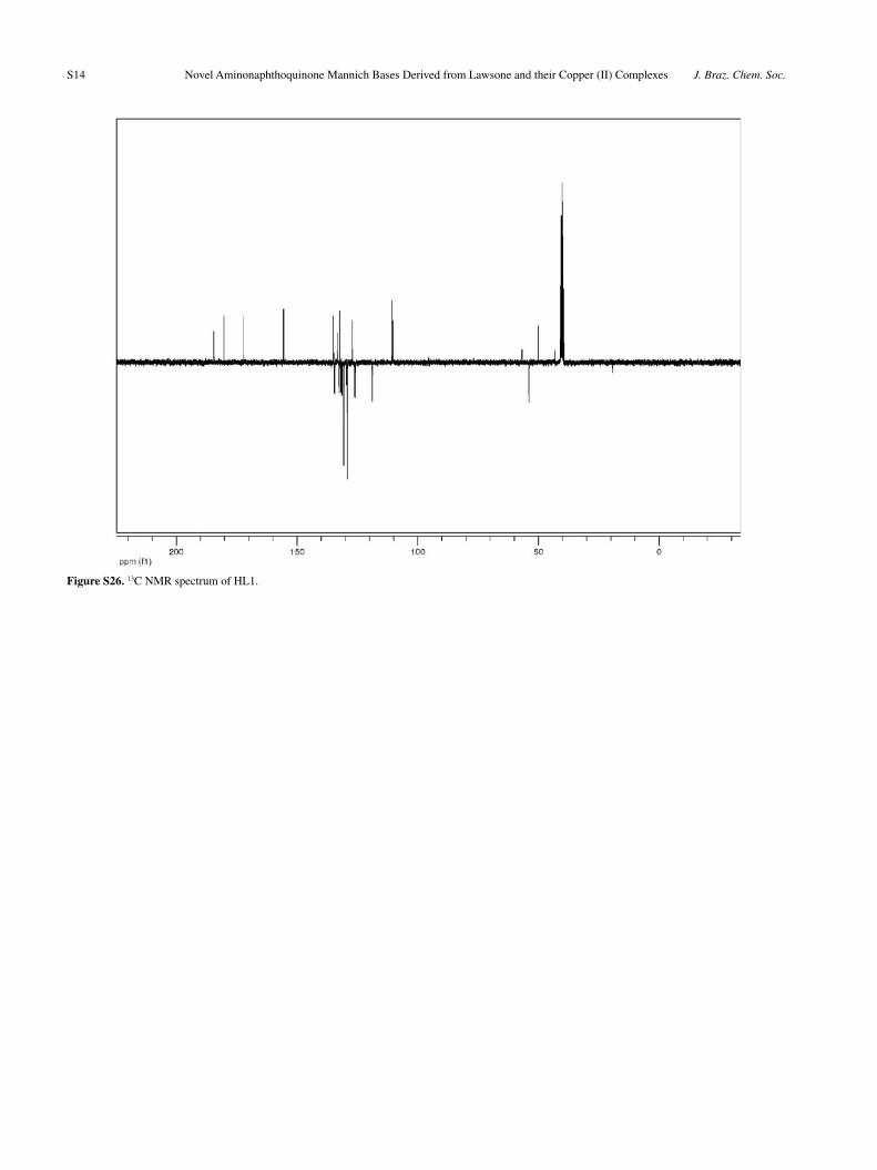
/cm-1: 3435 (O-H), 3060 (C-H), 2960 (C-H), 2933 (C-H), 1680 (C=O), 1588 (C=C), 1529 (d N-H), 1276 (C-O). 1H NMR (DMSO-d6, 300 MHz): d (ppm) 8.02 (ddd, J 7.60, 1.22, 0.45 Hz, 1H, H5 or H8); 7.93 (ddd, J 7.57, 1.41, 0.45 Hz, 1H, H8 or H5); 7.81 (td, J 7.60, 7.60, 1.41 Hz, 1H, H6 or H7); 7.73-7.66 (m, 3H, H7 or H6 and Ph); 7.49-7.38 (m, 3H, Ph); 5.59 (br s, 1H, H11); 2.97 (br t, J 7.65, 7.65 Hz, 2H, H19); 1.76-1.64 (m, 2H, H20); 1.48-1.34 (m, 2H, H21); 0.94 (t, J 7.36, 7.36 Hz, 3H, H22). 13C NMR (DMSO-d6, 75 MHz): d (ppm) 184.6, 178.7, 170.8, 138.9, 134.8, 134.0, 131.7, 131.1, 128.6, 128.1, 127.9, 125.6, 125.3, 111.5, 59.0, 45.6, 27.9, 19.5, 13.7. UV-Vis (CHCl
3) λ/nm, log ε: 267
(4.19), 338 (3.15), 437(3.20).
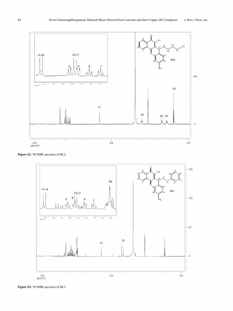
3-[N-(n-butyl)amino-3-nitrobenzyl]-2-hydroxy-1,4-naphthoquinone (HL2)
From 0.54 mL of butylamine and 0.907 g of p-nitro-benzaldehyde. Yield: 1.141 g, 60%, mp 172 ºC. Anal. Calc. for C
21H
20N
2O
5: C, 66.31; H, 5.30; N, 7.36%. Found: C,
66.20; H, 5.49; N, 7.51%. IR (KBr) νmax
/cm-1: 3437 (O-H), 3086 (C-H), 2962 (C-H), 1683 (C=O), 1588 (C=C), 1520
(d N-H), 1281 (C-O). 1H NMR (DMSO-d6, 300 MHz): d (ppm) 8.33 (d, J 8.85 Hz, 2H, H14; H16); 8.03 (ddd, J 7.62, 1.34, 0.49 Hz, 1H, H5 or H8); 7.97 (d, J 8.85 Hz, 2H, H13; H17): 7.93 (ddd, J 7.62, 1.39, 0.49 Hz, 1H, H8 or H5); 7.82 (td, J 7.62, 7.62, 1.39 Hz, 1H, H6 or H7); 7.70 (td, J 7.62, 7.62, 1.34 Hz, 1H, H7 or H6); 5.78 (s, 1H, H11); 3.02 (t, J 7.67, 7.67 Hz, 2H, H19); 1.73 (t, J 7.67, 7.67 Hz, 2H, H20); 1.40 (sx, J 7.33 Hz, 2H, H21); 0.95 (t, J 7.33, 7.33 Hz, 3H, H22). 13C NMR (DMSO-d6, 75 MHz): d (ppm) 184.2, 178.5, 170.7, 146.8, 146.0, 134.6, 133.9, 131.6, 131.1, 128.6, 125.5, 123.5, 110.3, 57.9, 45.6, 27.8, 19.4, 13.6. UV-Vis (CHCl
3) λ/nm, log ε: 268 (4.36), 336
(3.60), 427(3.26).
3-[N-(benzyl)amino-3-nitrobenzyl]-2-hydroxy-1,4-naphthoquinone (HL 3)
From 0.60 mL of benzyllamine and 0.907 g of p-nitro-benzaldehyde. Yield: (1.513 g, 73%); mp 139-140 ºC. Anal. Calc. for C
24H
18N
2O
5: C, 69.56; H, 4.38; N, 6.76%.
Found: C, 68.80; H, 4.39; N, 6.67%. IR (KBr) νmax
/cm-1:
3445 (O-H), 3066 (C-H), 3032 (C-H), 2969 (C-H), 1677 (C=O), 1592 (C=C), 1523 (d N-H), 1272 (C-O). 1H NMR (DMSO-d6, 300 MHz): d (ppm) 8.31 (d, J 8.86 Hz, 2H, H14; H16); 8.04 (ddd, J 7.60, 1.38, 0.40 Hz, 1H, H5 or H8); 7.95 (ddd, J 7.48, 1.41, 0.40 Hz, 1H, H8 or H5); 7.92 (d, J 8.86 Hz, 2H, H13; H17); 7.83 (td, J 7.60, 7.60, 1.41 Hz, 1H, H6 or H7); 7.70 (td, J 7.48, 7.48, 1.38 Hz, 1H, H7 or H6); 7.52-7.49 (m, 5H, Ph); 5.76 (s, 1H, H11); 4.28 (s, 2H, H19). 13C NMR (DMSO-d6, 75 MHz): d (ppm) 184.1, 178.6, 170.7, 146.9, 145.7, 134.6, 133.8, 132.1, 131.6, 131.1, 130.2, 128.9, 128.8, 128.6, 125.5, 125.2, 123.5,
Figure 1. Mannich bases HL1-HL13.
Neves et al. 715Vol. 20, No. 4, 2009
110.1, 57.8, 49.3. UV-Vis (CH2Cl
2) λ/nm, log ε: 273 (4.47),
337 (3.74), 389 (3.22).
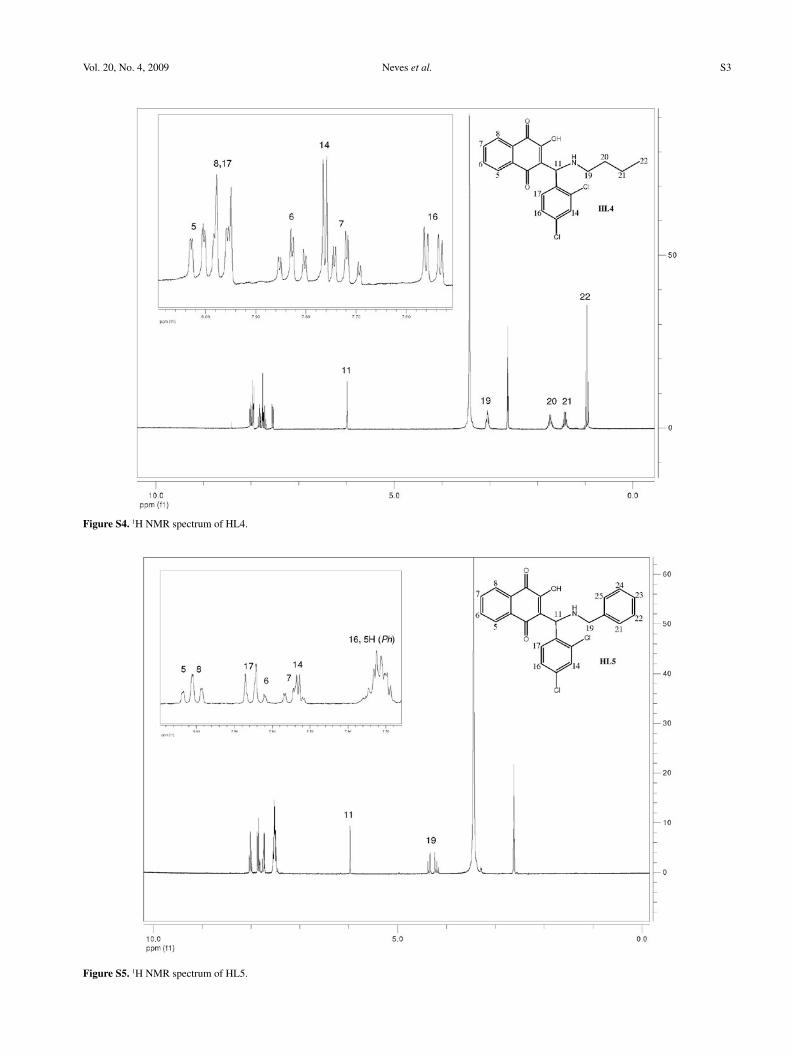
3-[N-(n-butyl)amino-2,4-diclorobenzyl]-2-hydroxy-1,4-naphthoquinone (HL4)
From 0.60 mL of benzylamine and 1.050 g of 2-4-dicloro-benzaldehyde. Yield: 1.071 g, 53%; mp 142-143 ºC. Anal. Calc. for C
21H
19Cl
2NO
3: C, 62.39; H, 4.74;
N, 3.46%. Found: C, 62.00; H, 4.64; N, 3.51%. IR (KBr) ν
max/cm-1: 3437 (O-H), 3064 (C-H), 2958 (C-H), 2870 (C-
H), 1678 (C=O), 1616 (C=C), 1588 (C=C), 1531 (d N-H), 1272 (C-O). 1H NMR (DMSO-d6, 300 MHz): d (ppm) 8.01 (dd, J 7.46, 1.33 Hz, 1H, H5 or H8); 7.97 (dd, J 7.46, 1.37 Hz, 1H, H8 or H5); 7.96 (d, J 8.51 Hz, 1H, H17); 7.83 (td, J 7.46, 7.46, 1.37 Hz, 1H, H6 or H7); 7.76 (d, J 2.19, 1H, H14); 7.72 (td, J 7.46, 7.46, 1.33 Hz, 1H, H7 or H6); 7.55 (dd, J 8.51, 2.19, 1H, H16); 5.99 (s, 1H, H11); 3.05 (br t, J 7.35, 7.35 Hz, 2H, H19); 1.80-1.65 (m, 2H, H20); 1.38-1.34 (m, 2H, H21); 0.96 (t, J 7.28, 7.28 Hz, 3H, H22). 13C NMR (DMSO-d6, 75 MHz): d (ppm) 184.1, 178.9, 171.3, 134.8, 134.5, 134.4, 133.9, 133.8, 131.9, 131.6, 131.1, 128.8, 127.7, 125.5, 125.2, 109.6, 55.4, 45.9, 27.7, 19.3, 13.5. UV-Vis (CHCl
3) λ/nm, log ε: 265 (4.29), 337 (3.42),
422 (3.29).
3-[N-(n-benzyl)amino-2,4-diclorobenzyl]-2-hydroxy-1,4-naphthoquinone (HL5)
From 0.54 mL of butylamine and 1.050 g of 2-4-dicloro-benzaldehyde. Yield: 1.731 g, 79%; mp 145-146 ºC. Anal. Calc. for C
24H
17Cl
2NO
3: C, 65.77; H, 3.91; N, 3.20%.
Found: C, 65.67; H, 3.87; N, 3.24%. IR (KBr) νmax
/cm-1: 3453 (O-H), 3065 (C-H), 3033 (C-H), 2969 (C-H), 1677 (C=O), 1592 (C=C), 1523 (d N-H), 1271 (C-O). 1H NMR (DMSO-d6, 300 MHz): d (ppm) 8.02 (ddd, J 7.37, 1.50, 0.47 Hz, 1H, H5 or H8); 8.00 (dd, J 7.37, 1.50, 0.47 Hz, 1H, H8 or H5); 7.86 (d, J 8.63 Hz, 1H, H17); 7.84 (td, J 7.46, 7.46, 1.50 Hz, 1H, H6 or H7); 7.74 (td, J 7.46, 7.46, 1.50 Hz, 1H, H7 or H6); 7.73 (d, J 2.16 Hz, 1H, H14); 7.55-7.49 (m, 6H, H16; Ph); 5.78 (s, 1H, H11); 4.35 (d, J 13.00 Hz, 1H, H19); 4.21 (d, J 13.00 Hz, 1H, H19’). 13C NMR (DMSO-d6, 75 MHz): d (ppm) 183.9, 179.3, 171.3, 134.6, 134.5, 134.4, 134.0, 133.9, 132.1, 132.0, 131.7, 131.2, 130.3, 128.9, 128.8, 128.6, 127.8, 125.6, 125.2, 109.4, 55.1, 49.7. UV-Vis (CHCl
3) λ/nm, log ε: 275 (4.14),
334 (3.40), 400 (3.10).
3-[N-(alyl)amino-2-hydroxybenzyl]-2-hydroxy-1,4-naphthoquinone (HL6)
From 0.41 mL of alylamine and 0.63 mL of 2-hydroxy-benzaldehyde. Yield: 1.475 g, 88%; mp 164-165 ºC. Anal. Calc. for C
20H
17NO
4: C, 71.63; H, 5.11; N, 4.18%.
Found: C, 71.50; H, 5.05; N, 4.27%. IR (KBr) νmax
/cm-1: 3255 (O-H), 3072 (C-H), 2980 (C-H), 2950 (C-H), 1678 (C=O), 1593 (C=C), 1561 (d N-H), 1277 (C-O). 1H NMR (DMSO-d6, 300 MHz): d (ppm) 8.02 (ddd, J 7.61, 1.35, 0.47 Hz, 1H, H5 or H8); 7.97 (dd, J 7.53, 1.41, 0.47 Hz, 1H, H8 or H5); 7.83 (td, J 7.47, 7.47, 1.41 Hz, 1H, H6 or H7); 7.73 (td, J 7.47, 7.47, 1.35 Hz, 1H, H7 or H6); 7.42 (dd, J 7.69, 1.65 Hz, 1H, H14 or H17); 7.26 (td, J 8.06, 8.06, 1.65 Hz, 1H, H16 or H15); 6.96 (dd, J 8.06, 0.99 Hz, 1H, H17 or H14); 6.86 (td, J 7.69, 7.69, 0.99 Hz, 1H, H15 or H16); 6.10-5.95 (m, 2H, H20); 5.85 (s, H11); 5.50-5.42 (m, 2H, H21); 3.74-3.60 (m, 2H, H19). 13C NMR (DMSO-d6, 75 MHz): d (ppm) 184.1, 179.6, 171.5, 155.5, 134.6, 133.9, 131.6, 131.1, 129.8, 129.4, 128.6, 125.5, 125.2, 123.8, 122.0, 119.1, 116.1, 110.1, 53.3, 47.9. UV-Vis (DMSO) λ/nm, log ε: 278 (4.34), 452 (3.33).
3-[N-(n-butyl)amino-2-hydroxybenzyl]-2-hydroxy-1,4-naphthoquinone (HL7)
From 0.54 mL of butylamine and 0.63 mL of 2-hydroxy-benzaldehyde. Yield: 1.405 g, 80%; mp 137-138 ºC (with dec.). Anal. Calc. for C
21H
21NO
4: C, 71.78; H, 6.02; N,
3.99%. Found: C, 71.12; H, 6.03; N, 3.92%. IR (KBr) νmax
/cm-1: 3233 (O-H), 3069 (C-H), 2959 (C-H), 2875 (C-H), 1681 (C=O), 1590 (C=C), 1528 (d N-H), 1275 (C-O). 1H NMR (DMSO-d6, 300 MHz): d (ppm) 8.02 (ddd, J 7.66, 1.35, 0.50 Hz, 1H, H5 or H8); 7.98 (ddd, J 7.47; 1.35; 0.50 Hz, 1H, H8 or H5); 7.84 (td, J 7.47, 7.47, 1.35 Hz, 1H, H6 or H7); 7.73 (td, J 7.47, 7.47, 1,35 Hz, 1H, H7 or H8); 7.44 (dd, J 7.74, 1.56 Hz, 1H, H14 or H17); 7.27 (td, J 8.03, 8.03, 1.56 Hz, 1H, H16 or H15); 6.99 (dd, J 8.03, 1.05 Hz, 1H, H17 or H14); 6.86 (td, J 7.74, 7.74, 1.05 Hz, 1H, H15 or H16); 5.86 (s, 1H, H11); 3.03 (t, J 7.50, 7.50 Hz, 2H, H19); 1.79-1.66 (m, 2H, H20); 1.50-1.38 (m, 2H, H21); 0.96 (br t, J 7.33, 7.33 Hz, 3H, H22). 13C NMR (DMSO-d6, 75 MHz): d (ppm) 184.1, 179.6, 171.6, 155.4, 134.5, 133.9, 131.6, 131.2, 129.6, 128.7, 125.5, 125.2, 123.6, 119.1, 115.9, 109.9, 54.1, 45.7, 27.8, 19.4, 13.5. UV-Vis (DMSO) λ/nm, log ε: 277 (4.33), 450 (3.30).
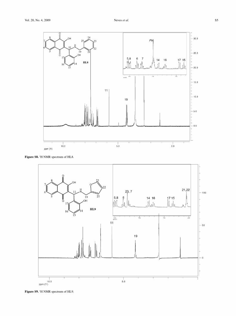
3-[N-(benzyl)amino-2-hydroxybenzyl]-2-hydroxy-1,4-naphthoquinone (HL8)
From 0.60 mL of benzylamine and 0.63 mL of 2-hydroxy-benzaldehyde. Yield: 1.792 g, 93%; mp 165-166 ºC. Anal. Calc. for C
24H
19NO
4 C, 74.79; H, 4.97; N,
3.63%. Found: C, 74.78; H, 4.90; N, 3.72%. IR (KBr) ν
max/cm-1: 3436 (O-H), 3067 (C-H), 2744 (C-H), 1681
(C=O), 1589 (C=C), 1516 (d N-H), 1276 (C-O). 1H NMR (DMSO-d6, 300 MHz): d (ppm) 8.04 (ddd, J 7.50, 1.36, 0.48 Hz, 1H, H5 or H8); 7.99 (ddd, J 7.50, 1.43, 0.48 Hz, 1H, H8 or H5); 7.84 (td, J 7.50, 7.50, 1.43 Hz, 1H, H6
Novel Aminonaphthoquinone Mannich Bases Derived from Lawsone and their Copper(II) Complexes J. Braz. Chem. Soc.716
or H7); 7.73 (td, J 7.50, 7.50, 1.36 Hz, 1H, H7 or H6); 7.51-7.46 (m, 5H, Ph); 7.38 (dd, J 7.71, 1.62 Hz, 1H, H14 or H17); 7.24 (td, J 8.05; 8.05, 1.62 Hz, 1H, H16 or H15); 6.94 (dd, J 8.05, 1.00 Hz, 1H, H17 or H14); 6.84 (td, J 7.71, 7.71, 1.00 Hz, 1H, H15 or H16); 5.82 (s, 1H, H11); 4.26 (d, J 13.09, 1H); 4.17 (d, J 13.09, 1H). 13C NMR (DMSO-d6, 75 MHz): d (ppm) 184.1, 179.7, 171.5, 155.6, 134.6, 133.9, 132.8, 131.7, 131.1, 130.0, 129.4, 128.9, 128.7, 128.5, 125.5, 125.2, 123.6, 119.0, 115.9, 110.0, 53.8, 49.4. UV-Vis (DMSO) λ/nm, log ε: 277 (4.29), 443 (3.25).
3-[N-(furfurylmethyl)amino-2-hydroxybenzyl]-2-hydroxy-1,4-naphthoquinone (HL9)
From furfurylamine (0.49 mL) and 2-hydroxy-benzaldehyde (0.63 mL). Yield: 1.653 g, 88%; mp 138 ºC. Anal. Calc. for C
24H
19NO
4.H
2O
: C, 67.17; H, 4.87; N,
3.56%. Found: C, 68.94; H, 4.99; N, 3.69%. IR (KBr) ν
max/cm-1: 3234 (O-H), 2943 (C-H), 1683 (C=O), 1591
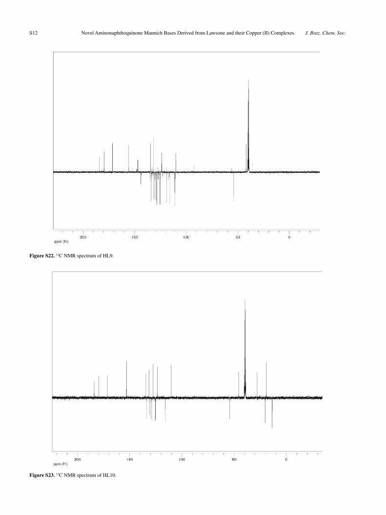
(C=C), 1551 (d N-H), 1279 (C-O). 1H NMR (DMSO-d6, 300 MHz): d (ppm) 8.02 (br d, J 7.61 Hz, 1H, H5 or H8); 7.97 (br d, J 7.56 Hz, 1H, H8 or H5); 7.83 (td, J 7.61, 7.61, 1.20 Hz, 1H, H6 or H7); 7.72 (td, J 7.56, 7.56, 1.20 Hz, 1H, H7 or H6); 7.51-7.46 (m, 5H, Ph); 7.32 (br d, J 7.89 Hz, 1H, H14 or H17); 7.23 (br td, J 7.89; 7.89, 1.62 Hz, 1H, H16 or H15); 6.92 (br d, J 7.89 Hz, 1H, H17 or H14); 6.82 (br t, J 7.48 Hz, 1H, H15 or H16); 5.78 (s, 1H, H11); 4.23 (d, J 14.51, 1H); 4.17 (d, J 14.51, 1H). 13C NMR (DMSO-d6, 75 MHz): d (ppm) 184.2, 179.7, 171.4, 155.7, 143.9, 134.6, 133.9, 131.7, 131.2, 129.4, 128.6, 125.5, 125.3, 123.7, 119.2, 119.0, 116.0, 111.6, 111.0, 110.1, 53.9, 41.9. UV-Vis (CHCl
3) λ/nm, log ε:
272 (4.03), 305 (3.93), 366 (3.74).
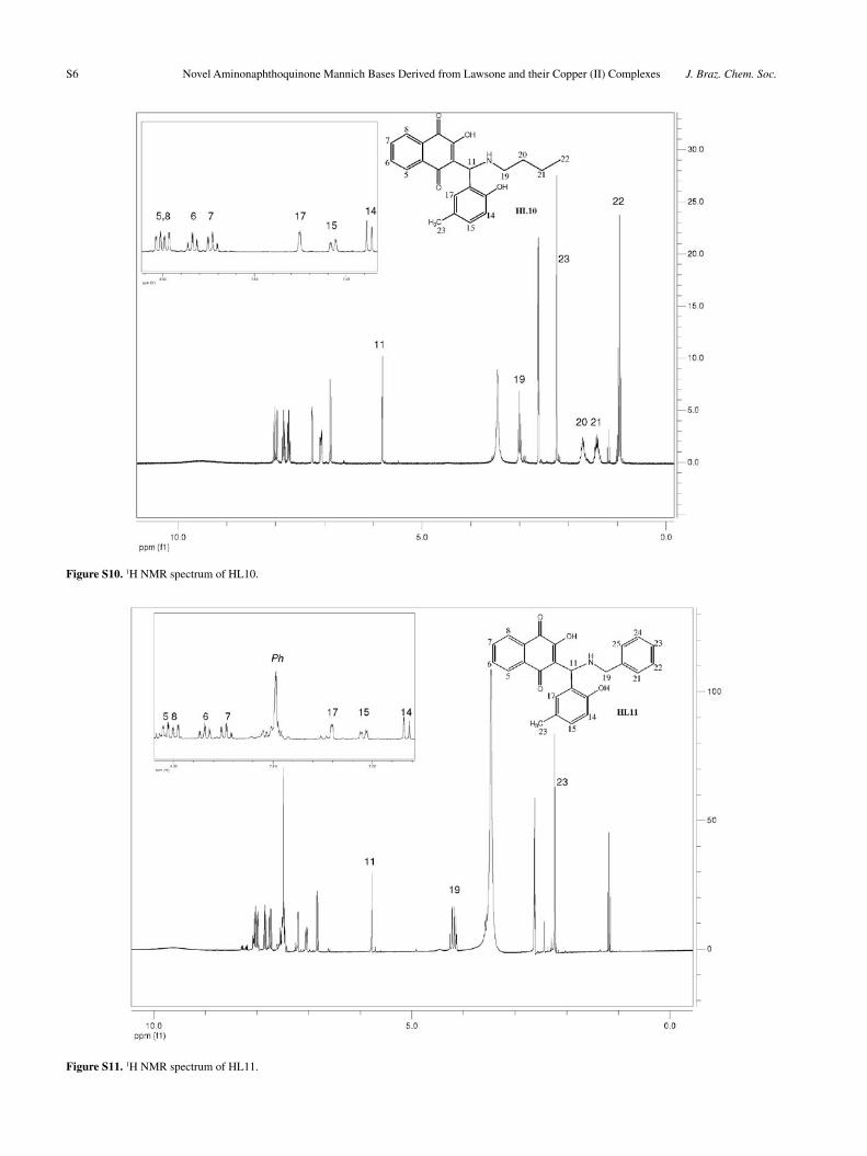
3-[N-(n-butyl)amino-2-hydroxy-5-methyl-benzyl]-2-hydroxy-1,4-naphthoquinone (HL10)
From butylamine (0.54 mL) and 2-hydroxy-5-methyl-benzaldehyde (0.817 g). Yield: 1.279 g, 70%; mp 154-155 ºC. Anal. Calc. for C
22H
23NO
4: C, 72.31; H, 6.34; N,
3.83. Found: C, 71.15; H, 6.28; N, 3.90%. IR (KBr) νmax
/cm-1: 3065, 2959, 2870, 1686, 1591, 1551, 1508, 1477, 1432, 1374, 1278. 1H NMR (DMSO-d6, 300 MHz): d (ppm) 8.02 (ddd, J 7.67; 1.30; 0.40 Hz, 1H, H5 or H8); 7.98 (ddd, J 7.54; 1.37; 0.40 Hz, 1H, H8 or H5): 7.84 (td, J 7.54; 7.54; 1.30 Hz, 1H, H6 or H7); 7.73 (td, J 7.47; 7.47; 1.37 Hz, 1H, H7 or H6); 7.25 (d, J 1.94 Hz, 1H, H17): 7.07 (dd, J 8.22, 1.94 Hz, 1H, H15); 6.87 (d, J 8.22 Hz, H14); 5.81 (s, 1H, H11); 3.00 (t, J 7.55, 7.55 Hz, 2H, H19); 2.25 (s, 3H, CH
3); 1.77-1.65 (m, 2H, H20); 1.48-1.34 (m, 2H, H21);
0.96 (t, J 7.34, 7.34 Hz, 3H, H22). 13C NMR (DMSO-d6, 75 MHz): d (ppm) 184.1, 179.5, 171.4, 153.1, 134.5, 133.9,
131.6, 131.1, 129.8, 128.6, 127.5, 125.5, 125.2, 123.5; 115.9; 110.2; 54.1; 45.7; 27.9; 20.3; 19.4; 13.5. UV-Vis (CHCl
3) λ/nm, log ε: 274 (4.10), 325 (3.63), 373 (3.38),
470 (2.93).
3-[N-(benzyl)amino-2-hydroxy-5-methyl-benzyl]-2-hydroxy-1,4-naphthoquinone (HL11)
From benzylamine (0.60 mL) and 2-hydroxy-5-methyl-benzaldehyde (0.817 g). Yield: 1.338 g, 67%; mp 147-148 ºC. Anal. Calc. for C
25H
21NO
4: C, 75.17; H, 5.30; N,
3.51. Found: C, 74.54; H, 5.36; N, 3.51%. IR (KBr) νmax
/cm-1: 3266; 3065; 2957; 2862; 1686; 1590; 1539; 1274; 1221. 1H NMR (DMSO-d6, 300 MHz): d (ppm) 8.04 (ddd, J 7.61, 1.39, 0.43 Hz, 1H, H5 or H8); 7.99 (ddd, J 7.54, 1.41, 0.43 Hz, 1H, H8 or H5); 7.84 (td, J 7.43, 7.43, 1.41 Hz, 1H, H6 or H7); 7.73 (td, J 7.43, 7.43, 1.39 Hz, 1H, H7 or H6); 7.50-7.47 (m, 5H, Ph); 7.20 (d, J 2.03 Hz, 1H, H17); 7.04 (dd, J 8.16, 2.03 Hz, 1H, H15); 6.83 (d, J 8.16 Hz, 1H, H14); 5.77 (s, H11); 4.24 (d, J 13.13 Hz, 1H, H19); 4.16 (d, J 13.13 Hz, 1H, H19’); 2.23 (s, 3H, CH
3). 13C NMR (DMSO-d6, 75 MHz): d
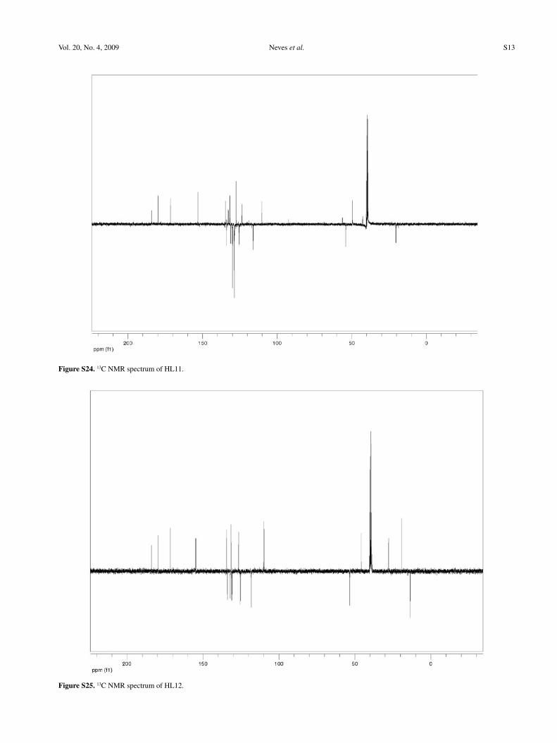
(ppm) 184.1, 179.9, 171.5, 153.2, 134.6, 133.9, 132.8, 131.7, 131.2, 130.1, 129.9, 128.9, 128.7, 128.6, 127.6, 125.6, 125.3, 123.5, 116.0, 110.2, 54.0, 49.5, 20.4. UV-Vis (CHCl
3) λ/nm,
log ε: 250 (4.46), 310 (4.15), 382 (3.90).
3-[N-(n-butyl)amino-2-hydroxy-5-bromo-benzyl]-2-hydroxy-1,4-naphthoquinone (HL12)
From butylamine (0.54 mL) and 2-hydroxy-5-bromo-benzaldehyde (1.206 g). Yield: 1.316 g, 61%; mp 166-167 ºC. Anal. Calc. for C
21H
20BrNO
4: C, 58.62; H, 4.68;
N, 3.26. Found: C, 58.74; H, 4.54; N, 3.42%. IR (KBr) ν
max/cm-1: 3202; 2961; 2934; 2867; 1679; 1591; 1524; 1273;
1229. 1H NMR (DMSO-d6, 300 MHz: d (ppm) 8.04 (d, J 7.20 Hz, 1H, H5 or H8); 7.98 (d, J 7.20 Hz, 1H, H8 or H5); 7.84 (t, J 7.20, 7.20 Hz, 1H, H6 or H7); 7.73 (t, J 7.20, 7.20 Hz, 1H, H7 or H6); 7.64 (d, J 2.15 Hz, 1H, H17); 7.42 (dd, J 8.58, 2.15 Hz, 1H, H15); 6.94 (d, J 8.58 Hz, 1H, H14); 5.82 (s, 1H, H11); 3.05-2.95 (m, 2H, H19); 1.78-1.65 (m, 2H, H20); 1.49-1.35 (m, 2H, H21); 0.96 (t, J 7.04, 7.04 Hz, 3H, H22). 13C NMR (DMSO-d6, 75 MHz): d (ppm) 183.9, 179.5, 171.1, 154.8, 134.4, 133.9, 132.0, 131.6, 131.2, 130.9, 126.5, 125.6, 125.2, 118.2, 110.2, 109.8, 53.4, 45.8, 27.8, 19.3, 13.6. UV-Vis (CHCl
3) λ/nm, log ε: 273 (3.12), 354 (2.27), 457 (2.05).
3-[N-(benzyl)amino-2-hydroxy-5-bromo-benzyl]-2-hydroxy-1,4-naphthoquinone (HL13)
From benzylamine (0.60 mL) and 2-hydroxy-5-bromo-benzaldehyde (1.206 g). Yield: 1.736 g, 75%; mp 160-161 ºC. Anal. Calc. for C
24H
18BrNO
4: C, 62.08; H, 3.91; N,
3.02. Found: C, 62,09; H, 4.01; N, 3.11%. IR (KBr) νmax
/cm-1: 3224 (O-H), 3068 (C-H), 2958 (C-H), 1688 (C=O), 1589

Neves et al. 717Vol. 20, No. 4, 2009
(C=C), 1539 (d N-H), 1275 (C-O). 1H NMR (DMSO-d6, 300 MHz): d (ppm) 8.04 (dd, J 7.62, 1.36 Hz, 1H, H5 or H8); 7.99 (d, J 7.54, 1.41 Hz, 1H, H8 or H5); 7.85 (td, J 7.44, 7.44, 1.41 Hz, 1H, H6 or H7); 7.74 (td, J 7.44, 7.44, 1.36 Hz, 1H, H7 or H6); 7.54 (d, J 2.37 Hz, 1H, H17); 7.50-7.46 (m, 5H, Ph); 7.39 (dd, J 8.63, 2.37 Hz, 1H, H15); 6.89 (d, J 8.63 Hz, 1H, H14); 5.77 (s, 1H, H11); 4.24 (d, J 13.15 Hz, 1H, H19); 4.15 (d, J 13.15 Hz, 1H, H19’). 13C NMR (DMSO-d6, 75 MHz) d (ppm): 184.5, 180.3, 172.1, 155.6, 135.1, 134.5, 133.3, 132.5, 132.3, 131.8, 131.5, 130.7, 129.5, 129.3, 129.1, 127.1, 126.2, 125.9, 118.8, 110.7, 110.4, 53.9, 50.1. UV-Vis (CHCl
3) λ/nm, log ε: 271 (3.10), 377 (2.60), 434 (1.94).
Synthesis of complexes [Cu(L)2] 1-13 from HL1-HL13,
respectively
To a suspension of 1 mmol of the ligand in 10 mL MeOH, was added a solution of CuCl
2.2H
2O (83 mg,
0.5 mmol) in 2 mL MeOH. After addition of Et3N (0.14 mL,
1 mmol), the suspension was left under stirring in the dark for 12h at room temperature. The resulting solids were filtered off, washed with methanol, diethyl ether and dried under vacuum (Figure 2).
[Cu(L1)2] (1)
From 335 mg of HL1. Yield: 337 mg, 92%; mp 198 ºC. Slow evaporation of a CHCl
3 solution yielded brown
crystals suitable for X-ray diffraction analysis. Anal. calc. for C
42H
40N
2O
6Cu.2H
2O: C, 65.65; H, 5.77; N, 3.65%.
Found: C, 64.69; H, 5.76; N, 3.54%. IR (KBr) νmax
/cm-1: 3468 (O-H), 3281 (N-H), 3064 (C-H), 2958 (C-H), 2928 (C-H), 1674 (C=O), 1621 (C=C), 1591 (C=C), 1273 (C-O). UV-Vis (CHCl
3) λ/nm, log ε: 315 (4.13), 425 (3.78),
538 (2.25).
[Cu(L2)2] (2)
From 380 mg of HL2. Yield: 280 mg, 68%; mp 208 ºC. Slow evaporation of the complex solution in a methanol/isopropanol mixture yielded brown crystals suitable for X-ray diffraction analysis. Anal. calc. for C
42H
38N
4O
10Cu.
H2O: C, 60.03; H, 4.80; N, 6.67%. Found: C, 59.21; H, 4.70;
N, 6.82%. IR (KBr) νmax
/cm-1: 3460 (O-H), 3273 (N-H), 3077 (C-H), 2956 (C-H), 2930 (C-H), 1675 (C=O), 1617 (C=C), 1592 (C=C), 1546 (d N-H), 1272 (C-O). UV-Vis (CHCl
3)
λ/nm, log ε: 298 (4.64), 324 (4.05), 415 (3.72), 536 (2.17).
[Cu(L3)2] (3)
From 414 mg of HL3. Yield: 280 mg, 63%; mp 176-177 ºC. Anal. Calc. for C
48H
34N
4O
10Cu.2H
2O: C, 62.23;
H, 4.13; N, 6.05%. Found: C, 61.19; H, 4.12; N, 6.16%. IR (KBr) ν
max/cm-1: 3459 (O-H), 3151 (N-H), 2933 (C-H),
1671 (C=O), 1591 (C=C), 1547 (d N-H), 1274 (C-O). UV-Vis (CHCl
3) λ/nm, log ε: 298 (4.51), 319 (4.16), 413
(3.70), 556 (2.17).
[Cu(L4)2] (4)
From 404 mg of HL4. Yield: 322 mg, 74%; mp 171 ºC. Anal. Calc. for C
42H
36Cl
4N
2O
6Cu.0.5H
2O: C, 57.38; H, 4.24;
N, 3.19%. Found: C, 56.49; H, 4.28; N, 3.28%. IR (KBr) ν
max/cm-1: 3446 (O-H); 3273 (N-H), 2959 (C-H), 2931 (C-
H); 2871 (C-H), 1678 (C=O), 1625 (C=C), 1591 (C=C), 1548 (d N-H), 1275 (C-O). UV-Vis (CHCl
3) λ/nm, log ε:
289 (4.40), 318 (3.97), 412 (3.70), 530 (2.22).
[Cu(L5)2] (5)
From 438 mg of HL5. Yield: 276 mg, 59%; mp 172-173 ºC. Anal. Calc. for C
48H
32Cl
4N
2O
6Cu.H
2O: C, 60.30;
H, 3.58; N, 2.93%. Found: C, 58.87; H, 3.56; N, 3.02%. IR (KBr) ν
max/cm-1: 3428 (O-H), 3266 (N-H), 3066 (C-H),
Figure 2. Complexes 1-13.
Novel Aminonaphthoquinone Mannich Bases Derived from Lawsone and their Copper(II) Complexes J. Braz. Chem. Soc.718
2926 (C-H), 1676 (C=O), 1625 (C=C), 1591 (C=C), 1549 (d N-H), 1277 (C-O). UV-Vis (CHCl
3) λ/nm, log ε: 298
(4.43), 320 (4.03), 411 (3.73), 553 (2.16).
[Cu(L6)2] (6)
From 335 mg of HL6. Yield: 296 mg, 81%; mp > 310 ºC. Anal. Calc. for C
40H
32N
2O
8Cu.0.5H
2O: C, 64.81;
H, 4.49; N, 3.78%. Found: C, 63.29; H, 4.60; N, 4.01%. IR (KBr) ν
max/cm-1: 3478 (O-H), 3153 (N-H), 1670 (C=O),
1593 (C=O), 1534 (d N-H), 1279 (C-O). UV-Vis (DMSO) λ/nm, log ε: 276 (4.65), 454 (3.63).
[Cu(L7)2] (7)
From 351 mg of HL7. Yield: 306 mg, 80%; mp 201 ºC. Slow evaporation of the complex solution in THF/dioxane yielded brown crystals suitable for X-ray diffraction analysis. Anal. Calc. for C
42H
40N
2O
8Cu.2H
2O: C, 63.03; H,
5.54; N, 3.50%. Found: C, 62.40; H, 5.63; N, 3.41%. IR (KBr) ν
max/cm-1: 3260 (N-H), 2954 (C-H), 2866 (C-H), 1683
(C=O), 1593 (C=C), 1530 (d N-H), 1276 (C-O). UV-Vis (DMSO) λ/nm, log ε: 277 (4.58), 455 (3.58).
[Cu(L8)2] (8)
From 385 mg of 8. Yield: 287 mg, 69%; mp 187 ºC. Anal. Calc. for C
48H
36N
2O
8Cu.2H
2O: C, 66.39; H, 4.64; N,
3.23%. Found: C, 66.50; H, 4.60; N, 3.31%. IR (KBr) νmax
/cm-1: 3474 (O-H), 3274 (N-H), 3067 (C-H), 2944 (C-H), 1668 (C=O), 1591 (C=C), 1533 (d N-H), 1279 (C-O). UV-Vis (DMSO) λ/nm, log ε: 276 (4.63), 449 (3.64).
[Cu(L9)2] (9)
From 375 mg of 9. Yield: 260 mg, 64%; mp > 310 ºC. Anal. Calc. for C
44H
32N
2O
10Cu.1.5H
2O: C, 62.97; H, 4.20;
N, 3.34%. Found: C, 61.93; H, 4.01; N, 3.25%. IR (KBr) ν
max/cm-1: 3478 (O-H), 3245 (N-H), 1669 (C=O), 1591
(C=C), 1534 (d N-H), 1281 (C-O). UV-Vis (DMSO) λ/nm, log ε: 276 (4.65), 449 (3.61).
[Cu(L10)2], (10)
From 365 mg of 10. Yield: 277 mg, 70%; mp 218 ºC. Anal. Calc. for C
44H
44N
2O
8Cu: C, 66.69; H, 5.60; N, 3.54%.
Found: C, 66.27; H, 5.64; N, 3.67%. IR (KBr) νmax
/cm-1: 3254 (N-H), 2954 (C-H), 2868 (C-H), 1693 (C=O), 1590 (C-C), 1500 (d N-H), 1277 (C-O). UV-Vis (DMSO) λ/nm, log ε: 277 (4.64), 452 (3.69).
[Cu(L11)2] (11)
From 399 mg of 11. Yield: 330 mg, 77%; mp 196 ºC. Anal. Calc. for C
50H
40N
2O
8Cu.H
2O: C, 68.37; H, 4.82; N,
3.19%. Found: C, 67.21; H, 4.68; N, 3.29%. IR (KBr) νmax
/cm-1: 3400 (O-H); 3251 (N-H); 3028 (C-H), 2918 (C-H),
1674 (C=O), 1590 (C=C), 1535 (d N-H), 1279 (C-O). UV-Vis (DMSO) λ/nm, log ε: 276 (4.63), 447 (3.63).
[Cu(L12)2] (12)
From 430 mg of HL12. Yield: 152 mg, 33%; mp 186-187 ºC. Anal. Calc. for C
42H
38Br
2N
2O
8Cu.1.5H
2O: C, 53.15;
H, 4.35; N, 2.95%. Found: C, 51.75; H, 4.22; N, 3.10%. IR (KBr) ν
max/cm-1: 3422 (O-H), 3253 (N-H), 2959 (C-H),
2933 (C-H), 2869 (C-H), 1675 (C=O), 1589 (C=C), 1532 (d N-H), 1277 (C-O). UV-Vis (CHCl
3) λ/nm, log ε: 276
(4.59), 347 (3.58), 428 (3.58).
[Cu(L13)2] (13)
From 464 mg of HL13. Yield: 294 mg, 60%; mp 202-203 ºC. Anal. Calc. for C
48H
34Br
2N
2O
8Cu.2H
2O: C, 56.18;
H, 3.73; N, 2.73%. Found: C, 55.77; H, 3.67; N, 2.80%. IR (KBr) ν
max/cm-1: 3454 (O-H), 3213 (N-H), 2946 (C-H),
1673 (C=O), 1591 (C=C), 1531 (d N-H), 1279 (C-O). UV-Vis (DMSO) λ/nm, log ε: 276 (4.65), 450 (3.65).
X-ray crystallography
The x-ray diffraction data for compounds were collected using a Bruker KAPPA CCD diffractometer,31 at 295K and Mo graphite monochromatic radiation. The cell parameters for the molecules were obtained and refined using the PHICHI32 and DIRAX33 programs, respectively, catching reflections with random orientation in hkl planes. Intensities were corrected by Lorentz polarization and absorption with the SADABS program.34 The structure was solved by Direct Methods using the SHELXS-97 program.35 The anisotropy parameters of non-H atoms were refined with the SHELXL-97 program.36 In 1, 2 and 7 the aromatic, methyl, methyne and methylene H-atoms were geometrically included in the refinement. Aromatic carbons were refined with U
iso(H) = 1.2 Ueq
Csp2, methylene carbons with Uiso
(H) = 1.2 Ueq Csp3, methine carbons with U
iso(H) = 1.2 Ueq Csp3
and methyl
carbons with Uiso
(H) = 1.5 Ueq Csp3. The hydrogen atom of the water molecules, N-H amine in the three compounds and O-H hydroxyl for 2 were localized experimentally in the Fourier map. For 2 the hydrogen atom coordinates corresponding to a water molecule could not be localized experimentally in the Fourier map. In view of this we opted for using the SQUEEZE37 tool contained in the WinGX38 package, in order to exclude any electronic density contributions relative to the disordered water molecules. This procedure is in accordance with the elemental analysis of the complex, confirming a species free from any crystallization solvate. Consequently we do not comment in this work on the hydrogen bonds for 2.
Neves et al. 719Vol. 20, No. 4, 2009
The solution and refinement of 1 suggested the presence of disordered C21carbon of the butyl moiety. X-ray data are listed in Table 1 and ORTEP-339 for Windows was used to draw the Figures.
Antibacterial assays
The antibacterial evaluation was performed with Gram-positive (Bacillus cereus ATCC 33019, Bacillus subtilis ATCC 6633, Enterococcus faecalis ATCC 29212, Staphylococcus aureus ATCC 25923) and Gram-negative (Escherichia coli ATCC 25922, Klebsiella pneumoniae ATCC 700603, Pseudomonas aeruginosa ATCC 27853) bacteria as test-microorganisms.
Minimum inhibitory concentration (MIC) was determined by the microdilution broth technique according to the M7-A6 document.40 The assays were carried in 96-well tissue culture microplates filled with Mueller Hinton broth (100 µL per well).41 The inoculum suspension of each strain was prepared in Mueller Hinton broth (108 bacteria cells per mL, corresponding to O. D. = 0.08-0.1 at 625 nm) and diluted to 1:10. All samples were tested in eighth concentrations from 3 to 0.02 × 10⁻³ mol L-1. The inoculum suspension (5 µL per well) was applied into the microplates which were incubated at 37 °C overnight. An aqueous solution of p-iodonitrotetrazolium violet (p-INT) (Sigma) (20 µL) was added42 and the microplates were incubated once more for 1-2 hours at 37 °C. The
Table 1. Crystallographic data and refinement parameters for 1, 2 and 7
Formula C42
H40
N2O
6Cu. 2H
2O (1) C
42H
38N
4O
10Cu. 2C
3H
8O
1 (2) C
42H
40N
2O
8Cu.H
8C
4O
2. 2H
2O (7)
Formula weight 768.37 942.49 888.44
T / K 295 295 295
Radiation, λ / Å 0.71073 0.71073 0.71073
Crystal System, space group Triclinic, P-1 Monoclinic, C2/c Monoclinic, P21/n
Unit cell dimensions,a, b, c / Åα, β, γ / degree
a = 9.473(2)b = 10.124(2)c = 11.984(2)α = 107.30(3)β = 90.81(3)γ = 117.02(3)
a = 26.187(5)b = 10.382(2)c = 21.158(4)β = 115.55(3)
a = 10.271(2)b = 17.274(4)c = 12.286(3)β = 97.97(3)
Volume / Å3 962.4(3) 5189.6(18) 2158.8(8)
Z, Calculated density / g cm-3 1/1.326 4/1.206 2/1.367
Absorption coefficient / mm-1 0.622 0.481 0.572
F(000) 403 1980 934
Crystal size / mm3 0.35 × 0.08 × 0.06 0.30 × 0.24 × 0.17 0.30 × 0.24 × 0.15
Theta range / degree 5.43 to 27.49 2.14 to 25.50 5.13 to 25.50
Index range −10 ≤ h ≤ 12,−13 ≤ k ≤ 13,−15 ≤ l ≤ 15
−31 ≤ h ≤ 29,−11 ≤ k ≤ 12,−24 ≤ l ≤ 25
−12 ≤ h ≤ 10,−20 ≤ k ≤ 20,−14 ≤ l ≤ 14
Reflections collected 14396 72031 20068
Independent reflections 4361[R(int)
= 0.0751] 4815[R(int)
= 0.0763] 3979[R(int)
= 0.0365]
Completeness to theta max. 98.6% 99.6% 99.0%
Max. and min. transmission 0.8116 and 0.9636 0.9228 and 0.8693 0.9192 and 0.8472
Refinement method Full-matrix least-squares on F2 Full-matrix least-squares on F2 Full-matrix least-squares on F2
Data / restraints / parameters 4361 / 0 / 258 4815 / 0 / 301 3979 / 0 / 277
Goodness-of-fit on F 2 1.059 1.109 1.060
Final R indices [I > 2 sigma(I)] R1 = 0.0572,
wR2 = 0.1473
R1 = 0.0608,
wR2 = 0.1469
R1 = 0.0465,
wR2 = 0.1246
R indices (all data) R1 = 0.0851,
wR2 = 0.1611
R1 = 0.0808,
wR2 = 0.1567
R1 = 0.0685,
wR2 = 0.1429
Extinction coefficient none 0.0002(2) None
Largest diff. peak and hole (e− Å-3) 0.538 and −0.762 0.397 and −0.219 0.900 and −0.722

Novel Aminonaphthoquinone Mannich Bases Derived from Lawsone and their Copper(II) Complexes J. Braz. Chem. Soc.720
MIC was defined as the lowest concentration of the extracts that inhibited the antibacterial visible growth as indicated by the p-INT colorimetric reagent. For sterility and growth control the Mueller Hinton broth was used without solvent or compounds. All strains were subcultured twice to verify the cell viability. Tests were performed in triplicate.
Results and Discussion
Syntheses
The Mannich bases HL1-HL13 (Figure 1) were synthesized from the reactions of 2-hydroxy-1,4-naphthoquinone (lawsone) with an various primary amines and aldehydes in ethanol under stirring at room temperature. The orange products are stable in the solid state, but undergo decomposition when left in solution for a long period of time. Compounds HL1-HL7 and HL11-HL13 are obtained in a pure state, but HL8-HL10 need to be recrystallised from hot ethanol. They were obtained in yields ranging from 53 (HL4) to 93% and formulated on the basis of analytical and spectroscopic data (see Experimental).
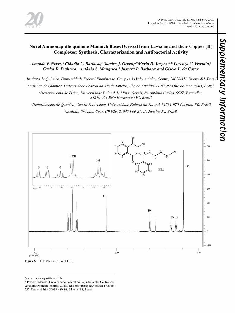
The 1H spectra of compounds HL1-HL13 exhibit peaks due to the four naphthoquinone aromatic hydrogens H5-H8 that appear in the d 7-8 ppm region as dd or ddd (H5 and H8) and td (H6 and H7) (see Figure 1 for numbering and experimental for data). The other chemical shifts are compatible with the structures proposed for these compounds. In general, the butyl hydrogens appear in the d 0.9 to 3.1 ppm region as multiplets (H19-H21) or triplets (methyl H22); in the case of the benzyl group; the CH
2
hydrogens H19 appear as a doublet around d 4.2 –4.6 and the phenyl H21-H25, as multiplets. The (substituted) phenyl group hydrogens H13-17 appear as expected, depending on the substitution pattern. Attributions were made on the basis of 1H × 1H (COSY experiments), J values and multiplicity. All expected resonances were observed in the 13C NMR spectra of compounds HL1-HL13. The resonances arising from the carbonyl carbons were found around d 185 and 179, and those attributed to C2 bound to the hydroxyl group, at about d 171.
Complexes 1-13 (Figure 2) were obtained by addition of trietylamine to a methanolic suspension of the ligand and CuCl
2.2H
2O (2:2:1), under stirring at room temperature for
12 h in yields varying from 60 to 92%, except for complex 12, isolated in 30%. Elemental analysis confirmed the proposed formulation. Due to low solubility in methanol, acetonitrile and water, conductivity measurements could not be carried out.
All compounds were also characterized by EPR and UV-Vis spectroscopy, and the structures of 1, 2 and 7, determined by X-ray diffraction analyses.
Description of the X-ray structures
Good quality crystals suitable for single crystal X-ray diffraction analyses were obtained for compounds 1, 2 and 7. The molecular structures of 1, 2 and 7 are shown in Figures 3, 4 and 5, respectively and selected bond lengths and angles are given in Table 2.
All complexes crystallize in centrosymmetric space groups, with a copper atom in the inversion centre. Two deprotonated ligands (L−) coordinate through the naphthalen-2-olate oxygen and secondary amine-N atoms, forming two six-membered chelate rings around the copper atom in a trans-N
2O
2 environment. Bond angles O(1)-
Cu-N(1) (and β parameters:43 88.43(9), 1, 91.33(10), 2, and 90.67(9)o, 7) and Cu-N and Cu-O distances indicate slightly distorted square-planar coordination of the complexes. The
Figure 3. ORTEP view of [Cu(L1)2].2H
2O 1 with labeled atoms and 50%
probability ellipsoids; H atoms were omitted for the sake of clarity.
Figure 4. ORTEP view of [Cu(L2)2].H
2O 2 with labeled atoms and 50%
probability ellipsoids; H atoms were omitted for the sake of clarity.
Neves et al. 721Vol. 20, No. 4, 2009
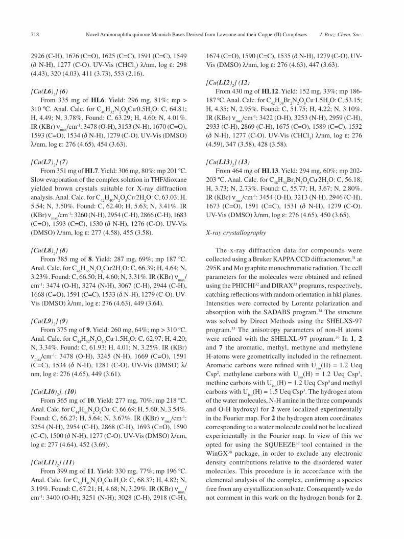
Cu–Namine
(2.023(2), 1, 2.023(3), 2, and 1.996(2) Å, 7) and Cu–O
phenolate distances (1.945(2), 1, 1.928(2), 2, and 1.942(2)
Å, 7) are in the normal range when compared to those observed for other copper(II) complexes containing the same coordination environment.44-46 In the same molecule, the two ligands have different absolute configurations at the chiral C11carbon. In the structures of compounds 1 and 2 the butyl and phenyl groups block the axial positions preventing further coordination to donor molecules (e.g. coordinating solvent), normally observed in the structures of analogous complexes.47,48
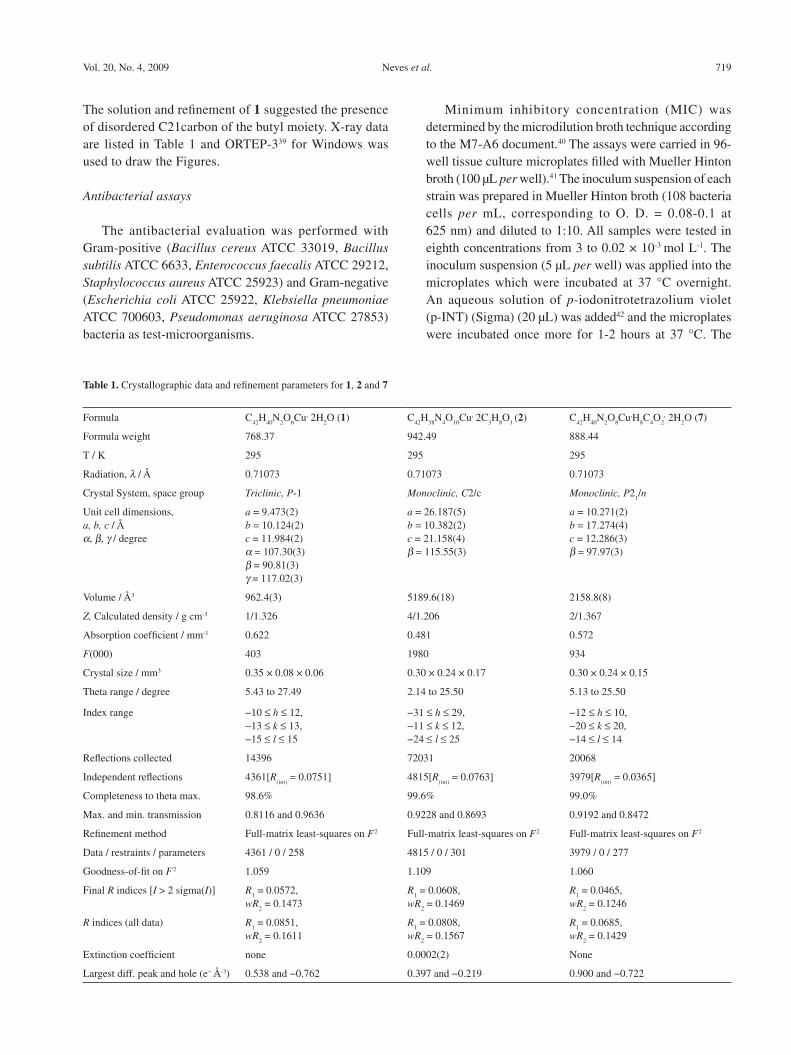
The packing arrangement of 1 exhibits molecules of 1 and water linked by N-H…O and O-H…O classic hydrogen bonds along the [100] crystallographic direction, resulting in a 1D supramolecular arrangement, Figure 6. The water
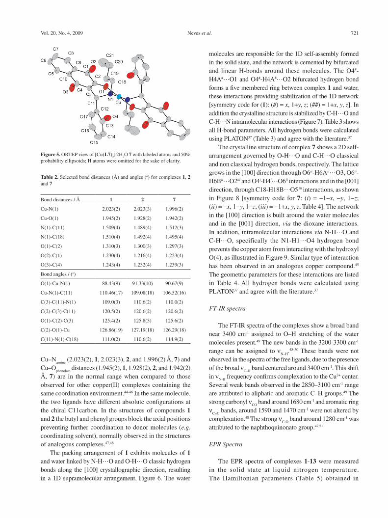
molecules are responsible for the 1D self-assembly formed in the solid state, and the network is cemented by bifurcated and linear H-bonds around these molecules. The O4#-H4A#…O1 and O4#-H4A#…O2 bifurcated hydrogen bond forms a five membered ring between complex 1 and water, these interactions providing stabilization of the 1D network [symmetry code for (1): (#) = x, 1+y, z; (##) = 1+x, y, z]. In addition the crystalline structure is stabilized by C-H…O and C-H…N intramolecular interactions (Figure 7). Table 3 shows all H-bond parameters. All hydrogen bonds were calculated using PLATON37 (Table 3) and agree with the literature.37
The crystalline structure of complex 7 shows a 2D self-arrangement governed by O-H…O and C-H…O classical and non classical hydrogen bonds, respectively. The lattice grows in the [100] direction through O6ii-H6Aii…O3, O6ii-H6Bii…O2iii and O4i-H4i…O6ii interactions and in the [001] direction, through C18-H18B…O5 iii interactions, as shown in Figure 8 [symmetry code for 7: (i) = −1−x, −y, 1−z; (ii) = −x, 1−y, 1−z; (iii) = −1+x, y, z, Table 4]. The network in the [100] direction is built around the water molecules and in the [001] direction, via the dioxane interactions. In addition, intramolecular interactions via N-H…O and C-H…O, specifically the N1-H1…O4 hydrogen bond prevents the copper atom from interacting with the hydroxyl O(4), as illustrated in Figure 9. Similar type of interaction has been observed in an analogous copper compound.45
The geometric parameters for these interactions are listed in Table 4. All hydrogen bonds were calculated using PLATON37 and agree with the literature.37
FT-IR spectra
The FT-IR spectra of the complexes show a broad band near 3400 cm-1 assigned to O–H stretching of the water molecules present.49 The new bands in the 3200-3300 cm-1 range can be assigned to ν
N–H.48-50 These bands were not
observed in the spectra of the free ligands, due to the presence of the broad ν
O-H band centered around 3400 cm-1. This shift
in νN-H
frequency confirms complexation to the Cu2+ center. Several weak bands observed in the 2850–3100 cm-1 range are attributed to aliphatic and aromatic C–H groups.49 The strong carbonyl ν
CO band around 1680 cm-1 and aromatic ring
νC=C
bands, around 1590 and 1470 cm-1 were not altered by complexation.48 The strong ν
C-O band around 1280 cm-1 was
attributed to the naphthoquinonato group.47,51
EPR Spectra
The EPR spectra of complexes 1-13 were measured in the solid state at liquid nitrogen temperature. The Hamiltonian parameters (Table 5) obtained in
Table 2. Selected bond distances (Å) and angles (o) for complexes 1, 2 and 7
Bond distances / Å 1 2 7
Cu-N(1) 2.023(2) 2.023(3) 1.996(2)
Cu-O(1) 1.945(2) 1.928(2) 1.942(2)
N(1)-C(11) 1.509(4) 1.489(4) 1.512(3)
N(1)-C(18) 1.510(4) 1.492(4) 1.495(4)
O(1)-C(2) 1.310(3) 1.300(3) 1.297(3)
O(2)-C(1) 1.230(4) 1.216(4) 1.223(4)
O(3)-C(4) 1.243(4) 1.232(4) 1.239(3)
Bond angles / (o)
O(1)-Cu-N(1) 88.43(9) 91.33(10) 90.67(9)
Cu-N(1)-C(11) 110.46(17) 109.08(18) 106.52(16)
C(3)-C(11)-N(1) 109.0(3) 110.6(2) 110.0(2)
C(2)-C(3)-C(11) 120.5(2) 120.6(2) 120.6(2)
O(1)-C(2)-C(3) 125.4(2) 125.8(3) 125.6(2)
C(2)-O(1)-Cu 126.86(19) 127.19(18) 126.29(18)
C(11)-N(1)-C(18) 111.0(2) 110.6(2) 114.9(2)
Figure 5. ORTEP view of [Cu(L7)2].2H
2O 7 with labeled atoms and 50%
probability ellipsoids; H atoms were omitted for the sake of clarity.
Novel Aminonaphthoquinone Mannich Bases Derived from Lawsone and their Copper(II) Complexes J. Braz. Chem. Soc.722
Figure 7. View of intramolecular interactions via non classical hydrogen bonds in complex 1.
Figure 6. View of the self-assembly 1D by classical hydrogen bonds along [100] crystallography direction. (Symmetry code for 1: (#) = x, 1+y, z; (##) = 1+x, y, z).
Table 3. Geometric parameters for non-classical H bonds for complex 1 (Å, degree)
D-H···A D-H H···A D···A ∠D-H···A
C(17)-H(17)···N(1)intra
0.93 2.60 2.926(5) 101
C(11)-H(11)···O(3)intra
0.99(4) 2.37(4) 2.856(5) 109(3)
O(4)a-H(4A)a···O(1) 0.76(7) 2.56(7) 3.079(5) 126(5)
O(4)a-H(4A)a···O(2) 0.76(7) 2.23(6) 2.972(5) 165(6)
O(4)a-H(4B)a···O(3)b 0.69(7) 2.24(7) 2.913(5) 168(7)
N(1)-H(1)···O(4)a 0.88(4) 2.16(4) 3.018(5) 167(4)
Symmetry code for 1: (a)x, 1+y, z; (b)1+x, y, z.

Neves et al. 723Vol. 20, No. 4, 2009
the simulations of the EPR spectra g|| > g⊥ > 2 and A
|| = (188 - 200) × 10-4 cm-1 are typical of elongated
octahedral or square planar geometry, suggesting copper(II) sites with axial symmetry.52 All complexes, except for 2 and 12, show values of g||/A|| between 110 and 122 thus confirming the square planar environment around the Cu2+ centre53 as established for compounds 1, 2 and 7 by X-ray analysis. The experimental values, g|| > g⊥, also indicate
Figure 8. View of the 2D self-arrangement governed by water and dioxane molecules along [100] and [001] crystallography directions, respectively (symmetry code for 7: (i) = −1−x, −y, 1−z; (ii) = −x, 1−y, 1−z; (iii) = −1+x, y, z).
Figure 9. View of the intramolecular interactions in complex 7.
that the unpaired electron is predominantly in the dx2-y2 orbital, which gives 2B
1g as the ground state. The very low
parallel and perpendicular components of the hyperfine coupling constant for complexes 2 and 12 (not resolved in the spectra) has been explained by considering a mixture of the Cu2+ dz2 and dx2–y2 orbitals as the ground state. It is found that a 10% mixture of dx2-y2 and dz2 results in a 20% reduction in dipolar anisotropy.54

Novel Aminonaphthoquinone Mannich Bases Derived from Lawsone and their Copper(II) Complexes J. Braz. Chem. Soc.724
The relatively low g|| values of the complexes are
consistent with a N2O
2 environment around the Cu2+ ions.52
As expected, the ligand field strength depends mainly on the nature of the R4 substituent on the nitrogen, the highest ligand field being observed for complexes of ligands containing R4 = butyl, independently of the nature of R2 and R3. The presence of the hydroxyl group (R1=OH), however, may lead to a decrease in the ligand field, as observed for complex 7, compared with complex 1 (Table 5), due to the presence of a O-H…N1 hydrogen bond that reduces the Lewis basicity of N1. This interaction is evidenced in the supramolecular arrangement of the structure of 7. This interaction may be present in all complexes containing R1 = OH (6-13).
UV-Vis spectra
The electronic spectra of complexes 1-5 and 12, recorded in CHCl
3 solution, are characterized by two
Table 4. Geometric parameters for non-classical H bonds for complex 7 (Å, degree)
D-H···A D-H H···A D···A ∠D-H···A
N(1)-H(1)···O(4)intra
0.93(4) 2.08(4) 2.818(3) 136(3)
C(11)-H(11)···O(3)intra
0.98 2.40 2.836(4) 107
O(4)i-H(4)i···O(6)ii 0.82 1.95 2.699(3) 151
O(6)ii-H(6A) ii ···O(3) 0.89 1.95 2.833(3) 169
O(6) ii-H(6B) ii ···O(2) iii 0.86 2.03 2.866(3) 163
C(18)-H(18B)···O(5) iii 0.97 2.53 3.461(8) 160
Symmetry code for (7): (i) = −1−x, −y, 1−z; (ii) = −x, 1−y, 1−z; (iii) = −1+x, y, z.
Table 5. Spin-Hamiltonian parameters used in the simulated spectra of the copper(II) ion in complexes 1-13 in decreasing order of field strength
Complexes A⊥ (×10-4 cm-1) A|| (× 10-4 cm-1) g⊥ g
||g
||/A
||
1 15 200 2.0500 2.2070 110
3 25 197 2.0350 2.2100 112
5 27 198 2.0350 2.2100 112
10 25 192 2.0470 2.2055 115
11 25 195 2.0470 2.2350 115
7 20 190 2.0550 2.2070 116
13 25 193 2.0440 2.2380 116
9 25 190 2.0450 2.2380 118
4 25 190 2.0450 2.2380 118
8 25 188 2.0480 2.2380 119
6 25 188 2.0488 2.2400 119
12 ---- ---- 2.0630 2.1880 -----
2 ---- ---- 2.0580 2.1880 ----
intense absorptions observed in the 425-315 nm range that are presumably due to a charge-transfer band from the naphthquinonato moiety to the metal ion, and to ligand based transitions.55 The band around 290 nm was not altered by coordination as it corresponds to π-π* transitions of the naphthoquinone ring.48-50. The d-d band appears between 530-550 nm and can be attributed to a 2A
1g←2B
1g
transition which supports the square planar geometry for the complexes in solution.50
The spectra of complexes 6-11 and 13 were recorded in DMSO solution due to their low solubility in CHCl
3. All
exhibit two intense absorption bands: the band observed around 450 nm was assigned to charge-transfer from the naphthoquinonate moiety to the metal ion and that at 277 nm, also present in the spectra of the respective free ligands, to π-π* transitions of the naphthoquinone ring. No d-d transition band was observed, which suggests coordination of DMSO molecules and distorted octahedral coordination environment of the Cu2+ ion.
Neves et al. 725Vol. 20, No. 4, 2009
Antibacterial activity
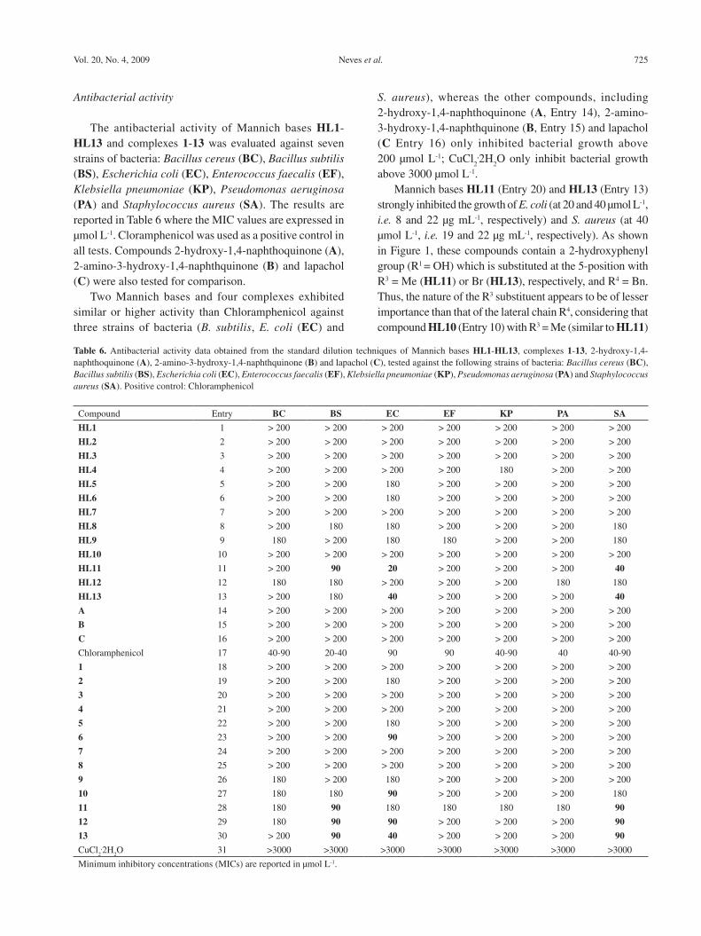
The antibacterial activity of Mannich bases HL1-HL13 and complexes 1-13 was evaluated against seven strains of bacteria: Bacillus cereus (BC), Bacillus subtilis (BS), Escherichia coli (EC), Enterococcus faecalis (EF), Klebsiella pneumoniae (KP), Pseudomonas aeruginosa (PA) and Staphylococcus aureus (SA). The results are reported in Table 6 where the MIC values are expressed in µmol L-1. Cloramphenicol was used as a positive control in all tests. Compounds 2-hydroxy-1,4-naphthoquinone (A), 2-amino-3-hydroxy-1,4-naphthquinone (B) and lapachol (C) were also tested for comparison.
Two Mannich bases and four complexes exhibited similar or higher activity than Chloramphenicol against three strains of bacteria (B. subtilis, E. coli (EC) and
S. aureus), whereas the other compounds, including 2-hydroxy-1,4-naphthoquinone (A, Entry 14), 2-amino-3-hydroxy-1,4-naphthquinone (B, Entry 15) and lapachol (C Entry 16) only inhibited bacterial growth above 200 µmol L-1; CuCl
2.2H
2O only inhibit bacterial growth
above 3000 µmol L-1. Mannich bases HL11 (Entry 20) and HL13 (Entry 13)
strongly inhibited the growth of E. coli (at 20 and 40 µmol L-1, i.e. 8 and 22 µg mL-1, respectively) and S. aureus (at 40 µmol L-1, i.e. 19 and 22 µg mL-1, respectively). As shown in Figure 1, these compounds contain a 2-hydroxyphenyl group (R1 = OH) which is substituted at the 5-position with R3 = Me (HL11) or Br (HL13), respectively, and R4 = Bn. Thus, the nature of the R3 substituent appears to be of lesser importance than that of the lateral chain R4, considering that compound HL10 (Entry 10) with R3 = Me (similar to HL11)
Table 6. Antibacterial activity data obtained from the standard dilution techniques of Mannich bases HL1-HL13, complexes 1-13, 2-hydroxy-1,4-naphthoquinone (A), 2-amino-3-hydroxy-1,4-naphthquinone (B) and lapachol (C), tested against the following strains of bacteria: Bacillus cereus (BC), Bacillus subtilis (BS), Escherichia coli (EC), Enterococcus faecalis (EF), Klebsiella pneumoniae (KP), Pseudomonas aeruginosa (PA) and Staphylococcus aureus (SA). Positive control: Chloramphenicol
Compound Entry BC BS EC EF KP PA SA
HL1 1 > 200 > 200 > 200 > 200 > 200 > 200 > 200
HL2 2 > 200 > 200 > 200 > 200 > 200 > 200 > 200
HL3 3 > 200 > 200 > 200 > 200 > 200 > 200 > 200
HL4 4 > 200 > 200 > 200 > 200 180 > 200 > 200
HL5 5 > 200 > 200 180 > 200 > 200 > 200 > 200
HL6 6 > 200 > 200 180 > 200 > 200 > 200 > 200
HL7 7 > 200 > 200 > 200 > 200 > 200 > 200 > 200
HL8 8 > 200 180 180 > 200 > 200 > 200 180
HL9 9 180 > 200 180 180 > 200 > 200 180
HL10 10 > 200 > 200 > 200 > 200 > 200 > 200 > 200
HL11 11 > 200 90 20 > 200 > 200 > 200 40
HL12 12 180 180 > 200 > 200 > 200 180 180
HL13 13 > 200 180 40 > 200 > 200 > 200 40
A 14 > 200 > 200 > 200 > 200 > 200 > 200 > 200
B 15 > 200 > 200 > 200 > 200 > 200 > 200 > 200
C 16 > 200 > 200 > 200 > 200 > 200 > 200 > 200
Chloramphenicol 17 40-90 20-40 90 90 40-90 40 40-90
1 18 > 200 > 200 > 200 > 200 > 200 > 200 > 200
2 19 > 200 > 200 180 > 200 > 200 > 200 > 200
3 20 > 200 > 200 > 200 > 200 > 200 > 200 > 200
4 21 > 200 > 200 > 200 > 200 > 200 > 200 > 200
5 22 > 200 > 200 180 > 200 > 200 > 200 > 200
6 23 > 200 > 200 90 > 200 > 200 > 200 > 200
7 24 > 200 > 200 > 200 > 200 > 200 > 200 > 200
8 25 > 200 > 200 > 200 > 200 > 200 > 200 > 200
9 26 180 > 200 180 > 200 > 200 > 200 > 200
10 27 180 180 90 > 200 > 200 > 200 180
11 28 180 90 180 180 180 180 90
12 29 180 90 90 > 200 > 200 > 200 90
13 30 > 200 90 40 > 200 > 200 > 200 90
CuCl2.2H
2O 31 >3000 >3000 >3000 >3000 >3000 >3000 >3000
Minimum inhibitory concentrations (MICs) are reported in µmol L-1.
Novel Aminonaphthoquinone Mannich Bases Derived from Lawsone and their Copper(II) Complexes J. Braz. Chem. Soc.726
and R4 = Bu is much less active than HL11 against all strains of bacteria. Solubility differences might be responsible for the changes in activity.
With a few exceptions, the complexes were less active than the respective pro-ligands, which is probably due to their lower solubility. Thus, the activity of HL11 (Entry 11) decreased upon complexation, from 20 to 90 µmol L-1 for 11 (E. coli) and from 20 to 90 µmol L-1 for 11 (S. aureus) (Entry 28), although slight increase in growth inhibition of all the other bacteria strains was observed (from >200 µmol L-1 for HL11 to 180 µmol L-1 for 11).
Improvement of the activity of HL10 and H12 that only inhibited the growth of all strains of bacteria above 180-200 µmol L-1 (Entries 10 and 12) was observed upon complexation: complex 10 (Entry 27) exhibits slight activity against B. cereus, B. subtilis and S. aureus (180 µmol L-1) and growth inhibition against E. coli (90 µmol L-1), and complex 12, against E. coli, E. faecalis and S. aureus above 90 µmol L-1 (Entry 29). Complexes 10 and 12 were formed from Mannich bases HL10 and HL12 that only differed from the very active ones, HL11 and HL13 with respect to the R4 group, butyl, instead of benzyl.
The effect of metal complexation on naphthoquinone antimicrobial agents has been discussed in the literature.56 Although metal chelation of the anion of 5-hydroxy-1,4-naphthoquinone (juglone) has resulted in complexes with similar antibacterial effect57 or higher antibacterial activity, e.g. against Bacillus ssp and S. aureus, than juglone, complexation of the anions of a series of 5-amino-8-hydroxy-1,4-naphthoquinones with M2+ (M = Ni, Co, Fe, Cu and Cr) resulted in reduced bacterial activity or lack of inhibition effect.55 Considering that redox active metals have been shown to be instrumental in naphthoquinone toxicity,11 the decreased activity observed in our work and by others is probably associated with decreased bioavailability of the aminonaphthoquinones as the result of decreased solubility upon complexation.
Conclusions
In conclusion, of the thirteen novel aminonaphthoquinones HL1-HL13 synthesized from lawsone, via the Mannich reaction, and their respective copper(II) complexes [Cu(L1)
2] – [Cu(L13)
2], those containing a 2-hydroxyphenyl
group (R1 = OH) which is substituted at the 5-position, either with R3 = Me or Br, and with R4 = benzyl or butyl groups have strongly inhibited the growth especially of Escherichia coli and Staphylococcus aureus. In general complexes were found to be less active than the respective pro-ligands, probably due to their lower solubility. Further
work is in progress to improve the bioavailability of the complexes of HL10-HL13.
Supplementary Information
Supplementary data are available free of charge at http://jbcs.sbq.org.br, as PDF file.
Crystallographic data for the structural analysis of the three complexes have been deposited with the Cambridge Crystallographic Data Center, CCDC No. 703509 (1), 703510 (2) and 703511 (7). Copies of this information may be obtained free of charge from The Director, CCDC, 12 Union Road, Cambridge, CB2 1EZ, UK (fax: +44 1233336 033; e-mail: [email protected]).
Acknowledgments
We thank CNPq, CAPES, FINEP, PRONEX-FAPERJ and FAPERJ for financial support, and the X-ray diffraction laboratory (LDRX) of Universidade Federal Fluminense for data collection.
References
1. Thompson, R. H.; Naturally Occurring Quinones IV: Recent
Advances, Champman & Hall: London, 1997.
2. Sacau, E. P.; Braun, A. E.; Ravelo, A. G.; Ferro, E. A.; Tokuda, H.;
Mukainaka, T.; Nishino, H.; Bioorg. Med. Chem. 2003, 11, 483.
3. Chen, J.; Huang, Y.; Liu, G.; Afrasiabi, Z.; Sinn, E.; Padhye,
S.; Ma. Y.; Toxicol. Appl. Pharmacol. 2004, 194, 40.
4. Ferreira, V. F.; Jorqueira, A.; Souza, A. M. T.; Silva, M. N.; de
Souza, M. C. B. V.; Gouvêa, R. M.; Rodrigues, C. R.; Pinto, A.
V.; Castro, H. C.; Santos, D. O.; Araújo, H. P.; Bourguignon, S.
C.; Bioorg. Med. Chem. 2006, 14, 5459.
5. Santos, A. F.; Ferraz, P. A. L.; Pinto, A. V.; Pinto, M. C. F. R.;
Goulart, M. O. F.; Sant’Ana, A. E. G.; Int. J. Parasitol. 2000,
30, 1199.
6. Santos, E. V. M.; Carneiro, J. W. M.; Ferreira, V. F.; Bioorg.
Med. Chem. 2004, 12, 87.
7. Kayser, O.; Kiderlen, A. F.; Laatsch, H.; Croft, S.; Acta Trop.
2000, 77, 307.
8. Gafner, S.; Wolfender, J. L.; Nianga, M.; Stoeckli-Evans, H.;
Hostettman, K.; Phytochemistry 1996, 42, 1315.
9. Machado, T. B.; Pinto, A. V.; Pinto, M. C. F. R.; Leal, I. C. R.;
Silva, M. G.; Amaral, A. C. F.; Kuster, R. M.; Netto-dosSantos,
K. R.; Int. J. Antimicrob. Agents 2003, 21, 279.
10. Medina, L. F. C.; Hertz, P. F.; Stefani, V.; Henriques, J. A. P.;
Zanotto-Filho, A.; Brandelli, A.; Biochem. Cell Biol. 2006, 84,
720.
11. Zang, R.; Hirsh, O.; Mohsen, M.; Samuni, A.; Arch. Biochem.
Biophys. 1994, 312, 385.
Neves et al. 727Vol. 20, No. 4, 2009
12. Vargas, M. D.; Pinto, A. C.; Echevarria, A.; Esteves-Souza,
A.; Camara, C. A.; Cunha, A. C.; Torres, J. C.; Lima, E. L. S.;
J. Braz. Chem. Soc. 2006, 17, 439.
13. Esteves-Souza, A.; Figueiredo, D. V.; Esteves, A.; Câmara, C.
A.; Vargas, M. D.; Pinto, A. C.; Echevarria, A.; Braz. J. Med.
Biol. Res. 2007, 30, 1399.
14. Cunha, A. S.; Vargas, M. D.; Gattass, C. R.; Pinto, A. C.;
Camara, C. A.; Esteves, A. S.; Lima, E. L. S.; Oncol. Rep. 2008,
20, 225.
15. Silva, M. S.; Camara, C. A.; Barbosa, T. P.; Soares, A. Z.; Cunha,
L. C.; Pinto, A. C.; Vargas, M. D.; Bioorg. Med. Chem. 2005,
13, 193.
16. Barbosa, T. P.; Camara, C. A.; Silva, T. M. S.; Martins, R. M.;
Pinto, A. C.; Vargas, M. D.; Bioorg. Med. Chem. 2005, 13,
6464.
17. Camara, C. A.; Silva, T. M. S.; Silva, T. G.; Barbosa, T. P.;
Martins, R. M.; Vargas, M. D.; Pinto, A. C.; An. Acad. Bras.
Ciênc. 2008, 80, 329.
18. Tandon, V. K.; Yadav, D. B.; Singh, R. V.; Chaturvedi, A. K.;
Shukla, P. K.; Bioorg. Med. Chem. Lett. 2005, 15, 5324.
19. Tandon, V. K.; Yadav, D. B.; Chaturvedi, A. K.; Shukla, P. K.;
Bioorg. Med. Chem. Lett. 2005, 15, 3288.
20. Riffel, A.; Medina, L. F.; Stefani, V.; Santos, R. C.; Bizani, D.;
Brandelli, A.; Braz. J. Med. Biol. Res. 2002, 35, 811.
21. Anacona, J. R.; Moreno, A.; Main Group Met. Chem. 1999, 22,
573.
22. Tandon, V. K.; Vaish, M.; Khanna, J. M.; Anand, N.; Arch.
Pharmazie 1990, 323, 383.
23. Leffer, M. T.; Hathaway, R. J.; J. Am. Chem. Soc. 1948, 70,
3222.
24. Baramee, A.; Coppin, A.; Mortuaire, M.; Pelinski, L.; Tomavo,
S.; Brocard, J.; Bioorg. Med. Chem. 2006, 14, 1294.
25. Santos, A. F.; Ferraz, P. A. L.; Pinto, A. V.; Pinto, M. C. F. R.;
Goulart, M. O. F.; Sant’Ana, A. E. G.; Int. J. Parasitol. 2000,
30, 1199.
26. Zang, O.; Hirsh, M.; Mohsen, A.; Samuni; Arch. Biochem.
Biophys. 1994, 312, 385.
27. Gokhale, N. H.; Shirisha, K.; Padhye, S. B.; Croft, S. L.;
Kendrick, H. D.; Mckee, V.; Bioorg. Med. Chem. Lett. 2006,
16, 430.
28. Gokhale, N. H.; Shirisha, K.; Padhye, S. B.; Croft, S. L.;
Kendrick, H. D.; Davies, W.; Anson, C. E.; Powell, A. K.;
J. Inorg. Biochem. 2003, 95, 249.
29. Chen, J.; Huang, Y.; Liu, G.; Afrasiabi, Z.; Sinn, E.; Padhye,
S.; Ma, Y.; Toxicol. Appl. Pharmacol. 2004, 197, 40.
30. Dalgliesh, C.E.; J. Am. Chem. Soc. 1949, 71, 1697.
31. Nonius; COLLECT, Nonius BV: Delft, The Netherlands,
1998.
32. Duisenberg, A. J. M.; Hooft, R. W. W.; Schreurs, A. M. M.;
Kroon, J.; J. Appl. Crystallogr. 2000, 33, 893.
33. Duisenberg, A. J. M.; J. Appl. Crystallogr. 1992, 25, 92.
34. Sheldrick, G. M.; SADABS; Program for Empirical Absorption
Correction of Area Detector Data, University of Göttingen:
Germany, 1996.
35. Sheldrick, G. M.; SHELXS97; Program for Crystal Structure
Solution, University of Göttingen: Germany, 1997.
36. Sheldrick, G. M.; SHELXL97; Program for Crystal Structure
Refinement, University of Göttingen: Germany, 1997.
37. Spek, A. L.; J. Appl. Crystallogr. 2003, 36, 7.
38. Farrugia, L. J.; J. Appl. Crystallogr. 1999, 32, 837.
39. Farrugia, L. J.; J. Appl. Crystallogr. 1997, 30, 565.
40. National Committee for Clinical Laboratory Standards Methods
for Dilution Antimicrobial Susceptibility Tests for Bacteria
that Grow Aerobically, 6th ed.; Approved Standard NCCLS
Document M7-A6: Wayne, PA, 2003.
41. Langfield, R. D.; Scarano, F. J.; Heitzman, M. E.; Kondo, M.;
Hammond, G. B.; Neto, C. C.; J. Ethnopharmacol. 2004, 94, 279.
42. Eloff, J. N.; Planta Med. 1998, 65, 711.
43. Addison, A. W. In Copper Coordination Chemistry: Biochemical
and Inorganic Perspectives; Karlin, K. D.; Zubieta, J., eds.,
Academic Press: New York, 1983.
44. Muppidi, V. K.; Das, S.; Raghavaiah, P.; Pal, S.; Inorg. Chem.
Commun. 2007, 10, 234.
45. Xie, Y.; Ni, J.; Liu, X.; Liu, Q.; Xu, X.; Transition Met. Chem.
2003, 28, 367.
46. Thakuria, H.; Das, G.; Polyhedron 2007, 26, 149.
47. Xie, Y.; Bu, W.; Chan, A. S. -C.; Xu, X.; Liu, Q.; Zhang, Z.; Yu,
J.; Fan, Y.; Inorg. Chim. Acta 2000, 310, 257.
48. Muppidi, V. K.; Zacharias, P. S.; Pal, S.; J. Solid State Chem.
2007, 180, 132.
49. Muppidi, V. K.; Das, S.; Raghavaiah, P.; Pal, S.; Inorg. Chem.
Commun. 2007, 10, 234.
50. Thakuria, H.; Das, G.; Polyhedron 2007, 26, 149.
51. Xie, Y.; Bu, W.; Chan, A. S. -C.; Xu, X.; Liu, Q.; Zhang, Z.; Yu,
J.; Fan, Y.; Inorg. Chim. Acta 2000, 310, 257.
52. Neves, A.; Rossi, L. M.; Bortoluzzi, A. J.; Mangrich, A. S.;
Haase, W.; Werner, R.; J. Braz. Chem. Soc. 2001, 12, 747.
53. Sakaguchi, U.; Addison, A. W.; J. Chem. Soc., Dalton Trans.
1979, 600.
54. Mendes, I. C.; Moreira, J. P.; Speziali, N. L.; Mangrich, A. S.;
Takahashi, J. A.; Beraldo, H.; J. Braz. Chem. Soc. 2006, 17, 1571.
55. Ma, S.; Zhu, W.; Xu, M.; Wang, Y.; Guo, Q.; Liu, Y.; Polyhedron
2003, 22, 3249.
56. Brandelli, A.; Bizani, D.; Martinelli, M.; Stefani, V.; Gerbase,
A. E.; Braz. J. Pharm. Sci. 2004, 40, 247.
57. Joshi, C. R.; Jagtap, G. S.; Chalgen, S. V.; Indian J. Pharmaceut.
Sci. 1988, 50, 107.
58. Bakolachristianopoulou, M. N.; Ecateriniadou, L. B.; Sarris,
K. J.; Eur. J. Med. Chem. 1986, 21, 385.
Received: March 3, 2009
Web Release Date: April 24, 2009
Supplementary Inform
ationJ. Braz. Chem. Soc., Vol. 20, No. 4, S1-S14, 2009.
Printed in Brazil - ©2009 Sociedade Brasileira de Química0103 - 5053 $6.00+0.00
*e-mail: [email protected]# Present Address: Universidade Federal do Espírito Santo, Centro Uni-versitário Norte do Espírito Santo, Rua Humberto de Almeida Franklin, 257, Universitário, 29933-480 São Mateus-ES, Brazil
Novel Aminonaphthoquinone Mannich Bases Derived from Lawsone and their Copper (II) Complexes: Synthesis, Characterization and Antibacterial Activity
Amanda P. Neves,a Cláudia C. Barbosa,a Sandro J. Greco,a,# Maria D. Vargas,a,* Lorenzo C. Visentin,b Carlos B. Pinheiro,c Antônio S. Mangrich,d Jussara P. Barbosae and Gisela L. da Costae
aInstituto de Química, Universidade Federal Fluminense, Campus do Valonguinho, Centro, 24020-150 Niterói-RJ, Brazil
bInstituto de Química, Universidade Federal do Rio de Janeiro, Ilha do Fundão, 21945-970 Rio de Janeiro-RJ, Brazil
cDepartamento de Física, Universidade Federal de Minas Gerais, Av. Antônio Carlos, 6627, Pampulha, 31270-901 Belo Horizonte-MG, Brazil
dDepartamento de Química, Centro Politécnico, Universidade Federal do Paraná, 81531-970 Curitiba-PR, Brazil
eInstituto Oswaldo Cruz, CP 926, 21045-900 Rio de Janeiro-RJ, Brazil
Figure S1. 1H NMR spectrum of HL1.
Novel Aminonaphthoquinone Mannich Bases Derived from Lawsone and their Copper (II) Complexes J. Braz. Chem. Soc.S2
Figure S2. 1H NMR spectrum of HL2.
Figure S3. 1H NMR spectrum of HL3.
Neves et al. S3Vol. 20, No. 4, 2009
Figure S4. 1H NMR spectrum of HL4.
Figure S5. 1H NMR spectrum of HL5.

Novel Aminonaphthoquinone Mannich Bases Derived from Lawsone and their Copper (II) Complexes J. Braz. Chem. Soc.S4
Figure S6. 1H NMR spectrum of HL6.
Figure S7. 1H NMR spectrum of HL7.
Neves et al. S5Vol. 20, No. 4, 2009
Figure S8. 1H NMR spectrum of HL8.
Figure S9. 1H NMR spectrum of HL9.
Novel Aminonaphthoquinone Mannich Bases Derived from Lawsone and their Copper (II) Complexes J. Braz. Chem. Soc.S6
Figure S10. 1H NMR spectrum of HL10.
Figure S11. 1H NMR spectrum of HL11.
Neves et al. S7Vol. 20, No. 4, 2009
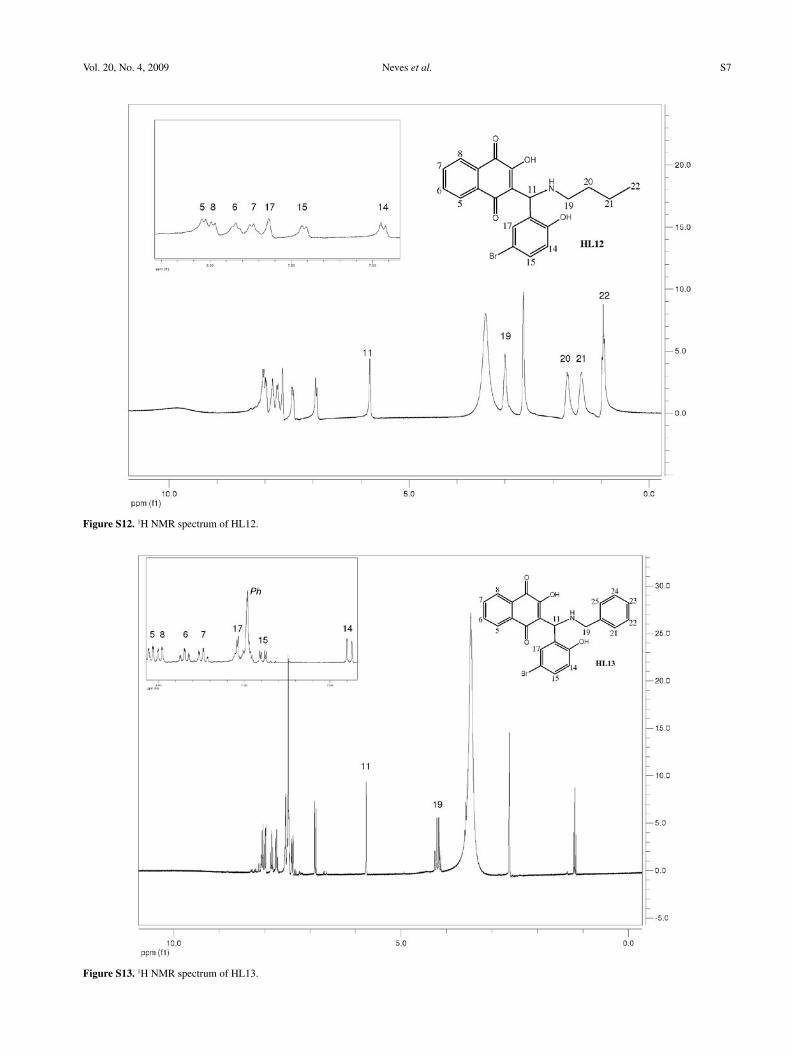
Figure S12. 1H NMR spectrum of HL12.
Figure S13. 1H NMR spectrum of HL13.
Novel Aminonaphthoquinone Mannich Bases Derived from Lawsone and their Copper (II) Complexes J. Braz. Chem. Soc.S8
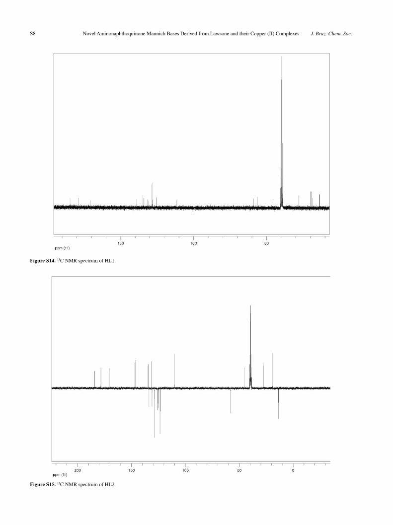
Figure S14. 13C NMR spectrum of HL1.
Figure S15. 13C NMR spectrum of HL2.
Neves et al. S9Vol. 20, No. 4, 2009
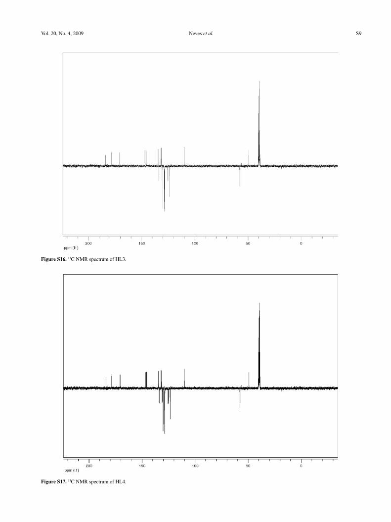
Figure S16. 13C NMR spectrum of HL3.
Figure S17. 13C NMR spectrum of HL4.

Novel Aminonaphthoquinone Mannich Bases Derived from Lawsone and their Copper (II) Complexes J. Braz. Chem. Soc.S10
Figure S18. 13C NMR spectrum of HL5.
Figure S19. 13C NMR spectrum of HL6.
Neves et al. S11Vol. 20, No. 4, 2009
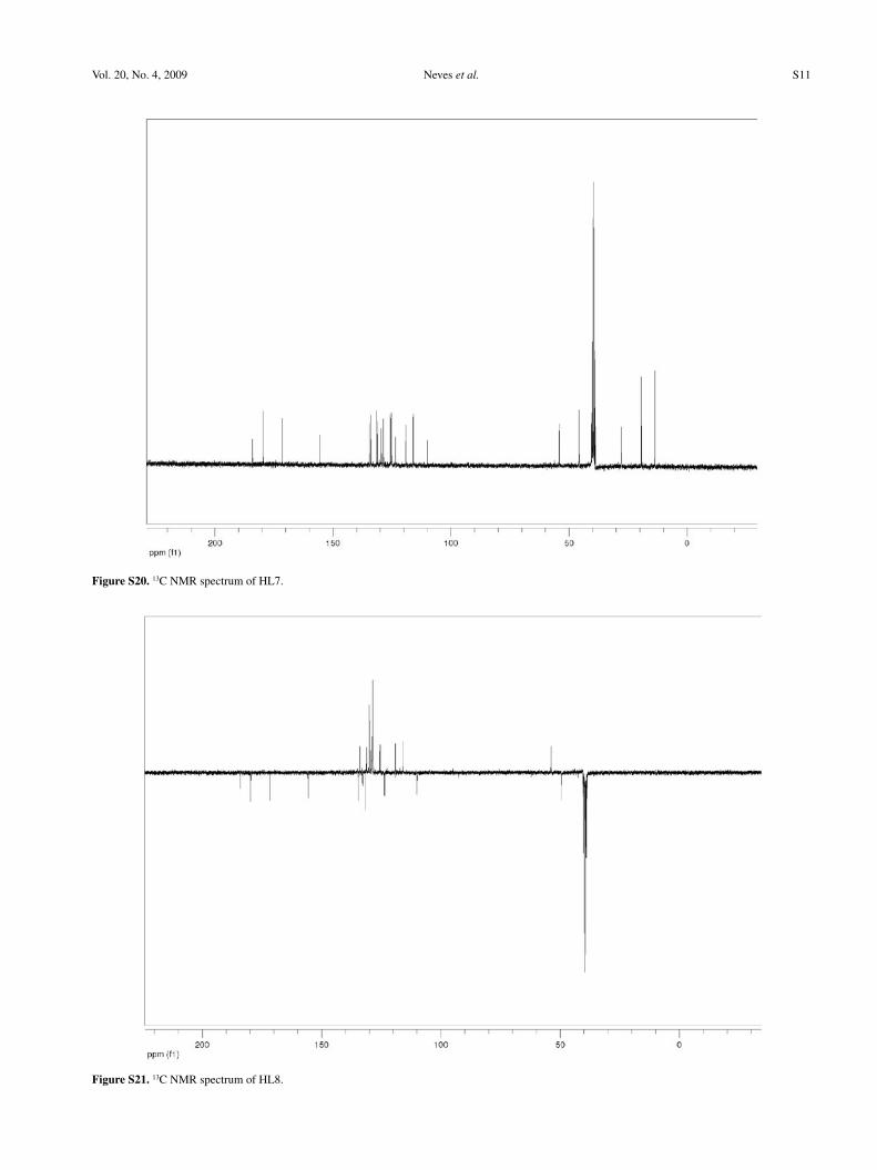
Figure S20. 13C NMR spectrum of HL7.
Figure S21. 13C NMR spectrum of HL8.
Novel Aminonaphthoquinone Mannich Bases Derived from Lawsone and their Copper (II) Complexes J. Braz. Chem. Soc.S12
Figure S22. 13C NMR spectrum of HL9.
Figure S23. 13C NMR spectrum of HL10.
Neves et al. S13Vol. 20, No. 4, 2009
Figure S24. 13C NMR spectrum of HL11.
Figure S25. 13C NMR spectrum of HL12.
Novel Aminonaphthoquinone Mannich Bases Derived from Lawsone and their Copper (II) Complexes J. Braz. Chem. Soc.S14
Figure S26. 13C NMR spectrum of HL1.