Saturation Mutagenesis of 5S rRNA in Saccharomyces cerevisiae
Upload
independentCategory
view
0download
0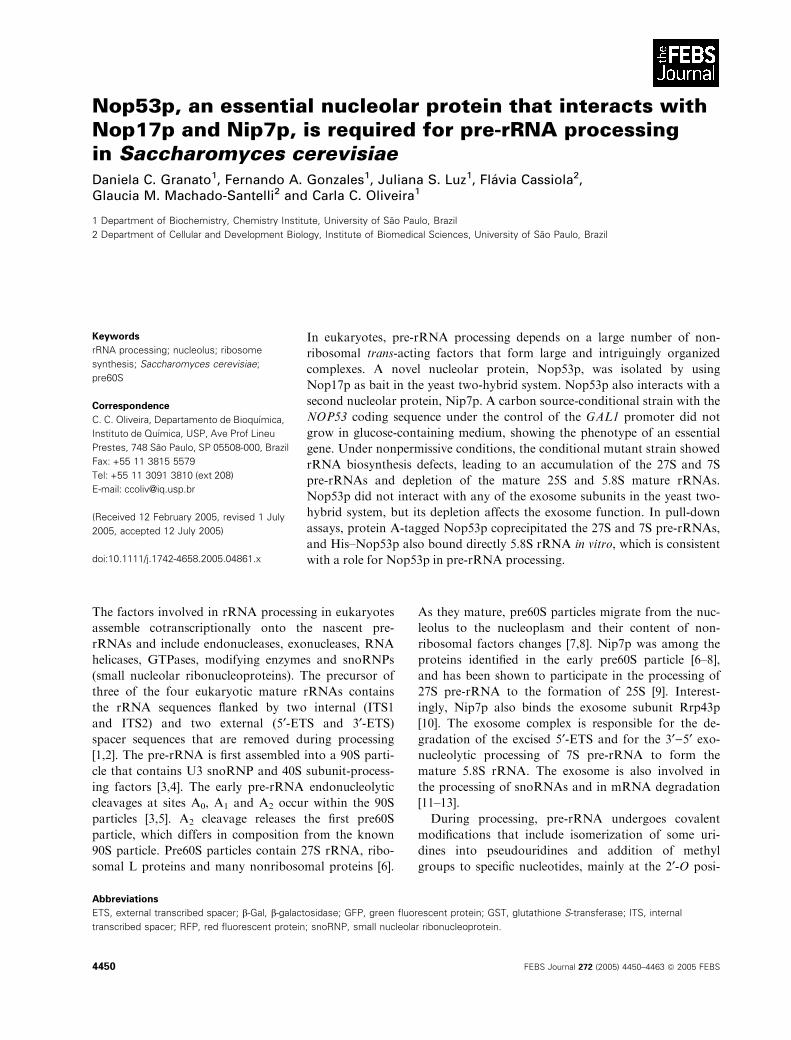
Nop53p, an essential nucleolar protein that interacts withNop17p and Nip7p, is required for pre-rRNA processingin Saccharomyces cerevisiaeDaniela C. Granato1, Fernando A. Gonzales1, Juliana S. Luz1, Flavia Cassiola2,Glaucia M. Machado-Santelli2 and Carla C. Oliveira1
1 Department of Biochemistry, Chemistry Institute, University of Sao Paulo, Brazil
2 Department of Cellular and Development Biology, Institute of Biomedical Sciences, University of Sao Paulo, Brazil
The factors involved in rRNA processing in eukaryotes
assemble cotranscriptionally onto the nascent pre-
rRNAs and include endonucleases, exonucleases, RNA
helicases, GTPases, modifying enzymes and snoRNPs
(small nucleolar ribonucleoproteins). The precursor of
three of the four eukaryotic mature rRNAs contains
the rRNA sequences flanked by two internal (ITS1
and ITS2) and two external (5¢-ETS and 3¢-ETS)spacer sequences that are removed during processing
[1,2]. The pre-rRNA is first assembled into a 90S parti-
cle that contains U3 snoRNP and 40S subunit-process-
ing factors [3,4]. The early pre-rRNA endonucleolytic
cleavages at sites A0, A1 and A2 occur within the 90S
particles [3,5]. A2 cleavage releases the first pre60S
particle, which differs in composition from the known
90S particle. Pre60S particles contain 27S rRNA, ribo-
somal L proteins and many nonribosomal proteins [6].
As they mature, pre60S particles migrate from the nuc-
leolus to the nucleoplasm and their content of non-
ribosomal factors changes [7,8]. Nip7p was among the
proteins identified in the early pre60S particle [6–8],
and has been shown to participate in the processing of
27S pre-rRNA to the formation of 25S [9]. Interest-
ingly, Nip7p also binds the exosome subunit Rrp43p
[10]. The exosome complex is responsible for the de-
gradation of the excised 5¢-ETS and for the 3¢)5¢ exo-nucleolytic processing of 7S pre-rRNA to form the
mature 5.8S rRNA. The exosome is also involved in
the processing of snoRNAs and in mRNA degradation
[11–13].
During processing, pre-rRNA undergoes covalent
modifications that include isomerization of some uri-
dines into pseudouridines and addition of methyl
groups to specific nucleotides, mainly at the 2¢-O posi-
Keywords
rRNA processing; nucleolus; ribosome
synthesis; Saccharomyces cerevisiae;
pre60S
Correspondence
C. C. Oliveira, Departamento de Bioquımica,
Instituto de Quımica, USP, Ave Prof Lineu
Prestes, 748 Sao Paulo, SP 05508-000, Brazil
Fax: +55 11 3815 5579
Tel: +55 11 3091 3810 (ext 208)
E-mail: [email protected]
(Received 12 February 2005, revised 1 July
2005, accepted 12 July 2005)
doi:10.1111/j.1742-4658.2005.04861.x
In eukaryotes, pre-rRNA processing depends on a large number of non-
ribosomal trans-acting factors that form large and intriguingly organized
complexes. A novel nucleolar protein, Nop53p, was isolated by using
Nop17p as bait in the yeast two-hybrid system. Nop53p also interacts with a
second nucleolar protein, Nip7p. A carbon source-conditional strain with the
NOP53 coding sequence under the control of the GAL1 promoter did not
grow in glucose-containing medium, showing the phenotype of an essential
gene. Under nonpermissive conditions, the conditional mutant strain showed
rRNA biosynthesis defects, leading to an accumulation of the 27S and 7S
pre-rRNAs and depletion of the mature 25S and 5.8S mature rRNAs.
Nop53p did not interact with any of the exosome subunits in the yeast two-
hybrid system, but its depletion affects the exosome function. In pull-down
assays, protein A-tagged Nop53p coprecipitated the 27S and 7S pre-rRNAs,
and His–Nop53p also bound directly 5.8S rRNA in vitro, which is consistent
with a role for Nop53p in pre-rRNA processing.
Abbreviations
ETS, external transcribed spacer; b-Gal, b-galactosidase; GFP, green fluorescent protein; GST, glutathione S-transferase; ITS, internal
transcribed spacer; RFP, red fluorescent protein; snoRNP, small nucleolar ribonucleoprotein.
4450 FEBS Journal 272 (2005) 4450–4463 ª 2005 FEBS
tion of the ribose. These nucleotide modifications are
directed by snoRNPs, which select the nucleotide
through complementary base-pairing between the
snoRNA and the rRNA substrate. The snoRNAs
involved in rRNA modification can be divided into
two major classes based on conserved sequence ele-
ments and on the association with evolutionarily con-
served core proteins [14–16]. The box C ⁄D class of
guide snoRNAs contains the core proteins Nop1p,
Nop58p, Nop56p and Snu13p, and is involved in clea-
vage and methylation of pre-rRNA. The box H ⁄ACA
guide snoRNAs are associated with the core proteins
Cbf5p, Gar1p, Nhp2p and Nop10p and function in
the conversion of uridine into pseudouridine [17–23].
In addition to the core snoRNP proteins, other
proteins have been found to be associated with the
snoRNPs and to participate in cleavage reactions as
well as methylation and pseudouridylation of specific
nucleotides of rRNA [24–28]. Among these proteins is
Nop17p, which interacts with the box C ⁄D snoRNP
subunit Nop58p and with the exosome subunit Rrp43p
[28]. Characterization of Nop17p function showed that
it is required for proper localization of the core pro-
teins of the box C ⁄D snoRNP Nop1p, Nop56p,
Nop58p and Snu13p [28]. In addition, cells depleted
of Nop17p show pre-rRNA processing defects that
include increased primer extension products at certain
box C ⁄D methylation sites, indicating that Nop17p is
required for proper pre-rRNA methylation [28]. A third
Nop17p-interacting partner isolated using the yeast
two-hybrid system is the protein encoded by the open
reading frame (ORF) YPL146C, Nop53p. Nop53p is
an essential nucleolar protein, which was also recently
identified as a subunit in pre60S particles [6,7].
In this study, we show that Nop53p is required for
the late steps of rRNA processing. Consistent with its
copurification with the pre60S particle, Nop53p deple-
tion affects exonucleolytic cleavage of the 3¢-end of the
7S pre-rRNA, a processing step that requires the func-
tion of the exosome [11]. In addition, protein A-tagged
Nop53p coprecipitated the 27S and 7S pre-rRNAs and
the mature 5.8S rRNA. Purified His–Nop53p also
bound in vitro transcribed 5.8S rRNA, showing that it
must play an important role in ribosome biogenesis,
possibly related to the exosome function.
Results
Nop53p interacts with the pre-rRNA processing
proteins Nop17p and Nip7p
Saccharomyces cerevisiae Nop53p, a previously unchar-
acterized essential protein (SGD), is encoded by the
YPL146C ORF and was identified in the yeast nuclear
pore complex [29] and as a component of the pre60S
complex [6,7]. In this study, Nop53p was isolated in
a two-hybrid screen as a protein interacting with
Nop17p, which is involved in the early steps of pre-
rRNA processing [28]. Nop17p and Nop53p interacted
in the two-hybrid system independently of the tag, but
the interaction was stronger when Nop17p was fused
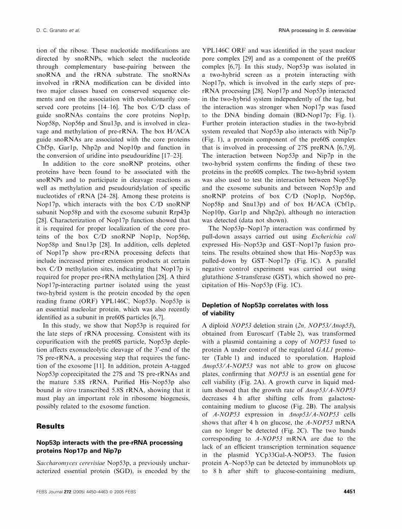
to the DNA binding domain (BD-Nop17p; Fig. 1).
Further protein interaction studies in the two-hybrid
system revealed that Nop53p also interacts with Nip7p
(Fig. 1), a protein component of the pre60S complex
that is involved in processing of 27S preRNA [6,7,9].
The interaction between Nop53p and Nip7p in the
two-hybrid system confirms the finding of these two
proteins in the pre60S complex. The two-hybrid system
was also used to test the interaction between Nop53p
and the exosome subunits and between Nop53p and
snoRNP proteins of box C ⁄D (Nop1p, Nop56p,
Nop58p and Snu13p) and of box H ⁄ACA (Cbf1p,
Nop10p, Gar1p and Nhp2p), although no interaction
was detected (data not shown).
The Nop53p–Nop17p interaction was confirmed by
pull-down assays carried out using Escherichia coli
expressed His–Nop53p and GST–Nop17p fusion pro-
teins. The results obtained show that His–Nop53p was
pulled-down by GST–Nop17p (Fig. 1C). A parallel
negative control experiment was carried out using
glutathione S-transferase (GST), which showed no pre-
cipitation of His–Nop53p (Fig. 1C).
Depletion of Nop53p correlates with loss
of viability
A diploid NOP53 deletion strain (2n, NOP53 ⁄Dnop53),obtained from Euroscarf (Table 2), was transformed
with a plasmid containing a copy of NOP53 fused to
protein A under control of the regulated GAL1 promo-
ter (Table 1) and induced to sporulation. Haploid
Dnop53 ⁄A-NOP53 was not able to grow on glucose
plates, confirming that NOP53 is an essential gene for
cell viability (Fig. 2A). A growth curve in liquid med-
ium showed that the growth rate of Dnop53 ⁄A-NOP53
decreases 4 h after shifting cells from galactose-
containing medium to glucose (Fig. 2B). The analysis
of A-NOP53 expression in Dnop53 ⁄A-NOP53 cells
shows that after 4 h on glucose, the A-NOP53 mRNA
can no longer be detected (Fig. 2C). The two bands
corresponding to A-NOP53 mRNA are due to the
lack of an efficient transcription termination sequence
in the plasmid YCp33Gal-A-NOP53. The fusion
protein A–Nop53p can be detected by immunoblots up
to 8 h after shift to glucose-containing medium,
D. C. Granato et al. RNA processing in S. cerevisiae
FEBS Journal 272 (2005) 4450–4463 ª 2005 FEBS 4451
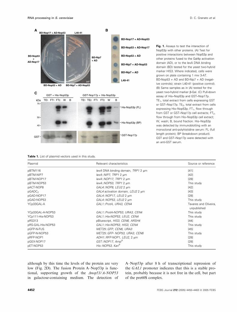
although by this time the levels of the protein are very
low (Fig. 2D). The fusion Protein A–Nop53p is func-
tional, supporting growth of the Dnop53 ⁄A-NOP53
in galactose-containing medium. The detection of
A–Nop53p after 8 h of transcriptional repression of
the GAL1 promoter indicates that this is a stable pro-
tein, probably because it is not free in the cell, but part
of the pre60S complex.
A B
BD-Nop53+
AD-Nop17
BD-Nop17 + AD-Nop53 L40-41
BD-Nip7 + AD
BD-Nip7 + AD-Nop53 BD-Nop53 + AD
3AT
BD-Nop17 + AD-Nop53
BD-Nip7 + AD-Nop53
BD-Nip7 + AD
BD-Nop53 + AD
BD-Nop53 + AD-Nop17
L40-41
CGST + His-Nop53p
FT2TE1 FT1 W B FT2TE1 FT1 W BTE2
GST-Nop17p + His-Nop53p
His-Nop53p (FL)
His-Nop53p (BP)
GST-Nop17pGST
40
50
75
kDa
Fig. 1. Assays to test the interaction of
Nop53p with other proteins. (A) Test for
positive interactions between Nop53p and
other proteins fused to the Gal4p activation
domain (AD), or to the lexA DNA binding
domain (BD) tested for the yeast two-hybrid
marker HIS3. Where indicated, cells were
grown on plate containing 1 mM 3-AT.
BD-Nop53 + AD and BD-Nip7 + AD (negat-
ive controls); strain L40-41 (positive control).
(B) Same samples as in (A) tested for the
yeast two-hybrid marker b-Gal. (C) Pull-down
assay of His–Nop53p and GST–Nop17p.
TE1, total extract from cells expressing GST
or GST–Nop17p; TE2, total extract from cells
expressing His–Nop53p; FT1, flow through
from GST or GST–Nop17p cell extracts; FT2,
flow through from His–Nop53p cell extract;
W, wash; B, bound fraction. His–Nop53p
was detected by immunoblotting with an
monoclonal anti-polyhistidine serum: FL (full
length protein); BP (breakdown product).
GST and GST–Nop17p were detected with
an anti-GST serum.
Table 1. List of plasmid vectors used in this study.
Plasmid Relevant characteristics Source or reference
pBTM116 lexA DNA binding domain, TRP1 2 lm [41]
pBTM-NIP7 lexA::NIP7, TRP1 2 lm [42]
pBTM-NOP17 lexA::NOP17, TRP1 2 lm [28]
pBTM-NOP53 lexA::NOP53, TRP1 2 lm This study
pACT-NOP8 GAL4::NOP8, LEU2 2 lm [42]
pGADC2 GAL4 activation domain, LEU2 2 lm [43]
pGAD-NOP17 GAL4::NOP17, LEU2 2 lm [28]
pGAD-NOP53 GAL4::NOP53, LEU2 2 lm This study
YCp33GAL-A GAL1::ProtA, URA3, CEN4 Tavares and Oliveira,
unpublished
YCp33GAL-A-NOP53 GAL1::ProtA-NOP53, URA3, CEN4 This study
YCp111-His-NOP53 GAL1::His-NOP53, LEU2, CEN4 This study
pRS313 pBluescript, HIS3, CEN6, ARSH4 [44]
pRS-GAL-His-NOP53 GAL1::His-NOP53, HIS3, CEN4 This study
pGFP-N-FUS MET25::GFP, CEN6, URA3 [45]
pGFP-N-NOP53 MET25::GFP- NOP53, URA3, CEN6 This study
pRFP-NOP1 ADH1::RFP-NOP1, LEU2, 2 lm [28]
pGEX-NOP17 GST::NOP17, AmpR [28]
pET-NOP53 His::NOP53, KanR This study
RNA processing in S. cerevisiae D. C. Granato et al.
4452 FEBS Journal 272 (2005) 4450–4463 ª 2005 FEBS
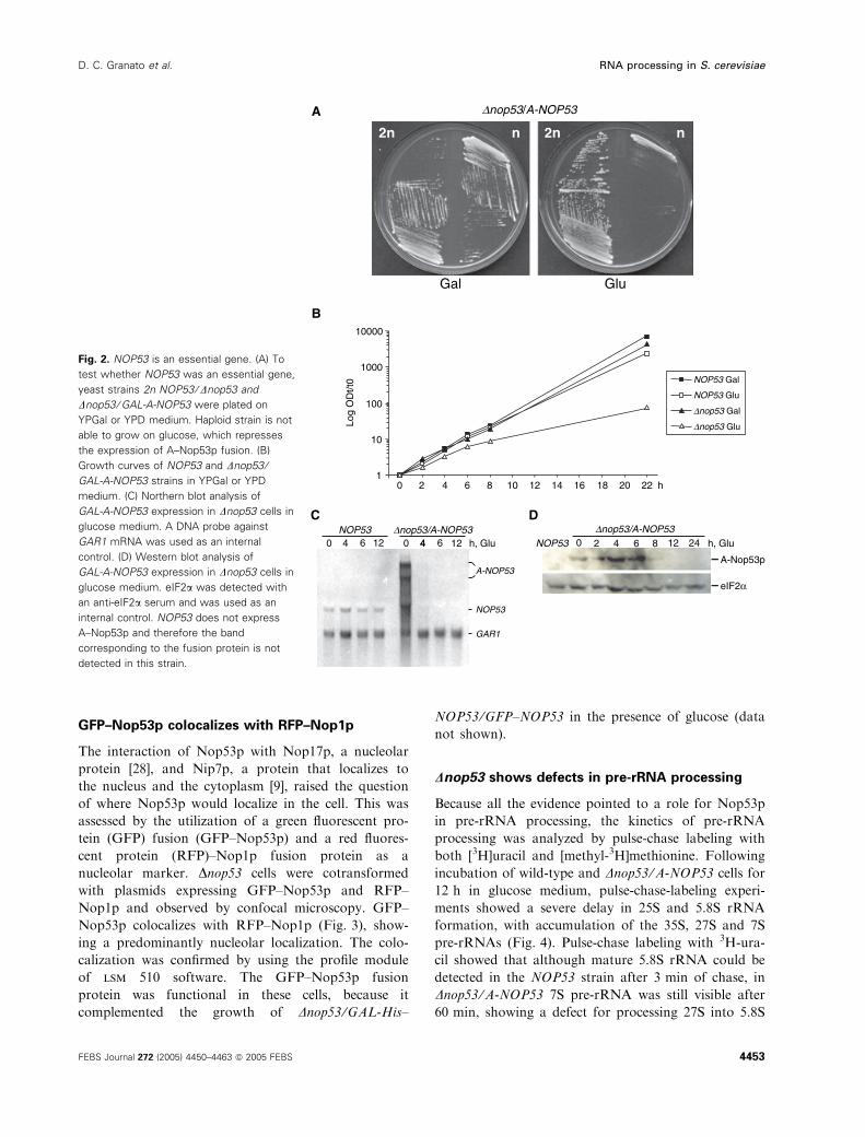
GFP–Nop53p colocalizes with RFP–Nop1p
The interaction of Nop53p with Nop17p, a nucleolar
protein [28], and Nip7p, a protein that localizes to
the nucleus and the cytoplasm [9], raised the question
of where Nop53p would localize in the cell. This was
assessed by the utilization of a green fluorescent pro-
tein (GFP) fusion (GFP–Nop53p) and a red fluores-
cent protein (RFP)–Nop1p fusion protein as a
nucleolar marker. Dnop53 cells were cotransformed
with plasmids expressing GFP–Nop53p and RFP–
Nop1p and observed by confocal microscopy. GFP–
Nop53p colocalizes with RFP–Nop1p (Fig. 3), show-
ing a predominantly nucleolar localization. The colo-
calization was confirmed by using the profile module
of lsm 510 software. The GFP–Nop53p fusion
protein was functional in these cells, because it
complemented the growth of Dnop53 ⁄GAL-His–
NOP53 ⁄GFP–NOP53 in the presence of glucose (data
not shown).
Dnop53 shows defects in pre-rRNA processing
Because all the evidence pointed to a role for Nop53p
in pre-rRNA processing, the kinetics of pre-rRNA
processing was analyzed by pulse-chase labeling with
both [3H]uracil and [methyl-3H]methionine. Following
incubation of wild-type and Dnop53 ⁄A-NOP53 cells for
12 h in glucose medium, pulse-chase-labeling experi-
ments showed a severe delay in 25S and 5.8S rRNA
formation, with accumulation of the 35S, 27S and 7S
pre-rRNAs (Fig. 4). Pulse-chase labeling with 3H-ura-
cil showed that although mature 5.8S rRNA could be
detected in the NOP53 strain after 3 min of chase, in
Dnop53 ⁄A-NOP53 7S pre-rRNA was still visible after
60 min, showing a defect for processing 27S into 5.8S
B
C D
2n n
Gal
2n n
Glu
Dnop53/A-NOP53A
1
10
100
1000
10000
1086420
0
12
8642 12 246 64 40 012
14 16 18 20 22 h
Log
OD
t/t0 NOP53 Gal
NOP53 Glu
Dnop53 Gal
Dnop53 Glu
h, GluNOP53Dnop53/A-NOP53
eIF2α
A-Nop53p
NOP53 Dnop53/A-NOP53
NOP53
A-NOP53
h, Glu
GAR1
124
Fig. 2. NOP53 is an essential gene. (A) To
test whether NOP53 was an essential gene,
yeast strains 2n NOP53 ⁄Dnop53 and
Dnop53 ⁄GAL-A-NOP53 were plated on
YPGal or YPD medium. Haploid strain is not
able to grow on glucose, which represses
the expression of A–Nop53p fusion. (B)
Growth curves of NOP53 and Dnop53 ⁄GAL-A-NOP53 strains in YPGal or YPD
medium. (C) Northern blot analysis of
GAL-A-NOP53 expression in Dnop53 cells in
glucose medium. A DNA probe against
GAR1 mRNA was used as an internal
control. (D) Western blot analysis of
GAL-A-NOP53 expression in Dnop53 cells in
glucose medium. eIF2a was detected with
an anti-eIF2a serum and was used as an
internal control. NOP53 does not express
A–Nop53p and therefore the band
corresponding to the fusion protein is not
detected in this strain.
D. C. Granato et al. RNA processing in S. cerevisiae
FEBS Journal 272 (2005) 4450–4463 ª 2005 FEBS 4453
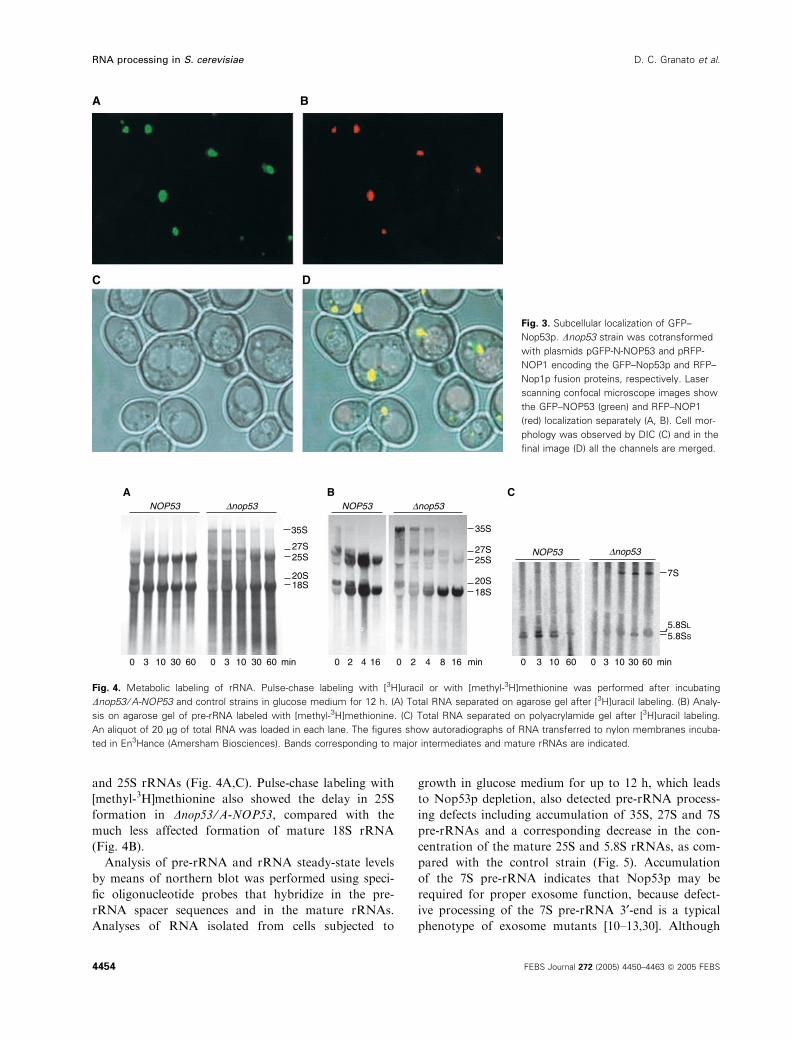
and 25S rRNAs (Fig. 4A,C). Pulse-chase labeling with
[methyl-3H]methionine also showed the delay in 25S
formation in Dnop53 ⁄A-NOP53, compared with the
much less affected formation of mature 18S rRNA
(Fig. 4B).
Analysis of pre-rRNA and rRNA steady-state levels
by means of northern blot was performed using speci-
fic oligonucleotide probes that hybridize in the pre-
rRNA spacer sequences and in the mature rRNAs.
Analyses of RNA isolated from cells subjected to
growth in glucose medium for up to 12 h, which leads
to Nop53p depletion, also detected pre-rRNA process-
ing defects including accumulation of 35S, 27S and 7S
pre-rRNAs and a corresponding decrease in the con-
centration of the mature 25S and 5.8S rRNAs, as com-
pared with the control strain (Fig. 5). Accumulation
of the 7S pre-rRNA indicates that Nop53p may be
required for proper exosome function, because defect-
ive processing of the 7S pre-rRNA 3¢-end is a typical
phenotype of exosome mutants [10–13,30]. Although
A B
C D
Fig. 3. Subcellular localization of GFP–
Nop53p. Dnop53 strain was cotransformed
with plasmids pGFP-N-NOP53 and pRFP-
NOP1 encoding the GFP–Nop53p and RFP–
Nop1p fusion proteins, respectively. Laser
scanning confocal microscope images show
the GFP–NOP53 (green) and RFP–NOP1
(red) localization separately (A, B). Cell mor-
phology was observed by DIC (C) and in the
final image (D) all the channels are merged.
A
0 2 4 16 0 2 4 8 16 min
25S
35S
27S
20S18S
NOP53 Dnop53B
3 10 30 60 0 3 10 30 60 min0
25S
35S
27S
20S18S
Dnop53NOP53
Dnop53
5.8SS
5.8SL
NOP53
7S
3 10 60 0 3 10 30 60 min0
C
Fig. 4. Metabolic labeling of rRNA. Pulse-chase labeling with [3H]uracil or with [methyl-3H]methionine was performed after incubating
Dnop53 ⁄A-NOP53 and control strains in glucose medium for 12 h. (A) Total RNA separated on agarose gel after [3H]uracil labeling. (B) Analy-
sis on agarose gel of pre-rRNA labeled with [methyl-3H]methionine. (C) Total RNA separated on polyacrylamide gel after [3H]uracil labeling.
An aliquot of 20 lg of total RNA was loaded in each lane. The figures show autoradiographs of RNA transferred to nylon membranes incuba-
ted in En3Hance (Amersham Biosciences). Bands corresponding to major intermediates and mature rRNAs are indicated.
RNA processing in S. cerevisiae D. C. Granato et al.
4454 FEBS Journal 272 (2005) 4450–4463 ª 2005 FEBS

the depletion of Nop53p does not seem to affect the
formation of 18S rRNA, an accumulation of 23S and
35S pre-rRNAs results in a slight decrease in the con-
centration of 18S rRNA (Fig. 5).
The lower concentrations of mature 25S and 5.8S
rRNAs detected by steady-state analysis are consistent
with the data obtained from the pulse-chase-labeling
experiments and indicate that Nop53p is involved in
the late steps of rRNA processing. To further investi-
gate the effects of Nop53p deficiency on pre-rRNA
cleavages we performed primer extension experiments
using primers that anneal in the regions of the mature
rRNAs close to the 5¢-end of those rRNAs. Extension
of the primer P2, that anneals to nucleotides 34–53
downstream of the 18S rRNA 5¢-end, showed that
depletion of Nop53p leads to shorter 18S rRNA at the
5¢-end (Fig. 6A). A similar decrease in the amount of
primer extension product is observed for the extension
reactions using primer P4 that anneals to nucleotides
42–64 downstream of the 5.8S rRNA 5¢-end (Fig. 6B).
Extension of primer P7 (complementary to nucleotides
80–105 downstream of 25S rRNA 5¢-end) also resulted
in a decrease of concentration of the band correspond-
ing to the 5¢-end of the 25S rRNA (Fig. 6C), although
in this case the effect of Nop53p depletion was not as
strong as observed for the 18S and 5.8S rRNAs. Con-
trol experiments were performed in parallel with total
RNA extracted from NOP53 cells. In these cells, the
primer extension products corresponded to the correct
5¢-ends of the rRNAs. Interestingly, when the same
experiments were performed with the mutant exosome
subunit strain rrp43-1 [13], the results were very similar
to those obtained from Dnop53 ⁄A-NOP53 cells
(Fig. 6). Therefore, the primer extension reactions with
total RNA from rrp43-1 cells growing under nonper-
missive conditions indicate that when the exosome is
P2 18S
NOP53 Dnop53
P1
23S
35S
P3 20S
P5 7S
P45.8S
7S
25SP7
P6 27S
12 h, Glu0 12 0 2 4 6 8
Actin
BA
35S
P4
DA2 A3
B1L/B1S EC2
C1
P3 P5
5´ETSA0
A1
18S 5.8S 25SITS1 3´ETSITS2
B2
P1P2 P7
A0/A1
Cleavage
20S 27S/A2
23S A2 Cleavage
27SBS/L
A3 Cleavage B2 Processing
B1 Processing
C1/ C2 Processing7SBS/L
Exosome
25S5.8S18S
32S
P6
Fig. 5. Northern blot analysis of pre-rRNA processing. (A) Total RNA was extracted from cells incubated in glucose medium for different time
intervals and hybridized against specific oligonucleotide probes. The relative positions of the probes on the 35S pre-rRNA are indicated in
(B). Bands corresponding to the major intermediates and to the mature rRNAs are indicated on the right-hand side. The lower panel shows a
northern blot detecting the actin mRNA, used as an internal control. (B) Structure of the 35S pre-rRNA and major intermediates of the rRNA
processing pathway in S. cerevisiae. The positions of the probes used for northern blot hybridizations are indicated below the 35S pre-rRNA.
Processing of 35S pre-rRNA starts with endonucleolytic cleavages at sites A0 and A1 in the 5¢-ETS, generating 32S pre-rRNA. The subse-
quent cleavage at site A2, in ITS1, generates the 20S and 27SA2 pre-rRNAs (dotted arrows indicate a possible pathway including the aber-
rant intermediate 23S). The 20S pre-rRNA is then processed at site D to the mature 18S rRNA. The major processing pathway of the 27SA2
pre-rRNA involves cleavage at site A3, producing 27SA3, which is digested quickly by exonucleases to generate the 27SBs (27SB short) pre-
rRNA. The subsequent processing step occurs at site B2, at the 3¢-end of the mature 25S rRNA. Processing at sites C1 and C2 separates
the mature 25S rRNA from the 7SS pre-rRNA. This pre-rRNA is subsequently processed exonucleolytically to generate the mature 5.8SS
rRNA. A fraction of the 27SA2 pre-rRNA is processed at the 5¢-end by a different mechanism and, following processing at the remaining
sites, gives rise to the 5.8SL (5.8S long) rRNA, which is 6–8 nucleotides longer than the 5.8SS rRNA at the 5¢-end.
D. C. Granato et al. RNA processing in S. cerevisiae
FEBS Journal 272 (2005) 4450–4463 ª 2005 FEBS 4455

not functional and rRNA processing is defective, pre-
cursor and intermediate rRNAs may undergo 5¢)3¢degradation. Interestingly, Dnop53 ⁄A-NOP53 cells
showed the same phenotype, indicating that Nop53p
affects exosome function.
Nop53p coprecipitates pre-rRNAs and binds 5.8S
rRNA
In order to find out whether Nop53p interacts with
pre-rRNAs, NOP53 strains expressing either Pro-
tein A or A–Nop53p fusion protein were constructed
to test coimmunoprecipitation of pre-rRNAs on IgG-
Sepharose affinity columns. The results obtained
showed that A–Nop53p coprecipitates the 27S and 7S
pre-rRNAs, and 5.8S mature rRNA (Fig. 7). A–
Nop53p also coprecipitated snR37, a box H ⁄ACA
snoRNA involved in pseudouridylation of the 25S
rRNA. A–Nop53p did not coprecipitate box C ⁄DsnoRNAs U3 and U14, involved in processing of 18S
rRNA (Fig. 7; data not shown). Compared with the
control Protein A, A–Nop53p coprecipitated 4.31-fold
more snR37, 4.67-fold more 5.8S, and 50-fold more
7S. These results indicate that Nop53p participates in
the pre60S complex, affecting the processing of the
27S and more strongly the processing of the 7S pre-
rRNA. Purified His–Nop53p was also tested for bind-
ing to in vitro transcribed 5.8S rRNA and the results
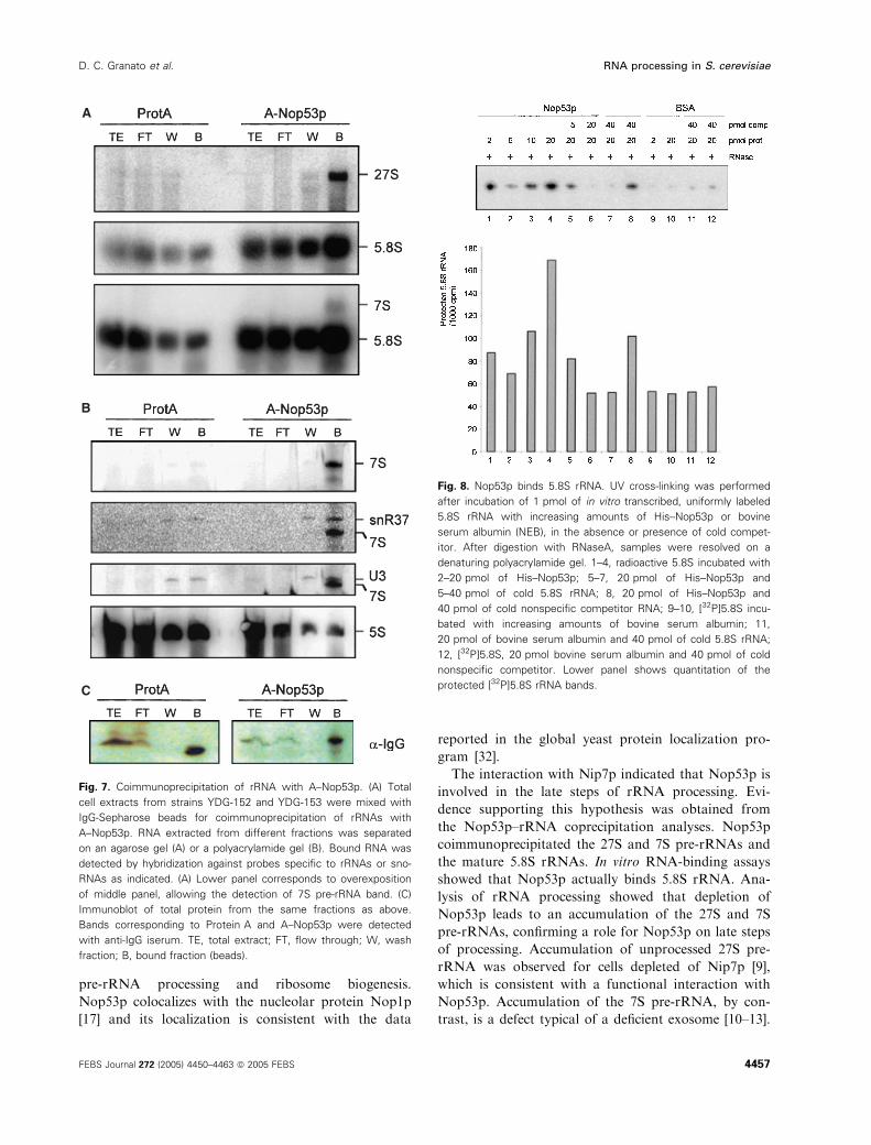
show that it binds directly to this RNA (Fig. 8).
These results support the hypothesis that Nop53p
depletion results in a defective function of the exo-
some.
Nop53p has a putative human homolog
Database searches were performed to identify possible
homologs of S. cerevisiae NOP53, and Nop53p was
found to be a conserved protein in eukaryotes, show-
ing a higher conservation in lower eukaryotes (Fig. 9).
Despite the fact that Nop53p binds RNA, no RNA
recognition motif was identified in its sequence. A
putative human ortholog (glioma tumor suppressor
candidate, Accession no. NP056525) shares 21% of
identity with its S. cerevisiae counterpart, but 41%
identity at the C-terminal region. Interestingly,
hNop53p was also localized to the nucleolus [31], sup-
porting the hypothesis of Nop53p having a conserved
function throughout evolution.
Discussion
Protein interaction studies have established a func-
tional link between several proteins involved in pre-
rRNA processing. The exosome subunit Rrp43p
interacts with Rrp46p, Nip7p and Nop17p [10,13,28].
Nop17p interacts with Nop58p and Nop53p [28]
(this study). The circle is closed by the interaction of
Nop53p and Nip7p, which was determined here. The
exosome subunits Rrp43p and Rrp46p and Nip7p
are found both in the nucleus and in the cyto-
plasm, whereas Nop58p, Nop17p and Nop53p are
restricted to the nuclear compartment, showing a
predominantly nucleolar localization [9,10,12,20,28].
The subcellular distribution and the interactions of
these proteins are consistent with their function in
A B C
Fig. 6. Analysis of pre-rRNA processing by primer extension. Total RNA was extracted from NOP53 and Dnop53 ⁄A-NOP53 cells growing in
glucose medium for different time intervals and used for primer extension experiments. RRP43 and rrp43-1 cells were incubated at 37 �Cfor the indicated periods prior to RNA extraction. Primer extension reactions were performed using oligonucleotides P2 (A), P4 (B) and P7 (C),
which are complementary to sequences downstream of the 5¢-end of the three mature rRNAs, 18S, 5.8S and 25S, respectively. Bands cor-
responding to mature 5¢-ends are indicated on the left-hand side. Arrows indicate main shorter primer extension products.
RNA processing in S. cerevisiae D. C. Granato et al.
4456 FEBS Journal 272 (2005) 4450–4463 ª 2005 FEBS
pre-rRNA processing and ribosome biogenesis.
Nop53p colocalizes with the nucleolar protein Nop1p
[17] and its localization is consistent with the data
reported in the global yeast protein localization pro-
gram [32].
The interaction with Nip7p indicated that Nop53p is
involved in the late steps of rRNA processing. Evi-
dence supporting this hypothesis was obtained from
the Nop53p–rRNA coprecipitation analyses. Nop53p
coimmunoprecipitated the 27S and 7S pre-rRNAs and
the mature 5.8S rRNAs. In vitro RNA-binding assays
showed that Nop53p actually binds 5.8S rRNA. Ana-
lysis of rRNA processing showed that depletion of
Nop53p leads to an accumulation of the 27S and 7S
pre-rRNAs, confirming a role for Nop53p on late steps
of processing. Accumulation of unprocessed 27S pre-
rRNA was observed for cells depleted of Nip7p [9],
which is consistent with a functional interaction with
Nop53p. Accumulation of the 7S pre-rRNA, by con-
trast, is a defect typical of a deficient exosome [10–13].
A
B
C
Fig. 7. Coimmunoprecipitation of rRNA with A–Nop53p. (A) Total
cell extracts from strains YDG-152 and YDG-153 were mixed with
IgG-Sepharose beads for coimmunoprecipitation of rRNAs with
A–Nop53p. RNA extracted from different fractions was separated
on an agarose gel (A) or a polyacrylamide gel (B). Bound RNA was
detected by hybridization against probes specific to rRNAs or sno-
RNAs as indicated. (A) Lower panel corresponds to overexposition
of middle panel, allowing the detection of 7S pre-rRNA band. (C)
Immunoblot of total protein from the same fractions as above.
Bands corresponding to Protein A and A–Nop53p were detected
with anti-IgG iserum. TE, total extract; FT, flow through; W, wash
fraction; B, bound fraction (beads).
Fig. 8. Nop53p binds 5.8S rRNA. UV cross-linking was performed
after incubation of 1 pmol of in vitro transcribed, uniformly labeled
5.8S rRNA with increasing amounts of His–Nop53p or bovine
serum albumin (NEB), in the absence or presence of cold compet-
itor. After digestion with RNaseA, samples were resolved on a
denaturing polyacrylamide gel. 1–4, radioactive 5.8S incubated with
2–20 pmol of His–Nop53p; 5–7, 20 pmol of His–Nop53p and
5–40 pmol of cold 5.8S rRNA; 8, 20 pmol of His–Nop53p and
40 pmol of cold nonspecific competitor RNA; 9–10, [32P]5.8S incu-
bated with increasing amounts of bovine serum albumin; 11,
20 pmol of bovine serum albumin and 40 pmol of cold 5.8S rRNA;
12, [32P]5.8S, 20 pmol bovine serum albumin and 40 pmol of cold
nonspecific competitor. Lower panel shows quantitation of the
protected [32P]5.8S rRNA bands.
D. C. Granato et al. RNA processing in S. cerevisiae
FEBS Journal 272 (2005) 4450–4463 ª 2005 FEBS 4457

Although Nop53p did not interact with any of the exo-
some subunits in the two-hybrid system (data not
shown), it might be connected to the exosome via
Nip7p. Similar to exosome mutants Dnop53 ⁄A-NOP53
strain showed higher levels of 7S pre-rRNA, indicating
a defective 3¢)5¢ exonucleolytic cleavage of this precur-
sor and therefore that the exosome is not fully active
in the absence of Nop53p. Interestingly, the accumu-
lated 7S pre-rRNA in cells depleted of Nop53p con-
tains aberrant 5¢-end, indicating that this pre-rRNA
is being degraded by a 5¢)3¢ exonuclease, probably
Rat1p or Xrn1p [33,34]. Rapid degradation of pre-
rRNAs has been reported for many strains with
defects in pre-rRNA processing [35–37]. The finding
that the depletion of Nop53p leads to the accumula-
tion of 7S pre-rRNA indicates that Nop53p could
mediate the signal for the processing of this pre-rRNA
to the exosome. Alternatively, the interaction of
Nop53p with Nip7p, that binds the exosome subunit
Rrp43p [10] could activate the exosome for processing
of the 7S pre-rRNA. However, since nip7 mutants do
not show accumulation of 7S pre-rRNA [9], the former
hypothesis seems more likely.
Nop53p also coprecipitated the box H ⁄ACA sno-
RNA snR37, but not box C ⁄D snoRNAs involved in
18S processing. This result raised the possibility that
Nop53p could participate in processing or assembly of
box H ⁄ACA snoRNPs. However, the deficiency of
Fig. 9. Multiple sequence alignment of
Nop53p. The full sequence of Nop53p and
its putative eukaryotic orthologs were
aligned. Numbers correspond to amino acid
position in each protein. Proteins access
numbers: C. glabrata, CAG62427; K. lac-
tis, XP_455604; E. gossypii, AAS51352;
S. pombe, CAB52719; Homo sapiens,
NP_056525; Mus musculus, AAH25810. *,
identity; :, strong similarity; ., weak similar-
ity. CLUSTALW was used for the sequence
alignment [50].
RNA processing in S. cerevisiae D. C. Granato et al.
4458 FEBS Journal 272 (2005) 4450–4463 ª 2005 FEBS

Nop53p did not affect box H ⁄ACA snoRNAs stability
(data not shown). It remains to be determined whether
Nop53p binds directly box H ⁄ACA snoRNAs, or whe-
ther snR37 coimmunoprecipitated as part of the
pre60S particle.
The data on the identification of Nop53p interaction
with Nop17p, a protein involved in the assembly
and ⁄or stabilization of box C ⁄D snoRNPs [28] indi-
cates that these interactions take place on the pre60S
particle. Interestingly, the modification of nucleotides
at the peptidyl transferase center has been reported to
occur late in processing, accounting for the copurifica-
tion of snoRNPs of box C ⁄D and H ⁄ACA with the
pre60S particles [7,27,38]. The interactions reported
here between Nop53p and Nop17p, and between
Nop53p and Nip7p could occur in the context of the
pre60S particles, which is formed by a different num-
ber of proteins associated with the 27S rRNA, depend-
ing on the phase of processing and transit from the
nucleolus to the cytoplasm.
In conclusion, the results obtained with the condi-
tional Dnop53 ⁄A-NOP53 strain showed that rRNA
processing is affected in the absence of Nop53p, lead-
ing to a reduction in rRNA synthesis and accumula-
tion of the pre-rRNAs 27S and 7S. The finding that
depletion of Nop53p affects more strongly the late
processing reactions responsible for the formation of
the mature 5.8S rRNA, indicates that this novel pro-
tein is important for proper exosome function.
During the final preparation of this article a study
was published on Nop53p [39]. In that study it is
reported that Nop53p is involved in the processing of
27S pre-rRNA, consistent with the data shown here.
However, contrary to our data, the authors found that
the depletion of Nop53p has stronger effects on the
maturation of the 25S rRNA, and not on the 5.8S.
Our data show that Nop53p coprecipitates the 27S
and 7S preRNAs and the mature 5.8S rRNA, binding
directly to the 5.8S rRNA region. These discrepancies
may be the result of the different strain background,
because Sydorskyy et al. [39] used their own deletion
strain, in which NOP53 was not essential, whereas the
strain we used was purchased from the yeast deletion
collection at Euroscarf.
Experimental procedures
DNA analyses and plasmid construction
DNA cloning and analyses were performed as described
elsewhere [40]. DNA was sequenced by using the Big Dye
method (Perkin-Elmer, USA). Plasmids used in this study
are summarized in Table 1, and cloning strategies are
briefly described below. The lexA::NOP53 fusion used in
the two-hybrid assay was constructed by inserting a 1.3 kb
BamHI ⁄SalI DNA fragment containing the PCR-amplified
NOP53 ORF into pBTM-116, which was previously diges-
ted with BamHI ⁄SalI restriction enzymes, generating the
plasmid pBTM-NOP53. Plasmid pACT-NOP53 (14–456,
numbers refer to Nop53p amino acid residues coded by this
cDNA clone) bears the gene encoding the hybrid protein of
the GAL4p activation domain and NOP53p. YCpGAL-A–
NOP53 was constructed by inserting the BamHI ⁄SalINOP53-containing fragment obtained from pBTM-NOP53
into Ycp33GALl-A vector previously digested with the
same restriction enzymes. Plasmid pGFP-N-NOP53 was
constructed by inserting the fragment XbaI ⁄SalI NOP53
obtained from the YCp111GAL-HIS–NOP53 vector diges-
ted with the same enzymes, into the pGFP-N-FUS vector
digested with SpeI ⁄XhoI restriction enzymes. pRS-GAL-
His–NOP53 was obtained by inserting the fragment (Bam-
HI ⁄SalI) containing NOP53 sequence and the fragment
(EcoRI ⁄BamHI) containing GAL1-HIS sequence into the
pRS313 vector digested with EcoRI and SalI. For the con-
struction of pET-NOP53, the PCR amplified NOP53 ORF
(BamHI ⁄SalI) was inserted into the pET-28a vector diges-
ted with BamHI and XhoI restriction enzymes.
Yeast transformation and maintenance
Yeast strains used in this work are listed in Table 2. Yeast
strains were maintained in yeast extract-peptone medium
(YP) or synthetic medium (YNB) as described previously
[47]. Glucose or galactose was added as carbon source to a
final concentration of 2% as indicated. Yeast cells were
transformed using the lithium acetate method as described
previously [47]. A Dnop53 strain was obtained from Euro-
scarf.
Yeast two-hybrid screen for proteins that interact
with Nop53p
The host strain for the two-hybrid screen, L40 [46], con-
tains both yeast HIS3 and E. coli lacZ genes as reporters
for two-hybrid interaction integrated into the genome.
Strain YDG146 is a derivative of L40, bearing plasmid
pBTM-NOP53, which encodes a hybrid protein containing
the lexA DNA binding domain and the full-length NOP53
ORF. Transformation of YDG146 was performed with
plasmid pGAD-NOP17 containing NOP17 ORF fused to
the GAL4 activation domain. Alternatively, L40 was trans-
formed with pBTM-NIP7 and pACT-NOP53. Transform-
ants were plated directly onto YNB medium lacking
histidine for immediate selection of Nop53p-interacting
proteins. His+ clones were tested for lacZ expression by
transferring cells to nitrocellulose filters and analyzing
b-galactosidase (b-Gal) activity [46]. b-Gal activity of
strains analyzed in two-hybrid experiments was quantitated
D. C. Granato et al. RNA processing in S. cerevisiae
FEBS Journal 272 (2005) 4450–4463 ª 2005 FEBS 4459

using cell extracts generated in buffer Z using ONPG as
substrate [41]. Strain L40-41 was used as a positive control
and strain YDG-146 ⁄ pGAD-C2 was used as negative con-
trol for two-hybrid interaction [42] (Table 2).
Protein pull-down and immunoblot analysis
Pull-down of His–Nop53p was assayed as follows: whole-
cell extracts from E. coli cells expressing either GST or
GST–Nop17p were generated in NaCl ⁄Pi buffer and mixed
with 500 lL of glutathione-Sepharose beads (Amersham
Biosciences). After washing bound material with NaCl ⁄Pi,
whole-cell extracts from E. coli cells expressing His–Nop53p
were added to the glutathione-Sepharose beads and incuba-
ted at 4 �C for 2 h. The glutathione-Sepharose beads were
precipitated and washed again with NaCl ⁄Pi and bound
proteins were eluted and resolved on SDS ⁄PAGE and
transferred to polyvinylidene difluoride membranes (Bio-
Rad Laboratories, Hercules, CA, USA), which were
incubated with an anti-(poly histidine) serum (Amersham
Biosciences) or with an anti-GST serum (Sigma, St. Louis,
MO, USA). The immunoblots were developed using the
ECL system (Amersham Biosciences).
RNA analysis
Exponentially growing cultures of yeast strains were shifted
from galactose to glucose medium. At various times, sam-
ples were collected and quickly frozen in a dry ice–ethanol
bath. Total RNA was isolated from yeast cells by a modi-
fied hot phenol method [48]. RNAs were separated by
electrophoresis on 1.3% agarose gels, following denatura-
tion with glyoxal [40] and transferred to Hybond nylon
membranes (Amersham Biosciences). Membranes were
probed with 32P-labeled oligonucleotides complementary
to specific regions of the 35S pre-rRNA (Table 3), or with32P-labeled DNA fragments corresponding to actin ORF,
using the hybridization conditions described previously [9]
and analyzed in a Phosphorimager (Molecular Dynamics,
Sunnyvale, CA, USA).
Metabolic labeling of rRNA
Metabolic labeling was performed as described previously
[9]. Exponentially growing cultures of strains NOP53 and
Dnop53 were incubated at 30 �C for 12 h in YNB–glucose
medium lacking methionine. Subsequently, cells were
pulse-labeled with 100 lCiÆmL)1 [methyl-3H]methionine
(Amersham Biosciences) for 2 min and chased with
100 lgÆmL)1 unlabeled methionine. At various times, sam-
ples were taken and quickly frozen in a dry ice–ethanol
bath. For metabolic labeling with [3H]uracil exponential
growing cultures of NOP53 and Dnop53 were shifted from
galactose to glucose medium and incubated for 12 h. Cells
were then pulse-labeled for 3 min at 37 �C with 50 lCiof [3H]uracil per mL and chased for up to 1 h after addit-
ion of unlabeled uracil to a final concentration of
300 lgÆmL)1. At various times samples were taken and
quickly frozen. Total RNA was isolated, separated by elec-
trophoresis and blotted as described above. Nylon mem-
branes were incubated in En3Hance (NEN) and submitted
to autoradiography.
Table 2. List of yeast strains used in this study.
Strain Relevant features
Source or
reference
L40 MATa his3d200 trp1–901 leu2–3311 ade2 lys2–801am [41]
URA3::(lexAop)8-lacZ LYS2::(lexAop)4-HIS3 [42]
L40-41 L40, pBTM-NIP7, pACT-NOP8 [28]
L40-61 L40, pBTM-NIP7, pACT-RRP43 This study
YFG-131 L40, pBTM-NOP17 [42]
YFG-247 L40, pBTM-NOP17, pACT-NOP53 [43]
YDG-146 L40, pBTM-NOP53 [28]
YDG-147 L40, pBTM-NOP53, pGAD-NOP17 This study
YDG-148 L40, pBTM-NIP7, pGAD-NOP53 This study
NOP53 MATa ⁄ a, his3D1 ⁄ his3D1 leu2D0 ⁄ leu2D0 lys2D0 ⁄ LYS2 ura3D0 ⁄ ura3D0 Euroscarf
MET15 ⁄met15D0 NOP53 ⁄NOP53
Dnop53 2n MATa ⁄ a, his3D1 ⁄ his3D1 leu2D0 leu2D0 ⁄ lys2D0 ⁄ LYS2 ura3D0 ⁄ ura3D0 Euroscarf
MET15 ⁄met15D0 NOP53 ⁄NOP53::KANR
Dnop53 MET15 his3D1 leu2D0 ura3D0 NOP53::KANR This study
YDG-149 Dnop53, pGFP-N-FUS, pRS-GAL-His-NOP53 This study
YDG-150 Dnop53, pGFP-N-FUS-NOP53 This study
YDG-151 Dnop53, YCp33GAL-A-NOP53 This study
YDG-152 NOP53, YCp33GAL-A This study
YDG-153 NOP53, YCp33GAL-A-NOP53 This study
RNA processing in S. cerevisiae D. C. Granato et al.
4460 FEBS Journal 272 (2005) 4450–4463 ª 2005 FEBS

Primer extension analysis
Total RNA extracted as described above was used for pri-
mer extension analysis. Reactions were performed by
annealing 1 pmol of [32P]-labeled oligonucleotide to 5 lg of
total RNA. Following annealing, extension was performed
with 100 U of MMLV reverse transcriptase (Invitrogen,
Carlsbad, CA, USA) and dNTPs (0.5 mm) for 30 min at
37 �C. cDNA products were precipitated, resuspended in
H2O, treated with RNase A, denatured and analyzed on
6% denaturing polyacrylamide gels. Gels were dried and
analyzed in a Phosphorimager. Oligonucleotides used in
primer extension analyses are listed in Table 3.
Coimmunoprecipitation of RNAs
Total cellular extracts were prepared from strains YDG152
and YDG153 expressing the ProtA or ProtA-Nop53p,
respectively, and added to IgG-Sepharose beads (Amer-
sham Biosciences) as described previously [49]. Immunopre-
cipitation was performed at 4 �C for 2 h. IgG-Sepharose
beads were washed with buffer A (20 mm Tris ⁄Cl pH 8,0,
0.5 mm magnesium acetate, 0.2% Triton X-100, 150 mm
potassium acetate, 1 mm dithiothrietol and protease inhibi-
tors) [49] and RNA was isolated from bound fractions by
adding phenol directly to the beads. After precipitation, the
recovered RNA was denatured and separated by electro-
phoresis on 6% polyacrylamide or 1.5% agarose gels and
transferred to nylon membranes. For comparison, 1% of
RNA recovered from total extract was loaded on gel.
Hybridization was performed as described above, using
probes specific to rRNAs and snoRNAs.
RNA binding assay
DNA fragment corresponding to 5.8S rRNA was cloned
into pGEM-T (Promega, Madison, WI, USA) vector and
in vitro transcription was performed with T7 RNA poly-
merase (Invitrogen), in the presence of 50 lCi of
[32P]UTP[aP]. One picomole of radiolabeled RNA was
incubated with different amounts of purified proteins in
the same buffer as used for coimmunoprecipitation of
RNAs [49] for 30 min at 37 �C. Cold competitor RNAs
were generated by parallel in vitro transcription of pGEM-
5.8S (generating 5.8S rRNA) or pBluescript (nonspecific
RNA) in the presence of 10 mm NTPs. UV cross-linking
was performed by placing RNA–protein complexes on ice
and irradiation for 15 min at 260 nm using a Fotodyne
transilluminator. They were then treated with 3 lg of
RNaseA for 30 min at 37 �C, resolved on a 6% denaturing
polyacrylamide gel and visualized on a Phosphorimager.
Subcellular localization of Nop53p
The subcellular localization of Nop53p was analyzed by
monitoring the fluorescence signal produced by a GFP
fusion to the N-terminal of Nop53p. The subcellular local-
ization of Nop1p was analyzed by monitoring the RFP,
which was fused to the N-terminus of this protein. GFP,
GFP–Nop53p and RFP–Nop1p proteins were expressed
from plasmids pGFP-N-FUS, pGFP-N-NOP53 and pRFP-
NOP1 (Table 1), respectively, transformed into the strain
Dnop53 (Table 2). Dnop53 cells were cotransformed with
vectors expressing GFP–Nop53p and RFP–Nop1p fusion
proteins. Living cells were immobilized on l-polylysine coa-
ted histological slides, in aqueous medium. The prepara-
tions were covered with cover slips, sealed and immediately
observed by confocal microscope. Ar (488 nm) and HeNe
(543 nm) lasers were used for image acquisition and the
confocal software used for image analysis.
Acknowledgements
We would like to thank the following people for their
support during the development of this work: Nilson
I.T. Zanchin for suggestions and critical reading of this
manuscript; Sandro R. Valentini for anti-GST serum;
Tereza C. Lima Silva and Zildene G. Correa for DNA
Table 3. DNA oligonucleotides used for northern blot hybridization and primer extension analyses.
Oligo Sequence Reference
P1 5¢-GGTCTCTCTGCTGCCGGAAATG-3¢ [9]
P2 5¢-CATGGCTTAATCTTTGAGAC-3¢ [8]
P3 5¢-GCTCTCATGCTCTTGCCAAAAC-3¢ [9]
P4 5¢-CGTATCGCATTTCGCTGCGTTC-3¢ [9]
P5 5¢-CTCACTACCAAACAGAATGTTTGAGAAGG-3¢ [13]
P6 5¢-GTTCGCCTAGACGCTCTCTTC-3¢ [9]
P7 5¢-GCCGCTTCACTCGCCGTTACTAAGGC-3¢ [28]
anti-U3 5¢-ATGGGGCTCATCAACCAAGTTGG-3¢ [49]
anti-U14 5¢-CTCAGACATCCTAGGAAGG-3¢ [28]
anti-snR11 5¢-GACGAATCGTGACTCTG-3¢ [20]
anti-snR37 5¢-GATAGTATTAACCACTACTG-3¢ [20]
D. C. Granato et al. RNA processing in S. cerevisiae
FEBS Journal 272 (2005) 4450–4463 ª 2005 FEBS 4461

sequencing; Celso R. Ramos for sequence alignment;
and Jose R. Tavares and Mauricio B. Goldfeder for
helping with yeast two-hybrid assays; Roberto Cabado
for confocal microscopy assistance. DCG, JSL and FC
were recipients of FAPESP fellowships, and FAG was
recipient of a CNPq fellowship. This work was suppor-
ted by FAPESP grant (03 ⁄ 06031-3 to CCO).
References
1 Venema J & Tollervey D (1995) Processing of pre-ribo-
somal RNA in Saccharomyces cerevisiae. Yeast 11,
1629–1650.
2 Kressler D, Linder P & Cruz J (1999) Protein trans-act-
ing factors involved in ribosome biogenesis in Saccharo-
myces cerevisiae. Mol Cell Biol 19, 7897–7912.
3 Grandi P, Rybin V, Baßler J, Petfalski E, Strauß D,
Marzioch M, Schafer T, Kuster B, Tschochner H,
Tollervey D et al. (2002) 90S pre-ribosomes include the
35S pre-rRNA, the U3 snoRNP, and 40S subunit
processing factors but predominantly lack 60S synthesis
factors. Mol Cell 10, 105–115.
4 Granneman S & Baserga SJ (2004) Ribosome biogen-
esis: of knobs and RNA processing. Exp Cell Res 296,
43–50.
5 Wehner KA, Gallagher JEG & Baserga SJ (2002) Com-
ponents of an interdependent unit within the SSU pro-
cessome regulate and mediate its activity. Mol Cell Biol
22, 7258–7267.
6 Baßler J, Grandi P, Gadal O, Leßmann T, Petfalski E,
Tollervey D, Lechner J & Hurt E (2001) Identification
of a 60S preribosomal particle that is closely linked to
nuclear export. Mol Cell 8, 517–529.
7 Nissan TA, Baßler J, Petfalski E, Tollervey D & Hurt E
(2002) 60S pre-ribosome formation viewed from assem-
bly in the nucleolus until export to the cytoplasm.
EMBO J 21, 5539–5547.
8 Fatica A, Cronshaw AD, Dlakiæ M & Tollervey D
(2002) Ssf1p prevents premature processing of an early
pre-60S ribosomal particle. Mol Cell 9, 341–351.
9 Zanchin NIT, Roberts P, DeSilva A, Sherman F &
Goldfarb DS (1997) Saccharomyces cerevisiae Nip7p is
required for efficient 60S ribosome subunit biogenesis.
Mol Cell Biol 17, 5001–5015.
10 Zanchin NIT & Goldfarb DS (1999) The exosome
subunit Rrp43p is required for the efficient maturation
of 5.8S, 18S and 25S rRNA. Nucleic Acids Res 27,
1283–1288.
11 Mitchell P, Petfalski E, Shevchenko A, Mann M &
Tollervey D (1997) The exosome: a conserved eukaryo-
tic RNA processing complex containing multiple 3¢-5¢exoribonucleases. Cell 91, 457–466.
12 Allmang C, Kufel J, Chanfreau G, Mitchell P, Petfalski
E & Tollervey D (1999) Functions of the exosome in
rRNA, snoRNA and snRNA synthesis. EMBO J 18,
5399–5410.
13 Oliveira CC, Gonzales FA & Zanchin NIT (2002) Tem-
perature-sensitive mutants of the exosome subunit
Rrp43p show a deficiency in mRNA degradation and
no longer interact with the exosome. Nucleic Acids Res
30, 4186–4198.
14 Maxwell ES & Fournier MJ (1995) The small nucleolar
RNAs. Annu Rev Biochem 35, 897–934.
15 Tollervey D & Kiss T (1997) Function and synthesis
of small nucleolar RNAs. Curr Opin Cell Biol 9, 337–
342.
16 Warner JR (2001) Nascent ribosomes. Cell 107, 133–
136.
17 Schimmang T, Tollervey D, Kern H, Frank R & Hurt
EC (1989) A yeast nucleolar protein related to mamma-
lian fibrillarin is associated with small nucleolar RNA
and is essential for viability. EMBO J 8, 4015–4124.
18 Bachellerie J-P & Cavaille J (1997) Guiding ribose
methylation of rRNA. Trends Biol Sci 22, 257–261.
19 Gautier T, Berges T, Tollervey D & Hurt E (1997)
Nucleolar KKE ⁄D repeat proteins Nop56p and Nop58p
interact with Nop1p and are required for ribosome bio-
genesis. Mol Cell Biol 17, 7088–7098.
20 Lafontaine DLJ & Tollervey D (1999) Nop58p is a
common component of the box C+D snoRNPs that is
required for snoRNA stability. RNA 5, 455–567.
21 Lafontaine DLJ & Tollervey D (2000) Synthesis and
assembly of the box C+D small nucleolar RNPs. Mol
Cell Biol 20, 2650–2659.
22 Filipowicz W & Pogacic V (2002) Biogenesis of small
nucleolar ribonucleoproteins. Curr Opin Cell Biol 14,
319–327.
23 Cahill NM, Friend K, Speckmann W, Li Z-H, Terns
RM, Terns MP & Steitz JA (2002) Site-specific cross-
linking analyses reveal an asymmetric protein distribu-
tion for a box C ⁄D snoRNP. EMBO J 21, 3816–3828.
24 Hong B, Wu K, Brockenbrough JS, Wu P & Aris JP
(2001) Temperature sensitive nop2 alleles defective in
synthesis of 25S rRNA and large ribosomal subunits in
Saccharomyces cerevisiae. Nucleic Acids Res 14, 2927–
2937.
25 Dragon F, Gallagher JE, Compagnone-Post PA,
Mitchell BM, Porwancher KA, Wehner KA, Wormsley S,
Settlage RE, Shabanowitz J, Osheim Y et al. (2002)
A large nucleolar U3 ribonucleoprotein required for 18S
ribosomal RNA biogenesis. Nature 417, 967–970.
26 Bonnerot C, Pintard L & Lutfalla G (2003) Functional
redundancy of Spb1p and a snR52-dependent mechan-
ism for the 2¢-O-ribose methylation of a conserved
rRNA position in yeast. Mol Cell 12, 1309–1315.
27 Dez C, Froment C, Noaillac-Depeyre J, Monsarrat B,
Caizergues-Ferrer M & Henry Y (2004) Npa1p, a
component of very early pre-60S ribosomal particles,
RNA processing in S. cerevisiae D. C. Granato et al.
4462 FEBS Journal 272 (2005) 4450–4463 ª 2005 FEBS

associates with a subset of small nucleolar RNPs
required for peptidyl transferase center modification.
Mol Cell Biol 24, 6324–6337.
28 Gonzales FA, Zanchin NIT, Luz JS & Oliveira CC
(2005) Characterization of Saccharomyces cerevisiae
Nop17p, a novel Nop58p-interacting protein that is
involved in pre-rRNA processing. J Mol Biol 346, 437–
455.
29 Rout MP, Aitchison JD, Suprapto A, Hjertaas K, Zhao
Y & Chait BT (2000) The yeast nuclear pore complex:
composition, architecture, and transport mechanism.
J Cell Biol 148, 635–651.
30 Mitchell P, Petfalski E & Tollervey D (1996) The 3¢ endof yeast 5.8S rRNA is generated by an exonuclease pro-
cessing mechanism. Genes Dev 10, 501–513.
31 Andersen JS, Lam YW, Leung AKL, Ong S-E, Lyon
CE, Lamond AI & Mann M (2005) Nucleolar proteome
dynamics. Nature 433, 77–83.
32 Huh W-K, Falvo JV, Gerke LC, Caroll AS, Howson
RW, Weissman JS & O’Shea EK (2003) Global analysis
of protein localization in budding yeast. Nature 425,
686–691.
33 Henry Y, Wood H, Morrisey JP, Petfalski E, Kearsey S
& Tollervey D (1994) The 5¢ end of yeast 5.8S rRNA is
generated by exonucleases from an upstream cleavage
site. EMBO J 13, 2452–2463.
34 Geerlings TH, Vos JC & Raue HA (2000) The final step
in the formation of 25S rRNA in Saccharomyces cerevi-
siae is performed by 5¢-3¢ exonucleases. RNA 6, 1698–
1703.
35 Venema J & Tollervey D (1999) Ribosome synthesis in
Saccharomyces cerevisiae. Annu Rev Genet 33, 261–311.
36 Allmang C, Mitchell P, Petfalski E & Tollervey D
(2000) Degradation of ribosomal RNA precursors by
the exosome. Nucleic Acids Res 28, 1684–1691.
37 Kufel J, Allmang C, Petfalski E, Beggs J & Tollervey D
(2003) Lsm proteins are required for normal processing
and stability of ribosomal RNAs. J Biol Chem 278,
2147–2156.
38 Lapeyre B & Purushothaman SK (2004) Spb1p-direc-
ted formation of Gm2922 in the ribosome catalytic
center occurs at a late processing stage. Mol Cell 16,
663–669.
39 Sydorskyy Y, Dilworth DJ, Halloran B, Yi EC,
Makhnevych T, Wozniak RW & Aitchison JD (2005)
Nop53p is a novel nucleolar 60S ribosomal subunit
biogenesis protein. Biochem J in press.
40 Sambrook J, Maniatis T & Fritsch EF (1989) Molecular
Cloning: A Laboratory Manual. Cold Spring Harbor
Laboratory Press, Cold Spring Harbor, NY.
41 Bartel PL & Fields S (1995) Analyzing protein–protein
interactions using two-hybrid system. Methods Enzymol
254, 241–263.
42 Zanchin NIT & Goldfarb DS (1999) Nip7p interacts
with Nop8p, an essential nucleolar protein required for
60S ribosome biogenesis, and the exosome subunit
Rrp43p. Mol Cell Biol 19, 1518–1525.
43 James P, Halladay J & Craig EA (1996) Genomic
libraries and a host strain designed for highly efficient
two-hybrid selection in yeast. Genetics 144, 1425–1436.
44 Sikorski RS & Hieter P (1989) A system of shuttle vec-
tors and yeast host strains designed for efficient manipu-
lation of DNA in Saccharomyces cerevisae. Genetics
122, 19–27.
45 Niedenthal RK, Riles L, Johnston M & Hegemann JH
(1996) Green fluorescent protein as a marker for gene
expression and subcellular localization in budding yeast.
Yeast 12, 773–786.
46 Vojtek AB & Hollenberg SM (1995) Ras–Raf interac-
tion: two · hybrid analysis. Methods Enzymol 255, 331–
342.
47 Sherman F, Fink GR & Hicks JB (1986) Laboratory
Course Manual for Methods in Yeast Genetics. Cold
Spring Harbor Laboratory Press, Cold Spring Harbor,
NY.
48 Dez C, Noaillac-Depeyre J, Caizergues-Ferrer M &
Henry Y (2002) Naf1p, an essential nucleoplasmic fac-
tor specifically required for accumulation of box
H ⁄ACA small nucleolar RNPs. Mol Cell Biol 22, 7053–
7065.
49 Oliveira CC & McCarthy JEG (1995) The relationship
between eukaryotic translation and mRNA stability. A
short upstream open reading frame strongly inhibits
translational initiation and greatly accelerates mRNA
degradation in the yeast Saccharomyces cerevisiae.
J Biol Chem 270, 8936–8943.
50 Altschul SF, Madden TL, Schaffer AA, Zhang J, Zhang
Z, Miller W & Lipman DJ (1997) Gapped BLAST and
PSI-BLAST: a new generation of protein database
search programs. Nucleic Acids Res 25, 3389–3402.
D. C. Granato et al. RNA processing in S. cerevisiae
FEBS Journal 272 (2005) 4450–4463 ª 2005 FEBS 4463
Copyright © 2022 FDOKUMEN