CHE 320: Organic Spectroscopy NMR Lecture Notes 2 Introduction to NMR
SOLID STATE NMR EVALUTION OF NATURAL RESIN/CLAY
NANOCOMPOSITES
Emerson O. da Silva1, Maria Inês B. Tavares1*, José S. Nogueira2
1* Instituto de Macromoléculas Professora Eloisa Mano/ UFRJ, Centro de
Tecnologia, Bloco J, Ilha do Fundão. PO Box: 68525, 21945-970, Rio de
Janeiro, RJ, Brazil,
[email protected], [email protected].
2DF/ICET/UFMT, Cuiabá, Mato Grosso, Brazil, [email protected]
Abstract
NMR nuclear relaxation times have been used by Tavares et al as a
methodology to characterize the nano materials, especially nanocomposites,
because NMR offers a great variety of relaxation parameters. The spin-lattice
relaxation time, with a time constant T1, have been explored to get as much
information as possible from the measurements of the spin-lattice proton
relaxation (T1H), which can measure the fraction of available polymer/clay
interface as well as the dispersion homogeneity of those interfaces actually
formed. The spin-lattice has been evaluated since this relaxation time confirms
the T1 and can give additional information to nanocomposite clay dispersion.
The NMR relaxation times are sensitive to the chemical environmental, changes
in the polymer matrix; chemical structure and interaction process, because they
depend on the domain distribution and sample homogeneity, since they are
measured in the solid state via intermolecular chains interaction and/or spin
diffusion. The T1 relaxation time of the nanocomposite decreased very much in
relation to the natural resin, according to the increase in the exfoliation clay
process, forming a nanocomposite with polymer matrix around the clay lamella.
Keywords: natural resin, NMR, Nanocomposite
Introduction
Nanotechnology permits obtaining very good and specific materials for
different areas. Generally, nanocomposites have their mechanical and thermal
properties improved comparing to virgin polymers. Modified clay produce
hierarchically ordered hybrid organic-inorganic nanocomposites. The
nanocomposites can be prepared by bulk polymerization; solution
polymerization; solution mixing and melt process. To prepare nanocomposites
some questions have to be answered: 1) to understand how the structure of the
clay relates to the structure of the nanocomposite; 2) to determine if there is a
relationship between the preparative method and the final structure of the
material, i.e., intercalated or exfoliated; and 3) to understand the pathway by
which nanocomposites enhance the thermal and fire stability of polymers, and
other properties. An enormous quantity of synthetic polymer nanocomposites
has been investigated, focusing different visions and necessity to obtain a good
nanocomposite material [1-4], because they often exhibit remarkable
improvement in materials properties when compared with virgin polymer or
conventional micro and macro composite. However, there are few works about
the nanocomposites based on natural polymeric matrix. As the synthetic
polymers are from non-renewable source, it is of strategic importance to study
natural polymer material as one of the possible substitute for some synthetic
polymers. Natural polymers can be extracted from trees in the form of
exsudates and they are called natural resins. Thus, the natural resins have very
good adhesion properties and they are normally used in composition of
varnishes, however, it can be used in different materials, for instance,
nanocomposites. The natural resin obtained from Amescla tree, which is the
subject of this study, which is a natural resin that is mainly constituted by
polyterpenoid compounds, and because of its particularity it is a good matrix to
prepare nanocomposite with organoclay.
Nanocomposites are basically characterized by X-ray diffraction; thermal
properties such as glass transition and thermal stability; electronic transmission
microscopy (TEM) [1-4] and mechanical properties. Therefore, other techniques
must be used to support and understand the nanostructured material. In this
case nuclear magnetic resonance (NMR) via nuclear relaxation times has been
used by Tavares et al [5-9], as a methodology to characterize the
nanomaterials, especially nanocomposites, because NMR offers a great variety
of relaxation parameters. The spin-lattice relaxation time have been explored to
get as much information as possible from the measurements of the spin-lattice
proton relaxation, which can measure the fraction of available polymer/clay
interface as well as the dispersion homogeneity of the those interfaces actually
formed [5-14].
According to the scope, this work evaluated an innovative methodology,
using solid state NMR to characterize the dispersion and the nanostructure of
the hybrid polymer nanocomposite. The NMR relaxation times are sensitive to
the chemical environmental; changes in the polymer matrix structure and
interaction process, because they depend on the domain distribution and
sample homogeneity, since they are measured in solid state via intermolecular
chains interaction and/or spin diffusion. Thus, the purpose of this work is the
preparation of a nanocomposite of NaRA and montmorillonite with and without
organic treatment and the characterization of formed nanocomposites, using X-
ray diffraction and nuclear magnetic resonance [15-17]. The NMR responses
were obtained through the determination of proton spin-lattice relaxation time.
T1H relaxation time was measured for each sample and they were interpreted in
terms of clay layer dispersion; homogeneity and interaction between clay layer
and resin polymer matrix.
Materials and Methods
Samples
Natural resin from Amescla tree was received from Universidade Federal
de Mato Grosso, collected in Sinop city, MT, Brazil. The pondered molar mass
was 56.000. The NaRA was purified by extraction by ethanol and precipitation
in water.
The nanoparticle used were nanoclay viscogel B7 (MMT), which was
treated with alquil ammonium salt with 12 atoms of carbon – C-12, and
homoinioc clay (MT), which were kindly supplied from Bentec.
Nanocomposite Preparation
Separated dispersions of NaRA and MMT or MT in chloroform were
prepared and stirred for 6 hours. The amounts of NaRA and MMT were
calculated to obtain a nanocomposite 5% (w/w). Then, the MMT suspension
was slowly added to NaRA solution and stirred for 3 days. Thus, this
nanocomposite was obtained by solution induced intercalation method.
Materials Characterization
The characterization methods were performed on XRD diffraction and
low field NMR, through the measurements of spin-lattice relaxation time.
X-Ray Diffraction
The extent of clay intercalation and / or exfoliation was determinate by X-
Ray Diffraction (XRD) analysis. The films were characterized using an X-ray
diffractometer, XRD Rigaku Miniflex, with CuK ( = 1,5418 Å) radiation
operated at 30 KV and 15 mA. The data were recorded at 2 rates of 2° per
minute. The basal spacing of nanocomposite was calculated by through the
Bragg’s relation: = 2 d sin.
Low Field NMR
Low field NMR MARAN ultra 23 spectrometer, operating at 23 MHz (for
protons), and equipped with an 18 mm variable temperature probe, was used
for the determination of relaxation measurements. Proton spin-lattice relaxation
times (T1H) were determined directly by the traditional inversion recovery pulse
sequence (180°- - 90°) the 90° pulse of 4.6µs was calibrated automatically by
the instrument software. The amplitude of the FID was sampled for twenty
data points, ranging from 0.1 to 5000 ms, with 4 scans for each point and 5s of
recycle delay. The relaxation values and relative intensities were obtained by
fitting the exponential data with the aid of the program WINFIT.
Results and Discussions
Figure 1 shows the X-ray pattern of the organofilic clay; natural resin and
its nanocomposite.
Figure 1 - X-ray diffraction of organofilic clay; natural resin and nano2
The nano2 exhibited no diffraction peaks in the 2 α range of 2-8 degree.
The absence of a Bragg scattering peak seems to indicate that the clay tactoids
were intercalated and/or exfoliated by the solution intercalated process.
However, the MMT can only delaminate into much smaller stacks, containing far
fewer silicate layers. The decrease and polydispersion of the clay layer number
NaRA
MMT
NANO2
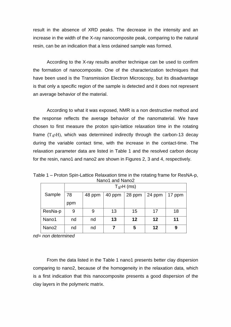
result in the absence of XRD peaks. The decrease in the intensity and an
increase in the width of the X-ray nanocomposite peak, comparing to the natural
resin, can be an indication that a less ordained sample was formed.
According to the X-ray results another technique can be used to confirm
the formation of nanocomposite. One of the characterization techniques that
have been used is the Transmission Electron Microscopy, but its disadvantage
is that only a specific region of the sample is detected and it does not represent
an average behavior of the material.
According to what it was exposed, NMR is a non destructive method and
the response reflects the average behavior of the nanomaterial. We have
chosen to first measure the proton spin-lattice relaxation time in the rotating
frame (T1H), which was determined indirectly through the carbon-13 decay
during the variable contact time, with the increase in the contact-time. The
relaxation parameter data are listed in Table 1 and the resolved carbon decay
for the resin, nano1 and nano2 are shown in Figures 2, 3 and 4, respectively.
Table 1 – Proton Spin-Lattice Relaxation time in the rotating frame for ResNA-p, Nano1 and Nano2
Sample
T1H (ms)
78
ppm
48 ppm 40 ppm 28 ppm 24 ppm 17 ppm
ResNa-p 9 9 13 15 17 18
Nano1 nd nd 13 12 12 11
Nano2 nd nd 7 5 12 9
nd= non determined
From the data listed in the Table 1 nano1 presents better clay dispersion
comparing to nano2, because of the homogeneity in the relaxation data, which
is a first indication that this nanocomposite presents a good dispersion of the
clay layers in the polymeric matrix.
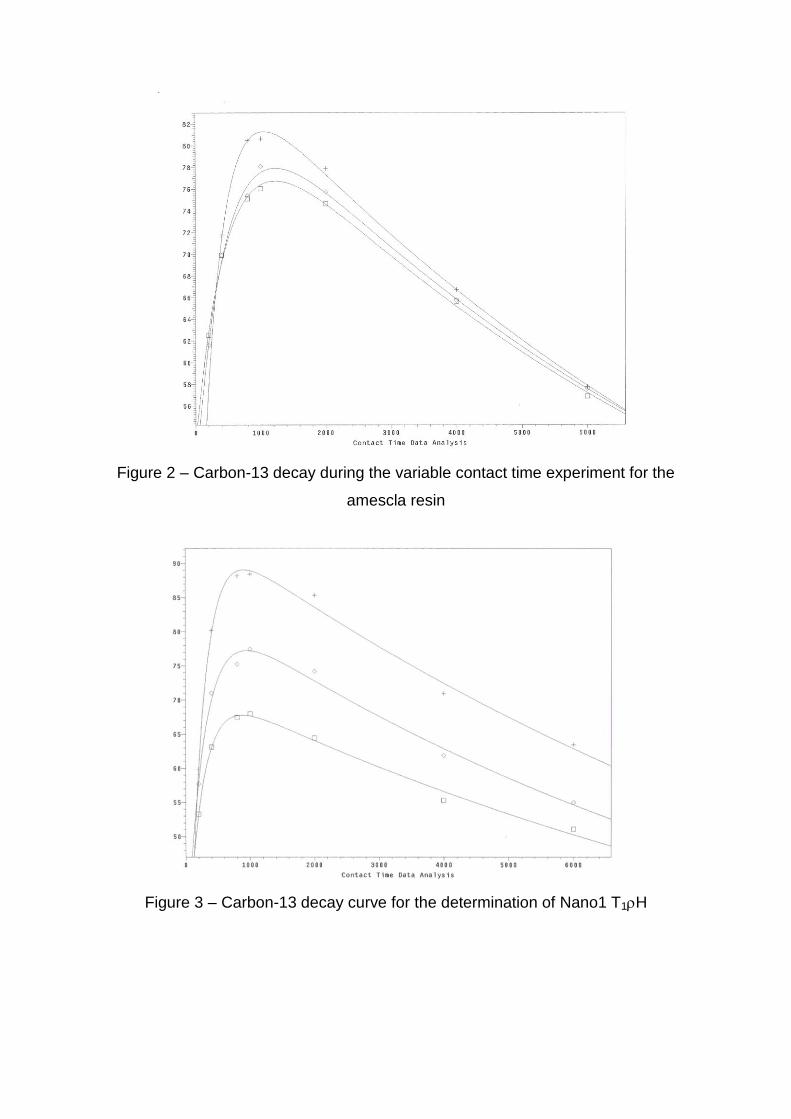
Figure 2 – Carbon-13 decay during the variable contact time experiment for the
amescla resin
Figure 3 – Carbon-13 decay curve for the determination of Nano1 T1H
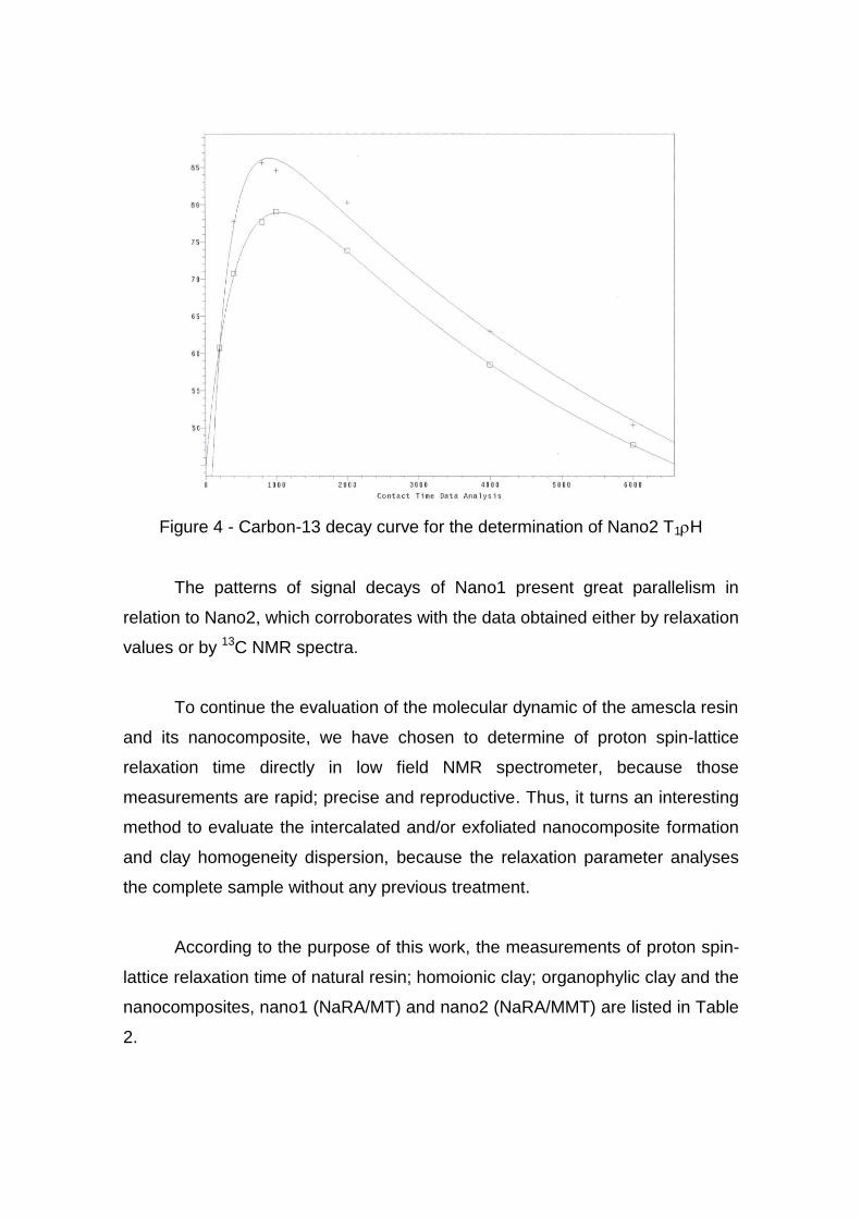
Figure 4 - Carbon-13 decay curve for the determination of Nano2 T1H
The patterns of signal decays of Nano1 present great parallelism in
relation to Nano2, which corroborates with the data obtained either by relaxation
values or by 13C NMR spectra.
To continue the evaluation of the molecular dynamic of the amescla resin
and its nanocomposite, we have chosen to determine of proton spin-lattice
relaxation time directly in low field NMR spectrometer, because those
measurements are rapid; precise and reproductive. Thus, it turns an interesting
method to evaluate the intercalated and/or exfoliated nanocomposite formation
and clay homogeneity dispersion, because the relaxation parameter analyses
the complete sample without any previous treatment.
According to the purpose of this work, the measurements of proton spin-
lattice relaxation time of natural resin; homoionic clay; organophylic clay and the
nanocomposites, nano1 (NaRA/MT) and nano2 (NaRA/MMT) are listed in Table
2.
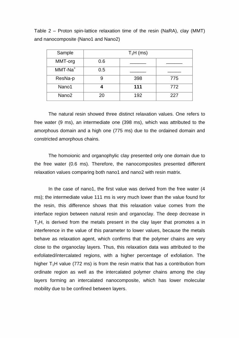
Table 2 – Proton spin-lattice relaxation time of the resin (NaRA), clay (MMT)
and nanocomposite (Nano1 and Nano2)
The natural resin showed three distinct relaxation values. One refers to
free water (9 ms), an intermediate one (398 ms), which was attributed to the
amorphous domain and a high one (775 ms) due to the ordained domain and
constricted amorphous chains.
The homoionic and organophylic clay presented only one domain due to
the free water (0.6 ms). Therefore, the nanocomposites presented different
relaxation values comparing both nano1 and nano2 with resin matrix.
In the case of nano1, the first value was derived from the free water (4
ms); the intermediate value 111 ms is very much lower than the value found for
the resin, this difference shows that this relaxation value comes from the
interface region between natural resin and organoclay. The deep decrease in
T1H, is derived from the metals present in the clay layer that promotes a in
interference in the value of this parameter to lower values, because the metals
behave as relaxation agent, which confirms that the polymer chains are very
close to the organoclay layers. Thus, this relaxation data was attributed to the
exfoliated/intercalated regions, with a higher percentage of exfoliation. The
higher T1H value (772 ms) is from the resin matrix that has a contribution from
ordinate region as well as the intercalated polymer chains among the clay
layers forming an intercalated nanocomposite, which has lower molecular
mobility due to be confined between layers.
Sample T1H (ms)
MMT-org 0.6 ______ ______
MMT-Na+ 0.5 ______ _____
ResNa-p 9 398 775
Nano1 4 111 772
Nano2 20 192 227
For the nano2, three values of relaxation parameter were also detected
and the low T1H value and the intermediate T1H value are higher than the
values compared for nano1. Moreover, the higher T1H value (227) for nano2 is
lower comparing to the higher T1H value determined for both natural resin and
nano1, which is due to the Na+ present in the clay structure that promotes the
decrease in the relaxation value, acting as a relaxation agent. But, some
intercalated and exfoliated nanocomposites can be found in nano2.
This affirmation can be supported by the fact that, if the nanocomposite
was only intercalated the relaxation value would increase due to the formation
of constricted polymer domains inside the clay layers. The exfoliation process
was facilitated because the natural resin presents chains with different sizes,
which can promote a plasticization effect promoting an exfoliation process. The
higher relaxation value does not change, because it belongs to the resin matrix
not affected by the organoclay presence.
Both nanocomposites showed to have good clay layer dispersion in the
resin matrix, but according to the relaxation values nano1 seems to be
homogeneous than nano2.
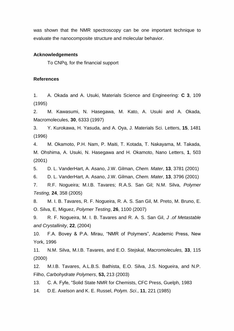
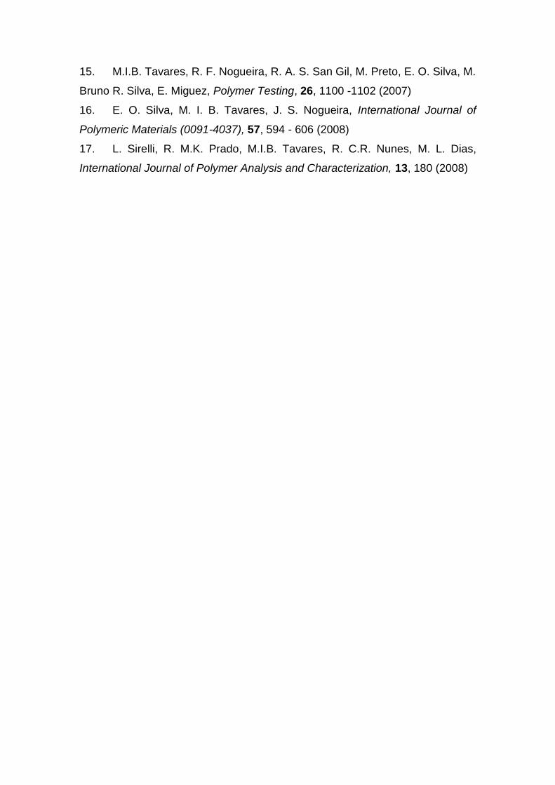
The clay structure was evaluated by the silicon-29 NMR spectrum, which
permits to evaluate changes in the clay structure due to proximity of polymer
chains. The silicon-29 spectra of clay, viscogel B7 and both nano1 and nano2
are showed in Figures 5, 6, 7 and 8, respectively. Comparing the Si-29 MAS
NMR spectra, the nanocomposites showed changes in the NMR signal form
and also a displacement in the chemical shift values. The nano1 and nano2 Si-
29 NMR spectra presented a pattern different than viscogel B7 and clay,
respectively, which can be due to the random clay layer dispersion in the
polymeric matrix, because of the break in the clay layer ordination. Polymer
chains could promote a clay layer intercalation and/or exfoliation. Confirming
the results obtained by the other techniques.
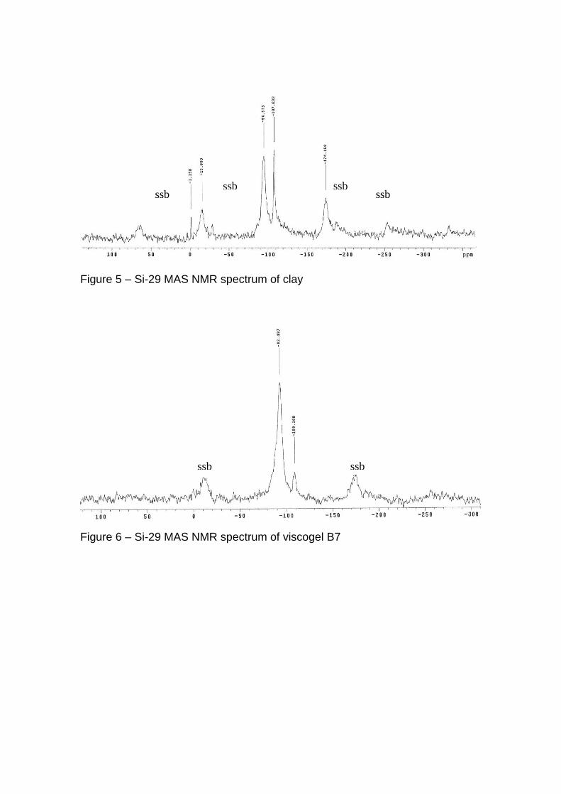
Figure 5 – Si-29 MAS NMR spectrum of clay
Figure 6 – Si-29 MAS NMR spectrum of viscogel B7
ssb ssb ssb ssb
ssb ssb
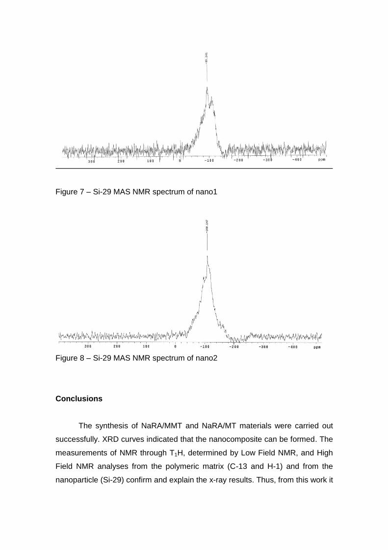
Figure 7 – Si-29 MAS NMR spectrum of nano1
Figure 8 – Si-29 MAS NMR spectrum of nano2
Conclusions
The synthesis of NaRA/MMT and NaRA/MT materials were carried out
successfully. XRD curves indicated that the nanocomposite can be formed. The
measurements of NMR through T1H, determined by Low Field NMR, and High
Field NMR analyses from the polymeric matrix (C-13 and H-1) and from the
nanoparticle (Si-29) confirm and explain the x-ray results. Thus, from this work it
was shown that the NMR spectroscopy can be one important technique to
evaluate the nanocomposite structure and molecular behavior.
Acknowledgements
To CNPq, for the financial support
References
1. A. Okada and A. Usuki, Materials Science and Engineering: C 3, 109
(1995)
2. M. Kawasumi, N. Hasegawa, M. Kato, A. Usuki and A. Okada,
Macromolecules, 30, 6333 (1997)
3. Y. Kurokawa, H. Yasuda, and A. Oya, J. Materials Sci. Letters, 15, 1481
(1996)
4. M. Okamoto, P.H. Nam, P. Maiti, T. Kotada, T. Nakayama, M. Takada,
M. Ohshima, A. Usuki, N. Hasegawa and H. Okamoto, Nano Letters, 1, 503
(2001)
5. D. L. VanderHart, A. Asano, J.W. Gilman, Chem. Mater, 13, 3781 (2001)
6. D. L. VanderHart, A. Asano, J.W. Gilman, Chem. Mater, 13, 3796 (2001)
7. R.F. Nogueira; M.I.B. Tavares; R.A.S. San Gil; N.M. Silva, Polymer
Testing, 24, 358 (2005)
8. M. I. B. Tavares, R. F. Nogueira, R. A. S. San Gil, M. Preto, M. Bruno, E.
O. Silva, E, Miguez, Polymer Testing, 26, 1100 (2007)
9. R. F. Nogueira, M. I. B. Tavares and R. A. S. San Gil, J .of Metastable
and Crystallinity, 22, (2004)
10. F.A. Bovey & P.A. Mirau, “NMR of Polymers”, Academic Press, New
York, 1996
11. N.M. Silva, M.I.B. Tavares, and E.O. Stejskal, Macromolecules, 33, 115
(2000)
12. M.I.B. Tavares, A.L.B.S. Bathista, E.O. Silva, J.S. Nogueira, and N.P.
Filho, Carbohydrate Polymers, 53, 213 (2003)
13. C. A. Fyfe, “Solid State NMR for Chemists, CFC Press, Guelph, 1983
14. D.E. Axelson and K. E. Russel, Polym. Sci., 11, 221 (1985)
15. M.I.B. Tavares, R. F. Nogueira, R. A. S. San Gil, M. Preto, E. O. Silva, M.
Bruno R. Silva, E. Miguez, Polymer Testing, 26, 1100 -1102 (2007)
16. E. O. Silva, M. I. B. Tavares, J. S. Nogueira, International Journal of
Polymeric Materials (0091-4037), 57, 594 - 606 (2008)
17. L. Sirelli, R. M.K. Prado, M.I.B. Tavares, R. C.R. Nunes, M. L. Dias,
International Journal of Polymer Analysis and Characterization, 13, 180 (2008)
Copyright © 2022 FDOKUMEN