
SMASH 2017 NMR Conference
191
Conference Program September 17th-20th 2017 Baveno, Italy
-
Upload
khangminh22 -
Category
Documents
-
view
2 -
download
0
Transcript of SMASH 2017 NMR Conference

SMASH 2017 NMR Conference
Dear SMASH 2017 attendees,
We are pleased to extend a warm welcome to the 2017 Small Molecules Are Still Hot NMR Conference. Once again, we find ourselves in the delightful setting of Baveno, on the shores of Lake Maggiore. Nestling in the foothills of the Alps, surrounded by natural wonders, it’s hard to believe we are only a short drive from Milan, one of Italy’s most important and vibrant cities. For those of you that attended the previous meeting here, we are certain you will agree with the above sentiments, for those who didn’t, we are sure you will appreciate this wonderful venue.
Of course, the attractive location for SMASH would be nothing if the scientific program was not strong and relevant to you; we think that we have put together an interesting and varied program that will be exactly that.
The formal program for SMASH begins on Sunday evening, with registration, a mixer and dinner (though we are sure many of you will have attended the various user meetings being held in the daytime of Sunday). This will be the perfect opportunity to catch up with old colleagues and make new acquaintances. The scientific sessions begin on Monday morning, and we have seven varied and exciting oral sessions covering a broad range of topics relevant to small molecules – from structural elucidation to solid state NMR, from reaction monitoring to exotic nuclei, from binding interactions to metabolomics (all tunefully named, as you may have noted). There is also a session which will revisit some of the ‘hot’ topics from previous SMASH meetings. As for previous meetings, each oral session also includes one short talk which is an ‘upgrade’ of a poster. We will also have three non-concurrent, interactive workshops, covering calculation of NMR parameters, solid-state NMR for solution-phase specialists and pure shift NMR. The formal program is completed by two poster sessions, which we are sure will provoke plenty of lively discussion – always a highlight at SMASH.
We are also very pleased to be presenting the 4th James Shoolery award to Dr James Keeler, who will give his award presentation on Monday evening before dinner. We look forward to a fascinating journey through the history of his involvement with NMR.
As always, key vendors in the NMR arena will be available at the meeting to showcase their products and discuss their use. For those of you wanting to explore the local area more, there is free time on Tuesday afternoon. Tuesday afternoon will also feature a roundtable discussion concerning ongoing initiatives in improving and standardising NMR data reporting, which although not part of the core program, may be of interest to many of you.
We are delighted that registrations for the meeting are as high as they've ever been, with record numbers of posters submitted and students attending, demonstrating the vibrancy of our field and - dare we say - that small molecules really are still hot. We sincerely hope that this meeting will continue the amazing tradition SMASH has forged since its establishment almost two decades ago.
On behalf of the organising committee, we extend a warm welcome to all of you and thank you for choosing to attend SMASH 2017. Gary Sharman and Tim Claridge Co-chairs, SMASH 2017 NMR conference
SMASH 2017 September 17th-20th, 2017 Baveno, Italy 2 of 193
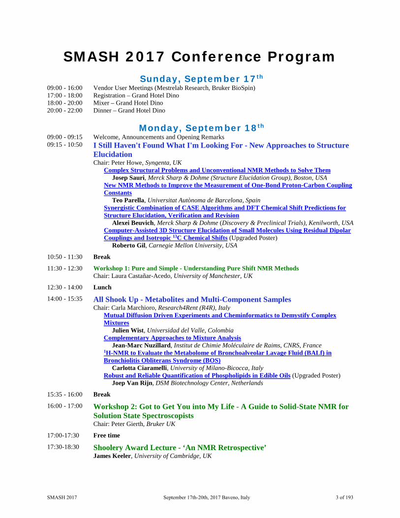
SMASH 2017 Conference Program
Sunday, September 17th 09:00 - 16:00 Vendor User Meetings (Mestrelab Research, Bruker BioSpin) 17:00 - 18:00 Registration – Grand Hotel Dino 18:00 - 20:00 Mixer – Grand Hotel Dino 20:00 - 22:00 Dinner – Grand Hotel Dino
Monday, September 18th 09:00 - 09:15 Welcome, Announcements and Opening Remarks 09:15 - 10:50 I Still Haven't Found What I'm Looking For - New Approaches to Structure
Elucidation Chair: Peter Howe, Syngenta, UK
Complex Structural Problems and Unconventional NMR Methods to Solve Them Josep Sauri, Merck Sharp & Dohme (Structure Elucidation Group), Boston, USA
New NMR Methods to Improve the Measurement of One-Bond Proton-Carbon Coupling Constants
Teo Parella, Universitat Autònoma de Barcelona, Spain Synergistic Combination of CASE Algorithms and DFT Chemical Shift Predictions for
Structure Elucidation, Verification and Revision Alexei Beuvich, Merck Sharp & Dohme (Discovery & Preclinical Trials), Kenilworth, USA
Computer-Assisted 3D Structure Elucidation of Small Molecules Using Residual Dipolar Couplings and Isotropic 13C Chemical Shifts (Upgraded Poster) Roberto Gil, Carnegie Mellon University, USA
10:50 - 11:30 Break
11:30 - 12:30 Workshop 1: Pure and Simple - Understanding Pure Shift NMR Methods Chair: Laura Castañar-Acedo, University of Manchester, UK
12:30 - 14:00 Lunch
14:00 - 15:35 All Shook Up - Metabolites and Multi-Component Samples Chair: Carla Marchioro, Research4Rent (R4R), Italy
Mutual Diffusion Driven Experiments and Cheminformatics to Demystify Complex Mixtures Julien Wist, Universidad del Valle, Colombia
Complementary Approaches to Mixture Analysis Jean-Marc Nuzillard, Institut de Chimie Moléculaire de Raims, CNRS, France
1H-NMR to Evaluate the Metabolome of Bronchoalveolar Lavage Fluid (BALf) in Bronchiolitis Obliterans Syndrome (BOS) Carlotta Ciaramelli, University of Milano-Bicocca, Italy
Robust and Reliable Quantification of Phospholipids in Edible Oils (Upgraded Poster) Joep Van Rijn, DSM Biotechnology Center, Netherlands
15:35 - 16:00 Break
16:00 - 17:00 Workshop 2: Got to Get You into My Life - A Guide to Solid-State NMR for Solution State Spectroscopists Chair: Peter Gierth, Bruker UK
17:00-17:30 Free time
17:30-18:30 Shoolery Award Lecture - ‘An NMR Retrospective’ James Keeler, University of Cambridge, UK
SMASH 2017 September 17th-20th, 2017 Baveno, Italy 3 of 193

18:30 – 20:00 Poster Session 1 - Even Better Than the Real Thing Even numbered posters to be presented - Drinks and snacks available
20:00 – 22:00 Dinner
Tuesday, September 19th
09:00 - 10:35 Let’s Dance - Reaction Monitoring and Kinetics Chair: Guy Lloyd-Jones, University of Edinburgh, UK
Catch Me If You Can - Watching Homogeneous Catalysis with Real-time High Resolution FlowNMR Ulrich Hintermair, University of Bath, UK
Quantitative NMR Spectroscopic Study of Highly Diluted Key Components in Complex Reactive Mixtures: Aqueous Amine Solutions Loaded with CO2 Erik von Harbou, University of Kaiserslautern, Germany
Chances and Pitfalls of In-Situ Irradiation NMR Spectroscopy Jonas Kind, Technische Universität Darmstadt, Germany
Ultrafast DOSY NMR of Hyperpolarised Mixtures (Upgraded Poster) Ludmilla Guduff, ICSN-CNRS, Gir-sur-Yvette, France
10:35 - 11:00 Break
11:00 - 12:35 We Will Rock You - Solid-State NMR Applications Chair: Steven Brown, University of Warwick, UK
No Heavy Metal - NMR Crystallography of Metal Salts and Organometallic Compounds Ann-Christin Poeppler, Universität Würzburg, Germany
Investigation of Powders at Natural Isotopic Abundance using Solid-State NMR and Dynamic Nuclear Polarization Pierre Thureau, University of Marseille, France
Combined Solid-State NMR, Diffraction and Modeling Studies of Small Molecule Pharmaceuticals Luis Mafra, University of Aveiro, Portugal
Insights into Amorphous Solid Dispersion of Felodipine Using Solid-state NMR Spectroscopy: Miscibility and Molecular Interactions (Upgraded Poster) Kanika Sarpal, University of Kentucky, USA
12:35 - 18:00 Lunch followed by free afternoon
17:00 - 18:00 Round Table Discussion on NMR Data Reporting (not a formal SMASH program event, but all interested parties are welcome to attend)
18:00 – 19:30 Poster session 2 – Against All Odds Odd numbered posters to be presented - Drinks and light buffet menu of starters available
Wednesday, September 20th
09:00 - 10:35 Should I Stay or Should I Go? - Non-Covalent Interactions and Complexes Chair: Elisabetta Chiarparin, AstraZeneca, UK
Fragment Evolution Without Routine Crystallography Ben Davis, Vernalis, UK
NMR2 for Fast 3D Structure Determination of Protein-Ligand Binding Site Without Protein Resonance Assignment Julien Orts, ETH, Switzerland
NMR Free Ligand Conformations for Enhanced Structure Based Drug Design Rodrigo Carbajo, AstraZeneca, UK
DiffErential EPitope Mapping (DEEP) STD NMR to Reveal the Pharmacophore of a Protein Target (Upgraded Poster) Serena Monaco, School of Pharmacy, University of East Anglia, UK
SMASH 2017 September 17th-20th, 2017 Baveno, Italy 4 of 193

10:35 - 11:00 Break
11:00 - 12:35 It’s Not Unusual - Multinuclear and Inorganic Methods Chair: Michael John, Georg-August Universität Göttingen, Germany
NMR Analysis of Small Paramagnetic Metal Complexes with Large Hyperfine Shifts Markus Enders, University of Heidelberg, Germany
Elucidation of the Topological and Chemical Order in Materials by Multi-Nuclear Solid-State NMR Pierre Florian, CEMHTI-CNRS Orleans, France
A Hyphenated Computational Protocol for Analysis of Natural Abundance 2H Residual Quadrupolar Couplings in (Chiral) Oriented Solvents Armando Navarro-Vázquez, Universidade Federal de Pernambuco, Brazil
Hyperpolarised Low-Field NMR for Industrial Reaction Monitoring (Upgraded Poster) Olga Semenova, Center of Hyperpolarisation of Magnetic Resonance, University of York, UK
12:35 - 14:00 Lunch, Free Time & Vendor Discussions
14:00 - 15:00 Workshop 3: We can work it out- Calculation of structures and NMR parameters Chair: Giuseppe Bifulco, Università di Salerno, Italy
15:00 - 15:30 Break
15:30 – 17:05 Go Your Own Way - Past SMASH Hot Topics Revisited Chair: Christina Thiele, Technische Universität Darmstadt, Germany
Broadband Pulses Revisited Burkhard Luy, Karlsruhe Insitut für Technologie, Germany
Small Microcoils Are Still Hot Aldrik Velders, Wageningen University, Netherlands
Hot Blooded, Check Out CPMG Sarah Robinson, Genentech, South San Francisco, USA
General Approach to Access Long-Range 1H-1H RDCs (Upgraded Poster) Davy Sinnaeve, Ghent University, Ghent, Belgium
17:05 - 17:15 Closing Remarks
Thursday, September 21st
09:30 – 15:30 qNMR Minisymposium (Not a formal SMASH event) Chairs: Michael Maiwald, BAM and Michael Bernstein, Mestrelab
This is not a formal SMASH program event, but all interested parties are welcome (no registration fee but please register).
Symposium program
SMASH 2017 September 17th-20th, 2017 Baveno, Italy 5 of 193

The James Shoolery Award In 2014, SMASH established the James Shoolery Award as a grant, in honor of James N. Shoolery, to recognize the important contributions by an individual to the field of small molecule NMR spectroscopy.
In 1952, Jim Shoolery joined Varian Associates to set up an applications laboratory for NMR spectroscopy. His main initial goals were to develop applications of NMR in chemistry and to educate the wider chemistry community in the potential value of NMR spectroscopy in their research. In pursuit of these goals during the 1950’s, he published a series of highly popular ads entitled “NMR at Work,” initially in Analytical Chemistry and later on the back page of the Journal of the American Chemical Society. These illustrated a wide range of applications of NMR in chemistry and were based on work that he carried out in the applications lab. He also wrote a number of “Technical
Information Bulletins” to help spectrometer owners in the operation of their instruments. Finally, he gave numerous lectures at conferences and research laboratories and at the annual NMR and EPR workshops that Varian Associates held in Palo Alto starting in 1958. In a 1993 article on the early history of NMR, he estimated that about 20,000 scientists had attended these different lectures by the end of the 1950’s.
At the same time, Jim interacted with the R & D division of Varian on NMR instrument improvement, including the progression of 1H operating frequency on Varian spectrometers from 30 to 40 to 60 and finally to 100 MHz by 1959. He was also involved in important technical improvements, including sample spinning, shim coils, spin decoupling, a flux stabilizer, and an electronic integrator. However, even with these improvements, the HR series of spectrometers were still extremely tricky to operate, requiring a significant amount of training, operating experience and patience. Jim realized that NMR spectroscopy would not reach its full potential as an analytical technique in chemistry until a spectrometer was developed that would be much easier to use, similar to the routine IR spectrometers that were already available from other manufacturers. Therefore, in 1957, Jim teamed with Emery Rogers of the marketing division of Varian to propose to the R & D division the development of a lower cost NMR spectrometer, which could use calibrated chart paper, which was rugged and reliable, and which could be run by graduate students and laboratory technicians with no training other than that provided by the spectrometer manual. He was heavily involved in this project, which resulted in 1961 in the introduction of the Varian A-60. This was a truly revolutionary development whose ease of operation triggered a dramatic increase in the use of NMR spectroscopy by chemists, in general, and by organic chemists, in particular. To illustrate its impact, the 1960 volume of the Journal of Organic Chemistry contained only one paper reporting the use of NMR while the 1967 volume included 220 papers, which used NMR data. In 2011, the seminal role of the A-60 in the development of NMR as a valuable analytical technique was recognized by the American Chemical Society as a National Historical Chemical Landmark in a ceremony at the Agilent facility in Santa Clara.
After the initial demonstration of FT NMR at Varian, Jim was involved in the development of the CFT-20 and FT-80 Varian spectrometers. These followed in the footsteps of the A-60 in being low cost and easy-to-use instruments for chemistry labs. In 1972, his book, “A Basic Guide to NMR,” was published by Varian Associates and helped to educate many young chemists in the use of NMR. Later, with the
SMASH 2017 September 17th-20th, 2017 Baveno, Italy 6 of 193

development of multi-pulse sequences and 2D NMR, Jim was among the first to recognize the great value of these techniques for identifying unknown organic chemical structures, particularly in the natural products field. Jim, along with Steve Patt, developed the APT sequence for spectral editing 13C spectra of organic compounds and, through the 1980’s, he collaborated with a number of natural products groups in establishing structures and assigning spectra of the compounds which they had isolated. He also, in 1984, published an important review article in the Journal of Natural Products, which clearly demonstrated the value of modern NMR techniques in the natural products field.
SMASH 2017 September 17th-20th, 2017 Baveno, Italy 7 of 193
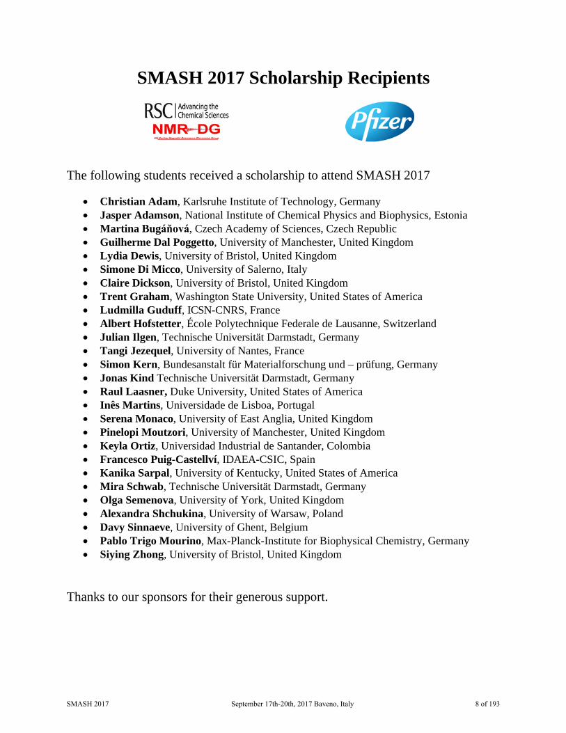
SMASH 2017 Scholarship Recipients
The following students received a scholarship to attend SMASH 2017
• Christian Adam, Karlsruhe Institute of Technology, Germany• Jasper Adamson, National Institute of Chemical Physics and Biophysics, Estonia• Martina Bugáňová, Czech Academy of Sciences, Czech Republic• Guilherme Dal Poggetto, University of Manchester, United Kingdom• Lydia Dewis, University of Bristol, United Kingdom• Simone Di Micco, University of Salerno, Italy• Claire Dickson, University of Bristol, United Kingdom• Trent Graham, Washington State University, United States of America• Ludmilla Guduff, ICSN-CNRS, France• Albert Hofstetter, École Polytechnique Federale de Lausanne, Switzerland• Julian Ilgen, Technische Universität Darmstadt, Germany• Tangi Jezequel, University of Nantes, France• Simon Kern, Bundesanstalt für Materialforschung und – prüfung, Germany• Jonas Kind Technische Universität Darmstadt, Germany• Raul Laasner, Duke University, United States of America• Inês Martins, Universidade de Lisboa, Portugal• Serena Monaco, University of East Anglia, United Kingdom• Pinelopi Moutzori, University of Manchester, United Kingdom• Keyla Ortiz, Universidad Industrial de Santander, Colombia• Francesco Puig-Castellví, IDAEA-CSIC, Spain• Kanika Sarpal, University of Kentucky, United States of America• Mira Schwab, Technische Universität Darmstadt, Germany• Olga Semenova, University of York, United Kingdom• Alexandra Shchukina, University of Warsaw, Poland• Davy Sinnaeve, University of Ghent, Belgium• Pablo Trigo Mourino, Max-Planck-Institute for Biophysical Chemistry, Germany• Siying Zhong, University of Bristol, United Kingdom
Thanks to our sponsors for their generous support.
SMASH 2017 September 17th-20th, 2017 Baveno, Italy 8 of 193

SMASH 2017 Conference Sponsors SMASH gratefully acknowledges the support from these companies:
Elite Sponsor
Sponsors
SMASH 2017 September 17th-20th, 2017 Baveno, Italy 9 of 193

SMASH 2017 NMR Conference Executive Board and Planning Committee
Program Co-Chairs
Tim Claridge
University of Oxford
Gary Sharman
Eli Lilly and Company
President President Outgoing Craig Butts Brian Marquez
University of Bristol Nalas Engineering
Logistics Carla Marchioro
Research 4 Rent (R4R)
Vendor Liaisons Margaret Levenberg Craig Butts
Stepan Company (Retired) University of Bristol
Treasurer & Registrar Robert Espina Magritek, Inc.
Secretary Secretary Outgoing Michael Hammer Daneen Angwin
Bruker Biospin GmbH SMASH - Small Molecule NMR Conference
Poster Co-Chairs Krish Krishnamurthy Roberto Gil
Chempacker LLC Carnegie Mellon University
SMASH 2017 September 17th-20th, 2017 Baveno, Italy 10 of 193

SMASH 2017 NMR Conference Organizing Committee
Daneen Angwin SMASH - Small Molecule NMR Conference
Craig Butts University of Bristol
Elisabetta Chiarparin AstraZeneca
Tim Claridge University of Oxford
Steve Coombes Astra Zeneca
Silvia Davalli Aptuit
Robert Espina Magritek, Inc.
David Foley Pfizer
Amy Freund Bruker Biospin
George Furst University of Pennsylvania
Laurine Galya Incyte Corporation
Roberto Gil Carnegie Mellon University
Michael Hammer Bruker Biospin
Andreas Kaerner Eli Lilly & Co
Krish Krishnamurthy Chempacker LLC
Dave Lankin University of Illinois Chicago
Margaret Levenberg Stepan Company (Retired)
Carla Marchioro Research 4 Rent (R4R)
Brian Marquez Mestrelab Research
Armando Navarro Vazquez Universidade Federal de Pernambu
Teo Parella Universitat Autònoma de Barcelona
Clark Ridge FDA
Maria Victoria Silva Elipe Amgen
Christina Thiele Technische Universitat Darmstadt
SMASH 2017 September 17th-20th, 2017 Baveno, Italy 11 of 193

Monday, September 18th 09:15 - 10:50
I Still Haven’t Found What I’m Looking for – New Approaches to
Structure Elucidation Chair: Peter Howe
Speakers:
Josep Sauri
Merck – Boston (US)
Teo Parella UAB (ES)
Alexei Buevich
Merck – Kenilworth (US)
Roberto Gil (Upgraded Poster) Carnegie Mellon University (US)
SMASH 2017 September 17th-20th, 2017 Baveno, Italy 12 of 193

Complex Structural Problems and Unconventional NMR Methods to Solve Them
Josep Saurí1, Yizhou Liu2, Kirk R. Gustafson3, Emily Mevers4, Jon Clardy4, Gary E. Martin2, and R. Thomas Williamson2
1. Structure Elucidation Group, Process and Analytical Research and Development, Merck Sharp & Dohme, Boston, MA 02115, USA
2. Structure Elucidation Group, Process and Analytical Research and Development, Merck Sharp & Dohme, Rahway, NJ 07065, USA
3. Molecular Targets Laboratory, Center for Cancer Research, National Cancer Institute, Frederick, MD 21702, USA
4. Department of Biological Chemistry and Molecular Pharmacology, Harvard Medical School, Boston, MA 02115, USA
Routine small and medium-sized molecules can generally be characterized by conventional NMR techniques such as 1D and 2D NMR experiments including COSY, NOESY/ROESY, HSQC and HMBC. However, when dealing with unknown and/or more complex molecular structures – especially proton-deficient compounds, an assignment strategy beyond the "normal" suite of NMR experiments is often required. In this presentation, we will discuss several examples of complex structural problems that were successfully addressed by the acquisition of novel NMR methods.
SMASH 2017 September 17th-20th, 2017 Baveno, Italy 13 of 193
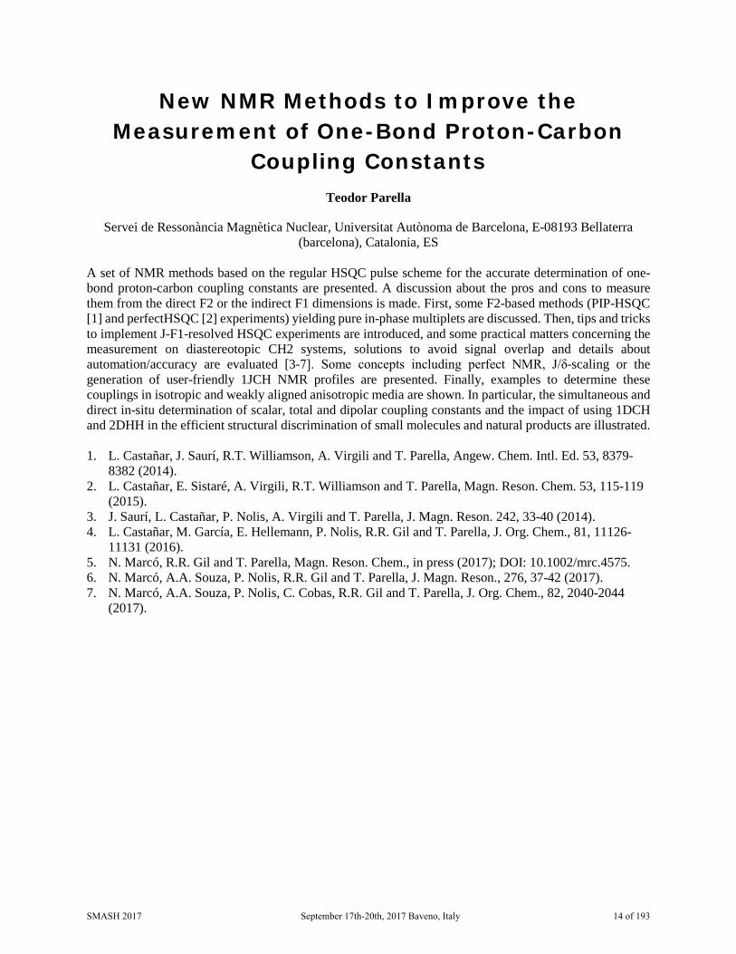
New NMR Methods to Improve the Measurement of One-Bond Proton-Carbon
Coupling Constants Teodor Parella
Servei de Ressonància Magnètica Nuclear, Universitat Autònoma de Barcelona, E-08193 Bellaterra (barcelona), Catalonia, ES
A set of NMR methods based on the regular HSQC pulse scheme for the accurate determination of one-bond proton-carbon coupling constants are presented. A discussion about the pros and cons to measure them from the direct F2 or the indirect F1 dimensions is made. First, some F2-based methods (PIP-HSQC [1] and perfectHSQC [2] experiments) yielding pure in-phase multiplets are discussed. Then, tips and tricks to implement J-F1-resolved HSQC experiments are introduced, and some practical matters concerning the measurement on diastereotopic CH2 systems, solutions to avoid signal overlap and details about automation/accuracy are evaluated [3-7]. Some concepts including perfect NMR, J/δ-scaling or the generation of user-friendly 1JCH NMR profiles are presented. Finally, examples to determine these couplings in isotropic and weakly aligned anisotropic media are shown. In particular, the simultaneous and direct in-situ determination of scalar, total and dipolar coupling constants and the impact of using 1DCH and 2DHH in the efficient structural discrimination of small molecules and natural products are illustrated. 1. L. Castañar, J. Saurí, R.T. Williamson, A. Virgili and T. Parella, Angew. Chem. Intl. Ed. 53, 8379-
8382 (2014). 2. L. Castañar, E. Sistaré, A. Virgili, R.T. Williamson and T. Parella, Magn. Reson. Chem. 53, 115-119
(2015). 3. J. Saurí, L. Castañar, P. Nolis, A. Virgili and T. Parella, J. Magn. Reson. 242, 33-40 (2014). 4. L. Castañar, M. García, E. Hellemann, P. Nolis, R.R. Gil and T. Parella, J. Org. Chem., 81, 11126-
11131 (2016). 5. N. Marcó, R.R. Gil and T. Parella, Magn. Reson. Chem., in press (2017); DOI: 10.1002/mrc.4575. 6. N. Marcó, A.A. Souza, P. Nolis, R.R. Gil and T. Parella, J. Magn. Reson., 276, 37-42 (2017). 7. N. Marcó, A.A. Souza, P. Nolis, C. Cobas, R.R. Gil and T. Parella, J. Org. Chem., 82, 2040-2044
(2017).
SMASH 2017 September 17th-20th, 2017 Baveno, Italy 14 of 193

Synergistic Combination of CASE Algorithms and DFT Chemical Shift Predictions for Structure Elucidation, Verification and
Revision Alexei V. Buevich1 and Mikhail E. Elyashberg2
1. Department of Discovery and Preclinical Sciences, Process Research and Development, NMR Structure Elucidation, Merck & Co., Inc., Kenilworth, NJ, USA
2. Moscow Department, Advanced Chemistry Development (ACD/Labs), Moscow, RU
Structure elucidation of complex natural products and organic compounds remains a challenging problem. Even when equipped with advanced spectroscopic methods, structure elucidation methodology is still not free from errors and, hence, the development of better, more robust methods remains in high demand. To support this endeavor, CASE (Computer-Assisted Structure Elucidation) expert systems were developed [1]. These systems are capable of generating an ensemble of all possible structures consistent with the molecular formula and the set of 2D NMR data followed by selection of the most probable structure on the basis of empirical NMR chemical shift prediction.
However, in some cases, CASE systems failed to distinguish the correct structure when average deviations of chemical shifts were too large or when the correct structure was among several top-ranked structures with acceptable but very similar deviations. Herein, we demonstrate for the first time that the combination of CASE and density functional theory (DFT) methods for NMR chemical shift prediction [2] allows unequivocal determination of correct structure even in such difficult situations [3]. This approach has been tested on three natural products: aquatolide, coniothyrione and epoxyroussoenone. All three cases represent rather challenging structural problems: the structures of the first two molecules were originally misassigned and the third structure, in addition to being a proton-deficient molecule, has four chiral centers.
We demonstrated that the proposed synergistic combination of CASE and DFT methodologies is an unbiased, reliable, and very efficient structure verification and de novo structure elucidation method that can be potentially applied to difficult structural problems when molecular systems are chiral, conformationally flexible and/or when other experimental methods (X-ray analysis, INADEQUATE, etc.) would be difficult or impossible to use.
1. Elyashberg M. E., Williams A. J. Computer-based Structure Elucidation from Spectral Data. The Art of Solving Problems; Springer, Heidelberg, 2015.
2. Lodewyk M. W., Siebert M. R., Tantillo D. J. Chem. Rev., 112, 1839-1862, 2012. 3. Buevich A. V., Elyashberg M. E. J. Nat. Prod., 79 (12), 3105–3116, 2016.
SMASH 2017 September 17th-20th, 2017 Baveno, Italy 15 of 193

Computer-Assisted 3D Structure Elucidation of Small Molecules Using Residual Dipolar
Couplings and Isotropic 13C Chemical Shifts Roberto R. Gil1, Kirill Blinov2, and Armando Navarro-Vazquez3
1. Department of Chemistry, Carnegie Mellon University, Pittsburgh, PA, US 2. MestReLab Research S. L., Santiago de Compostela, ES
3. Departamento de Química Fundamental, Universidade Federal de Pernambuco, Recife, BR
The 2D structure (molecular constitution) of most small molecules can be in principle straightforwardly determined by manual or automatic analysis of a set of experimental data that includes the molecular formula, a series of 1D and 2D NMR experiments providing trough-bond connectivity (COSY, TOCSY, HSQC, HMBC and ADEQUATE/INADEQUATE), and empirical chemical shift predictions. This is the main concept embedded in CASE (Computer Assisted Structure Elucidation) programs. Once the 2D structure is available, the determination of the relative spatial arrangement (configuration and preferred conformation) of all atoms in the molecule is a more challenging task commonly addressed by using NOE and 3J coupling constants analysis, as well as recent developments on DFT calculations of 13C chemical shifts (DP4). The development of the application of Residual Dipolar Couplings (RDCs) to the configurational and conformational analysis of small molecules has matured enough in the recent years to perform this task is an almost straightforward way, without even the need of using NOE and 3J coupling analysis, for the analysis of rigid and semi-rigid small molecules. We have recently shown that it is possible to go from molecular constitution to configuration without human intervention by feeding the program with an SDF structure file from a CASE-based program and a table containing only one-bond proton-carbon RDCs.[1] The process involves a) generation of the configurational space, b) conformational analysis for each configuration, c) automatic superposition of conformers for single-tensor analysis, d) fitting of RDC data to a set of conformation/configurations using model selection to prevent overfitting, e) plot of a bar diagram with quality factors indicating the selection of the correct 3D structure. This work led to the creation of StereoFitter, a complete computational module that can perform multiple-nmr parameters fitting (scalar couplings, NOE-derived distances, chemical shifts, RDCs, RCSAs) to combinations of configuration/conformations using model selection (Akaike Information Criterium (AIC)) to prevent overfitting. StereoFitter can be trivially extended to the use of chiroptics parameters (ECD, VCD) to determine absolute configuration. Generation of configurations, conformational analysis, DFT geometry optimizations, and analysis of DFT GIAO calculations of NMR parameters (J couplings, Chemical Shift Tensors) is also performed by StereoFitter by interacting with third party computational chemistry packages. In the present work we would like to show that, after the determination of the molecular constitution by the CASE program, a combined used of RDCs and isotropic 13C chemical shifts is enough to determine configuration, particularly useful for proton-deficient molecules. For the antimalarial drug Artemisinin,[2] the correct structure was determined using only 13C chemical shifts. The whole process from 2D structure to the generation of 38 diastereomers, followed by DFT energy minimization and DFT GIAO calculation of CSA tensor for each diastereomer, and finalizing with the fitting of chemical shifts to the set of diastereomers, took only 2 hours in a 5 years old PC equipped with 2 Xeon Processor with 6 cores each. Results shown in Figure 1.
SMASH 2017 September 17th-20th, 2017 Baveno, Italy 16 of 193

Figure 1: Determination of the configuraiton of Artemisinin using only isotropic sC chemical
shift. Fitting DFT calculated chemical shift with the experimental data using StereoFitter. The red asterisk shows the correct structure with the lowest χ2 value.
We have also applied the method to other relevant natural products such as the sesquiterpene lactone 10-epi-8-deoxycumambrin B [3] and the fungal metabolite Homodimericin A.[4] The structure of this natural product was determined using a combination of CASE, RDCs and RCSAs, since it has an hexacyclic core with fourteen quaternary carbons, eleven of them contiguous. For this particular compound we used the data reported by the authors.[4] For Homodimericin A we observed that it is possible to use 13C chemical shifts as an alternative to RCSAs to determine its configuration. In contrast to methodologies such as DP4 our method allows the fitting of populations to all the available experimental data including chemical shifts. Potential overfitting problems are treated by using the Akaike Information Criterion (AIC) where models with increasing number of populations are penalized. CASE analysis was performed using the MestReNova Structure Elucidator. Most of the results presented here used Poly(methylmethacrylate) (PMMA) based flexible gels, whose degree of alignment can be easily tuned by variable and reversible compression.[5] Acknowledgments: ANV thanks the UFPE for a visiting professor contract and FACEPE (APQ-0507-1.06/15) for financial support. NMR instrumentation at CMU was partially supported by the NSF(CHE-0130903 and CHE-1039870). R.R.G. acknowledges support from the NSF (CHE-1111684).We thanks Leandro F. Gil-Silva from Alignment Technologies LLC / Mestrelab Research SL for the provision of aligning gels. 1. Troche-Pesqueira, Eduardo; Anklin, Clemens; Gil, Roberto R.; Navarro-Vazquez, Armando.
Angewandte Chemie, International Edition, 56(13), 3660-3664, 2017. 2. Coordinating Group for Research on the Structure of Qing Hau Sau. Kexue Tongbao (Chinese
Edition) 22(3), 142, 1977. 3. Sosa, Virginia E.; Oberti, Juan C.; Gil, Roberto R.; Ruveda, Edmundo A.; Goedken, Virgil L.;
Gutierrez, Alicia B.; Herz, Werner. Phytochemistry 28(7), 1925-9, 1989.
SMASH 2017 September 17th-20th, 2017 Baveno, Italy 17 of 193

Monday, September 18th 11:30 - 12:30
Workshop 1
Pure and Simple – Understanding Pure Shift NMR Methodology
Coordinated by:
Laura Castañar-Acedo, The University of Manchester
SMASH 2017 September 17th-20th, 2017 Baveno, Italy 18 of 193

Understanding Pure Shift NMR Methodology Coordinated by:
Laura Castañar-Acedo, The University of Manchester Currently, pure shift NMR is an area of high interest. The aim of this workshop is to cover the practical aspects of these experiments. First, we will briefly describe the different methods available, their implementation in conventional 1D and multidimensional NMR experiments, and we will show several practical applications reported in recent years. The main part of the workshop will then deal with the practical features of pure shift experiments, such as optimal acquisition parameters and post-processing. No NMR experiment is perfect, and pure shift experiments are no exception. Some of the problems/limitations, such as sensitivity and spectral quality, will therefore also be discussed, as well as the techniques available for their removal/reduction. Finally, we will have an open question and answer session about all of the aspects covered, and about the challenges and possible next steps in the amazing adventure of the development and application of pure shift NMR experiments.
SMASH 2017 September 17th-20th, 2017 Baveno, Italy 19 of 193

Monday, September 18th 14:00 – 15:35
All Shook Up – Metabolites and Multi-
Component Samples Chair: Carla Marchioro
Speakers:
Julien Wist
Universidad del Valle, Cali (CO)
Jean-Marc Nuzillard Université de Reims Champagne-Ardenne, Reims (FR)
Carlotta Ciaramelle
University of Milano-Bicocca, Milan (IT)
Joep van Rijn (Upgraded Poster) DSM Biotechnology Center, Delft (NL)
SMASH 2017 September 17th-20th, 2017 Baveno, Italy 20 of 193

Mutual Diffusion Driven Experiments and Cheminformatics to Demystify Complex
Mixtures Christian Pantoja1, Michael Zasso2, Daniel Kostro2, Andrés Castillo1, Andrés Bernal1, Alejandro
Bolaños1, Luc Patiny2, and Julien Wist1
1. Chemistry Department, Universidad del Valle, Cali, CO 2. Institute of Chemical Sciences and Engineering, Ecole Polytechnique Fédérale de Lausanne
(EPFL), CH-1015 Lausanne, CH NMR earned its place in every chemistry department because it enabled the identification and characterization of compounds, better and quicker than any other technique. As NMR spectrometers got more mature, more complex samples such as macromolecules or mixtures of many compounds started being studied. We thus moved from the task of assigning each signal in a spectrum to atoms of a molecule to the task of assigning each signal in a spectrum to atoms in hundreds of molecules. Being successful at such a complex process requires combining efforts to improve both the resolution and overall quality of the experimental data and the accuracy of posterior analysis. Here we would like to present two contributions aiming at both directions; the first one being a new approach to experimentally discriminate different molecules in a mixture based on mutual diffusion coefficients, the second one being a collection of computational building blocks that can be used to elaborate specific tools to automate complex analysis pipelines. MDD experiments. Deconvolution of mixtures may be achieved in-situ with the help of a physical driving force. Think of DOSY, where the self-diffusion coefficient is used to discriminate signals from different molecules depending on their size. Recently we have shown that it is possible to establish a gradient of concentration for a binary solution of triethylamine-deuterium oxide inside an NMR tube. The gradient is induced by transition from a single phase to a biphasic state and will fade away as the system is allowed to return to its thermodynamical equilibrium (single phase). Dissolving species in such a system will have them spread along the tube according to their mutual or cooperative diffusion coefficients. Spatial encoding NMR experiments allow to capture the gradient for each individual species, thereby providing a mean to discriminate them. Results obtained in triethylamine-D2O were similar to those obtained by PFG-NMR, but not identical: water and methanol have similar self-diffusion coefficients and are poorly resolved in the DOSY experiment, while they have different mutual diffusion rates and appear well separated in the Mutual Diffusion Driven (MDD)-NMR experiment. Although still limited to a few binary systems (triethylamine-water, nitrobenzene-hexane, methanol-cyclohexane and others), such an approach offers the possibility to pick a system according to the affinity of the molecules that should be separated. Finally, initial conditions, such as molar fraction, mixing time and temperature can be tuned to improve separation. Cheminformatics. Despite the acquisition of NMR data is highly automated, posterior analysis is still mainly performed by hand and very few computational tools exist that perform them automatically. Assisted assignment has been around for a while but requires a lot of manual input from users. On the other hand, fully automatic assignment procedures do not rely on human intervention, but do rely on large amounts of manually-curated data. Similarly, only few tools exist that assist researchers in the task of matching signals with the different components of a mixture, or better, that perform it automatically. Here we present two examples of web applications build over our open source framework for cheminformatics.
SMASH 2017 September 17th-20th, 2017 Baveno, Italy 21 of 193

The first tool tries to assign 1H-NMR spectra in an iterative manner. Once atoms have been confidently assigned to signals, this knowledge is used to learn and predict expected chemical shifts. A new iteration can start and more atom-signal pairs are assigned. As a result, the accuracy of the predictions was found to increase after each iteration, as is the number of atoms assigned. Training this system with less 2341 pairs of molecule-spectrum let to predict over 60% of test pairs within 0.2 ppm, competing with the popular Spinus predictor. To the best of our knowledge, this is the first self-learning algorithm of its kind and it may in the future, once fed with enough data, compete with the best available predictors. As a second example, we present a tool to assist researchers in the task of identifying metabolites in mixtures. A database of reference compounds is used to propose putative candidates. First, a fully automated pipeline allows to schedule the acquisition of reference spectra and store them in the database, where they are duly peak-picked. A manual revision of the result is made easy by a complete user graphical interface. On the other end of the pipeline, metabolic profiles can be uploaded and analyzed using o-PLS and STOCSY approaches. PLS loadings and STOCSY rows trigger queries in the database resulting in a list of possible candidates. These tools are free to use and open source projects, written in JavaScript and thus natively designed for the web, the best place ever to share information.
SMASH 2017 September 17th-20th, 2017 Baveno, Italy 22 of 193

Complementary Approaches to Mixture Analysis
Ali Bakiri1, Ilhem Zebiri1, Jane Hubert1, Romain Reynaud2, Laurence Voutquenne1, Jean-Hugues Renault1, and Jean-Marc Nuzillard1
1. Institut de Chimie Moléculaire, Université de Reims, FR 2. Soliance-Givaudan, Pomacle, Marne, FR
The topic of mixture analysis is considered here for the identification of known compounds and for the discovery of new ones. The complementary methods we use or develop may not necessarily involve a physical separation step. The most straightforward way to get around the difficulty of analyzing a mixture is to fractionate it into pure compounds and to analyze them separately. The LC-NMR technique was created for this purpose. The availability of always higher static magnetic fields and of cyoprobes partly solved the NMR sensitivity problem that arises from the low amounts of sample injected in high resolution analytical LC columns. The concentration of LC effluents by their trapping in a solid phase extraction (SPE) cartridge led to the LC-SPE-NMR protocol. We used it for the re-investigation of the chemical content of a Peruvian plant, Dendrobangia boliviana, and it allowed us to characterize five new saponins [1]. Another way to solve the sensitivity problem is to increase the column effluent concentration. This approach was explored about 20 years ago by the coupling of Centrifugal Partition Chromatography (CPC) with NMR [2]. The combination of CPC fractionation (without any obligation to obtain a pure compound in any fraction), 13C NMR analysis of fractions, resonance binning and hierchical clustering of evolution profiles of 13C resonance intensities leads to the constitution of lists of chemical shifts values for the individualized components of a mixture. These lists are then used as search targets for a locally developped spectro-chemical database of natural products. The whole process, called CARAMEL (CARActérisation de MÉLanges, mixture caracterization in French), was already applied successfully, mostly to plant extracts selected for dermocosmetic activities [3]. The CARAMEL strategy identifies compounds already reported in literature and alerts its user for a possible discovery of new compounds. More recently, an algorithm was designed for the direct identification of known compounds from the 13C NMR spectrum of a complexe mixture, such as a plant extract, but without any purification step [4]. 1. Zebiri, I, Gratia, A, Nuzillard, J-M, Haddad, M, Cabanillas B, Harakat D, Voutquenne-Nazabadioko
L, Magn. Reson. Chem, 2017, submitted 2. Spraul, M, Braumann, U, Renault, J-H, Thépenier, P, Nuzillard, J-M,
J. Chromatogr. A, 776(1-2), 255-260, 1997. 3. Hubert, J, Nuzillard, J-M, Purson, S, Hamzaoui, M, Borie, N, Reynaud, R, Renault, J-H, Anal.
Chem., 86(6), 2955-2962, 2014 4. Bakiri, A, Hubert, J, Reynaud, R, Lanthony, S, Harakat, D, Renault, J-H, Nuzillard, J-M, J. Nat.
Prod., 2017, submitted
SMASH 2017 September 17th-20th, 2017 Baveno, Italy 23 of 193
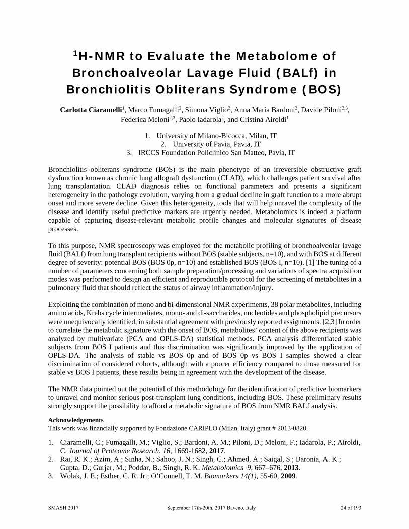
1H-NMR to Evaluate the Metabolome of Bronchoalveolar Lavage Fluid (BALf) in
Bronchiolitis Obliterans Syndrome (BOS) Carlotta Ciaramelli1, Marco Fumagalli2, Simona Viglio2, Anna Maria Bardoni2, Davide Piloni2,3,
Federica Meloni2,3, Paolo Iadarola2, and Cristina Airoldi1
1. University of Milano-Bicocca, Milan, IT 2. University of Pavia, Pavia, IT
3. IRCCS Foundation Policlinico San Matteo, Pavia, IT Bronchiolitis obliterans syndrome (BOS) is the main phenotype of an irreversible obstructive graft dysfunction known as chronic lung allograft dysfunction (CLAD), which challenges patient survival after lung transplantation. CLAD diagnosis relies on functional parameters and presents a significant heterogeneity in the pathology evolution, varying from a gradual decline in graft function to a more abrupt onset and more severe decline. Given this heterogeneity, tools that will help unravel the complexity of the disease and identify useful predictive markers are urgently needed. Metabolomics is indeed a platform capable of capturing disease-relevant metabolic profile changes and molecular signatures of disease processes. To this purpose, NMR spectroscopy was employed for the metabolic profiling of bronchoalveolar lavage fluid (BALf) from lung transplant recipients without BOS (stable subjects, n=10), and with BOS at different degree of severity: potential BOS (BOS 0p, n=10) and established BOS (BOS I, n=10). [1] The tuning of a number of parameters concerning both sample preparation/processing and variations of spectra acquisition modes was performed to design an efficient and reproducible protocol for the screening of metabolites in a pulmonary fluid that should reflect the status of airway inflammation/injury. Exploiting the combination of mono and bi-dimensional NMR experiments, 38 polar metabolites, including amino acids, Krebs cycle intermediates, mono- and di-saccharides, nucleotides and phospholipid precursors were unequivocally identified, in substantial agreement with previously reported assignments. [2,3] In order to correlate the metabolic signature with the onset of BOS, metabolites’ content of the above recipients was analyzed by multivariate (PCA and OPLS-DA) statistical methods. PCA analysis differentiated stable subjects from BOS I patients and this discrimination was significantly improved by the application of OPLS-DA. The analysis of stable vs BOS 0p and of BOS 0p vs BOS I samples showed a clear discrimination of considered cohorts, although with a poorer efficiency compared to those measured for stable vs BOS I patients, these results being in agreement with the development of the disease. The NMR data pointed out the potential of this methodology for the identification of predictive biomarkers to unravel and monitor serious post-transplant lung conditions, including BOS. These preliminary results strongly support the possibility to afford a metabolic signature of BOS from NMR BALf analysis. Acknowledgements This work was financially supported by Fondazione CARIPLO (Milan, Italy) grant # 2013-0820. 1. Ciaramelli, C.; Fumagalli, M.; Viglio, S.; Bardoni, A. M.; Piloni, D.; Meloni, F.; Iadarola, P.; Airoldi,
C. Journal of Proteome Research. 16, 1669-1682, 2017. 2. Rai, R. K.; Azim, A.; Sinha, N.; Sahoo, J. N.; Singh, C.; Ahmed, A.; Saigal, S.; Baronia, A. K.;
Gupta, D.; Gurjar, M.; Poddar, B.; Singh, R. K. Metabolomics 9, 667–676, 2013. 3. Wolak, J. E.; Esther, C. R. Jr.; O’Connell, T. M. Biomarkers 14(1), 55-60, 2009.
SMASH 2017 September 17th-20th, 2017 Baveno, Italy 24 of 193

Robust and Reliable Quantification of Phospholipids in Edible Oils
Joep van Rijn, Paul Groen, Remco Muntendam, and Adriana Carvalho de Souza
DSM Biotechnology Center, Delft, NL
Crude plant oils will form a solid sediment called gum upon storage. These gums contain mainly phospholipids. To obtain commercial edible oil the gum has to be removed, this process is known as degumming. The oldest form degumming is obtained by mixing crude oils with water and the solid formed is removed by centrifugation. Enzymatic degumming can be applied to reduce oil losses during degumming. To monitor degumming processes a robust and accurate phospholipid quantification is needed. Several techniques have been applied for the phospholipids quantification in edible oils, such as ICP, LC and TLC. However, in the past decade 31P NMR has shown to have advantages above the previously mentioned techniques because of the possibility to quantify different phospholipids directly from oil in an accurate and fast manner. In this report, we show the application and performance of 1D 31P NMR method for quantification of several phospholipids in crude and refined oils. The method is robust and has a variation lower than 5% for most phospholipids.
SMASH 2017 September 17th-20th, 2017 Baveno, Italy 25 of 193

Monday, September 18th 16:00 – 17:00
Workshop 2
Got to Get You into My Life – A Guide to Solid-State NMR for Solution State
Spectroscopists
Coordinated by: Peter Gierth, Bruker
SMASH 2017 September 17th-20th, 2017 Baveno, Italy 26 of 193

A Guide to Solid-State NMR for Solution State Spectroscopists
Coordinated by:
Peter Gierth, Bruker Solid state NMR is a tool with a large range of applications to small molecules, including providing spectra of insoluble compounds, polymorphism studies, analysis of solid mixtures, investigation of protonation states. Historically solid-state NMR has been seen as an exotic technique, requiring large amounts of specialised hardware and considerable operator experience, limiting its wider use. In recent years the improvements of general NMR hardware have allowed sophisticated solid-state NMR experiments on standard instruments with simple addition of a probe and pneumatic spinning unit, and this combined with an increasing array of relevant experimental techniques has increased interest in solid-state NMR among the general NMR community. This workshop will cover hardware requirements, some basic experimental procedures, and examples of applications of classical and recently developed techniques to small molecule problems.
SMASH 2017 September 17th-20th, 2017 Baveno, Italy 27 of 193

Monday, September 18th 18:30 – 20:00
Presentation of the James N. Shoolery Award
SMASH 2017 Recipient
James Keeler University of Cambridge, UK
James Keeler was born and raised in rural Norfolk, the second son of a farming family. He attended the local grammar school where an inspiring teacher first sparked an interest in chemistry and encouraged James to pursue this at University. He was an undergraduate in Oxford (at John’s College) and then won a scholarship from Merton College to continue with a doctorate under the supervision of Ray Freeman. On completing his doctorate in 1984, James was appointed to a ‘new blood’ lectureship in the Department of Chemistry, University of Cambridge and a Fellowship at Selwyn College. He has continued in these roles since that date.
James’ research interests have covered a wide range of topics in the broad area of ‘new techniques’ in high-resolution NMR. Particular themes have included improving lineshapes, the measurement of coupling constants, the suppression of zero-quantum coherence, the application of pulsed field gradients (especially to NOE experiments), and pure shift techniques. In recent years he has shifted his focus more to teaching and is the author of three undergraduate texts as well as the introductory NMR text Understanding NMR Spectroscopy. James has been awarded the Royal Society of Chemistry Meldola Medal, the University of Cambridge Pilkington Teaching Prize, and the Royal Society of Chemistry Silver Medal for contributions in Magnetic Resonance.
SMASH 2017 September 17th-20th, 2017 Baveno, Italy 28 of 193

Tuesday, September 19th 09:00 - 10:35
Let’s Dance – Reaction Monitoring
and Kinetics Chair: Guy Lloyd Jones
Speakers:
Ulrich Hintermair
University of Bath, Bath (UK)
Erik von Harbou University of Kaiserslautern, Kaiserslautern (DE)
Jonas Kind
TU Darmstadt, Darmstadt (DE)
Ludmilla Guduff (Upgraded Poster) ICSN-CNRS, Gir-sur-Yvette (FR)
SMASH 2017 September 17th-20th, 2017 Baveno, Italy 29 of 193

Catch Me If You Can - Watching Homogeneous Catalysis with Real-Time High
Resolution FlowNMR
Andrew M. R. Hall1, Peilong Dong2, Anna Codina3, John P. Lowe2, and Ulrich Hintermair1
1. Centre for Sustainable Chemical Technologies, University of Bath, UK 2. Department of Chemistry, University of Bath, UK
3. Bruker UK Ltd., Coventry, UK Molecular solution-phase catalysis is a key technology for addressing sustainability issues in the chemical industry. The development and optimization of homogeneous catalysts is, however, often hampered by limited insight into the kinetics of the reaction and the transformation of the catalyst, enforcing empirical optimization. Rational catalyst and reaction development is only possible through thorough understanding of catalyst activation and de-activation mechanisms, potential resting or dormant states, and the kinetics of the productive cycle (i.e. rate-limiting steps). While rather laborious techniques are available to investigate the afore-mentioned aspects separately, there is no readily applicable technique that may be used universally in early stages of catalyst development. We will present the development of such a technique based on NMR spectroscopy as one of the most informative solution-phase analysis method available. We have built a reaction setup in which a reaction vessel is coupled to a capillary NMR probe via small diameter HPLC tubing. With this we can continuously circulate a reaction mixture through the spectrometer, thereby follow the reaction progresses and catalyst transformation under catalytically relevant conditions in real time.
Figure 1. Schematic of the FlowNMR setup.
We have characterised the hydrodynamic flow characteristics of the setup and measured flow effects on continuous 1H NMR acquisition to quantify changes in T1, T2 and signal intensity as function of volumetric flow velocity. Application in real-time reaction and catalyst monitoring under strictly inert conditions has been demonstrated, and multiple solvent suppression and selective excitation techniques allow the detection of minor intermediates even in non-deuterated solvents. [1] Using the described setup, we have followed the asymmetric transfer-hydrogenation of acetophenone using Noyori’s chiral TsDPEN-ligated (arene)RuII complexes as catalysts in basic iso-propanol (figure 2).[2]
SMASH 2017 September 17th-20th, 2017 Baveno, Italy 30 of 193

Figure 2. Catalytic system investigated.
As continuous NMR acquisition can be started on pure solvent flow, and the reaction initiated by sequential injection of reagents, the entire reaction can be followed without any lag phases. High quality kinetic data is obtained for this air-sensitive transition metal catalysed reaction (figure 3). Importantly, both conversion and enantioselectivity at the end of the reaction are identical to the results obtained in a sealed Schlenk flask, demonstrating that several hundred pumping cycles do not affect the chemistry in solution.
Figure 3. Reaction kinetics of the system shown in fig. 2 as obtained by 1H FlowNMR.
Furthermore, using selective excitation techniques, we were able to observe metal-hydride intermediates during the reaction under the same conditions. These can be quantified over time and directly correlated to product formation kinetics (figure 4).
Time /min
0 50 100 150 200
Con
cent
ratio
n /M
0.0
0.1
0.2
0.3
0.4
0.5
ProductSubstrateSubstrate + Product
SMASH 2017 September 17th-20th, 2017 Baveno, Italy 31 of 193

Figure 4. Ru-H intermediates detected during the reaction shown in fig. 2.
We will present a full account of our data and discuss their implications for the mechanism of this widely used reaction. 1. A. M. R. Hall, J. C. Chouler, A. Codina, P. T. Gierth, J. P. Lowe, U. Hintermair, Catal. Sci. Technol.
2016, 6, 8406-8417. 2. K-J. Haack, S. Hashiguchi, A. Fujii, T. Ikariya, R. Noyori, Angew. Chem. Int. Ed. 1997, 36, 285.
SMASH 2017 September 17th-20th, 2017 Baveno, Italy 32 of 193
Quantitative NMR Spectroscopic Study of Highly Diluted Key Components in Complex
Reactive Mixtures: Aqueous Amine Solutions Loaded with CO2
Erik von Harbou, Richard Behrens, Elmar Kesler, and Hans Hasse
Laboratory of Engineering Thermodynamics, University of Kaiserslautern, Kaiserslautern, DE
NMR spectroscopy is widely applied in chemistry for example to elucidate chemical structures. Its potential for quantitative investigation of complex reactive multi-phasic systems, however, has been hardly exploited in chemical reaction engineering or thermodynamics so far even though it offers many advantages for quantitative investigations of complex systems. On the one hand, NMR spectroscopy features non-invasive measurements so that the investigation of reactive systems, which are typically very sensitive to changes of temperature or pressure, are not disturbed by sampling. On the other hand, in contrast to optical spectroscopy methods, the composition of the sample can be determined in most cases directly from the acquired NMR signal without the need for calibration prior to the analysis. Furthermore, the concentrations of several different analytes can be determined simultaneously even though their values differ by orders of magnitude. Thus, NMR spectroscopy is often not only the best but also the only applicable method to quantify complex reactive systems with intermediates or products that cannot be isolated from the mixture.
In this work, we will present an in-situ NMR spectroscopic method to investigate the liquid phase reactions in aqueous amine solutions loaded with CO2 under high pressure. These systems are widely applied in reactive absorption processes for example for scrubbing CO2 from synthesis or flue gas. In these processes, the CO2 is absorbed from the gas phase into the liquid phase where it undergoes immediately several consecutive reactions with the solvent. In order to be able to describe the absorption process reliably, a comprehensive picture of the reaction network is necessary. In addition, even though the concentration of the molecular CO2 dissolved in the liquid phase is very low, it determines both the rate of mass transfer from the gas to the liquid phase and the rate of reaction in the liquid phase. Therefore, its value together with the concentration of the reaction products has to been know precisely in order to be able to understand the overall reactive absorption process.
SMASH 2017 September 17th-20th, 2017 Baveno, Italy 33 of 193

Chances and Pitfalls of In-Situ Irradiation NMR Spectroscopy
Jonas Kind1, Ann-Kathrin Schönbein2, Jasper J. Michels2, Stefan Ortgies3, Alexander Breder3, and Christina M. Thiele1
1. Clemens-Schöpf-Institut für Organische Chemie und Biochemie, TU Darmstadt, DE 2. Max Planck Institute for Polymer Research, Department of Molecular Electronics, Mainz, DE 3. Institut für Organische und Biomolekulare Chemie, Georg-August-Universität, Göttingen, DE
NMR spectroscopy is widely used for (in-situ) reaction characterisation and reaction monitoring e.g. to identify intermediate species or to quantify side product formation. Irradiation of NMR samples inside the magnet is well known from photo-CIDNP experiments, where light is used to enhance NMR signal intensities. Furthermore, the combination of NMR and in-situ irradiation offers the possibility to characterise irreversible and reversible photochemical conversions on a large temporal scale [1,2].
By using recent results of two photochemical reactions we discuss benefits of in-situ irradiation NMR using LEDs and silica waveguides as well as unexpected pitfalls of this technique. We want to illustrate the scope of in-situ irradiation NMR in terms of irradiation intensities, sample homogenieties and quality of the resulting spectra as well as comparability to batch experiments. Firstly we show kinetic data of a reaction cascade yielding poly(2,5‐bis-(2′‐ethylhexyl)-1,4‐phenylene-vinylene) (BEH‐PPV), a well-known OLED emitter material. A reactive p-quinodimethane is produced in-situ from the premonomer by adding potassium tert-butoxide below the threshold temperature of the thermal polymerization reaction. Polymerization of the p-quinodimethane can be induced by irradiation of the reaction mixture with UV light [3]. Secondly we show results on a dye sensitized seleno mediated photo catalytic lactonization of an alkenoic acid in the presence of oxygen [4]. Production of the lacton occures via an isolatable seleno substituted lacton, which yields the final product after photo catalytic elimination. From batch experiments it is known that either the activation of the intermediate or the elimination to the product are rate limiting. Here we present data of an initial rate approach to identify the rate limiting step in the catalytic cycle. 1. Feldmeier, Christian; Bartling, Hanna; Riedle, Eberhard; Gschwind, Ruth M., J. Magn. Reson., 232,
39-44, 2013. 2. Kind, Jonas; Kaltschnee, Lukas; Leyendecker, Martin; Thiele, Christina M.; Chem. Commun., 52,
12506-12509, 2016. 3. Kuch, Serena; Vilbrandt, Nicole; Rehahn, Matthias, Macromol Rapid Commun., 37 (10), 820-825,
2016. 4. Ortgies, Stefan; Depken, Christian; Breder, Alexander; Org. Lett., 18, 2856, 2016.
SMASH 2017 September 17th-20th, 2017 Baveno, Italy 34 of 193
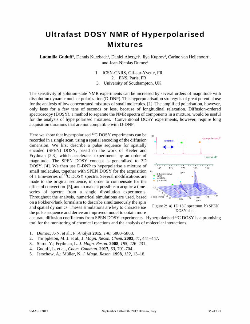
Ultrafast DOSY NMR of Hyperpolarised Mixtures
Ludmilla Guduff1, Dennis Kurzbach2, Daniel Abergel2, Ilya Kuprov3, Carine van Heijenoort1, and Jean-Nicolas Dumez1
1. ICSN-CNRS, Gif-sur-Yvette, FR 2. ENS, Paris, FR
3. University of Southampton, UK The sensitivity of solution-state NMR experiments can be increased by several orders of magnitude with dissolution dynamic nuclear polarization (D-DNP). This hyperpolarisation strategy is of great potential use for the analysis of low concentrated mixtures of small molecules. [1]. The amplified polarisation, however, only lasts for a few tens of seconds or less, because of longitudinal relaxation. Diffusion-ordered spectroscopy (DOSY), a method to separate the NMR spectra of components in a mixture, would be useful for the analysis of hyperpolarised mixtures. Conventional DOSY experiments, however, require long acquisition durations that are not compatible with D-DNP. Here we show that hyperpolarised 13C DOSY experiments can be recorded in a single scan, using a spatial encoding of the diffusion dimension. We first describe a pulse sequence for spatially encoded (SPEN) DOSY, based on the work of Keeler and Frydman [2,3], which accelerates experiments by an order of magnitude. The SPEN DOSY concept is generalised to 3D DOSY. [4]. We then use D-DNP to hyperpolarise a mixture of small molecules, together with SPEN DOSY for the acquisition of a time-series of 13C DOSY spectra. Several modifications are made to the original sequence, in order to compensate for the effect of convection [5], and to make it possible to acquire a time-series of spectra from a single dissolution experiments. Throughout the analysis, numerical simulations are used, based on a Fokker-Plank formalism to describe simultaneously the spin and spatial dynamics. Theses simulations are key to characterise the pulse sequence and derive an improved model to obtain more accurate diffusion coefficients from SPEN DOSY experiments. Hyperpolarised 13C DOSY is a promising tool for the monitoring of chemical reactions and the analysis of molecular interactions. 1. Dumez, J.-N. et al., P. Analyst 2015, 140, 5860–5863. 2. Thrippleton, M. J. et al., J. Magn. Reson. Chem. 2003, 41, 441–447. 3. Shrot, Y.; Frydman, L. J. Magn. Reson. 2008, 195, 226–231. 4. Guduff, L. et al., Chem. Commun. 2017, 53, 701-704. 5. Jerschow, A.; Müller, N. J. Magn. Reson. 1998, 132, 13–18.
Figure 2: a) 1D 13C spectrum. b) SPEN DOSY data.
SMASH 2017 September 17th-20th, 2017 Baveno, Italy 35 of 193
Tuesday, September 19th 11:00 - 12:35
We Will Rock You – Solid-State NMR
Applications Chair: Steven Brown
Speakers:
Ann-Christin Poeppler
Universität Würzburg, Würzburg (DE)
Pierre Thureau Marseille (FR)
Luis Mafra
University of Aveiro, Aveiro (PT)
Kanika Sarpal (Upgraded Poster) University of Kentucky, Lexington (US)
SMASH 2017 September 17th-20th, 2017 Baveno, Italy 36 of 193

No Heavy Metal - NMR Crystallography of Metal Salts and Organometallic Compounds
Ann-Christin Pöppler1,2, David Walker1, and Steven P. Brown1
1. Department of Physics, University of Warwick, Coventry, UK 2. Institute of Organic Chemistry, University of Würzburg, Würzburg, DE
Initially chosen as a robust proof of concept system for the study of very air- and moisture sensitive organolithium compounds, understanding the solid-state structure and properties of orotic acid monohydrate and its lithium and magnesium salts was not straightforward. All three compounds are known to form hydrate structures [1], an important class of chemicals due to the influence of the incorporated water molecules on the physicochemical properties, e.g. bioavailability and stability. Based on their connectivities, hydrate structures can be divided into three subsets: i) isolated hydrates, ii) channel hydrates and iii) (metal) ion assisted hydrates. The three compounds, represent a set of structures with, in principle, known single crystal X-ray structures, in which each individual compound belongs to a different class of hydrate. Orotic acid monohydrate contains water molecules in isolated sites, while both salt structures show water molecules coordinated to a metal ion. Additionally, in magnesium orotate octahydrate, water molecules are also arranged in channels. Their pharmaceutical potential is subject to discussion in literature [2]. A combined study by solid-state NMR, GIPAW (CASTEP) [3] calculations, powder X-ray diffraction and thermogravimetric analysis shows complexities in structure and dynamics of these compounds that go beyond the static view of the available crystal structures. Furthermore, first data for the analysis of the sensitive organolithium compounds is also presented. 1. a) G. Portalone, Acta Crystallogr. E, 64, o656, 2008; b) I. Bach, O. Kumberger, H. Schmidbaur,
Chem. Ber., 123, 2267-2271, 1990. 2. a) M. Gitlin, M. A. Frye, Bipolar Disorders, 14, 51-65, 2012; b) G. Jasmin, L. Proschek, Cardiovasc.
Drugs Ther., 12, 189-195, 1998; c) F. Rosenfeldt, S. Richards, Z. Lin, S. Pepe, R. J. Conyers, Cardiovasc. Drugs Ther., 12, 159-170, 1998.
3. a) S. J. Clark, M. D. Segall, C. J. Pickard, P. J. Hasnip, M. I. J. Probert, K. Refson, M. C. Payne, Z. Kristallogr., 220, 567, 2005; b) C. J. Pickard, F. Mauri, Phys. Rev. B, 63, 245101, 2001; c) J. R. Yates, C. J. Pickard, F. Mauri, Phys. Rev. B, 76, 024401, 2007.
SMASH 2017 September 17th-20th, 2017 Baveno, Italy 37 of 193

Investigation of Powders at Natural Isotopic Abundance using Solid-State NMR and
Dynamic Nuclear Polarization Pierre Thureau
Aix-Marseille University, CNRS, Marseille, France We demonstrate here that solid-state NMR can be used to investigate powders, which cannot be studied using standard techniques such as single-crystal X-ray diffraction. More specifically, an efficient procedure for the assigment of 13C resonances in natural abundance powders will be shown.[1] Furthermore, it will be shown that the sensitivity enhancement obtained using dynamic nuclear polarization (DNP) can be used to determine the conformation and crystal packing of pharmaceutical compounds.[2] The benefit of combining crystal structure prediction and DNP experiments will also be discussed. Finally, we will show that DNP experiments can be useful to reveal the early stage of polymorphic transformation in powders. 1. Dekhil, M., Mollica G., Texier Bonniot, T., Ziarelli, F., Thureau, P., Viel, S., Chem. Commun.,
52(55), 8565-8568, 2016. 2. Mollica G., Dekhil, M., Ziarelli, F., Thureau, P., Viel, S., Angew. Chem. Int. Ed., 54(20), 6028-6031,
2015.
SMASH 2017 September 17th-20th, 2017 Baveno, Italy 38 of 193

Combined Solid-State NMR, Diffraction and Modeling Studies of Small Molecule
Pharmaceuticals Luís Mafra
CICECO - Aveiro Institute of Materials, Department of Chemistry, University of Aveiro, Campus Universitário de Santiago, 3810-193 Aveiro, PT
Solid-state NMR (SSNMR) is a powerful atomic-level characterization technique able to study the local crystalline/amorphous structure of bulk and dosage forms of active pharmaceutical ingredients (API).[1] Toward a better understanding of how small molecule pharmaceuticals self-assemble in the solid-state to yield distinct polimorphic/pseudopolimorphic forms, a combined experimental 1D/2D SSNMR, X-ray diffraction (XRD), and computational study of selected small molecule pharmaceuticals is presented.[2-5] Although X-ray diffraction (XRD) provides a full description of the intra- and intermolecular distances and angles, the technique presents severe limitations in probing local interactions involving light atoms such as hydrogens. In contrast, SSNMR is particularly useful to study hydrogens and probe the strength and nature of protons engaged in hydrogen bond (HB) interactions, having the potential to discriminate among distinct polymorphs. Here we report the effect of crystal packing on the 1H and 13C chemical shifts (CS) of drug hydrates and anhydrates, including nonconventional HBs, π···π and CH···π contacts, are studied through periodic DFT calculations using the GIPAW-DFT formalism. It will be shown that NMR CSs can be sensitive detectors of hydration/dehydration states in highly insoluble antibiotics.[2] Current approaches to modify physicochemical properties of APIs, without impacting its pharmacological behavior, include the development of new “multicomponent crystalline solid forms” that can be obtained using mechanochemical processes, namely liquid assisted grinding (LAG).[3,4] This presentation highligh the NMR discrimination between cocrystals and salts of distinct APIs, revealing that 1H and 15N SSNMR combined with GIPAW-DFT calculations can locate “XRD-disordered” hydrogen atoms. 1H SSNMR detected unusually strong HBs associated with such disordered hydrogens through the presence of 1H resonances shifted to very high frequencies (up to ca. 20.1 ppm).[3] Recently, SSNMR became an important gadget in the process of crystal structure solution in powders. This is a non-trivial task and using powder XRD methods alone may often lead to the wrong structure solution. In this talk, a new hybrid approach for structure determination of crystalline solids, will be presented, based on the combination of SSNMR, XRD and an ensemble of computational-assisted structure solution tools including a genetic algorithm based on evolution-inspired operators repeatedly applied to populations of possible crystal structure solutions that evolve to eventually produce the best new offspring candidates. Such methodologies are demonstrated in a challenging multiple component crystal structure composed by flexible molecules - trihydrate β-lactamic antibiotic. [5] 1. Mafra, L., Santos, S. M., Sardo, S., Schmidt, H. F., In Computational Pharmaceutical Solid State
Chemistry, (Ed Abramov, Y.A.), Wiley, 2016, ISBN: 978-1-118-70074-7. 2. Mafra, L., Santos, S.M., Siegel, R., Alves, I., Paz, F. A. A., Dudenko, D., Spiess, H. W., J. Am.
Chem. Soc. 134, 71–74, 2012. 3. Martins, I.C.B., Sardo, M., Santos, S.M., Fernandes, A., Antunes, A., André, V., Mafra, L., Duarte,
M.T., Cryst. Growth Des. 16, 154–166, 2016.
SMASH 2017 September 17th-20th, 2017 Baveno, Italy 39 of 193

4. Fernandes, J. A., Sardo, M., Mafra, L., Choquesillo-Lazarte, D., Masciocchi, N., Cryst. Growth Des. 15, 3674–3683, 2015.
5. Santos, S.M., Rocha, J., Mafra, L., Cryst. Growth Des. 13, 2390–2395, 2013. [4] Fernandes, J. A., Sardo, M., Mafra, L., Choquesillo-Lazarte, D., Masciocchi, N., Cryst. Growth Des. 15, 3674–3683, 2015. [5] Santos, S.M., Rocha, J., Mafra, L., Cryst. Growth Des. 13, 2390–2395, 2013.
SMASH 2017 September 17th-20th, 2017 Baveno, Italy 40 of 193

Insights into Amorphous Solid Dispersions of Felodipine Using Solid-state NMR
Spectroscopy: Miscibility and Molecular Interactions
Kanika Sarpal and Eric J. Munson
College of Pharmacy, University of Kentucky, Lexington, KY, US
Phase separation in amorphous solid dispersions (ASDs) is still not clearly understood on the sub-nanometric scale [1], and further systematic investigations are still required. The phase separation behavior is influenced by many factors, such as the composition of drug-polymer blend, the type and the strength of drug-polymer interactions, and the method of preparation [2-4]. We demonstrated the use of solid-state NMR spectroscopy (SSNMR) to evaluate the role of the strength of drug-polymer hydrogen bonding (H-bonding) on the compositional homogeneity in ASDs of felodipine (FEL), a poorly water soluble drug, with poly(vinylpyrrolidone), or PVP, poly(vinylpyrrolidone-co-vinylacetate), or PVP/VA, and poly(vinylacetate) or PVAc. The dispersions were prepared at various drug loadings (50% to 90% w/w) via melt quenching. The blend scale miscibility was studied by examining the proton spin-lattice relaxation times in the laboratory and rotating frame (1H T1 and T1ρ) for the drug and the individual polymer for each set of ASDs. Domain sizes were estimated via spin diffusion. The experimental data was used to elucidate the influence of the strength of drug-polymer H-bonding on the phase behavior of resulting dispersions. It was found that FEL:PVP and FEL:PVP/VA systems exhibited weak signs of nano phase separation especially for the compositions with lower polymer loadings with considerably small domain sizes. Whereas FEL:PVAc system showed pronounced signs of nano phase separation as the polymer amount decreased with larger domains. The extent of H-bonding in amorphous FEL and FEL:Polymer blends within each set of ASDs was quantified via deconvolution. The carbonyl region was used to selectively analyze the evolution of the various populations of coexisting species in the samples. The order of the strength/extent of drug–polymer H-bonding interactions was PVP > PVP/VA > PVAc. It was suggested that the strength of drug-polymer H-bonding interaction is one of the key factors in controlling the phase behavior of ASDs. Our findings indicate that SSNMR is a useful tool for evaluating the spatial homogeneity with sub-50 nm resolution, where other conventional techniques fail, and drug-polymer interactions for the compositions of pharmaceutically relevant systems. 1. Qi, Sheng et al., Molecular Pharmaceutics, 10, 918-930, 2013 2. Nie, Haichen et al., Molecular Pharmaceutics, 13, 3964-3975, 2016 3. Chakravarty, Paroma et al., International Journal of Pharmaceutics, 519, 44-57, 2017 4. Yuan, Xiaoda et al., Molecular Pharmaceutics, 11, 329-337, 2014
SMASH 2017 September 17th-20th, 2017 Baveno, Italy 41 of 193

Tuesday, September 19th 17:00 – 18:00
Round table discussion
NMR Data Reporting
Coordinated by: Damien Jeannerat, Université de Genève
SMASH 2017 September 17th-20th, 2017 Baveno, Italy 42 of 193

NMR Data Reporting Coordinated by:
Damien Jeannerat, Université de Genève This will be a roundtable discussion (Damien Jeannarat, Chair) based around a number of ongoing initiatives in creating and NMR data storage/reporting standards. These include Damien's NMRedata initiative and there are also RSC and IUPAC discussion around this topic. All SMASH attendees are welcome.
SMASH 2017 September 17th-20th, 2017 Baveno, Italy 43 of 193

Wednesday, September 20th 09:00 - 10:35
Should I Stay or Should I Go? – Non-Covalent Interactions and
Complexes Chair: Elisabetta Chiarpin
Speakers:
Ben Davis
Vernalis (UK)
Julien Orts ETH Zurich, Zurich (CH)
Rodrigo Carbajo
AstraZeneca (UK)
Serena Monaco (Upgraded Poster) University of East Anglia, Norwich (UK)
SMASH 2017 September 17th-20th, 2017 Baveno, Italy 44 of 193

Fragment Evolution Without Routine Crystallography
Ben Davis
Vernalis, UK Fragment – and structure- based drug discovery typically require significant numbers of three dimensional structures of ligand/protein complexes. These structures have to be accurate, relevant and timely in order to guide medicinal chemistry efforts. However, many classes of biological target are not amenable to this approach due to a range of factors. For example, the target may not crystallise at all, or may crystallise only in an apo form, and high-resolution NMR structures of larger proteins remain time consuming to determine. In these cases conventional structure-guided methods cannot readily be applied, increasing the difficulty in discovering and developing novel drugs against these target classes. We have found that combining NMR data with modelling and chemical SAR can effectively steer a medicinal chemistry campaign from fragment to lead, particularly when accompanied by clear communication between the disparate groups involved. We will discuss our experiences and development of approaches to identifying and characterising ligands of PPI targets, with particular reference to the anti-apoptotic protein Bcl2. These methods have proven to be generally applicable and successful, with two compounds developed using these methods now in Phase I clinical trials.
SMASH 2017 September 17th-20th, 2017 Baveno, Italy 45 of 193

NMR2 for Fast 3D Structure Determination of Protein-Ligand Binding Site Without Protein
Resonance Assignment
Julien Orts1, Marielle Aulikki Wälti1, May Marsh2, Laura Vera2, Alvar D. Gossert3, Peter Güntert1,4, and Roland Riek1
1. ETH Zürich, Laboratory of Physical Chemistry, HCI F217, Vladimir-Prelog-Weg 2, Zürich, CH 2. Swiss Light Source, Paul Scherrer Institute, Villigen, CH
3. Novartis Institutes for BioMedical Research, Novartis AG, Basel, CH 4. Institute of Biophysical Chemistry Goethe University, Frankfurt, DE
X-ray crystallography molecular replacement (Rossmann et al. 1962) (MR) is a highly versatile tool for the detailed characterization of lead compound and binding modes in the pharmaceutical industry (Hillischet al 2004). The two major limitations of its application to drug research are (i) the availability of a similar protein structure, which, in the area of structure-based drug design, is most often a complex of the protein with a lead compound, and (ii) obtaining well-diffracting crystals of the ligand-protein complexes of interest. While nowadays the first point is often not a limitation anymore, obtaining well-diffracting crystals might be difficult. In such situations structure determination of protein-ligand complexes by liquid-state NMR is a good option. Unfortunately, the established standard structure determination protocol (Cavanagh et al. 2007) is in general time-consuming, and a shortcut using available structural data as in the case of MR in X-ray crystallography is not available. Here, we present NMR2 (NMR Molecular Replacement), a MR-like approach in NMR to determine the structures of the binding pockets of ligands at atomic resolution. The calculation of structures of protein-ligand complexes relies on the collection of unassigned semi-quantitative inter-molecular NOE distance restraints and on previously solved structures. The NMR2 method uses a high throughput structure calculation protocol, rather than a docking-scoring simulation. It is fast since it requires only a few days of measuring time and bypasses the time-consuming sequential assignment steps for the protein. When applied to the cancer-relevant HDMX protein, the NMR2 method yielded the structure of a ligand protein complex with an accuracy below 1 Ångstrom for the binding pocket irrespective of the starting protein structure templates used. We will present multiple NMR2 applications covering a peptidomimetic inhibitor and small molecules that bind strongly or weakly to protein receptors fully or partially labelled using methyl-specific isotope labelling. Our findings demonstrate that NMR2 may open an avenue for the fast and robust determination of the binding pocket structure of ligand-protein complexes at atomic resolution.
SMASH 2017 September 17th-20th, 2017 Baveno, Italy 46 of 193

NMR Free Ligand Conformations for Enhanced Structure Based Drug Design
Rodrigo J. Carbajo
AstraZeneca, Analytical & Structural Chemistry, Oncology IMED, Cambridge, UK
The increasing availability of protein-ligand x-ray crystallography data has transformed the ability to rationally design drugs and a number of success stories show how more quickly we can build ligand affinity in a structure enabled drug discovery project. However, despite the knowledge of the experimental bioactive ligand conformation and protein-ligand interactions, a challenge remains during a lead optimisation program to maintain or increase ligand affinity while tinkering with physical chemical properties. Although a number of computational tools are available to calculate the ligand conformational landscape in solution, these frequently fail to predict the actual most thermodynamically stable form, resulting in lack of progress with ligand affinity. We will show with examples from AstraZeneca oncology discovery projects, how measurement by NMR spectroscopy of the conformational landscape of free lead molecules in solution can aid and focus drug design hypothesis, leading to accurate predictions of affinity and physical chemistry properties.
1. Elyashberg M. E., Williams A. J. Computer-based Structure Elucidation from Spectral Data. The Art of Solving Problems; Springer, Heidelberg, 2015.
2. Lodewyk M. W., Siebert M. R., Tantillo D. J. Chem. Rev., 112, 1839-1862, 2012. 3. Buevich A. V., Elyashberg M. E. J. Nat. Prod., 79 (12), 3105–3116, 2016.
SMASH 2017 September 17th-20th, 2017 Baveno, Italy 47 of 193

DiffErential EPitope Mapping- (DEEP) STD NMR to reveal the pharmacophore of a
protein target
Serena Monaco1, Louise E. Tailford2, Nathalie Juge2, and Jesus Angulo1
1. School of Pharmacy, University of East Anglia, Norwich, UK 2. Institute of Food Research, Norwich Research Park, Norwich, UK
Saturation Transfer Difference (STD) NMR spectroscopy is extensively used to obtain epitope maps of ligands binding to protein receptors under fast exchange conditions [1], [2]. STD NMR reveals structural details of biomolecular recognition processes, which are fundamental to direct lead optimisation efforts in drug discovery. Standard procedures seek uniform saturation of the receptor to identify regions of the ligand contacting the protein binding pocket. However, in this way, the experiment does not provide information about the “nature” of the amino acids surrounding the ligand in the bound state. Here we report a novel protocol (DiffErential EPitope Mapping-STD NMR or DEEP-STD NMR) to identify the type of protein residues contacting the ligand [3]. We demonstrate that the approach constitutes a novel versatile method to orthogonally explore the nature (aliphatic, aromatic, polar or hydrophobic) of the amino acid residues lining the surface of the binding pocket and their orientation relative to the ligand. As a proof of principle, we selected two relevant protein-ligand interactions from different areas of interest: i) the interaction of 3-nitrophenyl-α-galactoside (3NPG) with subunit B of Cholera Toxin (CTB) [4], well known to CTB inhibitor designers; and ii) the interaction of 2,7 anhydro Neu5Ac with the glycosyl hydrolase GH33 from Ruminococcus Gnavus [5], of great interest in gut microbiota fundamental research, as it is over-represented in individuals affected by Inflammatory Bowel Disease [6]. For both systems, high resolution X-Ray structures were available, allowing us to validate the protocol. The approach overall seems solid, versatile and promising for expanding the view of STD-NMR and going beyond its current limitations. 1. Mayer, M. and B. Meyer, Angewandte Chemie International Edition, 38(12), 1784-1788, 1999. 2. Angulo, J. and Nieto, P., European Biophysics Journal, 40, 135-150, 2011. 3. Monaco, S., Tailford, E. L., Juge, N., and Angulo J., submitted. 4. Fan, E., et al., Biological Crystallography, 57(2), 201-212, 2001. 5. Tailford, E. L., et al., Nature Communication, 6, 7624-7641, 2015. 6. Berry, D. and Reinisch, W., Best Practice and Research: Clinical Gastroenterology, 27, 47–58, 2013.
SMASH 2017 September 17th-20th, 2017 Baveno, Italy 48 of 193

Wednesday, September 20th 11:00 – 12:35
It’s Not Unusual – Multinuclear and
Inorganic Methods Chair: Michael John
Speakers:
Markus Enders
Universität Heidelberg, Heidelberg (DE)
Pierre Florian CEMHTI-CNRS Orleans, Orleans (FR)
Armando Navarro-Vázquez
Universidade Federal de Pernambuco, Recife (BR)
Olga Semenova (Upgraded Poster) University of York, York (UK)
SMASH 2017 September 17th-20th, 2017 Baveno, Italy 49 of 193
NMR Analysis of Small Paramagnetic Metal Complexes with Large Hyperfine Shifts
Marko Damjanovic, Markus Hiller, and Markus Enders
Anorganisch-Chemisches Institut, Heidelberg University, Heidelberg, DE
Solution NMR spectra of paramagnetic molecules have been recorded already in the early days of NMR spectroscopy and the basic theory describing the interaction of the unpaired electron with the NMR nucleus was developed soon after [1]. This has led to many applications where paramagnetic centers influence the appearance of NMR signals (e.g. Lanthanide shift reagents, paramagnetic relaxation enhancement (pre), pseudocontact shifts (pcs), etc). Especially in large biomolecules, the introduction of paramagnetic centers adds a valuable tool for obtaining additional structural data. Some paramagnetic compounds show small paramagnetic shifts combined with small signal linewidths so that the typical toolbox of NMR experiments can be used for a straightforward interpretation of the data. However, many metal complexes show large paramagnetic shifts (i.e. hyperfine shifts) which makes the measurement and interpretation of these spectra more difficult. Therefore, NMR spectroscopy of small paramagnetic metal complexes has not become a standard technique in many laboratories. Large 1H or 13C NMR hyperfine shifts are the result of considerable unpaired electron delocalization to the measured nuclei leading to Fermi-contacts shifts (fcs) or non-averaged dipolar electron-nuclei interactions leading to pseudocontact shifts (pcs). The latter is large if the magnetic susceptibility is strongly anisotropic, which is the case for many Lanthanide complexes and for a few d-block complexes with spin orbit coupling. Anisotropy of the Magnetic Susceptibility (AMS) leads to partial alignment of the molecules in the magnetic field of the NMR spectrometer so that Residual Dipolar or Quadrupolar Couplings (RDC or RQC, respectively) become visible [2,3]. AMS is the prerequisite for so-called Single Molecule Magnets (SMMs). Many recently developed SMMs consist of one or only a few Lanthanoid or d-block metal ions and these systems usually give well-resolved 1H or 13C NMR spectra. The difficulty in the interpretation of such NMR spectra arises from the presence of more than one dominating NMR shift contribution. This problem can be solved by using quantum chemical calculations combined with strategies, which increase the number of available experimental NMR data. The latter is achieved by measuring 1H and 13C NMR spectra, by the introduction of a more complex ligand substitution pattern or by synthesis of an isostructural series of Lanthanide complexes [4]. The comprehensive paramagnetic NMR analysis, allows not only the extraction of many structural parameters in solution but also the determination of important magnetic properties like size and sign of the magnetic anisotropy [5], determination of zero-field splitting [6] or extraction of crystal field parameters. The recent developments for measuring and interpreting NMR spectra of small paramagnetic metal complexes helps to quickly evaluate new candidates for better SMMs but also improves the NMR method for paramagnetic molecules in general. 1. La Mar, G. N.; Horrocks Jr, W. D.; Holm, R. H. NMR of Paramagnetic Molecules: Principles and
Applications; Academic Press, INC.: New York and London, 1973. 2. Kruck, M.; Wadepohl, H.; Enders, M.; Gade, L. H. Chem. Eur. J. 19, 1599-1606, 2013. 3. Damjanovic, M.; Katoh, K.; Yamashita, M.; Enders, M. J. Am. Chem. Soc. 135, 14349-14358, 2013. 4. Hiller, M.; Maier, M.; Wadepohl, H.; Enders, M. Organometallics 35, 1916-1922, 2016.
SMASH 2017 September 17th-20th, 2017 Baveno, Italy 50 of 193

5. Damjanovic, M.; Morita, T.; Katoh, K.; Yamashita, M.; Enders, M. Chem. Eur. J. 21, 14421-14432, 2015.
6. Damjanovic, M.; Samuel, P. P.; Roesky, H. W.; Enders, M. Dalton Trans. 46, 5159-5169, 2017.
SMASH 2017 September 17th-20th, 2017 Baveno, Italy 51 of 193

Elucidation of the Topological and Chemical Order in Materials by Multi-Nuclear Solid-
State NMR Pierre Florian1, Franck Fayon1, Sohei Sukenaga2, Yann Morizet3, and Dominique Massiot1
1. Conditions Extrêmes et Matériaux: Haute température et Irradiation (CEMHTI-CNRS), Orléans, FR
2. Institute of Multidisciplinary Research for Advanced Materials (IMRAM), Sendai, JP 3. Université de Nantes, Laboratoire de Planétologie et Géodynamique, Nantes, FR
Local order, as opposed to the long-range order of the ideal crystalline structures, can be considered as an intrinsic characteristic of real materials, being often the clue to the tuning of their properties and their final applications. While ordering can be easily assessed in two-dimensional imaging techniques with resolution approaching the atomic level, the diagnostic, description, and qualification of local order in three dimensional systems is much more challenging.
Solid-state nuclear magnetic resonance and its panel of constantly developing new instruments and methods enable local, atom selective characterization of structures and assemblies ranging from atomic to nanometer length scales. This opens unique opportunities for characterizing a variety of materials, ranging from crystalline compounds to amorphous or glassy materials, for which we show that it becomes possible to separate topologic, geometric and chemical contributions to the order or disorder.[1]
Among the most efficient technics available in our toolbox are the multi-nuclear double-resonance experiments. In the context of material science, those approaches imply working with a wide type of nuclei such as 27Al, 11B, 29Si, 13P, 17O, 19F, etc. and can therefore appears to be somewhat difficult to. We will show that the quality of the retrieved information is worth the efforts especially when disorder is present in the system, hiding the fine structures of the solid-state NMR spectra.
Using examples extracted from our work on oxide glasses we will show how scalar- or dipolar-based sequences can decipher the complex spectral line shapes and provide new structural insights. Technics such as REDOR, refocused INEPT or HMQC involving two “X” nuclei will be illustrated for spectral editing as well as two-dimensional correlations addressing their specificities with regards to their use for solid-state materials.[2,3] The case of quadrupolar nuclei will be largely discussed in the context of dipolar recoupling schemes or presence of scalar couplings.[4]
1. D. Massiot, R. J. Messinger, S. Cadars, M. Deschamps, V. Montouillout, N. Pellerin, E. Veron, M. Allix, P. Florian and F. Fayon, Accounts of Chemical Research, 2013, 46, 1975-1984.
2. D. Massiot, F. Fayon, M. Deschamps, S. Cadars, P. Florian, V. Montouillout, N. Pellerin, J. Hiet, A. Rakhmatullin and C. Bessada, C. R. Chim., 2010, 13, 117-129.
3. G. Tricot, J. Trébosc, F. Pourpoint, R. Gauvin and L. Delevoye, in Annual Reports on NMR Spectroscopy, ed. A. W. Graham, Academic Press, 2014, vol. Volume 81, pp. 145-184.
4. M. Deschamps and D. Massiot, in NMR of Quadrupolar Nuclei in Solid Materials, eds. R. E. Wasylishen, S. E. Ashbrook and S. Wimperis, Wiley, 2012.
SMASH 2017 September 17th-20th, 2017 Baveno, Italy 52 of 193

A Hyphenated Computational Protocol for Analysis of Natural Abundance 2H Residual Quadrupolar Couplings in (Chiral) Oriented
Solvents Armando Navarro-Vázquez1,2, Philippe Berdagué2, and Philippe Lesot2
1. Departamento de Química Fundamental, Universidade Federal de Pernambuco, Recife, BR 2. Institute of Organic Chemistry and Institute for Biological Interfaces Karlsruhe, Institute of
Technology (KIT), Karlsruhe, DE 3. RMN en Milieu Orienté, ICMMO, UMR CNRS 8182, Université Paris-Sud /
Université Paris-Saclay, FR Natural abundance deuterium 2D-NMR (NAD 2D-NMR) in (chiral) oriented solvents is a powerful spectroscopic approach, essential for multiple and original analytical applications, from stereochemistry to the determination of site-specific natural (D/H) isotopic distribution.[1] Besides, due to the large value of the quadrupolar interaction the 2H residual quadrupolar couplings (2H-RQC) can furnish high-precision information about the structural, conformational and orientational properties of small organic chiral or prochiral molecules in aligning chiral media such as polyglutamate-based lyotropic systems (PBLG, PCBLL, PELG).[2] To fully exploit and further explore the potential of anisotropic NAD NMR, we developed a new integrated computational protocol, based on MSpin-RDC software,[3] was here specifically designed for the analysis of 2H-RQC extracted from anisotropic NAD-NMR spectra[3] in small molecules. 2H-RQC anisotropic data, under-exploited so far, can be easily merged in this approach with other anisotropic NMR observables such as residual dipolar couplings (RDC) or even residual chemical shift anisotropies (RCSA) for linear fitting of the alignment tensor components.[2,4] From experimental data recorded on a 14 T NMR spectrometer equipped with a 2H cryogenic probe,[1,2] subtle but detectable differences can be observed and quantified for the orientational distribution of enantiomers or enantiotopic directions of prochiral molecules interacting with chiral liquid crystals as presented here. Additionally, structural elucidation problems, such as prochiral assignment in methylene groups can be easily handled using the proposed approach.[3]
Figure 3. Relative orientation of inertia and alignment tensors principal frame in fenchone
enantiomers from 2H-RQC data obtained in PBLG.
SMASH 2017 September 17th-20th, 2017 Baveno, Italy 53 of 193

Acknowledgments P.L. and P.B. acknowledges both the CNRS and the University of Paris-Sud for their recurrent funding of fundamental research. A.N.-V. thanks KIT for a GästwissenschaftlerStipendium and UFPE for a visiting professorship as well as the HGF programme BIFTM, ChemPhysChem 10.1002/cphc.201601423 This article is protected by copyright. All rights reserved. - 25 - the DFG 555 (instrumentation facility Pro2NMR, LU 835/11), and FACEPE (APQ-0507- 1.06/15) for financial support. 1. Lesot, P., Encyclopedia of Magnetic Resonance (eMagRes), 2013, 2 (3), 315. 2. Lesot, P., Aroulanda, C.; Zimmermann, H. Luz, Z, Chem. Soc. Rev., 2015, 44, 230-275. 3. Navarro-Vázquez, A.; Berdagué, P.; Lesot, P. ChemPhysChem, 2017, DOI: 10.1002/cphc.201601423. 4. Navarro-Vázquez, A. Magn. Reson. Chem, 2012, 50 , S73–S79.
SMASH 2017 September 17th-20th, 2017 Baveno, Italy 54 of 193

Hyperpolarised Low-Field NMR for Industrial Reaction Monitoring
Olga Semenova1, Peter Richardson1, Andrew Parrott2, Alison Nordon2, Meghan Halse1, and Simon Duckett1
1. Centre of hyperpolarisation of Magnetic Resonance, Chemistry Department
University of York, York, UK 2. Chemistry Department, Strathclyde University, Glasgow, UK
Reaction monitoring is an important part of synthetic development, scale-up procedures and industrial quality control [1]. Process analytical tools (PAT) serve to control these steps in industry. Reliable analytical tools that can be used to follow a process are required. Low-cost benchtop NMR spectrometers that are based on permanent magnets with field strengths of around 1T offer the opportunity to develop NMR as an analytical tool for continuous reaction monitoring within an industrial site [2]. As a consequence of the relatively low magnetic field, these instruments suffer from low sensitivity and reduced chemical shift dispersion. Here we seek to investigate the viability of NMR reaction monitoring at low field by increasing sensitivity via the parahydrogen (p-H2) hyperpolarisation technique SABRE (Signal Amplification By Reversible Exchange) [3]. To illustrate this concept, we identify and follow a well-defined model reaction, such as the conversion of an amine into an amide. In this work, we describe studies on the level of SABRE enhancement seen for mixtures of reactant and product molecules in order to develop methods to quantitatively follow a reaction using hyperpolarisation. In addition, we investigate the effect of the SABRE process on the model reaction and demonstrate that the expected reaction products form in the presence of the SABRE catalyst. We show that the hyperpolarised NMR signals change with time as the starting material and product molecules concentrations in the reaction mixture evolve, indicating the viability of this method for reaction monitoring. In addition to this proof-of-concept work carried out on a standard high-field (400 MHz) NMR spectrometer, we present preliminary data from a benchtop (43 MHz) instrument to illustrate the feasibility of our approach. Key words: reaction monitoring, NMR, hyperpolarisation, benchtop. 1. Bakeev, K.A., Process Analytical Technology, Blackwell Publishing, 2005 2. Foley, D.A., Zell, .M.T., Marquez, B. L., Kaerner, A., Pharmaceutical Technology, Analytical
Technology and Laboratory Testing, Supplement to November Issue, p. 19-21, 2011 3. Adams, R. W., Aguilar, J. A., Atkinson, K. D., Cowley, M. J. Elliott, P. I., Duckett, S. B., Green, G.
G., Khazal, I. G., Lopez-Serrano, J., Williamson, D. C., Science, 323 (5922), p. 1708-1711, 2009.
SMASH 2017 September 17th-20th, 2017 Baveno, Italy 55 of 193

Wednesday, September 20th 14:00 – 15:00
Workshop 3
We Can Work It Out – Calculations of Structures and NMR Parameters
Coordinated by:
Giuseppe Bifulco, Università di Salerno (IT)
SMASH 2017 September 17th-20th, 2017 Baveno, Italy 56 of 193
We Can Work It Out – Calculations of Structures and NMR Parameters
Coordinated by:
Giuseppe Bifulco, Università di Salerno (IT) The recent progress in the fields of NMR and of quantum chemistry has contributed to a great acceleration in the structural determination of complex organic molecules. Recently, the substantial potential offered even from the common personal computer has made possible the use of DFT methods in the optimization of the geometries and calculation of the properties of medium and high molecular weight compounds. For this reason the world of quantum chemical NMR parameter calculation has attracted the interest not only of the theoretical chemists, but also of the experimental NMR spectroscopists. In this workshop the structural study of organic compounds by means of an integrated DFT-NMR approach will be described. In particular, both theoretical and practical aspects of NMR parameter calculation by quantum mechanical methods will be discussed, with special regard toward the modern protocols involved in the structural and stereochemical assignment of natural and synthetic organic compounds. Questions regarding problems/limitations/future directions are encouraged during the workshop.
SMASH 2017 September 17th-20th, 2017 Baveno, Italy 57 of 193

Wednesday, September 20th 15:30 - 17:05
Go Your Own Way – Past SMASH
Hot Topics Revisited Chair: Christina Thiele
Speakers:
Burkhard Luy
Karlsruhe Institut für Technologie, Karlsruhe (DE)
Aldrik Velders Wageningen University, Wageningen (NL)
Sarah Robinson
Genentech, South San Francisco (US)
Davy Sinnaeve (Upgraded Poster) Ghent University, Ghent (BE)
SMASH 2017 September 17th-20th, 2017 Baveno, Italy 58 of 193
Broadband Pulses Revisited Burkhard Luy
Institute of Organic Chemistry and Institute for Biological Interfaces 4 – Magnetic Resonance, Karlsruhe Institute of Technology, Karlsruhe, DE
Based on an overview talk, the various types of broadband compensated pulses are introduced and some hints to their usage given. In addition, several new developments in broadband pulse design, e.g. the design and application of broadband pulses with scalable effective flip angles, J-compensated broadband 1H,13C-pulse sandwiches and saturation pulses for ultrabroadband applications, are demonstrated.
SMASH 2017 September 17th-20th, 2017 Baveno, Italy 59 of 193

Small Microcoils Are Still Hot Aldrik Velders1,2, Vittorio Saggiomo1, Rosa Sanchez1,2, Miguel Mompean2, and Victoria Gomez2
1. Wageningen University & Research, Wageningen, NL 2. University of Castille-La Mancha, Ciudad Real, ES
Commercially-available NMR probe heads and tranceiver coils are very costly, impeding in-house modification of these for dedicated applications, such as on-flow and mass and/or volume-limited analyses. Since the pioneering work of Sweedler and collaborators on microliter solenoidal NMR coils two decades ago,[1] several groups have looked into alternative approaches to develop small-volume NMR detection devices exploiting different coil geometries.[2] In the past few years, we have exploited mainly lower nanoliter detection volume planar spiral microcoils for their excellent mass sensitivity, required among others for on-flow reaction monitoring, allowing the rapid determination of kinetic and thermodynamic parameters.[3,4] In addtion, these coils show fascinating broad-band properties allowing homo- and heteronuclear multidimensinoal NMR experiments with a single coil in a non-tuned circuit.[5,6] Although in-house designed for dedicated purposes, these glass NMR chips still require clean-room fabrication steps and we therefore started investigating alternative approaches. Recently, we developed a simple method for the manufacturing of microfluidic devices in a single block of PDMS, coined ESCARGOT: Embedded SCAffold RemovinG Open Technology.[7] Using this procedure it is possible to include not only channels in the PDMS device, but also stirring bars, electronics and even fully functional solenoidal NMR coils. This fabrication method is of interest for those willing to use microfluidic NMR devices without having much knowledge on the topic or access to microfabrication equipment and clean rooms. Here we will present our latest home-built NMR probes with different coil geometries and detection volumes in the nL-to-µL range for in-line and in-situ monitoring, with focus on improved concentration sensitivity, offering new horizons for home-built small-volume and low-cost NMR probe designs.[8] 1. Olson DL, Peck TL, Webb AG, Magin RL, Sweedler JV, Science, 1967-1970, 1995 2. Fratila RM, Velders AH, Ann.Rev.Anal.Chem.,4, 227-249, 2011 3. Gomez MV, Verputten HHJ, Diaz-Ortiz A, Moreno A, de la Hoz A, Velders AH, 4. Chem. Commun., 46, 4514-4516, 2010 5. Gomez MV, Rodriguez AM, de la Hoz A, Jimenez F, Fratila RM, Barneveld PA, Velders AH,
Anal.Chem., 10547-10555, 2015 6. Fratila RM, Gomez MV, Sykora S., Velders AH Nat. Commun., 5, 3025, 2014 7. Velders AH and Anders J, in preparation 8. Saggiomo V, Velders AH, Advanced Science, 2, 1500125, 2015 9. Velders AH et al. In preparation
SMASH 2017 September 17th-20th, 2017 Baveno, Italy 60 of 193
Hot Blooded, Check Out CPMG Sarah J Robinson and Gennady Khirich
Genentech Inc., South San Francisco, CA, USA Purity assessment of small molecules by NMR can be challenging when they exhibit multiple conformations at room temperature. Conformational isomers are observed when chemical exchange is slow on the chemical shift timescale (kex < Δω). This exchange is overcome and conformational isomers recognized by increasing the temperature until signal coalescence is observed (kex ~ Δω), followed by signal narrowing once the fast exchange regime is reached (kex > Δω). An alternative way to detect conformational changes is through the application of Carr-Purcell-Meiboom-Gill relaxation dispersion (CPMG-RD) sequences. These NMR experiments are traditionally used for protein conformational studies,1 although application to small molecules through ligand-target binding studies have been reported.2 Presented herein is the application of CPMG-RD to the dynamics that underlie chemical exchange phenomena in a class of structurally complex antibiotics. Some salient features of CPMG-RD are the ability to precisely quantitate the chemical exchange rate constants and populations, which is demonstrated with this compound class. 1. Whittier, S.; Hengge, A.; Loria, J.P. Conformational Motions Regulate Phosphoryl Transfer in
Related Protein Tyrosine Phosphatases. Science 2013, 341, 899-903. 2. Zintmaster, J. S.; Wilson, B. D.; Peng, J. W. Dynamics of Ligand Binding from 13C NMR Relaxation
Dispersion at Natural Abundance. J. Am. Chem. Soc. 2008, 130, 14060-14061.
SMASH 2017 September 17th-20th, 2017 Baveno, Italy 61 of 193

General Approach to Access Long-Range 1H-1H RDCs
Davy Sinnaeve1, Volker Schmidts2, Julian Ilgen2, Maria Enrica Di Pietro3, Christina M. Thiele2, and Burkhard Luy3
1. Department of Organic and Macromolecular Chemistry, Ghent University, Ghent, BE
2. Clemens Schöpf Institut für Organische Chemie und Biochemie, Technische Universität Darmstadt, Darmstadt, DE
3. Institut für Biologische Grenzflächen 4 – Magnetische Resonanz, Karlsruher Institut für Technologie, Karlsruhe, DE
Residual dipolar couplings (RDCs) are a powerful means for conformational and configurational analyses [1, 2]. For small molecules, 1H-13C one-bond couplings are mostly exploited. However, the quality of, for instance, configurational analysis greatly depends on the number of RDCs that sample independent orientations, which may not always be provided by one-bond couplings alone. Ways to obtain long-range RDCs are thus in high demand. Long-range 1H-1H RDCs would be an obvious choice, but are very challenging to access. The main reason is that, in alignment media such as PBLG, the high abundance of 1H-1H RDCs results in broad multiplets that overlap and from which individual splittings are not resolvable. Here, we propose a general approach to obtain 1H-1H RDCs. We demonstrate that the recent PSYCHEDELIC experiment [3] gives access to long-range nTHH couplings at a resolution close to the natural linewidth. PSYCHEDELIC delivers a 2D J-resolved spectrum with absorption mode lineshapes and with only 1H-1H couplings to one or more chosen spins, thus allowing coupling measurement as simple doublets at pure shift resolution and resolving both the issues of spectral overlap and multiplet complexity. The experiment is based on PSYCHE [4], which allows broadband homodecoupling at good sensitivity and tolerance to strong coupling. The tolerance to strong coupling can be further enhanced by introducing frequency-swept 180° pulses applied during gradients [5]. Although PSYCHEDELIC delivers the magnitude of the 1H-1H coupling, it offers no sign information, which is crucial for RDC interpretation. In isotropic samples, E.COSY or similar experiments can provide relative sign information, but in weakly aligned samples the overflow of 1H-1H couplings complicate cross-peak analysis. Solutions based on P.E.COSY or z-COSY using similar principles as PSYCHEDELIC to provide relative sign information of only selected couplings will be introduced. 1. Kummerlöwe, G., and Luy, B., Annu. Rep. NMR Spectrosc., 68, 193-230, 2009 2. V. Schmidts, Magn. Reson. Chem., 55, 54-60, 2017 3. Sinnaeve, D., Foroozandeh, M., Nilsson, M., and Morris, G. A., Angew. Chem. Int. Edit., 55, 1090-
1093, 2016 4. Foroozandeh, M., Adams, R. W., Meharry, N. J., Jeannerat, D., Nilsson, M., and Morris, G. A.,
Angew. Chem. Int. Edit., 53, 6990-6992, 2015 5. Thrippleton, M. J., Edden, R. A. E., and Keeler, J., J. Magn. Res., 174, 97-109, 2005
SMASH 2017 September 17th-20th, 2017 Baveno, Italy 62 of 193

Numerical Index to Posters
1 Roberto Rittner 1H NMR Experiments in the Conformational Analysis of Amino Acid Derivatives Containing a Sulfur Atom
2 Paolo Sabatino Identification of Non-Intentionally Added Substances in Packaging Materials by Pure-Shift NMR
3 Kirk Gustafson NMR Characterization of Complex Natural Products: Opportunities and Challenges in Structure Elucidation
4 Christian Adam Determination of Covalency in the Bonding of Trivalent Actinides with Soft N-Donor Ligands by NMR
5 Alan Kenwright Maximising Dispersion in COSY Experiments 6 Michael Bernstein Robust qNMR Automated Analysis 7 Olga Semenova Hyperpolarised Low-Field NMR for Industrial Reaction Monitoring
8 Dave Russell Overcoming Overlap in a Low-Field Reaction Monitoring System Using CRAFT (or Times, They Are A Changin’)
9 Pinelopi Moutzouri Simplifying the Analysis of Low Level Impurities in High Dynamic Range Mixtures
10 Pinelopi Moutzouri Ultraclean Pure Shift NMR: Cyclic Sideband Suppression
11 Guilherme Dal Poggetto Analyzing Highly Complex Mixtures: Combining Tailored Multilinear Relaxation and Diffusion Experiments with PARAFAC Tensor Analysis
12 Joep van Rijn Robust and Reliable Quantification of Phospholipids in Edible Oils 13 Ludmilla Guduff Ultrafast DOSY NMR of Hyperpolarised Mixtures
14 Serena Monaco DiffErential EPitope Mapping- (DEEP) STD NMR to Reveal the Pharmacophore of a Protein Target
15 Martin Leyendecker Scalable Chiral Alignment in Supramolecular Lyotropic Liquid Crystalline Phases
16 Krzysztof Kazimierczuk Sparse Sampling in Non-Frequency Dimensions
17 Julian Ilgen Novel Developments in Pure Shift NMR Using Perfect-Echo and Zangger-Sterk
18 Tangi Jezequel Internal Reference Method for Natural Abundance 13C Isotopomics 19 Keyla Maydee Ortiz Ortiz New Diagnostic Method for Chagas Disease Using 1H-NMR 20 Guilherme Dal Poggetto REST and Multivariate NMR Methods in Beer Analysis 21 Stanislav Sykora Molecular Spins and Persistent Magnetic Moments 22 Albert Hofstetter Rethinking NMR Crystallography 23 Elina Puljula NMR Metabolomics and Lyme Borreliosis
24 Roberto Gil Computer-Assisted 3D Structure Elucidation of Small Molecules Using Residual Dipolar Couplings and Isotropic 13C Chemical Shifts
25 M. Victoria Gomez Insitu illuminated Nanolitre NMR spectroscopy
26 Toru Miura Round Robin Test for Applying the qNMR Method to the Japanese Industrial Standards
27 Alexandra Shchukina Time-resolved NUS Interleaved Experiments with Online Processing and Analysis
28 Federico Rastrelli Chromatographic NMR Spectroscopy with Hollow Silica Spheres
SMASH 2017 September 17th-20th, 2017 Baveno, Italy 63 of 193

29 Jonathan Farjon Combining the best of NMR: Fast, Quantitative, Highly Resolved and Sensitive approach for metabolomics
30 Eriks Kupce NMR Experiments for Several Receivers
31 Elmar Kessler Structure-Property-Relationship for Amine Based CO2 Absorption by Quantitative NMR Spectroscopy
32 Mira Schwab Hot and Cool RDCs in One Alignment Medium
33 Francesc Puig-Castellví Time-Course Metabolomic Analysis of Yeast Unrestricted Growth by 2D 1H-13C HSQC NMR and 1D 1H NMR Spectroscopy
34 Michael Maiwald Design and Validation of a Compact Online NMR Module 35 Pablo Trigo Mourino Label-Free NMR-Based Dissociation Rates Determination
36 Simon Kern Reading Between the Lines – Automated Data Analysis for Low Field NMR Spectra
37 Jasper Adamson Chiral Hemicucurbit[8]uril Complexation with Anionic Guests: an NMR Titration and Variable Temperature NMR Study
38 Ariana Jones SHARPER NMR
39 Rita Bussei Towards the Optimization of the Identification and Quantification of Serum Metabolites
40 Siying Zhong Synergy of Computation, NMR and Synthesis to Elucidate the Correct Structure of Baulamycins
41 Lydia Dewis Conformationally Constrained Hydrocarbon Inhibitors of the p53-Mdm2 Protein-Protein Interaction
42 Claire Dickson Approaching DFT Accuracy for 3JCH Calculation Using Empirical Equations
43 Tobias Nickel Non-Animal Testing of the Skin Sensitization Potential of Chemicals
44 Paul Tan Characterisation of Catechol and (4-fluoro)-phenylboronic Acid Binding System: Investigation by F19-NMR and Modelling of Equilibria
46 Khodzhaberdi Allaberdiev Intramolecular Hydrogen Bond in p- Substituted o-(N- Diethyl)aminomethylphenols: NMR Study
47 Trent Graham Methanol Self-association and Preferential Solvation of Chelating Agents for the Extraction of Nuclear Fission products in Supercritical CO2
48 C. Benjamin Naman Molecular Networking Using 13C NMR Can Facilitate Natural Product Structure Elucidation
49 Davy Sinnaeve General Approach to Access Long-Range ¹H-¹H RDCs
50 Simone Di Micco Solution-State Structure Determination of Lactoferrin-Derived Peptides, Acting Against Influenza
51 Luiz Queiroz Junior Study of Ambroxol Hydrochloride and Excipients in Infant Syrups by ¹H NMR
52 Martina Bugáňová Interactions between Diet and Microbiota Studied by NMR-Based Metabolomics
53 Maria Enrica Di Pietro Probing Torsional Flexibility Via RDC-Driven Molecular Dynamics Simulations
54 Kanika Sarpal Insights into Amorphous Solid Dispersions of Felodipine Using Solid-state NMR Spectroscopy: Miscibility and Molecular Interactions
SMASH 2017 September 17th-20th, 2017 Baveno, Italy 64 of 193
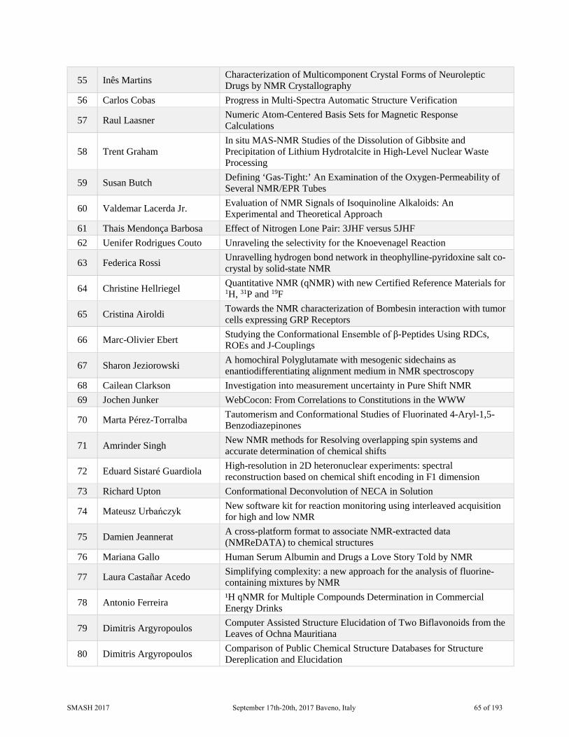
55 Inês Martins Characterization of Multicomponent Crystal Forms of Neuroleptic Drugs by NMR Crystallography
56 Carlos Cobas Progress in Multi-Spectra Automatic Structure Verification
57 Raul Laasner Numeric Atom-Centered Basis Sets for Magnetic Response Calculations
58 Trent Graham In situ MAS-NMR Studies of the Dissolution of Gibbsite and Precipitation of Lithium Hydrotalcite in High-Level Nuclear Waste Processing
59 Susan Butch Defining ‘Gas-Tight:’ An Examination of the Oxygen-Permeability of Several NMR/EPR Tubes
60 Valdemar Lacerda Jr. Evaluation of NMR Signals of Isoquinoline Alkaloids: An Experimental and Theoretical Approach
61 Thais Mendonça Barbosa Effect of Nitrogen Lone Pair: 3JHF versus 5JHF 62 Uenifer Rodrigues Couto Unraveling the selectivity for the Knoevenagel Reaction
63 Federica Rossi Unravelling hydrogen bond network in theophylline-pyridoxine salt co-crystal by solid-state NMR
64 Christine Hellriegel Quantitative NMR (qNMR) with new Certified Reference Materials for 1H, 31P and 19F
65 Cristina Airoldi Towards the NMR characterization of Bombesin interaction with tumor cells expressing GRP Receptors
66 Marc-Olivier Ebert Studying the Conformational Ensemble of β-Peptides Using RDCs, ROEs and J-Couplings
67 Sharon Jeziorowski A homochiral Polyglutamate with mesogenic sidechains as enantiodifferentiating alignment medium in NMR spectroscopy
68 Cailean Clarkson Investigation into measurement uncertainty in Pure Shift NMR 69 Jochen Junker WebCocon: From Correlations to Constitutions in the WWW
70 Marta Pérez-Torralba Tautomerism and Conformational Studies of Fluorinated 4-Aryl-1,5-Benzodiazepinones
71 Amrinder Singh New NMR methods for Resolving overlapping spin systems and accurate determination of chemical shifts
72 Eduard Sistaré Guardiola High-resolution in 2D heteronuclear experiments: spectral reconstruction based on chemical shift encoding in F1 dimension
73 Richard Upton Conformational Deconvolution of NECA in Solution
74 Mateusz Urbańczyk New software kit for reaction monitoring using interleaved acquisition for high and low NMR
75 Damien Jeannerat A cross-platform format to associate NMR-extracted data (NMReDATA) to chemical structures
76 Mariana Gallo Human Serum Albumin and Drugs a Love Story Told by NMR
77 Laura Castañar Acedo Simplifying complexity: a new approach for the analysis of fluorine-containing mixtures by NMR
78 Antonio Ferreira ¹H qNMR for Multiple Compounds Determination in Commercial Energy Drinks
79 Dimitris Argyropoulos Computer Assisted Structure Elucidation of Two Biflavonoids from the Leaves of Ochna Mauritiana
80 Dimitris Argyropoulos Comparison of Public Chemical Structure Databases for Structure Dereplication and Elucidation
SMASH 2017 September 17th-20th, 2017 Baveno, Italy 65 of 193
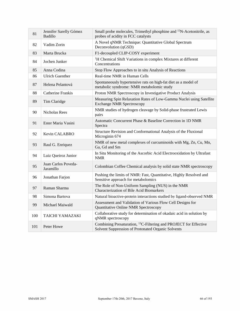
81 Jennifer Sarelly Gómez Badillo
Small probe molecules, Trimethyl phosphine and 15N-Acetonitrile, as probes of acidity in FCC catalysts
82 Vadim Zorin A Novel qNMR Technique: Quantitative Global Spectrum Deconvolution (qGSD)
83 Marta Brucka F1-decoupled CLIP-COSY experiment
84 Jochen Junker 1H Chemical Shift Variations in complex Mixtures at different Concentrations
85 Anna Codina Stop Flow Approaches to in situ Analysis of Reactions 86 Ulrich Guenther Real-time NMR in Human Cells
87 Helena Pelantová Spontaneously hypertensive rats on high-fat diet as a model of metabolic syndrome: NMR metabolomic study
88 Catherine Frankis Proton NMR Spectroscopy in Investigative Product Analysis
89 Tim Claridge Measuring Spin Relaxation Rates of Low-Gamma Nuclei using Satellite Exchange NMR Spectroscopy
90 Nicholas Rees NMR studies of hydrogen cleavage by Solid-phase frustrated Lewis pairs
91 Ester Maria Vasini Automatic Concurrent Phase & Baseline Correction in 1D NMR Spectra
92 Kevin CALABRO Structure Revision and Conformational Analysis of the Fluxional Microginin 674
93 Raul G. Enriquez NMR of new metal complexes of curcuminoids with Mg, Zn, Cu, Mn, Ga, Gd and Sm
94 Luiz Queiroz Junior In Situ Monitoring of the Ascorbic Acid Electrooxidation by Ultrafast NMR
95 Juan Carlos Poveda-Jaramillo Colombian Coffee Chemical analysis by solid state NMR spectroscopy
96 Jonathan Farjon Pushing the limits of NMR: Fast, Quantitative, Highly Resolved and Sensitive approach for metabolomics
97 Raman Sharma The Role of Non-Uniform Sampling (NUS) in the NMR Characterization of Bile Acid Biomarkers
98 Simona Bartova Natural bioactive-protein interactions studied by ligand-observed NMR
99 Michael Maiwald Assessment and Validation of Various Flow Cell Designs for Quantitative Online NMR Spectroscopy
100 TAICHI YAMAZAKI Collaborative study for determination of okadaic acid in solution by qNMR spectroscopy
101 Peter Howe Combining Presaturation, 13C-Filtering and PROJECT for Effective Solvent Suppression of Protonated Organic Solvents
SMASH 2017 September 17th-20th, 2017 Baveno, Italy 66 of 193
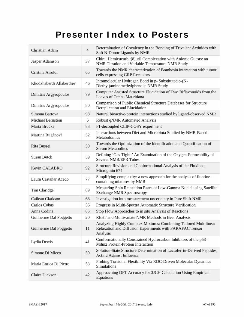
Presenter Index to Posters
Christian Adam 4 Determination of Covalency in the Bonding of Trivalent Actinides with Soft N-Donor Ligands by NMR
Jasper Adamson 37 Chiral Hemicucurbit[8]uril Complexation with Anionic Guests: an NMR Titration and Variable Temperature NMR Study
Cristina Airoldi 65 Towards the NMR characterization of Bombesin interaction with tumor cells expressing GRP Receptors
Khodzhaberdi Allaberdiev 46 Intramolecular Hydrogen Bond in p- Substituted o-(N- Diethyl)aminomethylphenols: NMR Study
Dimitris Argyropoulos 79 Computer Assisted Structure Elucidation of Two Biflavonoids from the Leaves of Ochna Mauritiana
Dimitris Argyropoulos 80 Comparison of Public Chemical Structure Databases for Structure Dereplication and Elucidation
Simona Bartova 98 Natural bioactive-protein interactions studied by ligand-observed NMR Michael Bernstein 6 Robust qNMR Automated Analysis Marta Brucka 83 F1-decoupled CLIP-COSY experiment
Martina Bugáňová 52 Interactions between Diet and Microbiota Studied by NMR-Based Metabolomics
Rita Bussei 39 Towards the Optimization of the Identification and Quantification of Serum Metabolites
Susan Butch 59 Defining ‘Gas-Tight:’ An Examination of the Oxygen-Permeability of Several NMR/EPR Tubes
Kevin CALABRO 92 Structure Revision and Conformational Analysis of the Fluxional Microginin 674
Laura Castañar Acedo 77 Simplifying complexity: a new approach for the analysis of fluorine-containing mixtures by NMR
Tim Claridge 89 Measuring Spin Relaxation Rates of Low-Gamma Nuclei using Satellite Exchange NMR Spectroscopy
Cailean Clarkson 68 Investigation into measurement uncertainty in Pure Shift NMR Carlos Cobas 56 Progress in Multi-Spectra Automatic Structure Verification Anna Codina 85 Stop Flow Approaches to in situ Analysis of Reactions Guilherme Dal Poggetto 20 REST and Multivariate NMR Methods in Beer Analysis
Guilherme Dal Poggetto 11 Analyzing Highly Complex Mixtures: Combining Tailored Multilinear Relaxation and Diffusion Experiments with PARAFAC Tensor Analysis
Lydia Dewis 41 Conformationally Constrained Hydrocarbon Inhibitors of the p53-Mdm2 Protein-Protein Interaction
Simone Di Micco 50 Solution-State Structure Determination of Lactoferrin-Derived Peptides, Acting Against Influenza
Maria Enrica Di Pietro 53 Probing Torsional Flexibility Via RDC-Driven Molecular Dynamics Simulations
Claire Dickson 42 Approaching DFT Accuracy for 3JCH Calculation Using Empirical Equations
SMASH 2017 September 17th-20th, 2017 Baveno, Italy 67 of 193
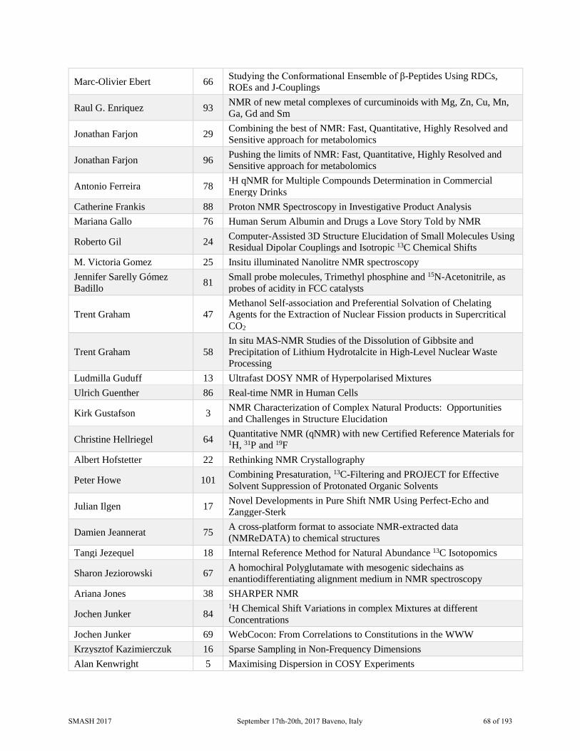
Marc-Olivier Ebert 66 Studying the Conformational Ensemble of β-Peptides Using RDCs, ROEs and J-Couplings
Raul G. Enriquez 93 NMR of new metal complexes of curcuminoids with Mg, Zn, Cu, Mn, Ga, Gd and Sm
Jonathan Farjon 29 Combining the best of NMR: Fast, Quantitative, Highly Resolved and Sensitive approach for metabolomics
Jonathan Farjon 96 Pushing the limits of NMR: Fast, Quantitative, Highly Resolved and Sensitive approach for metabolomics
Antonio Ferreira 78 ¹H qNMR for Multiple Compounds Determination in Commercial Energy Drinks
Catherine Frankis 88 Proton NMR Spectroscopy in Investigative Product Analysis Mariana Gallo 76 Human Serum Albumin and Drugs a Love Story Told by NMR
Roberto Gil 24 Computer-Assisted 3D Structure Elucidation of Small Molecules Using Residual Dipolar Couplings and Isotropic 13C Chemical Shifts
M. Victoria Gomez 25 Insitu illuminated Nanolitre NMR spectroscopy Jennifer Sarelly Gómez Badillo 81 Small probe molecules, Trimethyl phosphine and 15N-Acetonitrile, as
probes of acidity in FCC catalysts
Trent Graham 47 Methanol Self-association and Preferential Solvation of Chelating Agents for the Extraction of Nuclear Fission products in Supercritical CO2
Trent Graham 58 In situ MAS-NMR Studies of the Dissolution of Gibbsite and Precipitation of Lithium Hydrotalcite in High-Level Nuclear Waste Processing
Ludmilla Guduff 13 Ultrafast DOSY NMR of Hyperpolarised Mixtures Ulrich Guenther 86 Real-time NMR in Human Cells
Kirk Gustafson 3 NMR Characterization of Complex Natural Products: Opportunities and Challenges in Structure Elucidation
Christine Hellriegel 64 Quantitative NMR (qNMR) with new Certified Reference Materials for 1H, 31P and 19F
Albert Hofstetter 22 Rethinking NMR Crystallography
Peter Howe 101 Combining Presaturation, 13C-Filtering and PROJECT for Effective Solvent Suppression of Protonated Organic Solvents
Julian Ilgen 17 Novel Developments in Pure Shift NMR Using Perfect-Echo and Zangger-Sterk
Damien Jeannerat 75 A cross-platform format to associate NMR-extracted data (NMReDATA) to chemical structures
Tangi Jezequel 18 Internal Reference Method for Natural Abundance 13C Isotopomics
Sharon Jeziorowski 67 A homochiral Polyglutamate with mesogenic sidechains as enantiodifferentiating alignment medium in NMR spectroscopy
Ariana Jones 38 SHARPER NMR
Jochen Junker 84 1H Chemical Shift Variations in complex Mixtures at different Concentrations
Jochen Junker 69 WebCocon: From Correlations to Constitutions in the WWW Krzysztof Kazimierczuk 16 Sparse Sampling in Non-Frequency Dimensions Alan Kenwright 5 Maximising Dispersion in COSY Experiments
SMASH 2017 September 17th-20th, 2017 Baveno, Italy 68 of 193
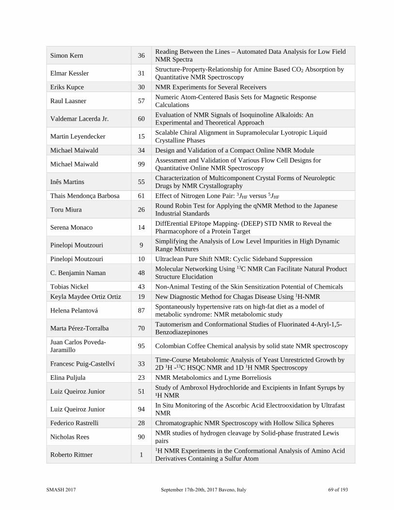
Simon Kern 36 Reading Between the Lines – Automated Data Analysis for Low Field NMR Spectra
Elmar Kessler 31 Structure-Property-Relationship for Amine Based CO2 Absorption by Quantitative NMR Spectroscopy
Eriks Kupce 30 NMR Experiments for Several Receivers
Raul Laasner 57 Numeric Atom-Centered Basis Sets for Magnetic Response Calculations
Valdemar Lacerda Jr. 60 Evaluation of NMR Signals of Isoquinoline Alkaloids: An Experimental and Theoretical Approach
Martin Leyendecker 15 Scalable Chiral Alignment in Supramolecular Lyotropic Liquid Crystalline Phases
Michael Maiwald 34 Design and Validation of a Compact Online NMR Module
Michael Maiwald 99 Assessment and Validation of Various Flow Cell Designs for Quantitative Online NMR Spectroscopy
Inês Martins 55 Characterization of Multicomponent Crystal Forms of Neuroleptic Drugs by NMR Crystallography
Thais Mendonça Barbosa 61 Effect of Nitrogen Lone Pair: 3JHF versus 5JHF
Toru Miura 26 Round Robin Test for Applying the qNMR Method to the Japanese Industrial Standards
Serena Monaco 14 DiffErential EPitope Mapping- (DEEP) STD NMR to Reveal the Pharmacophore of a Protein Target
Pinelopi Moutzouri 9 Simplifying the Analysis of Low Level Impurities in High Dynamic Range Mixtures
Pinelopi Moutzouri 10 Ultraclean Pure Shift NMR: Cyclic Sideband Suppression
C. Benjamin Naman 48 Molecular Networking Using 13C NMR Can Facilitate Natural Product Structure Elucidation
Tobias Nickel 43 Non-Animal Testing of the Skin Sensitization Potential of Chemicals Keyla Maydee Ortiz Ortiz 19 New Diagnostic Method for Chagas Disease Using 1H-NMR
Helena Pelantová 87 Spontaneously hypertensive rats on high-fat diet as a model of metabolic syndrome: NMR metabolomic study
Marta Pérez-Torralba 70 Tautomerism and Conformational Studies of Fluorinated 4-Aryl-1,5-Benzodiazepinones
Juan Carlos Poveda-Jaramillo 95 Colombian Coffee Chemical analysis by solid state NMR spectroscopy
Francesc Puig-Castellví 33 Time-Course Metabolomic Analysis of Yeast Unrestricted Growth by 2D 1H -13C HSQC NMR and 1D 1H NMR Spectroscopy
Elina Puljula 23 NMR Metabolomics and Lyme Borreliosis
Luiz Queiroz Junior 51 Study of Ambroxol Hydrochloride and Excipients in Infant Syrups by ¹H NMR
Luiz Queiroz Junior 94 In Situ Monitoring of the Ascorbic Acid Electrooxidation by Ultrafast NMR
Federico Rastrelli 28 Chromatographic NMR Spectroscopy with Hollow Silica Spheres
Nicholas Rees 90 NMR studies of hydrogen cleavage by Solid-phase frustrated Lewis pairs
Roberto Rittner 1 1H NMR Experiments in the Conformational Analysis of Amino Acid Derivatives Containing a Sulfur Atom
SMASH 2017 September 17th-20th, 2017 Baveno, Italy 69 of 193

Uenifer Rodrigues Couto 62 Unraveling the selectivity for the Knoevenagel Reaction
Federica Rossi 63 Unravelling hydrogen bond network in theophylline-pyridoxine salt co-crystal by solid-state NMR
Dave Russell 8 Overcoming Overlap in a Low-Field Reaction Monitoring System Using CRAFT (or Times, They Are A Changin’)
Paolo Sabatino 2 Identification of Non-Intentionally Added Substances in Packaging Materials by Pure-Shift NMR
Kanika Sarpal 54 Insights into Amorphous Solid Dispersions of Felodipine Using Solid-state NMR Spectroscopy: Miscibility and Molecular Interactions
Mira Schwab 32 Hot and Cool RDCs in One Alignment Medium Olga Semenova 7 Hyperpolarised Low-Field NMR for Industrial Reaction Monitoring
Raman Sharma 97 The Role of Non-Uniform Sampling (NUS) in the NMR Characterization of Bile Acid Biomarkers
Alexandra Shchukina 27 Time-resolved NUS Interleaved Experiments with Online Processing and Analysis
Amrinder Singh 71 New NMR methods for Resolving overlapping spin systems and accurate determination of chemical shifts
Davy Sinnaeve 49 General Approach to Access Long-Range ¹H-¹H RDCs
Eduard Sistaré Guardiola 72 High-resolution in 2D heteronuclear experiments: spectral reconstruction based on chemical shift encoding in F1 dimension
Stanislav Sykora 21 Molecular Spins and Persistent Magnetic Moments
Paul Tan 44 Characterisation of Catechol and (4-fluoro)-phenylboronic Acid Binding System: Investigation by F19-NMR and Modelling of Equilibria
Pablo Trigo Mourino 35 Label-Free NMR-Based Dissociation Rates Determination Richard Upton 73 Conformational Deconvolution of NECA in Solution
Mateusz Urbańczyk 74 New software kit for reaction monitoring using interleaved acquisition for high and low NMR
Joep van Rijn 12 Robust and Reliable Quantification of Phospholipids in Edible Oils
Ester Maria Vasini 91 Automatic Concurrent Phase & Baseline Correction in 1D NMR Spectra
TAICHI YAMAZAKI 100 Collaborative study for determination of okadaic acid in solution by qNMR spectroscopy
Siying Zhong 40 Synergy of Computation, NMR and Synthesis to Elucidate the Correct Structure of Baulamycins
Vadim Zorin 82 A Novel qNMR Technique: Quantitative Global Spectrum Deconvolution (qGSD)
SMASH 2017 September 17th-20th, 2017 Baveno, Italy 70 of 193

1 1H NMR Experiments in the
Conformational Analysis of Amino Acid Derivatives Containing a Sulfur
Atom
Roberto Rittner, Weslley G. D. P. Silva and Carolyne B. Braga
Chemistry Institute, University of Campinas, Campinas, SP, Brazil
The conformational analyses of L-methionine (L-Met) (1) and L-cysteine (L-Cys) (2), the two essential amino acids containing a sulfur atom, were performed including quantum theory calculations. Recent studies [1,2] have shown that an understanding of the interactions between the side chain and the backbone in an amino acid is essential to address the conformational preferences of peptides and proteins. Thus, the study of their N-acetylated derivatives using NMR data and quantum chemical calculations, including the solvent effect (IEF-PCM), combined with 3JHH spin-spin coupling constants analyses were carried out.
Theoretical calculations (ωB97X-D/aug-cc-pVTZ) resulted in 8 and 4 more stable conformers for 1 and 2, respectively, which are in three different dispositions (a, b and c). Furthermore, the calculations showed that the most stable conformers of 1 and 2 are in the form a, but when the dielectric constant (ε) of the medium is increased, these conformers are destabilized and have their populations decreased, indicating that the conformational equilibrium of 1 and 2 are affected by the solvent changes.
The solvent effect was also evaluated through experimental and theoretical 3JHH coupling constants. The observed values for 3JHaHb1 and 3JHaHb2 confirm the predominance of conformers a of 1 and 2 in the less polar solvent (chloroform). When ε is increased (from CDCl3 to DMSO), an increase in the 3JHaHb1 is observed and it indicates that conformers c of 1 are favoured in the conformational equilibrium, since this disposition has a hydrogen Ha anti to Hb1. Unlike 1, in more polar solvents conformers b of 2, which have Ha anti to Hb2, have their populations increased due to an increase in the 3JHaHb2. Thus, the changes in the values of 3JHaHb1 and 3JHaHb2 support our theoretical findings that the conformational equilibria of 1 and 2 are affected by the solvent change. The theoretically calculated 3JHH,calc are in agreement with the experimental data and reproduces well the trend observed by these compounds.
To investigate the effects (steric, hyperconjugation and hydrogen bonding) rulling the conformational preferences of the studied compounds, NBO, QTAIM and NCI analyses were carried out, as in a previous work in this laboratory [3]. These analyses showed that not only an specific interaction rules the conformational preferences of 1 and 2, but an interplay between steric repulsion and hyperconjugation
1. Newberry, R.W., Raines, R.T. Nat. Chem. Biol. (2016) 1–6. 2. Kecel, S., Ozel, A.E., Akyuz, S., Celik, S. Spectroscopy (2010) 24 219-232. 3. Braga, C. B., Rittner,
R., J. Phys. Chem. A (2017) 121 729-740. Acknowledgements: CNPq, FAPESP
SMASH 2017 September 17th-20th, 2017 Baveno, Italy 71 of 193

2 Identification of Non-Intentionally Added Substances in Packaging
Materials by Pure-Shift NMR
Paolo Sabatino1, Maria Oikonomou1, Jianbo Hou2 and Yiyong He2
1. Corporate R&D, Dow Benelux B.V., Terneuzen, The Netherlands 2. Corporate R&D, The Dow Chemical Company, Midland, MI, USA
Non-intentionally added substances (NIAS) are undesired chemical species present in formulated materials. The origin of the NIAS can vary; for example, impurities and/or decomposition of the starting materials, byproducts formed during side reactions etc. Independently of the source of contamination, the presence of NIAS in food packaging materials poses serious questions regarding the potential health risks associated with the consumer user. As consequence, the characterization of the low molecular weight fraction, especially as it relates to oligomers or reaction products of the starting monomers, is of fundamental importance.
Given the complexity of these formulated materials, the identification of NIAS, directly in the final packaging polymer or in the migration samples, represents one of the most challenging and complicated tasks performed by analytical groups within the chemical industry. The very low LOD required (in the ppb range) makes carbon measurements ineffective even when the samples are pre-concentrated several times. On the other hand, the concomitant presence of several species makes the proton NMR spectrum too crowded, limiting the amount of useful information that can be obtained.
In this work, we show a combination of 1D and 2D Pure-Shift techniques we could overcome the intrinsic resolution limitation of proton experiments and were able to identify several NIAS in a particularly challenging case. The implementation of these novel experiments enabled the use of NMR for the identification of NIAS in food packaging materials.
1. M. Foroozandeh, R. W. Adams, N. J. Meharry, D. Jeannerat, M. Nilsson, G. A. Morris, “Ultrahigh-Resolution NMR Spectroscopy”, Angew. Chem. Int. Ed., 53, (2014), 6990–6992.
SMASH 2017 September 17th-20th, 2017 Baveno, Italy 72 of 193

3 NMR Characterization of Complex Natural Products: Opportunities and Challenges in Structure Elucidation
Kirk R. Gustafson,1 Kentaro Takada,1 Naoya Oku,1 Yizhou Liu,2 Josep Saurí,2 R. Thomas Williamson,2
and Gary E. Martin2
1. Molecular Targets Laboratory, Center for Cancer Research, National Cancer Institute-Frederick, Maryland, USA
2. NMR Structure Elucidation, Process & Analytical Chemistry, Merck & Co. Inc., Rahway, New Jersey, USA
NMR provides powerful structural elucidation tools that are particularly well suited for natural products studies. Comprehensive spectroscopic characterization of a native metabolite may be sufficient to fully assign a planar structure. However assignment of the relative and absolute configuration of a molecule when there are multiple stereogenic centers often requires the development and application of additional experimental strategies. Many successful approaches in this regard rely on the formation of appropriate derivatives for more detailed NMR study. Since natural products are often obtained in very limited quantities, micro-scale chemical manipulations and the ability to analyze the structure of the resulting products is often key to the complete structural and configurational assignment of complex metabolites. Anisotropic NMR parameters such as residual dipolar coupling (RDC) and residual chemical shift anisotropy (RCSA) provide a powerful and complementary means to help assign and verify structures deduced from conventional NMR analyses.1-6 Application of a wide spectrum of NMR techniques and methodologies will be described in the structural elucidation of a new phosphomacrolide marine natural product with 8 stereogenic centers.
1. Liu, Y.; Saurí, J.; Mevers, E.; Peczuh, M. W.; Hiemstra, H.; Clardy, J.; Martin, G. E.; Williamson, R. T. Science, 356, eaam5349, 2017.
2. Nath, N.; Schmidt, M.; Gil, R. R.; Williamson, R. T.; Martin, G. E.; Navarro-Vázquez, A.; Griesinger, C.; Liu, Y. J. Am. Chem. Soc., 138, 9548-9556, 2016.
3. Castañar, L.; García, M.; Hellemann, E.; Nolis, P.; Gil, R. R.; Parella, T. J. Org. Chem., 81, 11126-11131, 2016.
4. Gil-Silva, L. F.; Santamaría-Fernández, R.; Navarro-Vázquez, A.; Gil, R. R. Chemistry Eur. J., 22, 472-476, 2016.
5. Schmidts, V. Magn. Reson. Chem., 55, 54-60, 2017. 6. Troche-Pesqueria, E.; Anklin, C.; Gil, R. R.; Navarro-Vázquez, A. Angew. Chem. Int. Ed., 56, 3660-
3664, 2017.
SMASH 2017 September 17th-20th, 2017 Baveno, Italy 73 of 193

4 Determination of Covalency in the Bonding of Trivalent Actinides with
Soft N-Donor Ligands by NMR Christian Adam1, Peter Kaden2, Viktoria Rohde3, Björn B. Beele4, Andreas Geist1, Horst Geckeis1 and
Petra J. Panak1,5
1. Karlsruhe Institute of Technology (KIT), Institute for Nuclear Waste Disposal (INE), Eggenstein-
Leopoldshafen, Germany 2. Helmholtz-Zentrum Dresden - Rossendorf e. V., Institute of Resource Ecology, Dresden, Germany
3. Fraunhofer Institute for Chemical Technology (ICT), Environmental Engineering, Pfinztal, Germany 4. University of Wuppertal, Institute of Inorganic Chemistry, Wuppertal, Germany
5. Heidelberg University, Institute of Physical Chemistry, Heidelberg, Germany In possible future actinide recycling processes the separation of the chemically highly similar trivalent actinide (An) ions from lanthanide (Ln) ions by liquid-liquid extraction is a challenging task. A number of extraction ligands with the required selectivity (separation factors >100) for the separation of An(III) from Ln(III) have been developed. Among those, soft N-donor ligands of the bis-triazinylpyridine type (BTP) have been very successful [1, 2]. However, the molecular origin of these ligands’ selectivity remains evasive.
NMR spectroscopy of the paramagnetic An(III) complexes and their comparison to structurally analogue Ln(III) complexes offers insights into the bonding situation. The overall paramagnetic chemical shift consists of the pseudo-contact shift (PCS), which is transmitted through-space, and the Fermi contact shift (FCS), transmitted through-bond. By separation of the two contributions, important information on the share of covalency in the An-ligand-bond is obtained [3]. As the coordination involves the nitrogen lone pair in the aromatic BTP core, 15N NMR is an especially valuable tool. By 15N isotope enrichment of several extraction ligands the chemical shifts even for the coordinating nitrogen atoms were observed throughout the Ln(III) series and for Am(III) complexes. Our results indicate that an increased share of covalency in the An-ligand bond is the driving force for the observed selectivity [4, 5].
The aliphatic side-chains of the aromatic cores of the most commonly used extraction agents do not only influence the solubility in organic solvents. They are crucial for the stability againts hydrolysis and radiolysis, and also have strong effects on the coordination kinetics and complex stability. We showed by comparison of extraction ligands with different side chains (nPr vs. iPr) that differences in coordination behaviour are not due to changes in the electronic structure but rather due to sterical reasons or different complex hydration [6].
While NMR is established as a routine method in inorganic and organic chemistry, it has so far found only limited use on complexes of paramagnetic metal ions in particular of the trans-uranium elements. We will show the merits of this method, which can deliver information that have previously not been available from other methods that have been used in actinide research for years.
1. Panak, Petra J., Geist, Andreas, Chem. Rev., 113(2), 1199-1236, 2013.
SMASH 2017 September 17th-20th, 2017 Baveno, Italy 74 of 193

2. Ekberg, Christian; Fermvik, Anna; Retegan, Teodora; Skarnemark, Gunnar; Foreman, M. R. S.; Hudson, M. J.; Englund, Sofie, Nilsson, Mikael, Radiochim. Acta, 96(4-5), 225-233, 2008.
3. Piguet, Claude, Geraldes, Carlos F. G. C., in Handbook on the Physics and Chemistry of Rare Earths, Vol. Volume 33 (Eds.: J. K.A. Gschneidner, J. C. G. Bünzli, V. K. Pecharsky), Elsevier, 2003, pp. 353-463.
4. Adam, Christian; Beele, Björn B.; Geist, Andreas; Müllich, Udo; Kaden, Peter, Panak, Petra J., Chem. Sci., 6(2), 1548-1561, 2015.
5. Adam, Christian; Kaden, Peter; Beele, Björn B.; Müllich, Udo; Trumm, Sascha; Geist, Andreas; Panak, Petra J., Denecke, Melissa A., Dalton Trans., 42(39), 14068-14074, 2013.
6. Adam, Christian; Rohde, Viktoria; Müllich, Udo; Kaden, Peter; Geist, Andreas; Panak, Petra J., Geckeis, Horst, Procedia Chem., 21, 38-45, 2016.
SMASH 2017 September 17th-20th, 2017 Baveno, Italy 75 of 193
5 Maximising Dispersion in COSY Experiments
Alan M. Kenwright1, Juan A. Aguilar1, Raquel Belda1, and Benjamin R. Gaunt1
1. Durham University Chemistry Department, Durham, UK.
Two-dimensional NMR experiments have traditionally been recorded with limited digital resolution, primarily to limit the size of data sets and the time taken to acquire the data. This has had the effect of limiting the dispersion of signals, and can lead to problems in assigning very crowded spectra.
Recent advances in computing power and in techniques such as Pure Shift methods [1] and compressed sensing [2,3] mean that it is now practical to acquire some two-dimensional NMR experiments at digital resolutions that approach the intrinsic resolution limits of the instrumentation and sample [4].
COSY experiments represent a particular challenge because, in contrast to experiments such as HSQC, the transfer function evolves in the same time period as the chemical shift. This means that if we look to extend the evolution period to achieve high digital resolution we also see correlations due to smaller couplings, often over more than 3 bonds, which make interpretation of the spectra of unknown structures more difficult.
Here we present an experiment that overcomes many of these problems and allows the acquisition of Pure Shift COSY spectra at very high digital resolution while minimising the introduction of unwanted long-range correlations. The utility of such an experiment is demonstrated.
1. M. Foroozandeh, R. W. Adams, N. J. Meharry, D. Jeannerat, M. Nilsson and G. A. Morris, Angew. Chem., Int. Ed., 2014, 53, 6990–6992.
2. D. L. Donoho, IEEE Trans. Inf. Theory, 2006, 52, 1289–1306 3. E. J. Candes and M. B. Wakin, IEEE Signal Process. Mag., 2008, 25, 21–30 4. J. A. Aguilar, R. Belda, A. Botana and A. M. Kenwright, RSC Adv., 2016, 6, 83380–83385
SMASH 2017 September 17th-20th, 2017 Baveno, Italy 76 of 193

6 Robust qNMR Automated Analysis
Michael Bernstein, Agustín Barba, Vadim Zorin, and Carlos Cobas
Mestrelab Research, S.L Feliciano Barrera 9B – Bajo, 15706 Santiago de Compostela (SPAIN)
The use of NMR for quantitation is an everyday occurrence. A popular application is the purity determination of single organic compounds, which is, for example, a staple requirement of the pharmaceutical industry.
The total uncertainty in the measurement is a summation of all stages in the analysis, and is well understood. Remarkably, quantitation by NMR (qNMR) routinely achieves results with <2% total uncertainty with only modest effort. With more effort, and skilled operators, this figure can be quite routinely reduced by an order of magnitude. qNMR is a validated primary procedure.
In many areas of NMR analysis the trend is towards automation, and implementation of more complex NMR analysis tasks to analysts having less NMR training and basic understanding. Indeed, the logical extension is to extend this to fully automated analysis. This imposes new challenges to NMR software capability because of the need for extremely reliable and sophisticated algorithms and work flows. An extension to automated qNMR adds a layer of necessary robustness, given the nature of the analysis and the regulated environments where it is often applied.
Here we will focus on NMR signal processing and analysis requirements in the context of qNMR. Many previously routine tasks require rethinking and new algorithms to ensure effective quantitation.
qNMR analysis itself is not an especially difficult procedure in principle, but there are many ways that it can be adversely influenced through naïve, improper operation. A surprisingly sophisticated analysis system is therefore required to ensure reliable results for a very high percentage of samples. We have developed algorithms and general capablities that addresses automated processing and analysis even with these quite challenging requirements. We show that fully automated qNMR processing and analysis is achievable when such a combination of elegant techniques are applied together.
SMASH 2017 September 17th-20th, 2017 Baveno, Italy 77 of 193
7 Hyperpolarised Low-Field NMR for Industrial Reaction Monitoring
Olga Semenova1, Peter Richardson1, Andrew Parrott2, Alison Nordon2, Meghan Halse1 and Simon
Duckett1
1. Centre of hyperpolarisation of Magnetic Resonance, Chemistry Department University of York, York, UK
2. Chemistry Department, Strathclyde University, Glasgow, UK Reaction monitoring is an important part of synthetic development, scale-up procedures and industrial quality control [1]. Process analytical tools (PAT) serve to control these steps in industry. Reliable analytical tools that can be used to follow a process are required. Low-cost benchtop NMR spectrometers that are based on permanent magnets with field strengths of around 1 T offer the opportunity to develop NMR as an analytical tool for continuous reaction monitoring within an industrial site [2]. As a consequence of the relatively low magnetic field, these instruments suffer from low sensitivity and reduced chemical shift dispersion. Here we seek to investigate the viability of NMR reaction monitoring at low field by increasing sensitivity via the parahydrogen (p-H2) hyperpolarisation technique SABRE (Signal Amplification By Reversible Exchange) [3].
To illustrate this concept, we identify and follow a well-defined model reaction, such as the conversion of an amine into an amide. In this work, we describe studies on the level of SABRE enhancement seen for mixtures of reactant and product molecules in order to develop methods to quantitatively follow a reaction using hyperpolarisation. In addition, we investigate the effect of the SABRE process on the model reaction and demonstrate that the expected reaction products form in the presence of the SABRE catalyst. We show that the hyperpolarised NMR signals change with time as the starting material and product molecules concentrations in the reaction mixture evolve, indicating the viability of this method for reaction monitoring. In addition to this proof-of-concept work carried out on a standard high-field (400 MHz) NMR spectrometer, we present preliminary data from a benchtop (43 MHz) instrument to illustrate the feasibility of our approach.
Key words: reaction monitoring, NMR, hyperpolarisation, benchtop.
1. Bakeev, K.A., Process Analytical Technology, Blackwell Publishing, 2005. 2. Foley, D.A., Zell, M.T., Marquez, B. L., Kaerner, A., Pharmaceutical Technology, Analytical
Technology and Laboratory Testing, Supplement to November Issue, p. 19-21, 2011. 3. Adams, R. W., Aguilar, J. A., Atkinson, K. D., Cowley, M. J. Elliott, P. I., Duckett, S. B., Green, G.
G., Khazal, I. G., Lopez-Serrano, J., Williamson, D. C., Science, 323 (5922), p. 1708-1711, 2009.
SMASH 2017 September 17th-20th, 2017 Baveno, Italy 78 of 193

8 Overcoming Overlap in a Low-Field Reaction Monitoring System Using
CRAFT (or Times, They Are A Changin’)
Dave Russell
Genentech, South San Francisco, CA, USA
There are significant advantages to using NMR in a Process Analytical/Reaction Monitoring role. Given the operating environment and financial constraints involved with superconducting magnets, low-field systems offer significant advantages. [1] Signal overlap is a problem even at high field, however, and the requirement to work in protonated solvents means that every analysis is challenged by the dynamic range of the system. CRAFT (Complete Reduction to Amplitude Frequency Table) [2, 3] offers a unique alternative to the traditional frequency-based approach to NMR data processing. It provides the advantage of returning high-quality qNMR results from complex spectra and powerful post-acquisition tools to handle large solvent resonances. This presentation will demonstrate results from our implementation of a 60 MHz NMR system using CRAFT-based data reduction to better understand reaction chemistries in a process chemistry environment, showing the advantages provided by time domain analysis.
1. “Monitoring chemical reactions by low-field benchtop NMR at 45MHz: pros and cons,” Maria Victoria Silva Elipe and Robert R. Milburn Magn. Reson. Chem. 2016, 54, 437–443.
2. “CRAFT (complete reduction to amplitude frequency table) – robust and time-efficient Bayesian approach for quantitative analysis by NMR” Krish Krishnamurthy, Magn. Reson. Chem., 2013, 51, 821-219.
3. “NMR Quantitative Analysis of Complex Mixtures” David J. Russell and Krish Krishnamurthy, eMagRes, 2014, Vol 3; 235-242. DOI 10.1002/9780470034590.emrstm1366.
SMASH 2017 September 17th-20th, 2017 Baveno, Italy 79 of 193

9 Simplifying the Analysis of Low Level Impurities in High Dynamic
Range Mixtures Pinelopi Moutzouri1, Peter Kiraly1, Andrew R. Phillips2, Steven R. Coombes3, Mathias Nilsson1, and
Gareth A. Morris1
1. School of Chemistry, University of Manchester, Manchester, UK,
2. Pharmaceutical Sciences, AstraZeneca, Macclesfield, UK 3. Pharmaceutical Technology and Development, AstraZeneca, Macclesfield, UK
The analysis of high dynamic range mixtures is always a challenge, and it is even more demanding when the main interest lies in the detection and quantification of low level components of the sample. In pharmaceutical chemistry, for example, all components above 0.1 % of a main active ingredient need to be identified and quantified. NMR can be very useful in this area, but the presence of 13C satellites complicates matters. 13C isotopomer signals have similar intensities, and often similar chemical shifts, to the resonances of interest. Broadband 13C decoupling is always a possibility, but can cause severe sample heating if good resolution is needed. Moreover, for nuclei with large secondary isotope shifts like 19F this approach does not solve the problem, merely halving the number of 19F-13C signals. Here, we suggest an alternative approach for dealing with 13C isotopomer signals in both 1H and 19F NMR, which relies on their suppression rather than decoupling.
In the case of 19F, a low-pass 1JCF filter followed by a heteronuclear 2DJ experiment allows both the one-bond and long range satellites to be suppressed [1]. This 2D acquisition scheme bypasses the problem caused by secondary isotope shifts, since the final 1D spectrum is extracted as an integral projection onto the F2 axis for an F1 range close to F1 = 0, which includes only 12C isotopomer signals. The final clean spectrum is obtained at only very modest cost in sensitivity. 19F-19F coupling is a potential problem, but can usually be dealt with by using selective 180° 19F pulses.
A related approach can be used in the case of 1H NMR. Here the problem of homonuclear coupling is dealt with by using a perfect echo [2], during which 13C 90° pulses are used to convert 1H coherence of 13C isotopomers into unobservable MQC. This is followed by a z-filter, to allow the acquisition of clean 1H spectra for 12C-bound protons only. The perfect echo provides time for four successive 90o carbon pulses; their timing is optimised to allow suppression over a wide range of 1JCH. Because 1H secondary isotope shifts are very small, long range 13C satellites are much less of a problem than in 19F NMR and are generally hidden under the parent signals, but if necessary low power 13C decoupling can be applied to collapse them.
In both the 19F and the 1H experiment the suppression of the 13C satellites signals leads to clean spectra with no visible 13C couplings, facilitating the analysis of high dynamic range mixtures.
1. Moutzouri P., Kiraly P., Phillips A. R., Coombes S. R., Nilsson M., and Morris G. A., Chem. Commun. 53, 123 (2017).
2. Takegoshi K., Ogura K., and Hikichi K., J. Magn. Reson. 84, 611 (1989); Zijl P. C. van, Moonen C. T. W., and Kienlin M. J. von, J. Magn. Reson., 89, 28 (1990).
SMASH 2017 September 17th-20th, 2017 Baveno, Italy 80 of 193
10 Ultraclean Pure Shift NMR: Cyclic Sideband Suppression
Pinelopi Moutzouri1, Yingxian Chen1, Mohammadali Foroozandeh1, Peter Kiraly1, Andrew R. Phillips2,
Steven R. Coombes3, Mathias Nilsson1, and Gareth A. Morris1
1. School of Chemistry, University of Manchester, Manchester, UK,
2. Pharmaceutical Sciences, AstraZeneca, Macclesfield, UK 3. Pharmaceutical Technology and Development, AstraZeneca, Macclesfield, UK
In all but the simplest 1H spectra, signal overlap is a problem. Pure shift NMR experiments can offer a vital resolution improvement, suppressing the effects of homonuclear couplings J, but the spectral simplicity that results comes at a price. Current pure shift experiments typically work by periodically refocusing the effects of J, either during a 1D acquisition (real-time methods) or in 2D mode (interferogram methods). The pure shift FID is recorded or constructed as a series of “chunks” of data of duration 1/sw1, in which J is refocused at the midpoint. The choice of sw1 requires a compromise between the quality of decoupling and either sensitivity (interferogram methods) or linewidth and spectral purity (real-time methods). Because J is only perfectly refocused at the midpoint of each data chunk, the pure shift FID retains a slight periodic modulation, which Fourier transforms into “chunking sidebands” spaced at multiples of sw1 either side of the pure shift peaks. With a typical sw1 of 40 Hz such sidebands are of the order of a few % or smaller and can generally be neglected, but in high dynamic range samples they can obscure real signals of interest.
The impact of such sidebands can be reduced by spreading them out, averaging spectra measured with different values of sw1 [1]. Here in contrast we propose to suppress them completely, by manipulating the phase of the residual J modulation. Small (ms) changes in pulse sequence timing are used to change the phases of sidebands over a 360° range while leaving the parent signals unaffected, so that averaging gives a clean spectrum. Using 2N different timings during time averaging allows sidebands to be suppressed to arbitraty order N.
1. Mauhart J., Glanzer S., Sakhaii P., Bermel W., and Zangger K., J. Magn. Reson., 259, 207 (2015).
SMASH 2017 September 17th-20th, 2017 Baveno, Italy 81 of 193

11 Analyzing Highly Complex Mixtures: Combining Tailored Multilinear
Relaxation and Diffusion Experiments with PARAFAC Tensor
Analysis
Guilherme Dal Poggetto, Laura Castañar, Ralph W. Adams, Gareth A. Morris and Mathias Nilsson
School of Chemistry, University of Manchester, Manchester, UK. To extract sensible NMR information from very complex mixtures is an important but also a formidable task. In this context PARAFAC (PARAllel FACtor analysis) [1], a powerful extension of component analysis to higher order arrays, has been used successfully for relatively simple mixtures. The power of PARAFAC stems from its ability to analyze multilinear data, i.e data that vary independently as a function of three or more variables. Previously, for NMR data, this has meant a combination of a spectroscopic dimension with diffusion encoding (as in a DOSY experiment) [2] and a third domain such as relaxation [3] or concentration variation [4]. Using changes in concentration can easily cause e.g. chemical shift changes, that are detrimental to multivariate analysis; and relaxation generally violates the multilinearity required for PARAFAC analysis as different spins in a given molecule have different relaxation times, which has limited the use of PARAFAC in earlier experiments. Recently, we have proposed a step towards NMR experiments specifically tailored for PARAFAC analysis: Relaxation-Encoded Selective TOCSY (REST) [5], in which differences in relaxation between spins can be exploited to separate the signals of different spin systems by producing multilinear data. It uses a combination of selective excitation and isotropic mixing to ensure that all the signals measured for a given species originate from a single proton [6]. Combining REST1 and REST2 in a single experiment (REST1-2), or REST and diffusion (REST-DOSY), should allow the application of PARAFAC to very complex mixtures. Extending the REST experiment to higher order data is significantly more efficient than using bilinear multivariate methods such as SCORE [7]. In principle, this approach could allow efficient analysis of highly complex mixtures with a large number of chemical species at a high dynamic range.
1. R. Bro. “PARAFAC. Tutorial and applications”. Chemom. Intell. Lab. Syst. 1997, 38, 149. 2. K. F. Morris and C. S. Johnson Jr. “Resolution of Discrete and Continuous Molecular Size
Distributions by Means of Diffusion-Ordered 2D NMR Spectroscopy”. J. Am. Chem. Soc., 1993, 115, 4291.
3. M. Nilsson, A. Botana and G. A. Morris. “T1-Diffusion-Ordered Spectroscopy: nuclear magnetic resonance mixture analysis using parallel factor analysis”. Anal. Chem. 2009, 81, 8119.
4. M. Khajeh, A. Botana, M. A. Bernstein, M. Nilsson and G. A. Morris. “Reaction Kinetics Studied Using Diffusion-Ordered Spectroscopy and Multiway Chemometrics”. Anal. Chem. 2010, 82, 2102.
5. G. Dal Poggetto, L. Castañar, R. W. Adams, G. A. Morris and M. Nilsson. Unpublished. 6. D. G. Davis and A. Bax. “Simplification of 1H NMR Spectra by Selective Excitation of Experimental
Subspectra”. J. Am. Chem. Soc. 1985, 107, 7197. 7. M. Nilsson and G. A. Morris, “Speedy component resolution: an improved tool for processing
Diffusion-Ordered spectroscopy data”. Anal. Chem. 2008, 80, 3777.
SMASH 2017 September 17th-20th, 2017 Baveno, Italy 82 of 193

12 Robust and Reliable Quantification of Phospholipids in Edible Oils
Joep van Rijn, Paul Groen, Remco Muntendam, and Adriana Carvalho de Souza
DSM Biotechnology Center, Delft, The Netherlands
Crude plant oils will form a solid sediment called gum upon storage. These gums contain mainly phospholipids. To obtain commercial edible oil the gum has to be removed, this process is known as degumming. The oldest form degumming is obtained by mixing crude oils with water and the solid formed is removed by centrifugation. Enzymatic degumming can be applied to reduce oil losses during degumming. To monitor degumming processes a robust and accurate phospholipid quantification is needed. Several techniques have been applied for the phospholipids quantification in edible oils, such as ICP, LC and TLC. However, in the past decade 31P NMR has shown to have advantages above the previously mentioned techniques because of the possibility to quantify different phospholipids directly from oil in an accurate and fast manner. In this report, we show the application and performance of 1D 31P NMR method for quantification of several phospholipids in crude and refined oils. The method is robust and has a variation lower than 5% for most phospholipids
SMASH 2017 September 17th-20th, 2017 Baveno, Italy 83 of 193

13 Ultrafast DOSY NMR of Hyperpolarised Mixtures
Ludmilla Guduff, 1 Dennis Kurzbach, 2 Daniel Abergel, 2 Ilya Kuprov, 3 Carine van Heijenoort1 and Jean-
Nicolas Dumez1
1. ICSN-CNRS, Gif-sur-Yvette, France 2. ENS, Paris, France
3. University of Southampton, United Kingdom
The sensitivity of solution-state NMR experiments can be increased by several orders of magnitude with dissolution dynamic nuclear polarization (D-DNP). This hyperpolarisation strategy is of great potential use for the analysis of low concentrated mixtures of small molecules. [1]. The amplified polarisation, however, only lasts for a few tens of seconds or less, because of longitudinal relaxation. Diffusion-ordered spectroscopy (DOSY), a method to separate the NMR spectra of components in a mixture, would be useful for the analysis of hyperpolarised mixtures. Conventional DOSY experiments, however, require long acquisition durations that are not compatible with D-DNP.
Here we show that hyperpolarised 13C DOSY experiments can be recorded in a single scan, using a spatial encoding of the diffusion dimension. We first describe a pulse sequence for spatially encoded (SPEN) DOSY, based on the work of Keeler and Frydman [2,3], which accelerates experiments by an order of magnitude. The SPEN DOSY concept is generalised to 3D DOSY. [4]. We then use D-DNP to hyperpolarise a mixture of small molecules, together with SPEN DOSY for the acquisition of a time-series of 13C DOSY spectra. Several modifications are made to the original sequence, in order to compensate for the effect of convection [5], and to make it possible to acquire a time-series of spectra from a single dissolution experiments. Throughout the analysis, numerical simulations are used, based on a Fokker-Plank formalism to describe simultaneously the spin and spatial dynamics. Theses simulations are key to characterise the pulse sequence and derive an improved model to obtain more accurate diffusion coefficients from SPEN DOSY experiments. Hyperpolarised 13C DOSY is a promising tool for the monitoring of chemical reactions and the analysis of molecular interactions.
1. Dumez, J.-N. et al., P. Analyst 2015, 140, 5860–5863. 2. Thrippleton, M. J. et al., J. Magn. Reson. Chem. 2003, 41, 441–447. 3. Shrot, Y.; Frydman, L. J. Magn. Reson. 2008, 195, 226–231. 4. Guduff, L. et al., Chem. Commun. 2017, 53, 701-704. 5. Jerschow, A.; Müller, N. J. Magn. Reson. 1998, 132, 13–18.
Figure 4:a) 1D 13C spectrum. b) SPEN DOSY data.
SMASH 2017 September 17th-20th, 2017 Baveno, Italy 84 of 193

14 DiffErential EPitope Mapping- (DEEP) STD NMR to Reveal the
Pharmacophore of a Protein Target
Serena Monaco1, Louise E. Tailford2, Nathalie Juge2, and Jesus Angulo1
1. School of Pharmacy, University of East Anglia, Norwich, UK
2. Institute of Food Research, Norwich Research Park, Norwich, UK Saturation Transfer Difference (STD) NMR spectroscopy is extensively used to obtain epitope maps of ligands binding to protein receptors under fast exchange conditions [1], [2]. STD NMR reveals structural details of biomolecular recognition processes, which are fundamental to direct lead optimisation efforts in drug discovery.
Standard procedures seek uniform saturation of the receptor to identify regions of the ligand contacting the protein binding pocket. However, in this way, the experiment does not provide information about the “nature” of the amino acids surrounding the ligand in the bound state.
Here we report a novel protocol (DiffErential EPitope Mapping-STD NMR or DEEP-STD NMR) to identify the type of protein residues contacting the ligand [3]. We demonstrate that the approach constitutes a novel versatile method to orthogonally explore the nature (aliphatic, aromatic, polar or hydrophobic) of the amino acid residues lining the surface of the binding pocket and their orientation relative to the ligand.
As a proof of principle, we selected two relevant protein-ligand interactions from different areas of interest: i) the interaction of 3-nitrophenyl-α-galactoside (3NPG) with subunit B of Cholera Toxin (CTB) [4], well known to CTB inhibitor designers; and ii) the interaction of 2,7 anhydro Neu5Ac with the glycosyl hydrolase GH33 from Ruminococcus Gnavus [5], of great interest in gut microbiota fundamental research, as it is over-represented in individuals affected by Inflammatory Bowel Disease [6]. For both systems, high resolution X-Ray structures were available, allowing us to validate the protocol. The approach overall seems solid, versatile and promising for expanding the view of STD-NMR and going beyond its current limitations.
1. Mayer, M. and B. Meyer, Angewandte Chemie International Edition, 38(12), 1784-1788, 1999. 2. Angulo, J. and Nieto, P., European Biophysics Journal, 40, 135-150, 2011. 3. Monaco, S., Tailford, E. L., Juge, N., and Angulo J., submitted. 4. Fan, E., et al., Biological Crystallography, 57(2), 201-212, 2001. 5. Tailford, E. L., et al., Nature Communication, 6, 7624-7641, 2015. 6. Berry, D. and Reinisch, W., Best Practice and Research: Clinical Gastroenterology, 27, 47–58, 2013.
SMASH 2017 September 17th-20th, 2017 Baveno, Italy 85 of 193

15 Scalable Chiral Alignment in Supramolecular Lyotropic Liquid
Crystalline Phases
M. Leyendecker, N.-C. Meyer and C. M. Thiele
Technische Universität Darmstadt, Clemens-Schöpf Institut für Organische Chemie, Darmstadt, Germany
Benzene-1,3,5-tricarboxamides (BTAs) are known to self-assemble into rod like and helical supramolecules.[1] These stiff aggregates act as mesogenes to form thermotropic or lyotropic liquid crystals (LLC).[2, 3] Achiral substituted BTAs aggregate to racemic mixtures of (M)- and (P)-helices. One handed helices can be obtained by introducing enantiopure sidechains to BTAs or via a sergeant-and-soldier-principle (SaS) by mixing chiral and achiral substituted BTAs.[4] In this work several BTAs were synthesized and tested for their behavior as SaS-LLCs.
These are used as weak orienting media, which give access to residual dipolar couplings (RDCs). RDCs offer complementary structure information to the conventional NMR parameters for structure determination (as 3J-couplings and NOEs). For structure determination of small organic molecules the LLCs should be compatible with organic solvents and introduce only a low degree of order.[5] Chiral alignment media can transfer the chiral information to the analyte molecule. As a result of diastereomorphous interactions with chiral alignment media enantiomers can be differentiated.[6]
Until now, primarily polymer based LLCs and anisotropically swollen gels are utilized as alignment media in organic solvents.[7] BTAs are a promising new approach for organic solvent compatible alignment media as synthetic steps for polymerisation[8] and/or crosslinking[9] are redundant. Challenges linked to this steps as well as time consuming sample preparation[7,9] of polymer gels can be avoided.
We show here, that mixtures of chiral and achiral BTAs form LLCs in organic NMR-solvents. To investigate the capabilities of the SaS-LLCs as chiral and enantiodifferentiating alignment-media, the enantiomers of β-pinene were analysed. The characterization of THF-d8 as molecular probe through ²H-NMR-spectroscopy indicated that SaS-LLCs act as chiral alignment media for small organic molecules with the potential of scalable chiral characteristics.
1. S. Cantekin, T. de Greef, A. Palmans, Chem. Soc. Rev. 2012, 41, 6125–6137. 2. Y. Matsunaga, N. Miyajima, Y. Nakayasu, Bull. Chem. Soc. Jpn. 1988, 61, 207. 3. D. Wang, Y. Huang, J. Li, Chem. Eur. J. 2013, 19, 685–690. 4. M. Smulders, A. Schenning, E. Meijer, J. Am. Chem. Soc. 2008, 130, 606-611. 5. B. Böttcher, C. M. Thiele, eMagRes 2012, 1, 169-180. 6. M. Sarfati et al., Chem. Commun. 2000, 21, 2069-2081. 7. V. Schmidts, Magn. Reson. Chem. 2017, 55, 54–60. 8. A. Marx, C. M. Thiele, Chem. Eur. J. 2009, 15, 254-260. 9. T. Montag, C. M. Thiele, Chem. Eur. J. 2013, 19, 2271-2274.
SMASH 2017 September 17th-20th, 2017 Baveno, Italy 86 of 193
16 Sparse Sampling in Non-Frequency Dimensions
Rupashree Dass1, Mateusz Urbańczyk1, Paweł Kasprzak2, Wiktor Koźmiński3, Krzysztof Kazimierczuk1
1. Centre of New Technologies, University of Warsaw, Poland 2. Faculty of Physics, University of Warsaw, Poland
3. Centre of Biological and Chemical Research, University of Warsaw, Poland Serial NMR experiments are often used in chemical and physical analysis. Experiments in a series can differ in temperature (monitoring phase transitions), pulsed field gradient setting (diffusion-ordered spectroscopy) or can be performed at different moments of time (monitoring chemical reactions). Multidimensional spectrscopy, although very informative, is difficult to employ in such experiments. The measurement of multidimensional spectra is time-consuming, because of costly sampling of indirect time dimensions. The non-uniform sampling (NUS) can help to shorten the experiment, but so far was limited to frequency dimensions. In this study we show how to extend it to non-frequency (pseudo)dimensions: temperature, diffusion-encoding gradients and others. A variety of different processing techniques will be discussed: time-resolved NUS[1,2], sparse inverse Laplace transform[3,4] and Radon transform[5,6].
1. Mayzel M, Rosenlöw J, Isaksson L, Orekhov VY. J. Biomol. NMR 2014;58:129–39. 2. Dass R, Kozminski W, Kazimierczuk K. Anal. Chem. 2015;87:1337–1343. 3. Urbańczyk M, Bernin D, Koźmiński W, Kazimierczuk K. Anal. Chem., 2013;85:1828–1833. 4. Urbańczyk M, Koźmiński W, Kazimierczuk K. Angew. Chemie 2014;126:6582–6585. 5. Kupče E, Freeman R. J. Am. Chem. Soc. 2013; 135:2871–2874. 6. Dass R, Kasprzak P., Kazimierczuk K., Submitted.
SMASH 2017 September 17th-20th, 2017 Baveno, Italy 87 of 193

17 Novel Developments in Pure Shift NMR Using Perfect-Echo and
Zangger-Sterk
Julian Ilgen,1 Lukas Kaltschnee1,2 and Christina M. Thiele1
1. Clemens-Schöpf-Institut, Technische Universität Darmstadt, Germany 2. National Institute of Diabetes and Digestive and Kidney Disease (NIDDK), National Institutes of
Health, Bethesda, Maryland, USA The interpretation of proton spectra of molecules with increasing complexity is often hampered by signal overlap due to homonuclear scalar coupling. Recently, the application of pure shift techniques successfully enhanced the resolution in one- and multidimensional proton detected experiments by collapsing multiplet structures into singlets.
A major drawback of methods for broadband homonuclear decoupling is their inherent sensitivity loss. In Zangger-Sterk[1] based techniques this is caused by a decrease in sample volume contributing to detectable signal. Various approaches have been presented to reduce this sensitivity penalty, such as multi-slice-excitation, nemo-Zangger-Sterk, sequential slice selection, ASAP or PSYCHE.[2-6]
Here we present a modification to Zangger-Sterk decoupling, which yields homonuclear decoupling for coupled pairs of spins even if they share the same volume element. This in turn can drastically improve the sensitivity of the experiment, as larger volume elements can be used for signal detection. These features are achieved by combination of Zangger-Sterk decoupling with the Perfect-Echo[7] experiment. Even in the presence of strong coupling the Perfect-Echo-Zangger-Sterk experiment produce clean spectra with good tolerance towards the occurrence of unwanted artefacts.
We show that this experiment is particularly suitable to study samples with strong signal clustering, a situation which can render classic Zangger-Sterk decoupling inefficient. Applications in one- and two-dimensional experiments are presented for oligomeric structures, which feature small chemical shift differences for atomic positions in the repetitive structural motives.
1. Zangger, K. and Sterk, H. J. Magn. Reson., 124, 486–489 1997. 2. Castañar, L., Nolis, P., Virgili, A. and Parella, T. Chem. Eur. J., 19(51), 15472–15475 2013. 3. Cotte, A. and Jeannerat, D. Angew. Chem. Int. Ed., 54, 6016–6018 2015. 4. Sakhaii, P., Haase, B., Bermel, W., Kerssebaum, R., Wagner, G. E. and Zangger, K., J. Magn. Reson.,
233, 92–95 2013. 5. Suryaprakash, L. and Suryaprakash, N. Chem. Commun., 50, 8550–8553 2014. 6. Foroozandeh, M., Adams, R. W., Meharry, N. J., Jeannerat, D., Nilsson, M. and Morris, G. A. Angew.
Chem. Int. Ed., 53, 6990–6992 2014. 7. Takegoshi, K., Ogura, K. and Hikichi, K. J. Magn. Reson., 84, 611–615 1989
SMASH 2017 September 17th-20th, 2017 Baveno, Italy 88 of 193
18 Internal Reference Method for Natural Abundance 13C Isotopomics
Tangi Jézéquel1, Katy Dinis1, Kévin Bayle1, Virginie Silvestre1, Serge Akoka1, and Patrick Giraudeau1,2
1. Université de Nantes, CNRS, CEISAM UMR 6230, Nantes, France
2. Institut Universitaire de France, Paris, France Isotope ratio monitoring by NMR spectrometry (irm-NMR) provides the complete 2H or 13C intramolecular position-specific composition at natural abundance, a strategy belonging to the recent concept of isotopomics [1]. Irm-2H has been developed in the 1980s [2], but the development of irm-13C NMR is much more recent. Indeed, this analytical tool requires an accuracy of 1‰ (against 1% for irm-2H NMR) due to the very small deviation range of 13C natural abundance values (only 50‰ against 500‰ for 2H). Numerous methodological developments have been necessary such as 1H decoupling using adiabatic pulses to reach such accuracy by 13C NMR [3].
Currently, measuring position-specific natural abundance 13C values relies on the combination of 13C acquisitions with isotope ratio mass spectrometry (IRMS) as a reference. This approach has two drawbacks: i) it requires two different analytical techniques on the same sample, ii) it suffers from the low sensitivity of 13C. To circumvent this constraint, we propose a new methodology to perform 13C isotopomics analysis on a single sample, via NMR only. In a first step, the concentration ratio of the chemical reference and the analyte must be determined by quantitative 1H NMR with a 1‰ accuracy.(1) Then, a 13C NMR analysis is performed on the same sample to determine the site-specific isotopic composition xi. The latter can either be performed via direct 13C detection or by polarization transfer methods. In both cases, the analyzed sample has to be highly concentrated in order to reach the required Signal-to-Noise Ratio in a realistic time in 13C NMR. Unfortunately, this is not compatible with the first step, since the analysis of highly concentrated samples by 1H NMR induces signal intensity and line shape distortions due to radiation damping (RD)(4) phenomenon which severely hampers the accuracy of quantitative 1H NMR.
In this context, we developed a new pulse sequence named DWET (Double-WET) to suppress RD and perform quantitative 1H NMR analysis on concentrated samples with a 1‰ accuracy. DWET detects only a slice from the sample by saturating the signal arising from lateral areas of the sensitive volume. This method has been successfully applied to isotopic analysis through the discrimination of vanillin samples as a function of their geographical origins based on the 13C isotopic profiles.(5) However, it appears that the DWET pulse sequence is unsuited for samples with short T1, which forms a serious limitation for irm-13C NMR experiments where a paramagnetic agent is added to decrease T1. In this context, we developed two variants of the DWET method, called Multi-WET (MWET) and Profiled-WET (PWET), to reach the same accuracy of 1‰ with a better immunity towards T1 variations. This new analytical package for 13C isotopomics at natural abundance opens a wide range of new applications in the fields of authentication and metabolism.
1. Jézéquel Tangi, Joubert Valentin, et. al. Magnetic Resonance in Chemistry, 55, 77-90, 2016. 2. Martin Gérard and Martin Maryvonne, Tetrahedron Letters, 22, 3525-3528, 1981 3. Tenailleau Estelle and Akoka Serge, Journal of Magnetic Resonance, 185, 50-58, 2007
SMASH 2017 September 17th-20th, 2017 Baveno, Italy 89 of 193

4. Krishnan Viswanathan and Murali Nagarajan, Progress in Nuclear Magnetic Resonance Spectroscopy, 68, 41-57, 2013
5. Bayle Kévin et. al. Analytical Chemistry, 87, 7550-7554, 2015
SMASH 2017 September 17th-20th, 2017 Baveno, Italy 90 of 193

19 New Diagnostic Method for Chagas Disease Using 1H-NMR
Keyla M. Ortiz1, Juan C. Poveda,1 Clara I. González,2 Mary H. Torres1, and Daniel R. Molina1
1. Escuela de Química. Universidad Industrial de Santander, Bucaramanga, Santander, Colombia
2. Escuela de Microbiología, Facultad de Salud, Universidad Industrial de Santander, Bucaramanga, Santander, Colombia
Chagas disease (CD) is caused by the protozoan parasite Trypanosoma cruzi[1]. This disease represents an important cause of morbidity and mortality in endemic countries, affects approximately 8 million people worldwide [2,3]. In Colombia, T. cruzi infection prevalence is around 5%, corresponding to 700,000 people, and in some areas of the department of Santander the seroprevalence is about 50% [4]. CD include an acute and a chronic phase, which presents a wide spectrum of clinical manifestations including cardiac, digestive and neurological forms [2,3]. The diagnosis of the infection with T. cruzi is made through serological and parasitological tests, which are used according to the clinical phase. The diagnosis is complex especially during the chronic phase, due to the lack of symptoms and the low or intermittent parasitemia that leads to direct parasitological methods having a low sensitivity. For this reason, the diagnosis is based on serological methods which detect the presence of specific antibodies directed against antigens of T. cruzi combined with clinical and epidemiological findings. However, serological tests present high sensitivity but lack specificity because of antigenic cross-reactivity with other parasites like Leishmania sp. and T. rangeli [5–7]. In this regard, World Health Organization suggested that at least two assays based on different techniques may be used in parallel to increase diagnostic accuracy because a single assay is not considered sufficiently sensitive and specific. Less invasive methods for diagnosis, such as determination of biomarkers from blood serum would be of significant advantage and useful for primary diagnosis, early detection and surveillance. Furthermore, non-invasive biomarkers could be used for knowledge of the metabolic alterations and it will help to understand the course of the infection. The aim of this work was to study the potential of NMR spectroscopy for the application of metabolomic studies in patients with CD to determine new metabolomic biomarkers of disease diagnostics.
In this work we use liquids state NMR spectroscopy to try to stablish a diagnosis method to patients of CD. The NMR experiments were performed on a Bruker Avance at 9.4T, 400 MHz for proton resonance. One-dimentional proton spectra were measure using a CPMG sequence [PRESET–90°–(δ–180°-)n-aq], which was optimized to remove the short T2 components arising due to the presence of high molecular weight proteins as well as to obtain a smooth baseline for multivariate analysis. All NMR spectra were binned to buckets including an equal width of 0.04 ppm. Water signal (4.6-4.8 ppm) was presaturated using low power level of 0.2 mW.
Chemical shift of 4.6 to 5.0 ppm was eliminated to remove water residual The Principal component analysis (PCA) and partial least-squares discriminant analysis (PLS-DA) was implemented to appraise grouping trends. For this purpose, groups of healthy volunteers (control group, CG) and patients with CD were compared. The potential biomarkers were found using the PLS-DA model. The results showed the metabolites important for the discrimination of the groups CG and CD. The predictive ability and validation of the PLS-DA model were determined using the predictive set of samples. According to the ROC curve
SMASH 2017 September 17th-20th, 2017 Baveno, Italy 91 of 193

and the AUC values, the PLS-DA model showed excellent discriminant properties for comparing CG and CD. This metabolites can be used like biomarkers of disease diagnostic.
The authors thanks the Vicerrectoria de Investigaciones of the Universidad Industrial de Santander for the analytical facilities of the nuclear magnetic resonance laboratory at Parque Tecnológico de Guatigura (Colombia).
1. Mantilla, J. C. Rev. Colomb. Cardiol. vol 18 (5), 249–261, 2011 2. Molina, I.; Salvador, F.; Sánchez-Montalvá, A. Enferm. Infecc. Microbiol. Clin. vol 34 (2), 132–138,
2016. 3. Rassi, A.; Rassi, A.; Marin-Neto, J. A. Lance. Vol 375 (9723), 1388–1402, 2010. 4. Duarte, L. F.; Flórez, O. R.; Rincón, G.; González, C. I. Colomb. Med. vol 45 (2), 61–66, 2014. 5. Ferrer, E.; Lares, M.; Viettri, M.; Medina, M. Enferm. Infecc. Microbiol. Clin. vol 31 (5), 277–282,
2012. 6. Castellanos, Y. Z.; Cucunubá, Z. M.; Flórez, A. C.; Orozco-Vargas, L. C. Biomedica vol 34 (2),
198–206, 2014. 7. Ponce, C.; Ponce, E.; Vinelli, E.; Aguilar, V. De; Gonzalez, A.; Rangel-aldao, R.; Levin, M. J.;
Umezawa, E. S.; Luquetti, A. O.; Montoya, A.; Zingales, B.; Esfandiari, J. J. Clin. Microbiol. vol 43 (10), 5065–5068., 2005.
SMASH 2017 September 17th-20th, 2017 Baveno, Italy 92 of 193
20 REST and Multivariate NMR Methods in Beer Analysis
Guilherme Dal Poggetto, Laura Castañar, Ralph W. Adams, Gareth A. Morris and Mathias Nilsson
School of Chemistry, University of Manchester, Manchester, UK
Diffusion-ordered spectroscopy (DOSY) [1] is one of the most commonly used NMR experiments in the analysis of mixtures. However DOSY is of limited use where mixture components have similar diffusion coefficients (i.e. similar hydrodynamic radii). Moreover, signal overlap can obscure diffusion information and complicate spectral interpretation, although this problem can be alleviated by multivariate analysis [2] if the compounds of interest have sufficiently different diffusion coefficients. When even this fails, differences in relaxation between different compounds can be exploited to separate signals if the combination of selective excitation and isotropic mixing is used to ensure that all the signals measured for a given species originate from a single proton. [3] Exploiting relaxation differences and focusing on selected spin connectivities can give great insight in mixture analysis, in a new class of experiments named Relaxation-Encoded Selective TOCSY (REST). The new method can for example use T1 encoding, e.g. by inversion or saturation recovery (REST1), or T2, e.g. using PROJECT [4] (REST2). REST has been used to investigate the carbohydrate content of a lager beer sample. The carbohydrate region of the 1H spectrum is very crowded and impossible to interpret directly, as is often the case for sugar mixtures. Using the REST2 experiment, followed by multivariate analysis with OUTSCORE, [5] spectra of individual components can be extracted.
1. K. F. Morris and C. S. Johnson Jr. “Resolution of Discrete and Continuous Molecular Size Distributions by Means of Diffusion-Ordered 2D NMR Spectroscopy”. J. Am. Chem. Soc., 1993, 115, 4291.
2. M. Nilsson and G. A. Morris. “Speedy Component Resolution: An Improved Tool for Processing Diffusion-Ordered Spectroscopy Data”. Anal. Chem., 2008, 80, 3777.
3. D. G. Davis and A. Bax. “Simplification of 1H NMR Spectra by Selective Excitation of Experimental Subspectra”. J. Am. Chem. Soc. 1985, 107, 7197.
4. J. A. Aguilar, M. Nilsson, G. Bodenhausen and G. A. Morris. “Spin echo NMR spectra without J modulation”. Chem. Commun., 2012, 48, 811.
5. A. A. Colbourne, S. Meier, G. A. Morris and M. Nilsson. “Unmixing the NMR spectra of similar species”. Chem. Commun., 2013, 49, 10510.
SMASH 2017 September 17th-20th, 2017 Baveno, Italy 93 of 193
21 Molecular Spins and Persistent Magnetic Moments
Stanislav Sykora
Extra Byte, Castano Primo, Italy
Some time ago [1,2], the author has proposed the theoretical concept of persistent currents in the electron shells of some diamagnetic molecules. If confirmed, these should be connected with the existence of permanent magnetic moments even in the absence of unpaired electrons, and of persistent molecular spins (angular momenta). A classical analogy for such quantum molecular situations might be, for example, the motorbike clutch: engaging in one sense, and free-running in the other. From the quantum mechanical point of view, this appears unavoidable, in particular in small molecules which lack cyclic symmetry, such as chloraldehyde or, in general C(XYZ), where C is a central atom and XYZ are three different atoms/groups arranged around it. A bonding electron partially localized near the central atom C, but affected by all three substituents X, Y, and Z, will necessarily behave as a wave moving differently in the cyclic sense (XYZ) rather than in the anti-cyclic sense (XZY). The result is a cyclically oriented ground state and a permanent current loop. In turn, the latter must give rise to a persistent magnetic moment and a persistent angular momentum (spin). Such persistent current loops are expected to exist also in a-cyclic fragments of large molecules, including proteins. In very small a-cyclic molecules, what makes them particularly interesting in the magnetic resonance context, is the fact that they would make the whole molecule behave as a unique spin particle, endowed with a magnetic moment, and therefore capable of exhibiting magnetic resonance at some (probably very low) Larmor frequency. If so, very-high-field magnetic resonance techniques could be used to study these phenomena. Another interesting aspect is that persistent electic currents in molecular electron shells are not an explicit part of the popular density function theory (though the DFT approximation might in part implicitly account for them). Making persistent electron current density, apart from elecron charge density, an explicit feature of DFT might be beneficial for its accuracy. In this presentation, the author would like to complement the original hypothesis with several quantitative estimates, as well as to point out the similarities and differences (considerable) between the hypothetical molecular spins and the spin-particles-as-we-know-them. 1. Stan Sykora, Gold Medal Talk at the 40th Annual Meeting of GIDRM, September 26-28, 2011, Parma,
Italy. 2. Stanislav Sykora: Molecular Spins: a novel Frontier of Chemical Physics and Magnetic Resonance?,
Presented at 53rd ENC, Miami, April 15-20, 2012, DOI: 10.3247/SL4Nmr12.005.
SMASH 2017 September 17th-20th, 2017 Baveno, Italy 94 of 193

22 Rethinking NMR Crystallography
Albert Hofstetter1, Martins Balodis1, Federico M. Paruzzo1, Arthur C. Pinon1, Gabriele Stevanato1, Cory
M. Widdifield2, Peter Bygrave3, Graeme M. Day3, and Lyndon Emsley1
1. Institut des Sciences et Ingénierie Chimiques, Ecole Polytechnique Fédérale de Lausanne (EPFL),
CH-1015 Lausanne, Switzerland. 2. Department of Chemistry, Durham University, Durham DH1 3LE, United Kingdom.
3. Division of Chemistry, University of Southampton, Southampton, United Kingdom. The physicochemical properties of crystalline solids are determined to a great extent by the atomic-level structural properties of the material of interest. This is especially important for pharmaceutically active materials, where bioavailability depends on crystal structure. This is also the case in many other areas, where exact knowledge of crystal structures is required, and as a result, the development of new and improved strategies for crystal structure determination will have a significant impact. Although single-crystal X-ray diffraction (SC-XRD) is the most powerful and routine technique for crystal structure determination, it is limited to single-crystals of appropriate quality and size. In the many cases, where the sample is either to some degree amorphous or a microcrystalline powder, structural characterization by SC-XRD is potentially not possible. Solid-state nuclear magnetic resonance (NMR) can overcome this limitation through its sensitivity to the local atomic environment regardless of the degree of long range order. Over the past decade the combination of NMR and computational methods has made tremendous progress [1] and today there are many examples of structure determination or validation by chemical shift measurements combined with density functional theory calculations (DFT)[2]. Recently there have been examples of de novo structure determination, combining NMR chemical shifts, DFT shift calculations and crystal structure prediction (CSP)[3]. However, there is currently a huge bottleneck in chemical shift based NMR crystallography, which is the time required for CSP calculations, which severely limits the complexity of the molecules that can be tackled. In general, CSP methods have two stages. First, an ensemble of gas-phase conformer structures is generated and sorted by their calculated internal energy. In the second stage the gas-phase conformers with the lowest internal energy are selected, and for each conformer an ensemble of possible crystal structures is generated. This procedure is based on the assumption that the low-energy gas-phase conformers are similar to those in the crystalline structures. Both stages involve searching the potential energy surface (PES) of the system, where the number of minima on the PES increases exponentially with the number of atoms. Often, only fully quantum mechanical calculations suffice to deliver the required level of accuracy[4]. These combined factors are responsible for the high computational cost associated with the CSP calculations. Here, we present a method that combines solid-state NMR measurements on powder samples with a computational analysis that allows the introduction of unbiased structural constraints restricting the area of interest of the PES that needs to be searched when using CSP in a structure determination process. These constraints are introduced during the gas-phase structure generation, where they can reduce the number of conformers by up to 90%, reducing the time required for the CSP procedure by an order of magnitude. This increase in efficiency allows the chemical shift based NMR crystal structure determination of structures of unprecedented complexity and size. We demonstrate the method on different pharmaceutically active
SMASH 2017 September 17th-20th, 2017 Baveno, Italy 95 of 193

compounds using constraints derived from a series of (1H-13C) HETCOR experiments. In particular, we show that the restrained CSP method allows the determination of the crystal structure of ampicillin, whereas unrestrained CSP fails to produce the correct structure. 1. Ashbrook, S. E. & McKay, D. Combining Solid-State NMR Spectroscopy with First-Principles
Calculations – A Guide to NMR Crystallography. Chem. Commun. 52, (2016). 2. Baias, M. u. a. Powder crystallography of pharmaceutical materials by combined crystal structure
prediction and solid-state 1H NMR spectroscopy. Phys. Chem. Chem. Phys. 15, 8069 (2013). 3. Baias, M. u. a. De novo determination of the crystal structure of a large drug molecule by crystal
structure prediction-based powder NMR crystallography. J. Am. Chem. Soc. 135, 17501–17507 (2013).
4. Pickard, C. J. & Needs, R. J. Ab initio random structure searching. J. Phys. Condens. Matter 23, 53201 (2011).
SMASH 2017 September 17th-20th, 2017 Baveno, Italy 96 of 193
23 NMR Metabolomics and Lyme Borreliosis
Elina Puljula1, Otto Glader2, Jukka Hytönen2, and Jari Sinkkonen1
1. Instrument Centre, Department of Chemistry, University of Turku, Turku, Finland
2. Medical Microbiology and Immunology, University of Turku, Turku, Finland Lyme borreliosis (LB), also known as Lyme disease, is the most important tick-transmitted disease globally [1, 2]. LB is caused by the spirochete Borrelia burgdorferi sensu lato. Borrelia is transferred to the human host via the tick’s saliva when the tick attaches itself to the skin of the host.
Clinically the LB infection can roughly be classified into early stage and disseminated stage of the disease. The early diagnosis of LB is possible without any laboratory testing if the patient is aware of the tick bite and a typical early rash lesion is visible at the tick bite site. However, it has been estimated that only 50 % of the patients with LB develop visible rash [3]. Thus, in atypical cases, with no or only a faint rash, laboratory confirmation of the infection would be most helpful. Unfortunately, no such test is currently available for the early stage of the disease, as the antibody production in early phase of the infection is too low for detection. The mainstay of LB laboratory testing today is the detection of borrelia specific antibodies in patient serum and cerebrospinal fluid. Direct diagnostic testing methods, such as cell culture or nucleic acid amplification, suffer from low sensitivity, and are therefore not practical.
Our aim was to develop a novel method for the diagnostics of LB by utilizing nuclear magnetic resonance spectroscopy (NMR) and/or mass spectrometry (MS). Both NMR and MS are suitable for metabolomics based research, i.e. studying the different substances formed in metabolic processes inside the body. With these methods, our aim is to detect biomarkers that are specific for LB.
To test the suitability of NMR metabolomics for the diagnostics of LB, we performed a preliminary study with Borrelia infected mice. Both urine and blood samples were collected from the infected mice. The samples were measured with a 600 MHz NMR instrument and samples from non-infected mice were used as controls. The data was analyzed by using multivariate analysis. From this data, clear differences between the Borrelia infected and the control mice could be seen, for example in the concentrations of organic acids, glucose and some amino acids. However, none of these molecules could be used as a specific biomarker for LB, as all of the molecular differences can be regarded as general biomarkers for infection.
1. Stanek, Gerold; Wormser, Gary P; Gray, Jeremy; Strle, Franc, The Lancet, 379, 461-473, 2012. 2. Hytönen, Jukka; Hartiala, Pauliina; Oksi, Jarmo; Viljanen, Matti K, Scandinavian Journal of
Rheumatology, 37, 161-172, 2008.
SMASH 2017 September 17th-20th, 2017 Baveno, Italy 97 of 193
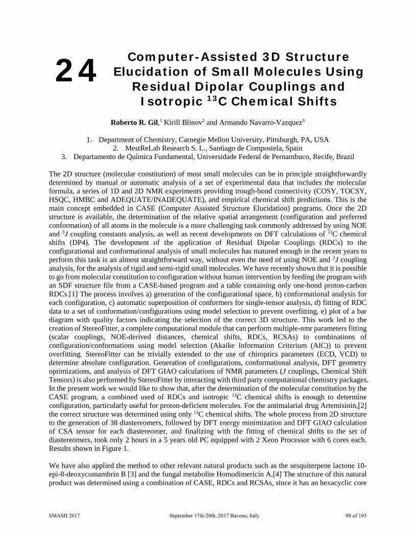
24 Computer-Assisted 3D Structure Elucidation of Small Molecules Using
Residual Dipolar Couplings and Isotropic 13C Chemical Shifts
Roberto R. Gil,1 Kirill Blinov2 and Armando Navarro-Vazquez3
1. Department of Chemistry, Carnegie Mellon University, Pittsburgh, PA, USA
2. MestReLab Research S. L., Santiago de Compostela, Spain 3. Departamento de Química Fundamental, Universidade Federal de Pernambuco, Recife, Brazil
The 2D structure (molecular constitution) of most small molecules can be in principle straightforwardly determined by manual or automatic analysis of a set of experimental data that includes the molecular formula, a series of 1D and 2D NMR experiments providing trough-bond connectivity (COSY, TOCSY, HSQC, HMBC and ADEQUATE/INADEQUATE), and empirical chemical shift predictions. This is the main concept embedded in CASE (Computer Assisted Structure Elucidation) programs. Once the 2D structure is available, the determination of the relative spatial arrangement (configuration and preferred conformation) of all atoms in the molecule is a more challenging task commonly addressed by using NOE and 3J coupling constants analysis, as well as recent developments on DFT calculations of 13C chemical shifts (DP4). The development of the application of Residual Dipolar Couplings (RDCs) to the configurational and conformational analysis of small molecules has matured enough in the recent years to perform this task is an almost straightforward way, without even the need of using NOE and 3J coupling analysis, for the analysis of rigid and semi-rigid small molecules. We have recently shown that it is possible to go from molecular constitution to configuration without human intervention by feeding the program with an SDF structure file from a CASE-based program and a table containing only one-bond proton-carbon RDCs.[1] The process involves a) generation of the configurational space, b) conformational analysis for each configuration, c) automatic superposition of conformers for single-tensor analysis, d) fitting of RDC data to a set of conformation/configurations using model selection to prevent overfitting, e) plot of a bar diagram with quality factors indicating the selection of the correct 3D structure. This work led to the creation of StereoFitter, a complete computational module that can perform multiple-nmr parameters fitting (scalar couplings, NOE-derived distances, chemical shifts, RDCs, RCSAs) to combinations of configuration/conformations using model selection (Akaike Information Criterium (AIC)) to prevent overfitting. StereoFitter can be trivially extended to the use of chiroptics parameters (ECD, VCD) to determine absolute configuration. Generation of configurations, conformational analysis, DFT geometry optimizations, and analysis of DFT GIAO calculations of NMR parameters (J couplings, Chemical Shift Tensors) is also performed by StereoFitter by interacting with third party computational chemistry packages. In the present work we would like to show that, after the determination of the molecular constitution by the CASE program, a combined used of RDCs and isotropic 13C chemical shifts is enough to determine configuration, particularly useful for proton-deficient molecules. For the antimalarial drug Artemisinin,[2] the correct structure was determined using only 13C chemical shifts. The whole process from 2D structure to the generation of 38 diastereomers, followed by DFT energy minimization and DFT GIAO calculation of CSA tensor for each diastereomer, and finalizing with the fitting of chemical shifts to the set of diastereomers, took only 2 hours in a 5 years old PC equipped with 2 Xeon Processor with 6 cores each. Results shown in Figure 1. We have also applied the method to other relevant natural products such as the sesquiterpene lactone 10-epi-8-deoxycumambrin B [3] and the fungal metabolite Homodimericin A.[4] The structure of this natural product was determined using a combination of CASE, RDCs and RCSAs, since it has an hexacyclic core
SMASH 2017 September 17th-20th, 2017 Baveno, Italy 98 of 193

with fourteen quaternary carbons, eleven of them contiguous. For this particular compound we used the data reported by the authors.[4] For Homodimericin A we observed that it is possible to use 13C chemical shifts as an alternative to RCSAs to determine its configuration.
Figure 1. Determination of the configuraiton of Artemisinin using only isotropic sC chemical shift. Fitting DFT calculated chemical shift with the experimental data using StereoFitter. The red asterisk shows the
correct structure with the lowest χ2 value. In contrast to methodologies such as DP4 our method allows the fitting of populations to all the available experimental data including chemical shifts. Potential overfitting problems are treated by using the Akaike Information Criterion (AIC) where models with increasing number of populations are penalized. CASE analysis was performed using the MestReNova Structure Elucidator. Most of the results presented here used Poly(methylmethacrylate) (PMMA) based flexible gels, whose degree of alignment can be easily tuned by variable and reversible compression.[5] Acknowledgments: ANV thanks the UFPE for a visiting professor contract and FACEPE (APQ-0507-1.06/15) for financial support. NMR instrumentation at CMU was partially supported by the NSF(CHE-0130903 and CHE-1039870). R.R.G. acknowledges support from the NSF (CHE-1111684).We thanks Leandro F. Gil-Silva from Alignment Technologies LLC / Mestrelab Research SL for the provision of aligning gels. 1. Troche-Pesqueira, Eduardo; Anklin, Clemens; Gil, Roberto R.; Navarro-Vazquez, Armando.
Angewandte Chemie, International Edition, 56(13), 3660-3664, 2017. 2. Coordinating Group for Research on the Structure of Qing Hau Sau. Kexue Tongbao (Chinese Edition)
22(3), 142, 1977. 3. Sosa, Virginia E.; Oberti, Juan C.; Gil, Roberto R.; Ruveda, Edmundo A.; Goedken, Virgil L.;
Gutierrez, Alicia B.; Herz, Werner. Phytochemistry 28(7), 1925-9, 1989.
SMASH 2017 September 17th-20th, 2017 Baveno, Italy 99 of 193

4. Mevers, Emily; Sauri, Josep; Liu, Yizhou; Moser, Arvin; Ramadhar, Timothy R.; Varlan, Maria; Williamson, R. Thomas; Martin, Gary E.; Clardy, Jon. Journal of the American Chemical Society 138(38), 12324-12327, 2016.
5. Gil, Roberto R.; Gayathri, Chakicherla; Tsarevsky, Nicolay V.; Matyjaszewski, Krzysztof. Journal of Organic Chemistry 73(3), 840-848, 2008.
6. By Gayathri, Chakicherla; Tsarevsky, Nicolay V.; Gil, Roberto R. Chemistry - A European Journal 16(12), 3622-3626, 2010.
SMASH 2017 September 17th-20th, 2017 Baveno, Italy 100 of 193

25 Insitu illuminated Nanolitre NMR spectroscopy
M. Victoria Gómez,1 Rosa Sanchez,1 Jose M. Mateo,1 Miguel Mompean,1 Antonio de la Hoz,1 and
Aldrik Velders1,2
1. University of Castilla-La Mancha, Ciudad Real, Spain
2. Wageningen University & Research, Wageningen, The Netherlands
In-situ NMR detection of UV-vis assisted reactions find application in many different fields as photocatalysis, photochromics, photoisomerizations, and hiperpolarization techniques, among others [1]. However, commercially-available NMR setups for in-situ illumination suffer from many limitations mainly related to the difficulty of an efficient and uniform illumination of the entire sample volume. These setups deal with hundreds of microliters of sample tipically contained in a 5 mm-NMR tube, therefore requiring high power light sources and complicated setups to bring the light uniforminly along the sample tube. The high intensity laser brings additional problems related to light-assisted sample degradation. Essentially two different methods of guiding the light into the coil region of a superconducting magnet have been developed over the years, light from the bottom of the NMR probe or from the top. The latter option is the most used nowdays, although still encounters several conflicts as the magnetic field distorsions caused by an insert into the sample volume and the light absorption by the solution above the radiofrequency coil region, which could decrease the stability of the sample and the observed signal, together with a loss of some sensitivity and resolution [2]. Here we present a novel setup for the illumination of nanolitre samples with low power light sources for the insitu NMR analysis of UV-vis light-assisted chemical reactions. The nanolitre sample are located underneath a reduced-diameter NMR coil, called microcoil, which is integrated on top of a microreactor allowing the light penetration typical of microchannels [3]. The use of these small NMR detectors not only offer an interesting approach to enhance NMR sensibility [4] but also, the cappabilities of the microcoils of analyzing sample volumes in the nL ranges enable the rapid optimization of reaction conditions and an extremely fast determination of the kinetic parameters of reactions [5, 6]. 1. He Z, Bae M, Wu J, Jamison T F, Angew. Chem. Int. Ed., 53, 14451-14455, 2014. 2. Kühn T, Schwalbe H, J. Am. Chem. Soc., 122, 6169-6174, 2000. 3. Cambie D, Bottecchia C, Straathof N J W, Hessel V, Noel T, Chem. Rev., 116, 10276-10341, 2016. 4. Fratila RM, Velders AH, Ann.Rev.Anal.Chem.,4, 227-249, 2011. 5. Gomez MV, Verputten HHJ, Diaz-Ortiz A, Moreno A, de la Hoz A, Velders AH, 6. Chem. Commun., 46, 4514-4516, 2010. 7. Gomez MV, Rodriguez AM, de la Hoz A, Jimenez F, Fratila RM, Barneveld PA, Velders AH,
Anal.Chem., 10547-10555, 2015.
SMASH 2017 September 17th-20th, 2017 Baveno, Italy 101 of 193
26 Round Robin Test for Applying the qNMR Method to the Japanese
Industrial Standards
Toru Miura
Laboratory and Specialty Chemicals Research Laboratories, Wako Pure Chemical Industries, Ltd. Introduction Verification of the method for quantitative analysis using 1H NMR (1H quantitative Nuclear Magnetic Resonance: 1H qNMR) recently began. By setting optimised measurement conditions for quantification, it will be possible to determine precise quantitative values.[1,2,3,4,5] 1H qNMR is a method for determining the quantities of substances based on the amount of proton nuclei. Therefore, it is used for deciding the accurate purities of reagents, residual pesticides, natural products, food additives, standard products, etc., and the absolute quantification of compounds that lack standards. In addition, we prepared a sample solution to which was accurately added an SI (International System of Units)-traceable Certified Reference Material (CRM), which was developed for 1H qNMR as calibration standard. When this sample solution is measured using the 1H qNMR conditions, accurate SI traceable quantitative values can be obtained. This method has been reported as the Accurate QuAntitative NMR with Internal reference substance (AQARI) method.[1,7,8] 1H qNMR in which this AQARI methodology is applied is already being adopted in Japan’s official analytical methods such as the Japanese Pharmacopoeia and Japan’s Specifications and Standards for Food Additives as an absolute purity determination method.[9] 1H qNMR is a novel assay method with principles that differ from those of conventional chromatography. Rather than identifying and quantifying all impurities contained in a product as in the mass balance method, it is a method whereby accurate SI traceable quantitative values can be obtained effectively. Therefore, it is expected to be applied not only in the field of pharmaceuticals and food products but also in various fields requiring precise quantitative values such as the industrial chemical field. Based on such a background, we performed a Round Robin Test using the same substance to be measured and calibration standard for qNMR, and assessed the accuracy of this assay method aiming at the inclusion of 1H qNMR in the Japanese Industrial Standards. ■Methods 1. Participating facilities The study was performed at five facilities, namely, the National Institute of Health Sciences, National Institute of Advanced Industrial Science and Technology, the National Research Institute of Fisheries Science, JEOL RESONANCE Co., Ltd, and Wako Pure Chemical Industries, Ltd. 2. Materials SI-traceable CRM, 1,4-bis(trimethylsilyl) benzene-d4 (1,4-BTMSB-d4, Wako Pure Chemical Industries, Ltd.; certified value: mass fraction 99.9 ± 0.6%) was used as the substance to be measured, and 3,5-bis (trifluoromethyl) benzoic acid (3,5-BTFMBA, the National Institute of Advanced Industrial Science and Technology; certified value: mass fraction 99.96 ± 0.06%) as the calibration standard for qNMR. Methanol-d4 (Wako Pure Chemical Industries, Ltd. (Purity (GC) mass fraction ≥ 98.5% and deuterium content of ≥ 99.8%) was the deuterated solvent used for dissolving the substance to be measured and the calibration standard for qNMR. 3. Preparation of sample solutions
SMASH 2017 September 17th-20th, 2017 Baveno, Italy 102 of 193

In preparing the sample solution, the mass of the substance to be measured and the calibration standard for qNMR were weighed such that at least twice the minimum weight Wmin[10] and the signal-to-noise ratio of the signals of the substance to be measured and the calibration standard for qNMR in the NMR spectrum were approximately equal. Methanol-d4 was added so that the substance to be measured was ~0.1 w/v%. The minimum weight Wmin of the precision balance was determined separately using formula 1.
Wmin=σ×2000 Formula 1: The minimum weight Wmin was evaluated using aluminium pan (approx. 60 mg) which is
used for the tare of the substance to be measured and the calibration standard for qNMR. The minimum weight Wmin was calculated by multiplying σ by 2000.σ: the standard deviation when the
measurement of the weight of the aluminium pan was repeated 10 times2000: the value when the safety coefficient (2) was divided by the permitted range (0.10%) in the measurement (= 2/(0.1/100))
4. NMR instrument, measurement conditions, and data process conditions Six machines with proton (1H) resonance frequency ≥ 300 MHz at each facility were used in the NMR instrument. The 1H qNMR measurement conditions were the standard measurement conditions that were set based on the Japanese Pharmacopoeia conditions. In short, the nucleus to be measured was 1H, digital resolution ≤ 0.25 Hz, spectral width ≥ 20 ppm including -5 to 15 ppm, spinning Off, flip angle of 90°, 13C decoupling, relaxation delay ≥ 60 s, number of transients ≥ 8, dummy scans ≥ 2, and probe temperature maintained at 20-30 °C. The 1H qNMR measurement conditions specific to each facility were set based on these standard measurement conditions to perform the measurements. The 1H qNMR data process conditions were those conditions suitable for measuring the respective quantitative values set independently using the software of each facility. 5. System Suitability Test A system suitability test was set up based on the documentations on the specification established in the purity determination for 1H qNMR in the Japanese Pharmacopoeia.[11] 6. Evaluation methods We checked whether the purity obtained for each facility was within the range of uncertainty of the substance to be measured (1,4-BTMSB-d4; certified value: mass fraction 99.9 ± 0.6%). ■Results The 1H qNMR measurement conditions were set based on the procedure documented in the Japanese Pharmacopoeia. 3,5-bis(trifluoromethyl) benzoic acid (3,5-BTFMBA; certified value: mass fraction 99.96 ± 0.06%) was used as the calibration standard for qNMR, and the system for determining the purity of 1,4-bis(trimethylsilyl) benzene-d4 (1,4-BTMSB-d4; certified value: mass fraction 99.9 ± 0.6%) was used for the evaluation. As 1H qNMR includes bias and variation factors in the respective operations for sample preparation, NMR measurement and data process, appropriate conditions were set for them. All analytical results lied within the range of uncertainty of 1,4-BTMSB-d4, confirming that 1H qNMR functions as a method for determining the purity or content rate of general organic compounds.
1. Pauli F. G., Chen S., Simmler C., Lankin C. D., Gödecke T., Jaki U. B., Friesen B. J., McAlpine B. J., Napolitano G. J., J. Med. Chem., 2014, 57 (22), pp 9220–9231.
2. Koike R., Jo S., Azuma M., Wakisaka T., BUNSEKI KAGAKU, 2004, 53 (10), pp 1133-1138. 3. Taichi Y., Ohtsuki T., Miura T., Suematsu T., Horinouchi T., Murakami M., Saito T., Ihara T., Tada
A., Tahara M., Goda Y., Akiyama H., Nakao S., Yamada Y., Koike R., Sugimoto N., BUNSEKI KAGAKU, 2014, 63 (4), pp 323-329.
4. Schoenberger T., Analytical and Bioanalytical Chemistry, April 2012, 403 (1), pp 247–254. 5. Watanabe R., Sugai C., Yamazaki T., Matsushima R., Uchida H., Matsumiya M., Takatsu A., Suzuki
T., Toxins, 2016, 8(10), 294; doi:10.3390/toxins8100294
SMASH 2017 September 17th-20th, 2017 Baveno, Italy 103 of 193

6. Tahara M., Sugimoto N., Ohtsuki T., Tada A., Akiyama H., Goda Y., Igarashi Y., Environmental Science, 2014, 27 (3), pp 142-150.
7. Ohtsuki T., Sato K., Abe Y., Sugimoto N., Akiyama H., Talanta, 2015, 131, pp 712–718. 8. Kato T., Saito M., Nagae M., Fujita K., Watai M., Igarashi T., Yasumoto T., Inagaki M., Analytical
Sciences, 2016, 32 (7) pp 729-734. 9. Hosoe J., Sugimoto N., Suematsu T., Yamada Y., Miura T., Hayakawa M., Suzuki H., Katsuhara T.,
Nishimura H., Kikuchi Y., Yamashita T., Goda Y., Pharmaceutical and Medical Device Regulatory Science, 2014, 45(3), pp 243-250. ().
10. USP General Chapter <41> “Balances”. 11. The Japanese Pharmacopoeia, Seventeenth Edition (E)-Cinnamic acid for assay 2)
SMASH 2017 September 17th-20th, 2017 Baveno, Italy 104 of 193

27 Time-Resolved NUS Interleaved Experiments with Online Processing
and Analysis
Alexandra Shchukina, Krzysztof Kazimierczuk, Mateusz Urbańczyk
University of Warsaw, Centre of New Technologies, Warsaw, Poland
The present work combines three key ideas: 1) implementing time-resolved non-uniform sampling (TR NUS) for monitoring changes in non-stable samples, 2) interleaving NMR experiments within the framework of TR NUS procedure (enables one to acquire various types of spectra simultaneously), 3) processing and analysing spectra on the fly. TR NUS [1, 2] is an approach to modify multidimensional NMR experiments for monitoring time-dependent processes occurring on the time-scale of tens of minutes to hours. As in conventional NUS, some random points of the indirect dimensions are probed, while others are skipped. However, since the sample and its spectrum are changing during the experiment, it is justified to use a very long, shuffled sampling schedule (even many times larger than the size of a full grid) and divide it into subsets that are “snapshots” of the process. Allowing for an overlap between these subsets, proper time resolution can be achieved. Each subset is then reconstructed with compressed sensing (CS) algorithms. The results of an N-dimensional experiment can then be presented as a N+1-dimensional array, where the actual, physical time serves as an extra quasi-dimension. Also, these results can be viewed a “movie” of N-dimensional spectra with peaks slightly changing from one frame to another, thus representing the changes in the sample. However, in the case of uncontrolled, irreversible changes in the sample the chosen measurement technique may turn out to be not optimal. Then, both sample and measurement time get wasted. Therefore, it would best to conduct several experiments at the same time: for instance, record one-dimensional spectrum of high sensitivity between increments of well resolved but insensitive multidimesional spectrum; or interleave signal sampling by several different multidimensional techniques, as was shown for NMR methods aimed at the resonance assignment of proteins [3]. To improve the procedure even further and make it convenient for the user, we have created a system which automatically carries out the CS reconstruction of the NUS spectra simultaneously to their acquisition. This system starts the reconstruction for every frame when the necessary number of indirect dimension FID points is acquired; during this time the data for the next frame are being acquired, etc. It also processes the data and shows the spectra with a possibility to switch between frames. The work on peak-picking and peak-tracking simultaneous to acquisition is in progress, as well as the generalisation of the system to make is capable of treating interleaved experiments.
1. Maxim Mayzel, Joakim Rosenlöw, Linnéa Isaksson, Vladislav Y. Orekhov. Journal of Biomolecular NMR, 58(2): 129–139, 2014.
2. Dass R, Koźmiński W, Kazimierczuk K. Anal. Chem., 87 (2):1337–1343, 2015. 3. V.A. Jaravine, A.V. Zhuravleva, P. Permi, I. Ibraghimov, and V.Yu. Orekhov. Journal of the
American Chemical Society, 130(12): 3927–3936, 2008.
SMASH 2017 September 17th-20th, 2017 Baveno, Italy 105 of 193

28 Chromatographic NMR Spectroscopy with Hollow Silica Spheres
Federico DeBiasi, Tania Gonzàlez-Garcìa, Tommaso Margola, Sara Springhetti, Fabrizio Mancin, and
Federico Rastrelli
Università degli Studi di Padova, Department of Chemical Sciences, Padova, Italy
NMR diffusometry comprises an ensemble of techniques designed to separate the signals of different molecules according to their apparent diffusion coefficients in solution. In some cases, the combination of dedicated pulse sequences and data processing can produce 2D “DOSY” maps displaying the chemical shift in the direct dimension and the apparent diffusion coefficient in the indirect dimension, something that closely reminds of a TLC plate. However useful in the analysis of complex mixtures, the success of this technique depends both on the absence of signal overlap, and on the existence of sizeable differences among the diffusion coefficients of the various species, a condition hardly met in crowded 1H NMR spectra. While signal overlap can be mitigated by pureshift spectroscopy, the separation in the diffusion dimension can be significantly improved by perturbing the analytes’ diffusion coefficient with an interacting matrix. To this aim, the use of a stationary phase made of solid silica has been proposed in order to exploit the partition equilibria which occur in a chromatographic column. [1] Owing to the intrinsic sample inhomogeneity, however, the practical implementation of this method, dubbed chromatographic NMR, relies on magic-angle spinning to remove the anisotropy of magnetic susceptibility across the example. In this context we have recently proposed the use of hollow microspheres to reduce the impact of field inhomogeneities. [2] The idea is founded on the observation that, in the presence of a uniform magnetic field, the effect of an isolated sphere on the external region is equivalent to that of a dipole located at the center of the sphere itself. When an NMR tube filled with silica powder and a solvent is put into the magnet, the magnetic field becomes inhomogeneous within small domains across the sample, a situation which is beyond the correction capabilities of the spectrometer shim system. However, if the same spheres are partly emptied to form a cavity, it can be proved that the magnitude of the associated dipolar field collapses as the shell of the sphere becomes thinner. In this communication we describe the use of hollow silica microspheres in chromatographic NMR, from early experimental observations up to a numerical solution of the magnetostatic problem in model samples. The obtained results provide a significant understanding of how the performances of such systems depend on the internal sample geometry, together with the surface properties of silica. Indeed, the possibilities offered by hollow spheres are manifold: we expect that new materials can be envisaged to simultaneously exploit different supramolecular recognition abilities based, for example, on the concurrence of hydrophobic and dipolar interactions. Along this line, the properties of hybrid hollow microspheres are currently being investigated in our lab. 1. S. Viel, F. Ziarelli, S. Caldarelli, Proc. Natl. Acad. Sci. USA, 100, 9696, 2003 b) G. Pages, C.
Delaurent, S. Caldarelli, Angew. Chem. Int. Ed., 45, 5950, 2006 c) S. Caldarelli, Magn Reson Chem, 45 Suppl 1, S48, 2007
2. T. Gonzalez-Garcia, T. Margola, A. Silvagni, F. Mancin, F. Rastrelli, Angew. Chem. Int. Ed., 55(8), 2733, 2016
SMASH 2017 September 17th-20th, 2017 Baveno, Italy 106 of 193
29 Combining the best of NMR: Fast, Quantitative, Highly Resolved and
Sensitive approach for metabolomics
J. Farjon1, E. Martineau1,2, S. Akoka1, P. Giraudeau1,3
1. Faculté des Sciences et Techniques de Nantes, UMR CNRS 6230 - CEISAM, Equipe EBSI,
LUNAM Université, France 2. SpectroMaitrise, CAPACITÉS SAS, 26 Bd Vincent Gâche, 44200 Nantes, France
3. Institut Universitaire de France, 1 rue Descartes, 75005 Paris cedex 05, France A better understanding of living organisms requires a detailed and quantitative analysis of complex biological samples such as extracts, biofluids for organisms under investigation. Among the most efficient analytical techniques, NMR is one of the most used and makes it possible to quantify complex mixtures with an excellent repeatability and precision. However, NMR is not the most sensitive technique despite its ability to give informative structural and quantitative data. In the context of metabolic studies, complex mixtures are usually hard to decipher on a 1D spectrum (see Figure). Therefore, 2D NMR is a nice alternative for improving the spectral resolution along two dimensions. For highly overcrowded spectra, the ultimate solution is to record heteronuclear 1H-13C 2D maps to benefit from the larger spectral window of 13C. However 13C nuclei are low abundant (1.1%), preventing the detection of low concentrated metabolites. Pushing the inherent limits of NMR is essential for investigating potential low concentrated metabolites within complex biological mixtures. We suggested a new approach based on new quantitative 1H-13C techniques called QUIPU HSQC [1] that can detect metabolites in the range of 100µM within leaf extracts thanks to the sensitivity gains of 2 to 3 for 1H-13C correlations in comparison with the conventional version. This technique has also been applied with success to better understand the photosynthetic and photorespiratory cycles by detecting sugar phosphates as biomarkers of these processes [2]. This approach has recently been improved through the Quick QUIPU HSQC or Q-QUIPU HSQC [3] combining spectral aliasing [4], NUS [5] and variable repetition times [6] methods. The time necessary to obtain quantitative data could be divided by a factor from 5 to 9 while preserving highly resolved spectra. The analytical performance of this new advanced tool dedicated to metabolomics is successfully shown on a model metabolite mixture and on breast cells cancer extracts. The new method opens perspectives in a variety of fields related to metabolomics.
1. Mauve C., Khlifi S., Gilard F., Mouille G. and Farjon J., Chem. Comm., 52, 6142-6145 (2016) 2. Khlifi S., Mauve C., Glab N., Gilard F., Hodges M., Mouille G. and Farjon J., under revision, (2017) 3. Farjon J., Martineau E., Akoka S., and Giraudeau P., submitted, (2017) 4. Foroozandeh M., Jeannerat D., Magn. Reson. Chem., 53, 894-900 (2015) 5. 5 Korzhnev D. M., Ibraghimov I. V., Billeter M., Orekhov V. Y., J. Biomol. NMR, 21, 263–268 (2001) 6. 6 Macura S., J. Am. Chem. Soc., 31, 9606-9607 (2009)
SMASH 2017 September 17th-20th, 2017 Baveno, Italy 107 of 193

30 NMR Experiments for Several Receivers
Helena Kovacs1 and Ēriks Kupče2
1. Bruker BioSpin AG, Fällanden, Switzerland
2. Bruker BioSpin, Coventry, UK 3.
NMR experiments involving multiple receivers provide a unique way of increasing the sensitivity and information content of data recorded in a given period of time [1-3]. We present a comprehensive series of such experiments designed for simultaneous detection of abundant nuclei, such as 1H, 19F and 31P, as well as samples enriched with magnetically active isotopes including 13C and 15N. The multiple receiver experiments are categorized into three main types – (a) parallel acquisition, (b) sequential acquisition and (c) interleaved experiments. The optimum implementation is shown to depend on the relaxation properties of the involved nuclei as well as the intrinsic sensitivity of the directly observed nuclei. We particularly focus on the basic NMR experiments involving the pairs of 1H / 19F nuclei and 1H / 31P nuclei not least because of the particularly important role that 19F and 31P NMR plays in drug discovery and pharmaceutical industry [3]. Essentially any of the basic 2D NMR experiments, such as COSY, NOESY, TOCSY, DOSY, HSQC, HMQC, HMBC, HETCOR or relaxation measurements that are routinely used in small molecule NMR can be easily adapted for and more efficiently recorded on systems equipped with multiple receivers. Many of these experiments are amenable to further reduction of experiment time by combining them with other fast NMR techniques, such as Hadamard NMR, non-uniform sampling, spatial encoding or rapid pulsing methods. We believe that the multi-receiver technology will boost the development of new NMR experiments as well as NMR research in general, making the NMR instruments more efficient and making the NMR spectroscopy even more unique in the universe of analytical tools and experimental techniques.
1. R. Freeman and Ē. Kupče, in Modern NMR Approaches To The Structure Elucidation of Natural Products, (Eds. A. J. Williams, G. E. Martin and D. Rovnyak), vol. 1, pp 117-145, Royal Soc. Chem. (2016).
2. Ē. Kupče, eMagRes, 4, 721–732, 2015. DOI 10.1002/9780470034590.emrstm1404. 3. H. Kovacs and Ē. Kupče, Magn. Reson. Chem., 54, 544-560, 2016.
SMASH 2017 September 17th-20th, 2017 Baveno, Italy 108 of 193
31 Structure-Property-Relationship for Amine Based CO2 Absorption by Quantitative NMR Spectroscopy
Elmar Kessler1, Luciana Ninni Schäfer1, Benjamin Willy3, Rolf Schneider2, Muhammad Irfan2,
Jörn Rolker3, Erik von Harbou1 and Hans Hasse1
1. Laboratory of Engineering Thermodynamics (LTD), University of Kaiserslautern,
Kaiserslautern, Germany 2. Evonik Technology & Infrastructure GmbH, Hanau, Germany
3. Evonik Performance Materials GmbH, Marl, Germany
In a cooperation project between Evonik Industries, thyssenkrupp, and the Laboratory of Engineering Thermodynamics (LTD) of University of Kaiserslautern, novel amines for syngas purification by reactive absorption are developed and investigated [1]. All studied amines are derivatives of triacetone amine (TAA) and will be named “Evonik Amines” (EvAs) in this project. This interesting group of substances has not been considered before for the present application. In a screening, a large number of EvAs are synthesized, i.e. the chemical structure is tailored. Thermodynamic properties of aqueous solutions of the novel amines, both loaded with CO2 and unloaded, which are particularly relevant for the industrial reactive absorption process are investigated: gas solubility and absorption kinetics, solid solubility, and liquid-liquid equilibria. The results show that even for very small differences in the chemical structure of the EvAs, major differences in their absorption behavior can be observed. To gain a better understanding of these findings, aqueous solutions of several EvAs, loaded with CO2 under high pressure, are studied with NMR spectroscopy. The experiments are carried out in a wide range of CO2 loadings and temperatures, which are relevant for industrial applications. For this purpose, a robust method for the in situ analysis of the chemical equilibria in CO2 loaded solutions, using quantitative NMR spectroscopy, is developed. Depending on the molecular structure of the amines, different reaction products are formed via different reaction paths when CO2 is being absorbed. Thus, the ability of NMR spectroscopy to combine directly structure elucidation with quantitative analysis is important in this project to gain insights into the reaction network and chemical equilibria and finally into the performance of the amines in a reactive absorption process. By comparing the results obtained for the different TAA derivatives the influence of changes in the molecular structure on the studied properties is elucidated. That knowledge gives a guideline for the synthesis of new amines.
1. Vasiliu, Dan, Yazdani, Amir, McCann, Nichola, Irfan, Muhammad, Schneider, Rolf, Rolker, Jörn, Maurer, Gerd, von Harbou, Erik, Hasse, Hans, J. Chem. Eng. Data, 61 (11), 3814–3826, 2016
SMASH 2017 September 17th-20th, 2017 Baveno, Italy 109 of 193
32 Hot and Cool RDCs in One Alignment Medium
Mira Schwab and Christina M. Thiele
Technische Universität Darmstadt, Clemens-Schöpf Institut für Organische Chemie und Biochemie,
Darmstadt, Germany In recent years it has become more and more popular to measure residual dipolar couplings (RDCs) providing global structural information as well as the relative configuration of molecules. For this approach suitable alignment media are necessary and as far as biomolecules are concerned, a wide range of water-based media are available. However, when it comes to small organic molecules, the compatibility of the analyte with the alignment-medium and the respective organic solvent limit the eligible media. Although, several alignment media based on either liquid crystalline phases or anisotropic swollen, streched or compressed gels (SAG) have been sucessfully applied in the RDC approach [1], the development of further alignment media is desirable. We synthesized Poly-ß-phenethyl-L-aspartate (PPLA), the constitutional isomer of Poly-γ-benzyl-L-glutamate (PBLG), which is a commonly used alignment medium in organic structure elucidation [2]. We have investigated the orientational properties of PPLA and determined excellent enantiodifferentiating abilities; by today the highest observed in a homopolypeptide-based alignment medium. Enantiodifferentiation is based on different diastereomorphous interactions between a chiral analyte and the P- and M-helix configuration or between both enantiomers of the analyte and one helix configuration, both leading to different mean orientations of the analyte. PPLA possesses the special temperature dependent feature to undergo a reversible helix screw-sense inversion in the LLC-state without perturbing the orientational order of the α-helical backbone in the nematic arrangement [3]. The transition originates from small energy differences between left- and right-handed helices in polyaspartates. This thermoresponsivity implies the possibility to measure different orientations of one enantiomer of the analyte within the same sample by just raising the temperature. Herein we report the synthesis of Polyphenethylaspartate and its outstanding abilities as thermoresponsive enantiodifferentiating alignment medium in NMR-spectroscopy.
1. Thiele, C. M., Concepts in Magnetic Resonance Part A, 30A, 65-80, 2007. 2. Marx, A., Schmidts, V., Thiele, C. M., Magnetic Resonance in Chemistry, 47, 734-740, 2009. 3. A. Abe, S. Okamoto, N. Kimura, K. Tamura, H. Onigawara, J. Watanabe, Acta Polymerica, 44, 54-56,
1993.
SMASH 2017 September 17th-20th, 2017 Baveno, Italy 110 of 193
33 Time-Course Metabolomic Analysis of Yeast Unrestricted Growth by 2D
1H-13C HSQC NMR and 1D 1H NMR Spectroscopy
Francesc Puig-Castellví1, Yolanda Pérez2, Benjamín Piña1, Ignacio Alfonso2, and Romà Tauler1
1. Department of Environmental Chemistry, Institute of Environmental Assessment and Water Research
(IDAEA-CSIC), Barcelona, Spain 2. Department of Biological Chemistry and Molecular Modelling, Institute of Advanced Chemistry of
Catalonia (IQAC-CSIC), Barcelona, Spain Metabolomics is a field of ‘omics’ research that is primarily focused on the identification and characterization of small molecule metabolites in cells, tissues, organs and organisms[1]. Metabolomic approaches have been used for biomarker discovery in clinical studies[2], food analysis[3], environmental assessment[4], and phenotyping[5], among others. Due to the important role that metabolomics plays in bridging the phenotype-genotype gap in all these areas, it has become one of the most ambitious ‘hot topic’ at present. One of the preferred instrumental techniques in metabolomics is Nuclear Magnetic Resonance (NMR) spectroscopy, due to the inherent advantages of the technique (simple sample preparation, non-destructive, robust,…). Most of the studies have been focused on the analysis of one-dimensional proton (1D 1H) NMR, while the analysis of other nuclei (such as 13C and 31P) and other NMR experiments (1H-13C HSQC[6], 2D 1H INADEQUATE[7],…) are still underrepresented. The preference of 1D 1H NMR in NMR metabolomics lies in the fact that has good sensitivity in a short acquisition time, albeit it lacks spectral resolution, since it presents a high overlapping degree. In this study, we have characterized the metabolome of a yeast strain grown in two different liquid media for a 3-day period with both 1D 1H NMR and 2D 1H-13C HSQC experiments. Dozens of metabolites, including amino acids, nucleotides, sugars and organic acids, among others, have been detected and quantified. The metabolic response over time has been connected to the particular composition of each culture medium, proving that cell adaptation does not only occurs under unfavourable or stress conditions, but also in order to minimize the energy cost of growth and development (i.e. by uptaking a metabolite from the extracellular medium instead of producing it by de-novo biosynthesis). Finally, with this empirical example, we have concluded that the combined analysis of both 1D 1H NMR and 2D 1H-13C HSQC datasets is much preferrable than the analysis of the two separately, since a better interpretation of the overall metabolome can be obtained with the combined approach analyzing the two datasets simultaneously.
1. German, J.B., B. Hammock, and S. Watkins, Metabolomics: building on a century of biochemistry to guide human health. Metabolomics, 1(1): p. 3-9, 2005.
2. Mavel, S., et al., 1H–13C NMR-based urine metabolic profiling in autism spectrum disorders. Talanta, 114: p. 95-102, 2013.
SMASH 2017 September 17th-20th, 2017 Baveno, Italy 111 of 193

3. Monakhova, Y.B., et al., Independent component analysis (ICA) algorithms for improved spectral deconvolution of overlapped signals in 1H NMR analysis: application to foods and related products. Magnetic Resonance in Chemistry, 52(5): p. 231-240, 2014.
4. Lankadurai, B.P., E.G. Nagato, and M.J. Simpson, Environmental metabolomics: an emerging approach to study organism responses to environmental stressors. Environmental Reviews, 21(3): p. 180-205, 2013.
5. Puig-Castellví, F., et al., 1H NMR metabolomic study of auxotrophic starvation in yeast using Multivariate Curve Resolution-Alternating Least Squares for Pathway Analysis. Scientific Reports, 6: p. 30982, 2016.
6. Vergara, F., A. Shino, and J. Kikuchi, Cannibalism Affects Core Metabolic Processes in Helicoverpa armigera Larvae—A 2D NMR Metabolomics Study. International Journal of Molecular Sciences, 17(9): p. 1470, 2016.
7. Martineau, E., et al., Absolute quantification of metabolites in breast cancer cell extracts by quantitative 2D 1H INADEQUATE NMR. NMR in Biomedicine, 25(8): p. 985-992, 2012.
SMASH 2017 September 17th-20th, 2017 Baveno, Italy 112 of 193

34 Design and Validation of a Compact Online NMR Module
Simon Kern, Klas Meyer, Svetlana Guhl, Andrea Paul, Michael Maiwald
Bundesanstalt für Materialforschung und -prüfung (BAM), Berlin, Germany
Monitoring chemical reactions is the key to process control. Today, mainly optical online methods are applied, which are calibration intensive. NMR spectroscopy has a high potential for direct loop process control [1, 2] while cutting the calibration and validation needs to an minimum and thus exhibiting short set-up times. Compact NMR instruments make NMR spectroscopy accessible in industrial and harsh environments for advanced process monitoring and control. Intensified continuous processes are in focus of current research. Flexible (modular) chemical plants can produce different products using the same equipment with short down-times between campaigns and quick introduction of new products to the market. In continuous flow processes online sensor data and tight closed-loop control of the product quality are mandatory. Data analysis techniques are available but currently mostly used for off-line data analysis to detect the causes of variations in the product quality. This is addressed within the EU’s Research Project CONSENS [3] by the development and integration of a smart NMR module for process monitoring. The presented NMR module is provided in a mobile explosion proof housing and involves a compact spectrometer together with an acquisition unit and a programmable logic controller for automated data preparation (phasing, baseline correction), and evaluation. Such “smart sensors” provide the basis for the future project “Industrie 4.0”, and Industrial Internet of Things (IIoT), along with current requirements to process control, model based control, or soft sensing. The module transforms the acquired online spectra of various technically relevant reactions to either conventional 4‒20 mA signals as well as WiFi based OPC-UA communication protocols. This enables NMR-based advanced process control for the progressive or result in bloodcurdling discussions with plant managers along with automation and safety engineers.
1. Michalik-Onichimowska, A., Kern, S., Riedel, J., Panne, U., King, R., Maiwald, M.: Journal of Magnetic Resonance (2017)., 277, 154–161.
2. Meyer, K., Kern, S., Zientek, N., Guthausen, G., Maiwald, M.: Trends in Analytical Chemistry 83 (2016), 39–52.
3. CONSENS – Integrated Control and Sensing for Sustainable Operation of Flexible Intensified Processes, funded by the European Union’s Horizon 2020 research and innovation programme under grant agreement N° 636942; www.consens-spire.eu; 31.05.2017
SMASH 2017 September 17th-20th, 2017 Baveno, Italy 113 of 193

35 Label-Free NMR-Based Dissociation Rates Determination
Pablo Trigo-Mourino,1 Donghan Lee1,2 and Christian Griesinger.1
1. Max-Planck-Institute for Biophysical Chemistry, Göttingen, Germany.
2. James Graham Brown Cancer Center, Lousville, KY, USA Protein functions can be accurately modified with small-molecule ligands. In addition to the equilibrium binding affinity of such ligands to biomacromolecules the kinetics and lifetime of the complex formation are of interest. Indeed, there is now growing experimental evidence linking ligand efficiency with ligand-receptor complex lifetime (τ), raising the interest in the determination of dissociation rate constants (koff = 1/τ).[1] Recently, the High Power Relaxation Dispersion (RD) technique has been developed in our group, allowing the detection of single-digit-microsecond chemical exchange events.[2] Here we present the use of a 1D 1H R1ρ experiment on unmodified ligands, from which binding dissociation can be easily extracted.[3] This method has advantages over previously proposed CPMG RD techniques.[4] First, 1H spectra does not require expensive, and unpractical, isotope-labeled samples; second, the experimental time is significantly shorter; third, the 1H nucleus is differently sensitive from 13C to the conformational state, e.g. rotameric states, reducing the weight of bound ligand slow dynamics (if any) that might obscure the binding event; and fourth, the use of R1ρ experiment instead of CPMG makes possible the use of any multiplet from the ligand due to the absence of J coupling modulation during the spin-lock period. Additionally, the use of the High-Power RD approach improves the detection limit from residence times of 500 µs to the single digit µs.
Fig. 1. Relaxation Dispersion profiles of Epothilone A in the absence (red, flat line) and in presence
(blue) of tubulin heterodimers. The solid lines indicate the individual fit of each resonance. Globally, both resonances fitted to a koff = 6.88 ± 0.47 104 s-1, which translates into a half-life τ = 14.52 ± 0.93 μs.
We show the applicability of our method with different small-molecule binding to two different target systems: the soluble bovine serum albumin and stabilized microtubules. Due to the inherent high sensitivity of 1H 1D sampling different ratios of ligand to target biomolecule can be quickly acquired, allowing the deconvolution of the term and therefore the extraction of the KD together with the koff. 1. R. A. Copeland, Nat. Rev. Drug Discov. 2016, 15, 87-95. 2. a D. Ban, A. D. Gossert, K. Giller, S. Becker, C. Griesinger, D. Lee, J. Magn. Reson. 2012, 221, 1-4; b
D. Ban, M. T. Sabo, C. Griesinger, D. Lee, Molecules 2013, 18; c S. Pratihar, T. M. Sabo, D. Ban, R. B. Fenwick, S. Becker, X. Salvatella, C. Griesinger, D. Lee, Angew. Chem. 2016, 128, 9719-9722.
SMASH 2017 September 17th-20th, 2017 Baveno, Italy 114 of 193

3. D. G. Davis, M. E. Perlman, R. E. London, J. Magn. Reson. B 1994, 104, 266-275. 4. a J. S. Zintsmaster, B. D. Wilson, J. W. Peng, J. Am. Chem. Soc. 2008, 130, 14060-14061; b J. W. Peng,
B. D. Wilson, A. T. Namanja, J. Biomol. NMR 2009, 45, 171-183; c T. Moschen, S. Grutsch, M. A. Juen, C. H. Wunderlich, C. Kreutz, M. Tollinger, J. Med. Chem. 2016, 59, 10788-10793.
SMASH 2017 September 17th-20th, 2017 Baveno, Italy 115 of 193

36 Reading Between the Lines – Automated Data Analysis for Low
Field NMR Spectra
Simon Kern, Svetlana Guhl, Klas Meyer, Andrea Paul, and Michael Maiwald
Bundesanstalt für Materialforschung und -prüfung (BAM), Berlin, Germany
For reaction monitoring and process control using NMR instruments, in particular, after acquisition of the FID the data needs to be corrected in real-time for common effects using fast interfaces and automated methods. When it comes to NMR data evaluation under industrial process conditions, the shape of signals can change drastically due to nonlinear effects. Additionally, the multiplet structure becomes more dominant because of the comparably low-field strengths which results in overlapping of multiple signals [1]. However, the structural and quantitative information is still present but needs to be extracted by applying predictive models. We present a range of approaches for the automated spectra analysis moving from statistical approach, (i.e., Partial Least Squares Regression) to physically motivated spectral models (i.e., Indirect Hard Modeling [2]). By using the benefits of traditional qNMR experiments data analysis models can meet the demands of the PAT community (Process Analytical Technology) regarding low calibration effort/calibration free methods, fast adaptions for new reactants, or derivatives and robust automation schemes.
1. Meyer, K., Kern, S., Zientek, N., Guthausen, G., & Maiwald, M. (2016). Process control with compact NMR. TrAC Trends in Analytical Chemistry, 83, 39-52.
2. Michalik-Onichimowska, A., Kern, S., Riedel, J., Panne, U., King, R., & Maiwald, M.: Journal of Magnetic Resonance (2017), 277, 154-161.
SMASH 2017 September 17th-20th, 2017 Baveno, Italy 116 of 193
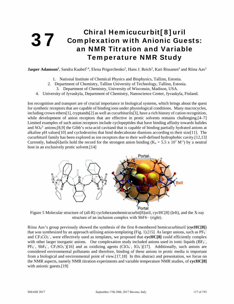
37 Chiral Hemicucurbit[8]uril Complexation with Anionic Guests:
an NMR Titration and Variable Temperature NMR Study
Jasper Adamson1, Sandra Kaabel2,4, Elena Prigorchenko2, Hans J. Reich3, Kari Rissanen4 and Riina Aav2
1. National Institute of Chemical Physics and Biophysics, Tallinn, Estonia.
2. Department of Chemistry, Tallinn University of Technology, Tallinn, Estonia. 3. Department of Chemistry, University of Wisconsin, Madison, USA.
4. University of Jyvaskyla, Department of Chemistry, Nanoscience Center, Jyvaskyla, Finland. Ion recognition and transport are of crucial importance in biological systems, which brings about the quest for synthetic receptors that are capable of binding ions under physiological conditions. Many macrocycles, including crown ethers[1], cryptands[2] as well as cucurbiturils[3], have a rich history of cation recognition, while development of anion receptors that are effective in protic solvents remains challenging.[4–7] Limited examples of such anion receptors include cyclopeptides that have binding affinity towards halides and SO4
2- anions;[8,9] the Gibb’s octa-acid cavitand that is capable of binding partially hydrated anions at alkaline pH values[10] and cyclodextrins that bind dodecaborate dianions according to their size[11]. The cucurbituril family has been explored as ion receptors due to their well-defined hydrophobic cavity.[12,13] Currently, babus[6]urils hold the record for the strongest anion binding (Ka = 5.5 x 10⁷ M-1) by a neutral host in an exclusively protic solvent.[14]
Figure 5 Molecular structure of (all-R) cyclohexanohemicucurbit[8]uril, cycHC[8] (left), and the X-ray
structure of an inclusion complex with SbF6− (right).
Riina Aav’s group previously showed the synthesis of the first 8-membered hemicucurbituril (cycHC[8]) that was synthesized by an approach utilising anion-templating (Fig. 1).[15] As larger anions, such as PF6
− and CF3CO2
−, were effectively used as templates, we proposed that cycHC[8] could efficiently complex with other larger inorganic anions. Our complexation study included anions used in ionic liquids (BF4
−, PF6
−, SbF6−, CF3SO3
−)[16] and as oxidizing agents (ClO4−, IO4
−)[17]. Additionally, such anions are considered environmental pollutants and therefore, binding of these anions in protic media is important from a biological and environmental point of view.[17,18] In this abstract and presentation, we focus on the NMR aspects, namely NMR titration experiments and variable temperature NMR studies, of cycHC[8] with anionic guests.[19]
SMASH 2017 September 17th-20th, 2017 Baveno, Italy 117 of 193
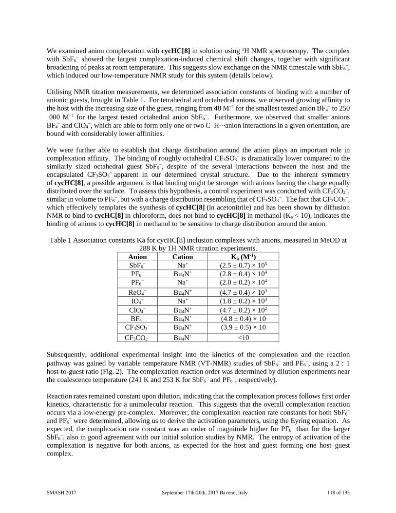
We examined anion complexation with cycHC[8] in solution using 1H NMR spectroscopy. The complex with SbF6
− showed the largest complexation-induced chemical shift changes, together with significant broadening of peaks at room temperature. This suggests slow exchange on the NMR timescale with SbF6
−, which induced our low-temperature NMR study for this system (details below). Utilising NMR titration measurements, we determined association constants of binding with a number of anionic guests, brought in Table 1. For tetrahedral and octahedral anions, we observed growing affinity to the host with the increasing size of the guest, ranging from 48 M−1 for the smallest tested anion BF4
− to 250000 M−1 for the largest tested octahedral anion SbF6
−. Furthermore, we observed that smaller anions BF4
− and ClO4−, which are able to form only one or two C–H⋯anion interactions in a given orientation, are
bound with considerably lower affinities. We were further able to establish that charge distribution around the anion plays an important role in complexation affinity. The binding of roughly octahedral CF3SO3
− is dramatically lower compared to the similarly sized octahedral guest SbF6
−, despite of the several interactions between the host and the encapsulated CF3SO3
− apparent in our determined crystal structure. Due to the inherent symmetry of cycHC[8], a possible argument is that binding might be stronger with anions having the charge equally distributed over the surface. To assess this hypothesis, a control experiment was conducted with CF3CO2
−, similar in volume to PF6
−, but with a charge distribution resembling that of CF3SO3−. The fact that CF3CO2
−, which effectively templates the synthesis of cycHC[8] (in acetonitrile) and has been shown by diffusion NMR to bind to cycHC[8] in chloroform, does not bind to cycHC[8] in methanol (Ka < 10), indicates the binding of anions to cycHC[8] in methanol to be sensitive to charge distribution around the anion. Table 1 Association constants Ka for cycHC[8] inclusion complexes with anions, measured in MeOD at
288 K by 1H NMR titration experiments. Anion Cation Ka (M-1) SbF6
− Na+ (2.5 ± 0.7) × 105 PF6
− Bu4N+ (2.8 ± 0.4) × 104 PF6
− Na+ (2.0 ± 0.2) × 104 ReO4
− Bu4N+ (4.7 ± 0.4) × 103 IO4
− Na+ (1.8 ± 0.2) × 103 ClO4
− Bu4N+ (4.7 ± 0.2) × 102 BF4
− Bu4N+ (4.8 ± 0.4) × 10 CF3SO3
− Bu4N+ (3.9 ± 0.5) × 10 CF3CO2
− Bu4N+ <10 Subsequently, additional experimental insight into the kinetics of the complexation and the reaction pathway was gained by variable temperature NMR (VT-NMR) studies of SbF6
− and PF6−, using a 2 : 1
host-to-guest ratio (Fig. 2). The complexation reaction order was determined by dilution experiments near the coalescence temperature (241 K and 253 K for SbF6
− and PF6−, respectively).
Reaction rates remained constant upon dilution, indicating that the complexation process follows first order kinetics, characteristic for a unimolecular reaction. This suggests that the overall complexation reaction occurs via a low-energy pre-complex. Moreover, the complexation reaction rate constants for both SbF6
− and PF6
− were determined, allowing us to derive the activation parameters, using the Eyring equation. As expected, the complexation rate constant was an order of magnitude higher for PF6
− than for the larger SbF6
−, also in good agreement with our initial solution studies by NMR. The entropy of activation of the complexation is negative for both anions, as expected for the host and guest forming one host–guest complex.
SMASH 2017 September 17th-20th, 2017 Baveno, Italy 118 of 193

Figure 6 Evolution of proton resonances in the variable temperature NMR study (a) for PF6− and (b) for
SbF6−. The cycHC[8] and guest concentrations were 2.6 mM and 1.5 mM in MeOD solution, respectively.
1. Pedersen CJ. The discovery of crown ethers (Noble Lecture). Angew. Chemie Int. Ed. English. 27(8), 1021–1027 (1988).
2. Lehn J. Supramolecular chemistry—scope and perspectives molecules, supermolecules, and molecular devices (Nobel Lecture). Angew. Chemie Int. Ed. English. 27(1), 89–112 (1988).
3. Freeman WA, Mock WL, Shih NY. Cucurbituril. J. Am. Chem. Soc. [Internet]. 103(24), 7367–7368 (1981). Available from: http://dx.doi.org/10.1021/ja00414a070.
4. Langton MJ, Serpell CJ, Beer PD. Anion recognition in water: recent advances from a supramolecular and macromolecular perspective. Angew. Chemie Int. Ed. 55(6), 1974–1987 (2016).
5. Gale PA, Caltagirone C. Anion sensing by small molecules and molecular ensembles. Chem. Soc. Rev. [Internet]. 44(13), 4212–4227 (2015). Available from: http://dx.doi.org/10.1039/C4CS00179F.
6. Evans NH, Beer PD. Advances in Anion supramolecular chemistry: from recognition to chemical applications. Angew. Chemie Int. Ed. 53(44), 11716–11754 (2014).
7. Sessler JL, Gale PA, Cho W-S. Anion receptor chemistry. Royal Society of Chemistry. 8. Kubik S, Kirchner R, Nolting D, Seidel J. A Molecular Oyster: A Neutral Anion Receptor Containing
Two Cyclopeptide Subunits with a Remarkable Sulfate Affinity in Aqueous Solution. J. Am. Chem. Soc. [Internet]. 124(43), 12752–12760 (2002). Available from: http://dx.doi.org/10.1021/ja026996q.
9. Sommer F, Kubik S. Anion binding of a neutral bis (cyclopeptide) in water–methanol mixtures containing up to 95% water. Org. Biomol. Chem. 12(44), 8851–8860 (2014).
10. Sokkalingam P, Shraberg J, Rick SW, Gibb BC. Binding Hydrated Anions with Hydrophobic Pockets. J. Am. Chem. Soc. [Internet]. 138(1), 48–51 (2016). Available from: http://dx.doi.org/10.1021/jacs.5b10937.
11. Assaf KI, Ural MS, Pan F, et al. Water Structure Recovery in Chaotropic Anion Recognition: High‐Affinity Binding of Dodecaborate Clusters to γ‐Cyclodextrin. Angew. Chemie. 127(23), 6956–6960 (2015).
12. Masson E, Ling X, Joseph R, Kyeremeh-Mensah L, Lu X. Cucurbituril chemistry: a tale of supramolecular success. RSC Adv. [Internet]. 2(4), 1213–1247 (2012). Available from: http://dx.doi.org/10.1039/C1RA00768H.
13. Barrow SJ, Kasera S, Rowland MJ, del Barrio J, Scherman OA. Cucurbituril-Based Molecular
SMASH 2017 September 17th-20th, 2017 Baveno, Italy 119 of 193

Recognition. Chem. Rev. [Internet]. 115(22), 12320–12406 (2015). Available from: http://dx.doi.org/10.1021/acs.chemrev.5b00341.
14. Yawer MA, Havel V, Sindelar V. A bambusuril macrocycle that binds anions in water with high affinity and selectivity. Angew. Chemie Int. Ed. 54(1), 276–279 (2015).
15. Prigorchenko E, Oeren M, Kaabel S, et al. Template-controlled synthesis of chiral cyclohexylhemicucurbit[8]uril. Chem. Commun. [Internet]. 51(54), 10921–10924 (2015). Available from: http://dx.doi.org/10.1039/C5CC04101E.
16. Olivier-Bourbigou H, Magna L, Morvan D. Ionic liquids and catalysis: Recent progress from knowledge to applications. Appl. Catal. A Gen. [Internet]. 373(1–2), 1–56 (2010). Available from: http://www.sciencedirect.com/science/article/pii/S0926860X09007030.
17. Urbansky ET. Perchlorate Chemistry: Implications for Analysis and Remediation. Bioremediat. J. [Internet]. 2(2), 81–95 (1998). Available from: http://dx.doi.org/10.1080/10889869891214231.
18. Frade RFM, Afonso CAM. Impact of ionic liquids in environment and humans: an overview. Hum. Exp. Toxicol. 29(12), 1038–1054 (2010).
19. Kaabel S, Adamson J, Topić F, et al. Chiral hemicucurbit [8] uril as an anion receptor: selectivity to size, shape and charge distribution. Chem. Sci. (2017).
SMASH 2017 September 17th-20th, 2017 Baveno, Italy 120 of 193

38 SHARPER NMR
Ariana B. Jones, Guy C. Lloyd-Jones, and Dusan Uhrin
University of Edinburgh, School of Chemistry, Edinburgh, Scotland, UK
To maximise the NMR signal intensity, it is beneficial to remove the splitting due to J-couplings. However, limitations of NMR probe technology mean that decoupling in the traditional sense is not always possible. To overcome these limitations, a new NMR method referred to as SHARPER (Sensitive, Homogeneous And Resolved PEaks in Real time) was developed. The method removes all homo- and heteronuclear couplings of a selected signal, producing narrow singlets with linewidths approaching limits dictated by the spin-spin relaxation of the observed nucleus. The resulting increase in the signal-to-noise ratio, lowers the detection threshold, allowing minimal concentrations of solutions to be studied.
In addition, the SHARPER method eliminates limitations of the magnetic field inhomogeneity. This allows detection of inherently inhomogeneous systems, such as biphasic solutions, agitated gas liquid mixtures and gels.
SMASH 2017 September 17th-20th, 2017 Baveno, Italy 121 of 193
39 Towards the Optimization of the Identification and Quantification of
Serum Metabolites
Rita Bussei,1 Silvia Castegnaro,1 Carlotta Ciaramelli,1 Silvia Mari,2 and Cristina Airoldi1
1. Department of Biotechnology and Biosciences University of Milano-Bicocca, Milan, Italy.
2. Mestrelab Research S.L., Santiago de Compostela, Spain. The metabolic profiling of serum can provide important information about physiological and pathological states and may aid understanding of the mechanism of disease occurrence [1]. A quantitative analysis of the metabolites contained in serum samples can be performed using 1H NMR spectroscopy [2]. However, due to the complexity of this matrix, NMR-based profiling has remained manual, resulting in a slow, expensive, and error-prone procedures that have hindered clinical and industrial adoption of metabolomics via this analytical technique. Here we present an approach that exploits MestreNova software, and in particular its SMA (Simple Mixture Analysis) plugin [3], which can quickly, accurately, and autonomously produce serum sample metabolic profile in a very short time. In particular, we have implemented a library of about 40 compounds which, given a 1D 1H NMR spectrum, can autonomously determine and quantify the metabolic profile in less than 1 minute. One of the greatest limitation related the NMR analysis serum samples depends on the fact that spectra include both sharp/narrow peaks, from small molecule metabolites, and broad peaks, from proteins and lipids. The size difference between macromolecules and small molecule metabolites provides an excellent basis for ultrafiltration of the serum. Nowadays, the easiest and most reliable method is to pass the sample through a 3 kDa molecular weight cut off (MWCO) micro-centrifuge filter in order to separate metabolites from proteins and other large molecules [4]. Nevertheless, this is a time-demanding procedure that implies risks in sample manipulation and loose of information regarding macromolecules contained therein. For this reason, we are exploring the possibility to filter NMR peaks a posteriori on the basis of peak linewidths as determined by globals spectral deconvolution [5]. This approach will allow to save time in sample preparation and to retain information from the high molecular weight components of serum. Acknowledgements We thank Fondazione CARIPLO for funding project 2015-0763 and Regione Lombardia for funding project “VIBRO” 1. Zhang A, Sun H, Wang X, Anal Bioanal Chem. 404(4):1239-45, 2012 2. Wishart D., TrAC, 27(3), 228–237, 2008 3. http://mestrelab.com/software/mnova/sma/ 4. Pascal Mercier, Michael J. Lewis, David Chang, David Baker, David S. Wishart, Journal of
Biomolecular NMR, 49, 307–323, 2011 5. Carlos Cobas, Felipe Seoane, Santiago Domínguez, Stan Sykora, Antony N. Davies,
Spectroscopy Europe, 23 (1), 2010
SMASH 2017 September 17th-20th, 2017 Baveno, Italy 122 of 193

40 Synergy of Computation, NMR and Synthesis to Elucidate the Correct
Structure of Baulamycins
Siying Zhong, Jingjing Wu, Paula Lorenzo, Muhammad Ali, Craig P. Butts, Eddie L. Myers and Varinder K. Aggarwal
School of Chemistry, University of Bristol, Cantock’s Close, Bristol, BS8 1TS
Structural elucidation of acyclic natural products, in particular the determination of relative configuration, still remains a challenge[1] because the observed time-averaged NMR parameters, such as scalar coupling constants, are strongly influenced by the molecules’ complex dynamic conformation(s) in solution state.[2]
The configuration of a recently isolated polyketide Baulamycin A,[3] which exhibits potent antibacterial activity, was reassigned using a combination of Density Functional Theory (DFT) calculations and synthesis. In this work, DFT calculations were employed to predict Boltzmann-averaged 1H-1H, 1H-13C scalar coupling constants and 1H-1H distances. Comparison between the computed NMR parameters with the experimentally determined values eliminated up to 120 out of the possible 128 diastereomeric candidates. Finally, synthesis[4] allowed the relative and absolute configurations of Baulamycin A to be positively identified.
TIBO
Li
MeH TIBO
Li
HMe
Cl Li
Baulamycin A (R = CH2CH3)Baulamycin B (R = CH3)
Proposed structure
Revised structure
HO
OH
OH OH OH
R
O
HO
OH
OH OH OH
R
O
Synthesis
Computation& NMR
Our work demonstrates the power of using a combination of computation and NMR in structural elucidation of acyclic natural products. Further developments in accurate conformational analysis by computation, together with new NMR experiments for accessing hard-to-measure NMR properties, would enable us to confidently and reliably determine the structures of flexible open chain-molecules exclusively in silico.
1. G. Bifulco, P. Dambruoso, L. Gomez-Paloma and R. Riccio, Chem. Rev.107, 3744, 2007 2. G. J. Sharman and I. C. Jones, Magn. Reson. Chem. 39, 549, 2001 3. D. H. Sherman and co-workers, J. Am. Chem. Soc., 136, 1579, 2014. (correction: 10541, 2014) 4. V. K. Aggarwal and co-workers, Nature, 489, 278, 2012.
SMASH 2017 September 17th-20th, 2017 Baveno, Italy 123 of 193

Leu26
Trp23
Phe19
Phe19
Trp23
Leu26
41 Conformationally Constrained Hydrocarbon Inhibitors of the p53-Mdm2 Protein-Protein Interaction
Lydia I. Dewis1, Craig P. Butts1, Varinder K. Aggarwal1, Adrian Mulholland1 and Elizabetta Chiaparin2
1. School of Chemistry, University of Bristol, Cantock’s Close, Bristol, UK
2. AstraZeneca, Cambridge Science Park, Milton Road, Cambridge, UK We aim to develop a library of conformationally controlled hydrocarbons that can inhibit the p53-Mdm2 protein–protein interaction (PPI). Previously, we have established that contiguously methyl-substituted hydrocarbons, accessible through iterative lithiation–borylation, are conformationally constrained into either helical or linear conformations depending on the stereochemistry of the methyl groups. NMR data based on NOE-derived interproton distances and scalar coupling constants confirmed the conformation of the molecules in solution.1
Both the linear and helical conformations project side chains onto the same face of the molecule with distances between them varying from 5-7 Å. These distances closely match those observed between the residues on an α-helix, making these potential α-helix mimetics, capable of inhibiting PPIs that are mediated through an α-helix, such as the p53-Mdm2 PPI. The p53-Mdm2 PPI is dominated by three ‘hot-spot’ residues on p53: Phe19, Trp23 and Leu26 which insert into a hydrophobic cleft on Mdm2.2 We envisage carrying out an iterative lithiation−borylation sequence using the appropriate substituents at judicious points in the sequence to design a hydrocarbon framework that is capable of mimicking the three ‘hot-spot’ residues on p53. The conformation of the molecule must be retained on addition of the ‘hot-spot’ residues to ensure that the groups are located at analogous distances and angular relationships to those on p53.
To experimentally confirm their conformation in solution, we will use a combined NMR and computational approach, comparing experimental interproton distances derived from 1D-NOE and scalar coupling constants to those calculated at the DFT level of theory. EXSIDE and IPAP-HSQMBC will be used to
OH OH
Helical Linear
SMASH 2017 September 17th-20th, 2017 Baveno, Italy 124 of 193

extract nJCH coupling constants. Finally, we will use well established biological assays to determine their binding affinity and specificity to the Mdm2-p53 interface. We hope that this project will serve as a strong foundation for the use of conformationally constrained hydrocarbons as scaffolds for the design of proteomimetics targeting a number of other important PPIs.
1. Burns, M. et. al. Nature. 2014, 513, 183-188 2. Jayatunga, K. P. et. al. Bioorg. Med. Chem. Lett. 2014. 24. 717-724
SMASH 2017 September 17th-20th, 2017 Baveno, Italy 125 of 193
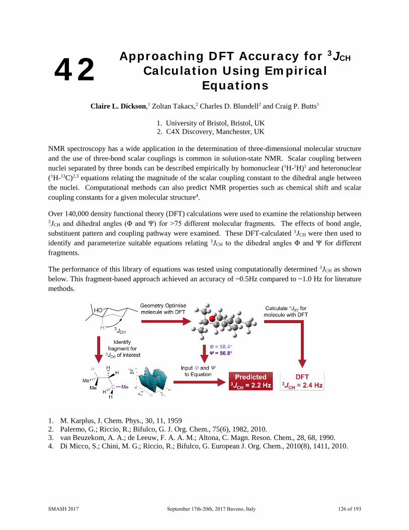
42 Approaching DFT Accuracy for 3JCH Calculation Using Empirical
Equations
Claire L. Dickson,1 Zoltan Takacs,2 Charles D. Blundell2 and Craig P. Butts1
1. University of Bristol, Bristol, UK 2. C4X Discovery, Manchester, UK
NMR spectroscopy has a wide application in the determination of three-dimensional molecular structure and the use of three-bond scalar couplings is common in solution-state NMR. Scalar coupling between nuclei separated by three bonds can be described empirically by homonuclear (1H-1H)1 and heteronuclear (1H-13C)2,3 equations relating the magnitude of the scalar coupling constant to the dihedral angle between the nuclei. Computational methods can also predict NMR properties such as chemical shift and scalar coupling constants for a given molecular structure4.
Over 140,000 density functional theory (DFT) calculations were used to examine the relationship between 3JCH and dihedral angles (Φ and Ψ) for >75 different molecular fragments. The effects of bond angle, substituent pattern and coupling pathway were examined. These DFT-calculated 3JCH were then used to identify and parameterize suitable equations relating 3JCH to the dihedral angles Φ and Ψ for different fragments.
The performance of this library of equations was tested using computationally determined 3JCH as shown below. This fragment-based approach achieved an accuracy of ~0.5Hz compared to ~1.0 Hz for literature methods.
1. M. Karplus, J. Chem. Phys., 30, 11, 1959 2. Palermo, G.; Riccio, R.; Bifulco, G. J. Org. Chem., 75(6), 1982, 2010. 3. van Beuzekom, A. A.; de Leeuw, F. A. A. M.; Altona, C. Magn. Reson. Chem., 28, 68, 1990. 4. Di Micco, S.; Chini, M. G.; Riccio, R.; Bifulco, G. European J. Org. Chem., 2010(8), 1411, 2010.
SMASH 2017 September 17th-20th, 2017 Baveno, Italy 126 of 193

43 Non-Animal Testing of the Skin Sensitization Potential of Chemicals
Tobias Nickel1, Erica Brendler1, Alexander Böhme2 and Gerrit Schüürmann1,2
1. TU Bergakademie Freiberg, Freiberg, Germany.
2. Helmholtz Centre for Environmental Research, Leipzig, Germany The main step in skin sensitization is the reaction of small molecules with the proteins and peptides. These adducts are recognized by the immune system which results in the disease known as contact allergy. The current approach to detect the sensitization power of chemicals, the local lymph node assay [1], includes the use of laboratory animals. For this reason development of alternative testing methods for the skin sensitization potential of chemicals is desired. Several approaches have been developed, like QSAR modelling [2] or the Direct Peptide Reactivity Assay.[3]
This work aims to achieve a better understanding of the different mechanisms in the reaction of a chemical with a protein. In principle five different types can occur: Acylation, Michael-Addition, Imine forming and Nucleophilic substitution (either SN2 or SNAr). Amino acids and small peptides are used as test substances to identify the reaction mechanism as well as the reaction rate. For the structural characterization mainly NMR spectroscopy is used. Kinetic studies are performed with either NMR spectroscopy or with a HPLC chemoassay. For several chemicals it is not clear which reaction is the rate determining step, because in the HPLC chemoassay only the loss of the nucleophile signal is monitored. For the identification of products and mechanism NMR spectroscopy is very important. One example is the reaction of Trinitrobenzenesulfonic acid (TNBS) with amino acids or small peptides. This system had already been investigated and it was shown that the skin sensitization potency was underpredicted in correlation to the rate constant [4]. We could show that for TNBS different reactions occur, depending on the pH. The reaction is ten times faster at pH=7.4 then at pH=6 and more products can be observed.
Other molecules, like Acrolein, can have more than one reacting center. This substance can either react with Imin-formation or with Michael Addition. By NMR spectroscopy we were able to identify the preferred reaction mechanism, as well as the corresponding products.
As mentioned above many reactions are generating a product mixture, which depends on the number of reacting centers or the mechanism. For these mixtures it is important to use a separation processes before the structural studies. Therefore we try to develop a coupling method of HPLC with ultra-high resolution FT-ICR-MS. Together with NMR, this will improve our understanding of the reactions.
1. Basketter D. A. et al., Food Chem. Toxicol., 40, 593–598, 2002. 2. Dearden J. C. et al., Chem. Res. Toxicol., 28, 1975-1986, 2015. 3. Gerberick G. F. et al., Toxicol. Sci., 81, 332−343, 2004. 4. Roberts D. W., Aptula A. O., Chem. Res. Toxicol., 27, 240-246,2014.
SMASH 2017 September 17th-20th, 2017 Baveno, Italy 127 of 193

44 Characterisation of Catechol and (4-fluoro)-phenylboronic Acid Binding System: Investigation by F19-NMR
and Modelling of Equilibria
Gary Sharman and Paul Tan Eli Lilly and Company Limited, Analytical Technologies, Windlesham, Surrey, United Kingdom
The multi-step equilibria which are hallmarks of the reaction [1] between catechol and (4-fluoro)-phenylboronic acid are measured and characterised by F19-NMR in buffered aqueous media across a range of pH. The recorded species concentrations show very good agreement with the data values from a model of a linked set of equilibria for the reaction. Global curve fitting yields equilibrium constants for all steps which are consistent across all conditions tested for this reaction. This process provides a template for robust and facile interrogation of analogous equilibria in other systems.
1. Hall, Dennis G. (ed.), Boronic Acids. 2nd Edition, 2011, Wiley-VCH, Weinheim ISBN: 978-3-527-32598-6
SMASH 2017 September 17th-20th, 2017 Baveno, Italy 128 of 193

46 Intramolecular Hydrogen Bond in p- Substituted o-(N-
Diethyl)aminomethylphenols: NMR Study
Khodzhaberdi Allaberdiev
Ukraine State Scientific Research Institute for Plastics,Donetsk,Ukraine
The aminomethylphenols are commonly used as catalysts for supramolecular systems, epoxy resins and in the synthesis of medicaments with multiple pharmacological activities. The availability of intramolecular H-bonds causes the specificity of physicochemical properties of the aminomethylphenols.
A series p-substituted o-(N-diethyl)aminomethylphenols (o-DEAMPH), XC6H5CH2Y(X=p-OCH3, CH3, H, F, Cl, Br, COOCH3, CN and NO2, Y=o-N(C2H5)2) were studied using Nuclear Magnetic Resonance spectroscopy(NMR). The chemical shifts of the bridging hydrogen atom in o-DEAMPH have been measured in CCl4 solutions. Moreover,1H NMR spectra of compounds were obtained at B3LYP/6–311+G(d,p) level using the GIAO method. Excellent linear correlation (R2= 0.9996) observed between values the chemical shift of hydrogen atom obtained experimentally of 1H NMR and calculated B3LYP/6–311+G(d,p). A linear relationship revealed the chemical shift of the hydrogen atom,δOH with the charge on the hydrogen atom, QA(H), calculated by the quantum theory atoms in molecules (QTAIM) (R2=0.9902). It is found that a linear correlation between experimental assignment phenol hydroxyl proton chemical shifts, δOH and theoretical calculated values frequencies of the O-H stretching mode, νOH : δOH = 58.4151-0.0149νOH (R2=0.9872. The values hydroxyl proton chemical shifts,δOH also correlates linearly with the para-substituent constants, σp
- (R2= 0.9882). In addition,the intramolecular O-H…N bond energy was estimated from the empirical relationship. It is shown that from the standpoint of their strength the intramolecular O-H…N hydrogen bonds can be classified as strong interactions. The intramolecular hydrogen bonding energies of the considered systems range from 6.5 to 8.5 kcal/mol.Intramolecular hydrogen bond energy is linearly correlated with Hammett constants, σp
- (R2= 0.9903).
Moreover, numerous relationships between the hydrogen bond energy obtained from empirical 1H NMR data and predicted geometric descriptors of hydrogen bond: d H…N (R2= 0.9984), ∠ O-H…N (R2= 0.9979) as well as topological descriptors of hydrogen bond strength: total energy density ρBCP (R2= 0.9964) and the density of potential energy V (R2=0.9980) at the bond critical point of the proton-acceptor (H-N) distance (QTAIM) and the core-valence bifurcation index CVB (based in ELF, R2= 0.9945) were obtained. From these dependencies excluded the point corresponding to R=H. Relationships between of values hydrogen bond energy and various quantities characterizing of o-DEAMPH are discussed.
SMASH 2017 September 17th-20th, 2017 Baveno, Italy 129 of 193

47 Methanol Self-association and Preferential Solvation of Chelating
Agents for the Extraction of Nuclear Fission products in Supercritical CO2
Trent R. Graham, Daniel Pope, Aurora Clark, and Steven R. Saunders
Washington State University, Pullman, Washington, United States of America
While supercritical carbon dioxide (sCO2) is a promising solvent in heavy metal extractions due to its low toxicity, surface tension and viscosity, many chelating molecules exhibit limited solubility in sCO2. The addition of volatile polar molecules to sCO2, such as methanol (MeOH), improves the solubility of chelating molecules and metal-ligand complexes. MeOH self-assembly and preferential solvation of 2-ethylhexylphosphonic acid mono-2-ethylhexyl ester (HEHEHP), a chelating ligand, in sCO2 at 19.7 MPa and 323 K was investigated with diffusion-ordered nuclear magnetic resonance spectroscopy (DOSY-NMR) and molecular dynamics simulations. DOSY-NMR pulse sequences contain a pair of pulsed field gradients that attenuate the NMR resonance and enable calculation of diffusivity for each NMR active component in the mixture. Diffusometry results were analyzed using a model that integrates the Stokes-Einstein relationship with self-association models to approximate the size-dependent, energy of cluster formation landscape. DOSY-NMR results are consistent with molecular dynamics simulations and suggest the formation of small clusters of MeOH limited to a composition of under 10 molecules in a cluster at high concentrations of MeOH.
Next, self-assembly of HEHEHP into dimers was quantified with DOSY-NMR in sCO2 at 19.7 MPa and 323 K. The propensity of HEHEHP to self-associate into dimers in sCO2 was evaluated with the integrated Stokes-Einstein, self-association model. The results suggest that HEHEHP is primarily composed of dimers at concentrations of over 40 mM in sCO2. Finally, we compare the extent of dimerization of HEHEHP in sCO2 containing 4 mole % of MeOH to the dimerization of HEHEHP in neat sCO2 with a series of DOSY-NMR experiments. The experiments indicate that MeOH preferentially solvates HEHEHP, increasing both the solubility of HEHEHP in the supercritical fluid and the weight fraction of monomeric HEHEHP. Our results suggests that the increased amount of monomeric chelators may be an additional, synergistic factor in the enhanced heavy metal extraction in MeOH modified sCO2.
SMASH 2017 September 17th-20th, 2017 Baveno, Italy 130 of 193
48 Molecular Networking Using 13C NMR Can Facilitate Natural Product
Structure Elucidation
C. Benjamin Naman,1 Christopher A. Leber,1 Chen Zhang,2 and William H. Gerwick1,3 1. Center for Marine Biotechnology and Biomedicine, Scripps Institution of Oceanography, University of
California – San Diego, San Diego, California USA 2. Department of Nanoengineering, University of California – San Diego
3. Skaggs School of Pharmacy, University of California – San Diego
Structure elucidation of natural products is a complicated and challenging research problem, with variables of subjectivity including the experience of the spectroscopist, the particular structure(s) to be determined, and instrumental capabilities, to name a few. Modern methods, such as the incorporation of RDC and LR-HSQMBC experiments, advanced data analysis using computer-assisted structure elucidation, and other innovative approaches are changing the way that unknowns are characterized (when cost-permissive or otherwise feasible).[1-2] Similarly, the dereplication of natural products in mixtures using NMR, LC-MS, and other methodologies has had a profound impact on the efficiency and pace of new natural product isolation and discovery.[3-4] Recently, a method has emerged for the simplification of 13C NMR data that allowed construction of a large-scale data library from values obtained experimentally, computationally, or even by extraction from literature reports.[5] Thus we have undertaken an informatics-based research project to evaluate whether or not the use of 13C fingerprinting, followed by comparative array analyses, can facilitate natural product structure elucidation. Indeed, the evaluation of these data can be completed by several different metrics, including the calculation of Tanimoto coefficients, Pearson correlation coefficients, Jaccard indeces, normalized euclidian distances, and cosine values. As a proof of principle, each of these computations was completed for a set of previously isolated and already characterized natural products (including many analogues from the same structural class). The results to be shown in this presentation indicate that not only can the simplified 13C NMR data rapidly facilitate the classification of known natural producs and analogues thereof, but furthermore that specific variations in functional group substitutions will also lead to molecular network connections that can provide other clues to the structure of unknowns.
1. Blinov, K. A.; Buevich, A. V.; Williamson, R. T., Martin, G. E. Org. Biomol. Chem., 2014, 12, 9505-9509.
2. Troche-Pesqueira, E.; Anklin, C.; Gil, R. R. Angewandte Chem. Int. Ed., 2017, 56, 3660-3664. 3. Bakiri, A. Hubert, J., Reynaud, R.; Lanthony, S.; Harakat, D.; Renault, J.-H.; Nuzillard, J.-M. J. Nat.
Prod., 2017, 80, 1387–1396. 4. Wang, M.; Carver, J. J.; Phelan, V. V; Sanchez, L. M.; Garg, N.; Peng, Y.; Nguyen, D. D.; Watrous,
J.; Kapono, C. A.; Luzzatto-Knaan, T.; Porto, C.; al et. Linington, R. G.; Gutiérrez, M.; Lopes, N. P.; Gerwick, W. H.; Moore, B. S.; Dorrestein, P. C.; Bandeira, N. Nat. Biotechnol., 2016, 34, 828–837.
5. Churanova, T.; Blinov, K. A. ENC, 2017.
SMASH 2017 September 17th-20th, 2017 Baveno, Italy 131 of 193

49 General Approach to Access Long-Range ¹H-¹H RDCs
Davy Sinnaeve1, Volker Schmidts2, Julian Ilgen2, Maria Enrica Di Pietro3, Christina M. Thiele2, and
Burkhard Luy3
1. Department of Organic and Macromolecular Chemistry, Ghent University, Ghent, Belgium 2. Clemens Schöpf Institut für Organische Chemie und Biochemie, Technische Universität
Darmstadt, Darmstadt, Germany 3. Institut für Biologische Grenzflächen 4 – Magnetische Resonanz, Karlsruher Institut für
Technologie, Karlsruhe, Germany Residual dipolar couplings (RDCs) are a powerful means for conformational and configurational analyses [1, 2]. For small molecules, 1H-13C one-bond couplings are mostly exploited. However, the quality of, for instance, configurational analysis greatly depends on the number of RDCs that sample independent orientations, which may not always be provided by one-bond couplings alone. Ways to obtain long-range RDCs are thus in high demand. Long-range 1H-1H RDCs would be an obvious choice, but are very challenging to access. The main reason is that, in alignment media such as PBLG, the high abundance of 1H-1H RDCs results in broad multiplets that overlap and from which individual splittings are not resolvable. Here, we propose a general approach to obtain 1H-1H RDCs.
We demonstrate that the recent PSYCHEDELIC experiment [3] gives access to long-range nTHH couplings at a resolution close to the natural linewidth. PSYCHEDELIC delivers a 2D J-resolved spectrum with absorption mode lineshapes and with only 1H-1H couplings to one or more chosen spins, thus allowing coupling measurement as simple doublets at pure shift resolution and resolving both the issues of spectral overlap and multiplet complexity. The experiment is based on PSYCHE [4], which allows broadband homodecoupling at good sensitivity and tolerance to strong coupling. The tolerance to strong coupling can be further enhanced by introducing frequency-swept 180° pulses applied during gradients [5]. Although PSYCHEDELIC delivers the magnitude of the 1H-1H coupling, it offers no sign information, which is crucial for RDC interpretation. In isotropic samples, E.COSY or similar experiments can provide relative sign information, but in weakly aligned samples the overflow of 1H-1H couplings complicate cross-peak analysis. Solutions based on P.E.COSY or z-COSY using similar principles as PSYCHEDELIC to provide relative sign information of only selected couplings will be introduced.
1. Kummerlöwe, G., and Luy, B., Annu. Rep. NMR Spectrosc., 68, 193-230, 2009 2. V. Schmidts, Magn. Reson. Chem., 55, 54-60, 2017 3. Sinnaeve, D., Foroozandeh, M., Nilsson, M., and Morris, G. A., Angew. Chem. Int. Edit., 55, 1090-
1093, 2016 4. Foroozandeh, M., Adams, R. W., Meharry, N. J., Jeannerat, D., Nilsson, M., and Morris, G. A.,
Angew. Chem. Int. Edit., 53, 6990-6992, 2015 5. Thrippleton, M. J., Edden, R. A. E., and Keeler, J., J. Magn. Res., 174, 97-109, 2005
SMASH 2017 September 17th-20th, 2017 Baveno, Italy 132 of 193

50 Solution-State Structure Determination of Lactoferrin-Derived
Peptides, Acting Against Influenza Simone Di Micco1, Maria Carmina Scala1, Marina Sala1, Agostina Pietrantoni2, Antonia Spensiero1,
Mariangela Agamennone3, Alessia Bertamino1, Ettore Novellino4, Giuseppe Bifulco1, Isabel M. Gomez-Monterrey4, Fabiana Superti2 and Pietro Campiglia1
1. Department of Pharmacy, University of Salerno, Via Giovanni Paolo II 132, Fisciano, Italy2. National Institute of Health, Viale Regina Elena 299, Rome, Italy
3. Department of Pharmacy, University of Chieti “G. d’Annunzio”, Via dei Vestini 31, Chieti Italy
4. Department of Pharmacy, University of Naples “Federico II”, Via D. Montesano 49, Napoli, Italy
Bovine lactoferrin is a glycoprotein playing a key role in innate immunity against infections. Previously, we have demonstrated that selected peptides from bovine lactoferrin C-lobe are able to prevent both cell infection and Influenza virus hemagglutination [1]. These findings prompted us to further investigate the ability of lactoferrin derived peptides to inhibit Influenza virus infection. In this study, we present the structural investigation of new bovine lactoferrin C-lobe derived sequences and their ability to inhibit infection and viral hemagglutination.
By means of NMR spectroscopy, we have investigated the solution structures of new five peptides (1-5) derived from loop Ser418-Pro429 of Bovine lactoferrin (PDB ID: 3IB0) [2], in order to determine the structural elements responsible for binding towards the macromolecule. In details, we have resolved the solution structure (HFA/H2O) of 1 (SKHSSLDCVLRP) and 2 (SLDCVLRP), finding a γ-turn for 1, and β-turn for 2. The global turn observed for 1 and are in agreement with conformation of the lactoferrin loop Ser418-Pro429 (PDB ID: 3IB0). In particular, the helix 310 formed by Cys425-Leu427 of protein loop is overlapped with the γ-turn of 1 (Val9-Arg11) and β-turn of 2 (Cys4-Arg7). The tetrapeptides 3 (VLRP), 4 (SLDC) and 5 (SKHS) gave rise to very low number of inter-residue NOE effects, due to their expected highly flexibility, hampering the solution structure determination. However, we may suggest that the preferred conformation of 3-5 is similar to the spatial arrangement observed for 1, 2 and the loop Ser418-Pro429 as highlighted by the biological activity of 3-5. Indeed, The tetrapeptides showed an anti-Influenza activity in a concentration range femto- to picomolar. Our data indicate that these peptides may constitute a non-toxic tool for potential applications as anti-Influenza therapeutics.
1. Rafia, Singh, Nagendra, Vikram, Gopalakrishnapillai, Kumar, Ramasamy P., Sinha, Mau, Bhushan,Ammendolia, Maria G., Agamennone, Mariangela, Pietrantoni, Agostina, Lannutti, Fabio, Siciliano,Rosa A., De Giulio, Beatrice, Amici, Fabiana Superti, Pathog. Glob. Health., 106, 12-19, 2012.
2. Mir, Asha, Kaur, Punit, Srinivasan, Alagiri, Sharma, Sujata, Singh, Tej P., Biophys.J. 97, 3178-3186,2009.
SMASH 2017 September 17th-20th, 2017 Baveno, Italy 133 of 193

51 Study of Ambroxol Hydrochloride and Excipients in Infant Syrups by
¹H NMR
Luiz Henrique Keng Queiroz Junior1,3, Danilo Monteiro de Carvalho1, Vinicius Sousa Ferreira1,2, Vinícius Silva Pinto, Luciano Morais Lião1 and Luiz Alberto Colnago3
1. Federal University of Goiás, Chemical Institute, Goiânia-GO,Brazil
2. Federal Institute of Goiás, Campus Formosa-GO, Brazil 3. Embrapa Instrumentação, São Carlos-SP, Brazil
Ambroxol Hydrochloride (AH) is a drug with mucolytic activity used as an expectorant and found in commercially available pharmaceutical preparations in the form of drops, injections, syrups and tablets and is widely used in pediatric patients. International pharmacopoeias advocate methods for the determination of AH only in pure form, which makes it impossible to apply these methods in formulations, and the excipients may interfere with the quality control analysis. A promising technique for this type of case is Nuclear Magnetic Resonance1-3. The objective of the present study is to use the Nuclear Magnetic Resonance (NMR) technique in the characterization and quantification of the active principle, in addition to investigating the presence of ethanol in children's syrups with ambroxol hydrochloride as the active compound. The ERETIC method provides a reference signal, synthesized by an electronic device, which can be used for the determination of absolute concentrations after a previous calibration.4 Ten commercial samples were analyzed in triplicate, and were performed the characterization of the main compounds present in the formulations. Seven samples showed characteristic ethanol signals and based on the qualitative analysis of the spectra the triplet (CH3 of ethanol) was selected as quantifiable signal for ethanol. For AH, a singlet was selected as a quantifiable signal. According to National Formulation of the Brazilian Pharmacopoeia, 2nd edition Rev. 02, syrups must not present ethanol in their composition, and their minimum presence must be declared in the package leaflet. However, through the study in question, it was possible to quantify the ethanol in the formulation without the declaration, and one of the samples had a value of 1.8 oGL, therefore being classified as an alcoholic beverage according to a Brazilian decree 6.117/2007. There was also the quantification of the AH content, where it was possible to observe that only one of the samples had the declared content of 2.7 mg/mL in 120 mL of solution, what could affect the therapeutic effect of these syrups. 1. Heinänen, C. Barbas J. Pharm. Biom. Anal. 24 (2001) 1005. 2. United States Pharmacopeia, 26a Ed. Rockvillle, 2003. 3. European Pharmacopoeia, 7a Ed. Ambroxol hydrochloride 2011 4. Akoka, L. Barantin, M. Trierweiler, Anal. Chem. 71 (1999) 2554–2557.
SMASH 2017 September 17th-20th, 2017 Baveno, Italy 134 of 193

52 Interactions between Diet and Microbiota Studied by NMR-Based
Metabolomics Martina Bugáňová1,2, Helena Pelantová1, Miloslav Kverka1,3, Štěpán Coufal1, Tomáš Hudcovic1 and
Marek Kuzma1
1. Institute of Microbiology of the Czech Academy of Sciences, Prague or Nový Hrádek,
Czech Republic 2. Faculty of Chemical Technology, University of Chemistry and Technology, Prague, Czech Republic
3. Institute of Experimental Medicine of the Czech Academy of Sciences, Prague, Czech Republic
The gut microbiome is a comprehensive ecosystem that regulates many physiological functions. It affects the evolution of the immune system, influences cell proliferation, and ensures metabolism of bile salts as well as indigestible polysaccharides and proteins [1]. An individual’s diet is one of the main factors affecting the gut microbial composition, and the catalytic activity of intestinal microbiota determines an individual´s health status. Hence, investigating the interactions between the gut microbiome and diet is crucial for understanding how serious metabolic diseases develop, e.g. metabolic syndrome, colorectal cancer, and others. Lately, NMR-based metabolomic analysis of fecal extracts has become a well-established platform for the study of the gut microbiota and host metabolism through the analysis of the endproducts of their metabolism. It is advantageous to analyze feces as it has immediate contact with the colon and is easily sampled, which makes it a relevant material for exploring both the gut microbiota metabolism as well as its co-metabolism with the host. In this study, we focused on the impact of different microbial colonizations and diets on the metabolic composition of fecal samples in the experimental model of BALB/c and C57BL/6 mice respectively. Water fecal extracts were analyzed by 600 MHz NMR spectrometer using 1D-NOESY pulse sequence. Metabolites identification was supported by information extracted from J-resolved, COSY, and HSQC spectra. Statistical analysis of the acquired data was performed using a combination of univariate (parametric and non-parametric tests) and multivariate (principal component analysis (PCA) in addition to the partial least square discriminant analysis (PLS-DA)) approach. Firstly, we compared the fecal metabolome of conventionally colonized (CV) male mice fed by two different diets: a standard murine diet and a diet with excess protein and fat (“Western-type diet”, WTD). The WTD significantly increased the levels of short-chain fatty acids (SCFAs; namely acetate, propionate and butyrate), which are an important energy source for cells and possess other properties favourable for the host organism. Enhanced protein breakdown might explain the increased concentrations of alanine, glutamate, and 5-aminovalerate in WTD-treated mice. Additionally, we observed the elevation of fumarate and succinate, indicating more intensive carbohydrate breakdown in mice on WTD. Secondly, we were looking for differences in fecal metabolome of germ-free (GF), monocolonized (Escherichia coli Nissle 1917) and CV mice, all fed the same standard diet. Statistical analysis of NMR spectra was able to unequivocally differentiate among samples from these three groups. Obviously, the largest differences were observed between CV and GF mice. These included elevated levels of SCFAs (butyrate, propionate and acetate) in CV mice, as their production from polysaccharides or proteins is conditioned by the presence of specific bacteria. On the contrary, the absence of bacteria in GF mice resulted in higher levels of saccharides, e.g. raffinose. Therefore, raffinose, which can be digested only by
SMASH 2017 September 17th-20th, 2017 Baveno, Italy 135 of 193

certain bacterial strains, may serve as a good marker of microbial activity [2]. Additionally, NMR spectroscopy allowed us to distinguish CV mice from E. coli Nissle colonized mice. This observation is of particular importance since the probiotic bacterium E. coli Nissle has been reported to have a positive effect on maintaining remission of ulcerative colitis [3]. In summary, our data show several interactions between diet and microbiota beyond the obvious relations between substrates and enzymes. Next, our metabolomic results will be correlated with abundance of particular colonies residing the gut or gut mucosa transcriptome. A follow-up study will then focus on monitoring how these differences in microbial metabolism influence the development of intestinal inflammation and colorectal cancer in this mouse model.
Acknowledgements This work is supported by the Czech Science Foundation (Grant No. 16-06326S) and the Operational Program Prague – Competitiveness (Project No.: CZ.2.16/3.1.00/24023). The authors gratefully acknowledge the project LO1509 of the Ministry of Education, Youth and Sports of the Czech Republic.
1. Blaut M., and Clavel T., Journal of Nutrition, 137(3), 751S-755S, 2007. 2. Heinzmann S. S., Schmitt-Kopplin P., Journal of Proteome Research, 14(5), 2267-2277, 2015. 3. Schultz M., Inflammatory Bowel Diseases, 14(7), 1012-1018, 2008.
SMASH 2017 September 17th-20th, 2017 Baveno, Italy 136 of 193
53 Probing Torsional Flexibility Via RDC-Driven Molecular Dynamics
Simulations
Maria E. Di Pietro1, Ulrich Sternberg2, and Burkhard Luy1,3
1. Institute of Organic Chemistry, Karlsruhe Institute of Technology (KIT), Karlsruhe, Germany
2. COSMOS GbR, Jena, Germany 3. Institute for Biological Interfaces 4 - Magnetic Resonance, Karlsruhe Institute of Technology
(KIT), Eggenstein-Leopoldshafen, Germany
Residual Dipolar Couplings (RDCs) have established themselves as an efficient NMR tool for the structure elucidation of organic molecules [1]. Unlike standard short-range NMR parameters such as chemical shifts, 3J-couplings and NOEs, RDCs make it generally possible to correlate nuclear pairs that are far apart in the molecular structure, hence providing global constitutional, configurational and conformational information. Interpretation of experimentally determined RDCs is, however, far from being a trivial task, especially in the presence of conformational flexibility. To this end, different strategies have been developed. A promising and conceptually new approach is based on Molecular Dynamics simulations with Orientational Constraints (MDOC) [2] implemented in the COSMOS software [3] and successfully applied to stereochemical assignment of rigid and flexible molecules [4]. In this contribution, the potential of RDC-driven MD simulations is explored in the study of the torsional flexibility of small non-steroidal anti-inflammatory drugs, members of the salicylate and profen families. Realistic conformational distributions are obtained with high-quality fits of the experimental one-bond and long-range couplings used as constraints. Contrary to most existing methods, the MDOC approach does not require any optimized structure or a priori knowledge of the conformational surface. This feature, together with the possibility of combining isotropic and anisotropic constraints as well as the reasonable computational cost, make the COSMOS software an appealing candidate in the search of a versatile and general tool for the structure elucidation of any flexible organic molecule.
SMASH 2017 September 17th-20th, 2017 Baveno, Italy 137 of 193

Fig. a) Torsional distributions obtained for the fluorinated anti-inflammatory drug flurbiprofen in 20-ns MDOC simulations with COSMOS software, starting from three different geometries and using as
constraints RDCs measured in PBLG/THF-d8 together with 3J-couplings, and comparison with results obtained via theoretical calculations at DFT level and a different conformational model known as AP-
DPD approach [5]. b) Experimental versus back-calculated one-bond and long-range RDCs for the same MDOC simulations.
1. a) Schmidts, V., Magn. Reson. Chem., 55(1), 54-60, 2017; b) Gil, R. R., Angew. Chem. Int. Ed., 50(32), 7222-7224, 2011.
2. a) Sternberg, U., Witter, R., Ulrich, A. S., J. Biomol. NMR, 38(1), 23-39, 2007; b) Sternberg U. and Witter R., J. Biomol. NMR, 63(3), 265-274, 2015.
3. Sternberg, U., Koch, F.-Th., Losso, P., http://www.cosmos-software.de/ce_intro.html. 4. Tzvetkova, P., Sternberg, U., Gloge, T., Navarro-Vazquéz, A., Luy, B., in preparation. 5. Di Pietro, M. E., Aroulanda, C., Celebre, G., Merlet, D., De Luca, G., New J. Chem., 39(12), 9086-
9097, 2015.
SMASH 2017 September 17th-20th, 2017 Baveno, Italy 138 of 193

54 Insights into Amorphous Solid Dispersions of Felodipine Using Solid-state NMR Spectroscopy:
Miscibility and Molecular Interactions
Kanika Sarpal and Eric J. Munson
College of Pharmacy, University of Kentucky, Lexington, Kentucky
Phase separation in amorphous solid dispersions (ASDs) is still not clearly understood on the sub-nanometric scale [1], and further systematic investigations are still required. The phase separation behavior is influenced by many factors, such as the composition of drug-polymer blend, the type and the strength of drug-polymer interactions, and the method of preparation [2-4]. We demonstrated the use of solid-state NMR spectroscopy (SSNMR) to evaluate the role of the strength of drug-polymer hydrogen bonding (H-bonding) on the compositional homogeneity in ASDs of felodipine (FEL), a poorly water soluble drug, with poly(vinylpyrrolidone), or PVP, poly(vinylpyrrolidone-co-vinylacetate), or PVP/VA, and poly(vinylacetate) or PVAc. The dispersions were prepared at various drug loadings (50% to 90% w/w) via melt quenching. The blend scale miscibility was studied by examining the proton spin-lattice relaxation times in the laboratory and rotating frame (1H T1 and T1ρ) for the drug and the individual polymer for each set of ASDs. Domain sizes were estimated via spin diffusion. The experimental data was used to elucidate the influence of the strength of drug-polymer H-bonding on the phase behavior of resulting dispersions. It was found that FEL:PVP and FEL:PVP/VA systems exhibited weak signs of nano phase separation especially for the compositions with lower polymer loadings with considerably small domain sizes. Whereas FEL:PVAc system showed pronounced signs of nano phase separation as the polymer amount decreased with larger domains. The extent of H-bonding in amorphous FEL and FEL:Polymer blends within each set of ASDs was quantified via deconvolution. The carbonyl region was used to selectively analyze the evolution of the various populations of coexisting species in the samples. The order of the strength/extent of drug–polymer H-bonding interactions was PVP > PVP/VA > PVAc. It was suggested that the strength of drug-polymer H-bonding interaction is one of the key factors in controlling the phase behavior of ASDs. Our findings indicate that SSNMR is a useful tool for evaluating the spatial homogeneity with sub-50 nm resolution, where other conventional techniques fail, and drug-polymer interactions for the compositions of pharmaceutically relevant systems.
1. Qi, Sheng et al., Molecular Pharmaceutics, 10, 918-930, 2013 2. Nie, Haichen et al., Molecular Pharmaceutics, 13, 3964-3975, 2016 3. Chakravarty, Paroma et al., International Journal of Pharmaceutics, 519, 44-57, 2017 4. Yuan, Xiaoda et al., Molecular Pharmaceutics, 11, 329-337, 2014
SMASH 2017 September 17th-20th, 2017 Baveno, Italy 139 of 193
55 Characterization of Multicomponent Crystal Forms of Neuroleptic Drugs
by NMR Crystallography
Inês C. B. Martins1,2,3, José R. Gomes2, Martin U. Schmidt3, Maria T. Duarte1 and Luís Mafra2
1. CQE - Centro de Química Estrutural, Instituto Superior Técnico, Universidade de Lisboa,
Lisboa, Portugal 2. CICECO - Aveiro Institute of Materials, University of Aveiro, Aveiro, Portugal 3. Institut fuer Anorganische und Analytische Chemie, J. W. Goethe-Universitaet,
Frankfurt am Main, Germany
Launching new products in the market is very expensive and extremely time consuming. Envisaging these aspects, pharmaceutical industry has shown a high interest on improving the efficacy of “old” drugs by enhancing their physicochemical properties (solubility, dissolution rate and bioavailability) without changing their pharmacological behavior. This has been adressed using several approaches including the preparation of multicomponent drug crystal forms (salts and co-crystals), developed over the last years. [1-3] Understanding how molecular systems self-assemble in the solid-state is a great challenge, with particular relevance in pharmaceutical sciences.[4] Herein, we present an experimental NMR, single-crystal / powder X-ray diffraction (XRD), and computational study of the supramolecular assemblies of Gabapentin (GBP) and amantadine (AMA); two active pharmaceutical ingredients used to treat neurodegenerative diseases, such as epilepsy and Parkinson, respectively.[5,6] For GBP, an amino-acid based drug, there are few co-crystals reported to date[7] and here we report the preparation of two organic “solid” ionic liquid drug systems featuring an unusual lamellar arrangement. The effect of crystal packing interactions was investigated and quantified using SSNMR in tandem with periodic and cluster DFT calculations, by means of a stepwise in silico dismantlement of the 3D packing. To better understand the synergy between weak and strong hydrogen bonds, several computer cluster fragments were evaluated based on calculated packing-induced chemical shifts.[8] In the case of AMA, the very low aqueous solubility and the bioavailability problems related to the use of the hydrochloride and sulfate salt forms,[6] lead us to prepare a set of new molecular salts through the combination with safe organic co-formers. The structural determination was carried out using powder XRD for the compounds where it was not possible to grow crystals. DFT calculations were also used in tandem with SSNMR for structure validation.[9] Thermal stability analysis were perfomed, in both systems, using DSC-TGA and hot-stage microscopy. For Ama salts, preliminary solubility studies were carried out, showing an improvement of the drug aqueous solubility.
1. Braga, D., Maini, L., Grepioni, F., Chem. Soc. Rev., 42(18), 7638–7648, 2013. 2. Duggirala, N. K., Perry, M. L., Almarsson, O., Zaworotko, M. J., Chem. Commun., 52(4), 640-655,
2016. 3. Domingos, S., André, V., Quaresma, S., Martins, I. C. B., Piedade, M. F. M., Duarte, M. T., J. Pharm.
Pharmacol., 67(6), 830-846, 2015. 4. Martins, I. C. B., Santos, M. S., Fernandes, A., Antunes, A., André, V., Mafra, L., Duarte, M. T.,
Cryst. Growth Des., 16(1), 156-166, 2016.
SMASH 2017 September 17th-20th, 2017 Baveno, Italy 140 of 193
5. Braga, D., Grepioni, F., Maini, L., Rubini, K., Polito, M., Brescello, R., Cotarca, L., Duarte, M. T., André, V., Piedade, M. F. M., New J. Chem., 32(10), 1788-1795, 2008.
6. Jankovic, J., Aguilar, L. G., Neuropsychiatr. Dis. Treat., 4(4), 743–757, 2008. 7. Reddy, L. S., Nethune, S. J., Kampf, J. W., Hornedo, N. R., Cryst. Growth Des., 9(1), 378-385, 2009. 8. Martins I. C. B., Sardo, M., Cendak, T., Gomes, J. R. B., Duarte, M. T., Mafra, L., article in
submission. 9. Martins I. C. B., Bodach, A., Alig, E., Sardo, M., Mafra, L., Fink, L., Schmidt, M. U., Duarte, M. T.,
article in preparation.
SMASH 2017 September 17th-20th, 2017 Baveno, Italy 141 of 193

56 Progress in Multi-Spectra Automatic Structure Verification
Stanislav Sykora1 and Carlos Cobas2
1. Extra Byte, Castano Primo, Italia
2. Mestrelab Research, Santiago de Compostela, Spain
Automatic Structure Verification is fast becoming an important part of NMR data evaluation software packages such as Mnova and others. Its basic goal is to answer, in a qualitative as well as quantitative way, the question
“Is this molecular structure compatible with these NMR data?” Naturally, the query one makes, the answer one gets. These things are fuzzy in logic because of imperfections and impurities in the spectra, nmr parameters prediction errors, solvent effects, etc. The complexity is close to that of an artificial intelligence, and the scoring is critically dependent on the query itself. But the problems extend beyond that, involving what one intends by “NMR data”. As we know well, it is one thing to have a single 1H spectrum, for example, and another one to have a pair of spectra of different kinds, such as 1H and 13C, or 1H and HSQC, and a still more different one if the spectra (of presumably the same compound) include an arbitrary subset of any of the “200 and more” NMR experiments such as, for example 1H, 13C, HSQC, COSY, and HMBC. How does one proceed with the automatic analysis in such cases? It is well known that the likelihood of multiple “solutions” decreases sharply when one combines several spectra of different kinds. We also know that even ‘human’ analysis is in these cases anything but linear and standard: it requires multiple “passes” through the spectra and a non-trivial search for correlations (or lack of correlations) of various orders. We also expect that critical points can get exacerbated and build up beyond a threshold (false negatives). Since no spectrum is perfect, how many spectra it takes before any structure gets ruled out? Over the last few years we have built a considerable body of experience [1] with these problems. How to design a software machinery to iteratively analyse a set of any number of NMR spectra of various kinds but presumably of the same compound (or better the same sample) and make it extract and refine all information they might contain in a synergetic way, and then score that information database against an assumed, hypothetical molecular structure. In this presentation we would like to share with you some of this.
1. Cobas Gómez C.J., Bernstein M.A., Sykora S., An Integrated Approach to Structure Verification Using Automated Procedures, Chapter 12 in Structure Elucidation in Organic Chemistry: The Search for the Right Tools, edited by Bravo J., Cid M., Wiley-VCH 2015. DOI: 10.1002/9783527664610.ch12.
SMASH 2017 September 17th-20th, 2017 Baveno, Italy 142 of 193

57 Numeric Atom-Centered Basis Sets for Magnetic Response Calculations
Raul Laasner1, William Huhn1, Johannes Colell2, Thomas Theis2, Warren Warren2,3, and Volker Blum1,2
1. Department of Mechanical Engineering and Materials Science, Duke University, Durham, NC,
United States 2. Department of Chemistry, Duke University, Durham, NC, United States
3. Department of Physics, Duke University, Durham, NC, United States First principles calculations have become extremely useful for assigning the chemical shift peaks and J-couplings to specific nuclei. One of the difficulties that theoretical NMR calculations usually face is having good quality basis sets that are accurate in the region near the atomic nuclei, which has great influence on the NMR parameters. To date, several all-electron models have been proposed, which are based on commonly used basis functions such as Gaussian-type orbitals (GTOs), projector-augmented waves (PAW), linearized augmented plane waves (LAPW), and Slater-type orbitals (STOs). In this work, we explore the use of numeric atom-centered orbitals (NAOs) [1] as basis sets for magnetic response calculations, and compare their accuracy to Gaussian-type orbitals. The NAOs have numerically tabulated radial functions and therefore their radial shape is fully flexible. This includes retaining the correct shape of the orbitals near the nucleus without a significant increase in the computational cost. Furthermore, following [1], the NAOs can be constructed to be strictly localized, which can lead to linear scaling with system size, which becomes beneficial for large systems. We focus on three magnetic response properties that are characterized by different degrees of localization of the perturbation: the magnetizability (defined as the second-order electronic response to an external field), NMR shieldings, and J-couplings. We present details of our implementation, in a nonrelativistic formalism, and use a test set of molecules with light atoms up to chlorine. For magnetizabilities, the NAOs demonstrate faster convergence than GTOs. For shieldings, the general-purpose NAOs show similar convergence as GTOs that are specifically optimized for the calculation of shieldings. J-couplings pose a significant challenge for basis set convergence, because the Fermi contact term requires extreme flexibility of the basis orbitals near the nucleus, as has been illustrated by strategies pursued by the most advanced GTO basis sets available [3].
1. Blum, V., Gehrke, R., Hanke, F., Havu, P., Havu, V., Ren, X., Reuter, K., Scheffler, M., Comput. Phys. Comm, 180(11), 2175-2196, 2009.
2. Zhang, I., Ren X., Rinke, P., Blum, V., and Scheffler, M., New J. Phys., 15, 123033, 2013. 3. Jensen, F., J. Chem. Theory Comput., 2(5), 1360-1369, 2006.
SMASH 2017 September 17th-20th, 2017 Baveno, Italy 143 of 193

58 In situ MAS-NMR Studies of the Dissolution of Gibbsite and
Precipitation of Lithium Hydrotalcite in High-Level Nuclear Waste
Processing
Trent R. Graham, Jian Z. Hu, Xin Zhang, Carolyn I. Pearce, and Kevin M. Rosso
Pacific Northwest National Laboratory, Richland, Washington, United States The Hanford site contains high-level radioactive waste (HLRW) that was produced during World War II and the Cold War during nuclear weapon manufacturing. HLRW is composed of dynamic colloids perturbed far from equilibrium from exposure to both ionizing radiation and extreme alkalinity. Colloidal aluminum hydroxides, known as gibbsite, is a major HLRW component. Hanford personal are actively looking for solutions to process HLRW. One potential solution is to store the waste as glass through a process called vitrification. However, vitrification of material with high aluminum content forms polyphasic glasses with poor material properties. The vitrification strategy therefore necessitates removal of large amounts of aluminum species through dissolution processes. The aluminate in solution would then be precipitated as lithium hydrotalcite. We have completed in situ 27Al, magic angle spinning, nuclear magnetic resonance spectroscopy (MAS-NMR) studies to investigate the dissolution of gibbsite and precipitation of lithium hydrotalcite to emulate the processing of HLRW. Gibbsite dissolution and precipitation of lithium hydrotalcite in aqueous solutions of lithium hydroxide (LiOH) was observed with in situ 27Al MAS-NMR. In situ 27Al MAS-NMR measurements were carried out on a Varian-Inova 500 MHz NMR spectrometer at 25°C using a 7.5 mm MAS probe and a homemade 7.5 mm zirconia rotor sealed with Teflon tape. Gibbsite (0.5 M) had a particle size of 400-700 nm and was dispersed in 3 M LiOH and loaded into the NMR spectrometer. Analysis of in situ NMR spectra suggests gibbsite dissolution produces tetrahedral aluminate species that interact with lithium ions in solution. Aluminate interactions with lithium are consistent with the progressive deshielding of the tetrahedral resonance. Line deconvolution was utilized to separate the broad gibbsite signal from the sharp lithium hydrotalcite resonance. The presence of two kinetic regimes during the dissolution of gibbsite suggests the phase transformation occurs through the intercalation of Li ions. Work is underway to conduct in situ 7Li studies to investigate the formation of ion pairs and Li intercalation during the dissolution and precipitation process. A fundamental understanding of the dissolution of gibbsite and precipitation of lithium hydrotalcite will assist facilities in HLRW processing.
SMASH 2017 September 17th-20th, 2017 Baveno, Italy 144 of 193

59 Defining ‘Gas-Tight:’ An Examination of the Oxygen-Permeability of
Several NMR/EPR Tubes
Susan E. Butch
Wilmad-Labglass, Vineland, NJ, USA
Numerous research applications exist which require exclusion of air and/or moisture from an NMR sample. In the absence of a glovebox or similar inert atmosphere, these approaches become even more challenging. J. Young tubes (or comparable LPV tubes) are the most widely used gas-tight NMR tubes. These tubes are simple to use and seal reliably, however, they do not allow for syringe puncture, require a special glass tube to accommodate the valve, and have other limitations. In this investigation, we employed redox indicators to determine the rate of O2 diffusion into NMR/EPR tubes with J. Young/LPV valves, screw-caps, omni-fit valves, and septa. We compared these difussion rates to the innate rate of O2 diffusion into an uncapped tube. Experiments with uncapped tubes demonstrated that O2 diffusion through aqueous solutions is moderately slow. Samples in LPV tubes remained below the experimental limit of O2 detection for hours, only showing minor oxygen contamination after one day. Tubes sealed with screw-caps and septa showed intermediate oxygen permeability. The implications of this experimental data and the practical constraints associated will be discussed.
SMASH 2017 September 17th-20th, 2017 Baveno, Italy 145 of 193
60 Evaluation of NMR Signals of Isoquinoline Alkaloids: An
Experimental and Theoretical Approach
Layla R. Barbosa, Warley S. Borges, Alvaro C. Neto, and Valdemar Lacerda Jr.
Chemistry Department, Federal University of Espírito Santo, Vitória, Brazil
The Amaryllidaceae family lies between the 20 most applied plant families as therapeutic agents against various diseases. An important characteristic of this family is the presence of isoquinoline alkaloids, exhibiting several biological activities such as antitumor, antibacterial, antifungal, antimalarial, antiviral, analgesic, and antiparasitic and acetylcholinesterase inhibition [1]. The innumerable studies in the literature on the occurrence, isolation and structural determination of natural products, has made them interesting objects of study in nuclear magnetic resonance (NMR) spectroscopy [2], which is a very important and well-established tool for the structural analysis of organic compounds [2-4]. These isoquinoline alkaloids homolyorine-type isolated from Amaryllidaceae plants are reported in the literature when substituted at C-2, with an OH or OCH3 group in α position. However, the spectra of these compounds are not simple, there are overlapping 1H and 13C NMR signals and many couplings that hinder the unequivocal assignment of all 1H and 13C NMR signals and the determination of the relative stereochemistry of this group. The development of experimental NMR techniques has been accompanied by advances in computing and theoretical methods (especially the DFT methods), which permit shielding tensors and spin-spin coupling constants to be calculated with highly reliable results [3,4]. We present here a complete NMR assignment for some isoquinoline alkaloids. These studies were performed using 1D and 2D NMR techniques and compared with the theoretical predictions of the chemical shifts and coupling constants using DFT calculations. GIAO model at DFT/B3LYP level of theory using the cc-pVTZ basis (with and without solvent) set was employed for calculations of 1H and 13C NMR chemical shifts (δ), using Gaussian03 program [6]. From correlation between the theoretical and experimental data, it was concluded that the model used was effective for calculating the shielding tensors for the compounds studied. From hippeastrine, for example, it was found that the mean deviation (MD), standard deviation (SD) and linear correlation factor (R) for all the calculations were generally better when using solvent from δ 1H and without solvent from δ 13C. In addition, the results showed a slight indication that the OH group is in α position, as reported in the literature. Acknowledgements The authors thank to CNPq, CAPES and FAPES for financial support and fellowships. 1. Bastida, J. et al. Chemical and biological aspects of Narcissus alkaloids In: The Alkaloids; Cordell,
G. A.2006, 63, 87; 2. Salles, R. C., et al, Journal of Molecular Structure, 1007, 191–195, 2012; 3. Queiroz, L. H. K., et al, Magn. Reson. Chem., 49, 140–144, 2011; 4. Barbosa, L. R., et al, Magn. Reson. Chem., 52, 318-328, 2014. 5. GaussView 4. version 4.1.2, Gaussian Inc.: Wallingford, CT, 2007. 6. Frish, M. J. et al., Gaussian 03, Revision C.02, Gaussian, Inc., Wallingford CT, 2004.
SMASH 2017 September 17th-20th, 2017 Baveno, Italy 146 of 193
61 Effect of Nitrogen Lone Pair: 3JHF versus 5JHF
Thaís M. Barbosa, Renan V. Viesser, Lucas G. Martins, Roberto Rittner, and
Cláudio F. Tormena
Institute of Chemistry, University of Campinas – UNICAMP – Brazil
Fluorine atom is present in a large range of pharmaceutical compounds such as voriconazole, an antifungal drug. As 19F is an interesting probe in NMR studies, the JHF is a suitable parameter for correct structure assignment of those drugs.[1] Despite the voriconazole assignment has been reported,[2] information about the magnitude and sign of nJHF are not provided. In the case of the pyrimidinyl moiety of voriconazole, the long-range scalar coupling between proton and fluorine nuclei, 5JHF (2.40 Hz), is observed to be larger than 3JHF (1.86 Hz). Model molecules, such as fluorobenzene, N-methyl-2-fluoropyridine, N-methyl-3-fluoropyridine, 3-fluoropyridine, 5-pyrimidine, and 2-fluoropyridine were chosen to evaluate the trend of 3JHF and 5JHF. Spectral-aliased HSQC [3] was used to determine the relative sign between JCF and JHF. Theoretical calculation of JHF coupling showed a similar trend observed experimentally. The coupling pathways for 3JHF and 5JHF couplings were decomposed using NLMO approach.[4] The main electronic factor that explain the observed trend is the high delocalization character of nitrogen lone pair, which has positive and negative contributions to 5JHF and 3JHF, respectively.
1. Hierso, J., Chem. Rev., 114, 4838-4867, 2014. 2. Barbosa, T. M., Morris, G. A., Nilsson, M., Rittner, R., Tormena, C. F., RSC Adv., 2017, 7, 34000. 3. Marcó, N., Fredi, A., Parella, T., Chem. Comm., 51, 3262-3265, 2015. 4. Viesser, R. V., Ducati, L. C., Autschbach, J., Tormena, C. F., Phys. Chem. Chem. Phys., 2016, 18,
24119.
SMASH 2017 September 17th-20th, 2017 Baveno, Italy 147 of 193
62 Unraveling the Selectivity for the Knoevenagel Reaction
Uenifer R. Couto and Cláudio F. Tormena
Institute of Chemistry, University of Campinas – UNICAMP – Brazil
Aldol condensations represents a versatile class of reactions that involve the formation of carbon-carbon bond. Among these, the Knoevenagel reaction [1] is the condensation of carbonyl (usually aldehydes) and activated methylene compounds, leading to unsaturated products. Reactions with ethyl cyanoacetate as methylenic compound usually leads only to E isomer. Some authors [2,3] suggest that selectivity is due to steric constraints between substituents in the reaction intermediate. In this work, reactions of 2-methoxybenzaldehyde were conducted with both ethyl cyanoacetate and ethyl acetoacetate, to evaluate the effect of bulkiness of substituents.
In the reaction of ethyl cyanoacetate, only E isomer was observed. While a mixture of isomers (E:Z, 4:6) was observed in the reaction of ethyl acetoacetate. A sample analyzed (1H NMR) one day after its purification shown enriched E isomer (E:Z, 9:1). The same sample was analyzed after four days and the 4:6 (E:Z) ratio was restored. This result suggests a thermodynamic equilibrium between E and Z isomers of Knoevenagel reaction. The selectivity observed for cyano-derivatives should be due to a highly favored isomer in such equilibrium. To test this hypothesis, Metternich and Gilmour methodology [4] was applied to induce conversion of (E)- to (Z)-cyano-derivatives, and then the behavior of this mixture was monitored by 1H NMR (17.1 h). It was observed spontaneous interconversion of these isomers, corroborating to the hypothesis of thermodynamic equilibrium.
The product ratios of Knoevenagel reactions under study can be assigned to thermodynamic equilibrium between isomers. The existence of such equilibrium suppresses any preferences due to reaction mechanism, since after enough period, the ratio between isomers can be predicted considering only the relative energies of each isomer.
1. Knoevenagel, E. Ber. Dtsch. Chem. Ges. 27, 2345-2346, 1894. 2. Tanikaga, R.; Konya, N.; Kaji, A. Chem. Lett. 14, 1583-1586, 1985. 3. Tanikaga, R.; Konya, N.; Tamura, T.; et al. J. Chem. Soc. Perkin Trans. 1. 825-830, 1987. 4. Metternich, J. B.; Gilmour, R. J. Am. Chem. Soc. 137, 11254-11257, 2015
SMASH 2017 September 17th-20th, 2017 Baveno, Italy 148 of 193

63 Unravelling hydrogen bond network in theophylline-pyridoxine salt co-
crystal by solid-state NMR
F. Rossi, V. Giorgio, M.R. Chierotti, R. Gobetto, and E. Priola
Department of Chemistry and NIS Centre, University of Torino, Italy Pharmaceutical co-crystals represent an emerging class of crystalline solids formed by at least one API (active pharmaceutical ingredient) and other one or more appropriate components (GRAS), laying in the same crystal lattice and held together by weak interactions (hydrogen bond, halogen bond, π-stacking).[1,2] Solid-state nuclear magnetic resonance (SSNMR) spectroscopy is a versatile technique that can provide essential information about structure, packing and intermolecular interactions.[3] Hence, SSNMR is increasingly proving to be a valuable complementary method to single crystal X-ray diffraction (SCXRD), usually considered as the best tool for structural studies.[4,5] Herein, we report an experimental and computational investigation by means of SSNMR and DFT (density functional theory) calculations of a theophylline-pyridoxine co-drug, a co-crystal formed by two APIs usually given in co-therapy. The co-drug has been initially characterized by SCXRD, IR and Raman spectroscopy and then an exhaustive SSNMR study has been performed. SSNMR spectra have provided several information, such as the number of independent molecule in the unit cell (Z’), the purity and crystallinity of the sample, the neutral or ionic nature of the adduct and hydrogen bonding properties. Several advanced 2D SSNMR spectra such as 1H DQ MAS, 13C-1H HETCOR, 14N-1H J- and D-HMQC were acquired, taking advantage of the resolution and sensitivity improvement provided by indirect detection pulse sequences and very fast MAS at 70 kHz. These experiments, supported and completed by DFT calculations, have been fundamental to accurately determine the position of hydrogen atoms and thus to elucidate the complex HB network and to define the ionic character of the drug-drug salt co-crystal.
1. C. Aakeröy, Is there any point in making co-crystals?. Acta Crystallogr. Sect. B Struct. Sci. Cryst. Eng. Mater., 71(4): 387–391, 2015.
2. B. S. Sekhon, Drug-drug co-crystals. DARU J. Pharm. Sci., 20: 45, 2012. 3. K. J. D. MacKenzie, M. E. Smith, Multinuclear Solid-State Nuclear Magnetic Resonance of Inorganic
Materials. ed. R. W. Cahn, Pergamon, Oxford, 2002. 4. X. Filip, G. Borodi, and C. Filip, Testing the limits of sensitivity in a solid-state structural
investigation by combined X-ray powder diffraction, solid-state NMR, and molecular modelling. Phys. Chem. Chem. Phys., 13(40): 17978–17986, 2011.
5. D. V. Dudenko et al., Exploiting the Synergy of Powder X-ray Diffraction and Solid-State NMR Spectroscopy in Structure Determination of Organic Molecular Solids. J. Phys. Chem. C, 117(23): 12258–12265, 2013.
SMASH 2017 September 17th-20th, 2017 Baveno, Italy 149 of 193

64 Quantitative NMR (qNMR) with new Certified Reference Materials for 1H,
31P and 19F
Romana Rigger, Christine Hellriegel, Robert Sauermoser, Alexander Rueck and Markus Obkircher
Merck, Sigma-Aldrich Production GmbH, 9471 Buchs, Switzerland
Over the last decades quantitative NMR (qNMR) spectroscopy has become an important tool for the content determination of organic substances and the quantitative evaluation of impurities. Since the signal intensity is directly proportional to the number of protons contributing to the resonance, qNMR is considered as a relative primary method [1-3]. Quantitative NMR in combination with metrological weighing was optimized to show the power of the measurement method [4]. The implementation of qNMR in new fields of application (e.g. metabolomics, biomarker discovery, physiological pathways) brings along complex molecules and systems, thus making the usage of 1H-qNMR more challenging. The answer to the problem is given by the use of other NMR active nuclei, namely 31P and 19F. We discuss three classes of qNMR CRM, based on different NMR active nuclei. The certification concept for 31P-qNMR CRM shows how traceability to the SI can be established by using different nuclei [5]. We could adapt this approach to establish traceability for 19F to primary Standard Reference Material (SRM) from the National Institute of Standards and Technology (NIST) and to primary Certified Reference Material (CRM) from the National Metrology Institute of Japan (NMIJ). The suitability of 19F for qNMR is underlined by a detection sensitivity that is comparable to that of protons and a large chemical shift range (300ppm) [6].
1. Malz F, Jancke H, Journal of Pharmaceutical and Biomedical Analysis, 38(5), 813-823, 2005 2. Saito T, Ihara T, Koike M, Kinugasa S, Fujimine Y, Nose K, Hirai T, Accreditation and Quality
Assurance, 14(2), 79-86, 2009 3. De Bievre P, Dybkaer R, Fajgelj A, Hibbert BD, Pure and Applied Chemistry, 83(10), 1873-1935, 2011 4. Weber M, Hellriegel C, Rueck A, Sauermoser R, Wuethrich J, Accreditation and Quality Assurance,
18(2), 91–98, 2013 5. Weber M, Hellriegel C, Rueck A, Wuethrich J, Jenks P, Obkircher M, Analytical and Bioanalytical
Chemistry, 407(11), 3115-3123, 2015 6. Yu JX, Hallac RR, Chiguru S, Mason RP, Progress in nuclear magnetic resonance spectroscopy, 70,
25–49, 2013
SMASH 2017 September 17th-20th, 2017 Baveno, Italy 150 of 193
65 Towards the NMR characterization of Bombesin interaction with tumor cells expressing GRP Receptors
C. Airoldi,1 C. Ceresa,2 M. Rocchetti,1 and A. Palmioli1
1. Dept of Biotechnology and Biosciences, University of Milano-Bicocca, P.zza della
Scienza 2, 20126, Milano, Italy 2. Dept of Medicine and Surgery, University of Milano-Bicocca, Via Cadore, 48, 20900,
Monza, Italy Bombesin (BN) is a 14-residue peptide originally isolated from the amphibian Bombina bombina [1]. It belongs to a family of peptides showing a variety of biological activities in numerous tissues and cell types, [2] exerted through their interaction with the Gastrin-Releasing Peptide Receptors (GRPR), transmembrane G-proteins coupled receptors triggering different signaling transduction pathways, resulting, among which, in the stimulation of cell proliferation. GRPRs are significantly involved in the pathogenesis of different human cancers [3], and are recently emerged as tumoral markers in early prostate and breast cancers diagnosis [4]. For these reasons, the research of new GRPR ligands as antagonists or carriers for cytotoxic and imaging molecular tools might be a promising strategy for the treatment and diagnosis of human tumoral malignancies [5]. In this scenario, structural data about BN binding to GRPR are required for the design and synthesis of high affinity receptor ligands, but, unfortunately, they are not yet available. BN conformation has been studied in various solvents demonstrating that it adopts an unordered structure in aqueous media and in dimethyl sulfoxide [6], while a partial helical structure has been observed in aqueous solutions containing TFE [7]. According to proposed models, this is the conformation that, probably, BN presents when anchored to biological membranes. With the aim to verify the truthfulness of this hypothesis, we studied the effect of d25-SDS (a biological membrane mimetic) on BN by CD and NMR spectroscopy. As for BN-GPCR interaction, the heptapeptide BN(8–14) has been shown to be the minimal carboxyl fragment interacting with the receptor, the same experiment were performed also on the BN C-terminal heptapeptide. Moreover, to discover the structural determinants of BN interaction with GRPR, the binding of both BN and BN(8-14) to human prostate carcinoma cell line (PC-3) over-expressing the receptor has been studied thought on-cell STD-NMR experiments. Acknowledgements Authors acknowledge AIRC for funding project 17030 - Targeting of Gastrin-Releasing Peptide receptor expressing tumors: NMR characterization of Bombesin/GRP-R interaction 1. Anastasi, A.; Erspamer, V.; Bucci, M. Experientia 27, 166-167 (1971). 2. Bruzzone, R.; Eur. J. Biochem. 179, 323-331 (1989). 3. Ramos-Álvarez, I.; Moreno, P.; Mantey, S. A.; Nakamura, T.; Nuche-Berenguer, B.; Moody, T. W.;
Coy, D. H.; Jensen, R. T.; Peptides 72, 128-144 (2015). 4. Mansi, R.; Fleischmann, A.; Macke, H. R.; Reubi, J. C.; Nat Rev Urol 10, 235-244 (2013). 5. Hohla, F.; Schally, A. V.; Cell Cycle 9, 1738-1741 (2010). 6. Carver, J. A.; Eur. J. Biochem. 168, 193-199 (1987). 7. a) Cavatorta, P.; Farrugia, G.; Masotti, L.; Sartor, G.; Szabo, A. G.; Biochem. Biophys. Res. Commun
141, 99-105 (1986); b) Dìaz, M. D.; Fioroni, M.; Burger, K.; Berger, S.; Chem. Eur. J. 8, 7, 1663-1669 (2002).
SMASH 2017 September 17th-20th, 2017 Baveno, Italy 151 of 193

66 Studying the Conformational Ensemble of β-Peptides Using RDCs,
ROEs and J-Couplings
Marc-Olivier Ebert1, Carla Rigling1, and Beata Kolesinska2
1. ETH Zürich, Laboratory of Organic Chemistry, Zürich, Switzerland 2. Lodz University of Technology, Institute of Organic Chemistry, Lodz, Poland
NMR spectroscopy is the method of choice for determination of the three-dimensional structure of
molecules in solution. Most commonly, the available NMR data are thereby attributed to a single dominant molecular conformation. But molecules in solution are constantly subjected to conformational changes and representing the conformational ensemble as one single structure can lead to over-restraining and thus to misinterpretation of the data. Efforts to overcome this problem have mainly been focused on large biomolecules. For small and medium-sized molecules the low density of available restraints still renders a more adequate description of the conformational ensemble difficult. The incorporation of RDCs in the structure determination protocol and the use of more exact ROE- and NOE-derived distance restraints are promising measures to get closer to this goal.
We have studied the solution structure in methanol of representative members from two families of β-peptides exhibiting differential flexibility: The β3-heptapeptide 1 and the mixed β2/β3-nonapeptides 2a and 2b. Peptide 1 forms a stable 314 helix in methanol [1]. Earlier studies of 2a suggest that a 12/10 helix is the dominant conformation of the terminally protected β2/β3-peptide while deprotection to 2b is believed to lead to an equilibrium between 10/12 and 314 helix [2, 3]. It is known that β2/β3-peptides can exhibit antimicrobial activity, and only recently they were found to penetrate the lipid bilayer of eukaryotic cells [4, 5].
1: NH2-β3hVal-β3hAla-β3hLeu-β3hAla(αMe)-β3hVal-β3hAla-β3hLeu-COOH 2a: Boc-β3hVal-β2hAla-β3hLeu-β2hVal-β3hAla-β2hLeu-β3hVal-β2hAla-β3hLeu-Bn 2b: NH2-β3hVal-β2hAla-β3hLeu-β2hVal-β3hAla-β2hLeu-β3hVal-β2hAla-β3hLeu-COOH We have determined RDCs of compounds 1 as well as 2a and 2b in stretched polyvinyl acetate gel in
methanol. We have used this data together with ROE- and J-coupling derived dihedral angle restraints in a series of simulated annealing calculations in Xplor-NIH. The results are discussed with respect to our ability to more accurately define long-range structure and detect local and more extended flexibility in these compounds [6].
1. Seebach, D., Ciceri, P. E., Overhand, M., Jaun, B., Rigo, D., Oberer, L., Hommel, U., Amstutz, R.,
Widmer, H., Helv. Chim. Acta, 79 (8), 2043, 1996. 2. Rüping, M., Schreiber, J., Lelais, G., Jaun, B., Seebach, D., Helv. Chim. Acta, 85 (9), 2577, 2002. 3. Trzesniak, D., Glättli, A., Jaun, B., van Gunsteren, W. F., J. Am. Chem. Soc., 127 (41), 14320, 2005.
SMASH 2017 September 17th-20th, 2017 Baveno, Italy 152 of 193
4. Arvidsson, P., Ryder, N., Weiss, H., Gross, G., Kretz, O., Woessner, R., Seebach, D., Chembiochem, 4 (12), 1345, 2003
5. Kolesinska, B., Eyer, K., Robinson, T., Dittrich, P. S., Beck, A. K., Seebach, D., Walde, P., Chem. Biodiversity, 12 (5), 697, 2015.
6. Rigling, C., Ebert, M.-O., Magn. Reson. Chem., 55, 655, 2017.
SMASH 2017 September 17th-20th, 2017 Baveno, Italy 153 of 193

67 A homochiral Polyglutamate with mesogenic sidechains as
enantiodifferentiating alignment medium in NMR spectroscopy
Sharon Jeziorowski and Christina M. Thiele
Technische Universität Darmstadt, Clemens-Schöpf-Institut für Organische Chemie und Biochemie,
Darmstadt, Germany
Within classical NMR spectroscopy in isotropic solution there are only few established possibilities to elucidate the spatial structure. The use of anisotropic NMR parameters – especially residual dipolar couplings (RDCs) – offers access to further structural information. However, partial alignment of an analyte with respect to the magnetic field is required [1]. This can be achieved by inter alia dissolving the analyte in lyotropic liquid crystalline (LLC) phases. These can be based on homopolyglutamates, i.e. poly(-γ-benzyl-L-glutamate) (PBLG) or poly(-γ-ethyl-L-glutamate) (PELG). Their helical chirality allows for enantiodiffentiation as different diastereomorphous interactions take place [2]. To obtain a better understanding of these interactions and the orientation process we decided to introduce a mesogenic biphenyl group into the sidechain. As mesogenes themselves can form LLC-Phases, we were wondering whether the rigidity and therefore the tendency to form LLC Phases can be increased. In previous works it was reported that the polymer with biphenylgroups shows surprising temperature- and solvent-dependent chiroptical changes [3]. This allows for the investigation of the impact of the sidechain on the analyte’s orientation. Therefore Poly(-p-biphenylmethyl-glutamate) (PBPMG) was synthesized and investigated towards its orienting properties. Herein we report about the performance of PBPMG as a versatile new enantiodifferentiating alignment medium with temperature tunable properties.
1. Böttcher, B.; Thiele, C. M.; eMagRes., 1, 169-180, 2012. 2. Aroulanda, C.; Sarfati, M.; Courtieu, J.; Lesot, P.; Enantiomer, 6, 281-287, 2001. 3. Reidy, M. P.; Green, M. M.; Macromolecules, 23, 4225-4234, 1990.
SMASH 2017 September 17th-20th, 2017 Baveno, Italy 154 of 193

68 Investigation into measurement uncertainty in Pure Shift NMR
Cailean Clarkson, Fahmina Fardus-Reid and John Warren
LGC, Science and Innovation, Organic Analysis, Purity & Calibration, Teddington, UK
After many decades as a fringe analytical technique, qNMR has been gaining mainstream traction, primarily due to its acceptance as a primary reference technique and thus able to fulfil ISO Guide 34 criteria for generation of certified reference materials. LGC recently obtained ISO17025 calibration status for quantification of pure (>90%) organic materials by qNMR, allowing faster and more cost effective characterisation of reference materials. The application of 1D qNMR in situations where the NMR spectra are less well resolved, due to structural complexity or multicomponent mixtures, can present significant challenges when high accuracy is required. To allow resolution of analyte signals, users are turning to alternate NMR experiments that afford improved resolution through additional dimensions and/or spectral editing. Unfortunately, the disadvantage of using such techniques for quantitative purposes is that they typically introduce analyte specific biases that are not present in classic 1D qNMR experiments. These techniques therefore do not offer the same degree of traceability to the international system of units required for ISO guide 34 reference materials. Understanding the limitations in the measurement techniques and the associated measurement uncertainty is vital in ensuring that the analysis is fit for purpose. In order to claim SI traceability of multiple pulse NMR techniques, these biases and additional sources of measurement uncertainty need to be quantified. The use of quantitative HSQC based experiments, to alleviate signal overlap by dispersion of along the 13C dimension, is well documented as its inherent biases have been thoroughly investigated. Pure shift NMR techniques have recently attracted a lot of attention and can improve signal resolution by removing homonuclear coupling. The majority of the literature relating to pure shift methods is focused on the qualitative features of this technique. This poster will address the implementation of pure shift methods for quantitative analysis and provide an assessment of its strengths and limitations in comparison to quantitative 1D and HSQC methods. It will discuss biases and uncertainties arising from a range of sources including method parameters and physical and spectral properties of the analyte and standard.
SMASH 2017 September 17th-20th, 2017 Baveno, Italy 155 of 193
69 WebCocon: From Correlations to Constitutions in the WWW
Matthias Köck1, Thomas Lindel2, and Jochen Junker3
1. Alfred-Wegener-Institut, Bremerhaven, Germany
2. TU Braunschweig, Braunschweig, Germany 3. Fundação Oswaldo Cruz – CDTS, Rio de Janeiro, Brazil
The use of computers for structure elucidation has been shown numerous times to help reserachers to improve the quality of the determined structures. Most of these Softwares are integrated into comercial packages, commonly combined with NMR data handling or data analysis. But there are a few Softwares available for download, which mostly lack in user interface capabilities. Usually they come alone, without integration with other tools that could help in the improvement of their output. Almost 15 years ago we presented WebCOCON (Web - Correlations to Constitutions), which is based on COCON [1,2], a closed source software for structure elucidation. Until today, it’s the only service that is offered for free in the WWW, and continiously undergoes changes. It was the first CASE software that used molecular modeling in order to improve the results. MD was selected due to it’s speed (<1s per structure), and all suggested molecules are subjected to it. If the MD fails, the structure is excluded from the result set (our statistical filter), otherwise the total energy calculated is used for the final ranking [3]. Today several CASE packages offer the integration with DFT calculations, which results in a much higher overhead (>2h per structure). WebCOCON has been integrated with DFT for the best 5 structures only, after the MD calculation. But, the quality of the NMR data calculated by freely available DFT packages is much lower when compared to comercial packages, so that for now we gave up on it. From early on WebCOCON offered a unique feature: “Structure Discussion” based on theoretical NMR correlation data [4]. For this purpose a molecule is submitted to the server, which first generates theoretical NMR correlation data and then runs a structure elucidation with this data. Interestingly, for many molecules alternatives can be found that can not easily be distinguished. This kind of application can be regarded as procheck (and others), but for small molecules. By now the complete WebCOCON interface is done in Javascript, resulting in a much improved user’s experience.
1. Lindel, T; Junker, J; Köck, M; Journal of Molecular Modeling, 3 (8), 364-368, 1997. 2. Lindel, T; Junker, J; Köck, M; European Journal of Organic Chemistry, (3), 573-577, 1999. 3. Junker, J; Journal of Cheminformatics, 3(1), 31, 2011. 4. Junker, J; Journal of Cheminformatics, 3(1), 27, 2011.
SMASH 2017 September 17th-20th, 2017 Baveno, Italy 156 of 193
70 Tautomerism and Conformational Studies of Fluorinated 4-Aryl-1,5-
Benzodiazepinones
Marta Pérez-Torralba1, Mª Ángeles García1, Rosa M. Claramunt1, Ibon Alkorta2 and José Elguero2
1. Departamento de Química Orgánica y Bio-Orgánica, Facultad de Ciencias, UNED,
Madrid, Spain 2. Instituto de Química Médica, Centro de Química Orgánica Manuel Lora-Tamayo, CSIC,
Madrid, Spain
Although 1,5-benzodiazepines are compounds with interesting biological properties, they can be considered orphan molecules, in the sense that their pharmacological potential has not been fully explored [1].
Continuing with our studies on new scaffolds, we present here our results concerning three 4-aryl-6,7,8,9-tetrafluoro-NH-1,5-benzodiazepine-2-ones (Ar= phenyl, 2-fluorophenyl, 2-chlorophenyl) and their corresponding N-methyl derivatives [2,3]. The NH parent compounds have been prepared by reaction of 1,2-diamino- 3,4,5,6-tetrafluorobenzene with ethyl benzoylacetate, ethyl (2-fluorobenzoyl)acetate and ethyl (2-chlorobenzoyl)acetate [4]. Iodomethane/K2CO3/KI in N,N-dimethylformamide was used to obtain the N-methyl compounds [5]. The structural characterization by multinuclear NMR (1H, 13C, 15N, 19F) both in solution and in the solid state, permitted us to study the imine/enamine tautomerism concluding that in the NH compounds a mixture of both tautomers exists in solution. In solid state, only the imine (-N=C(Ar)-CH2-CO-) tautomer is present. To understand these results B3LYP/6-311++G(d,p) theoretical calculations were carried out. The imine tautomers correspond to the most stable structures and the calculated geometries are similar to the three experimental ones obtained by X-ray diffraction analysis, but the aryl rings are more twisted. Concerning conformational studies, the experimental inversion barriers have been determined by variable temperature 1H NMR. The values for the NH compounds are rather low, about 48 kJ·mol-1 and the measurement was carried out in THF-d8 at low temperatures. For the N-methyl ones the barriers are quite high (about 70 kJ·mol-1) and the experiments were performed in DMSO-d6 at high temperatures. In both cases theoretical calculations provided consistent values [6]. The application of this methodology, which combines the use of NMR spectroscopy and DFT theoretical calculations, to novel 1,5-benzodiazepinones is now in progress.
1. Valdés, C.; Bayod, M. The Chemistry of Benzodiazepines in Modern Heterocyclic Chemistry, Eds Alvarez-Builla, J.; Vaquero, J. J.; Barluenga, J., Wiley-VCH Verlag GmbH & Co. KGaA, Weinheim, Germany. doi: 10.1002/9783527637737.ch25, 2011.
2. Privileged Scaffolds in Medicinal Chemistry, Ed. S. Bräse, Royal Society of Chemistry, Cambridge, 2016.
3. Pérez-Torralba, M.; Claramunt, R. M.; García, M. A.; López, C.; Torralba, M. C.; Torres, M. R.; Alkorta, I.; Elguero, J. Beilstein J. Org. Chem., 9, 2156–2167, 2013.
4. Saber, A.; Zouihri H.; Essassi, E. M.; Ng, S. W. Acta Crystallogr. Sect. E, E66, o1408 (sup-1 to 6), 2010.
SMASH 2017 September 17th-20th, 2017 Baveno, Italy 157 of 193

5. Meanwell, N. A.; Sit, S. Y.; Gao, J.; Wong, H. S.; Gao, Q.; St. Laurent, D. R.; Balasubramanian N. J. Org. Chem., 60, 1565–1582, 1995.
6. Martín, O; Pérez-Torralba, M; García, M. A.; Claramunt, R. M.; Torralba, M. C.; Torres, M. R.; Alkorta, I.; Elguero, J. ChemistrySelect, 4, 861-870, 2016.
This work has been financed by the Spanish Ministerio de Economía y Competitividad (MINECO, CTQ2014-56833R).
SMASH 2017 September 17th-20th, 2017 Baveno, Italy 158 of 193

71 New NMR methods for Resolving overlapping spin systems and
accurate determination of chemical shifts
Amrinder Singh and Hanudatta S. Atreya
NMR Research Centre, Indian Institute of Science, Bangalore-560012, India
Nuclear magnetic resonance (NMR) spectroscopy is a powerful technique widely used not only for structure determination but also for dynamic studies of molecules. Its contribution in the field of metabolomics has been significant despite its lower sensitivity. An important step in the analysis of NMR spectroscopic data is accurate estimation of the chemical shift values of peaks observed in the spectrum and the identification of the individual components. However, it is a challenging task hindered by severe signal overlap, with weak intensity of some resonances further compounding the issue. This problem is frequently encountered in metabolomics and systems involving large proteins and their complexes, where the sample contains a mixture of a large number of compounds with varying S/N. The resolution in NMR is directly proportional to the duration for which the signal is acquired and is considered to be limited by the intrinsic linewidth of the peaks. For achieving good resolution, the NMR signal or the free induction decay (FID) is acquired typically with a large number of sampling points and processed using apodization, linear prediction or line-shape fitting. However, these methods become unsuccessful if the signal decays rapidly due to transverse relaxation resulting in large linewidths. While approaches such as involving dual reciever system have been established for rapid NMR data acquisition but resolving broad or overlapping peaks remains a bottleneck for application of NMR. We address this problem on the basis of higher-order spectra estimation technique to accurately estimate the chemical shifts and achieve high resolution. Particularly for metabolomics field we have developed a new phase modulated HSQC-TOCSY NMR experiment which facilitate identification of metabolites for resonance assignments. By suitably adjusting the 1H chemical shift evolution period to a specified value in a conventional 2D[13C-1H] HSQC-TOCSY experiment, the phase of the cross peaks of different metabolites are modulated facilitating their recognition. The results of these studies will be presented.
SMASH 2017 September 17th-20th, 2017 Baveno, Italy 159 of 193
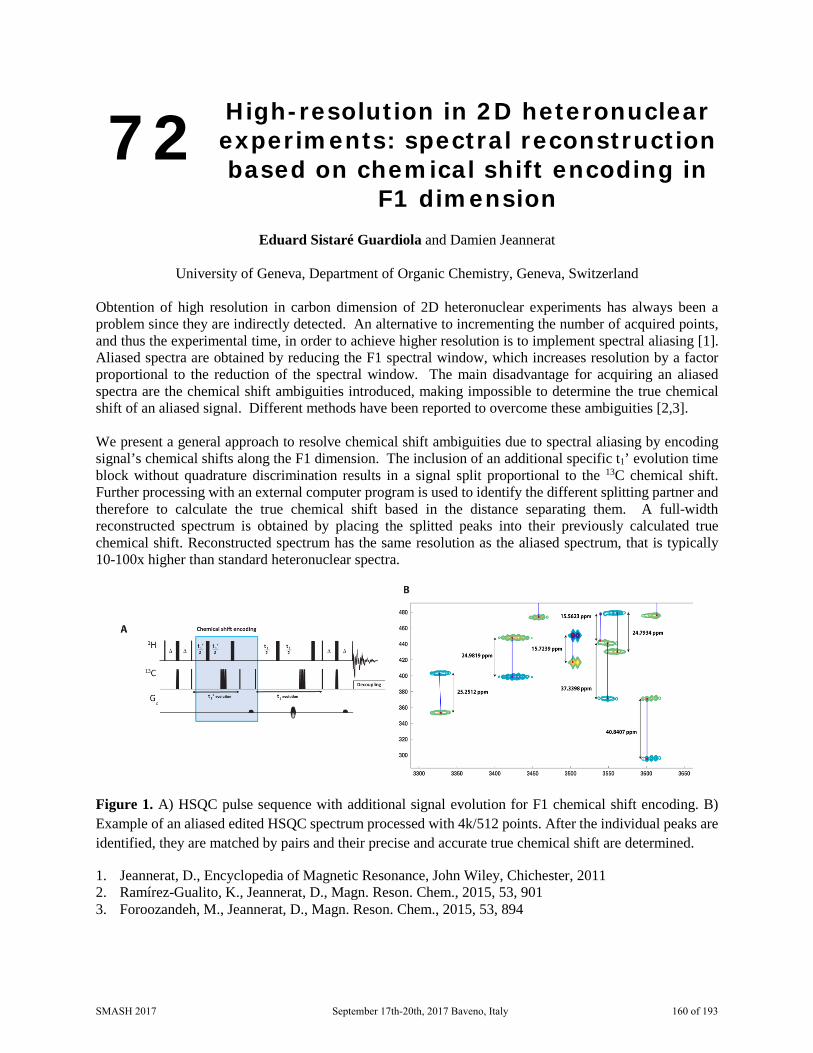
72 High-resolution in 2D heteronuclear experiments: spectral reconstruction based on chemical shift encoding in
F1 dimension
Eduard Sistaré Guardiola and Damien Jeannerat
University of Geneva, Department of Organic Chemistry, Geneva, Switzerland
Obtention of high resolution in carbon dimension of 2D heteronuclear experiments has always been a problem since they are indirectly detected. An alternative to incrementing the number of acquired points, and thus the experimental time, in order to achieve higher resolution is to implement spectral aliasing [1]. Aliased spectra are obtained by reducing the F1 spectral window, which increases resolution by a factor proportional to the reduction of the spectral window. The main disadvantage for acquiring an aliased spectra are the chemical shift ambiguities introduced, making impossible to determine the true chemical shift of an aliased signal. Different methods have been reported to overcome these ambiguities [2,3]. We present a general approach to resolve chemical shift ambiguities due to spectral aliasing by encoding signal’s chemical shifts along the F1 dimension. The inclusion of an additional specific t1’ evolution time block without quadrature discrimination results in a signal split proportional to the 13C chemical shift. Further processing with an external computer program is used to identify the different splitting partner and therefore to calculate the true chemical shift based in the distance separating them. A full-width reconstructed spectrum is obtained by placing the splitted peaks into their previously calculated true chemical shift. Reconstructed spectrum has the same resolution as the aliased spectrum, that is typically 10-100x higher than standard heteronuclear spectra.
Figure 1. A) HSQC pulse sequence with additional signal evolution for F1 chemical shift encoding. B) Example of an aliased edited HSQC spectrum processed with 4k/512 points. After the individual peaks are identified, they are matched by pairs and their precise and accurate true chemical shift are determined.
1. Jeannerat, D., Encyclopedia of Magnetic Resonance, John Wiley, Chichester, 2011 2. Ramírez-Gualito, K., Jeannerat, D., Magn. Reson. Chem., 2015, 53, 901 3. Foroozandeh, M., Jeannerat, D., Magn. Reson. Chem., 2015, 53, 894
SMASH 2017 September 17th-20th, 2017 Baveno, Italy 160 of 193

73 Conformational Deconvolution of NECA in Solution
Richard Upton
GSK R&D Stevenage, UK
NECA is a 5’ modified adenosine analogue and shows interesting biological properties as a synthetic non selective adenosine receptor agonist. The NECA conformation with the purine ring syn to the ribose together with an internal hydrogen bond, was indicated to be a major conformer in solution (DMSO) by NMR and hence was suggested as being the form responsible for receptor biological activity [1]. This conformer is also the form found in the single crystal x-ray of NECA, further supporting it as a low energy form [2]. However, recent work demonstrated that NECA bound to a Thermostabilised Human Adenosine A2A Receptor [3] with a very different anti orientation of the purine ring. Hence, further NMR data was collected on NECA in solution and the restraints obtained were deconvoluted to yield a conformational ensemble. The preliminary results of this work are outlined.
1. Prasad, R. N.; Bariana, D. S.; Fung, A.; Savic, M.; Tietje, K.; Stein, H. H.; Brondyk, H. D.; Egan, R. S. J. Med. Chem. 1980, 23, 313-319.
2. Moos, W. H.; Hamilton, H. W.; Ortwine, D. F.; Taylor, M. D.; McPhail, A. T. Nucleosides Nucleotides 1989, 8, 449-461.
3. G. Lebon, T. Warne, P.C. Edwards, K. Bennett, C.J. Langmead, A.G. Leslie, C.G. Tate Nature 2011, 474, 521-525.
SMASH 2017 September 17th-20th, 2017 Baveno, Italy 161 of 193

74 New software kit for reaction monitoring using interleaved
acquisition for high and low NMR
Mateusz Urbańczyk, Alexandra Shchukina, Krzysztof Kazimierczuk
University of Warsaw, Centre of New Technologies, Warsaw, Poland The reaction monitoring is crucial and booming field of NMR. However most available solutions concentrate on utilizing series of 1D spectra to monitor the changes in the system. Different approach is present in the poster by Alexandra Shchukina titled “Time-resolved NUS interleaved experiments with online processing and analysis”. In this work she presents the idea of acquiring of 2D randomly sampled spectrum interleaved with 1D. Allowing to have better insight in the changing system. The work presented here shows the new software kit allowing user to utilize the idea presented by A. Shchukina on their own systems. The new program consists of two main parts: 1. Module/macros controlling the spectrometer acquisition allowing user to acquire interleaved spectra using simple GUI. 2. Processing module (combined with CS processing from MDDNMR) which allows online processing, displaying and analysis of spectra during, or post acquistion. The program is written in python allowing use of it on almost any OS and is providing user-friendly GUI.
Currently the software toolkit is working on Agilent and Magritek spectrometers (Bruker version will be available later) therefore working on both High-field and benchtop systems. The first beta version of toolkit will be released on SMASH 2017.
Figure 7: Screenshot of interface during development S h t f i t f d i d l t
SMASH 2017 September 17th-20th, 2017 Baveno, Italy 162 of 193

75 A cross-platform format to associate NMR-extracted data (NMReDATA) to
chemical structures
Julien Wist1, Jean-Marc Nuzillard2, Nils E. Schloerer3, Stefan Kuhn3, Marion Pupier4, Mate Erdelyi5, Christoph Steinbeck6, Patrick Giraudeau7, Roberto R. Gil8, Antony J. Williams9, Craig Butts10, Kessler
Pavel11, Fabrice Moriaud11, Mikhail Elyashberg12, Dimitris Argyropoulos12, Manuel Perez13, Gary Martin14, Paul Trevorrow15, and Damien Jeannerat4
1. University of Valle, Cali, Colombia
2. CNRS and University of Reims-Champagne-Ardenne, Reims, France 3. IDNMR, University of Cologne, Cologne, Germany
4. Department of organic chemistry, University of Geneva, Geneva, Switzerland 5. Department of Chemistry - BMC, Uppsala University, Sweden
6. Cheminformatics and Chemometrics, Friedrich-Schiller-University Jena, Germany 7. CEISAM, University of Nantes, Nantes, France
8. Department of Chemistry, Carnegie Mellon University, Pittsburgh, USA 9. National Center for Computational Toxicology, EPA, USA
10. University of Bristol, United Kingdom 11. Bruker BioSpin GmbH, Rheinstetten, Germany
12. ACD/Labs, Toronto, Canada 13. Mestrelab Research, Santiago de Compostela, Spain
14. Merck and co. New-Jersey, USA 15. Magnetic Resonance in Chemistry, Wiley, Chichester, UK
An open initiative involving all the major players of computer-assisted structure elucidation (CASE), including methodology specialists, software and database developers and the editorial board of Magnetic Resonance in Chemistry, is addressing the old problem of reporting and sharing the assignment of 1D and 2D NMR spectra of organic molecules.[1] Our approach aims to solve some of the problems encountered with the “full analysis” of organic compounds. Usually, they are reported in chemistry journals using an image of the chemical structure, a text-based assignment of the 1D 1H and 13C spectra and a set of tables listing the correlations found in 2D spectra such as COSY, HSQC and HMBC. In the best case, images of the spectra (of uneven quality and resolution) can be found as Supplementary material. This is unsatisfactory.[2,3] We introduced a data format to associate the data extracted from the “full NMR analysis” (1D 1H and 13C, COSY, HSQC, HMBC, etc.) and the structure of the identified compound. The file uses the SD format, that is compatible with .mol files (a quite commonly format used to draw chemical structures). The NMR-extracted data (chemical shift, coupling and assignment) are encoded as so-called “tags”, that are included in the .sdf files. These “tags” are not visible when displaying the molecules but can be accessed by specialized software such as CASE software and analyzed by the database during the importation of the data. These .sdf files including NMReDATA will be generated by future releases of computer-assisted structure elucidation software and have multiple roles: 1) They make the link between the atoms of the structure and the signals found in the spectra (assignment). 2) They list, for each 1D and 2D spectrum, the spectral parameters in a defined format (chemical shifts, couplings, integrals, 2D correlations)
SMASH 2017 September 17th-20th, 2017 Baveno, Italy 163 of 193

3) They combine the data extracted from the spectra into an aggregated table (list of chemical shifts, coupling network, etc.). 4) They include links from the spectral data to the original files of the spectra (located in a local files folder in a database). These files will be uploaded on a database together with the associated spectra as embargoed NMR data. A link to the data will be included in the manuscript submitted to scientific journals. The reviewing will be facilitated by the fact that the spectra, the extracted data and the structure will be accessible in an usable electronic format. The reviewers will use their favorite CASE software to assess the assignment. Once the paper is accepted, the spectra and the extracted spectral data become openly accessible to everybody.
Fig. 1 Schematic representation of an NMR record including the files of the NMR spectra, and the SDF file containing the structure and the assigned NMR data extracted from the spectra.
1. www.nmredata.org 2. Jeannerat, D. Magn. Reson. Chem. 55(1) 7-14, 2017 3. Castillo, A. M., Bernal, A., Dieden, R., Patiny, L., Wist, J., J. Cheminform., 8(1), 26, 2016
SMASH 2017 September 17th-20th, 2017 Baveno, Italy 164 of 193

76 Human Serum Albumin and Drugs a Love Story Told by NMR
Mariana Gallo,1 Nadine Alaimo,2 Sara Matteucci,2 Edith S. Monteagudo,1 and Daniel O. Cicero2
1. Università di Roma Tor Vergata, Roma, Italia
2. IRBM Science Park SpA, Pomezia (RM), Italia
Plasma protein binding affects the time that a drug stays in the body and can also influence the drug's efficiency. Only the unbound fraction can have a biological effect or be metabolised/excreted. Because of that, estimating the unbound fraction of a drug candidate is of capital importance in the drug discovery process. Human serum albumin (HSA) is the most abundant protein in the blood circulation, it is 600 μM concentrated in plasma and most drugs have some affinity for HSA that results in sequestration of compound in serum. Furthermore, this 65 kDa protein presents various and diverse drug-binding sites: specific and no specific. In fact, HSA is able of soaking up ligands in a non-specific manner. This conduces to apparently discordant results of diverse individual ligand binding studies, which are difficult to generalize, and the great quantity of data are perplexing. Thus, considering the relevance of evaluating the HSA binding of pharmacological compounds, we present here a systematic NMR study for valutating the binding site and the affinity of several representative drugs to HSA. We have used 1D 1H line-broadening [1] and competition-STD experiments [2] for quantitative affinity measurement, and 2D 1H-13C correlation experiments for determining the binding site in HSA [3], with the aim to have a clearer picture of the interaction of diverse drugs with this abundant protein in plasma.
1. Shortridge Matthew D., Hage David S., Harbison Gerard S., and Powers Roebert, J. Comb. Chem., 10(6), 948-958, 2008
2. Wang Yu-Sen, Liu Dingjiang, and Wiss Daniel F., Magn. Reson. Chem., 42, 485-489, 2004 3. Krenzel Eileen S., Zhongjing Chen, and Hamilton James A., Biochemistry, 52, 1559-1567, 2013
SMASH 2017 September 17th-20th, 2017 Baveno, Italy 165 of 193
77 Simplifying complexity: a new approach for the analysis of
fluorine-containing mixtures by NMR
Laura Castañar,1 Pinelopi Moutzouri,1 Thais M. Barbosa,2 Mathias Nilsson,1 Claudio F. Tormena,2 Roberto Rittner,2 Andrew R. Phillips,3 Steven R. Coombes,4 and Gareth A. Morris1
1. School of Chemistry, University of Manchester, Manchester, UK. 2. Chemistry Institute, University of Campinas, Campinas, Brazil.
3. Pharmaceutical Sciences, AstraZeneca, Macclesfield, UK. 4. Pharmaceutical Technology & Development, AstraZeneca, Macclesfield, UK.
In recent years, interest in fluorine-containing compounds has significantly increased, mainly due to their biological activity as anticancer, antidepressant and antiviral drugs, amongst others.[1] NMR spectroscopy has proven to be a powerful tool for the analysis of fluorine-containing mixtures.[2,3] However, most current methods fail to extract structural information for individual components in a mixture, and new methods to tackle such problems are needed. Here we propose a novel two-step 1D experiment for obtaining structural information from simplified 1H spectra showing only those protons that are in a spin system coupled to a selected fluorine. The first step is a doubly selective 19F-1H reverse INEPT block, transferring coherence from a selected fluorine to a selected proton. The second is a TOCSY transfer to all the protons that correlate with the selected HF pair. We show the power of this method for a mixture containing four fluorinated pharmaceuticals. From this complex mixture, a set of clean 1D 1H TOCSY subspectra is obtained, one for each fluorine resonance.
1. J. Wang, M. Sánchez-Roselló, J.L. Aceña, C. del Pozo, A. E. Sorochinsky, S. Fustero, V. A. Soloshonok and H. Liu, Chem. Rev., 114, 2432-2506, 2014.
2. J. E. Power, M. Foroozandeh, P. Moutzouri, R. W. Adams, M. Nilsson, S. R. Coombes, A. R. Phillips and G. A. Morris, Chem. Commun., 52, 6892-6894, 2016.
3. P. Moutzouri, P. Kiraly, A. R. Phillips, S. R. Coombes, M. Nilsson, G. A. Morris, Chem. Commun., 53, 123-125, 2017.
SMASH 2017 September 17th-20th, 2017 Baveno, Italy 166 of 193

78 ¹H qNMR for Multiple Compounds Determination in Commercial Energy
Drinks
Maiara da S. Santos, Thayana da C. Alves, Ariane S. D. Silva, Tiago Venâncio and Antonio G. Ferreira
Chemistry Department, Federal University of São Carlos, São Carlos-SP, Brazil
The global market of energy drinks was valued at USD 43 billion in 2016 and it is expected to witness a high growth in the next years [1] and due to this, the quality of the products is essential. Considering the analytical techniques that are employed for this purpose, the advantages of NMR spectroscopy can provide a great differential in the energy drink analysis. In this context, the aims of this work were the development and validation of a ¹H qNMR method to determinate multiple analytes in commercial energy drinks and to explore the NMR technique potentiality for analyses of non-targeted compounds, in order to identify possible some undeclared compounds. The development of qNMR method was carried out according to EUROLAB Technical Report 1/2014 “Guide to NMR Method Development and Validation – Part 1: Identification and Quantification” [2]. The procedure of validation and its performance criteria were based on the Q2 R1 Guideline and FDA recommendation, respectively [3,4]. The procedures of analytical methodology (accuracy, repeatability, intermediary precision, specificity, range, linearity, detection and quantification limits) were satisfactory evaluated since the values obtained were with the FDA performance criteria [4]. Six compounds (caffeine, sucrose, glucose, citrate, threonine and ethanol) were identified and simultaneously quantified in several commercial products. The quantification was carried out directly in the matrix diluted in D2O (1:1 v/v) employing maleic acid and quinine (Certified Reference Material – RMs) as internal and external standard, respectively. The ethanol identified and quantified was not declared in the label of any products. Therefore, the identification of undeclared compound show as the NMR technique can be useful to investigate possible adulteration. Besides, the ¹H qNMR method developed in this work can contribute for the quality control improvements of commercial energy drinks in a simple and fast way.
1. Market Research Report. Available in: <http://www.grandviewresearch.com/industry-analysis/energy-drinks-market>.
2. EUROLAB Technical Report 1/2014. “Guide to NMR Method Development and Validation – Part 1: Identification and Quantification”, 2014.
3. International Conference on Harmonisation (ICH) Harmonised Tripartite Guideline. Text on Validation of Analytical Procedures Q2(R1), 2005.
4. ORA Laboratory Procedure. FDA. Methods, Method Verification and Validation, 2014.
SMASH 2017 September 17th-20th, 2017 Baveno, Italy 167 of 193
79 Computer Assisted Structure Elucidation of Two Biflavonoids from
the Leaves of Ochna Mauritiana
Godwin A. Dziwornu1, Nabeelah R. Toorabally2, Minu G. Bhowon2, Sabina Jhaumeer-Laulloo2, Suthananda N. Sunassee1,3, Arvin Moser4 and Dimitris Argyropoulos5
1. Chemistry Department, University of Cape Town, Rondebosch, South Africa
2. Department of Chemistry, University of Mauritius, Réduit, Mauritius 3. South African Medical Research Council Drug Discovery and Development Research Unit,
University of Cape Town, Rondebosch, South Africa 4. Advanced Chemistry Development, Inc., Toronto, ON, Canada
5. Advanced Chemistry Development UK Ltd., Bracknell, UK
As part of a phytochemical investigation of the leaves of O.mauritiana we have isolated, using a combination of silica gel flash chromatography and preparative layer chromatography (PLC), two biflavonoid compounds (1 and 2) from the organic extract of this plant. Approximately 12.5 mg of 1 and 2.5 mg of 2 were placed in NMR tubes and dissolved with DMSO-d6 in order to elucidate their structures. High-resolution mass spectra were also recorded in order to establish the molecular formulae.
Both compounds were relatively proton deficient resulting in few correlations in long range 2D NMR experiments. The repeated ring structure made some of the 13C peaks appear very close together, which also limited the usefulness of conventional 2D experiments. Together with the presence of many exchangeable protons, the elucidation of the structures proved to be rather challenging. Here we describe the advanced computation method used for elucidating the structures using a series of 1D and 2D NMR experiments. Initially, a search was performed on a database with predicted 13C spectra, or ca. 92 million compounds in search of a spectrum similar to the 13C spectra of 1 and 2. The spectrum of 1 matched that of a previously reported structure, while there was no match for the spectrum of 2. Computer Assisted Structure Elucidation (CASE), together with band-selective versions of the HMBC experiment were employed to assign the NMR data and unequivocally determine the structures and thus confirming the molecules. Additionally, several closely related structures were ruled out either by poor agreement between predicted and experimental 13C chemical shifts, or by additional derivatization experiments.
1. Elyashberg, Mikhail E., Williams, Antony J., Blinov, Kiril, Contemporary Computer-Assisted Approaches to Molecular Structure Elucidation, Cambridge: RSC Publishing, 2012
2. Pawlak, Tomasz, Jaworska, Magdalena, Potrzebowski, Marek J., Phys. Chem. Chem. Phys. 2013, 3137–3145
3. Pawlak, Tomasz, Potrzebowski, Marek J., J. Phys Chem. B 2014, 118 (12), 3298–3309
SMASH 2017 September 17th-20th, 2017 Baveno, Italy 168 of 193

80 Comparison of Public Chemical Structure Databases for Structure
Dereplication and Elucidation
Dimitris Argyropoulos, Sergey Golotvin, Rostislav Pol, Joe DiMartino, Arvin Moser
Advanced Chemistry Development, Toronto, Canada The capability to quickly and reliably separate and identify active components in natural product mixtures—identified through bio-assay and/or mass spectrometry guided fractionation—is critical to successful natural product-based drug discovery. Dereplication refers to the process of screening active compounds early in the development process in order to recognize and eliminate those compounds that have been studied in the past. This process enables scientists to proactively decrease the number of structures that will need to be fully elucidated and focuses testing on true ‘unknowns’. One question that should be addressed before starting a dereplication project is ‘where can a comprehensive list of all the known structures be found’? It is generally recognized that the largest collections of such structures exist in PubChem and ChemSpider. PubChem [1] is a database of chemical molecules and their activities against biological assays. It is maintained by the National Centre for Biotechnology Information, is part of the National Library of Medicine, and contains more than 93 million structures. ChemSpider [2] is a database of chemicals, that is owned by the Royal Society of Chemistry and currently contains approximately 59 million structures. The 13C NMR spectrum of a compound can be considered a fingerprint since it is virtually unaffected by conditions such as pH, concentration, and solvent effects that can cause 1H NMR spectra to vary. It is also largely magnetic field independent, since there are no couplings that could cause variations in stronger or weaker fields. Searching databases of predicted 13C NMR spectra for similarity to the unknown is, therefore, a very powerful and reliable strategy [3] for structure dereplication. The search can be enhanced by including search terms such as molecular formula (expanded to cover MF ranges) and by accommodating for missing or extraneous peaks in the NMR spectrum. It is also very common to use such databases to identify structural fragments in the case of genuinely unknown structures. In this work we will compare PubChem and ChemSpider in terms of overlap between the two databases, the types and variety of compounds contained therein, and their overall effectiveness in structure dereplication and elucidation
1. Kim, Sunghwan, Thiessen, Paul.A., Bolton, Evan.E., Chen, Jie, Fu, Gang, Gindulyte, Asta, Han, Lianyi, He, Jane, He, Siqian, Shoemaker, Benjamin A, Wang, Jiyao, Yu, Bo, Zhang, Jian, Bryant, Stephen.Nucleic Acids Res., 4; 44(D1):D1202-1213, Jan 2016
2. Pence, Harry E., Williams, Antony J., J. Chem. Educ., 87(11), 1123-1124, 2010 3. Hubert, Jane, Chollet, Sebastien, Purson, Sylvain, Reynaud, Romain, Harakat, Dominique, Martinez,
Agathe, Nuzzilard, Jean-Marc, Renault, Jean-Hugues, J. Nat. Prod., 78 (7), 1609–1617, 2015
SMASH 2017 September 17th-20th, 2017 Baveno, Italy 169 of 193

81 Small probe molecules, Trimethylphosphine and 15N-Acetonitrile, as probes of acidity in FCC catalysts
Jennifer Gómez1, Juan C. Poveda1*, Daniel R. Molina1 and Alexander Guzmán2
1. Laboratorio de Espectroscopia Atómica Molecular, Universidad Industrial de Santander,Bucaramanga, Colombia
2. Instituto Colombiano del Petróleo, ICP-ECOPETROL S.A., Piedecuesta, Colombia
FCC catalysts responsible for the conversion of heavy fractions of crude oil into useful products like fuels (gasoline, naphtha, LPG). They are composed of zeolite Y and a matrix of silica-alumina, kaolin and clay. Of them, the zeolite Y plays an important role in the catalytic activity through the Brønsted and Lewis acid sites.
One of the most important aspects in the behavior of these catalysts is their continuous loss of activity due to the clogging of the pores by the formation of coke and the poisoning of the active sites by the deposition of metals such as Ni and V, causing the inactivation of the same. To counteract these effects, the catalyst is subjected to regeneration cycles to improve it and again is feedback it to the FCC unit.
Small molecules can be used as probes to measure the acidity in these samples. 15N-Acetonitrile in function with ssNMR spectroscopy is a powerful technique for the structural characterization. The isotropic chemical shifts of 1H and 15N of acetonitrile depends of the interaction with acid sites (Brønsted and Lewis). An upfield shift of 15N resonance will be related with an increase in acid strength of the Brønsted sites.
The acidity of FCC catalysts hydrothermally deactivated and different regeneration cycles was analyzed. Acetonitrile (15N/99%) and trimethyl phosphine were used as probe molecules. CPTOSS experiments allow us to identify the different Brønsted and Lewis acid sites in the samples, 31P-one pulse experiments give us the quantity of acids sited presents in the materials and, HETCOR 15N{1H} and 31P{1H}, allows the unambiguous assignment of the Brønsted sites. Also, was revealed decreasing structural properties and change of the number of Brönsted acid sites of the FCC catalysts after hydrothermal deactivation.
Keywords: FCC catalyst, solid state NMR, 15N-acetonitrile, deactivation.
Acknowledgments: Authors thanks the financial support of the National Research Program in Energy and Mining, Ecopetrol, Colciencias and the Universidad Industrial de Santander through the contract 403 + 203 and the Vicerectoria de Investigaciones y Extensión (VIE) Grant number 8821.
1. Behera, B.; Ray. S. S. Catalysis Today, 141 (2009), 195-204.2. Occelli, M. L.; Voigt, U.; Eckert, H. Applied Catalysis A: General, 259 (2004), 245-251.3. Chen, N-Y.; Liu, M-C.; Yang, S-C.; Sheu, H-S.; Chang, J-R. Ind. Eng. Chem. Res. 54 (2015), 8456-
8468.4. Yi, D.; Zhang, H.; Deng, Z. Journal of Molecular Catalysis A: Chemical. 326 (2010), 88-93.5. Simperler, A.; Bell, R. G.; Foster, M. D.; Gray, A. E.; Lewis, D. W.; Anderson, M. W. J. Phys. Chem.
108 (2004), 7152-7161.
SMASH 2017 September 17th-20th, 2017 Baveno, Italy 170 of 193

82 A Novel qNMR Technique: Quantitative Global Spectrum
Deconvolution (qGSD)
Vadim Zorin,1 Mike Bernstein,1 Felipe Seoane,1 Stanislav Sykora,2 Carlos Cobas1
1. Mestrelab Research, Santiago de Compostela, Spain 2. Extra Byte, Castano Primo (Mi), Italy
Global Spectrum Deconvolution (GSD) is a technique for an automatic peak analysis of NMR spectra (1). GSD analyses frequency-domain NMR spectra and produces a highly accurate list of NMR peaks with refined peak parameters including position, amplitude, linewidth and shape. GSD has been at the core of Mnova data analysis for many years, and has proved to be highly reliable and fast. It is widely used in Mnova as a preparation step for many advanced analysis routines like multiplet analysis, automatic assignments, automatic structure verification, etc.
Deconvolution of NMR spectra is an attractive technique for quantitative analysis: accuracy is often adequate, and it can provide realistic integrals even for overlapping lines. However, the accuracy of the quantitation depends on how well theoretical models describe experimental NMR resonances. As a default, GSD uses a Generalized Lorentzian function to model the experimental lineshapes. This is a very flexible model that covers a broad range of shape variations for NMR resonances. However, careful analysis (2) shows that even such a flexible model cannot precisely fit all possible experimental lineshapes, as evidenced by non-zero fitting residuals. As a consequence, due to its model-free nature, for well resolved resonances, the classical point-by-point integration method may produce higher accuracy quantitation compared to model-dependant procedures.
To have the “best of both worlds”, we would like to determine integrals that have the accuracy of traditional integration with the resolving power that comes from GSD, together with access to an analysis capability of partially overlapping signals.
To improve GSD quantitation, we propose a new technique of “quantitative GSD”: qGSD. It is based on careful analysis of the residuals after GSD processing, and correcting GSD lineshapes in a way which minimizes the residuals. Preliminary results of qGSD applied to real 1D NMR data will be presented. We will show that for well-resolved spectral lines qGSD can offer quantitation precision which is significantly improved over conventional GSD, and approaches or meets the precision of the classical sum integration. At the same time, the technique also provides a good quality quantitative analysis of the lines within overlapped regions.
1. Schoenberger, T.; Menges, S.; Bernstein, M. A.; Pérez, M.; Seoane, F.; Sýkora, S.; Cobas, C. Anal. Chem. 2016, 88 (7), 3836–3843
2. http://resources.mestrelab.com/getting-really-fussy-about-integration/
SMASH 2017 September 17th-20th, 2017 Baveno, Italy 171 of 193

83 F1-decoupled CLIP-COSY experiment
Marta Brucka1 and Damien Jeannerat1
1. Department of Organic Chemistry, University of Geneva, Geneva, Switzerland
The presented research work focuses on the novel F1-decoupled CLIP-COSY [1] experiment. The experiment is very attractive for a structural chemist seeking for a fast assignment of signals in highly resolved spectra and in addition it is also an interesting case study for a NMR methodologist.
Based on in-phase magnetization transfer during the mixing, it is compatible with different kind of decoupling blocks to be applied in the middle of the t1 evolution. While the use of the selective modulated pulse (nemoZS) [2,3] or the PSYCHE [4] element ensures optimal sensitivity, the application of the BIRD [5] filter introduces some interesting features in this experiment.
The BIRD, selectively inverting only the 13C satellites and thus decoupling them from all other protons, inserts an additional 13C isotope into the spin system under investigation. This alters the J coupling interactions and very often allows to neatly decouple signals which would otherwise be classified as strongly coupled. The presence of a distinct isotope allows to draw some parallel with heteronuclear experiments, in particular it gives some flexibility on the type of J coupling interaction to be observed and/or eliminated either by the homonuclear decoupling or broadband decoupling during acquisition.
It will be shown on the example of the BIRD-CLIP-COSY how this can be used to fully reveal multiplet structure of signals not easily accessible by other methods.
We also propose a solution to diminish the one drawback of the CLIP-COSY experiment related to the dependence of the signals intensity on the length of the in-phase transfer period. We suggest to increment the constant delay into a third dimension and Fourier transform the data. The sum of signals along the new dimension results in the CLIP-COSY where the intensity variation is drastically reduced
1. Koos, M. R. M.; Kummerlowe, G.; Kaltschnee, L.; Thiele, C. M.; Luy, B. Angew. Chem. Int. Ed., 55, 7655-7659, 2016.
2. Zangger, K.; Sterk, H. J. Magn. Reson., 124, 486-489, 1997. 3. Cotte, A.; Jeannerat, D. Angew. Chem. Int. Ed., 54, 6016-6018, 2015. 4. Faroozandeh, M.; Adams, R. W.; Meharry, N. J.; Jeannerat, D.; Nilson, M.; Morris, G. A. Angew.
Chem. Int. Ed., 53, 6990-6992, 2014. 5. Aguilar, J. A.; Nilsson, M., B.; Morris G. A. Angew. Chem. Int. Ed., 50, 9716-9717, 2011.
SMASH 2017 September 17th-20th, 2017 Baveno, Italy 172 of 193

84 1H Chemical Shift Variations in complex Mixtures at different
Concentrations
Emanuelle F. da Silveira and Jochen Junker
Fundação Oswaldo Cruz – CDTS, Rio de Janeiro, Brazil The chemical shifts of a compound are known to vary due to certain experimental conditions, specially the temperature and the solvent used. Only in some cases variatons have been observed with changing concentrations, usually due to aromatic interactions of the observed compound. Also, changes of chemical shifts have been observed due to binding to proteins. But, the general expectation is that for normal experimental conditions, chemical shifts vary only slighly with the concentration of the analyte. Hence, spectra of mixtures can easily be compared among each other and compounds in mixtures can be identified.
This approach works, for example, for lyophilized blood plasma, but it did not work with coffee preparations. Our coffee preparations were produced using a normal household coffee machine, a fixed volume of coffee was lyophilized, and later a known amount was solubilized in D2O. Our first observation was that the chemical shifts of the caffeine signals of the mixture did not match the values observed in the spectrum of pure caffeine in D2O. Next we aquired NMR spectra with varrying concentrations.
In none of the spectra the chemical shifts of the caffeine signals matched. We observed that the signals that were shifting significantly, >0.15 ppm, could be found in the aliphatic region, whereas in the aromatic region the shift was only about 0.05 ppm. No other signals shifted in the aliphatic region, different from the aromatic region, where more changes were observed. In no concentration of our coffee preparations we could observe the chemical shifts of caffeine observed in a reference spectrum in D2O.
This has two consequences: first, the assignment of the peaks can not be done by simple comparison of the pure compound spectra. Second, estatistical analysis of this spectra is not possible, in this case even excluding the aromatic region completely does not resolve this, since changes in the aliphatic region were observed as well. This takes away one of the most powerfull tools that spectroscopists have at hands for analysis and comparison.
SMASH 2017 September 17th-20th, 2017 Baveno, Italy 173 of 193

85 Stop Flow Approaches to in situ Analysis of Reactions
Guy Lloyd-Jones,1 Edward King,2 Ruth Dooley,1Ariana Jones,1 Dusan Uhrin1, and Anna Codina,3
1. University of Edinburgh, Edinburgh, EH9 2FJ
2. TgK Scientific Ltd, Bradford on Avon, BA15 1DH 3. Bruker UK Ltd, Banner Lane, Coventry, CV4 9GH
The work presented will centre around our development of in situ NMR analysis techniques for reactions of interest to the synthetic chemist; these will be exemplified in the context of our recent investigations of reactions employing reagents based on boron and silicon.1-3 The overall project that these developments have contributed to, aims to gain a better grasp of the fundamental physical and chemical processes that facilitate and govern these reactions, thereby allowing "informed control" of the processes, either directly, or indirectly via modulation of undesired side reactions. Mechanistic interrogation via analysis of reaction kinetics and other physical organic parameters is the key to this insight, and gaining sufficient quantity and quality of data is pivotal. The bulk of our data is obtained by UV, IR, MS, and most especially NMR, in concert with strategic isotopic labelling and computational analysis.
We will focus on the problems we faced when reactions became too fast, or too sensitive to be reproducible. This resulted in the conventional techniques for in situ analysis of reactions by IR and NMR either being of limited use in terms of reaction life-time, precision, or highly inefficient in terms of the physical handling aspects of accurate reaction assembly.
We thus began to explore new approaches and devices to give us traction in the analysis of these systems. These developments have allowed the design, assembly and interfacing of new devices with IR and NMR instruments. The presentation will reveal details of these new devices. We will show how they allow the exploration of synthetic organic reaction kinetics in situ, not just quickly and efficiently, but also in a way that the devices can be rapidly coupled / decoupled to standard IR and NMR instruments, thus requiring instrument access only when the device is being used.
1. Gonzalez, J. A.; Ogba, O. M.; Morehouse, G. F.; Rosson, N.; Houk, K. N.; Leach, A. G.; Cheong, P. H.-Y.; Burke, M. D.; Lloyd- Jones, G. C. Nature Chem. 2016 DOI: NCHEM-16020310.
2. 2.Cox, P. A.; Leach, A. G.; Campbell, A. D.; Lloyd-Jones, G. C. J. Am. Chem. Soc. 2016 DOI: 10.1021/jacs.6b03283.
3. 3.Cox, P. A.; Reid, M.; Leach, A. G.; Campbell, A. D.; King, E. G.; Lloyd-Jones, G. C. Chem. Sci., submitted, 2016.
SMASH 2017 September 17th-20th, 2017 Baveno, Italy 174 of 193

86 Real-time NMR in Human Cells
Jennie Roberts1, Matteo Pennestri2, Anna Codina2, Constanze Bonifer2, Ulrich L Günther1
1. Institute of Cancer and Genomic Sciences, University of Birmingham, Birmingham, UK
2. Bruker UK Ltd, Banner Lane, Coventry, UK NMR has for a long time been used to study metabolism in cells. However human cells have always been challenging as the conditions needed to keep cells viable are more difficult to achieve in an NMR system than for Escherichia coli bacterial or yeast cells. We have recently described an experiment where we have embedded primary chronic lymphocytic leukaemia cells in an agarose matrix and measured metabolic turn-over for an elongated period of time [1]. In this system, we observed fast and reversible adaptation of these cells to lowering oxygen levels.
In order to be able to better control the conditions of such real-time experiments we have now explored a flow-based approach where cells are placed in a flow-tube, and medium is circulated over a period of 12h. In collaboration with Bruker we have adapted the InsightMRTM flow-unit for this purpose. By adding a bioreactor in the flow-path we can control oxygen and CO2 tension. This approach has been tested for different human cell lines.
This new approach offers unprecedented opportunities to test the response of patient derived cells to altering conditions or the exposure of drugs, and may open new avenues in the context of personalised medicine.
1. Koczula KM, Ludwig C, Hayden R, Cronin L, Pratt G, Parry H, et al. Metabolic plasticity in CLL: adaptation to the hypoxic niche. Leukemia 30:65–73 (2016).
SMASH 2017 September 17th-20th, 2017 Baveno, Italy 175 of 193

87 Spontaneously hypertensive rats on high-fat diet as a model of metabolic syndrome: NMR metabolomic study
Helena Pelantová1, Martina Bugáňová1,2, Blanka Šedivá3, Martina Holubová4, Marek Kuzma1, Lenka
Maletínská4, and Jaroslav Kuneš4,5
1. Institute of Microbiology of the Czech Academy of Sciences, Prague, Czech Republic 2. University of Chemistry and Technology, Prague, Czech Republic
3. University of West Bohemia, Plzeň, Czech Republic 4. Institute of Organic Chemistry and Biochemistry of the Czech Academy of Sciences, Prague, Czech
Republic 5. Institute of Physiology of the Czech Academy of Sciences, Prague, Czech Republic
Metabolic syndrome is a cluster of pathologies that includes insulin resistance, type 2 diabetes mellitus, obesity, dyslipidemia, hypertension which when present in combination markedly increase the risk of cardiovascular morbidity and mortality [1]. Spontaneously hypertensive rats (SHR) are one of the most common animal models used to study essential hypertension in humans. However, it was demonstrated that high-fat (HF) diet increased body weight, serum cholesterol and blood pressure in SHR and these changes in metabolic parameters were associated with cardiovascular dysfunction in these animals [2]. SHR on HF diet can be used as a model of metabolic syndrome [3]. The aim of our study was to characterize for the first time the SHR-HF model using NMR-based metabolomics in combination with the standard biochemical parameters.
SHR-HF model was studied in the context of both obesity and hypertension. The normotensive Wistar-Kyoto (WKY) rats served as the closest genetic control for the SHR. Male SHR and WKY rats were supplied with a standard low-fat (LF) and a HF diet, establishing four experimental groups: hypertensive and normotensive, both lean and obese. Urine samples were collected overnight after 12 weeks of HF diet. NMR spectra were acquired using Carr-Purcell-Meiboom-Gill pulse sequence with the presaturation. Pre-processed spectra were subjected to multivariate statistical analysis (Principal component analysis (PCA), Partial least square – discriminant analysis (PLS-DA)) and univariate parametric and nonparametric tests.
PCA and PLS-DA models display substantial separation of all experimental groups: diets are clearly differentiated by the first principal component, genetic strains by the second one. Urinary metabolic profiles of obese animals show attenuated concentration of hippurate, trigonelline, fumarate, phenylacetylglycine, 3-indoxylsulfate, citrate, and methylamine, and increased levels of tartrate, methylnicotinamide, glucose, taurine, dimethylamine, and allantoin compared to the corresponding lean controls. Changes of these metabolites are manifested to a different extent depending on the genetic strain. To identify a potential relationship between the urine metabolic composition and pathologies clustered in metabolic syndrome, the metabolite concentrations were also correlated with significantly changed biometric and biochemical parameters.
Acknowledgements: The authors gratefully acknowledge the project LO1509 of the Ministry of Education, Youth and Sports of the Czech Republic.
SMASH 2017 September 17th-20th, 2017 Baveno, Italy 176 of 193
1. Chan J.C.N, Tong P.C.Y., Critchley J.A.J.H., Semin. Vasc. Med. 2(1), 45-47, 2002. 2. Cao J., Sodhi K., Puri N., Monu S.R., Rezzani R., Abraham N.G., Diabetol. Metab. Syndr. 3(1), 37,
2011. 3. Chung S., Park C.W., Shin S.J., Lim J.H., Chung H.W., Youn D.-Y., Kim H.W., Kim B.S., Lee J.-H.,
Kim G.-H., Chang Y.S., Nephrol. Dial. Transplant 25(2), 389-399, 2010.
SMASH 2017 September 17th-20th, 2017 Baveno, Italy 177 of 193

88 Proton NMR Spectroscopy in Investigative Product Analysis
Catherine J. Frankis
Reading Scientific Services Limited, Reading Science Centre, Berkshire, RG6 6LA, United Kingdom
The speedy and reliable identification and quantification of unknown contaminants in both finished products and raw materials is crucial in understanding production issues and quickly solving potential quality issues. It can be critical to quickly acquire a top-level screen of a contaminated substance, where comparison to a control sample can identify the class of compounds that could be present. In addition, there is also a need to understand the composition of products however, successful de-formulation of particular complex matrices can require application of numerous techniques, each providing information about a different class of compound, which can take time.
NMR spectroscopy is a flexible technique with varied applications, which can provide useful information about a large number of chemical compounds that may be present in situations such as these. Simple sample preparation procedures and short analysis times also mean results can be obtained quickly. Examples of the application of NMR spectroscopy in the analysis of food, cosmetic and pharmaceutical products will be displayed, including contaminants screening, product de-formulation and investigation into product quality issues.
SMASH 2017 September 17th-20th, 2017 Baveno, Italy 178 of 193

89 Measuring Spin Relaxation Rates of Low-Gamma Nuclei using Satellite
Exchange NMR Spectroscopy
Gerard P. Robertson1 Barbara Odell1, Ilya Kuprov2, Darren J. Dixon1 and Tim D. W. Claridge1
1. Department of Chemistry, University of Oxford, Chemistry Research Laboratory, Oxford (UK)
School of Chemistry, University of Southampton, Highfield Campus, Southampton (UK) NMR spectroscopy is well established as the leading technique for the structure elucidation of small molecules and for studying their dynamic behaviour in solution. In particular, spin relaxation rates can be correlated with the global and local dynamics of a molecule. Measurement of the longitudinal relaxation time constants (T1s) for transition metals has the potential to provide valuable information on their complexes relating to their co-ordination geometry, motional properties and molecular interactions.
However, there exists scant evidence of longitudinal relaxation data of low magnetogyric ratio (γ) transition metal nuclei in the literature, much of which was collected many decades ago. There has been limited progress in this area due to a number of factors, including the poor detection sensitivity of low-γ nuclei and often the need for specialised NMR probes operating at the low resonance frequencies appropriate to observe metals such as 103Rh, 107/109Ag, 183W and 187Os. These factors make the direct measurement of the relaxation properties of such nuclei largely impractical using standard instrumentation.
In this work we demonstrate the potential to measure relaxation rates of low-γ nuclei without the need to observe them directly or even to apply rf pulses to them, making these measurements accessible with conventional broadband probes. This can be achieved through the observation of “spin-state satellite exchange” in 2D EXSY (Exchange Spectroscopy) spectra of a J-coupled, high-γ partner nucleus. We demonstrate this approach by measurement of the longitudinal relaxation rates of 195Pt (as a model system) and 107/109Ag metal centres in organometallic complexes through observation of scalar-coupled 31P nuclei. Excellent agreement is demonstrated for T1 data collected for 195Pt using direct observation with the conventional inversion-recovery sequence.
SMASH 2017 September 17th-20th, 2017 Baveno, Italy 179 of 193

90 NMR studies of hydrogen cleavageby Solid-phase frustrated Lewis
pairs Nicholas H. Rees1 Jun-Yi Xing1 and Dermot O’Hare1
1. Department of Chemistry, University of Oxford, Oxford, UK
Hydrogenation is important in many chemical transformations and frustrated Lewis acid pairs (FLP) offer a metal-free catalyst for such reactions.1 In the solution phase FLPs have shown extensive catalytic hydrogenation activity, but for industrial processes a solid catalyst is prefered for reasons such as; catalyst seperation, the use of continuous gas or liquid feed fixed bed reactors or slurry-phase reactors.2 Here we report the preparation and characterisation of a silica supported FLP [≡Si-OB(C6F5)2][PtBu3] and its reactivity on exposure to hydrogen, deuterium and CD3OD characterised by 1H, 2H, 11B, 19F and 31P Solid State NMR spectroscopy.3
1. Stephan, D. W., Science, 354 (6317), aaf7229, 20162. J. R. Severn, J. C. Chadwick, R. Duchateau and N. Friederichs, Chem. Rev., 105, 4073–4147, 20053. Xing J., Buffet J., Rees N. H., Norby P., and O’Hare D., Chem. Commun., 52, 10478-10481, 2016
SMASH 2017 September 17th-20th, 2017 Baveno, Italy 180 of 193
91 Automatic Concurrent Phase & Baseline Correction in 1D NMR
Spectra Ester Maria Vasini1, 3, C. Carlos Cobas2 and Stanislav Sykora3
1. DICAM, University of Bologna, Italy2. Mestrelab Research, Santiago de Compostela, Spain
3. Extra Byte, Castano Primo, Italy
The algorithmic problem of phase correction (PC), and that of baseline correction (BC), of 1D NMR spectra have been both tackled many times over the last half a century, by many authors. There are many algorithms which emulate the manual procedure. Basically, they all ‘fit’ the parameters which describe the phase (ph0, ph1) and the baseline (various parametrized models) so as to maximize some quality assessment of the corrected spectrum. Historically, the employed ‘quality functions’ included peak heights, negative peak lobes, DISPA patterns symmetry, selected baseline points, peak ablation, etc. Here we propose a radically different type of ‘quality function’ Q to be optimized. It is based on the histograms of the spectrum (real and imaginary parts) which turn out to be very sensitive to both phase and baseline distortions. This permits us to:
1. Carry out the phase and baseline corrections simultaneously. In traditional approaches these arealways intended as separate evaluation steps and carried out separately (first phase and thenbaseline), even though all NMR spectroscopists know that the two corrections interfere with other.We think that our approach neatly overcomes this problem.
2. Carry out both corrections on both the real and imaginary parts of a spectrum. So far, the baselinecorrection was always done only on the real part, a fact that can have various adverse effects onother evaluation tasks.
3. Enhance the objectivity of the corrections, especially considering that in practice one often facessituations with multiple acceptable ‘solutions’.
We describe the algorithm we have developed and we illustrate the results achieved. 1. Cobas C., Bernstein M.A, Martìn-Pastor M., Tahoces P.G., 2006. A new general-purpose fully
automatic baseline-correction procedure for 1D and 2D NMR data. Journal of Magnetic Resonance183, 145-151;
2. Binczyk F., Tarnawski R., Polanska J., 2015. Strategies for optimizing the phase correctionalgorithms in Nuclear Magnetic Resonance spectroscopy. Biomedical Engineering On line 14 (S2);
3. Cobas C. and Sykora S., 2007. Extended DISPersion-Absorption (eDISPA) approach to AutomaticPhasing of HR-NMR Spectra. Poster presented at 48th ENC Conference, Daytona Beach, FL (USA),April 22-27, 2007.
4. Zorin V., Bernstein M.A. and Cobas C., 2017. a robust, general automatic phase correction algorithmfor high-resolution NMR data, Magnetic Resonance in Chemistry 55, 738-746.
SMASH 2017 September 17th-20th, 2017 Baveno, Italy 181 of 193
92 Structure Revision and Conformational Analysis of the
Fluxional Microginin 674
Kevin Calabro,1,2 Olivier P. Thomas*1 and Grégory Genta-Jouve*3
1. Marine Biodiscovery, National University of Ireland Galway, Galway, Ireland.
2. Cosmo International Ingredients, 855 Avenue du Docteur Maurice Donat, 06250 Mougins, France. 3. C-TAC UMR-CNRS 8638 COMETE, Université Paris Descartes, Paris, France.
The cyanobacterium Microcystis aeruginosa is well-known for the production of numerous bioactive modified peptides such as the hepatotoxic microcystins or the proteases inhibitors cyanopeptolins and microginins.1 The latter metabolites are a class of linear peptides that contain generally four to six amino acid residues including the 3-amino-2-hydroxydecanoic acid (ahda). To date, around 50 structural variants are known but only 13 microginins have been fully characterized by NMR.2-4 Microginin 674 (1) was obtained from a culture of Microcystis aeruginosa (SAG 14-85). This tetrapeptide exhibited two tyrosines, the ahda residue and an N-methyl-methionine. Interestingly at room temperature, most of the 1D NMR signals of 1 were splitting, suggesting the existence of diastereoisomers or rotamers. The presence of the latter was first confirmed by changing the NMR solvent and then by variable-temperature NMR experiments (VT-NMR). Moreover, structures of 1 and a commercially available microginin 674 differ in the amino acids assemblage with an inversion between the N-Me-Met and Tyr2.5 The lack of NMR data provided by the companies and the difficulties in NMR data analysis induced by the presence of rotamers, question the structure proposed for this natural product. We report herein the structure revision, but also the experimental and theoretical conformational analysis of microginin 674 allowing the establishment of the thermodynamic parameters involved in the exchange process between the two rotamers.
1. George, E. C.; Shunyan, M.; Jimmy, O., Chemodiversity in Freshwater and Terrestrial Cyanobacteria – A Source for Drug Discovery. Current Drug Targets 2011, 12 (11), 1654-1673.
2. Ishida, K.; Kato, T.; Murakami, M.; Watanabe, M.; Watanabe, M. F., Microginins, Zinc Metalloproteases Inhibitors from the Cyanobacterium Microcystis aeruginosa. Tetrahedron 2000, 56 (44), 8643-8656.
3. 3Reshef, V.; Carmeli, S., Protease inhibitors from a water bloom of the cyanobacterium Microcystis aeruginosa. Tetrahedron 2001, 57 (14), 2885-2894.
4. Gesner-Apter, S.; Carmeli, S., Three novel metabolites from a bloom of the cyanobacterium Microcystis sp. Tetrahedron 2008, 64 (28), 6628-6634.
5. Group, M. D. Microginin 674. http://www.marbionc.org/gallery/detail.aspx?id=203509.
SMASH 2017 September 17th-20th, 2017 Baveno, Italy 182 of 193

93 NMR of new metal complexes of curcuminoids with Mg, Zn, Cu, Mn,
Ga, Gd and Sm
William Meza,1 Yair Alvarez, 1 Marco A. Obregón,1 Mirian Estévez,1 Simón Hernández,1 Carolina Escobedo,2 Shane Pawsey,3 Julia Cassani4 and Raúl G. Enríquez1
1. Instituto de Química, UNAM, México, [email protected];
2. Universidad Autónoma de Guanajuato, Departamento de farmacia, México
3. Bruker BioSpin Corporation, Billerica, USA; 4. Universidad Autónoma Metropolitana, UAM, Unidad Xochimilco, Méxicoffiliations
Curcumin is a secondary metabolite with historical roots in its usage for culinary or therapeutic purposes. It has gained an outstanding relevance for its implications as a systemic antiinflammatory agent and its potential as an anti-cancer agent.[1,2] Also, derivatives diaceylcurcumin (DAC) and tetrahydrogenated diacetylcurcumin (DAC-H) have attracted wide attention due to their biological and complexation properties.[3]
Following our chemical studies of both ligands we prepared new metal complexes of DAC-H with Mg, Zn and Cu, which were studied in liquid state by high-resolution NMR. Also, new metal complexes of DAC with Mn, Gd, Ga and Sm were prepared and studied by NMR in both liquid and solid state. DAC-H complexes showed clear spectral changes caused by the metal binding to the diketone site. The metal ions were choosen after their presence in other complexes with biological properties.
Our previous crystal homoleptic structures showed the stoichiometry ML2 for the complexes including the new Manganese complex reported herein. The vinylic proton signal of the ligand was used as a probe for complexation in DMSO, while in CP-MAS NMR, the profile of carbonyl signals and signal broadening provided a useful criteria of complexation. The case of trivalent metal ions is discussed.
1. Sahdeo Prasad, Subash C. Gupta, Amit K. Tyagi, Bharat B. Aggarwal. Curcumin a component of golden spice: From bedside to bench and back, Biotechnology Advances, 32, 1053–1064, 2014
2. Amalraj A, Pius A, Gopi S, Gopi S. Biological activities of curcuminoids, other biomolecules from turmeric and their derivatives – A review, Journal of Traditional and Complementary Medicine, 7, 205 – 233, 2016.
3. Simon Wanninger, Volker Lorenz, Abdus Subhanb, Frank T. Edelmann. Metal complexes of curcumin – synthetic strategies, structures and medicinal applications, The Royal Society of Chemistry, 4986 – 5002, 2015.
SMASH 2017 September 17th-20th, 2017 Baveno, Italy 183 of 193
94 In Situ Monitoring of the Ascorbic Acid Electrooxidation by Ultrafast
NMR
Luiz Henrique Keng Queiroz Júnior1,3, Pollyana Ferreira da Silva Vianna1,2 and Luiz Alberto Colnago1
1. Embrapa Instrumentação, São Carlos-SP, Brazil 2. Instituto de Química, Universidade de São Paulo, São Carlos-SP,Brazil
3. Instituto de Química, Universidade Federal de Goias, Goiânia-GO,Brazil Ascorbic acid (AA) electrooxidation is a known reaction, however this compound has many applications as the antioxidant agent in food and drink indrustry.1 In this work we chose this reaction to be applied to a Electrochemistry and Nuclear Magnetic Resonance (EC-NMR) coupled system. The magnetoelectrolisys effect was also evaluated because the electrochemical reaction was performed insitu.2 A promising methodology for the real-time monitoring of these reactions is the Ultrafast NMR (UF-NMR)3,4, capable of recording 2D NMR correlations in a fraction of a second. The objective of the present study is to apply UF-NMR experiments to the real-time monitoring of in-situ AA electrooxidation reaction. The electrochemical system consisted in a glass capillary with two coaxials Pt wire coils as work and auxiliary electrodes, the pseudo-reference electrode was also a glass capillary, but with Ag wire. Cyclic voltammetry and chronoamperommetry techniques were used to evaluate the magnetoeletrolisys effect and oxidate the AA in real time, respectively. During all the electrochemical process, in order to monitor in real-time the evolution of the oxidation of the AA, many ultrafast COSY (UF-COSY) were continuously performed every 41s, each one corresponding to an eight-scan experiment. Analyzing the most representative UF-COSY spectra, it was possible to note the gradual consumption of the AA during the course of the electrooxidation. After 15 minutes, the ¹H NMR correlations of the product, dehydroascorbic acid, were observed in the UF-COSY spectrum. It was confirmed by a HSQC experiment that the dehydroascorbic acid detected was present in the hydrated bicyclic and monocyclic forms. Also, it was detected another stable product formed on this condition and it still remains uncharacterized. We are performing other kinds of UF-NMR experiments to continue to investigate this electrochemical process, and also conventional NMR experiments to perform the characterization of the formed unknown compound.
1. BREZINA, M., KORYTA, J., LOUCKA, T., MARSIKOVA, D., PRADAC, J., J. Electroanal. Chem. Interfacial Electrochem. 40 (1) (1972) 13-17.
2. GOMES, B. F., SILVA, P. F., LOBO, C. M. S., SANTOS, M. S., COLNAGO, L. A. Strong magnetoelectrolysis effect during electrochemical reaction monitored in situ by high-resolution NMR spectroscopy. Anal. Chim. Acta (in press), (2017)
3. FRYDMAN, L., SCHERF, T. LUPULESCU, A., Proc. Natl. Acad. Sci., 99(25), 15858-15862, 2002. 4. Boisseau, R., Bussy, U., Giraudeau, P., Boujtita, M., Anal. Chem. 87 (1), 372-375, 2015.
SMASH 2017 September 17th-20th, 2017 Baveno, Italy 184 of 193
95 Colombian Coffee Chemical analysis by solid state NMR spectroscopy
Juan Carlos Poveda1 and Daniel Ricardo Molina1
1. Laboratorio de Resonancia Magnética Nuclear, Universidad Industrial de Santander, Bucaramanga,
Colombia CP 681002 In the Colombian mountains, local farmers produce one of the best coffees in the world. The varieties of Arabica coffee and Robusta coffee are grown in special areas of the country and their quality, flavor and fragrance are recognized all over the world. Different methodologies by NMR and IR have been developed for the chemical analysis2,3 of aquous extracts of Colombian coffee to confirm its origin1 and distinguish it from adulterated coffee. Also, low field NMR has been used to establish the origin of Robusta and Arabica varieties.4
In this work, colombian coffees were analizated by ssNMR and their chemical compositions were correlationated with the quality properties.
Samples used for solid state NMR spectroscopy do not requiere extraction steps previously to the analysis. All the samples were measured using a 13C/CP-TOSS pulse sequence to suppres the rotational echoes, contact time was optimized to obtain the best conditions to the 1H polarization transfer at 10kHz rotation speed. Because in solids the line broadening by 1H dipolar coupling results in an excesive line broadening, the chemical analysis can´t indentify individual chemical compounds, however is possible to analysis the chemical changes of the samples on basis the main chemical groups.
ssNMR results in combination with statistical analysis shows that is possible to classify the samples on basis of quality criteria.
Authors thanks to Vicerectoria de Investigaciones of Universidad Industrial de Santander
1. Arana, V.A., J. Wist et al., Coffe´s country of origin determination by NMR: The Colombian case. Food Chemistry 175 (2015) 500-506.
2. Feldman.J.R., Ryder, W.S. and Kung, J.T. Importance of nonvolatile compounds to the flavor of coffe. J. Agr. Foof. Chem. 17 (1969) 733-739.
3. Arana, V.A., Wist, J. et al. Classification of Coffe Beans by GC-C-IRMS, GC-MS, and 1H-NMR, J. Analytical Methods in Chemistry, (2016) http://dx.doi.org/10.1155/2016/8564584
4. Defernez. M., Kemsley, E.K. et al.Low-field 1H-NMR spectroscopy for distinguishing between arabica and robusta ground roast coffes, Food Chemistry, 216 (2017) 106-113.
SMASH 2017 September 17th-20th, 2017 Baveno, Italy 185 of 193

97 The Role of Non-Uniform Sampling (NUS) in the NMR Characterization
of Bile Acid Biomarkers Raman Sharma1, Gregory S. Walker1, Kathleen A. Farley2, Frank Delaglio3, Matt Cerny1 and David
Rodrigues1
1. Pharmacokinetics, Dynamics & Metabolism 2. Structural & Molecular Sciences
3. National Institute of Science and Technology 2-D NMR is the quintessential analytical technique in the structural characterization of organic compounds. However, these experiments often have long acquisition times and can be limited in sensitivity and/or resolution. Reducing the required number of data points used in the second and third dimension through various non-uniform sampling (NUS) schemes has been used with protein and peptide samples to enhance resolution and decrease the acquisition time of multidimensional experiments. In 2-D and 3-D experiments, these models can result in a 75% reduction in acquisition time. Many of these gains can also be realized in the analysis of small molecules. The reduction in acquisition time by using NUS can be utilized to promote higher sample throughput (through a reduction in total acquisition time), increased sensitivity (by using more pulses per f1 data point) or increasing resolution by adding more data points in the f1 dimension. All of these approaches have been evaluated using common homonuclear and heteronuclear experiments used in the structural elucidation of small molecules (1H-1H COSY, 1H-13C HSQC and 1H-13C HMBC).
Recently, a biomarker project highlighted the utility of NUS in 2D small molecule NMR. The search for novel bile acid (BA) biomarkers of liver organic anion-transporting polypeptides (OATPs) lead to a LC-MS method that supported the analysis of 30 different BAs, which included 15 different 3-O-sulfates. Many of these O-sulfates were not commercially available and required either chemical or biological synthesis. The resulting synthetic products need to be structurally characterized and in some cases quantitated via NMR prior to their use in a quantitative LC-MS assay. Because of the structural redundancies within bile acids high resolution 2D NMR was required. Using NUS acquisition schemes on these samples facilitated structural elucidation of the site of sulfation while saving a factor of seven or more in acquisition time.
SMASH 2017 September 17th-20th, 2017 Baveno, Italy 188 of 193

98 Natural bioactive-protein interactions studied by ligand-
observed NMR
Simona Bartova and Sofia Moco
Nestle Institute of Health Sciences, Metabolomics, Lausanne, Switzerland Molecular recognition of small molecules by proteins is fundamental to most processes in living organisms. Small molecule-protein interactions are involved in transport, regulation, and metabolism in the cell, having direct implications in the molecular aetiology of diseases, as well as in therapeutic target discovery.
Natural compounds have long inspired drug development given their potential bioactivities. Plants are therefore an important source of bioactives with a wide chemical diversity. Cytokinins, N6- substituted purines, are a class of phytohormones promoting plant growth and development. Besides their role in plant physiology, little is known about the biological effects of cytokinins in humans.
To study the potential interactions of bioactives with human proteins, ligand-observed NMR was set-up. The interaction of a series of cytokinins N6-benzyladenine (BA), N6-benzyladenosine (BAR) and N6-benzyladenosine phosphate (BARMP) with the model protein, human serum albumin (HSA) was monitored. Different approaches were tested for this purpose: Water Ligand Observed via Gradient Spectroscopy (WaterLOGSY), relaxation-edited (T1rho) NMR and saturation transfer difference (STD) NMR. In all approaches, the 3 studied bioactives coordinate to HSA, with slightly different binding propensity, according to the different substituents. In addition, to minimize the amount of required protein, miniaturisation of the sample from 5 to 1.7 mm tubes was attempted.
With this study on cytokinins-HSA interactions, we hope to demonstrate the potential of ligand-observed NMR screening in the field of natural bioactives and bioactive mixtures.
SMASH 2017 September 17th-20th, 2017 Baveno, Italy 189 of 193

99 Assessment and Validation of Various Flow Cell Designs for
Quantitative Online NMR Spectroscopy
Lukas Wander, Simon Kern, Klas Meyer, Svetlana Guhl, Michael Maiwald
Bundesanstalt für Materialforschung und -prüfung (BAM), Berlin, Germany
Compact nuclear magnetic resonance (NMR) instruments make NMR spectroscopy and relaxometry accessible in industrial and harsh environments for reaction and process control. Robust field integration of NMR systems have to face explosion protection or integration into process control systems with short set-up times. This paves the way for industrial automation in real process environments. [1, 2]
The design of failsafe, temperature and pressure resistant flow through cells along with their NMR-specific requirements is an essential cornerstone to enter industrial production plants and fulfill explosion safety requirements. NMR-specific requirements aim at full quantitative pre-magnetization and acquisition with maximum sensitivity while reducing sample transfer times and dwell-times. All parameters are individually dependent on the applied NMR instrument. Luckily, an increasing number of applications are reported together with an increasing variety of commercial equipment. However, these contributions have to be reviewed thoroughly.
The performance of sample flow cells commonly used in online analytics and especially for low-field NMR spectroscopy was experimentally and theoretically investigated by 1H-NMR experiments and numerical simulations [3]. Here, we demonstrate and discuss an automated test method to determine the critical parameters of flow through cells for quantitative online NMR spectroscopy. The setup is based on randomized setpoints of flow rates in order to reduce temperature related effects. Five flow cells and tubings were assessed and compared for high-field as well as low-field NMR spectrometers.
1. Meyer, K., Kern, S., Zientek, N., Guthausen, G., Maiwald, M.: Trends in Analytical Chemistry 83 (2016), 39–52.
2. CONSENS – Integrated Control and Sensing for Sustainable Operation of Flexible Intensified Processes, funded by the European Union’s Horizon 2020 research and innovation programme under grant agreement N° 636942; www.consens-spire.eu; 31.05.2017.
3. Dalitz, F., Maiwald, M., Guthausen, G., Chemical Engineering Science 75 (2012) 318–326.
SMASH 2017 September 17th-20th, 2017 Baveno, Italy 190 of 193

100 Collaborative study for determination of okadaic acid in solution by qNMR spectroscopy
Taichi Yamazaki1, Tsuyoshi Kato2, Ryuichi Watanebe3, Maki Saito2, Toshiyuki Suzuki3 and Akiko
Takatsu1
1. National Metrology Institute of Japan (NMIJ)/AIST, Tsukuba, Ibaraki, Japan 2. Japan Food Research Laboratories (JFRL), Tama, Tokyo, Japan
3. National Research Institute of Fisheries Science (NRIFS-FRA), Yokohama, Kanagawa, Japan Okadaic acid is one of the diarrhea shellfish toxins. An instrumental analysis has been introduced into the testing of diarrhea shellfish toxins in many laboratories all over the world, where the regulatory level of the okadaic acid group is set as 0.16 mg/kg at CODEX in 2008. Certified reference materials (CRMs) of the diarrhea shellfish toxin are required for the instrumental analysis, and CRMs which concentration is determined by qNMR with external standard method [1] are provided from NRC Canada. qNMR uses a different structural compound as a reference material, and the method is practically used for rapid purity determination. The internal standard method is considered more accurate than the external standard method in qNMR, and there are some reports about comparison between these two methods [2, 3]. We already established qNMR with internal standard method for standard solution [4]. This approach is expected to provide an accurate determination for low concentration solution of precious sample such as diarrhea shellfish toxins.
In this research, we studied the optimization of sample preparation and measurement conditions of qNMR spectroscopy with internal standard (IS) for the determination of low concentration of okadaic acid in solution. Moreover, a collaborative study within three laboratories was performed and the accuracy and variation factors of qNMR spectroscopy were discussed.
Okadaic acid was produced and purified by the optimized method in previous work [5]. Purified okadaic acid was dissolved in methanol and prepared to approximately 350 mg/L methanol solution. 1,4-bis(trimethylsilyl) benzene-d4 (1,4-BTMSB-d4) as an IS from Wako Pure Chemical Industries, Ltd. was the CRMs whose purity was (0.999±0.005) kg/kg (k=2). The measurement sample with an internal standard was prepared based on our previous work [4]. All of mass were weighed by micro balance (XP56V, METTLER TOLEDO, Giessen, Germany). This sample solution was measured by NMR spectroscopy. Measurement temperature was regulated at 23 °C. Typical qNMR experimental parameters were as follows: 59523.8 Hz (99.2 ppm) spectral width, 4.0 s acquisition time, 13.0 s (90°) pulse width, 60 s relaxation delay, and 512 transients acquired. 13C decoupling was used in all measurement. Data processing was performed using MestReNova ver. 8.1.
The concentration of okadaic acid solution of approximately 350 mg/L was successfully determined with only 2.5 % of standard uncertainty by qNMR with using the internal standard method. This result was equal to the uncertainty reported from NRC Canada in spite of a low concentration sample of about 1/10 in the report. The measurement accuracy in this approach was confirmed from this result. In this inter-laboratory test, the determined values of okadaic acid were in good agreement among three laboratories by
SMASH 2017 September 17th-20th, 2017 Baveno, Italy 191 of 193

selecting adequate signals with sufficient resolution. The obtained value was validated because the difference from collaborative study was equal to the measurement standard uncertainty.
1. Burton I. W., et. al., Anal. Chem., 77, 3123, 2005. 2. Cullen C. H., et al., Magn. Reson. Chem., 51(11), 705, 2013 3. Ryuichi W., et. al., Toxins, 8(10), 294, 2016. 4. Taichi Y., et. al., Ana. Sci., 33, 369, 2017. 5. Toshiyuki, S., et. al., in Proceedings of the Sixteenth International conference on Harmful Algae, ed.
A. L. MacKenzie 2014, Wellington, New Zealand, 34.
SMASH 2017 September 17th-20th, 2017 Baveno, Italy 192 of 193

101 Combining Presaturation, 13C-Filtering and PROJECT for
Effective Solvent Suppression of Protonated Organic Solvents
Peter W. A. Howe
Syngenta Ltd, Crop Protection Research, Bracknell, UK.
Presaturation is one of the most popular solvent suppression methods in the analysis of small organic molecules. This appears to be because it is highly frequency-selective, requires no specialist spectrometer hardware and requires minimal customization for any specific solvent. The frequency selectivity of presaturation leads directly to two significant disadvantages. First, solvent signal from regions of magnetic field inhomogeneity is not suppressed [1]. Second, 13C satellites of solvent resonances are not suppressed.
This poster demonstrates that the PROJECT spin-echo sequence [2] can be used to overcome both of these disadvantages. First, phase cycling of the 180° pulses suppresses solvent signal from regions of magnetic field inhomogeneity in the same way as in NOESY-PRESAT, PURGE and FLIPSY. Second, adding only three 13C pulses to the PROJECT sequence re-introduces evolution of JCH allowing conversion of 13C satellite signals into unobservable or dephased coherences. The resulting 13C filtering suppresses a broad range of JCH. These two features allow acquisition of high-quality proton spectra in protonated organic solvents such as DMSO and acetonitrile.
The main disadvantage of presaturation-PROJECT is that, like NOESY-PRESAT, it requires a 90° excitation pulse. Therefore, a long relaxation delay is required to obtain resonance integrals independent of T1 relaxation rates. However, this also allows a longer and hence more selective presaturation pulse [3].
1. McKay, Ryan T., Concepts in Magnetic Resonance, 38A(5), 197-220, 2011. DOI: 10.1002/cmr.a.20223
2. Aguilar, Juan A., Nilsson, Mathias, Bodenhausen, Geoffrey, Morris, Gareth A., Chem. Commun., 48, 811-813, 2012. DOI: 10.1039/c1cc16699a
3. Hoult, David, Journal of Magnetic Resonance, 21, 337-347, 197
SMASH 2017 September 17th-20th, 2017 Baveno, Italy 193 of 193


![[ CONFERENCE TENTATIVE ]](https://static.fdokumen.com/doc/165x107/6332727f3108fad7760ea090/-conference-tentative-.jpg)