Transcatheter aortic valve implantation with a self-expanding nitinol bioprosthesis

Nitinol Embolic Protection Filters: Design Investigationby Finite Element Analysis
Michele Conti, Matthieu De Beule, Peter Mortier, Denis Van Loo, Pascal Verdonck, Frank Vermassen,Patrick Segers, Ferdinando Auricchio, and Benedict Verhegghe
(Submitted October 16, 2008; in revised form February 17, 2009)
The widespread acceptance of carotid artery stenting (CAS) to treat carotid artery stenosis and itseffectiveness compared with surgical counterpart, carotid endarterectomy (CEA), is still a matter of debate.Transient or permanent neurological deficits may develop in patients undergoing CAS due to distalembolization or hemodynamic changes. Design, development, and usage of embolic protection devices(EPDs), such as embolic protection filters, appear to have a significant impact on the success of CAS.Unfortunately, some drawbacks, such as filtering failure, inability to cross tortuous high-grade stenoses,malpositioning and vessel injury, still remain and require design improvement. Currently, many differentdesigns of such devices are available on the rapidly growing dedicated market. In spite of such a growingcommercial interest, there is a significant need for design tools as well as for careful engineering investi-gations and design analyses of such nitinol devices. The present study aims to investigate the embolicprotection filter design by finite element analysis. We first developed a parametrical computer-aided designmodel of an embolic filter based on micro-CT scans of the Angioguard� XP (Cordis Endovascular, FL)EPD by means of the open source pyFormex software. Subsequently, we used the finite element method tosimulate the deployment of the nitinol filter as it exits the delivery sheath. Comparison of the simulationswith micro-CT images of the real device exiting the catheter showed excellent correspondence with oursimulations. Finally, we evaluated circumferential basket-vessel wall apposition of a 4 mm size filter in astraight vessel of different sizes and shape. We conclude that the proposed methodology offers a useful toolto evaluate and to compare current or new designs of EPDs. Further simulations will investigate vessel wallapposition in a realistic tortuous anatomy.
Keywords Angioguard�, carotid artery stenting, embolic pro-tection device, finite element analysis, nitinol, wallapposition
1. Introduction
Cardiovascular disease (CVD) is the main cause of death inWestern countries. Each year CVD causes over 4.35 milliondeaths in Europe (accounting for 49% of all deaths in 2005)(Ref 1).
CVD is often related to atherosclerosis, a degeneration ofthe vessel wall which has the main consequence of narrowingthe vessel lumen, i.e. a stenosis, leading to blood flow reductionor blockage.
At present, the deployment of an intravascular stent hasbecome a common and widely used minimally invasivetreatment for stenotic arteries, including mainly coronaryarteries but also peripheral stenotic vessels such as the carotidarteries.
ln recent years, carotid artery stenting (CAS) has emerged asa less invasive treatment as compared to the conventionalsurgical approach, carotid endarterectomy (CEA) (Ref 2), butthe efficacy of CAS relative to surgery is still a matter of debate(Ref 3).
One major concern related to CAS is the possible distalembolization potentially leading to stroke or other severeneurological complications. Embolization is due mainly to theplaque debris and thrombi generated during the stenosisdilatation and stent apposition. Consequently, embolic protec-tion devices (EPDs) have been developed to capture suchreleased debris and appear to have a significant impact on thesuccess of CAS (Ref 4-8).
EPDs can be classified in the following three categories(Ref 9):
• distal balloon occlusion;• proximal balloon occlusion;• embolic protection filters (EPFs).
The balloon occlusion systems block the flow in the internalcarotid artery (ICA) and emboli are aspirated before balloondeflation and catheter removal, whereas EPFs maintain bloodflow while emboli are captured.
This article is an invited paper selected from presentations at ShapeMemory and Superelastic Technologies 2008, held September 21-25,2008, in Stresa, Italy, and has been expanded from the originalpresentation.
Michele Conti and Ferdinando Auricchio, Universita degli Studi diPavia, Pavia, Italy; Matthieu De Beule, Peter Mortier, PascalVerdonck, Patrick Segers, and Benedict Verhegghe, IBiTech,Institute of Biomedical Technology, Ghent University, Ghent,Belgium; Denis Van Loo, UGCT, Center for X-ray Tomography,Ghent University, Ghent, Belgium; and Frank Vermassen, Departmentof Vascular Surgery, Ghent University Hospital, Ghent, Belgium.Contact e-mail: [email protected].
JMEPEG �ASM InternationalDOI: 10.1007/s11665-009-9408-8 1059-9495/$19.00
Journal of Materials Engineering and Performance

Such a class of filters most often consists of a metallicbasket-like structure coated with a membrane made of poly-meric material containing numerous pores. Filters are usuallymounted on a 0.014 in. (0.036 mm) guidewire, generally30 mm proximal to a flexible tip and are delivered through avery small profile catheter (<3 French, i.e. 1 mm). During aCAS procedure, before the lesion dilatation and the stentapposition, the filter is opened in the ICA lumen distal to thetarget lesion, by withdrawing the delivery sheath. At the end ofthe CAS procedure, a retrieval catheter is advanced onto theguidewire to capture and remove the filter (Ref 10).
EPFs have both advantages and disadvantages with respectto balloon occlusion procedures. On one hand, EPFs have theadvantage of preserving cerebral flow throughout the procedurewhile capturing embolic debris allowing distal perfusion andangiography during the CAS procedure. On the other hand,filters have disadvantages ranging from difficulty in navigatingseverely stenosed or tortuous vessels to possible incorrect filterapposition against the vessel wall (Ref 11, 12). Currently,several embolic filter designs are available on a fast growingdedicated market (Ref 11).
Because of the relevant differences in EPF designs, it islikely that there are situations in which individual EPFs may bebetter suited to avoid adverse events during CAS and,consequently, the investigation and the comparison of theperformance of different EPFs can play a key role inthe improvement of filtering outcomes and potentially on thecomplete CAS procedure.
Engineering investigation and computer models have shownthe capability to provide useful information to understand themechanics of cardiovascular devices, e.g. stents, and to improvedevice design (Ref 13-16). To the best of our knowledge, theavailable comparative investigations about advantages or rolesthat specific EPDsmay have in CAS aremainly based on in vitro,ex vivo, or clinical studies (Ref 12, 17-20).
The purpose of this study is to use finite element analysis(FEA) in the design evaluation of basket-like embolic protec-tion filters. In particular, we investigated the effects of sizing oncircumferential basket-vessel wall apposition of a widelyadopted embolic filter, i.e. the Angioguard� XP (CordisEndovascular, FL).
In order to achieve this purpose, we organized the study asfollows:
• Using a script-based procedure, we created a 3D finite ele-ment model of the Angioguard� XP taking into accountseveral geometrical features (such as number of struts,strut diameter, filter length, filter diameter, etc.).
• To validate the model, we qualitatively compared thenumerical prediction of a free filter expansion, i.e. as itexits out of the delivery sheath, with a micro-CT scanimage of the real device deployment.
• To evaluate the circumferential basket-vessel wall apposi-tion, we simulated the deployment of a 4 mm size Angio-guard� XP in a vessel having different sizes and shape.
2. Materials and Methods
2.1 Model Geometry
The Angioguard� XP consists of a 0.014 in. (0.036 mm)guidewire with a nitinol basket frame enveloped by a porous
polymeric membrane (Fig. 1). The current Angioguard� XPbasket sizes range from 4 to 8 mm in diameter in order todeploy within vessels with diameters ranging from 3.0 to7.5 mm (Ref 11, 21).
We used the in-house developed pyFormex software(version 0.6.5) (Ref 22) as a pre-processing tool to build ageometrical and finite element model of the Angioguard� XP.
The model includes the following parts:
• filter struts defining the basket frame;• membrane;• markers.
The mesh generation procedure was defined by fouressential steps (Fig. 2):
1. creation of path reproducing the shape of one filter strut;2. sweeping of planar section along the defined path in
order to create one 3D filter strut;3. replication and rotation of the filter strut around the filter
axis in order to create the whole basket frame;4. definition of the whole model assembling the filter struts
with the membrane and markers.
From a geometrical point of view, the model is defined byseveral parameters as depicted in Fig. 2 and reported in Table 1.
Fig. 1 The Angioguard� XP embolic protection device
R1
Raxis
R2
L1
L2
Part1
Part2
Dstrut
Lmarker
Dmarker1
2
43
Fig. 2 Example of filter model generation in four steps
Journal of Materials Engineering and Performance
The pyFormex script-based mesh generation procedureallows the creation of a finite element model in a quick wayfacilitating parametrical finite element analyses; moreover,pyFormex generates a finite element mesh that can be easilyimported into several commercial finite element solvers. In thisstudy, we used ABAQUS/EXPLICIT 6.8 (Abaqus Inc., Prov-idence, RI).
In order to assess accurately the value of the definedgeometrical parameters, a micro-CT scan of a real device inopen configuration was performed and the image was analyzedby Materialise miniMagics V12.0.5.1 (see Table 1).
2.2 Material Properties
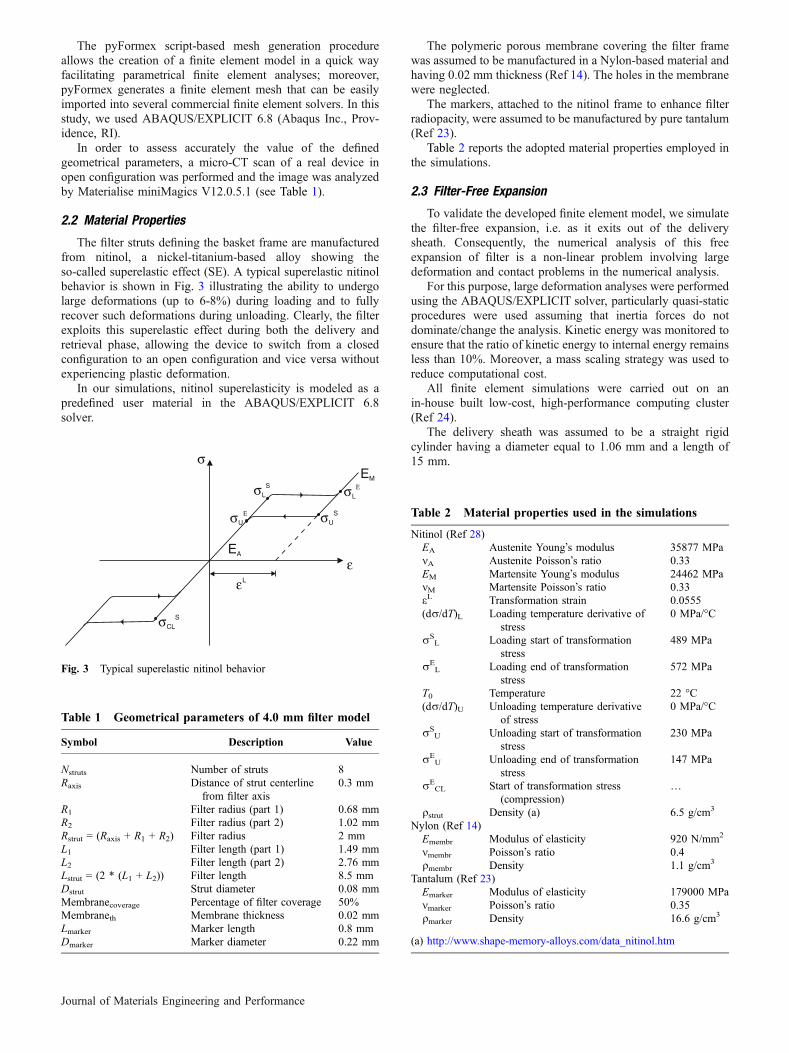
The filter struts defining the basket frame are manufacturedfrom nitinol, a nickel-titanium-based alloy showing theso-called superelastic effect (SE). A typical superelastic nitinolbehavior is shown in Fig. 3 illustrating the ability to undergolarge deformations (up to 6-8%) during loading and to fullyrecover such deformations during unloading. Clearly, the filterexploits this superelastic effect during both the delivery andretrieval phase, allowing the device to switch from a closedconfiguration to an open configuration and vice versa withoutexperiencing plastic deformation.
In our simulations, nitinol superelasticity is modeled as apredefined user material in the ABAQUS/EXPLICIT 6.8solver.
The polymeric porous membrane covering the filter framewas assumed to be manufactured in a Nylon-based material andhaving 0.02 mm thickness (Ref 14). The holes in the membranewere neglected.
The markers, attached to the nitinol frame to enhance filterradiopacity, were assumed to be manufactured by pure tantalum(Ref 23).
Table 2 reports the adopted material properties employed inthe simulations.
2.3 Filter-Free Expansion
To validate the developed finite element model, we simulatethe filter-free expansion, i.e. as it exits out of the deliverysheath. Consequently, the numerical analysis of this freeexpansion of filter is a non-linear problem involving largedeformation and contact problems in the numerical analysis.
For this purpose, large deformation analyses were performedusing the ABAQUS/EXPLICIT solver, particularly quasi-staticprocedures were used assuming that inertia forces do notdominate/change the analysis. Kinetic energy was monitored toensure that the ratio of kinetic energy to internal energy remainsless than 10%. Moreover, a mass scaling strategy was used toreduce computational cost.
All finite element simulations were carried out on anin-house built low-cost, high-performance computing cluster(Ref 24).
The delivery sheath was assumed to be a straight rigidcylinder having a diameter equal to 1.06 mm and a length of15 mm.
Table 1 Geometrical parameters of 4.0 mm filter model
Symbol Description Value
Nstruts Number of struts 8Raxis Distance of strut centerline
from filter axis0.3 mm
R1 Filter radius (part 1) 0.68 mmR2 Filter radius (part 2) 1.02 mmRstrut = (Raxis + R1 + R2) Filter radius 2 mmL1 Filter length (part 1) 1.49 mmL2 Filter length (part 2) 2.76 mmLstrut = (2 * (L1 + L2)) Filter length 8.5 mmDstrut Strut diameter 0.08 mmMembranecoverage Percentage of filter coverage 50%Membraneth Membrane thickness 0.02 mmLmarker Marker length 0.8 mmDmarker Marker diameter 0.22 mm
εL
ε
σ
σUΕ
σLS σL
Ε
σUS
EM
EA
σCLS
Fig. 3 Typical superelastic nitinol behavior
Table 2 Material properties used in the simulations
Nitinol (Ref 28)EA Austenite Young�s modulus 35877 MPamA Austenite Poisson�s ratio 0.33EM Martensite Young�s modulus 24462 MPamM Martensite Poisson�s ratio 0.33eL Transformation strain 0.0555(dr/dT)L Loading temperature derivative of
stress0 MPa/�C
rSL Loading start of transformation
stress489 MPa
rEL Loading end of transformation
stress572 MPa
T0 Temperature 22 �C(dr/dT)U Unloading temperature derivative
of stress0 MPa/�C
rSU Unloading start of transformation
stress230 MPa
rEU Unloading end of transformation
stress147 MPa
rECL Start of transformation stress
(compression)…
qstrut Density (a) 6.5 g/cm3
Nylon (Ref 14)Emembr Modulus of elasticity 920 N/mm2
mmembr Poisson�s ratio 0.4qmembr Density 1.1 g/cm3
Tantalum (Ref 23)Emarker Modulus of elasticity 179000 MPammarker Poisson�s ratio 0.35qmarker Density 16.6 g/cm3
(a) http://www.shape-memory-alloys.com/data_nitinol.htm
Journal of Materials Engineering and Performance

The parts defining filter/catheter systems (see Fig. 4) weremodelled as follows:
• filter struts: 33,792 three-dimensional 8-node brick�reduced-integration� elements (C3D8R);
• membrane: 8320 three-dimensional 4-node quadrilateralmembrane elements with reduced integration (M3D4R);
• markers: 1296 three-dimensional 8-node brick �reduced-integration� elements (C3D8R);
• catheter: 3806 three-dimensional 4-node bilinear quadrilat-eral rigid elements (R3D4).
Mesh tie constraints of type surface-to-surface were appliedto tie the markers and the membrane to the filter struts.
Furthermore, the filter frame struts are tied to marker bandsdefining the relative movement of the filter ends with respect tothe guidewire (Ref 21), particularly the proximal marker bandis fixed to the guidewire while the distal marker band is free toslip along the guidewire. Therefore, mesh tie constraints, typerigid body, are imposed between the nodes lying on each filterframe end and the predefined reference points. Boundaryconditions are then applied to the reference points (RPs) torealistically simulate the movement of the marker bands. Inparticular, at the proximal end RP, all the degrees of freedomwere constrained; at the distal end RP, only displacement alongfilter axis and rotations were allowed.
The analysis strategy assumes that the filter frame defor-mation during deployment is comparable to the deformationduring the filter frame insertion into the delivery sheath.Consequently, a progressive rigid translation along the filteraxis is imposed to the delivery sheath in order to induce thenecessary filter deformation to switch from the open configu-ration (filter out of delivery sheath) to the closed configuration(filter within the delivery sheath).
A frictionless general contact algorithm has been used inorder to handle the interactions between the filter frame and thedeployment sheath. In this case, the membrane elements wereexcluded from the contact strategy in order to simplify thesimulations assuming that it has a minor role in the overall filterdeployment/retrieval mechanics.
2.4 Circumferential Basket-Vessel Wall Apposition
In order to evaluate the circumferential basket-vessel wallapposition, we carried out the simulations of a filter deployingin a straight vessel modelled as a rigid cylinder having differentsizes and shape. In particular, two different scenarios wereinvestigated: (i) filter expansion in three circular vessels having
respectively 3.0, 3.25, and 3.5 mm diameter as suggested by themanufacturer for a 4 mm size filter (Ref 21); (ii) filter expansionin two oval vessels having both 1.5 mm major semi axis andrespectively an ovality* of 0.85 and 0.75.
Preliminary simulations showed that, in the analyzed case,final filter/vessel configuration is not influenced by thedeploying mode and consequently the interaction betweenfilter and sheath has not been taken into account.
We defined a two-step simulation strategy as follows:(i) filter diameter reduction is obtained by elongating the basketframe by appropriate axial displacement boundary conditions tothe distal end RP, only self contact of membrane was includedin this step; (ii) previous boundary conditions are deactivatedallowing the filter re-expansion and a global contact algorithmwas included allowing the filter/vessel interaction.
The effect of blood flow pressure acting on the filtermembrane was included in the analyses applying a uniformpressure of 8.80 mmHg on the inner surface of the membrane(Ref 25).
Circumferential basket-vessel wall apposition was evaluatedin terms of gap between the vessel lumen and the circumfer-ential area covered by the filter.
In order to keep the computational cost acceptable and tominimize the impact of the membrane mesh on the gapmeasurement, a final membrane mesh of M3D4R 24960elements was chosen by a preliminary mesh sensibility analysisabout filter deployment simulation in the 3 mm circular vessel(a finer membrane mesh—29,120 elements—results in a 5.4%gap measurement differing 6% compared to the 5.7% gapobtained with the coarser mesh—24,960 elements).
3. Results
3.1 Filter-Free Expansion
The simulation of the filter-free expansion showed that thebasket-frame configuration experiences a severe change. Theradial compression of the filter frame is accomplished by anaxial elongation bringing the diameter to a final value of0.96 mm when the filter is completely inserted into the sheathas shown in Fig 5. Moreover, the filter frame experiences aradial deformation that is not uniform along the filter length,resulting in non-uniform adhesion between the strut and thedelivery sheath wall.
Comparing the previously described adopted mesh to a finerone (i.e. 67,584, 33,024, and 2,592 elements respectively forfilter struts, membrane, and markers) showed a negligibledivergence (i.e. difference in maximum axial elongation lessthan 1%).
Delivery
Marker
Membrane
Z X
Y
Filter struts
Delivery sheath
Proximal end RP
Distal end RP
Filter axis
Fig. 4 Finite element model of Angioguard� XP and part of thedelivery sheath
ZX
Y
Fig. 5 Longitudinal cut view of the filter frame in the deliverysheath
*Ovality being defined as the change in cross section roundness and definedas O = b/a, where a is cross section semi-major axis length and b is crosssection semi-minor axis length.
Journal of Materials Engineering and Performance

Moreover, Fig. 6 shows a good qualitative agreementbetween the numerical results and the micro-CT image of thepartially deployed Angioguard� XP.
3.2 Circumferential Basket-Vessel Wall Apposition
Simulations of the filter deployment in the straight vesselshowed that the filter is not able to completely cover the vessellumen and gaps are present between the vessel wall and themembrane (see Fig. 7), confirming the experimental resultsrecently reported by Sieworek et al. (Ref 26).
As reported in Table 3, the vessel size seems to have aminor impact on the circumferential basket-vessel wall appo-sition which is in contrast to the impact of the vessel shape. Infact, an increase in the vessel ovality dramatically influencesthe gap while a change in the vessel diameter does notsignificantly modify the gap area.
Consequently, the simulations of filter deployment in thestraight rigid vessel suggest that, in this case, filter malappo-sition is caused by inability of the filter struts to accomplish thevessel asymmetry.
4. Limitations
The filter-free expansion simulation showed qualitativeagreement with micro-CT images but limitations pertaining to
the frictionless contact between the filter and the deliverysheath are present.
Clearly, the assumption of a uniform pressure distributionalong the filter membrane should be improved using moreaccurate and realistic values achieved, for example, byexperiments and computational fluid dynamics analyses.
Material properties of the filter components should also bederived from experimental data to define the mechanicalproperties more accurately.
The major limitation of this study is the rigid bodyassumption for the vessel. In reality, the radial outward forceof the filter is likely to reshape the vessel and this effect canconsequently influence the filter-vessel wall apposition. How-ever, modeling filter apposition in a realistic vessel (i.e. internalcarotid artery) introduces substantial computational challenges,such as the incorporation of the variability of the constitutionand mechanical properties of the diseased arterial wall, andshould be accomplished by adequate experimental validation.This challenge is considered to be beyond the scope of thispreliminary investigation.
5. Conclusions
In this study, we proposed and validated a finite elementmodel in order to investigate the basket-like design of anembolic protection filter device. In particular, the circumferen-tial filter/vessel wall apposition was numerically evaluatedconfirming the inability of the filter to completely adapt toasymmetric vessels.
Clearly, this study needs to be considered as a preliminaryproof of concept of the use of finite element based modeling
Fig. 6 Partially deployed filter: microCT image (top panel); numer-ical simulation (bottom panel)
Fig. 7 A 4 mm size filter deployed in a 3 mm circular vessel (on the left) and in 3 mm vessel having 0.75 ovality (on the right)
Table 3 Numerical results of filter/vessel wall apposition
Filter size, mm—Ovality
3.5-0.0 3.25-0.0 3.0-0.0 3.0-0.85 3.0-0.75
Gap (% ofvessel lumen)
5.7 4.7 5.7 8.7 14.7
Journal of Materials Engineering and Performance

strategies to investigate and understand the mechanics ofembolic protection filters. The proposed model is a base tofurther investigate the impact of design parameters (filterlength, diameter, number of struts, etc.) on filter mechanics(flexibility, radial strength, etc.) and the filter apposition in amore realistic tortuous anatomy taking also into account vesselwall material properties.
Finally, we finally conclude that the proposed methodologycould be useful to evaluate and to compare current or newdesigns of EPDs in the early design phase as recentlyrecommended by the FDA (Ref 27).
References
1. S. Petersen, V. Peto, M. Rayner, J. Leal, R. Luengo-Fernandez, andA.Gray, EuropeanCardiovascularDisease Statistics, BHFLondon, 2005
2. M.H. Wholey, N. Al-Mubarek, and M.H. Wholey, Updated Review ofthe Global Carotid Artery Stent Registery, Catheter. Cardiovasc.Interv., 2003, 60, p 259–266
3. A.J. Furlan, Carotid-Artery Stenting—Case Open or Closed? N. Engl.J. Med., 2006, 355(16), p 1726–1729
4. N. Al-Mubarak, G.S. Roubin, J.J. Vitek, S.S. Iyer, G. New, and M.B.Leon, Effect of the Distal Balloon Protection System on Microemb-olization During Carotid Stenting, Circulation, 2001, 104, p 1999–2002
5. J.S. Yadav, M.H. Wholey, R.E. Juntz, P. Fayad, B.T. Katzen, G.J.Mishkel, T.K. Bajwa, P. Whitlow, N.E. Strickman, M.R. Jaff, J.J.Popma, D.B. Snead, D.E. Cutlip, B.G. Firth, and K. Ouriel, ProtectedCarotid Artery Stenting Versus Endarterectomy in High-Risk Patients,N. Engl. J. Med., 2004, 351, p 1493–1501
6. P.P. Goodney, M.L. Schermerhorn, and R.J. Powell, Current Status ofCarotid Artery Stenting, J. Vasc. Surg., 2006, 43, p 406–411
7. CaRESS Steering Committee, Carotid Revascularization Using End-arterectomy or Stenting Systems (CaRESS) Phase I Clinical Trial:1-year Results, J. Vasc. Surg., 2005, 42, p 213–219
8. M.H. Yen, D.S. Lee, S. Kapadia, R. Sachar, D.L. Bhatt, C.T. Bajzer,and J.S. Yadav, Symptomatic Patients Have Similar OutcomesCompared with Asymptomatic Patents After Carotid Artery Stentingwith Emboli Protection, Am. J. Cardiol., 2005, 95, p 297–300
9. M.K. Eskandari, S.F. Najjar, J.S. Matsumura, M.R. Kibbe, and M.D.Morasch, Technical Limitations of Carotid Filter Embolic ProtectionDevices, Ann. Vasc. Surg., 2007, 21, p 403–407
10. F. Fanelli, M. Bezzi, E. Boatta, and R. Passariello, Techniques inCerebral Protection, Eur. J. Radiol., 2006, 60, p 26–36
11. K. Kasirajan, P.A. Schneider, and K.C. Kent, Filter Devices forCerebral Protection During Carotid Angioplasty and Stenting,J. Endovasc. Ther., 2003, 10, p 1039–1045
12. G.M. Siewiorek, M.H. Wholey, and E.A. Finol, In Vitro PerformanceAssessment of Distal Protection Devices for Carotid Artery Stenting:Effect of Physiological Anatomy on Vascular Resistance, J. Endovasc.Ther., 2007, 14(5), p 712–724
13. F. Auricchio, M. Di Loreto, and E. Sacco, Finite Element Analysis of aStenotic Artery Revascularization Through Stent Insertion, Comput.Methods Biomech. Biomed. Eng., 2000, 0, p 1–15
14. M. De Beule, P. Mortier, S.G. Carlier, B. Verhegghe, R. Van Impe, andP. Verdonck, Realistic Finite Element-Based Stent Design: The Impactof Balloon Folding, J. Biomech., 2008, 41(2), p 383–389
15. C. Lally, F. Dolan, and P. Prendergast, Cardiovascular Stent Design andVessel Stresses: A Finite Element Analysis, J. Biomech., 2005, 38(8),p 1574–1581
16. P. Mortier, M. De Beule, S.G. Carlier, R. Van Impe, B. Verhegghe, andP. Verdonck, Numerical Study of the Uniformity of Balloon-Expand-able Stent Deployment, J. Biomech. Eng., 2008, 130(2), p 021018
17. S. Muller-Hulsbeck, T. Jahnke, C. Liess, C. Glass, F. Paulsen, J.Grimm, and M. Heller, In Vitro Comparison of Four CerebralProtection Filters for Preventing Human Plaque Embolization DuringCarotid Interventions, J. Endovasc. Ther., 2002, 9(6), p 793–802
18. S. Muller-Hulsbeck, T. Jahnke, C. Liess, C. Glass, J. Grimm, and M.Heller, Comparison of Various Cerebral Protection Devices Used forCarotid Artery Stent Placement: An In Vitro Experiment, J. Vasc.Interv. Radiol., 2003, 14(5), p 613–620
19. S. Muller-Hulsbeck, P. Stolzmann, C. Liess, J. Hedderich, F. Paulsen,T. Jahnke, and M. Heller, Vessel Wall Damage Caused by CerebralProtection Devices: Ex Vivo Evaluation in Porcine Carotid Arteries,Radiology, 2005, 235(2), p 454–460
20. R.J. Powell, C. Alessi, B. Nolan, E. Rzucidlo, M. Fillinger, D. Walsh,M. Wyers, R. Zwolak, and J.L. Cronenwett, Comparison of Emboli-zation Protection Device Specific Technical Difficulties during CarotidArtery Stenting, J. Vasc. Surg., 2006, 44, p 56–61
21. Instructions for use Angioguard XP� Emboli Capture Guidewiresystem. www.cordislabeling.com/pdf/24408544_3.pdf. Accessed 16March 2009
22. http://pyFormex.berlios.de. Accessed 16 March 200923. http://www.cabot-corp.com. Accessed 16 March 200924. http://bumps.ugent.be/bumper. Accessed 16 March 200925. J.M. Hendriks, J.D. Zindler, A. van der Lugt, P. Pattynama, M. van
Sambeek, J. Bosch, and L. van Dijk, Embolic Protection Filters forCarotid Artery Stenting: Differences in Flow Obstruction Dependingon Filter Construction, J. Endovasc. Ther., 2006, 13(1), p 47–50
26. G.M. Sieworek, M.K. Eskandari, and E.A. Finol, The Angioguard�Embolic Protection Device, Expert Rev. Med. Devices, 2008, 5(3),p 287–296
27. Guidance for Industry and FDA Staff, Coronary and Carotid EmbolicProtection Devices—Premarket Notification [510(k)] Submissions.http://www.fda.gov/cdrh/ode/guidance/1658.html. Accessed 16 March2009
28. A.R. Pelton, J. DiCello, and S. Miyazaki, Optimisation of Processingand Properties of Medical Grade Nitinol Wire, Minim. Invasive Ther.Allied Technol., 2000, 9(1), p 107–118
Journal of Materials Engineering and Performance
Copyright © 2022 FDOKUMEN