Measurement and analysis of the Am-243 neutron capture cross section at the n_TOF facility at CERN
Upload
khangminh22Category
view
0download
0
cells
Article
A Boron Delivery Antibody (BDA) with Boronated SpecificResidues: New Perspectives in Boron Neutron Capture Therapyfrom an In Silico Investigation
Alessandro Rondina 1,† , Paola Fossa 2,†, Alessandro Orro 1 , Luciano Milanesi 1 , Antonella De Palma 1 ,Davide Perico 1 , Pier Luigi Mauri 1,* and Pasqualina D’Ursi 1,*
�����������������
Citation: Rondina, A.; Fossa, P.; Orro,
A.; Milanesi, L.; De Palma, A.; Perico,
D.; Mauri, P.L.; D’Ursi, P. A Boron
Delivery Antibody (BDA) with
Boronated Specific Residues: New
Perspectives in Boron Neutron
Capture Therapy from an In Silico
Investigation. Cells 2021, 10, 3225.
https://doi.org/10.3390/cells10113225
Academic Editor: Kei Nakai
Received: 26 October 2021
Accepted: 16 November 2021
Published: 18 November 2021
Publisher’s Note: MDPI stays neutral
with regard to jurisdictional claims in
published maps and institutional affil-
iations.
Copyright: © 2021 by the authors.
Licensee MDPI, Basel, Switzerland.
This article is an open access article
distributed under the terms and
conditions of the Creative Commons
Attribution (CC BY) license (https://
creativecommons.org/licenses/by/
4.0/).
1 Institute for Biomedical Technologies, National Research Council (ITB-CNR), 20054 Segrate (MI), Italy;[email protected] (A.R.); [email protected] (A.O.); [email protected] (L.M.);[email protected] (A.D.P.); [email protected] (D.P.)
2 Department of Pharmacy, Section of Medicinal Chemistry, School of Medical and Pharmaceutical Sciences,University of Genoa, 16132 Genoa, Italy; [email protected]
* Correspondence: [email protected] (P.L.M.); [email protected] (P.D.)† These authors contributed equally to this work.
Abstract: Boron Neutron Capture Therapy (BNCT) is a tumor cell-selective radiotherapy based ona nuclear reaction that occurs when the isotope boron-10 (10B) is radiated by low-energy thermalneutrons or epithermal neutrons, triggering a nuclear fission response and enabling a selectiveadministration of irradiation to cells. Hence, we need to create novel delivery agents containing10B with high tumor selectivity, but also exhibiting low intrinsic toxicity, fast clearance from normaltissue and blood, and no pharmaceutical effects. In the past, boronated monoclonal antibodies havebeen proposed using large boron-containing molecules or dendrimers, but with no investigationsin relation to maintaining antibody specificity and structural and functional features. This workaims at improving the potential of monoclonal antibodies applied to BNCT therapy, identifying insilico the best native residues suitable to be substituted with a boronated one, carefully evaluatingthe effect of boronation on the 3D structure of the monoclonal antibody and on its binding affinity.A boronated monoclonal antibody was thus generated for specific 10B delivery. In this context, wehave developed a case study of Boron Delivery Antibody Identification Pipeline, which has beentested on cetuximab. Cetuximab is an epidermal growth factor receptor (EGFR) inhibitor used inthe treatment of metastatic colorectal cancer, metastatic non-small cell lung cancer, and head andneck cancer.
Keywords: Boron Neutron Capture Therapy; 4-borono-L-phenylalanine; Boron Delivery Antibodystrategy; docking; molecular dynamics
1. Introduction
One of the greatest and still unsolved challenges in cancer therapy is specificallytargeting tumor cells without damaging the surrounding healthy cells. Chemotherapyproduces severe side effects to normal cells due to toxicity, and radiation therapy causesdestruction of the neighboring safe tissues and of those crossed by the radiation beam.Boron Neutron Capture Therapy (BNCT) is an emerging tumor cell-selective radiotherapybased on a nuclear reaction that occurs when the isotope boron-10 (10B) is radiated bylow-energy thermal neutrons or epithermal neutrons, triggering a nuclear fission responsethat produces an alpha particle (4He) and a lithium-7 (7Li) nucleus with a high LinearEnergy Transfer (LET) [1]. The LET particles have a path length of 5−10 µm; this is veryclose to the diameter of a cell, thus limiting their destructive effects on the boron-containingcells. To obtain an appropriate generation of radiation from the boron neutron capturereaction, which means a successful therapy, high amounts of boron (at least 109 boron
Cells 2021, 10, 3225. https://doi.org/10.3390/cells10113225 https://www.mdpi.com/journal/cells

Cells 2021, 10, 3225 2 of 13
atoms per cell) have to be accumulated in cancer cells. Only two compounds are currentlyused in clinical applications for BNCT, namely 10B-boronophenylalanine (BPA) and sodiummercaptoundecahydro-closo-dodecaborate (BSH) [2].
In recent years, accelerator-based neutron sources have been proposed; they are morecompact and less expensive than a reactor, and can be installed in hospitals permitting anincrease in clinical trials [3]. In 2020, the Ministry of Health, Labor, and Welfare of Japanapproved the world’s first medical boron drug and devices for BNCT, specifically for thetreatment of locally unresectable recurrent or unresectable advanced head and neck cancers(HNC), based on borofalan (Steboronine®) and the accelerator neutron source [4,5].
In addition to the optimization of epithermal neutron spectrum of accelerator-basedBNCT, which has become comparable to nuclear reactor-based BNCT [6], of primaryimportance is the improvement of the boron carriers (such as nanoparticles and proteins)in increasing the uptake into target cells [7–9]. Moreover, the heterogeneity of tumors andnew boron carriers should be accounted for, so as to deduce a priori their distribution in thebody and their concentration in specific tissues [10].
In order to gain a more selective and efficient therapy, we were interested in targetingonly specific proteins located in/on cancer cells. In the past, boronated Epidermal GrowthFactor EGF was chemically linked to a heavily boronated polyamidoamine dendrimer(BD) [11,12]. However, despite the mild reaction conditions used to conjugate EGF to theBD, a significant decrease in the KA of the bioconjugate was observed, probably due bothto EGF conformational changes and to steric hindrance by the bulky BD groups, whichimpaired EGF binding to the epidermal growth factor receptor, EGFR [13].
In this context, we developed a computational protocol to evaluate if a specific mono-clonal antibody with boronated residues was still capable of recognizing its specific targetprotein in/on tumor cells. This computational approach is based on reduced antibodyconformational changes and steric hindrance interactions with the biological target, tomaintain a significant binding affinity between the two proteins. The protocol is gener-alizable and may be applied to any monoclonal antibody used in cancer therapy. In thepresent work, cetuximab—a chimeric monoclonal antibody capable of inhibiting EGFRand decelerating tumor growth—is discussed as a case study. Cetuximab is used for thetreatment of metastatic colorectal cancer, metastatic non-small cell lung cancer, and headand neck cancer. Of note, the amount of EGF receptor increases up to 106 times on tumorcells than on normal cells, demonstrating a significant accumulation of cetuximab [14–16].EGFR is a transmembrane glycoprotein that belongs to the ErbB receptor family [17]. SinceEGFR activation induces macropinocytosis, it is suitable for BNCT, which requires highselectivity to maximize 10B concentrations in cancer cells. The efficient cellular uptake ofboron atoms inserted into the antibody is in fact guaranteed.
The results obtained within this new approach will be discussed in light of theirpotential applications in therapy.
2. Materials and Methods2.1. Pipeline Description
The pipeline has been developed to identify (a) the best candidates from a subset ofboron-containing ligands obtained from the literature and DrugBank (see Section 2.2) and(b) the most suitable residues to be boronated. Based on the ligand scaffold similarity withside chains of amino acids, we selected 4-borono-L-phenylalanine and the L-enantiomerof cis-1-amino-3-borono-cyclopentanecarboxylic acid for their similarity with tryptophan,histidine, phenylalanine, and tyrosine residues. In the first step to evaluate the mostsuitable residues to be modified/boronated on the protein, all histidine and tyrosineresidues were mutated into glycine and then into alanine. In this way, we created twosubsets of cavities to be explored for boronation. Two selected boron ligands were thensimplified into fragments and used as exploring probes in docking studies using AutoDockVina [18] to identify a pool of the best cavities capable of hosting boronated side-chainresidues. The docking results were automatically filtered by a Python script based on

Cells 2021, 10, 3225 3 of 13
energetic ranking and steric overlapping between original residues and modified ligand,in terms of distance and directionality.
Validation of the results via visual inspection was also performed, which took intoaccount not only affinity score levels but also ligand orientation, degree of overlap, andligand distance from the respective side chains of the mutated residues.
Biophysical feature characterization based on functional group, spatial constraints,and the chemical properties of side chain groups allowed us to identify the candidateresidues for boronation.
Molecular dynamics (MD) simulations with the native and the modified mAb werethen performed and compared to evaluate whether the new mutation was acceptable andensure it did not impact protein folding.
2.2. Fragment Probe for Docking
Molecules from the BNCT literature and a subset of 75 drugs containing boron atomsfrom DrugBank [19] were taken into account. The two best candidates, 4-borono-L-phenylalanine and L-enantiomer of cis-1-amino-3-borono-cyclopentanecarboxylic acid,were selected. Three fragment probes, namely phenylboronic acid, p-toluene boronic acid,and cyclopentylboronic acid, were generated. The 3D structure of the fragment probes wasthen built in mol2 format from the SMILE linear representation, using Ligprep. Molecularcharges were then computed with Epik under unspecified pH conditions. Finally, thepdbqt file for the docking procedure was created with Autodock MGLTools. The boronatom was converted into a carbon atom, which best approximates boron and is the mostcommonly used substitute in computational studies, because boron is not parametrized inAutodock Vina. Adjustment to the lengths and bond angles between connected boron andatoms were carried out according to measurements provided by scientific literature [20]. Inthis way, the final ligands retained all the geometric structural characteristics and electricalcharges of a molecule containing a boron atom (Supplementary Figure S1).
2.3. Case Study of Cetuximab Fab
We downloaded the crystallographic structure of cetuximab Fab (PDB ID: 1YY8) fromthe Protein Data Bank (PDB). The cetuximab residues best mimicked by the probes, forboth their structural and chemico-physical features, were Phe, Tyr, Trp, and His. Eachresidue was mutated to Gly and subsequently to Ala. We selected chain A and chain B aslight and heavy chains, respectively. Any mutation around the binding site with the EGFRmembrane protein was excluded from mutation, as this interaction should be maintainedfor the desired antibody selectivity and activity. Docking simulations were carried out foreach proposed mutation. Results were analyzed via visual inspection and a Python script,selecting the best residues to be mutated, taking into account not only affinity score levelsbut also orientation, degree of overlap, and ligand distance from the respective side chainsof the mutated residues. Four candidate residues for substitution were identified: threelocated on the light chain (chain A Tyr140, chain A Tyr173, and chain A Tyr186) and onelocated on the heavy chain (chain B Tyr200). The impact of mutation on antibody structurestability and folding was evaluated through MD simulations on both mutated and nativeproteins for comparison. Procedural steps are described in the following subsections.
2.4. Fab Mutagenesis
Phe, Tyr, Trp, and His residues of cetuximab were mutated to Gly and subsequentlyto Ala using a wizard tool by Pymol 2.3.4 (PyMOL Molecular Graphics System, DeLanoScientific LLCSouth, San Francisco, CA, USA), consequently obtaining eight differentmutated structures.
2.5. Docking Analysis
The eight structures were prepared for docking using Autodock MGLTools: watermolecules and ions were removed, hydrogen atoms were added, and charges were assigned.
Cells 2021, 10, 3225 4 of 13
Blind docking was performed between each of the three fragment probes and the mutatedcetuximab proteins using AutoDock Vina. Phenylboronic acid, p-toluene boronic acid, andcyclopentylboronic acid were docked on modified cetuximab where Gly or Ala replacedPhe, Tyr, Trp, and His. Grid box parameters were set to a size of 56 × 64 × 88 Å3 and to acoordinate center of (32.771, 37.27, 32.376 Å). Exhaustiveness was set to the default valueand the energy range was set to 4. The results obtained were filtered based on energeticranking and steric overlapping between the original residues and the probes, in terms ofdistance, directionality, and steric clash by visual inspection with Pymol 2.3.4 (PyMOLMolecular Graphics System, DeLano Scientific LLCSouth, San Francisco, CA, USA) and aspecific Python 3.8 script, implemented to derive the distance between a reference carbonatom on the original residue and the corresponding carbon atom on the probe (details arereported in Results, Section 3.2).
2.6. Boronated Amino Acid Residue Parametrization
To obtain the topological file for the boronated residue to be used in MD simulations,a mutated residue library and force field parameters were used. The cetuximab 3D struc-ture was downloaded and processed through pdb4amber (AMBER Software, California,San Francisco, CA, USA), a script included in Amber. In the absence of starting coordi-nates to create a new boronated residue (BPA), the native coordinates of the backbone Tyrresidues were extracted from the antibody to constitute the reference coordinates. The N-and C-terminal residues were reconstituted and the side chain oxygen was replaced by thefunctional group B(OH)2. Bond lengths and angles for the B(OH)2 moiety were obtainedfrom experimental measurements [20]. Charges for the modified residue were calculatedusing the Epik tool, Schrodinger [21].
The ac file template for the prepgen [22] program was manually edited for the modifiedresidues. The template was used to remove the excess atoms at the N- and C-terminalswhile the residue was ready to connect with the other amino acids in the antibody. Then,it was used to generate the prepin file. The latter was processed by tleap to generate themodified residue fcrmod file [23,24]. Once the new parameterized residue was obtained,the original tyrosines were replaced with the new mutated residue within the pdb files,called BPA. The new files were then uploaded to tleap to create topological and coordinatefiles useful in MD simulations.
2.7. MD Simulations
To study the dynamic behavior of modified residues within the protein context, fivedifferent MD simulation analyses were performed: one simulation for the native proteinand then four simulations for the boronated proteins, one for each singular mutation. TheMD simulations were carried out with the Amber18 Molecular Dynamics package [22,25]using the ff14SB force fields and parameters file of the modified residues described in theprevious paragraph. The simulations were carried out with TIP3P water models in octa-hedral boxes; they were neutralized with counter ions and a salt concentration of 150mMwas maintained based on the number of water molecules present in the model and on thecharge of the solute [26]. Subsequently, an energy minimization of the system was carriedout in four consecutive steps of 5000 steps with steepest descendent method to allow thesystem to stabilize gradually. Starting from a completely restrained system, except for thehydrogen atoms (restraint wt = 2.0), restraint was gradually removed, first from the watermolecules (restraint wt = 1.0) and then from the native protein residues (restraint wt = 0.5).Finally, all restraints were released, with a cutoff for non-bonded interactions of 8 Å. Thesystem was then heated in an NVT ensemble in Langevijn thermostat in two consecutivesteps of 100,000 steps each, first from 0 K up to 200 K, and then up to 300 K. The systemwas then equilibrated in the NPT ensemble for 3 ns. Ultimately, each system was subjectedto a simulation of 100 ns.

Cells 2021, 10, 3225 5 of 13
3. Results3.1. Pipeline Description
We developed a BDA (Boron Delivery Antibody) strategy, which improves the po-tential of monoclonal antibodies applied to BNCT therapy by identifying the antibodyresidues that can be replaced by a boronated analogue. The complete scheme of the com-putational procedure is shown in Figure 1. The pipeline has a modular design for theidentification of the best amino acids that could be substituted by a boronated analogue,without impairment of the monoclonal antibody folding and its target protein recognition.The following main steps are discussed here.
Figure 1. The BDA pipeline.
3.1.1. Selection of Best Boronated Compounds and Antibody Mutation
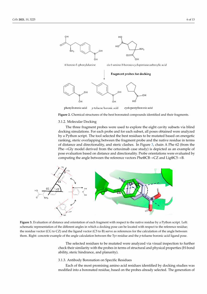
In a preliminary step, the fragment probes that best mimicked the chemico-physicalfeatures of some antibody amino acid residues were identified. A dataset of drugs wasassembled from the BNCT literature and DrugBank boronated compounds, obtaining75 molecules. Among these, 4-borono-L-phenylalanine and L-enantiomer of cis-1-amino-3-borono-cyclopentanecarboxylic acid, both already used in BNCT, were selected for theiroptimal scaffold similarity with Phe, Tyr, Trp, and His residues. From the two scaffolds,three fragments were generated. In the case of 4-borono-L-phenylalanine, the α- andβ-carbon were removed and p-toluene boronic acid and phenylboronic acid were obtained,respectively. In the case of cis-1-amino-3-borono-cyclopentanecarboxylic acid case, theamino and the carboxyl groups were removed, thereby obtaining cyclopentylboronic acid(Figure 2).
To represent the 3D structure of the fragment probes, we used measurements providedby scientific literature. Lengths and bond angles between boron and atoms connected toit were properly set (Figure S1). In this way, the final ligands displayed all the geometricstructural characteristics and electrical charges of a molecule containing a boron atom.
Each antibody residue able to mimic the chemico-physical properties of the probe frag-ments was mutated to Gly and then to Ala, in order to check whether the probe/candidateligand side chain was capable of repositioning itself exactly in the region previously occu-pied by the side chain of the native residue. To evaluate the impact of the residue β-carbonatom in influencing the fragment probe pose, Gly mutation was additionally includedto the Ala scanning mutation. The two mutations created two sets of cavities, furthersubdivided into four cavity subsets, one for each of the four mutated amino acid residues(Phe, Tyr, Trp, and His).
Cells 2021, 10, 3225 6 of 13
Figure 2. Chemical structures of the best boronated compounds identified and their fragments.
3.1.2. Molecular Docking
The three fragment probes were used to explore the eight cavity subsets via blinddocking simulations. For each probe and for each subset, all poses obtained were analyzedby a Python script. The tool selected the best residues to be mutated based on energeticranking, steric overlapping between the fragment probe and the native residue in termsof distance and directionality, and steric clashes. In Figure 3, chain A Phe 62 (from thePhe→Gly model derived from the cetuximab case study) is depicted as an example ofpose evaluation based on distance and directionality. Probe orientations were evaluated bycomputing the angle between the reference vectors Phe@CB→CZ and Lig@C5→B.
Figure 3. Evaluation of distance and orientation of each fragment with respect to the native residue by a Python script. Left:schematic representation of the different angles in which a docking pose can be located with respect to the reference residue;the residue vector (CG to CZ) and the ligand vector (C5 to B) serve as references for the calculation of the angle betweenthem. Right: concrete example of the angle calculation between the Tyr residue and the p-toluene boronic acid ligand pose.
The selected residues to be mutated were analyzed via visual inspection to furthercheck their similarity with the probes in terms of structural and physical properties (H-bondability, steric hindrance, and planarity).
3.1.3. Antibody Boronation on Specific Residues
Each of the most promising amino acid residues identified by docking studies wasmodified into a boronated residue, based on the probes already selected. The generation of

Cells 2021, 10, 3225 7 of 13
the new boronated residue took place starting from the initial coordinates of the α-carbonof the candidate residue. Since the boron atom is not parameterized in Amber18 force field,it was necessary to add the proper parameters and generate the corresponding residuetopological file and coordinate file for the subsequent simulations (see the Materials andMethods section for details, Supplementary Figures S2 and S3 and Tables S1–S5).
3.1.4. Modified Antibody Folding Evaluation
To evaluate the modified monoclonal antibody folding in comparison with the nativefolding, MD simulations were performed. In fact, it is necessary to preserve the originalprotein folding to retain the antibody functionality; thus, the new boronated residuesshould not cause folding alterations. RMSD and RMSF parameters were then calculatedto check whether there were any alterations in the mutated protein stability compared tothe wild-type. Subsequently, H-bond analysis allowed us to ascertain if the new residuesmaintained the native H-bond network. Finally, cluster analysis let us identify the mostlikely conformation of the modified monoclonal antibody by comparison with the native.
3.2. Case Study
Cetuximab, a monoclonal antibody capable of inhibiting epidermal growth factorreceptor (EGFR), was selected as a case study to test our strategy and was mutated fordelivering boron atoms. The XRay structure of cetuximab Fab (PDB id: 1YY8) alone andbound to the EGFR (PDB id: 1YY9) receptor were retrieved from PDB [27]. Both the heavyand the light chains of cetuximab participate in the interaction with the complementaritydetermining regions (CDRs) of the Fab fragment. The binding surface of the Fab fragmentis rich in tyrosine and tryptophan, residues mimicked by the chemico-physical featuresof the probe fragments used in the docking. As a consequence, only the residues notinvolved in the interaction with the receptor were mutated by us to Gly and Ala (Figure 4),producing for each residue type (namely Phe, Tyr, Trp, and His) eight cavity subsets to beexplored by different fragment probes as potential binding cavities.
In detail, when Phe, Tyr, Trp, and His were substituted with Gly or Ala, 16, 19, 8 and6 cavities were obtained, respectively. In Figure 4, the mutated residues are accentuatedusing balls of different colors.
Figure 4. Cetuximab Fab: spheres indicate the location of Tyr (yellow), Trp (green), Phe (red), and His (blue) residues thatwere mutated into Gly and Ala. The contact area with the receptor, enclosed by a box, was not considered in this study.
Cells 2021, 10, 3225 8 of 13
3.3. Docking Results
From the docking results, we only considered fragment probes poses localized insidethe cavities left from the mutation of Phe, Tyr, Trp, and His into Gly and Ala.
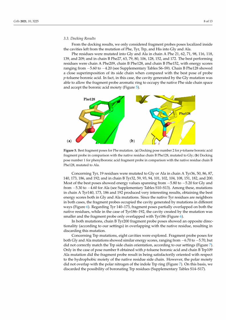
Phe residues were mutated into Gly and Ala in chain A Phe 21, 62, 71, 98, 116, 118,139, and 209; and in chain B Phe27, 63, 79, 80, 106, 128, 152, and 172. The best performingresidues were chain A Phe209, chain B Phe128, and chain B Phe152, with energy scoresranging from −5.60 to −4.20 (see Supplementary Tables S6–S9). Chain B Phe128 showeda close superimposition of its side chain when compared with the best pose of probep-toluene boronic acid. In fact, in this case, the cavity generated by the Gly mutation wasable to allow the fragment probe aromatic ring to occupy the native Phe side chain spaceand accept the boronic acid moiety (Figure 5).
Figure 5. Best fragment poses for Phe mutation. (a) Docking pose number 2 for p-toluene boronic acidfragment probe in comparison with the native residue chain B Phe128, mutated to Gly; (b) Dockingpose number 1 for phenylboronic acid fragment probe in comparison with the native residue chain BPhe128, mutated to Ala.
Concerning Tyr, 19 residues were mutated to Gly or Ala in chain A Tyr36, 50, 86, 87,140, 173, 186, and 192; and in chain B Tyr32, 59, 93, 94, 101, 102, 104, 108, 151, 182, and 200.Most of the best poses showed energy values spanning from −5.80 to −5.20 for Gly andfrom −5.30 to −4.60 for Ala (see Supplementary Tables S10–S13). Among these, mutationsin chain A Tyr140, 173, 186 and 192 produced very interesting results, obtaining the bestenergy scores both in Gly and Ala mutations. Since the native Tyr residues are neighborsin both cases, the fragment probes occupied the cavity generated by mutations in differentways (Figure 6). Regarding Tyr 140–173, fragment poses partially overlapped on both thenative residues, while in the case of Tyr186–192, the cavity created by the mutation wassmaller and the fragment probe only overlapped with Tyr186 (Figure 6).
In both mutations, chain B Tyr200 fragment probe poses showed an opposite direc-tionality (according to our settings) in overlapping with the native residue, resulting indiscarding this mutation.
Concerning Trp mutations, eight cavities were explored. Fragment probe poses forboth Gly and Ala mutations showed similar energy scores, ranging from−6.70 to−5.70, butdid not correctly match the Trp side chain orientation, according to our settings (Figure 7).Only in the case of pose number 8 obtained with p-toluene boronic acid and chain B Trp109Ala mutation did the fragment probe result in being satisfactorily oriented with respectto the hydrophobic moiety of the native residue side chain. However, the polar moietydid not overlap with the polar nitrogen of the indole Trp ring (Figure 7). On this basis, wediscarded the possibility of boronating Trp residues (Supplementary Tables S14–S17).

Cells 2021, 10, 3225 9 of 13
Figure 6. Best fragment poses for Tyr mutation. (a) Docking pose numbers 3, 4, and 7 for p-tolueneboronic acid fragment probe in comparison with the native residues chain A Tyr173 and 140, mutatedto Gly; (b) Docking pose numbers 4, 5, 7, 9, and 20 for phenylboronic acid fragment probe incomparison with the native residues chain A Tyr173 and 140, mutated to Ala; (c) Docking posenumber 10 for phenylboronic acid fragment probe in comparison with the native residues chainA Tyr192 and 186, mutated to Ala; (d) Docking pose numbers 11 and 16 for phenylboronic acidfragment probe in comparison with the native residue chain B Tyr200, mutated to Ala; (e) Dockingpose numbers 11 and 14 for p-toluene boronic acid fragment probe in comparison with the nativeresidue chain B Tyr200.
Figure 7. Fragment probe pose in comparison with the native residue—the case of Trp. Docking posenumber 8 for p-toluene boronic acid fragment probe in comparison with the native residue chain BTyr109, mutated to Gly.
When His was mutated into Gly or Ala, fragment probe poses obtained with phenyl-boronic acid and p-toluene boronic acid were located outside the cavities derived frommutation (Supplementary Tables S18–S21).
The cyclopentil boronic acid probe, although having a partial structural similaritywith His and Trp and, even lower, with Phe and Tyr, demonstrated the worst performance.In fact, it positioned itself outside the cavities generated by the mutations.
In conclusion, from the above reported docking results and their analysis, the bestnative residues were predicted to be boronated chain A Tyr140, chain A Tyr173, chain ATyr 186, and chain B Tyr200.
3.4. Monoclonal Antibody Folding Evaluation Using MD Simulations
Finally, to evaluate whether the boronated residues are able to keep the wild typemonoclonal antibody folding, MD simulations were performed on mutated cetuximab atthe residues listed above. RMSD values are reported in Supplementary Figure S4.

Cells 2021, 10, 3225 10 of 13
The comparison between wild type and boronated residues was performed usingRMSD analysis. Both wild type (Tyr140, 173, 186, and 200) and corresponding boronatedresidues stabilized at an average of about 1.5 Å in both chains (Figure S4). The similarRMDS trends for wild type and boronated protein suggested a negligible effect on structuralchanges determined by the boronated residue insertion. A structural analysis betweenwild type and boronated residues was successively performed using cluster analysis tohighlight minimal differences in protein rearrangement. RMSF values were calculated andthe corresponding plots are reported in Supplementary Figure S5. Comparing the wildtype protein chains with those that were boronated, RMSF values remained almost equallyfluctuating. In addition, clustering analysis was performed on each trajectory to capturethe representative conformation of wild type and boronated residues of the monoclonalantibody. Clusters with the highest population were distributed in the following way:30% for wild type, 77% for Tyr140, 38% for Tyr173, 72% for Tyr186 and 70% for Tyr200.Representative structures for each cluster were chosen to compare the conformationalchanges among wild type and corresponding boronated residues. As shown in Figure 8, thenative overall structure was maintained in the boronated protein, and as was foreseeable,the monoclonal antibody interaction region with EGFR was fully preserved.
Figure 8. Comparison of protein structures: 3D superimposition of representative structures obtained from cluster analysisshows structural similarity among wild type and boronated proteins, especially in the EGFR interaction region.
Moreover, the total number of hydrogen bonds of wild type and boronated residueswere also conserved. In fact, the analysis of the hydrogen bonds along the trajectories forboth wild type and boronated proteins indicates a total hydrogen bond number of about350, while the boronated residues made the same hydrogen bonds as Tyr140, 173, 186, and200, indicating a stable interaction network as in the wild type (Supplementary Figure S6and Tables S22–S25).
4. Discussion and Conclusions
The results obtained from this study suggest the possibility of using boronated mon-oclonal antibodies in BNCT as innovative tools in anticancer therapy. The boronatedantibody in fact is able to display the same selectivity in comparison with the standardmonoclonal antibody, but is more powerful against cancer cells thanks to the boronation.
To develop this kind of double acting monoclonal antibody, an innovative and dedi-cated pipeline was implemented, capable of screening, supporting, and identifying the bestamino acids that could be substituted by a boronated analogue. By doing so, particularattention was paid to potential 3D protein structure modifications and to potential sterichindrance interactions determined by the boronation. For this purpose, based on previousliterature studies [13], only a suitable and small boronated moiety, namely B(OH)2, was

Cells 2021, 10, 3225 11 of 13
inserted in specific amino acid residues. In addition, the residues far from this regionwere selected to preserve the mAb–target receptor interaction area and avoid the negativeimpact caused by steric hindrance interactions. The pipeline we developed can be used foroptimizing any protein of interest as a specific interactor for cancer cells in BNCT and isbased on a library of boronated compounds which display a scaffold similarity with thenatural amino acid residues. The pipeline has a modular design, with an automatic fluxof data in the boronation simulation, e.g., evaluation of the most suitable residue typesand residue positions to be boronated, evaluation of the monoclonal antibody folding afterinsertion of the specific boronated residue, and ranking of the best results after boronationusing a Python script.
The pipeline applied to the monoclonal antibody cetuximab, as a case study, allowedus to identify by means of molecular docking four Tyr residues as the best to be mutated,among the several present. The similarity of Tyr with phenyl boronic acid, p-tolueneboronic acid, and cyclopentil boronic acid relies on similar steric and polar features. Infact, the only hydrophobic feature (e.g., an aliphatic (Hys) or an aromatic ring (Phe, Tyr,and Trp)) is insufficient by itself to perform the best pocket occupancy, and the polarcomponent (e.g., OH, nitrogen atom) should integrate it. MD simulations proved to be avery efficient tool to evaluate the correct protein folding, letting us predict whether themutations impact the 3D mAb structure. Specifically, the four Tyr residues suggested bythese docking studies and confirmed by MD simulations were, among the others, the bestcapable of retaining the native protein folding and guaranteed the high binding specificityof cetuximab to EGFR.
In the past, boronated mAbs were prepared using large boron-containing molecules ordendrimers, but the boronated antibody specificity and structural and functional featureswere not preserved.
In this context, we developed a pipeline useful for a fast and precise evaluation ofthe effect of boronated modification on the 3D structure of monoclonal antibodies. Thedeveloped pipeline was tested on cetuximab, inserting an increased number of boronitems without consequent conformational changes of the mAb. The preservation of themAb 3D structure ensures the mAb specificity and strength in the binding to target. Theprotocol can be generalized and applied to any monoclonal antibody used in cancer therapy.In the present work, cetuximab, a chimeric monoclonal antibody capable of inhibitingEGFR and decelerating tumor growth, has been discussed as a case study. Of note, theexpression of EGFR is estimated to be around 0.5–1 × 10⁵ per each normal cell [28], andit is overexpressed 106 times more per cancer cell [29]. Thus, it is potentially possible toachieve more than 109 10B atom per cancer cell.
Thus, the boronated mAb can perform double anti-tumor activity: chemotherapy,related to its typical action, and radiotherapy, as a sort of boost, due to the neutron irradia-tion on 10B. The mAb could be obtained based on unnatural amino acid technology, usingtyrosine building block 4-borono-L-phenylalanine and applying a solid phase synthesisusing an automated peptide synthesizer [30].
In conclusion, based on these findings, this innovative computational pipeline and thisapplication on cetuximab as a case study provide evidence that BNCT treatment can benefitfrom the experience of using Monoclonal Antibodies as anti-tumor drugs; specifically,mAbs are suitable tools to drive boron on tumor targets.
Supplementary Materials: The following are available online at https://www.mdpi.com/article/10.3390/cells10113225/s1.
Author Contributions: Conceptualization, P.F., P.L.M. and P.D.; Methodology, A.O. and P.D., investi-gation, A.R., A.O., P.F. and P.D.; Writing—original draft preparation, A.R., P.F., A.D.P., D.P., P.L.M.and P.D.; Writing—review and editing, L.M., A.O., P.L.M., P.F. and P.D. All authors have read andagreed to the published version of the manuscript.

Cells 2021, 10, 3225 12 of 13
Funding: This research was funded by EU project EOSC-Pillar, grant number 857650; pan-Europeanre-search infrastructure for Biobanking and BioMolecular Re-sources Research In-frastructure (BBMRI),grant number BBMRI; EGI-Advanced Computing for Eosc, grant number EGI-ACE;Progetti ordinaridi ricerca finalizzata, Ministero della salute, grant number RF-2019-12370396; CENTRO NAZIONALEDI RICERCA IN BIOINFORMATICA PER LE SCI-ENZE “OMICHE” CNRBIOMICS, grant numberPON R&I PIR01_00017.
Data Availability Statement: Not applicable.
Acknowledgments: This work was supported by EU project EOSC-Pillar (Grant number 857650)(towards A.O.), pan-European research infrastructure for Biobanking and BioMolecular ResourcesResearch Infrastructure (BBMRI) and EGI-ACE (towards P.D.), RF-2019-12370396: Metabolic syn-drome and risks of breast cancer and cardiovascular disease: a systems biology approach appliedto epidemiological studies to identify predictive biomarkers and pathological molecular pathways(towards P.L.M.), and PON R&I PIR01_00017 CENTRO NAZIONALE DI RICERCA IN BIOINFOR-MATICA PER LE SCIENZE “OMICHE” CNRBIOMICS (towards P.L.M. and A.O.). The authors thankJohn Hatton of the Institute of Biomedical Technologies (CNR-ITB) for proofreading the manuscript.
Conflicts of Interest: The authors declare no conflict of interest.
References1. Worm, D.J.; Hoppenz, P.; Els-Heindl, S.; Kellert, M.; Kuhnert, R.; Saretz, S.; Köbberling, J.; Riedl, B.; Hey-Hawkins, E.; Beck-
Sickinger, A.G. Selective Neuropeptide Y Conjugates with Maximized Carborane Loading as Promising Boron Delivery Agentsfor Boron Neutron Capture Therapy. J. Med. Chem. 2020, 63, 2358–2371. [CrossRef] [PubMed]
2. Sauerwein, W.A.G.; Bet, P.M.; Wittig, A. Neutron Capture Therapy; Wittig, A., Moss, R., Nakagawa, Y., Eds.; Springer:Berlin/Heidelberg, Germany, 2012. [CrossRef]
3. Miyatake, S.I.; Kawabata, S.; Hiramatsu, R.; Kuroiwa, T.; Suzuki, M.; Kondo, N.; Ono, K. Boron neutron capture therapy formalignant brain tumors. Neurol. Med. Chir. (Tokyo) 2016, 56, 361–371. [CrossRef] [PubMed]
4. Hirose, K.; Konno, A.; Hiratsuka, J.; Yoshimoto, S.; Kato, T.; Ono, K.; Otsuki, N.; Hatazawa, J.; Tanaka, H.; Takayama, K.; et al.Boron neutron capture therapy using cyclotron-based epithermal neutron source and borofalan (10B) for recurrent or locallyadvanced head and neck cancer (JHN002): An open-label phase II trial. Radiother Oncol. 2021, 155, 182–187. [CrossRef] [PubMed]
5. STELLA PHARMA. Available online: https://stella-pharma.co.jp/cp-bin/wordpress5/wp-content/uploads/2020/05/Steboronine-launched_ENG.pdf (accessed on 19 October 2021).
6. Yang, W.; Barth, R.F.; Wu, G.; Kawabata, S.; Sferra, T.J.; Bandyopadhyaya, A.K.; Tjarks, W.; Ferketich, A.K.; Moeschberger, M.L.;Binns, P.J.; et al. Molecular targeting and treatment of EGFRvIII-positive gliomas using boronated monoclonal antibody L8A4.Clin. Cancer Res. 2006, 12, 3792–3802. [CrossRef] [PubMed]
7. Yang, W.; Wu, G.; Barth, R.F.; Swindall, M.R.; Bandyopadhyaya, A.K.; Tjarks, W.; Tordoff, K.; Moeschberger, M.; Sferra, T.J.; Binns,P.J.; et al. Molecular targeting and treatment of composite EGFR and EGFRvIII-positive gliomas using boronated monoclonalantibodies. Clin. Cancer Res. 2008, 14, 883–891. [CrossRef] [PubMed]
8. Yang, W.; Barth, R.F.; Wu, G.; Tjarks, W.; Binns, P.; Riley, K. Boron neutron capture therapy of EGFR or EGFRvIII positive gliomasusing either boronated monoclonal antibodies or epidermal growth factor as molecular targeting agents. Appl. Radiat Isot. 2009,67, S328–S331. [CrossRef]
9. Torres-Sánchez, P.; Porras, I.; Ramos-Chernenko, N.; Arias de Saavedra, F.; Praena, J. Optimized beam shaping assembly fora 2.1-MeV proton-accelerator-based neutron source for boron neutron capture therapy. Sci. Rep. 2021, 11, 7576. [CrossRef][PubMed]
10. Wu, G.; Barth, R.F.; Yang, W.; Chatterjee, M.; Tjarks, W.; Ciesielski, M.J.; Fenstermaker, R.A. Site-specific conjugation of boron-containing dendrimers to anti-EGF receptor monoclonal antibody cetuximab (IMC-C225) and its evaluation as a potential deliveryagent for neutron capture therapy. Bioconjug. Chem. 2004, 15, 185–194. [CrossRef]
11. Wu, G.; Yang, W.; Barth, R.F.; Kawabata, S.; Swindall, M.; Bandyopadhyaya, A.K.; Tjarks, W.; Khorsandi, B.; Blue, T.E.; Ferketich,A.K.; et al. Molecular targeting and treatment of an epidermal growth factor receptor-positive glioma using boronated cetuximab.Clin. Cancer Res. 2007, 13, 1260–1268. [CrossRef]
12. Sauerwein, W.A.G.; Sancey, L.; Hey-Hawkins, E.; Kellert, M.; Panza, L.; Imperio, D.; Balcerzyk, M.; Rizzo, G.; Scalco, E.; Herrmann,K.; et al. Theranostics in Boron Neutron Capture Therapy. Life (Basel) 2021, 11, 330. [CrossRef]
13. Capala, J.; Barth, R.F.; Bendayan, M.; Lauzon, M.; Adams, D.; Soloway, A.H.; Carlsson, J. Boronated epidermal growth factor as apotential targeting agent for boron neutron capture therapy of brain tumors. Bioconjug. Chem. 1996, 7, 7–15. [CrossRef] [PubMed]
14. Yamaguchi, A.; Achmad, A.; Hanaoka, H.; Heryanto, Y.D.; Bhattarai, A.; Khongorzul, E.; Shintawati, R.; Kartamihardja, A.A.P.;Kanai, A.; Sugo, Y.; et al. Immuno-PET imaging for non-invasive assessment of cetuximab accumulation in non-small cell lungcancer. BMC Cancer 2019, 19, 1000. [CrossRef] [PubMed]
15. Wee, P.; Wang, Z. Epidermal Growth Factor Receptor Cell Proliferation Signaling Pathways. Cancers 2017, 9, 52. [CrossRef][PubMed]
Cells 2021, 10, 3225 13 of 13
16. Barth, R.F.; Yang, W.; Adams, D.M.; Rotaru, J.H.; Shukla, S.; Sekido, M.; Tjarks, W.; Fenstermaker, R.A.; Ciesielski, M.; Nawrocky,M.M.; et al. Molecular targeting of the epidermal growth factor receptor for neutron capture therapy of gliomas. Cancer Res. 2002,62, 3159–3166.
17. Seshacharyulu, P.; Ponnusamy, M.P.; Haridas, D.; Jain, M.; Ganti, A.K.; Batra, S.K. Targeting the EGFR signaling pathway incancer therapy. Expert Opin. Ther. Targets 2012, 16, 15–31. [CrossRef]
18. Trott, O.; Olson, A.J. AutoDock Vina: Improving the speed and accuracy of docking with a new scoring function, efficientoptimization, and multithreading. J Comput. Chem. 2010, 31, 455–461. [CrossRef] [PubMed]
19. Wishart, D.S.; Knox, C.; Guo, A.C.; Shrivastava, S.; Hassanali, M.; Stothard, P.; Chang, Z.; Woolsey, J. DrugBank: A comprehensiveresource for in silico drug discovery and exploration. Nucleic Acids Res. 2006, 34, D668–D672. [CrossRef] [PubMed]
20. Rettig, S.J.; Trotter, J. Crystal and molecular structure of phenylboronic acid, C6H5B(OH)2. Can. J. Chem. 2011, 55, 3071–3075.[CrossRef]
21. Greenwood, J.R.; Calkins, D.; Sullivan, A.P.; Shelley, J.C. Towards the comprehensive, rapid, and accurate prediction of thefavorable tautomeric states of drug-like molecules in aqueous solution. J. Comput. Aided Mol. Des. 2010, 24, 591–604. [CrossRef][PubMed]
22. Case, D.A.; Ben-Shalom, I.Y.; Brozell, S.R.; Cerutti, S.R.; Cheatham, T.E., III; Cruzeiro, V.W.D.; Darden, T.A.; Duke, R.E.; Ghoreishi,D.; Gilson, M.K.; et al. AMBER 2018; University of California: San Francisco, CA, USA, 2018.
23. Kurt, B.; Temel, H. Parameterization of Boronates Using VFFDT and Paramfit for Molecular Dynamics Simulation. Molecules2020, 25, 2196. [CrossRef]
24. Kurt, B.; Temel, H. Development of AMBER parameters for molecular dynamics simulations of boron compounds containingaromatic structure. Chem. Phys. Lett. 2021, 775, 138656. [CrossRef]
25. Salomon-Ferrer, R.; Case, D.A.; Walker, R.C. An overview of the Amber biomolecular simulation package. WIREs Comput. Mol.Sci. 2013, 3, 198–210. [CrossRef]
26. Machado, M.R.; Pantano, S. Split the Charge Difference in Two! A Rule of Thumb for Adding Proper Amounts of Ions in MDSimulations. J Chem Theory Comput. 2020, 16, 1367–1372. [CrossRef] [PubMed]
27. Li, S.; Schmitz, K.R.; Jeffrey, P.D.; Wiltzius, J.J.; Kussie, P.; Ferguson, K.M. Structural basis for inhibition of the epidermal growthfactor receptor by cetuximab. Cancer Cell 2005, 7, 301–311. [CrossRef] [PubMed]
28. Carpenter, G.; Cohen, S. Epidermal growth factor. Annu. Rev. Biochem. 1979, 48, 193–216. [CrossRef] [PubMed]29. Gullick, W.J.; Marsden, J.J.; Whittle, N.; Ward, B.; Bobrow, L.; Waterfield, M.D. Expression of epidermal growth factor receptors
on human cervical, ovarian, and vulval carcinomas. Cancer Res. 1986, 46, 285–292. [PubMed]30. Soor, H.S.; Hansen, J.; Diaz, D.B.; Appavoo, S.; Yudin, A.K. Solid-phase synthesis of peptide β-aminoboronic acids. Pept. Sci.
2019, 111, e24072. [CrossRef]
Copyright © 2022 FDOKUMEN