Saturation Mutagenesis of 5S rRNA in Saccharomyces cerevisiae
Upload
independentCategory
view
0download
0
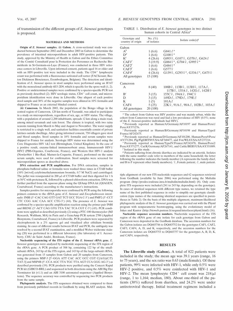
JOURNAL OF CLINICAL MICROBIOLOGY, Aug. 2007, p. 2580–2589 Vol. 45, No. 80095-1137/07/$08.00�0 doi:10.1128/JCM.02554-06Copyright © 2007, American Society for Microbiology. All Rights Reserved.
New Highly Divergent rRNA Sequence among Biodiverse Genotypesof Enterocytozoon bieneusi Strains Isolated from Humans in
Gabon and Cameroon�
Jacques Breton,1,4 Emmanuelle Bart-Delabesse,2 Sylvestre Biligui,1,2 Alessandra Carbone,1,7
Xavier Seiller,1† Madeleine Okome-Nkoumou,5 Chantal Nzamba,6 Maryvonne Kombila,4Isabelle Accoceberry,3 and Marc Thellier1,2*
Unite INSERM 511, CHU Pitie-Salpetriere, 91 Bd de l’Hopital, 75013 Paris, France1; Assistance Publique Hopitaux de Paris,Laboratoire de Parasitologie-Mycologie, CHU Pitie-Salpetriere, 47 Bd de l’Hopital, 75013 Paris, France2; Laboratoire de
Parasitologie-Mycologie, CHU de Saint Andre, 1 rue Jean Burguet, 33075 Bordeaux, France3; Departement deParasitologie-Mycologie, Universite des Sciences de la Sante, B.P. 4009, Libreville, Gabon4; Unite des
Maladies Infectieuses, Fondation Jeanne Ebori, B.P. 861, Libreville, Gabon5; Centre deTraitement Ambulatoire, Hopital General de Libreville, Gabon6; and Genomique Analytique,
Universite Pierre et Marie Curie-Paris 6, 4 Place Jussieu, 75005 Paris, France7
Received 20 December 2006/Returned for modification 27 March 2007/Accepted 22 May 2007
Intestinal microsporidiosis due to Enterocytozoon bieneusi is a leading cause of chronic diarrhea in severelyimmunocompromised human immunodeficiency virus (HIV)-positive patients. It may be a public healthproblem in Africa due to the magnitude of the HIV pandemic and to poor sanitary conditions. We designed twoprevalence studies of E. bieneusi in Central Africa, the first with HIV-positive patients from an urban settingin Gabon and the second with a nonselected rural population in Cameroon. Stool samples were analyzed by animmunofluorescence antibody test and PCR. Twenty-five out of 822 HIV-positive patients from Gabon and 22out of 758 villagers from Cameroon were found to be positive for E. bieneusi. The prevalence rates of the twostudies were surprisingly similar (3.0% and 2.9%). Genotypic analysis of the internal transcribed spacer regionof the rRNA gene showed a high degree of diversity in samples from both countries. In Gabon, 15 isolatesshowed seven different genotypes: the previously reported genotypes A, D, and K along with four new genotypes,referred to as CAF1, CAF2, CAF3, and CAF4. In Cameroon, five genotypes were found in 20 isolates: the knowngenotypes A, B, D, and K and the new genotype CAF4. Genotypes A and CAF4 predominated in Cameroon,whereas K, CAF4, and CAF1 were more frequent in Gabon, suggesting that different genotypes present differingrisks of infection associated with immune status and living conditions. Phylogenetic analysis of the newgenotype CAF4, identified in both HIV-negative and HIV-positive subjects, indicates that it represents a highlydivergent strain.
As many as 90% of AIDS patients experience diarrhea inAfrica, where “slim disease” (prolonged diarrhea and wasting)is a pathognomonic trait of AIDS (30). While early investiga-tion revealed that coccidian parasites were a leading cause ofchronic diarrhea, the development of improved microscopic,immunological, and molecular diagnostic methods has high-lighted the importance of microsporidian infections (11). Twospecies of microsporidia can cause gastrointestinal disease:Enterocytozoon bieneusi (10) and Encephalitozoon intestinalis(7, 18), although E. bieneusi is the more frequently reportedspecies. Data on the prevalence of E. bieneusi in human im-munodeficiency (HIV)-positive patients in Africa is scarce, andvalues differ greatly, from 5% to 50%, depending on the pop-ulation studied (whether or not it is selected for diarrhea) and
the diagnostic methods (2, 5, 17, 21, 27, 38, 43). Besides, E.bieneusi also can infect immunocompetent patients, especiallychildren, although the disease is less serious and the infectionis self-limited in the absence of immune abnormalities (42).Since mature spores are shed in the host’s feces, the transmis-sion routes of this pathogen may involve person-to-person aswell as waterborne or food-borne contaminations, especially indeveloping countries with poor sanitation (3). E. bieneusi alsohas been isolated from a large number of domestic and wildmammals, as well as birds (29). The potential of zoonotictransmission is supported by phylogenetic studies showing thatseveral genotypes can infect humans as well as animals (9, 12,26, 37, 39–41).
To address the importance of E. bieneusi infections inAfrica, we conducted two studies of prevalence in differentpopulations, the first with HIV-infected patients from an urbansetting in Gabon and the second with a rural population inCameroon. The presence of E. bieneusi spores in stool sampleswas detected by an immunofluorescent antibody test (IFAT)and PCR. Genotypic analysis of E. bieneusi isolates was per-formed by sequencing the internal transcribed spacer (ITS)portion of the rRNA gene. On the basis of these results, aphylogenetic interpretation regarding the sources and modes
* Corresponding author. Mailing address: Assistance PubliqueHopitaux de Paris, Laboratoire de Parasitologie-Mycologie, CHUPitie-Salpetriere, 47 boulevard de l’Hopital, 75013 Paris, France.Phone: (33) 01 42 16 01 13. Fax: (33) 01 42 16 01 65. E-mail: [email protected].
† Present address: Institut Catholique d’Etudes Superieures, LaRoche-sur-Yon, France.
� Published ahead of print on 30 May 2007.
2580
on January 8, 2016 by guesthttp://jcm
.asm.org/
Dow
nloaded from
of transmission of the different groups of E. bieneusi genotypesis proposed.
MATERIALS AND METHODS
Origin of E. bieneusi samples. (i) Gabon. A cross-sectional study was con-ducted between September 2002 and December 2003 in Gabon to determine theprevalence of intestinal microsporidiosis in adult HIV-positive patients. Thisstudy, approved by the Ministry of Health in Gabon and the Ethics Committeeof the Comite Consultatif pour la Protection des Personnes en Recherche Bio-medicale in St-Germain-en-Laye (France), was conducted in three HIV refer-ence centers in Libreville. Upon informed consent, patients aged 16 or older andwith an HIV-positive test were included in the study. The CD4� lymphocytecount was performed with a fluorescence-activated cell sorter (FACScount; Bec-ton Dickinson Biosciences, Erembodegem, Belgium). The detection and identi-fication of E. bieneusi spores in stool samples were performed using an IFATwith the monoclonal antibody 6E5-2D9, which is specific for the spore wall (1, 2).Positive or undetermined samples were confirmed by a species-specific PCR testas previously described (2). HIV serologic status, CD4� cell count, and micros-poridium identification were done in Libreville. One aliquot of each positivestool sample and 30% of the negative samples were diluted in 10% formalin andshipped to France as an external blinded control.
(ii) Cameroon. In March 2003, the population of the Bongo village in thecentral region of Cameroon, 120 km north of Yaounde, was asked to participatein a study on microsporidiosis, regardless of sex, age, or HIV status. The village,with a population of around 2,200 inhabitants, spreads 12 km along a main road,among mixed savannah and rain forest. The climate is tropical, with two rainyseasons running from March to May and August to November. The water supplyis restricted to a single well, and sanitation facilities essentially consist of privatelatrines outside dwellings. After giving informed consent, 758 villagers gave stooland blood samples. Stool samples in 10% formalin and serum samples wereshipped to France for analysis. Serologic HIV status was determined using theCore Diagnostics HIV 1&2 test (Birmingham, United Kingdom). In the case ofa positive result, enzyme-linked immunosorbent assay, Immunocomb HIV1/HIV2 (PBS-Orgenics, Courbevoie, France), and Western blot HIV-1 analysis(Newlavblot 1 Bio-Rad, Marnes-la-Coquette, France), performed on the sameserum sample, were used for confirmation. Stool samples were screened formicrosporidium spores as described above.
DNA extraction and PCR amplification. For DNA extraction, samples inphosphate-buffered saline or formalin were washed three times in CTAB buffer(10% [wt/vol] cetyltrimethylammonium bromide, 0.7 M NaCl) and centrifuged.The pellet was resuspended in 200 �l of CTAB buffer and then digested for 1 hat 56°C with proteinase K, followed by a phenol-chloroform extraction. DNA wasfurther purified from the aqueous phase using the QIAmp DNA kit (QIAGEN,Courtaboeuf, France) according to the manufacturer’s instructions.
Samples positive for microsporidia were confirmed by PCR using the followingprimers common to the rRNA gene of E. bieneusi and Encephalitozoon intesti-nalis: INBI (5�-CAC CAG GTT GAT TCT GCC TGA C-3�) and PMP2 (5�-CCTCTC CGG AAC CAA ACC CTG-3�) (44). The presence of E. bieneusi wasconfirmed by a species-specific amplification reaction using the primer pair INBIand BIENE (5�-ACT CAG GTG TTA TAC TCA CGT C-3�) (45). PCR condi-tions were applied as described previously (2) using a PTC-100 thermocycler (MJResearch, Waltham, MA) in Paris and a GeneAmp PCR system 2700 (AppliedBiosystems, Courtaboeuf, France) in Libreville. PCR products were separated byelectrophoresis in a 2% agarose gel and visualized after ethidium bromidestaining. In cases of different results between IFAT and PCR, the ambiguity wasresolved by a second IFAT examination, and a modified Weber trichrome stain-ing (20) was performed in a different laboratory (the laboratory of I. Accoce-berry, CHU de Saint Andre, Bordeaux, France).
Nucleotide sequencing of the ITS region of the E. bieneusi rRNA gene. E.bieneusi genotypes were analyzed by nucleotide sequencing of the ITS region ofthe rRNA gene. A PCR product of 508 bp, containing 122 bp of the small-subunit rRNA, 243 bp of the ITS region, and 143 bp of the large-subunit rRNA,was generated from 15 samples from Gabon and 20 samples from Cameroon,using the primers MSP-3 [5�-GGA ATT CAC ACC GCC CGT C(A/G)(C/T)TAT-3] and MSP4B (5�-CCA AGC TTA TGC TTA AGT CCA GGG AG-3�) asdescribed previously (19). PCR products were purified using the Concert RapidPCR kit (GIBCO-BRL) and sequenced in both directions using the ABI Big DyeTerminator kit (v1.1) and an ABI 3100 automated sequencer (Applied Biosys-tems). The sequence accuracy was controlled by sequencing two PCR productsfrom the same sample.
Phylogenetic analysis. The ITS sequences obtained were compared to thosefrom previously published records in GenBank by using BLAST analysis. Mul-
tiple alignment of our new ITS nucleotide sequences and 62 sequences retrievedfrom GenBank (available by June 2006) was performed using the Multalinprogram (http://prodes.toulouse.inra.fr/multalin/multalin.html) (8). Only com-plete ITS sequences were included (241 to 243 bp, depending on the genotype).In cases of identical sequences with different type names, we retained the typename of the first published sequence in order to simplify our genotypic classifi-cation (type names of the remaining identical sequences are specified in paren-theses in Table 2). On the basis of this multiple alignment, maximum-likelihoodphylogenetic analysis of the E. bieneusi genotypes was carried out with the Phymlprogram with nonparametric bootstrapping, using the evolutionary model ofJukes and Kantor (http://bioweb.pasteur.fr/seqanal/interfaces/phyml.html) (16).
Nucleotide sequence accession numbers. Nucleotide sequences of the ITSregion of the rRNA gene of one isolate for each genotype from Gabon andCameroon were deposited in the GenBank database. The accession numbers forthe Gabon isolates are DQ683746 to DQ683752 for the genotypes CAF1, CAF2,CAF3, CAF4, A, D, and K, respectively, and the accession numbers for theCameroon isolates are DQ683753 to DQ683757 for the genotypes A, B, D, K,and CAF4, respectively.
RESULTS
The Libreville study (Gabon). A total of 822 patients wereincluded in the study; the mean age was 39.1 years (range, 16to 75 years), and the sex ratio was 0.63 (male/female). Of thesepatients, 99% were infected with HIV-1, while only 0.5% wereHIV-2 positive, and 0.5% were coinfected with HIV-1 andHIV-2. The mean lymphocyte CD4� cell count was 218/�l(range, 1 to 1,164; median, 180). About one-third of the pa-tients (30%) suffered from diarrhea, and 24.2% were underantiretroviral therapy. Initial treatment regimens included a
TABLE 1. Distribution of E. bieneusi genotypes in two distincthuman cohorts in Central Africaa
Genotype andcountry of origin
No. (%)of isolates Isolate code(s)g
GabonAb 1 (6.6) G841�*Dd 1 (6.6) G100�*Ke 4 (26.6) G093�, G107�, G370�, G624�CAF1f 3 (19.9) G084�*, G704�, G995�*CAF2f 1 (6.6) G484�*CAF3f 1 (6.6) G577�CAF4f 4 (26.6) G139�, G293�*, G334�*, G475�All genotypes 15 (100)
CameroonA 8 (40) 108B�, 113B�, 113H�, 117A�,
117B�, 135A�, 142G�, 142H�Bc 3 (15) 155C�, 194A�, 194C�D 3 (15) 149D�, 179D�, 179E�K 1 (5) 181A�CAF4 5 (25) 22K�, 91A�, 98A�, 102B�, 103A�All genotypes 20 (100)
a The cohort from Gabon was HIV positive and was mainly urban, while thecohort from Cameroon was rural and had a low prevalence of HIV (0.5%; noneof the E. bieneusi-positive individuals had HIV).
b Previously reported as Human/A/Germany/AF10197 and Human/Peru1/Peru/AY371276.
c Previously reported as Human/B/Germany/AF10198 and Human/TypeI/France/AF242475.
d Previously reported as Human/D/Germany/AF101200, Human/Peru9/Peru/AY371284, Pig/PigEBITS9/USA/AF348477, and Fox/WL8/USA/AY237216.
e Previously reported as Human/TypeIV/France/AF242478, Human/Peru2/Peru/AY371277, Cat/K/Germany/AF267141, and Cattle/BEB5/USA/AY331009.
f Isolated in this study.g Patients suffering from chronic diarrhea are indicated by an asterisk. Isolates
with the same number code refer to members of the same household; the letterfollowing the number indicates the family member (A represents the family chief,and B to F represent other family members). �, Female patient; �, male patient.
VOL. 45, 2007 E. BIENEUSI GENOTYPES FROM CENTRAL AFRICA 2581
on January 8, 2016 by guesthttp://jcm
.asm.org/
Dow
nloaded from
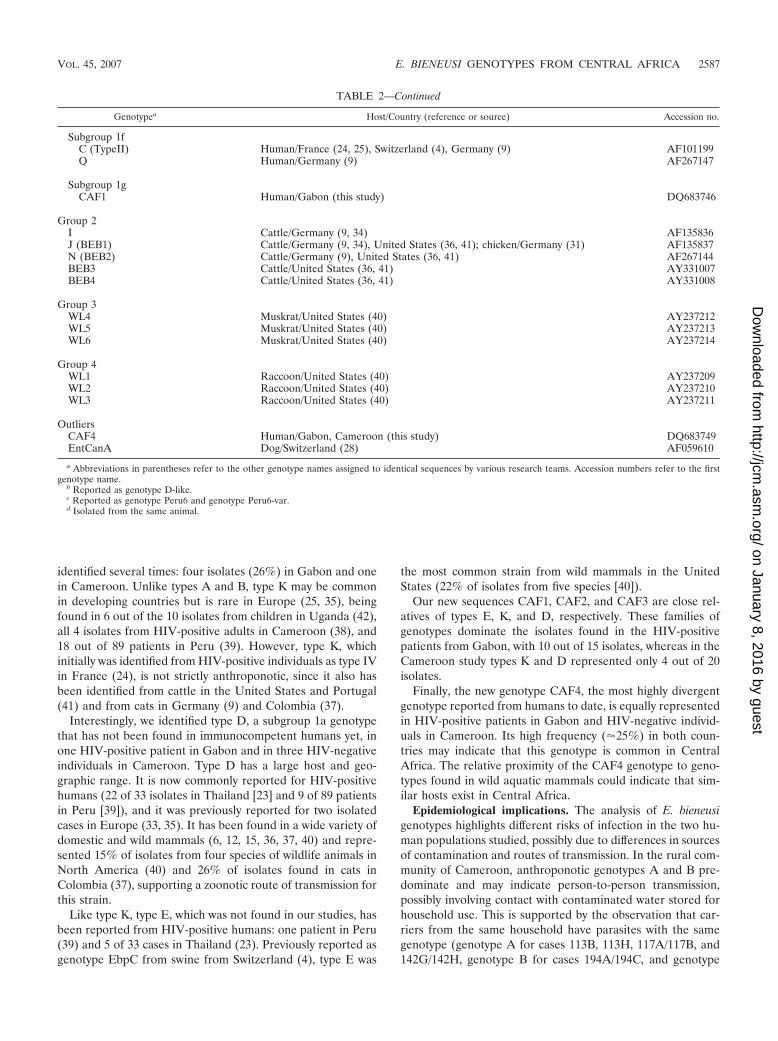
FIG. 1. Sequence alignment of the ITS region of the rRNA gene of E. bieneusi genotypes using the Multalin software. New sequences fromGabon and Cameroon (CAF1 to CAF4) are compared to sequences representative of each group. (Representative sequences are the following:group 1, subgroup 1a, genotype D; group 1, subgroup 1b, genotype Peru6; group 1, subgroup 1c, genotype K; group 1, subgroup 1d, genotype E;group 1, subgroup 1e, genotype O; group 1, subgroup 1f, genotype C; group 2, genotype N; group 3, genotype WL4; group 4, genotype WL1; andthe outlier sequence EntCanA). Dots indicate identity to CAF1. Point mutations of the new sequences CAF1, CAF2, and CAF3 compared to theirclosest homologues, K and D, are shaded gray (for accession numbers, see Table 2) (note that the upper ruler indicates the true sequence positionfor group 1 genotypes [243 bp], and the lower ruler indicates the Multalin alignment position number, with gaps counted).
2582 BRETON ET AL. J. CLIN. MICROBIOL.
on January 8, 2016 by guesthttp://jcm
.asm.org/
Dow
nloaded from
combination of one protease inhibitor (usually Indinavir) or anonnucleoside reverse transcriptase inhibitor (Efavirenz) andtwo nucleoside reverse transcriptase inhibitors (NRTIs) (d4T,3TC, AZT, or ddI). The majority (85%) lived in the urban areaof Libreville and originated from Gabon (86.5%), whereas theremaining patients were migrant workers from West Africa.
By IFAT, 25 out of the 822 stool samples were positive forE. bieneusi (prevalence, 3%). Of the positive stools, all exceptthree were confirmed to be positive by the species-specificPCR. Negative PCR samples contained very few spores inmicroscopic examination and remained negative despite re-peated internal and external PCR controls. All 25 E. bieneusi-positive patients were HIV-1 positive, and one was coinfectedwith HIV-2. The mean age was 35.4 years (range, 21 to 57years), and the sex ratio was 0.47 (male/female). Only fourpatients (16%; class B2) were not at the AIDS stage of theinfection. The average CD4� cell count of 23 patients was98.5/�l (range, 1 to 384/�l; values were not available for twopatients). A cell count below 100/�l (mean, 29.3/�l) was scoredfor 15 (65%) out of 23 patients. All except three patients fromthe class B2 HIV group suffered from digestive symptoms.These symptoms corresponded to diarrhea (14 cases), weightloss (15 cases), anorexia (6 cases), abdominal pain (5 cases),and nausea (5 cases). Six patients were undergoing antiretro-viral therapy (three patients were receiving a combination of anonnucleoside reverse transcriptase inhibitor and two NRTIs,and three patients were receiving a combination of a proteaseinhibitor and two NRTIs) at least 3 months before the diag-nosis of intestinal microsporidiosis. Twenty of these patients(80%) resided in Libreville, while the other five came fromdistant provinces of the country.
The Bongo study (Cameroon). The mean age of the Bongostudy population was 28.2 years (758 individuals; range, �1 to80 years), and the sex ratio was 0.81 (male/female). The com-bination of IFAT and PCR detection of microsporidia showed22 inhabitants positive for E. bieneusi (prevalence, 2.9%). TheCore Diagnostics HIV 1&2 screening showed that only foursubjects were HIV-1 positive. Sera of the latter patients wereconfirmed to be HIV-1 positive by enzyme-linked immunosor-bent assay and Western blot analysis, resulting in a prevalencerate of 0.5% in the Bongo population. None of the microspo-ridium-positive individuals were found to be positive for HIV.Data on the clinical status (diarrhea) of these individuals werenot available.
Analysis of genotypes. Thirty-five (15 from Gabon and 20from Cameroon) E. bieneusi isolates yielded sufficient PCRproduct for the sequencing of the ITS region of the rRNAgene. Both series showed a high degree of diversity of geno-types, as shown in Table 1.
In Gabon, seven different genotypes were identified: thepreviously reported genotypes A, D, and K as well as four newgenotypes. The four new genotypes are referred to as CAF1,CAF2, CAF3, and CAF4 (CAF being short for Central Africa).The genotypes K, CAF4, and CAF1 seemed to be more com-mon among the isolates from Gabon, with four, four, and threeisolates each, respectively, whereas genotypes A, D, CAF2, andCAF3 were found only once each.
In Cameroon, five genotypes were identified: the knowngenotypes A, B, D, and K and the new genotype CAF4. How-ever, genotype A was the most common, with eight isolates,
followed by CAF4, which was detected in five cases. Types Band D were scored three times each, and genotype K wasfound only once.
Out of the four new genotypes, three consisted of sequencesdisplaying very high identity to sequences of the known geno-types K and D. Types CAF1 and CAF2 differed from K by onlyone position (CAF1, position 113 [C3T]; CAF2, position 193[G3A]), and type CAF3 differed from D at positions 160 and224 (G3C at both positions) (Fig. 1). The last genotype,CAF4, is 1 bp shorter (242 bp) than all of the others. It was alsovery divergent from previously reported sequences; the besthomology, i.e., 76% identity, was found with the set of geno-types WL1 to WL3, isolated from raccoons in the UnitedStates (40). Indeed, the identities of the new sequences to allother previously published sequences were between 61 and64% and as low as 46% with genotype EntCanA.
The ITS flanking regions (122 bp of the small-subunit rRNAand 143 bp of the large-subunit rRNA) of the nine CAF4isolates from both studies showed 100% identity to the corre-sponding regions of the E. bieneusi rRNA sequences fromother genotypes.
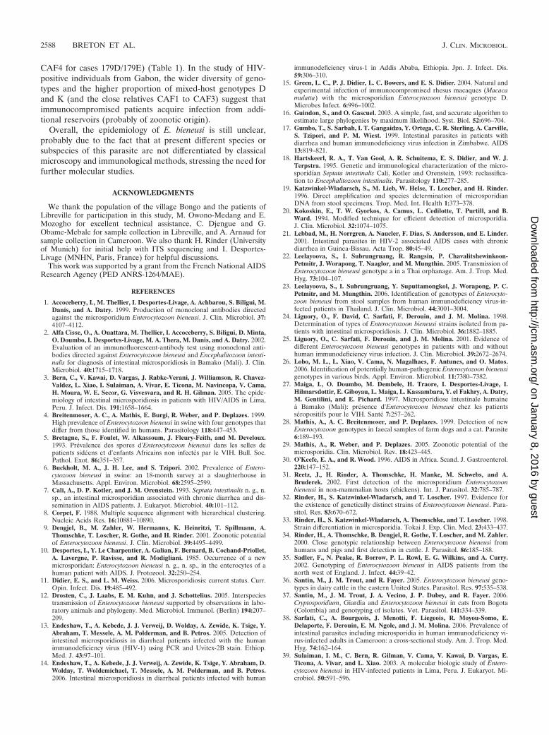
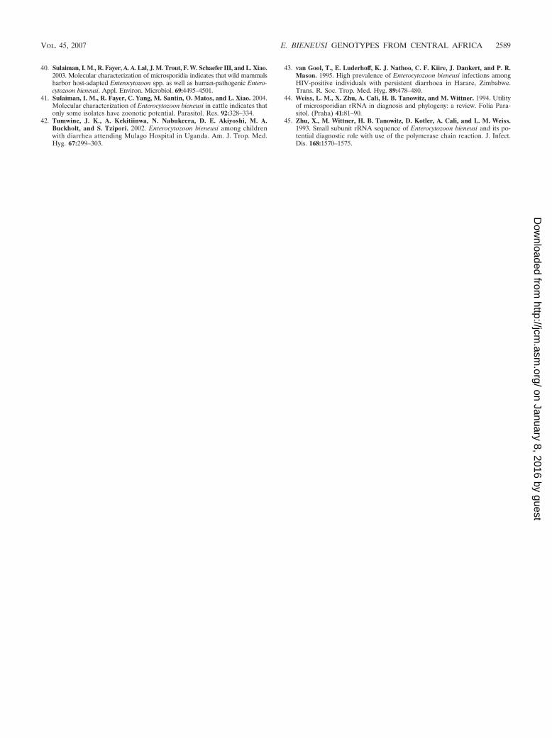
Phylogenetic analysis. An unrooted phylogram and a phy-logenetic unrooted tree inferred by the maximum-likelihoodanalysis of the ITS region were constructed using the Phymlsoftware (Fig. 2 and 3). As seen in previous studies (12, 40),four main groups, numbered 1 to 4, are segregated from themost divergent sequence, EntCanA, which was isolated fromdogs (28). Group 1 contains most of the sequences previouslypublished as well as the new sequences CAF1, CAF2, andCAF3 (53 out of 66 sequence) (Fig. 2). These sequences havebeen isolated from a wide diversity of hosts worldwide, includ-ing humans and animals (Table 2). The other three groupscontain sequences isolated from one host species. Group 2contains five sequences from cattle (N to I) (9, 31, 34, 36, 41).Group 3 is made up of three sequences from muskrat (WL4 toWL6), and group 4 is made up of three divergent sequencesfrom raccoon (WL1 to WL3) (40). Our new human sequenceCAF4 is placed, along with EntCanA, between group 3 andgroup 4, far away from group 1, which contains all the geno-types isolated from humans so far.
Maximum-likelihood analysis shows that group 1 is furthersubdivided into at least seven clades (Fig. 2). To provide amore comprehensive classification of group 1 E. bieneusi geno-types, the simplified nomenclature of Drosten et al. (12) wasused, i.e., subgroups 1a to 1f, and subgroup 1g was added. Theidentities within this group are high: 79.4% of the nucleotides(193/243) are conserved among the 53 sequences.
There are apparent differences in the range of host specific-ity within the group 1 clades, which are divided into two mainbranches and one isolated sequence (CAF1). The right branchcontains subgroups with more restricted host specificity thanthose of the left branch. Subgroup 1f is human specific (mainlygenotype C cases), subgroup 1d is found in wild mammals(apart from genotype E, which has a wide host range), andsubgroup 1e is found mainly in farm animals (11 out of 13sequences were isolated from swine), although some genotypesof this group (O, U, and W) recently have been reported inThailand from HIV-positive patients (23). The left branchsupporting subgroups 1a, 1b, and 1c shows the greatest hostdiversity, including commensal birds, commensal and wild
VOL. 45, 2007 E. BIENEUSI GENOTYPES FROM CENTRAL AFRICA 2583
on January 8, 2016 by guesthttp://jcm
.asm.org/
Dow
nloaded from
mammals, and HIV-negative and HIV-positive individuals.Genotypes from humans (including those also found in animalhosts) predominate, with 21 out of 32 sequences in subgroups1a, 1b, and 1c. These sequences from the left-branch subgroupsrepresent 72% of a total of 29 genotypes reported to be foundin humans so far.
The new genotype CAF1 is found at the base of the twomain branches. It was identified in three isolates from Gaboncollected over a period of 12 months and is proposed to con-stitute the new subgroup 1g. CAF1’s position on the tree isintermediate between the subgroups 1c and 1e, containingtypes K and E, respectively. It differs by one point mutationeach from K (position 113) and E (position 141) (Fig. 1).
DISCUSSION
Results of both studies showed (i) that prevalence rates of E.bieneusi were similar between immunocompetent people in theBongo village and HIV-positive patients from Libreville; (ii)that despite the limited number of isolates analyzed, a substan-tial amount of genotypic diversity was observed in both ruraland urban contexts; and (iii) that new genotypes that differedfrom known types by one or more nucleotides were identified,in particular, type CAF4, which is the most divergent genotypereported from humans to date.
Prevalence of E. bieneusi in Africa. In previous studies con-ducted in Africa, the prevalence of E. bieneusi in HIV-infected
FIG. 2. Unrooted phylogram inferred by the maximum-likelihood analysis of the ITS of E. bieneusi genotypes. The phylogram was constructedusing the Phyml software. Groups and subgroups are indicated in parentheses. New genotypes from this study are in boldface (for accessionnumbers, see Table 2).
2584 BRETON ET AL. J. CLIN. MICROBIOL.
on January 8, 2016 by guesthttp://jcm
.asm.org/
Dow
nloaded from
adults with diarrhea was around 10% (2, 5, 13, 14, 21, 43), butvalues as high as 32% and 51% have been reported (17, 27).Compared to these rates, the prevalence rates of E. bieneusiinfections we report for Cameroon and Gabon (2.9% and3.0%) seem to be low and, considering the difference of HIVstatus in the populations studied, are surprisingly similar. How-ever, the prevalence rate we observed in the Gabonese HIV-infected population is comparable to those reported for theurban areas of Yaounde, Cameroon (5.2%) (38), and Lima,Peru (3.9%) (39). These studies were also conducted withHIV-infected adults not selected for diarrhea. On the contrary,the E. bieneusi prevalence rate of 2.9% we observed in Cam-eroon may be surprising, since it could be expected to be lowerin a population with low HIV prevalence. This is the firstreport of E. bieneusi prevalence in an African population notselected for age, diarrhea status, or HIV status, since onlytwo previous studies addressed this issue on the continent.Bretagne et al. reported a prevalence for E. bieneusi of 0.8% inchildren in Niger, for whom HIV status could not be assessed(5), whereas Tumwine et al. found a high prevalence in Ugan-dan children with or without diarrhea (17.4% or 16.%, respec-tively); the rate of positive HIV status was estimated to be 18to 20% (42). Therefore, intestinal infection of immunocompe-
tent subjects with E. bieneusi might be common in Africa,especially among children.
Genotypic diversity and host specificity. Although a highdegree of diversity of genotypes was found in both populationsstudied, differences in their relative distribution may be rele-vant for the epidemiology of E. bieneusi.
Our studies revealed for the first time the presence of ge-notypes A and B in Africa. These subgroup 1c anthroponoticgenotypes previously have been reported from HIV-positiveand HIV-negative populations in Europe (4, 24, 25, 32, 35),Peru (39), and Thailand (22). Moreover, we found quantitativedifferences between these types A and B and the previouslydescribed types A and B, depending on the study cohort, astypes A and B were identified in 40% and 15%, respectively, ofthe isolates from Cameroon, whereas only one type A isolatewas retrieved in the Gabonese study. These genotypes appearunequally present in other parts of the world: type B is thedominant strain in France (24), Germany (32), Switzerland (4),and the United Kingdom (35), making up 50 to 85% of theisolates, whereas type A was the most frequent strain in Peru(35% of isolates) and type B was absent (39). Type A also hasbeen reported from Thailand, but not type B (22).
Genotype K is another member of subgroup 1c that we
FIG. 3. Representation of group and host associations of the phylogenetic tree inferred by the maximum-likelihood analysis of the ITS of E.bieneusi genotypes. The tree was constructed using the Phyml software. Groups are circled with a solid line; subgroups composing group 1 aredelineated by a dotted line. New genotypes from this study are denoted by an open circle. Only a few genotype names are indicated for clarity andto permit comparison with the positions in the phylogram shown in Fig. 2. Abbreviations: CanA, EntCanA; Pig7, EBITSPig7. For accessionnumbers, see Table 2. Hosts were classified as HIV-positive or HIV-negative humans, commensal mammals (cat, dog, cattle, swine, and zooanimals), commensal birds (chicken, urban park pigeon, pet, and zoo birds), and wild mammals (beaver, otter, muskrat, fox, and raccoon).
VOL. 45, 2007 E. BIENEUSI GENOTYPES FROM CENTRAL AFRICA 2585
on January 8, 2016 by guesthttp://jcm
.asm.org/
Dow
nloaded from
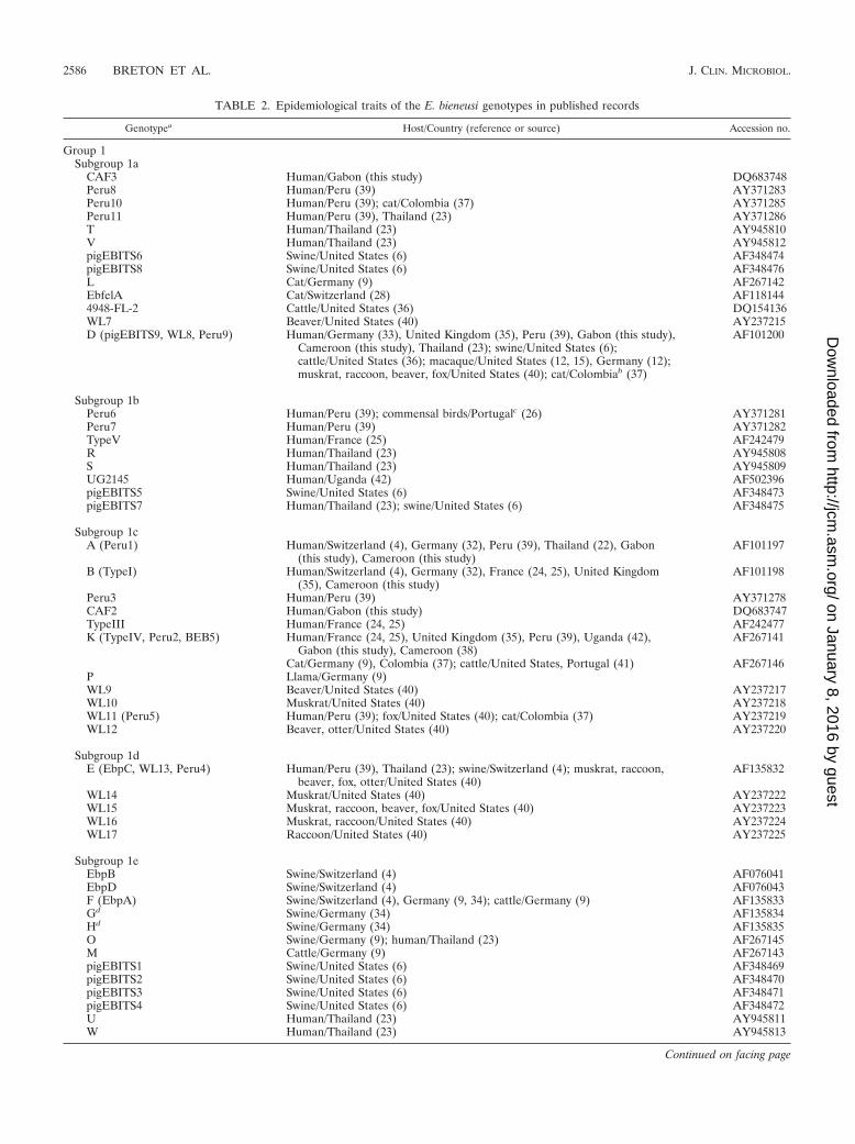
TABLE 2. Epidemiological traits of the E. bieneusi genotypes in published records
Genotypea Host/Country (reference or source) Accession no.
Group 1Subgroup 1a
CAF3 Human/Gabon (this study) DQ683748Peru8 Human/Peru (39) AY371283Peru10 Human/Peru (39); cat/Colombia (37) AY371285Peru11 Human/Peru (39), Thailand (23) AY371286T Human/Thailand (23) AY945810V Human/Thailand (23) AY945812pigEBITS6 Swine/United States (6) AF348474pigEBITS8 Swine/United States (6) AF348476L Cat/Germany (9) AF267142EbfelA Cat/Switzerland (28) AF1181444948-FL-2 Cattle/United States (36) DQ154136WL7 Beaver/United States (40) AY237215D (pigEBITS9, WL8, Peru9) Human/Germany (33), United Kingdom (35), Peru (39), Gabon (this study),
Cameroon (this study), Thailand (23); swine/United States (6);cattle/United States (36); macaque/United States (12, 15), Germany (12);muskrat, raccoon, beaver, fox/United States (40); cat/Colombiab (37)
AF101200
Subgroup 1bPeru6 Human/Peru (39); commensal birds/Portugalc (26) AY371281Peru7 Human/Peru (39) AY371282TypeV Human/France (25) AF242479R Human/Thailand (23) AY945808S Human/Thailand (23) AY945809UG2145 Human/Uganda (42) AF502396pigEBITS5 Swine/United States (6) AF348473pigEBITS7 Human/Thailand (23); swine/United States (6) AF348475
Subgroup 1cA (Peru1) Human/Switzerland (4), Germany (32), Peru (39), Thailand (22), Gabon
(this study), Cameroon (this study)AF101197
B (TypeI) Human/Switzerland (4), Germany (32), France (24, 25), United Kingdom(35), Cameroon (this study)
AF101198
Peru3 Human/Peru (39) AY371278CAF2 Human/Gabon (this study) DQ683747TypeIII Human/France (24, 25) AF242477K (TypeIV, Peru2, BEB5) Human/France (24, 25), United Kingdom (35), Peru (39), Uganda (42),
Gabon (this study), Cameroon (38)AF267141
Cat/Germany (9), Colombia (37); cattle/United States, Portugal (41) AF267146P Llama/Germany (9)WL9 Beaver/United States (40) AY237217WL10 Muskrat/United States (40) AY237218WL11 (Peru5) Human/Peru (39); fox/United States (40); cat/Colombia (37) AY237219WL12 Beaver, otter/United States (40) AY237220
Subgroup 1dE (EbpC, WL13, Peru4) Human/Peru (39), Thailand (23); swine/Switzerland (4); muskrat, raccoon,
beaver, fox, otter/United States (40)AF135832
WL14 Muskrat/United States (40) AY237222WL15 Muskrat, raccoon, beaver, fox/United States (40) AY237223WL16 Muskrat, raccoon/United States (40) AY237224WL17 Raccoon/United States (40) AY237225
Subgroup 1eEbpB Swine/Switzerland (4) AF076041EbpD Swine/Switzerland (4) AF076043F (EbpA) Swine/Switzerland (4), Germany (9, 34); cattle/Germany (9) AF135833Gd Swine/Germany (34) AF135834Hd Swine/Germany (34) AF135835O Swine/Germany (9); human/Thailand (23) AF267145M Cattle/Germany (9) AF267143pigEBITS1 Swine/United States (6) AF348469pigEBITS2 Swine/United States (6) AF348470pigEBITS3 Swine/United States (6) AF348471pigEBITS4 Swine/United States (6) AF348472U Human/Thailand (23) AY945811W Human/Thailand (23) AY945813
Continued on facing page
2586 BRETON ET AL. J. CLIN. MICROBIOL.
on January 8, 2016 by guesthttp://jcm
.asm.org/
Dow
nloaded from
identified several times: four isolates (26%) in Gabon and onein Cameroon. Unlike types A and B, type K may be commonin developing countries but is rare in Europe (25, 35), beingfound in 6 out of the 10 isolates from children in Uganda (42),all 4 isolates from HIV-positive adults in Cameroon (38), and18 out of 89 patients in Peru (39). However, type K, whichinitially was identified from HIV-positive individuals as type IVin France (24), is not strictly anthroponotic, since it also hasbeen identified from cattle in the United States and Portugal(41) and from cats in Germany (9) and Colombia (37).
Interestingly, we identified type D, a subgroup 1a genotypethat has not been found in immunocompetent humans yet, inone HIV-positive patient in Gabon and in three HIV-negativeindividuals in Cameroon. Type D has a large host and geo-graphic range. It is now commonly reported for HIV-positivehumans (22 of 33 isolates in Thailand [23] and 9 of 89 patientsin Peru [39]), and it was previously reported for two isolatedcases in Europe (33, 35). It has been found in a wide variety ofdomestic and wild mammals (6, 12, 15, 36, 37, 40) and repre-sented 15% of isolates from four species of wildlife animals inNorth America (40) and 26% of isolates found in cats inColombia (37), supporting a zoonotic route of transmission forthis strain.
Like type K, type E, which was not found in our studies, hasbeen reported from HIV-positive humans: one patient in Peru(39) and 5 of 33 cases in Thailand (23). Previously reported asgenotype EbpC from swine from Switzerland (4), type E was
the most common strain from wild mammals in the UnitedStates (22% of isolates from five species [40]).
Our new sequences CAF1, CAF2, and CAF3 are close rel-atives of types E, K, and D, respectively. These families ofgenotypes dominate the isolates found in the HIV-positivepatients from Gabon, with 10 out of 15 isolates, whereas in theCameroon study types K and D represented only 4 out of 20isolates.
Finally, the new genotype CAF4, the most highly divergentgenotype reported from humans to date, is equally representedin HIV-positive patients in Gabon and HIV-negative individ-uals in Cameroon. Its high frequency (�25%) in both coun-tries may indicate that this genotype is common in CentralAfrica. The relative proximity of the CAF4 genotype to geno-types found in wild aquatic mammals could indicate that sim-ilar hosts exist in Central Africa.
Epidemiological implications. The analysis of E. bieneusigenotypes highlights different risks of infection in the two hu-man populations studied, possibly due to differences in sourcesof contamination and routes of transmission. In the rural com-munity of Cameroon, anthroponotic genotypes A and B pre-dominate and may indicate person-to-person transmission,possibly involving contact with contaminated water stored forhousehold use. This is supported by the observation that car-riers from the same household have parasites with the samegenotype (genotype A for cases 113B, 113H, 117A/117B, and142G/142H, genotype B for cases 194A/194C, and genotype
TABLE 2—Continued
Genotypea Host/Country (reference or source) Accession no.
Subgroup 1fC (TypeII) Human/France (24, 25), Switzerland (4), Germany (9) AF101199Q Human/Germany (9) AF267147
Subgroup 1gCAF1 Human/Gabon (this study) DQ683746
Group 2I Cattle/Germany (9, 34) AF135836J (BEB1) Cattle/Germany (9, 34), United States (36, 41); chicken/Germany (31) AF135837N (BEB2) Cattle/Germany (9), United States (36, 41) AF267144BEB3 Cattle/United States (36, 41) AY331007BEB4 Cattle/United States (36, 41) AY331008
Group 3WL4 Muskrat/United States (40) AY237212WL5 Muskrat/United States (40) AY237213WL6 Muskrat/United States (40) AY237214
Group 4WL1 Raccoon/United States (40) AY237209WL2 Raccoon/United States (40) AY237210WL3 Raccoon/United States (40) AY237211
OutliersCAF4 Human/Gabon, Cameroon (this study) DQ683749EntCanA Dog/Switzerland (28) AF059610
a Abbreviations in parentheses refer to the other genotype names assigned to identical sequences by various research teams. Accession numbers refer to the firstgenotype name.
b Reported as genotype D-like.c Reported as genotype Peru6 and genotype Peru6-var.d Isolated from the same animal.
VOL. 45, 2007 E. BIENEUSI GENOTYPES FROM CENTRAL AFRICA 2587
on January 8, 2016 by guesthttp://jcm
.asm.org/
Dow
nloaded from
CAF4 for cases 179D/179E) (Table 1). In the study of HIV-positive individuals from Gabon, the wider diversity of geno-types and the higher proportion of mixed-host genotypes Dand K (and the close relatives CAF1 to CAF3) suggest thatimmunocompromised patients acquire infection from addi-tional reservoirs (probably of zoonotic origin).
Overall, the epidemiology of E. bieneusi is still unclear,probably due to the fact that at present different species orsubspecies of this parasite are not differentiated by classicalmicroscopy and immunological methods, stressing the need forfurther molecular studies.
ACKNOWLEDGMENTS
We thank the population of the village Bongo and the patients ofLibreville for participation in this study, M. Owono-Medang and E.Mozogho for excellent technical assistance, C. Djengue and G.Obame-Mebale for sample collection in Libreville, and A. Arnaud forsample collection in Cameroon. We also thank H. Rinder (Universityof Munich) for initial help with ITS sequencing and I. Desportes-Livage (MNHN, Paris, France) for helpful discussions.
This work was supported by a grant from the French National AIDSResearch Agency (PED ANRS-1264/MAE).
REFERENCES
1. Accoceberry, I., M. Thellier, I. Desportes-Livage, A. Achbarou, S. Biligui, M.Danis, and A. Datry. 1999. Production of monoclonal antibodies directedagainst the microsporidium Enterocytozoon bieneusi. J. Clin. Microbiol. 37:4107–4112.
2. Alfa Cisse, O., A. Ouattara, M. Thellier, I. Accoceberry, S. Biligui, D. Minta,O. Doumbo, I. Desportes-Livage, M. A. Thera, M. Danis, and A. Datry. 2002.Evaluation of an immunofluorescent-antibody test using monoclonal anti-bodies directed against Enterocytozoon bieneusi and Encephalitozoon intesti-nalis for diagnosis of intestinal microsporidiosis in Bamako (Mali). J. Clin.Microbiol. 40:1715–1718.
3. Bern, C., V. Kawai, D. Vargas, J. Rabke-Verani, J. Williamson, R. Chavez-Valdez, L. Xiao, I. Sulaiman, A. Vivar, E. Ticona, M. Navincopa, V. Cama,H. Moura, W. E. Secor, G. Visvesvara, and R. H. Gilman. 2005. The epide-miology of intestinal microsporidiosis in patients with HIV/AIDS in Lima,Peru. J. Infect. Dis. 191:1658–1664.
4. Breitenmoser, A. C., A. Mathis, E. Burgi, R. Weber, and P. Deplazes. 1999.High prevalence of Enterocytozoon bieneusi in swine with four genotypes thatdiffer from those identified in humans. Parasitology 118:447–453.
5. Bretagne, S., F. Foulet, W. Alkassoum, J. Fleury-Feith, and M. Develoux.1993. Prevalence des spores d’Enterocytozoon bieneusi dans les selles depatients sideens et d’enfants Africains non infectes par le VIH. Bull. Soc.Pathol. Exot. 86:351–357.
6. Buckholt, M. A., J. H. Lee, and S. Tzipori. 2002. Prevalence of Entero-cytozoon bieneusi in swine: an 18-month survey at a slaughterhouse inMassachusetts. Appl. Environ. Microbiol. 68:2595–2599.
7. Cali, A., D. P. Kotler, and J. M. Orenstein. 1993. Septata intestinalis n. g., n.sp., an intestinal microsporidian associated with chronic diarrhea and dis-semination in AIDS patients. J. Eukaryot. Microbiol. 40:101–112.
8. Corpet, F. 1988. Multiple sequence alignment with hierarchical clustering.Nucleic Acids Res. 16:10881–10890.
9. Dengjel, B., M. Zahler, W. Hermanns, K. Heinritzi, T. Spillmann, A.Thomschke, T. Loscher, R. Gothe, and H. Rinder. 2001. Zoonotic potentialof Enterocytozoon bieneusi. J. Clin. Microbiol. 39:4495–4499.
10. Desportes, I., Y. Le Charpentier, A. Galian, F. Bernard, B. Cochand-Priollet,A. Lavergne, P. Ravisse, and R. Modigliani. 1985. Occurrence of a newmicrosporidan: Enterocytozoon bieneusi n. g., n. sp., in the enterocytes of ahuman patient with AIDS. J. Protozool. 32:250–254.
11. Didier, E. S., and L. M. Weiss. 2006. Microsporidiosis: current status. Curr.Opin. Infect. Dis. 19:485–492.
12. Drosten, C., J. Laabs, E. M. Kuhn, and J. Schottelius. 2005. Interspeciestransmission of Enterocytozoon bieneusi supported by observations in labo-ratory animals and phylogeny. Med. Microbiol. Immunol. (Berlin) 194:207–209.
13. Endeshaw, T., A. Kebede, J. J. Verweij, D. Wolday, A. Zewide, K. Tsige, Y.Abraham, T. Messele, A. M. Polderman, and B. Petros. 2005. Detection ofintestinal microsporidiosis in diarrheal patients infected with the humanimmunodeficiency virus (HIV-1) using PCR and Uvitex-2B stain. Ethiop.Med. J. 43:97–101.
14. Endeshaw, T., A. Kebede, J. J. Verweij, A. Zewide, K. Tsige, Y. Abraham, D.Wolday, T. Woldemichael, T. Messele, A. M. Polderman, and B. Petros.2006. Intestinal microsporidiosis in diarrheal patients infected with human
immunodeficiency virus-1 in Addis Ababa, Ethiopia. Jpn. J. Infect. Dis.59:306–310.
15. Green, L. C., P. J. Didier, L. C. Bowers, and E. S. Didier. 2004. Natural andexperimental infection of immunocompromised rhesus macaques (Macacamulatta) with the microsporidian Enterocytozoon bieneusi genotype D.Microbes Infect. 6:996–1002.
16. Guindon, S., and O. Gascuel. 2003. A simple, fast, and accurate algorithm toestimate large phylogenies by maximum likelihood. Syst. Biol. 52:696–704.
17. Gumbo, T., S. Sarbah, I. T. Gangaidzo, Y. Ortega, C. R. Sterling, A. Carville,S. Tzipori, and P. M. Wiest. 1999. Intestinal parasites in patients withdiarrhea and human immunodeficiency virus infection in Zimbabwe. AIDS13:819–821.
18. Hartskeerl, R. A., T. Van Gool, A. R. Schuitema, E. S. Didier, and W. J.Terpstra. 1995. Genetic and immunological characterization of the micro-sporidian Septata intestinalis Cali, Kotler and Orenstein, 1993: reclassifica-tion to Encephalitozoon intestinalis. Parasitology 110:277–285.
19. Katzwinkel-Wladarsch, S., M. Lieb, W. Helse, T. Loscher, and H. Rinder.1996. Direct amplification and species determination of microsporidianDNA from stool specimens. Trop. Med. Int. Health 1:373–378.
20. Kokoskin, E., T. W. Gyorkos, A. Camus, L. Cedilotte, T. Purtill, and B.Ward. 1994. Modified technique for efficient detection of microsporidia.J. Clin. Microbiol. 32:1074–1075.
21. Lebbad, M., H. Norrgren, A. Naucler, F. Dias, S. Andersson, and E. Linder.2001. Intestinal parasites in HIV-2 associated AIDS cases with chronicdiarrhea in Guinea-Bissau. Acta Trop. 80:45–49.
22. Leelayoova, S., I. Subrungruang, R. Rangsin, P. Chavalitshewinkoon-Petmitr, J. Worapong, T. Naaglor, and M. Mungthin. 2005. Transmission ofEnterocytozoon bieneusi genotype a in a Thai orphanage. Am. J. Trop. Med.Hyg. 73:104–107.
23. Leelayoova, S., I. Subrungruang, Y. Suputtamongkol, J. Worapong, P. C.Petmitr, and M. Mungthin. 2006. Identification of genotypes of Enterocyto-zoon bieneusi from stool samples from human immunodeficiency virus-in-fected patients in Thailand. J. Clin. Microbiol. 44:3001–3004.
24. Liguory, O., F. David, C. Sarfati, F. Derouin, and J. M. Molina. 1998.Determination of types of Enterocytozoon bieneusi strains isolated from pa-tients with intestinal microsporidiosis. J. Clin. Microbiol. 36:1882–1885.
25. Liguory, O., C. Sarfati, F. Derouin, and J. M. Molina. 2001. Evidence ofdifferent Enterocytozoon bieneusi genotypes in patients with and withouthuman immunodeficiency virus infection. J. Clin. Microbiol. 39:2672–2674.
26. Lobo, M. L., L. Xiao, V. Cama, N. Magalhaes, F. Antunes, and O. Matos.2006. Identification of potentially human-pathogenic Enterocytozoon bieneusigenotypes in various birds. Appl. Environ. Microbiol. 11:7380–7382.
27. Maiga, I., O. Doumbo, M. Dembele, H. Traore, I. Desportes-Livage, I.Hilmarsdottir, E. Giboyau, L. Maiga, L. Kassambara, Y. el Fakhry, A. Datry,M. Gentilini, and E. Pichard. 1997. Microsporidiose intestinale humainea Bamako (Mali): presence d’Enterocytozoon bieneusi chez les patientsseropositifs pour le VIH. Sante 7:257–262.
28. Mathis, A., A. C. Breitenmoser, and P. Deplazes. 1999. Detection of newEnterocytozoon genotypes in faecal samples of farm dogs and a cat. Parasite6:189–193.
29. Mathis, A., R. Weber, and P. Deplazes. 2005. Zoonotic potential of themicrosporidia. Clin. Microbiol. Rev. 18:423–445.
30. O’Keefe, E. A., and R. Wood. 1996. AIDS in Africa. Scand. J. Gastroenterol.220:147–152.
31. Reetz, J., H. Rinder, A. Thomschke, H. Manke, M. Schwebs, and A.Bruderek. 2002. First detection of the microsporidium Enterocytozoonbieneusi in non-mammalian hosts (chickens). Int. J. Parasitol. 32:785–787.
32. Rinder, H., S. Katzwinkel-Wladarsch, and T. Loscher. 1997. Evidence forthe existence of genetically distinct strains of Enterocytozoon bieneusi. Para-sitol. Res. 83:670–672.
33. Rinder, H., S. Katzwinkel-Wladarsch, A. Thomschke, and T. Loscher. 1998.Strain differentiation in microsporidia. Tokai J. Exp. Clin. Med. 23:433–437.
34. Rinder, H., A. Thomschke, B. Dengjel, R. Gothe, T. Loscher, and M. Zahler.2000. Close genotypic relationship between Enterocytozoon bieneusi fromhumans and pigs and first detection in cattle. J. Parasitol. 86:185–188.
35. Sadler, F., N. Peake, R. Borrow, P. L. Rowl, E. G. Wilkins, and A. Curry.2002. Genotyping of Enterocytozoon bieneusi in AIDS patients from thenorth west of England. J. Infect. 44:39–42.
36. Santin, M., J. M. Trout, and R. Fayer. 2005. Enterocytozoon bieneusi geno-types in dairy cattle in the eastern United States. Parasitol. Res. 97:535–538.
37. Santin, M., J. M. Trout, J. A. Vecino, J. P. Dubey, and R. Fayer. 2006.Cryptosporidium, Giardia and Enterocytozoon bieneusi in cats from Bogota(Colombia) and genotyping of isolates. Vet. Parasitol. 141:334–339.
38. Sarfati, C., A. Bourgeois, J. Menotti, F. Liegeois, R. Moyou-Somo, E.Delaporte, F. Derouin, E. M. Ngole, and J. M. Molina. 2006. Prevalence ofintestinal parasites including microsporidia in human immunodeficiency vi-rus-infected adults in Cameroon: a cross-sectional study. Am. J. Trop. Med.Hyg. 74:162–164.
39. Sulaiman, I. M., C. Bern, R. Gilman, V. Cama, V. Kawai, D. Vargas, E.Ticona, A. Vivar, and L. Xiao. 2003. A molecular biologic study of Entero-cytozoon bieneusi in HIV-infected patients in Lima, Peru. J. Eukaryot. Mi-crobiol. 50:591–596.
2588 BRETON ET AL. J. CLIN. MICROBIOL.
on January 8, 2016 by guesthttp://jcm
.asm.org/
Dow
nloaded from
40. Sulaiman, I. M., R. Fayer, A. A. Lal, J. M. Trout, F. W. Schaefer III, and L. Xiao.2003. Molecular characterization of microsporidia indicates that wild mammalsharbor host-adapted Enterocytozoon spp. as well as human-pathogenic Entero-cytozoon bieneusi. Appl. Environ. Microbiol. 69:4495–4501.
41. Sulaiman, I. M., R. Fayer, C. Yang, M. Santin, O. Matos, and L. Xiao. 2004.Molecular characterization of Enterocytozoon bieneusi in cattle indicates thatonly some isolates have zoonotic potential. Parasitol. Res. 92:328–334.
42. Tumwine, J. K., A. Kekitiinwa, N. Nabukeera, D. E. Akiyoshi, M. A.Buckholt, and S. Tzipori. 2002. Enterocytozoon bieneusi among childrenwith diarrhea attending Mulago Hospital in Uganda. Am. J. Trop. Med.Hyg. 67:299–303.
43. van Gool, T., E. Luderhoff, K. J. Nathoo, C. F. Kiire, J. Dankert, and P. R.Mason. 1995. High prevalence of Enterocytozoon bieneusi infections amongHIV-positive individuals with persistent diarrhoea in Harare, Zimbabwe.Trans. R. Soc. Trop. Med. Hyg. 89:478–480.
44. Weiss, L. M., X. Zhu, A. Cali, H. B. Tanowitz, and M. Wittner. 1994. Utilityof microsporidian rRNA in diagnosis and phylogeny: a review. Folia Para-sitol. (Praha) 41:81–90.
45. Zhu, X., M. Wittner, H. B. Tanowitz, D. Kotler, A. Cali, and L. M. Weiss.1993. Small subunit rRNA sequence of Enterocytozoon bieneusi and its po-tential diagnostic role with use of the polymerase chain reaction. J. Infect.Dis. 168:1570–1575.
VOL. 45, 2007 E. BIENEUSI GENOTYPES FROM CENTRAL AFRICA 2589
on January 8, 2016 by guesthttp://jcm
.asm.org/
Dow
nloaded from
Copyright © 2022 FDOKUMEN