Activity profiles for marine sponge-associated bacteria obtained by 16S rRNA vs 16S rRNA gene...
11
ORIGINAL ARTICLE Activity profiles for marine sponge-associated bacteria obtained by 16S rRNA vs 16S rRNA gene comparisons Janine Kamke, Michael W Taylor and Susanne Schmitt School of Biological Sciences, The University of Auckland, Auckland, New Zealand The phylogenetic diversity of microorganisms in marine sponges is becoming increasingly well described, yet relatively little is known about the activities of these symbionts. Given the seemingly favourable environment provided to microbes by their sponge hosts, as indicated by the extra- ordinarily high abundance of sponge symbionts, we hypothesized that the majority of sponge- associated bacteria are active in situ. To test this hypothesis we compared, for the first time in sponges, 16S rRNA gene- vs 16S rRNA-derived bacterial community profiles to gain insights into symbiont composition and activity, respectively. Clone libraries revealed a highly diverse bacterial community in Ancorina alata, and a much lower diversity in Polymastia sp., which were identified by electron microscopy as a high- and a low-microbial abundance sponge, respectively. Substantial overlap between DNA and RNA libraries was evident at both phylum and phylotype levels, indicating in situ activity for a large fraction of sponge-associated bacteria. This active fraction included uncultivated, sponge-specific lineages within, for example, Actinobacteria, Chloroflexi and Gemmatimonadetes. This study shows the potential of RNA vs DNA comparisons based on the 16S rRNA gene to provide insights into the activity of sponge-associated microorganisms. The ISME Journal (2010) 4, 498–508; doi:10.1038/ismej.2009.143; published online 7 January 2010 Subject Category: Microbe–microbe and microbe–host interactions Keywords: activity; bacteria; marine sponges; microbial symbionts; 16S rRNA Introduction Many marine sponges harbour dense and diverse microbial communities of considerable ecological and biotechnological importance (Hentschel et al., 2006; Taylor et al., 2007b). These communities, which can include bacteria, archaea and eukaryotic microorganisms, are often quite specific to sponges, with many microbial phylotypes appearing to live exclusively within sponge hosts and not in the surrounding seawater (Hentschel et al., 2002; Taylor et al., 2007b; Schmitt et al., 2008). Although our understanding of the microbial diversity in sponges is rapidly improving, much remains unknown about the activity of these microbes (Taylor et al., 2007a). Specific microbially mediated processes within sponges, such as photosynthesis, sulphate reduc- tion, nitrogen fixation and nitrification, have been quantified and in many cases the relevant microbes have been identified (Wilkinson and Fay, 1979; Wilkinson, 1983; Diaz and Ward, 1997; Wilkinson et al., 1999; Hoffmann et al., 2005, 2009; Hallam et al., 2006; Bayer et al., 2008; Mohamed et al., 2008a, 2009; Steger et al., 2008). These studies, which have utilized methods such as isotope enrichments, metagenomics and functional gene analyses, have extended our knowledge of symbiont function in sponges, yet they remain focused on specific pro- cesses or particular functional groups of organisms. What is lacking to date is a community-wide assess- ment of microbial activity in marine sponges. This would be useful, as identification of those sponge- associated microbes that are active in situ is an important step towards elucidating their ecological role and contribution to the host. All living organisms contain ribosomal RNA, and these molecules have become the gold standard for microbial ecology and taxonomy (Ludwig and Schleifer, 1999; Tringe and Hugenholtz, 2008). The analysis of 16S rRNA genes through clone libraries and fingerprinting approaches such as denaturing gradient gel electrophoresis (DGGE) has greatly extended our knowledge about the phylogenetic richness of sponge-associated bacteria and archaea (Webster et al., 2001, 2004; Hentschel et al., 2002; Taylor et al., 2004, 2007b; Holmes and Blanch, 2006; Longford et al., 2007; Schmitt et al., 2007, 2008; Thiel et al., 2007; Mohamed et al., 2008b; Zhu et al., 2008). Researchers in other systems have taken this approach one step further, yielding insights into both richness and activity by comparing 16S rRNA gene- and 16S rRNA-derived sequences, Received 5 October 2009; revised 25 November 2009; accepted 28 November 2009; published online 7 January 2010 Correspondence: MW Taylor, School of Biological Sciences, The University of Auckland, Private Bag 92019, Auckland 1142, New Zealand. E-mail: [email protected] The ISME Journal (2010) 4, 498–508 & 2010 International Society for Microbial Ecology All rights reserved 1751-7362/10 $32.00 www.nature.com/ismej
-
Upload
independent -
Category
Documents
-
view
2 -
download
0
Transcript of Activity profiles for marine sponge-associated bacteria obtained by 16S rRNA vs 16S rRNA gene...

ORIGINAL ARTICLE
Activity profiles for marine sponge-associatedbacteria obtained by 16S rRNA vs 16S rRNA genecomparisons
Janine Kamke, Michael W Taylor and Susanne SchmittSchool of Biological Sciences, The University of Auckland, Auckland, New Zealand
The phylogenetic diversity of microorganisms in marine sponges is becoming increasingly welldescribed, yet relatively little is known about the activities of these symbionts. Given the seeminglyfavourable environment provided to microbes by their sponge hosts, as indicated by the extra-ordinarily high abundance of sponge symbionts, we hypothesized that the majority of sponge-associated bacteria are active in situ. To test this hypothesis we compared, for the first time insponges, 16S rRNA gene- vs 16S rRNA-derived bacterial community profiles to gain insights intosymbiont composition and activity, respectively. Clone libraries revealed a highly diverse bacterialcommunity in Ancorina alata, and a much lower diversity in Polymastia sp., which were identified byelectron microscopy as a high- and a low-microbial abundance sponge, respectively. Substantialoverlap between DNA and RNA libraries was evident at both phylum and phylotype levels, indicatingin situ activity for a large fraction of sponge-associated bacteria. This active fraction includeduncultivated, sponge-specific lineages within, for example, Actinobacteria, Chloroflexi andGemmatimonadetes. This study shows the potential of RNA vs DNA comparisons based on the16S rRNA gene to provide insights into the activity of sponge-associated microorganisms.The ISME Journal (2010) 4, 498–508; doi:10.1038/ismej.2009.143; published online 7 January 2010Subject Category: Microbe–microbe and microbe–host interactionsKeywords: activity; bacteria; marine sponges; microbial symbionts; 16S rRNA
Introduction
Many marine sponges harbour dense and diversemicrobial communities of considerable ecologicaland biotechnological importance (Hentschel et al.,2006; Taylor et al., 2007b). These communities,which can include bacteria, archaea and eukaryoticmicroorganisms, are often quite specific to sponges,with many microbial phylotypes appearing to liveexclusively within sponge hosts and not in thesurrounding seawater (Hentschel et al., 2002; Tayloret al., 2007b; Schmitt et al., 2008). Although ourunderstanding of the microbial diversity in spongesis rapidly improving, much remains unknown aboutthe activity of these microbes (Taylor et al., 2007a).Specific microbially mediated processes withinsponges, such as photosynthesis, sulphate reduc-tion, nitrogen fixation and nitrification, have beenquantified and in many cases the relevant microbeshave been identified (Wilkinson and Fay, 1979;Wilkinson, 1983; Diaz and Ward, 1997; Wilkinsonet al., 1999; Hoffmann et al., 2005, 2009; Hallamet al., 2006; Bayer et al., 2008; Mohamed et al., 2008a,
2009; Steger et al., 2008). These studies, which haveutilized methods such as isotope enrichments,metagenomics and functional gene analyses, haveextended our knowledge of symbiont function insponges, yet they remain focused on specific pro-cesses or particular functional groups of organisms.What is lacking to date is a community-wide assess-ment of microbial activity in marine sponges. Thiswould be useful, as identification of those sponge-associated microbes that are active in situ is animportant step towards elucidating their ecologicalrole and contribution to the host.
All living organisms contain ribosomal RNA, andthese molecules have become the gold standard formicrobial ecology and taxonomy (Ludwig andSchleifer, 1999; Tringe and Hugenholtz, 2008). Theanalysis of 16S rRNA genes through clone librariesand fingerprinting approaches such as denaturinggradient gel electrophoresis (DGGE) has greatlyextended our knowledge about the phylogeneticrichness of sponge-associated bacteria and archaea(Webster et al., 2001, 2004; Hentschel et al., 2002;Taylor et al., 2004, 2007b; Holmes and Blanch, 2006;Longford et al., 2007; Schmitt et al., 2007, 2008;Thiel et al., 2007; Mohamed et al., 2008b; Zhu et al.,2008). Researchers in other systems have takenthis approach one step further, yielding insightsinto both richness and activity by comparing 16SrRNA gene- and 16S rRNA-derived sequences,
Received 5 October 2009; revised 25 November 2009; accepted 28November 2009; published online 7 January 2010
Correspondence: MW Taylor, School of Biological Sciences, TheUniversity of Auckland, Private Bag 92019, Auckland 1142, NewZealand. E-mail: [email protected]
The ISME Journal (2010) 4, 498–508& 2010 International Society for Microbial Ecology All rights reserved 1751-7362/10 $32.00
www.nature.com/ismej

respectively (Moeseneder et al., 2001, 2005; Winteret al., 2001; Troussellier et al., 2002; Mills et al.,2005; Gentile et al., 2006; Martinez et al., 2006;Brinkmann et al., 2008; McIlroy et al., 2008; Westet al., 2008; Rodriguez-Blanco et al., 2009). Ingeneral, cellular concentrations of rRNA are corre-lated with growth rate and activity (DeLong et al.,1989; Poulsen et al., 1993), hence—with acknowl-edgement of certain caveats (e.g., for ammonia-oxidizing bacteria; Morgenroth et al., 2000)—therRNA itself can yield useful information aboutwhich community members are active.
In this study we investigated bacterial communitycomposition (16S rRNA gene) and activity (16SrRNA) in two marine sponges from north-eastern New Zealand. Clone libraries, generatinga total of 313 sequences, were constructed from thehigh-microbial-abundance sponge Ancorina alata(Demospongiae: Astrophorida: Ancorinidae) andthe low-microbial-abundance sponge Polymastia sp.(Demospongiae: Hadromerida: Polymastiidae). Theexistence of both high- and low-microbial-abundancesponges is well documented (Vacelet and Donadey,1977; Reiswig, 1981; Hentschel et al., 2006; Weiszet al., 2008), although the exact reasons for thesedifferences in microbial loads are uncertain. Inaddition to the well-characterized taxa, such as theAlpha- and Gammaproteobacteria, activity wasinferred for uncultivated, sponge-specific lineageswithin phyla, including the Gemmatimonadetes,Chloroflexi and a taxon of uncertain affiliation relatedto the sponge-specific ‘Poribacteria’ (Fieseler et al.,2004). Moreover, our results show the potential ofrRNA gene vs rRNA comparisons to provide insightsinto the activity of sponge-associated microorganisms.
Materials and methods
Sponge samplingSmall samples were taken from three individualsof each of the sponges A. alata and Polymastia sp.(both class Demospongiae). Sampling was carriedout by SCUBA diving at depths of 3–10 m atMathesons Bay (361180S, 1741470E) and Jones Bay(361230S, 1741490E), northeastern New Zealand, inNovember/December 2008. Tissue samples weretransferred into RNAlater (Applied Biosystems/Ambion, Foster City, CA, USA), then transportedto the laboratory on ice before freezing at �80 1C.Samples were subsequently freeze-dried and storedagain at �80 1C. Tissue samples for electron micro-scopy were cut into small pieces of about 1 mm3,fixed in 2.5% glutaraldehyde double-distilled water,and kept overnight at 4 1C before processing.
Transmission electron microscopy (TEM)Fixed sponge samples (three individuals persponge) were washed five times in cacodylate buffer(50 mM, pH 7.2), fixed in 2% osmium tetroxide for90 min, washed five times in double-distilled water,
and incubated overnight in 0.5% uranyl acetate.After dehydration in an ethanol series (30%, 50%,70%, 90%, 96%, and three times at 100% for 30 mineach), samples were incubated three times for30 min in 1� propylene oxide, maintained over-night in 1:1 (vol/vol) propylene oxide-Epon 812(EM bed-812, Electron Microscopy Science,Hatfield, PA, USA), incubated twice for 2 h in Epon812, and finally embedded in Epon 812 for 48 h at60 1C. Samples were then sectioned with an ultra-microtome (Leica, Wetzlar, Germany; EM UC6) andexamined by TEM (Philips CM12, Fei Company,Hillsboro, OR, USA).
Nucleic acid extraction and construction of 16S rRNAgene/rRNA clone librariesTotal DNA and RNA were co-extracted from 5–6 mgof freeze-dried sponge tissue using the AllPrepDNA/RNA mini kit (Qiagen, Hilden, Germany).Extractions were performed separately for all threeindividuals of both species (A. alata and Polymastiasp.). The RNA extract was subsequently purified byDNA digestion for 60 min using RQ1 RNase-freeDNase (Promega, Madison, WI, USA) and RNA wasreverse-transcribed into cDNA using random hexa-mers in the SuperScript III First Strand SynthesisSystem (Invitrogen, Carlsbad, CA, USA). After-wards, DNA, cDNA and RNA were PCR-amplifiedwith the universal bacterial primers 616V (50-AGAGTTTGATYMTGGCTC-30) and 1492R (50-GGYTACCTTGTTACGACTT-30) (Kane et al., 1993;Juretschko et al., 1998), spanning a B1500 bp regionof the 16S rRNA gene. The RNA template served as acontrol in the PCR and did not give any products.Cycling conditions on a T1 Thermocycler (Biometra,Goettingen, Germany) were as follows: initial dena-turing step at 95 1C for 5 min, 30 cycles of denaturingat 95 1C for 1 min, primer annealing at 54 1C for 1 minand elongation at 72 1C for 90 s, followed by a finalextension step at 72 1C for 10 min. The PCR productsfrom each sponge species representing the respec-tive DNA and cDNA fractions were pooled andligated into the pGEM-T-easy vector (Promega) andclone libraries were constructed according to themanufacturer’s instructions (four libraries in total).Pooling of PCR products was justified given thatcorresponding denaturing gradient gel electro-phoresis profiles based on DNA and RNA revealedhigh inter-individual similarities (data not shown).Clones that contained an insert (as evaluated byblue/white colony screening) were grown overnightat 37 1C. After a lysis step for 30 min at 94 1C, a PCRwas performed with the vector-specific primersPGEM-F and PGEM-R (Aislabie et al., 2009), todetermine which clones contained a correct-sizedinsert. Cycling conditions were the same as de-scribed above. In total, 375 (192 for Polymastia sp.and 183 for A. alata) PCR products were sequencedby Macrogen (Seoul, Korea), and, after the removalof poor quality sequences and chimeric sequences
Activity profiles for marine sponge-associated bacteriaJ Kamke et al
499
The ISME Journal
(n¼ 62) detected with Pintail (Ashelford et al.,2005), the final sequence data (81 near-full length(41200 bp) and 232 partial sequences) were sub-mitted to the DDBJ/EMBL/GenBank databases underaccession numbers FJ900272–FJ900584.
Statistical and phylogenetic analysesThe nonparametric richness estimator Chao1, used toevaluate how much of the richness in each library wassequenced, was calculated at different operationaltaxonomic unit (OTU) thresholds using DOTUR im-plented in Mothur (Schloss and Handelsman, 2005).Preliminary phylogenetic affiliations were obtained forall DNA- and cDNA (RNA)-derived sequences usingthe NCBI’s BLAST server (http://blast.ncbi.nlm.nih.gov/Blast.cgi). Sequences from A. alata and Polymastiasp., as well as their closest relatives identified byBLAST, were aligned using the web-based SINAaligner, then imported into a 16S rRNA ARB-SILVAdatabase (Ludwig et al., 2004; Pruesse et al., 2007)(version 96, containing 271543 bacterial rRNA se-quences) for subsequent manual refinement of thealignment (inspection of the automatic alignment byeye and manual corrections where appropriate usingthe editor tool in the ARB software package). Maximumlikelihood, maximum parsimony and neighbor-joiningtrees were calculated in ARB using long (X1200 bp)sequences only. Shorter sequences were added usingthe parsimony interactive tool in ARB without alteringthe tree topology. Phylogenetic consensus trees, usingthe maximum likelihood tree as a backbone, weremanually constructed (Ludwig et al., 1998). Maximumparsimony bootstraps (1000 resamplings) were per-formed to further assess the stability of observedbranching patterns.
Results
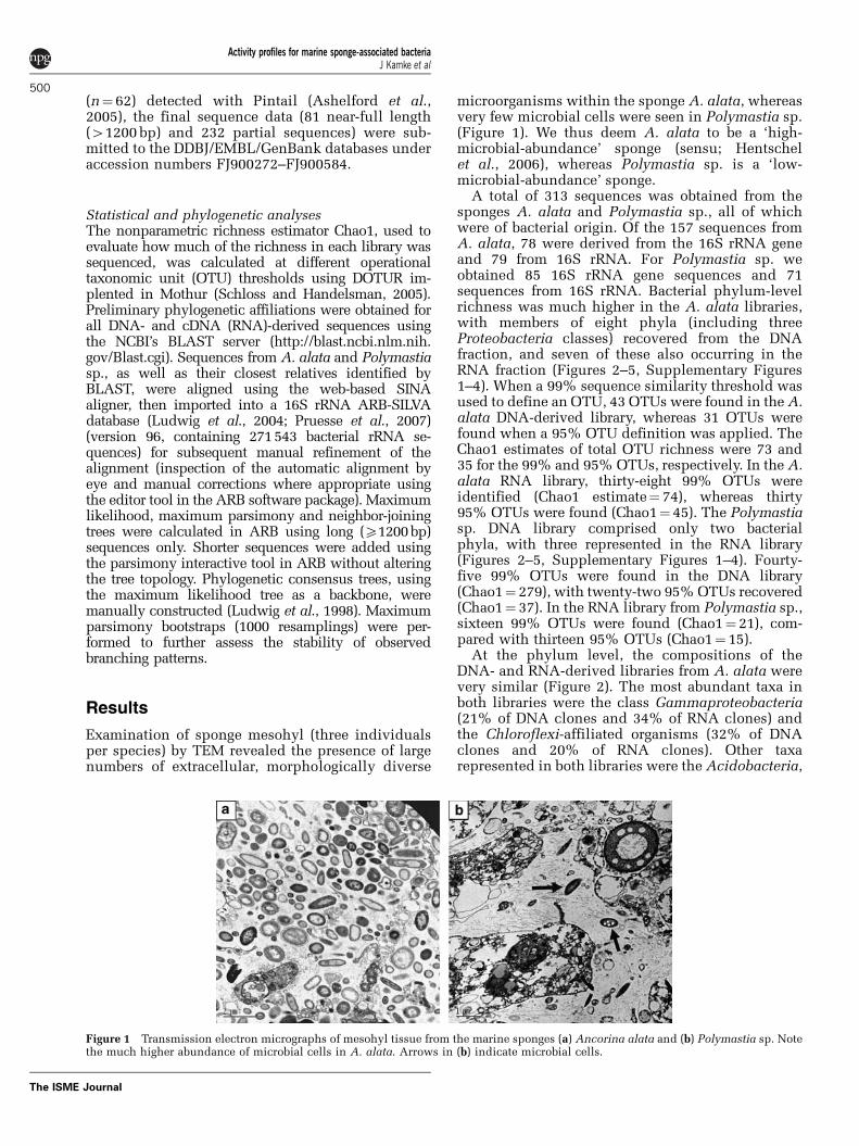
Examination of sponge mesohyl (three individualsper species) by TEM revealed the presence of largenumbers of extracellular, morphologically diverse
microorganisms within the sponge A. alata, whereasvery few microbial cells were seen in Polymastia sp.(Figure 1). We thus deem A. alata to be a ‘high-microbial-abundance’ sponge (sensu; Hentschelet al., 2006), whereas Polymastia sp. is a ‘low-microbial-abundance’ sponge.
A total of 313 sequences was obtained from thesponges A. alata and Polymastia sp., all of whichwere of bacterial origin. Of the 157 sequences fromA. alata, 78 were derived from the 16S rRNA geneand 79 from 16S rRNA. For Polymastia sp. weobtained 85 16S rRNA gene sequences and 71sequences from 16S rRNA. Bacterial phylum-levelrichness was much higher in the A. alata libraries,with members of eight phyla (including threeProteobacteria classes) recovered from the DNAfraction, and seven of these also occurring in theRNA fraction (Figures 2–5, Supplementary Figures1–4). When a 99% sequence similarity threshold wasused to define an OTU, 43 OTUs were found in the A.alata DNA-derived library, whereas 31 OTUs werefound when a 95% OTU definition was applied. TheChao1 estimates of total OTU richness were 73 and35 for the 99% and 95% OTUs, respectively. In the A.alata RNA library, thirty-eight 99% OTUs wereidentified (Chao1 estimate¼ 74), whereas thirty95% OTUs were found (Chao1¼ 45). The Polymastiasp. DNA library comprised only two bacterialphyla, with three represented in the RNA library(Figures 2–5, Supplementary Figures 1–4). Fourty-five 99% OTUs were found in the DNA library(Chao1¼ 279), with twenty-two 95% OTUs recovered(Chao1¼ 37). In the RNA library from Polymastia sp.,sixteen 99% OTUs were found (Chao1¼ 21), com-pared with thirteen 95% OTUs (Chao1¼ 15).
At the phylum level, the compositions of theDNA- and RNA-derived libraries from A. alata werevery similar (Figure 2). The most abundant taxa inboth libraries were the class Gammaproteobacteria(21% of DNA clones and 34% of RNA clones) andthe Chloroflexi-affiliated organisms (32% of DNAclones and 20% of RNA clones). Other taxarepresented in both libraries were the Acidobacteria,
Figure 1 Transmission electron micrographs of mesohyl tissue from the marine sponges (a) Ancorina alata and (b) Polymastia sp. Notethe much higher abundance of microbial cells in A. alata. Arrows in (b) indicate microbial cells.
Activity profiles for marine sponge-associated bacteriaJ Kamke et al
500
The ISME Journal

Actinobacteria, Bacteroidetes, Gemmatimonadetes,Alpha- and Deltaproteobacteria, and a lineage ofuncertain affiliation that seems to fall within thePlanctomycetes–Verrucomicrobia–Chlamydiae (PVC)superphylum (Wagner and Horn, 2006). The onlymajor taxon to be recovered from only one librarywas the Nitrospirae, for which a single sequencewas found in the DNA-derived library. Phylogenetic
consensus trees were constructed for all obtainedsequences, and examination of these reveals theextent of overlap between DNA and RNA libraries atthe phylotype level as indicated by clusters A1–A16in Figures 3–5 and Supplementary Figures 1–4. Forexample, concordance between the libraries washigh within the Actinobacteria (Figure 3), in whichboth DNA and RNA clones (cluster A1) occurredwithin a sponge-specific cluster. In addition, therewas an RNA phylotype without a correspondingDNA sequence, and DNA phylotypes with nomatching RNA sequences. Similar results wereevident for the other bacterial phyla (Figures 4 and5, Supplementary Figures 1–4), in which therewere typically some overlapping DNA and RNAsequences but also examples of DNA or RNAsequences on their own.
The Polymastia sp. clone libraries were much lessdiverse than those of A. alata (Figure 2). The DNAlibrary was dominated (84% of clones) by a singleAlphaproteobacteria lineage (Supplementary Figure2), with the remaining sequences falling elsewherewithin the Alphaproteobacteria and within oneActinobacteria lineage (Figure 3). Both the Alpha-proteobacteria (cluster P2; Supplementary Figure 2)and Actinobacteria (cluster P1; Figure 3) were alsorepresented in the RNA-derived library, althoughthe Alphaproteobacteria comprised only 24% of thesequenced clones. Although absent from the DNAlibrary, the Gammaproteobacteria were well repre-sented in the RNA library, with several phylotypestogether making up 45% of the recovered clones(Supplementary Figure 1). Spirochaetes were alsoabundant in the RNA library, comprising 18% ofclones (Supplementary Figure 4), but were absentfrom the DNA library.
Consistent with previous studies, many of thesequences obtained in this study fell into mono-phyletic, sponge-specific sequence clusters(Hentschel et al., 2002). These occurred to varyingextents in all recovered bacterial phyla, althoughsome clusters within the Chloroflexi (Figure 4),Gemmatimonadetes (Figure 5), Nitrospirae andDeltaproteobacteria (both Supplementary Figure 4)also contained at least one coral-derived sequence.
Discussion
This study represents the first community-wideapproach to investigating bacterial activity inmarine sponges. Previous studies have providedvaluable information on specific metabolic processesand/or the organisms involved, but to gain a broaderpicture of microbial diversity and activity withinany system it is essential to consider the wholecommunity. By comparing 16S rRNA gene and 16SrRNA profiles of bacterial identity and activity,respectively, we were able to provide insights intothe in situ activity of uncultivated, sponge-specificbacterial lineages. Taxa such as the Actinobacteria,
0
10
20
30
40
Proteobacteria
A. alata
# of
clo
nes
Proteobacteria
Acidob
acte
ria
Actino
bacte
ria
Acidob
acte
ria
Actino
bacte
ria
Bacte
roide
tes
Chloro
flexi
Gemm
atim
onad
etes
Nitros
pira
Alpha
Delta
Gamm
a
Uncer
t. lin
eage
Spiroc
haet
es
Bacte
roide
tes
Chloro
flexi
Gemm
atim
onad
etes
Nitros
pira
Alpha
Delta
Gamm
a
Spiroc
haet
es
Uncer
t. lin
eage
0
10
20
30
80
90
70
Polymastia sp.
Figure 2 Phylogenetic distribution of bacteria clones recoveredfrom the DNA and RNA libraries from the marine spongesAncorina alata (top) and Polymastia sp. (bottom). For eachbacterial phylum, up to three bars are shown on the graphs. Theleftmost (grey) bar indicates the number of clones representingphylotypes recovered exclusively from the DNA-derived library;the centre (white) bar indicates clones recovered exclusively fromthe RNA-derived library; the rightmost bar indicates clonesrecovered from both the DNA- and RNA-derived libraries (DNA,grey, RNA, white).
Activity profiles for marine sponge-associated bacteriaJ Kamke et al
501
The ISME Journal

Chloroflexi, Gemmatimonadetes and Acidobacteriacontain large sponge-specific clusters from diversehost sponges; yet, a failure to obtain most of these
organisms in pure culture has led to a paucity ofinformation about their activities and likely functionwithin the host.
Polymastia sp. DNA clone Pol144
Polymastia sp. RNA clone Pol116
Corticium sp. clone CC07, DQ247957Ancorina alata RNA clone AncL43Ancorina alata DNA clone AncD18Ancorina alata DNA clone AncD10Ancorina alata DNA clone AncA1Ancorina alata DNA clone AncE13Ancorina alata DNA clone AncD29Ancorina alata DNA clone AncC21Ancorina alata DNA clone AncE12
Ancorina alata DNA clone AncD19Aplysina fulvaclone i137, FM160879
seafloor lava clone P9X2b3E09, EU491133marine basalt glass clone 9NBGBact_68, DQ070807
seafloor lava clone EPR4059-B2-Bc74, EU491575Ancorina alata DNA clone AncD11
Rhopaloeides odorabile clone R106, AF333524Ancorina alata DNA clone AncA12
Discodermia dissoluta clone Dd-spT-C33, AY897088Erythropodium caribaeorum (coral) clone, DQ889881
Agelas dilatata clone AD035, EF076144Ancorina alata DNA clone AncD8
Ancorina alata DNA clone AncE1Xestospongia muta cloneXmE298, EF159928
“Microthrix parvicella” , DQ147278Arctic sediment clone B78-84, EU287048
Ancorina alata RNA clone AncK22Ancorina alata RNA clone AncL22Ancorina alata RNA clone AncL39
Rhopaloeides odorabile clone R19, AF333523Pseudoceratina clavata clone pcclon08, AY568033
Xestospongia testudinaria clone XA1D01, AY954047Xestospongia muta clone XmA301, EF159861
Aplysina aerophoba clone TK06, AJ347026Theonella swinhoei clone RSWS12, AF434941
seafloor lava clone EPR3968-O8a-Bc68, EU491735marine clone AEGEAN_113, AF406537
Antarctic water clone Ant4E12, DQ295238Acidimicrobium ferrooxidans, U75647
prairie soil clone FFCH4532, EU132523Kocuria polaris, AJ278868
Micrococcus luteus, AJ536198Actinomyces canis, AJ243891
Micromonospora rosaria, X92631Salinispora arenicola, AY040619
Streptomyces cinnamonensis, AY999746Conexibacter woesei, AJ440237
0.10
deep-sea coral clone ctg_CG0AA22, DQ395502
Smenospongia aurea clone SAA35-1, EF159801
Polymastia sp. RNA clone Pol159Polymastia sp. RNA clone Pol761
Polymastia sp. RNA clone Pol812
Polymastia sp. RNA clone Pol301Polymastia sp. RNA clone Pol87
Polymastia sp. RNA clone Pol405
Polymastia sp. RNA clone Pol939Polymastia sp. RNA clone Pol904
Polymastia sp. DNA clone Pol207
Polymastia sp. DNA clone Pol112Polymastia sp. DNA clone Pol103
Polymastia sp. DNA clone Pol178Polymastia sp. DNA clone Pol199
Polymastia sp. DNA clone Pol46
Polymastia sp. DNA clone Pol115Polymastia sp. DNA clone Pol208
Polymastia sp. DNA clone Pol117Polymastia sp. DNA clone Pol74
Polymastia sp. DNA clone Pol126P1
A1
Figure 3 16S rRNA-based phylogenetic consensus tree of sponge-associated Actinobacteria. Sequences obtained during this study fromthe New Zealand sponges Ancorina alata or Polymastia sp. are shown in bold, and their clone names are labelled with either DNA orRNA. Polytomies indicate that respective branching order could not be unambiguously resolved using different treeing methods. Dashedlines represent short sequences (o1200 bp) that were added using the parsimony interactive tool in ARB. Shaded boxes representsponge-specific monophyletic clusters, as defined previously (Hentschel et al., 2002). Boxes labelled with ‘A’ for A. alata or ‘P’ forPolymastia sp. signify monophyletic groupings (‘phylotypes’) of DNA- and RNA-derived clones from the respective sponge species.Filled circles indicate bootstrap support of X90%, and open circles represent X75% support. The outgroup (not shown) consisted of arange of sequences representing other bacterial phyla. Scale bar signifies 10% sequence divergence.
Activity profiles for marine sponge-associated bacteriaJ Kamke et al
502
The ISME Journal

Ancorina alata RNA clone AncK19Ancorina alata DNA clone AncB5Ancorina alata DNA clone AncD1
Ancorina alata RNA clone AncK32Ancorina alata RNA clone AncL10
Aplysina aerophoba clone 263, AY485298Plakortis sp. clone PK053, EF076110Svenzea zeai clone E68, FJ529330
Ancorina alata DNA clone AncB8Ancorina alata RNA clone AncL8
Agelas dilatata clone AD020, EF076163marine clone HF130_C1_P1, DQ300598
Plakortis sp. clone PK106, EF076083Ancorina alata DNA clone AncE27
Ancorina alata DNA clone AncE4Agelas dilatata clone AD031, EF076128
Ancorina alata RNA clone AncL23Ancorina alata DNA clone AncA14
Plakortis sp. clone PK042, EF076101Svenzea zeai clone A67, FJ529286
marine clone SAR307, U20798soil clone C083, AF507700
deep-sea clone Urania-1B-09, AY627520Arctic sediment clone B78-32, EU286996
Ancorina alata RNA clone AncK45Ancorina alata DNA clone AncE9
Ancorina alata DNA clone AncB10Ancorina alata RNA clone AncL11Ancorina alata DNA clone AncA15Ancorina alata DNA clone AncB22
Ancorina alata DNA clone AncB21Ancorina alata DNA clone AncB4
Ancorina alata RNA clone AncL34Theonella swinhoei clone PAWS52f, AF186417
Ancorina alata DNA clone AncD16Agelas dilatata clone AD047, EF076135
Cymbastela concentrica clone Cc072, AY942767seafloor lava clone P9X2b8B08, EU491321
marine bacterioplankton clone D92_36, AY534100
Ancorina alata RNA clone AncL36Ancorina alata RNA clone AncL41
Ancorina alata RNA clone AncL33Ancorina alata DNA clone AncB18Ancorina alata DNA clone AncD14Ancorina alata DNA clone AncD3
Ancorina alata DNA clone AncD7Plakortis sp. clone PK010, EF076074Antho chartacea clone AnCha11f, EF076229
Svenzea zeai clone E146, FJ529347marine mud volcano clone Kazan-1B-18/BC19-1B-18, AY592095
Svenzea zeai clone A124, FJ529310Ancorina alata RNA clone AncK5
Discodermia dissoluta clone Dd-spT-92, AY897076marine bacterioplankton clone SAR317, AY534093
oceanic crust clone CTD005-79B-02, AY704386
deep-sea crustal fluids clone FS118-32B-02, AY869681
Crater Lake clone CL500-9, AF316763
A2
A3
A4
A5
Ancorina alata RNA clone AncL15Ancorina alata RNA clone AncL12Ancorina alata RNA clone AncL45
Oculina patagonica (coral) clone c1uc49, EF206877Aplysina aerophoba clone TK16, AJ347035
Plakortis sp. clone PK044, EF076096Xestospongia muta clone XmE179, EF159906
stream clone S-Rwb_62, DQ017911hypersaline microbial mat clone 01D2Z74, DQ329882
Ancorina alata DNA clone AncD5anaerobic sludge clone 31b08, EF515624
hydrothermal vent worm enrichment clone BHI80-52, AJ431247
*Ancorina alata DNA clone AncD21Ancorina alata DNA clone AncD30
Ancorina alata DNA clone AncD45
0.10
Svenzea zeai clone A68, FJ529287Xestospongia muta clone XmE051, EF159873
Aplysina aerophoba clone TK23, AJ347038Ancorina alata DNA clone AncD23
Agelas dilatata clone AD076, EF076192hot spring clone FD21, AB354630
farm soil clone AKYH929, AY921924Dehalococcoides ethenogenes , CP000027
Ancorina alata RNA clone AncK29oil seep clone 101-103, EF157231
Thermobaculum terrenum, AF391972Sphaerobacter thermophilus, AJ420142
Thermomicrobium roseum, M34115Chloroflexus aurantiacus, D38365
Oscillochloris trichoides, AF093427Roseiflexus castenholzii, CP000804
Herpetosiphon aurantiacus, CP000875Ktedonobacter racemifer, AM180156
Ancorina alata DNA clone AncC44Ancorina alata DNA clone AncC25Ancorina alata DNA clone AncC20
Figure 4 16S rRNA-based phylogenetic consensus tree of sponge-associated Chloroflexi and affiliated organisms. Details are the same asthose provided for Figure 3, with the following addition: boxes with an asterisk represent monophyletic clusters containing sponge- andcoral-derived sequences.
Activity profiles for marine sponge-associated bacteriaJ Kamke et al
503
The ISME Journal

Bacterial 16S rRNA gene vs 16S rRNA comparisonsin spongesEarlier studies have successfully used the 16S rRNAgene vs rRNA approach to investigate the activity of,
for example, marine plankton, sediment and gashydrate communities (Moeseneder et al., 2001, 2005;Mills et al., 2005; Gentile et al., 2006; Rodriguez-Blanco et al., 2009). Similar to earlier studies, we
Ancorina alata RNA clone AncL9Ancorina alata DNA clone AncC16Ancorina alata RNA clone AncL31Ancorina alata RNA clone AncL28Ancorina alata RNA clone AncK40Ancorina alata RNA clone AncL2
Plakortis sp. clone PK018, EF076082Theonella swinhoei clone PAUC34f, AF186412Theonella swinhoei clone PAWS53, AF186442
Discodermia dissoluta clone Dd-spT-C29, AY897110marine metagenome clone HF4000_E5_P1, DQ300922
marine sediment clone HCM3MC78_9D_FL, EU374035Ancorina alata RNA clone AncL29Ancorina alata DNA clone AncB25Ancorina alata RNA clone AncL19Plakortis sp. clone PK019, EF076072
marine clone 4C229720, EU802398marine clone EF100-108A04, AY627378
Ancorina alata RNA clone AncL47
cave biofilm clone CV39, DQ499291Ancorina alata RNA clone AncK16
Ancorina alata RNA clone AncK6Discodermia dissoluta clone Dd-spT-A14, AY897080
hypersaline microbial mat clone GN01-8.062, DQ154826
Chlamydiae
Lentisphaerae
Haloanaerobiales
“Poribacteria”
Planctomycetes
Verrucomicrobia
prairie soil clone FFCH9145, EU135575farm soil clone AKYG868, AY921903
Caldithrix abyssi, AJ430587
Ancorina alata RNA clone AncL37Ancorina alata DNA clone AncB6Ancorina alata DNA clone AncE14Ancorina alata RNA clone AncK3
Ancorina alata RNA clone AncK27Aplysina aerophoba clone TK19, AJ347028
Ancorina alata RNA clone AncK43Montastrea annularis (coral) clone CD204C05, DQ200426
Ancorina alata RNA clone AncK9Agelas dilatata clone AD004, EF076125
Tethya aurantium clone TAA-10-101, AM259897Chondrilla nucula clone CN82, AM259917
oyster shell clone MBIOS-20, EU369136hypersaline microbial mat clone MAT-CR-M1-F03, EU245438prairie soil clone FFCH1365, EU134886marine sediment clone 3G1820-56, DQ431899
Ancorina alata RNA clone AncK20Xestospongia muta clone XmA116, EF159842Svenzea zeai clone E153, FJ529350
Gemmatimonas aurantiaca, AB072735forest soil clone DUNssu032, AY913252
rhizosphere clone Amb_16S_479, EF018145
"OP9"
0.10
deep-sea sediment clone MBAE32, AJ567590
deep-sea crustal fluid clone FS142-11B-02, DQ513022
saltmarsh clone LCP-89, AF286032
Gem
matim
onadetes
A6
A7
A8
*
Figure 5 16S rRNA-based phylogenetic consensus tree of sponge-associated Gemmatimonadetes and a lineage of uncertain affiliationrelated to the Planctomycetes–Verrucomicrobia–Chlamydiae (PVC) superphylum (Wagner and Horn, 2006). Details are the same as thoseprovided for earlier figures, and some clusters (not containing sequences from this study) are grouped (shown as wedges) in the interestsof saving space.
Activity profiles for marine sponge-associated bacteriaJ Kamke et al
504
The ISME Journal

found substantial overlap between the DNA- andRNA-derived libraries (clusters A1–16 and P1 and 2 inFigures 3–5, and Supplementary Figures 1–4), but alsomany cases in which a particular DNA or RNAsequence occurred alone (e.g., both phenomena canbe seen for the Actinobacteria in Figure 3). Each ofthese cases can be readily explained—or at leastspeculated upon (Moeseneder et al., 2005). First,phylotypes from the same sponge species representedin both DNA and RNA libraries are clearly presentand—as indicated by the detection of their rRNA—arepresumably active as well. This is the case for, forexample, cluster A1 in Figure 3. On this same tree, theA. alata DNA clones AncD11, AncA12, AncD8 andAncE1 may represent abundant bacteria and/or thosethat contain multiple 16S rRNA gene operons. Theabsence of matching clones in the RNA library impliesthat the bacteria represented by these sequences mayhave only low activity. RNA clones without corre-sponding DNA clones (e.g., AncK22, AncL22 andAncL39 in Figure 3) may represent bacteria that areuncommon but metabolically highly active. It is worthnoting here that a conservative approach was taken,with any inferences about bacterial activity remainingstrictly qualitative.
Discrepancies between DNA- and RNA-derivedlibraries can often be explained within a biologicalcontext, as discussed above. However, it is alsoworthwhile to consider methodological factors thatcould potentially contribute to such variation amonglibraries. Most critically, the entire approach isbased on the use of rRNA as a proxy for activity.Although it is now known that ammonia-oxidizingbacteria retain appreciable cellular concentrations ofrRNA even during idle periods when activity isexpected to be minimal (Morgenroth et al., 2000), itis widely accepted that in most bacteria rRNA levelsare typically correlated with cellular growth rate andactivity (DeLong et al., 1989; Poulsen et al., 1993).The approach therefore seems valid for mostbacteria in most situations, although further inves-tigation of host-associated microbes is required toestablish definitively the relationship betweenrRNA level and activity for symbionts. In addition,small cells are likely to have a lower ribosomal RNAcontent when compared with larger cells, eventhough they could be metabolically more active.Another point to consider is that a specific sequencecould be detected in one (i.e., either DNA or RNA)library but missed in the corresponding librarybecause of insufficient numbers of clones beingsequenced. Thus, a difference between DNA andRNA libraries would seem to be present, when infact there is none. Given the considerable overlapbetween our DNA- and RNA-derived libraries, wedo not believe this to be a major factor in our case,although this does depend on the phylotype defini-tion used. If an exclusively monophyletic groupingof DNA- or RNA-derived sequences is used to definea phylotype, then concordance between the librariesis high. However, one can also consider the case in
which phylotypes are defined based on a—some-what arbitrary—sequence similarity threshold (e.g.,99% may represent ‘species’, whereas 95% mayapproximate ‘genus’ level). Using a 95% threshold,observed OTU richness was almost as high in theA. alata DNA library as the total richness predictedby the Chao1 estimator, suggesting that furthersequencing of this library would have revealed veryfew additional genera. Predictably, the observed andpredicted OTU richness values diverge more when amore stringent (99%) OTU threshold is used.Similar results can be seen for other libraries,indicating that further sequencing could potentiallyeliminate some of the observed differences betweenDNA- and RNA-derived libraries. Irrespective ofthis, the overall conclusion, that a large fraction ofsponge-associated bacteria are active in situ, isnot affected. What is worth noting is the seeminglylow coverage of the DNA-derived library fromPolymastia sp. The observed number of 99% OTUsin this library was 45, whereas an OTU richness of279 was estimated by the Chao1 analysis. Interest-ingly, only twenty-two 95%-OTUs were found,whereas the corresponding Chao1 estimate fell toonly 37. The vast majority of sequences in thislibrary fall into one monophyletic cluster within theAlphaproteobacteria, with a minimum pairwisesimilarity of 93.7%; thus, there is evidently a highlevel of microdiversity (‘species-level’) in thiscluster, but almost all sequences belong to the same‘genus’ (95%).
We used clone libraries to provide the highestdegree of phylogenetic information (through thegeneration of full-length 16S sequences) whiledemonstrating the effectiveness of the combinedDNA vs RNA approach for sponges. However, othertechniques are more appropriate in situationsrequiring high sample numbers, for example whenexamining biological and/or environmental varia-bility. Fortunately, the DNA vs RNA approach isequally applicable to high-throughput communityfingerprinting techniques such as denaturinggradient gel electrophoresis (Winter et al., 2001;Troussellier et al., 2002), terminal restrictionfragment length polymorphism (T-RFLP) (Moesenederet al., 2001) and single-stranded conforma-tional polymorphism (SSCP) (West et al., 2008;Rodriguez-Blanco et al., 2009). It could also be usedin conjunction with next-generation sequencingmethods such as tag pyrosequencing (Sogin et al.,2006; Huse et al., 2008). Our laboratory is currentlyinvestigating this for sponges, with the aimof detecting bacterial activity in both rare andabundant community members.
New insights into the in situ activity of sponge-associated bacteriaMarine sponges fall into one of two main categorieswith respect to their associated microorganisms.High-microbial-abundance sponges contain very
Activity profiles for marine sponge-associated bacteriaJ Kamke et al
505
The ISME Journal

dense communities of diverse microorganisms. Inthese sponges, microbes occur at densities of up to1010 cells per gram wet weight of sponge and theircollective biomass may rival that of the host spongecells (Vacelet, 1975; Friedrich et al., 2001). Our TEMdata indicate that the New Zealand sponge A. alatabelongs to this category (Figure 1A). Our secondsponge, Polymastia sp., exhibited much lowerdensities of microbes in the mesohyl (Figure 1B)and therefore seems typical of a low-microbial-abundance sponge, which tend to have microbialdensities of 105–106 cells per gram, similar to that ofseawater (Hentschel et al., 2006). In addition, A.alata and Polymastia sp. also differ in the fact thatthe latter sponge has a much lower bacterialsequence richness, with only three phyla detectedcompared with eight in A. alata (Figure 2).
Within each sponge, the DNA and RNA librariesshowed considerable overlap, indicating that asubstantial fraction of the bacterial communitywithin both sponges was physiologically active.This is evident at the levels of both phylum andspecific sequence types, with overlapping phylo-types marked by boxes in Figures 3–5 and Supple-mentary Figures 1–4. In Polymastia sp.,corresponding DNA and RNA phylotypes werefound in two major clusters, one within theActinobacteria (P1, Figure 3) and the other withinthe Alphaproteobacteria (P2, Supplementary Figure2). Interestingly, about one-third of all RNA clones,and almost all DNA clones, fell within these twoclusters. Phylotypes P1 and P2 might thereforerepresent true symbionts of Polymastia sp., althoughthis needs to be confirmed by future studies. Incontrast, many remaining RNA clones, mainlywithin Gammaproteobacteria and Spirochaetes,could represent highly active bacteria serving asfood or being contaminants from seawater. Many ofthese clones cluster with sequences from non-spongesources (Figures 3–5, Supplementary Figures 1–4).In A. alata, corresponding DNA and RNA phylo-types were distributed among seven different phyla,thus encompassing almost the entire bacterialdiversity found in this sponge. Activity was there-fore not restricted to a specific phylogenetic group ofbacteria, but rather to a phylogenetically complexbacterial community.
Many of the detected phylotypes in A. alata(including those found at both DNA and RNAlevels) were similar to sequences derived from othersponges, or even fell within monophyletic sponge-specific clusters. Much recent attention has beenfocused on the phenomenon that even distantlyrelated sponges from different oceans share a subsetof their microbial communities that is not foundoutside sponge hosts (Hentschel et al., 2002; Tayloret al., 2007b). This is particularly the case for thehigh-microbial-abundance sponges, which containnumerous lineages that are apparently absent fromother environments. However, despite this interest,there is still little known about the nature of these
symbioses. The function of sponge-specificmicrobes has only been determined for certainmicrobial groups that are responsible for, forexample, photosynthesis, nitrification or sulphatereduction in sponges (Wilkinson, 1983; Hoffmannet al., 2005; Bayer et al., 2008). This study suggeststhat in fact many of the sponge-specific symbiontsare active within their respective host sponges.
The activity patterns described here for microbialconsortia in A. alata and Polymastia sp. representonly a snapshot at a single time point. Microbialactivities may be heavily influenced by host biologyand other environmental factors. For example, thepumping activity of a sponge influences the oxyge-nation of its mesohyl matrix (Hoffmann et al., 2008).The mesohyl of the Mediterranean sponge Aplysinaaerophoba was well oxygenated while the spongewas pumping water through its body, but becameanoxic minutes after pumping ceased (Hoffmannet al., 2008). It is easily conceivable that in the firstcircumstance aerobic microbes are active, whereasunder anoxic conditions anaerobic microbes wouldbe more active. Additional, chemical and physicalfactors that might influence the activity of sponge-associated microbes include changes in tempera-ture, salinity, light or turbulence. The combined 16SrRNA and 16S rRNA gene approach offers a way toevaluate activity changes within the overall spongemicrobial community, whereas analyses of mRNAcould give more insights into which specific path-ways are being affected.
Concluding remarksIn this study we have successfully shown theapplication of the 16S rRNA gene vs rRNA approachto marine sponge-associated bacteria. In the process,we were able to provide the first insights into thein situ activity of uncultivated, sponge-specificbacterial lineages. There is a compelling argumentfor including both rRNA gene and rRNA analyses infuture investigations of sponge-associated micro-organisms, as the combined approach allows identi-fication of phylotypes that would remain hiddenwhen DNA or RNA clones are examined in isolation.Furthermore, it helps us to tackle one of the keyfocal points for sponge microbiology research(Taylor et al., 2007a), the challenge of elucidatingsymbiont activity and function.
Acknowledgements
We gratefully acknowledge the help of K Lau, G Lear andS Boycheva with RNA analyses, A Turner with TEM, MMawdsley for assistance with sample collection, and PDeines (all University of Auckland) for helpful discus-sions. This research was supported by a University ofAuckland New Staff Research Fund grant (Project: 93413609286) to MWT and a German Research Foundation(DFG) grant (SCHM 2559/1-1) to SS.
Activity profiles for marine sponge-associated bacteriaJ Kamke et al
506
The ISME Journal

References
Aislabie J, Jordan S, Ayton J, Klassen JL, Barker GM,Turner S. (2009). Bacterial diversity associated withornithogenic soil of the Ross Sea region, Antarctica.Can J Microbiol 55: 21–36.
Ashelford KE, Chuzhanova NA, Fry JC, Jones AJ, Weight-man AJ. (2005). At least 1 in 20 16S rRNA sequencerecords currently held in public repositories isestimated to contain substantial anomalies. ApplEnviron Microbiol 71: 7724–7736.
Bayer K, Schmitt S, Hentschel U. (2008). Physiology,phylogeny and in situ evidence for bacterial andarchaeal nitrifiers in the marine sponge Aplysinaaerophoba. Environ Microbiol 10: 2942–2955.
Brinkmann N, Martens R, Tebbe CC. (2008). Origin anddiversity of metabolically active gut bacteria fromlaboratory-bred larvae of Manduca sexta (Sphingidae,Lepidoptera, Insecta). Appl Environ Microbiol 74:7189–7196.
DeLong EF, Wickham GS, Pace NR. (1989). Phylogeneticstains: ribosomal RNA-based probes for the identifica-tion of single cells. Science 243: 1360–1363.
Diaz MC, Ward BB. (1997). Sponge-mediated nitrificationin tropical benthic communities. Mar Ecol Prog Ser156: 97–107.
Fieseler L, Horn M, Wagner M, Hentschel U. (2004).Discovery of the novel candidate phylum ‘Poribacteria’in marine sponges. Appl Environ Microbiol 70: 3724–3732.
Friedrich AB, Fischer I, Proksch P, Hacker J, Hentschel U.(2001). Temporal variation of the microbial commu-nity associated with the Mediterranean spongeAplysina aerophoba. FEMS Microbiol Ecol 38: 105–113.
Gentile G, Giuliano L, D0Auria G, Smedile F, Azzaro M, DeDomenico M et al. (2006). Study of bacterial commu-nities in Antarctic coastal waters by a combination of16S rRNA and 16S rDNA sequencing. Environ Micro-biol 8: 2150–2161.
Hallam SJ, Mincer TJ, Schleper C, Preston CM, Roberts K,Richardson PM et al. (2006). Pathways of carbonassimilation and ammonia oxidation suggested byenvironmental genomic analyses of marine Crenarch-aeota. PLoS Biol 4: e95.
Hentschel U, Usher KM, Taylor MW. (2006). Marinesponges as microbial fermenters. FEMS Microbiol Ecol55: 167–177.
Hentschel U, Hopke J, Horn M, Friedrich AB, Wagner M,Hacker J et al. (2002). Molecular evidence for auniform microbial community in sponges from differ-ent oceans. Appl Environ Microbiol 68: 4431–4440.
Hoffmann F, Larsen O, Thiel V, Rapp HT, Pape T,Michaelis W et al. (2005). An anaerobic world insponges. Geomicrobiol J 22: 1–10.
Hoffmann F, Radax R, Woebken D, Holtappels M, Lavik G,Rapp HT et al. (2009). Complex nitrogen cycling inthe sponge Geodia barretti. Environ Microbiol 11:2228–2243.
Hoffmann F, Roy H, Bayer K, Hentschel U, PfannkuchenM, Brummer F et al. (2008). Oxygen dynamics andtransport in the Mediterranean sponge Aplysinaaerophoba. Mar Biol 153: 1257–1264.
Holmes B, Blanch H. (2006). Genus-specific associationsof marine sponges with group I crenarchaeotes.Mar Biol 150: 759–772.
Huse SM, Dethlefsen L, Huber JA, Welch DM, Relman DA,Sogin ML. (2008). Exploring microbial diversity and
taxonomy using SSU rRNA hypervariable tag sequen-cing. PLoS Genet 4: e1000255.
Juretschko S, Timmermann G, Schmid M, Schleifer KH,Pommerening-Roser A, Koops HP et al. (1998).Combined molecular and conventional analyses ofnitrifying bacterium diversity in activated sludge:Nitrosococcus mobilis and Nitrospira like bacteria asdominant populations. Appl Environ Microbiol 64:3042–3051.
Kane MD, Poulsen LK, Stahl DA. (1993). Monitoringthe enrichment and isolation of sulfate-reducingbacteria by using oligonucleotide hybridizationprobes designed from environmentally derived16S rRNA sequences. Appl Environ Microbiol 59:682–686.
Longford SR, Tujula NA, Crocetti GR, Holmes AJ,Holmstrom C, Kjelleberg S et al. (2007). Comparisonsof diversity of bacterial communities associated withthree sessile marine eukaryotes. Aquat Microb Ecol 48:217–229.
Ludwig W, Schleifer K-H. (1999). Phylogeny of Bacteriabeyond the 16S rRNA standard. ASM News 65:752–757.
Ludwig W, Strunk O, Klugbauer S, Klugbauer N, Weize-negger M, Neumaier J et al. (1998). Bacterial phylo-geny based on comparative sequence analysis.Electrophoresis 19: 554–568.
Ludwig W, Strunk O, Westram R, Richter L, Meier H,Yadhukumar et al. (2004). ARB: a softwareenvironment for sequence data. Nucleic Acids Res32: 1363–1371.
Martinez RJ, Mills HJ, Story S, Sobecky PA. (2006).Prokaryotic diversity and metabolically active micro-bial populations in sediments from an active mudvolcano in the Gulf of Mexico. Environ Microbiol 8:1783–1796.
McIlroy S, Porter K, Seviour RJ, Tillett D. (2008). Simpleand safe method for simultaneous isolation of micro-bial RNA and DNA from problematic populations.Appl Environ Microbiol 74: 6806–6807.
Mills HJ, Martinez RJ, Story S, Sobecky PA. (2005).Characterization of microbial community structure inGulf of Mexico gas hydrates: comparative analysis ofDNA- and RNA-derived clone libraries. Appl EnvironMicrobiol 71: 3235–3247.
Moeseneder MM, Arrieta JM, Herndl GJ. (2005). Acomparison of DNA- and RNA-based clone librariesfrom the same marine bacterioplankton community.FEMS Microbiol Ecol 51: 341–352.
Moeseneder MM, Winter C, Herndl GJ. (2001). Horizontaland vertical complexity of attached and free-livingbacteria of the eastern Mediterranean Sea, determinedby 16S rDNA and 16S rRNA fingerprints. LimnolOceanogr 46: 95–107.
Mohamed NM, Colman AS, Tal Y, Hill RT. (2008a).Diversity and expression of nitrogen fixation genesin bacterial symbionts of marine sponges. EnvironMicrobiol 10: 2910–2921.
Mohamed NM, Rao V, Hamann MT, Kelly M, Hill RT.(2008b). Monitoring bacterial diversity of the marinesponge Ircinia strobilina upon transfer into aqua-culture. Appl Environ Microbiol 74: 4133–4143.
Mohamed NM, Saito K, Tal Y, Hill RT. (2009). Diversity ofaerobic and anaerobic ammonia-oxidizing bacteria inmarine sponges. ISME J, doi:10.1038/ismej.2009.84.
Morgenroth E, Obermayer A, Arnold E, Bruhl A, WagnerM, Wilderer PA. (2000). Effect of long-term idle
Activity profiles for marine sponge-associated bacteriaJ Kamke et al
507
The ISME Journal

periods on the performance of sequencing batchreactors. Water Sci Technol 41: 105–113.
Poulsen LK, Ballard G, Stahl DA. (1993). Use of rRNAfluorescence in situ hybridization for measuring theactivity of single cells in young and establishedbiofilms. Appl Environ Microbiol 59: 1354–1360.
Pruesse E, Quast C, Knittel K, Fuchs BM, Ludwig W,Peplies J et al. (2007). SILVA: a comprehensive onlineresource for quality checked and aligned ribosomalRNA sequence data compatible with ARB. NucleicAcids Res 35: 7188–7196.
Reiswig HM. (1981). Partial carbon and energy budgetsof the bacteriosponge Verongia fistularis (Porifera:Demospongiae) in Barbados. PSZNI Mar Ecol 2:273–293.
Rodriguez-Blanco A, Ghiglione J-F, Catala P, CasamayorEO, Lebaron P. (2009). Spatial comparison of total vsactive bacterial populations by coupling geneticfingerprinting and clone library analyses in the NWMediterranean Sea. FEMS Microbiol Ecol 67: 30–42.
Schloss PD, Handelsman J. (2005). Introducing DOTUR, acomputer program for defining operational taxonomicunits and estimating species richness. Appl EnvironMicrobiol 71: 1501–1506.
Schmitt S, Angermeier H, Schiller R, Lindquist N,Hentschel U. (2008). Molecular microbial diversitysurvey of sponge reproductive stages and mechanisticinsights into vertical transmission of microbial sym-bionts. Appl Environ Microbiol 74: 7694–7708.
Schmitt S, Weisz JB, Lindquist N, Hentschel U. (2007).Vertical transmission of a phylogenetically complexmicrobial consortium in the viviparous sponge Irciniafelix. Appl Environ Microbiol 73: 2067–2078.
Sogin ML, Morrison HG, Huber JA, Welch DM, Huse SM,Neal PR et al. (2006). Microbial diversity in the deepsea and the underexplored ‘rare biosphere’. Proc NatlAcad Sci USA 103: 12115–12120.
Steger D, Ettinger-Epstein P, Whalan S, Hentschel U, deNys R, Wagner M et al. (2008). Diversity and mode oftransmission of ammonia-oxidizing archaea in marinesponges. Environ Microbiol 10: 1087–1094.
Taylor MW, Hill RT, Piel J, Thacker RW, Hentschel U.(2007a). Soaking it up: the complex lives of marinesponges and their microbial associates. ISME J 1:187–190.
Taylor MW, Radax R, Steger D, Wagner M. (2007b).Sponge-associated microorganisms: evolution, ecologyand biotechnological potential. Microbiol Mol Biol Rev71: 295–347.
Taylor MW, Schupp PJ, Dahllof I, Kjelleberg S, SteinbergPD. (2004). Host specificity in marine sponge-associated bacteria, and potential implications for marinemicrobial diversity. Environ Microbiol 6: 121–130.
Thiel V, Neulinger SC, Staufenberger T, Schmaljohann R,Imhoff JF. (2007). Spatial distribution of sponge-associated bacteria in the Mediterranean spongeTethya aurantium. FEMS Microbiol Ecol 59: 47–63.
Tringe SG, Hugenholtz P. (2008). A renaissance for thepioneering 16S rRNA gene. Curr Opin Microbiol 11:442–446.
Troussellier M, Schafer H, Batailler N, Bernard L, CourtiesC, Lebaron P et al. (2002). Bacterial activity andgenetic richness along an estuarine gradient (RhoneRiver plume, France). Aquat Microb Ecol 28: 13–24.
Vacelet J. (1975). Etude en microscopie electroniquede l’association entre bacteries et spongiaires dugenre Verongia (Dictyoceratida). J Microsc Biol Cell23: 271–288.
Vacelet J, Donadey C. (1977). Electron microscope study ofthe association between some sponges and bacteria.J Exp Mar Biol Ecol 30: 301–314.
Wagner M, Horn M. (2006). The Planctomycetes, Verruco-microbia, Chlamydiae and sister phyla comprise asuperphylum with biotechnological and medicalrelevance. Curr Opin Biotechnol 17: 241–249.
Webster NS, Negri AP, Munro MM, Battershill CN. (2004).Diverse microbial communities inhabit Antarcticsponges. Environ Microbiol 6: 288–300.
Webster NS, Wilson KJ, Blackall LL, Hill RT. (2001).Phylogenetic diversity of bacteria associated with themarine sponge Rhopaloeides odorabile. Appl EnvironMicrobiol 67: 434–444.
Weisz JB, Lindquist N, Martens CS. (2008). Do associatedmicrobial abundances impact marine demospongepumping rates and tissue densities? Oecologia 155:367–376.
West NJ, Obernosterer I, Zemb O, Lebaron P. (2008). Majordifferences of bacterial diversity and activity insideand outside of a natural iron-fertilized phytoplanktonbloom in the Southern Ocean. Environ Microbiol 10:738–756.
Wilkinson CR. (1983). Net primary productivity in coralreef sponges. Science 219: 410–412.
Wilkinson CR, Fay P. (1979). Nitrogen fixation in coral reefsponges with symbiotic cyanobacteria. Nature 279:527–529.
Wilkinson CR, Summons RE, Evans E. (1999). Nitrogenfixation in symbiotic marine sponges: ecologicalsignificance and difficulties in detection. MemQueensland Mus 44: 667–673.
Winter C, Moeseneder MM, Herndl GJ. (2001). Impact ofUV radiation on bacterioplankton community compo-sition. Appl Environ Microbiol 67: 665–672.
Zhu P, Li Q, Wang G. (2008). Unique microbial signaturesof the alien Hawaiian marine sponge Suberites zeteki.Microb Ecol 55: 406–414.
Supplementary Information accompanies the paper on The ISME Journal website (http://www.nature.com/ismej)
Activity profiles for marine sponge-associated bacteriaJ Kamke et al
508
The ISME Journal