Neuropsychology of Facial Expression, 1984 paper
26
Psychological Bulletin 1984, Vol. 95, No. 1,52-77 Copyright 1984 by the American Psychological Association, Inc. The Neuropsychology of Facial Expression: A Review of the Neurological and Psychological Mechanisms for Producing Facial Expressions William E. Rinn State University of New York at Stony Brook This report examines the neuromotor and behavioral aspects of facial expressions. It begins with an exploration of methodological considerations. This is followed by a detailed description of the facial musculature and the lower motor neuron innervation of this musculature. Upper motor neuron innervation from pyramidal and extrapyramidal circuits is explored, with special attention given to the respective roles of these two systems in voluntary versus emotional facial movements. Also discussed are the evolution of volitional and emotional motor systems, the behavioral and neurological differences between the upper and lower face, the mechanisms of proprioceptive feedback from the face, and asymmetry in facial expression. Facial expressions are principally the result of stereotyped movements of facial skin and fascia (connective tissue) due to contraction of the facial muscles in certain combinations. Such contractions create folds, lines, and wrinkles in the skin and cause movement of facial landmarks such as mouth corners and eyebrows. Although such factors as skin coloration and perspiration may contribute to a few expressions, the most salient aspects of most expressions are the direct result of muscle action. The face muscles are not the only muscles that respond to emotion. The striate muscles in the neck, back, arms, and so forth, also contract in response to emotion, as do the smooth muscles of the blood vessels and alimentary tract. Nowhere in the body, however, are the emotions so clearly differentiated from each other as in the pattern of facial muscle tension. This should not be surprising given the role of the face in communication and the role of emotional communication in social exchange. The communication of emotion is central to early social experience in humans and other mammals and has been described as lying at the core of the social process (Angle, 1963; Hamburg, 1963). Some authorities have even suggested that this social-communicative function of emotion has played a central role in shaping the evolution of human facial expression (Andrew, 1965). So powerful is the communicative impact of the face that it is difficult to separate the message from the medium. We tend to describe facial behaviors not in anatomical terms but in terms of the emotions portrayed. This tendency has been a pronounced impact on recent research. Objective Description and Measurement of Facial Expression Most methods used in research to describe and quantify facial expressions rely heavily on the subjective impressions of an observer. Campbell (1978), for example, asked raters to gauge which of two faces "looked happier." Sackiem and Gur (1978) had raters judge the "intensity" of emotional expressions. There are several problems with this approach. To begin with, constructs such as "happiness" and "intensity" of emotion refer to internal states that are not directly observable. Most such studies employ multiple raters, thereby increasing the reliability of the impression, but I wish to express my appreciation to Carroll Izard and Joan Borod for their very helpful suggestions concerning this manuscript. Requests for reprints should be sent to William Rinn, who is now at the Spaulding Rehabilitation Hospital, 125 Nashua Street, Boston, MA 02114. email: [email protected] it is not clear that all raters used the same criteria in arriving at their judgments (e.g., brightness of the eyes, movements of the brows, Neuropsychology of Facial Expression William E. Rinn 1
-
Upload
hms-harvard -
Category
Documents
-
view
0 -
download
0
Transcript of Neuropsychology of Facial Expression, 1984 paper
Psychological Bulletin 1984, Vol. 95, No. 1,52-77 Copyright 1984 by the American Psychological Association, Inc.
The Neuropsychology of Facial Expression: A Review of
the Neurological and Psychological Mechanisms for Producing Facial Expressions
William E. Rinn
State University of New York at Stony Brook
This report examines the neuromotor and behavioral aspects of facial expressions. It begins with an exploration of methodological considerations. This is followed by a detailed description of the facial musculature and the lower motor neuron innervation of this musculature. Upper motor neuron innervation from pyramidal and extrapyramidal circuits is explored, with special attention given to the respective roles of these two systems in voluntary versus emotional facial movements. Also discussed are the evolution of volitional and emotional motor systems, the behavioral and neurological differences between the upper and lower face, the mechanisms of proprioceptive feedback from the face, and asymmetry in facial expression.
Facial expressions are principally the result of stereotyped movements of facial skin and fascia (connective tissue) due to contraction of the facial muscles in certain combinations. Such contractions create folds, lines, and wrinkles in the skin and cause movement of facial landmarks such as mouth corners and eyebrows. Although such factors as skin coloration and perspiration may contribute to a few expressions, the most salient aspects of most expressions are the direct result of muscle action.
The face muscles are not the only muscles that respond to emotion. The striate muscles in the neck, back, arms, and so forth, also contract in response to emotion, as do the smooth muscles of the blood vessels and alimentary tract. Nowhere in the body, however, are the emotions so clearly differentiated from each other as in the pattern of facial muscle tension.
This should not be surprising given the role of the face in communication and the role of emotional communication in social exchange. The communication of emotion is central to early social experience in humans and other
mammals and has been described as lying at the core of the social process (Engel, 1963; Hamburg, 1963). Some authorities have even
mammals and has been described as lying at the core of the social process (Angle, 1963; Hamburg, 1963). Some authorities have even suggested that this social-communicative function of emotion has played a central role in shaping the evolution of human facial expression (Andrew, 1965).
So powerful is the communicative impact of the face that it is difficult to separate the message from the medium. We tend to describe facial behaviors not in anatomical terms but in terms of the emotions portrayed. This tendency has been a pronounced impact on recent research.
Objective Description and Measurement of Facial Expression
Most methods used in research to describe and quantify facial expressions rely heavily on the subjective impressions of an observer. Campbell (1978), for example, asked raters to gauge which of two faces "looked happier." Sackiem and Gur (1978) had raters judge the "intensity" of emotional expressions. There are several problems with this approach. To begin with, constructs such as "happiness" and "intensity" of emotion refer to internal states that are not directly observable. Most such studies employ multiple raters, thereby increasing the reliability of the impression, but
I wish to express my appreciation to Carroll Izard and Joan Borod for their very helpful suggestions concerning this manuscript.
Requests for reprints should be sent to William Rinn, who is now at the Spaulding Rehabilitation Hospital, 125 Nashua Street, Boston, MA 02114. email: [email protected]
it is not clear that all raters used the same criteria in arriving at their judgments (e.g., brightness of the eyes, movements of the brows,
Neuropsychology of Facial Expression William E. Rinn 1

position of mouth corners, skin coloration, etc.). Subjective inferences about the internal states of others can easily become influenced by idiosyncracies of the raters and their relation to the poser (e.g., prejudices, sex role expectations, raters' mood, and social perceptiveness, etc.). Although the findings of studies using such methods are by no means invalid (e.g., Sackiem & Gur, 1978), such methods add substantially to error variance and make statistically significant findings more difficult to obtain.
The development of objective measures of facial expression has come about mainly through a de-emphasis on inferring the "meaning" of the expression and an increase in emphasis on direct description. One can objectively describe facial expressions by simply listing the position and movements of various lines, wrinkles, folds, and facial landmarks (see Figure 1) without-regard to the semantic or emotional meaning expressed.
The measurement system described by Blurton-Jones (1971) illustrates this approach. In this system the expression is divided into nine anatomical components (e.g., brow position, mouth shape, lip position, eye openness, tongue position, eye direction, lip separation, teeth showing, and other miscellaneous). For each component, a choice of descriptors is provided for the rater to select among. For eye openness, for example, the choices are (a) wide, (b) bit wide, (c) normal, (d) bit narrow, (e) very narrow, and (f) upper lid down. Detailed descriptions are provided of the changes that occur in the appearance of various facial landmarks during each possible movement.
Another way to objectively describe a facial expression is in terms of the particular facial muscles that produce it. Although the muscles are not directly visible, the facial musculature underlying various movements of .the skin is well established (Darwin, 1872/1965; Ekman & Friesen, 1978). (See Figures 1 and 2.)
Description in terms of muscles has much to recommend it. It is, first of all, a more direct reflection of the actions of the nervous system than is movement of the skin. Second, it allows a more revealing description of the behavior than is permitted by accounts of skin movement. For example, when a person attempts to mask a sorrowful expression with a smile, two opposing muscles are brought into play:
Figure 1. Principle landmarks of the face. the zygomatic major (see Figure 2), which raises the mouth corners, and the depressor anguli oris, which lowers them (Oster & Ekman, 1978). The resultant expression could be described in terms of the emotion it seems to portray, (e.g., a "brave smile" or an "ironic smile"), or it could be described strictly in terms of skin movement. But these approaches miss the most essential feature of this expression: the competition between two antagonistic muscle groups for control of the expression.
Description in terms of muscles also generally produces a more detailed account of the event than systems based solely on external facial features. The "eye openness" component of Blurton- Jones's protocol (described above) is a case in point. That system allows descrip
Figure 2. Principle muscles of facial expression.
Neuropsychology of Facial Expression William E. Rinn 2

Neuropsychology of Facial Expression William E. Rinn 3
tion of the eyes only in terms of the height of the eye opening. This distance, however, can be influenced by a number of different muscles— each of which produces a strikingly different facial appearance. The eyes can become "narrowed" because the upper lid is drooping due to relaxation of the levator palpebrae, or because the inner ring of the orbicularis oculi is contracted causing a slight elevation of the lower lid. (This is often seen in anger expressions.) The eyes can also narrow because the outer ring of the orbicularis oculi is contracted, producing a squint, or even because the zygomatic muscle is contracted, elevating the check and producing a secondary elevation of the lower eye lid. All of these distinctions are missed by Blurton-Jones's system, but would be picked up by a system based purely on muscle activity.
Since the muscles are not directly visible, in order to describe their actions, one needs a method of translating skin movement into the muscle patterns that produce them. The most elaborate instrument for this purpose is the Facial Action Coding System (FACS; Ekman & Friesen, 1978). The FACS is a catalog of all perceptible "action units" (AUs) that the face is capable of producing and the muscular basis of these AUs. Action units are not facial gestalts but discrete movements of some part of the face. For example, action unit number 5 (AU 5) = upper eyelids raised; AU 4 = brows lowered and drawn together.
Some AUs can be performed by more than one muscle. Indeed, some groups of muscles always act as a unit. For example, the corrugator, the procerus, and the depressor supercilii (part of the orbicularis oculi) all act to draw the brows medially and downward. Conversely, some muscles are capable of performing more than one action unit. The medial and lateral aspects of the frontalis muscle, for example, can contract independently and raise the nasal and temporal aspects of the eyebrows, respectively. Therefore, action units do not correspond precisely to specific facial muscles. They are defined strictly in terms of observable movements of the facial skin. However, the muscular basis of each AU is well established, and the FACS permits reliable inferences about the muscles involved in any expression.
Reducing the expression to its simplest elements for objective description does not, of
course, preclude observations regarding combinations of movements that frequently appear together. Indeed, the FACS manual lists the most frequently observed combinations of AUs for each of six common emotional expressions. However, the reduction of the expression to a list of AUs has the advantage of providing a means of describing any facial configuration, even one that does not readily fit into a preconceived category.
Another muscularly based facial coding system is Izard's (1979) Maximally Discriminative Facial Movement Coding System (Max). Unlike the FACS, the Max is not a comprehensive catalog of all possible facial movements. Appearance changes that do not discriminate between different emotions are not dealt with. The Max manual describes nine fundamental emotional expressions (those posited by Izard's Differential Emotion Theory) in terms of appearance changes detectable by the Max. Although somewhat less detailed than the FACS, the Max is easier to use and may be preferred when one is interested particularly in examining emotion-based facial movements. For the study of nonemotional facial movements (e.g., brow and lip movements related to speech), or when a highly detailed description of facial behavior is desired, a more comprehensive system such as the FACS is required. Affectograms Izard recommends organizing facial expression data in a format he calls an affectogram (Izard & Dougherty, 1982). An affectogram is a graphic representation that illustrates the sequence and duration of expressions occurring on a subject's face over a given period of time. The expressions are represented by various symbols, with time of occurrence represented along the abscissa. (See Figure 3.)
The affectogram is an extremely useful way to organize data on facial behavior. It can facilitate a detailed comparison of the emotional reactions of different individuals (e.g., brain damaged vs. normal) to a given emotion evoking incident. With repeated measures, it allows one to analyze maturational changes in emotional reactions of infants or children to specific emotion-evoking stimuli. By representing other neurologically relevant events (e.g., speech, speech related brow movements,

Figure 3. Individual affectograms. (Two 4-month-old white male infants' affect expressions in response to the pain of inoculation. From "Two complementary systems for measuring facial expressions in infants and children" by C. E. Izard and L. M. Dougherty, in C. E. Izard (Ed.), Measuring emotions in infants and children. Cambridge, England: Cambridge University Press, 1982. Copyright 1982 by Cambridge University Press. Reprinted by permission.) gestures, blinks, gaze shifts, physiological measures, etc.) along the same time line, one can readily recognize and quantify the rela-tionships between these variables and various facial behaviors. Affectograms are simple to construct and permit a highly detailed description of a great deal of data in a very succinct format. (For a discussion of other uses of affectograms and statistical treatment of affectogram data, see Izard & Dougherty, 1982.) Electromyograph (EMG)
It is clear that not all emotional states are visibly reflected in the facial expression. It has been shown, however, that the face muscles often respond to such states even in the absence of movement of the facial skin. Using electromyographic (EMG) measures of electrical potentials in muscles, Schwartz, Fair, Salt, Mandel, and Klerman (1976a, 1976b) have demonstrated that various patterns of facial muscle activity reliably accompany the experience of different emotional states. For example,
following instructions to imagine a "happy" situation, increases are seen in the zygomatic, depressor angular oris, and mentalis muscles compared with resting baseline, whereas corrugator activity decreases slightly. Sad imagery produces increased activity principally in the corrugators. Angry imagery produces increases most predominantly in depressor angular oris activity. With EMG measures, these patterns are detectable even When there is little or no movement of the facial skin (Schwartz, 1982).
Because of this extreme sensitivity, facial EMG recording may eventually have clinical utility. A study by Schwartz et al. (1976a), for example, found that in clinically depressed patients, the facial muscle response to happy imagery showed the same patterns as for nondepressed subjects but in markedly attenuated form. Conversely, sad imagery produced a comparatively exaggerated version of the normal sad pattern. Additionally, normals and depressives responded differently when asked to simply imagine a "typical day." Normals
Neuropsychology of Facial Expression William E. Rinn 4

produced patterns similar to their patterns for "happy" imagery but in attenuated form. De-pressives produced an attenuated version of their "sad" pattern. Video
Although most observers would agree that facial behavior plays an important role in communication, few appreciate the immense complexity and richness of the messages conveyed. It is virtually impossible to apprehend all of it in a single exposure. For this reason, with the possible exception of EMG studies, there is no substitute for video recording of facial behavior.
On reviewing a videotape of a conversation, the first thing one is struck with is the surprising frequency of facial gestures. For many subjects, the face and head are almost constantly generating expressions and gestures. These expressive movements are generally missed in casual conversation. Some expressions are of very low amplitude and cause only subtle changes in the overall expression. Many of these are the results of highly controlled, skillful suppression of the facial reaction to emotion. A second reason why many facial behaviors are missed is that their duration is too short or too long. Some expressions endure for as little as 40 msec (Ekman & Friesen, 1975). Others go undetected because they are virtually constant. It is not uncommon, for example, for persons to keep their eyebrows elevated (frontalis muscle) or knitted (corrugators) for several minutes at a time while engaged in conversation or mental effort. These patterns of muscle tension may become visible only when the face is subsequently relaxed.
Video recording is also necessary to ensure reliability in observations. Correct descriptions generally require considerable scrutiny. Ekman (1980) suggests that the tape be reviewed twice: once to look for activity in the upper face (eyes and forehead), and once for the lower face. Additionally, all movements that are questionable should be confirmed by reviewing the relevant segments. It is best to have more than one judge to establish the reliability of the observation. Additionally, when viewing the monitor, it is essential that the observer sit directly in front of the screen. When multiple observers are used, they should crowd close
together in front of the monitor. Viewing angles of more than a few degrees can produce very convincing illusions of asymmetry in expression. Artifactual Asymmetry
Studies of asymmetry in facial behavior that use subjective measures such as intensity of the emotion displayed must control for certain perceptual artifacts. It is now well established that the right hemisphere is superior to the left in the processing of faces and emotional expressions (Benton, 1980; Landis, Assal, & Ferret, 1979; Ley & Bryden, 1979; Patterson & Bradshaw, 1975; Rizzolatti, Umilta, & Berlucchi, 1971; Suberi & McKeever, 1977). This is presumably due to the right hemisphere's greater facility in dealing with visual-spatial stimuli.
Stimuli in the left side of one's visual field (everything to the left of fixation) are processed initially in the right hemisphere. When an observer looks directly at a poser, the right side of the poser's face appears in the left visual field and thus is processed initially by the right hemisphere. Thus, unless controlled for, the right hemisphere superiority for processing faces can lead to the incorrect judgment that the right face dominates the facial appearance. (Of course, left visual field information eventually becomes available to the left hemisphere because of transmission through the corpus callosum, but not until considerable right hemisphere processing and probably some signal decay have taken place.) Early studies, which did not control for this artifact, uniformly found right face dominance in most subjects (Lindzey, Prince, & Wright, 1952; McCurdy, 1949; Wolff, 1933). When these artifacts are controlled for, right face dominance is much less common (Gilbert & Bakan, 1973). In fact, recent studies, nearly all of which have controlled for perceptual asymmetries, have generally found that the left face dominates the emotional expression.
Studies using more objective measures such as facial EMG or anatomically based scoring of facial movements, without interpretations of the emotional "message" or "intensity" of the expression, eliminate these artifacts. The right hemisphere (left visual field) superiority is for processing facial gestalts. Neither hemi-
Neuropsychology of Facial Expression William E. Rinn 5

sphere is known to be superior to the other at detecting movement or estimating distance. The Muscles of the Face
The muscles of the face may be divided into two groups according to the function they serve. Most of the facial muscles attach to the facial skin or fascia (a subcutaneous sheet of fibrous material) and manipulate the facial features into meaningful expressions. However, on each side of the face, there are four muscles that attach to bone and ligament and move these skeletal structures around. These are the temporalis, the masseter, and the internal and external pterygoid muscles. These muscles do respond to emotion, as do the muscles of the neck and back, and they may even have a minor effect on facial expression. However, their primary function is not the communication of affect. Essentially, these muscles are responsible for chewing movements of the jaws. These two groups of muscles (mastication muscles and expressive or "mimitic" muscles) use different nerve tracts and have somewhat different evolutionary origins. Phylogeny and Ontogeny Nowhere is the principle that ontogeny re-capitulates phylogeny better exemplified than in the facial muscles. The muscles of facial expression have their evolutionary origin in the muscles of the breathing apparatus (the gill arches) of vertebrate fish. Although these muscles have a striate appearance, their func- tion in fish is respiratory, and some of the autonomic connections to these muscles (through the facial nerve) remain intact in hu-mans. Because of this, many anatomists classify the face muscles as viscera (Crelin, 1981; Noback & Demarest, 1975).
In submammalian land-dwelling animals, these gill muscles become the sphincter colli muscle that encircles the neck. In mammals, the muscles take their place on the head and form investments in the freely moveable facial skin and fascia. The mammalian face is, there-fore, flexible and expressive, whereas the submammalian face presents a rigid mask (Huber, 1931).
In the human embryo, vestigial gill arches (called the branchial arches) are present in the
first few weeks of life. The muscles of facial expression arise from the second of the five branchial arches, which is located in the site of the future neck. The muscles of mastication arise from the first branchial arch, which corresponds to the jaws in fish. Both sets of muscles are present by the 5th week of development. Gradually, the arches develop into other structures, and the muscles migrate to new locations, carrying their nerve tracts with them, so that by the 8th week they have arrived at their final positions—that is, around the mouth, nose, and eyes for the muscles of expression; on the side of the head for the muscles of mastication (Crelin, 1981). Behavior Ekman and Friesen (1975) divide the face into three regions: (a) the brows and forehead, (b) the eyes, lids, and root of the nose, and (c) the lower face, including the cheeks, mouth, lower nose, and chin. This division is based on the fact that these regions are largely motorically independent of each other and make somewhat independent contributions to the facial message. Their roles in language illustrate this point. The lower face participates in language by articulating the lips, while the upper face participates by adding a kind of visual punctuation of the speech, reflecting semantic emphasis and/or pitch and stress contours of intonation (Ekman & Friesen, 1975). Additionally, Darwin (1872/1965) has suggested that the brows are involved in attention/ concentration and mental effort, frequently being held high (frontalis) while listening intently to conversation and drawn down and together (corrugator) during mental effort, such as calculation, pondering, and so forth.
The lower and upper face also differ in the degree of fine motor control. The oral region is manipulated by many small muscles and can be moved in almost any direction. The brows are manipulated by fewer muscles and can move only up, down, and together (i.e., medially).
The lower face also shows considerable lateral independent of action; that is, one can unilaterally retract one corner of the mouth while the other remains relatively still. Few people have this degree of unilateral movement in the brows or forehead. Rather the muscles
Neuropsychology of Facial Expression William E. Rinn 6

Neuropsychology of Facial Expression William E. Rinn 7
of the left forehead are neurologically yoked to those of the right so that independent voluntary movement is extremely difficult (DeMyer, 1980;Cf.,Ekman&Friesen, 1978).
These behavioral differences between the upper and lower face are the result of important differences in neural innervation of these two facial regions. These differences will be discussed later in this report. Neural Innervation of the Face Neurons through which the brain innervates muscles are called motor neurons and are dis-tinguished from those called sensory neurons, which bring information to the brain from sense receptors. Motor neuron circuits have two parts. Upper motor neurons (UMNs) carry motor impulses from motor centers in the brain to the brain stem or spinal cord. Lower motor neurons (LMNs) carry the impulses from the brain stem or cord to the muscle itself. The LMN tract that innervates the muscles of facial expression is called the seventh cranial nerve, or simply, the facial nerve.
Another LMN tract that innervates certain facial muscles is the fifth cranial nerve—the trigeminal nerve. The functions of these two nerves differ markedly. The trigeminal nerve innervates the temporalis, masseter, and the internal and external pterygoid muscles. As noted above, these muscles manipulate the mandibles in chewing movements. By contrast, the muscles innervated by the facial nerve do not move any skeletal structures at all. In humans, the primary function of the facial nerve is not to perform operations on the environment but to arrange the facial features in meaningful configurations. That is, it is specialized for communication. Course and Functions of the Facial Nerve
In all, three brain stem nuclei contribute fibers to the facial nerve tract. Fibers from the superior salivatory nucleus innervate the lacrimal and salivary glands in the face. The nucleus solitarius contributes sensory fibers that carry taste information from the anterior two thirds of the tongue. These sensory and autonomic functions are served by a particular group of fibers within the facial nerve, called the nervus intermedius. Although the fibers of
the nervus intermedius share a fiber bundle with the facial nerve, their functions are essentially independent. The fibers innervating the muscles of expression all begin in a small cluster of cell bodies, the motor nucleus of the facial nerve, located in the brain stem at the level of the pons.
The LMN tract of the left face is completely independent of that of the right face, and their respective nuclei in the pons are symmetrically paired and likewise independent. Thus, when the left and right side of the face behave more or less identically, it is because more or less identical signals have been sent to both LMN nuclei; that is, the integration is accomplished by UMN circuits. Additionally, when parts of the face are described as contralaterally or bilaterally innervated, this means that UMN fibers from either the opposite (contralateral) or both (bilateral) hemispheres supply impulses to the LMN nucleus. The final common pathway (the LMN) supplies the muscles of only one side of the face.
The intracranial course of the facial nerve is well established. Fibers leaving the motor nucleus loop around the nucleus of the sixth cranial nerve (involved with eye movements), then leave the pons, and are joined by fibers from nuclei salivary and solitarius. The first major branch given off by the facial nerve proper is the stapedius branch, which innervates the stapedius muscle in the middle ear. The function of the stapedius muscle is to damp vibrations of the ossicles in the middle ear. A lesion of this branch will produce a loss of this damping and cause an oversensitivity to sound (hyperacusis).
The organization of the peripheral portions of the facial nerve shows considerable variability between individuals, both in the course of its branches and in the specific muscles innervated by each branch. Five major branches are usually present, but the adjacent branches communicate with each other through a network called the pes anserinus or parotid plexus. Thus, the precise innervation of any given muscle in any individual cannot be stated with certainty (Marker & McCabe, 1977). Figure 4 represents a composite impression of the facial nerve, based on drawings in Gray (1977), DeJong (1979), Chusid & McDonald (1962), and Borland's Medical Dictionary (1974). The organization depicted in Figure 4 is fairly typical, but by no means universal.

Figure 4, The five main peripheral branches of the facial nerve and the muscles innervated by each branch.
In nearly all humans, and in most mammals,
the main trunk divides into an upper and lower division (the temporofacial and cervicofacial divisions, respectively) shortly after emerging onto the face just in front of the ear through a hole in the skull known as the stylomastoid foramen. The cervicofacial division gives rise to the cervical branch, the mandibular branch, and the buccal branch, all of which supply muscles of the lower face, as shown in Figure 4. The temporofacial division gives off the zygomatic and temporal branches, which innervate the muscles in the middle and upper face, respectively. Numerous smaller branches exist, but these five are responsible for most visible facial expressions.
The temporofacial division differs from the cervicofacial division in that the latter carries impulses from only the contralateral hemisphere of the brain, while the temporofacial division—especially the temporal branch — carries impulses originating in either hemisphere. This will be explained more fully in the ensuing discussion of the corticobulbar pathways. ' The Facial Nerve Nucleus
The motor nucleus of the facial nerve is a column of cell bodies, about 4 mm long, situated in the brain stem, about one-third of the way up from the base of the pons. It is the largest of the cranial nerve motor nuclei and contains some 7,000 to 10,000 nerve cells plus
numerous glial cells, (Brodal, 1981; Courville, 1966b). Different anatomists have described different numbers of cell body groups within the nucleus. Figure 5 is a schematic representation of these cell groups and is based on drawings from five independent studies that were presented by Courville (1966b).
It is now clear that the various cell groups in the nucleus map to specific peripheral branches of the facial nerve: Note in Figure 5 that the various peripheral branches of the nerve are topographically represented in the organization of the laterally situated cell groups. The ventromedial group contains the cell bodies of the neurons that innervate the platysma muscle of the neck. The ventrolateral group contains cell bodies of fibers innervating muscles that move the lower lip. The lateral group maps to muscles moving the upper lip. The dorsolateral (or "intermediate") group supplies the muscles of the upper face, including some of the auricular muscles of the external ear. The dorsomedial and medial groups innervate the stapedius muscles of the middle ear and the rest of the auricular muscles of the external ear.
Some authorities have reported that the facial muscles map onto the nucleus in a rostral- caudal (up-down) organization; that is, that the muscles of the lower face are represented in the most rostral (highest) regions of the nucleus, and the upper face muscles map to the caudal (lower) portion. These reports seem to be based on the findings of a single study (Szentagothai, 1948). Courville (1966b) failed to replicate this finding and attributed the original discovery to inappropriate methodology.
Figure 5 is based on studies conducted on lower animals (rabbits, dogs, cats). Similar groupings of cell bodies are present in the facial nucleus in humans. Almost certainly, the basic mapping of face muscles onto the nucleus is the same as in these other mammals (Vraa- Jensen, 1942). However, the human facial nucleus differs from that of lower mammals in two important respects. First, the laterally situated groups of cell bodies are much larger in humans than in other animals, and the differentiation of various muscles is much finer (Brodal, 1981; Vraa-Jensen, 1942). This is especially true of those groups concerned with movements of the mouth and lower face. This probably relates to superior fine motor control
Neuropsychology of Facial Expression William E. Rinn 8

Figure 5. The facial nerve nucleus. (This is a schematic composite drawing of a cross section of the facial nerve motor nucleus, based loosely on five such drawings culled from various studies and appearing in Courville, 1966a. I have indicated the peripheral branches of the facial nerve that map to each grouping of cell bodies within the nucleus.)
in this region in humans and our ability to convey messages through subtle manipulations of these muscles, both in speech and in facial expression.
Second, the medial and dorsal medial groups of cell bodies, which are quite large in lower mammals, are insignificant in humans. Fibers from these cells innervate the upper face and auricular muscles, which move the external ear in lower mammals as part of the orienting response. In humans, the auriculars serve the far less vital function of enabling us to wiggle the ears. The Cerebral Cortex
The frontal lobes of the brain are separated from the parietal lobes by the central sulcus. The anterior lip of this sulcus is known as the motor strip and contains a topographical map of the muscles of the body (see Figure 6). In general, muscles of the left side of the body are mapped onto the right motor strip, whereas the right side of the body is mapped onto the left motor strip. In each motor strip, the contra-
lateral lower extremity, from the knee down, is represented on the medial aspects of the hemisphere. The rest of the musculature is systematically laid out on the superior and lateral surface in an organization that corresponds to the layout of these muscles in the body.
Neural fibers from each region of the motor strip follow a systematic course through the internal capsule and brain stem and form synapses with brain stem and spinal cord motor nuclei. Impulses emanating from the motor strip travel through these pathways to the LMNs and finally to the muscles themselves. Electrical stimulation of any given region of the motor strip will produce contractions of the muscles represented. In humans, surgical ablation of any given region of the motor strip will usually lead to a paralysis of the muscles represented.
The face representation is on the lateral aspects of the motor strip. Note in Figure 6 that the face representation is "upside down" with respect to the rest of the motor strip. That is, if one examines the organization of the motor
Neuropsychology of Facial Expression William E. Rinn 9

Figure 6. The primary motor strip of the cerebral cortex. (The diagram schematically shows a front view of a cross section of the left frontal lobe, cut just anterior to the Rolandic fissure. The relative proportion of cortical tissue controlling the muscles of various body parts is illustrated by the caricature, as well as by the length of the dark lines adjacent to the caricature. (From The Cerebral Cortex of Man, by W. Penfield and T. Rasmussen, New \fork: Macmillan, 1950. Copyright 1950 by MacMillan Publishing Co., Inc., renewed 1978 by Theodore Rasmussen. Reprinted by permission.)
strip, beginning in the median fissure, and follows its course along the curvature of the brain's surface, it can be seen that successively higher (more rostral) body regions are represented as one progresses toward the temporal lobe (e.g., toes, calves, thighs, buttocks, stomach, chest, arms, fingers, neck). The motor representation of the face reverses this trend so that the progression is neck, upper face, middle face, lower face, tongue, larynx. This reversal is probably due to the fact that the motor strip emerged very early in mammalian evolution. Note that in most lower mammals, the ears and upper face are adjacent to the neck, whereas the lips and snout are further rostral.
Note also that the motor strip representations of the face and hands are disproportionately
large compared with the trunk and legs. In general, the size of the motor strip representation reflects the degree of fine motor control of the region in question (Guyton, 1976). In this regard, note that the lower face, and particularly the region representing the lip area, is disproportionately larger than the upper face representation. This almost certainly reflects the relative importance of fine movements of the lower face in communication. Corticobulbar Connections
The efferent fibers from the motor strip are generally referred to as the pyramidal tract. Fibers going to nuclei in the brain stem are more specifically labeled corticobulbar tracts, (This term has its origin in the now obsolete
Neuropsychology of Facial Expression William E. Rinn 10

use of the term bulb for the brain stem.) The specific organization of the corticobulbar connections to the facial nerve nucleus are interesting and reflect important functional differences between the upper and lower face.
Corticobulbar pathways to the facial nucleus are of two types: direct and indirect (Brodal, 1981; Noback & Demarest, 1975). Direct fibers synapse directly on the facial nucleus and topographically map the motor strip face representation to the face representation in the facial nerve nucleus. The indirect pathways convey cortical influences first to interneurons in the brain stem reticular formation. From these, relay neurons deliver the impulses to the facial nerve nucleus.
Of the direct fibers, those synapsing on cell body groups that innervate the lower half of the face (i.e., the ventrolatetal and lateral groups) emanate exclusively from the contralateral cortex. However, only about 75% of the direct corticobulbar fibers innervating the nuclear representation of the orbicularis oculi muscle (surrounding the eye) are of contralateral origin. The remaining 25% project from the ipsilateral cortex. This trend toward bilateral innervation is even more pronounced for the upper face. Direct corticobulbar fibers to the brows/forehead representation in the nucleus are about evenly divided between those emanating from the ipsilateral and contralateral motor strips (Noback & Demarest, 1975; DeMyer, 1980). Because of this UMN organization, unilateral lesions of the motor strip or corticobulbar pathways will cause contralateral paralysis of the lower face, although the upper face muscles, with their bilateral cortical connections, are relatively unaffected.
The significance of the upper face versus lower face differences in UMN organization is best illustrated by considering some general characteristics of contralaterally versus bilat- erally innervated muscles elsewhere in the body. Not all of the body's muscles receive exclusive, direct contralateral innervation. Indeed, the other major motor nerve of the face, the trigeminal, is completely bilaterally innervated (DeJong, 1979), In general, muscles of the most distal aspects of the extremities (the fingers and toes) are completely contralaterally innervated, whereas the axial muscles (those along the stomach and back)
are bilaterally innervated (Barr, 1974; DeMyer, 1980; Gardner, 1975).
The contralaterally innervated (distal) muscles are generally bilaterally independent. This allows the fingers of the left and right hand, for example, to simultaneously perform very different behaviors (e.g., as in playing a musical instrument.) Bilaterally innervated (axial) muscles are largely yoked to each other. One cannot generally contract only one half of one's stomach muscles, for example (DeMyer, 1980).
The muscles of the lower face, like other contralaterally innervated muscles, are bilaterally independent in their functioning. All normal humans can easily perform unilateral movements of the lips. Muscles higher on the face (which are increasingly bilaterally supplied) are increasingly bilaterally yoked. Many people, for example, have difficulty with unilateral winking, that is, contraction of the orbicularis oculi. Many more are incapable of deliberately raising just one eyebrow (frontalis muscles). Moreover, when unilateral brow movements do occur, they probably utilize only the contralateral pathways. Penfield and Jasper (1954) found that unilateral electrical stimulation of the motor strip representation of the frontalis produced either contralateral or else bilateral brow movements. Ipsilateral movements were never seen.
Another difference between contralaterally and bilaterally innervated muscles concerns degree of voluntary control. Contralaterally innervated muscles have the capacity for fine, discrete, highly controlled movements, whereas bilaterally innervated muscles perform only more gross movements (Leukel, 1978). Similarly, the muscles that manipulate the lips show considerable voluntary control, whereas the frontalis and corrugator do not (Cf., Ekman & Friesen, 1978). Instructions to manipulate the eyebrows up or down, for example, frequently lead to coarse, jerky movements, often in the wrong direction, or no movement at all (Rinn, Friedman, & Meller, 1982).
Perhaps because of this superior degree of fine motor control, contralaterally innervated muscles are involved in learned, skilled motor behaviors much more commonly than are bilaterally innervated muscles. The participation of the lower face in articulating speech is an
Neuropsychology of Facial Expression William E. Rinn 11

example of its role in learned, skilled behaviors. The upper face shows no comparable behavior. As noted earlier in this report, the participation of the upper face in language is restricted to adding "punctuation" and prosody.
Because they are involved in learned behaviors, highly contralaterally innervated muscles tend to have disproportionately large representations in the cortical motor strip. Bilaterally supplied (axial) muscles are minimally represented. Hence, the motor strip representation of the upper face muscles, although larger than that of other axial structures such as the stomach muscles, is modest compared with the very substantial cortical map of the lower face. Other UMN Connections to the Facial Nerve Nucleus
Indirect corticobulbar pathways carry impulses from the cortex to interneurons in the brain stem reticular formation which, in turn, relay the impulses to the facial nucleus. These interneuron connections presumably do more than relay cortical influences. Probably, they provide for subcortical modulation of cortical influences and carry direct motor impulses from subcortical motor areas of the brain. Unlike the direct corticobulbar pathways, these interneurons carry impulses to both the left and right facial nuclei. Thus, whereas the direct corticobulbar paths to the face are contralateral in origin, indirect paths are bilateral (Courville, 1966a; Holstege, Kuypers, & Dekker, 1977). These indirect, bilateral pathways are more common for the nuclear representation of the upper than lower face.
In addition to being bilaterally rather than contralaterally innervated, the upper face has another neurological feature that distinguishes it from the lower face. The cell bodies representing the upper face in the facial nerve nucleus (i.e., the dorsomedial and intermediate groups) receive direct fibers from the contralateral red nucleus. These fibers (called the rubrofacial pathways) have no connections at all with the nuclear cell bodies representing the lower face (Courville, 1966a). Although it receives fibers from the ipsilateral frontal cortex, the red nucleus is not simply a cluster of interneurons from the indirect corticobulbar
pathways. It is a large, well-defined structure in the brain stem that gets much of its input from the contralateral cerebellum (Netter, 1977; Noback & Demarest, 1975). The red nucleus contains a topographical map of the contralateral muscles of the body and is believed to play a role in conveying cerebellar influences to the extremities. These pathways probably help to modulate reaching movements of the arms to various points in space by constantly adjusting the movement to compensate for slight errors in trajectory or distance, that is, they help one to motorically "zero in" on a given point in space.
The fact that the red nucleus sends fibers to the nuclear representation of the upper face (and not to that of the lower face) is difficult to interpret until one considers that, in lower mammals, this region of the facial nucleus also controls ear movements. Such ear movements are part of the orienting response and help the animal to focus its attention on a given point in space. Courville (1966a) has suggested that the red nucleus conveys cerebellar influences to help the ears motorically locate targets. The cerebellum is believed to play a similar role (albeit without the red nucleus) in regulating targeted saccadic eye movements (Gardner, 1975). Volitional Versus Emotional Innervation of the Face
Volitionally induced movements of the face use different UMN pathways than those used for emotionally induced movements. Impulses for volitionally induced movements emanate from the cortical motor strip and course to the facial nucleus through the pyramidal tract (or more specifically, the corticobulbar projections). Impulses for emotional facial movements arise from a phylogenically older motor system known as the extrapyramidal motor system. The extrapyramidal "system" is not actually a unitary system but a group of highly interactive neural circuits, each of which contributes its own specialized influences to the final motor response. Although neuroanatomists generally include some cells of the frontal and prefrontal cortex in the extrapyramidal system, the system involves mostly subcortical nuclei, and its influences are conveyed to the
Neuropsychology of Facial Expression William E. Rinn 12
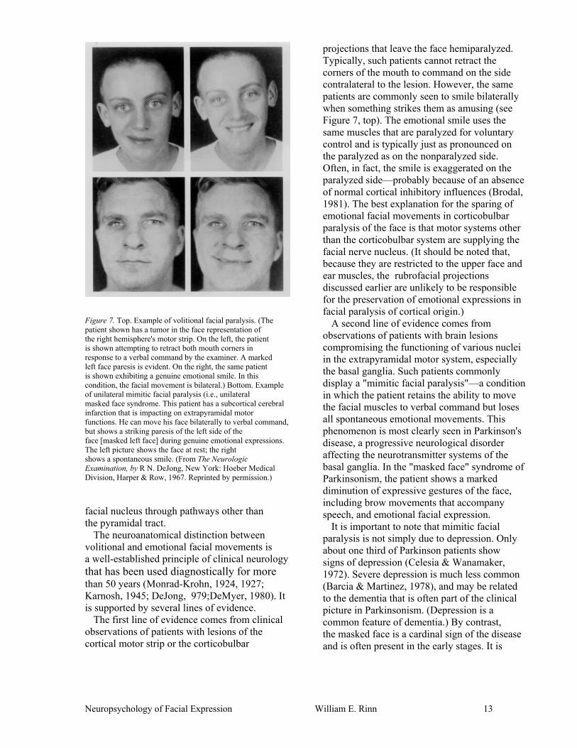
Figure 7. Top. Example of volitional facial paralysis. (The patient shown has a tumor in the face representation of the right hemisphere's motor strip. On the left, the patient is shown attempting to retract both mouth corners in response to a verbal command by the examiner. A marked left face paresis is evident. On the right, the same patient is shown exhibiting a genuine emotional smile. In this condition, the facial movement is bilateral.) Bottom. Example of unilateral mimitic facial paralysis (i.e., unilateral masked face syndrome. This patient has a subcortical cerebral infarction that is impacting on extrapyramidal motor functions. He can move his face bilaterally to verbal command, but shows a striking paresis of the left side of the face [masked left face] during genuine emotional expressions. The left picture shows the face at rest; the right shows a spontaneous smile. (From The Neurologic Examination, by R N. DeJong, New York: Hoeber Medical Division, Harper & Row, 1967. Reprinted by permission.)
facial nucleus through pathways other than the pyramidal tract.
The neuroanatomical distinction between volitional and emotional facial movements is a well-established principle of clinical neurology that has been used diagnostically for more than 50 years (Monrad-Krohn, 1924, 1927; Karnosh, 1945; DeJong, 979;DeMyer, 1980). It is supported by several lines of evidence.
The first line of evidence comes from clinical observations of patients with lesions of the cortical motor strip or the corticobulbar
projections that leave the face hemiparalyzed. Typically, such patients cannot retract the corners of the mouth to command on the side contralateral to the lesion. However, the same patients are commonly seen to smile bilaterally when something strikes them as amusing (see Figure 7, top). The emotional smile uses the same muscles that are paralyzed for voluntary control and is typically just as pronounced on the paralyzed as on the nonparalyzed side. Often, in fact, the smile is exaggerated on the paralyzed side—probably because of an absence of normal cortical inhibitory influences (Brodal, 1981). The best explanation for the sparing of emotional facial movements in corticobulbar paralysis of the face is that motor systems other than the corticobulbar system are supplying the facial nerve nucleus. (It should be noted that, because they are restricted to the upper face and ear muscles, the rubrofacial projections discussed earlier are unlikely to be responsible for the preservation of emotional expressions in facial paralysis of cortical origin.)
A second line of evidence comes from observations of patients with brain lesions compromising the functioning of various nuclei in the extrapyramidal motor system, especially the basal ganglia. Such patients commonly display a "mimitic facial paralysis"—a condition in which the patient retains the ability to move the facial muscles to verbal command but loses all spontaneous emotional movements. This phenomenon is most clearly seen in Parkinson's disease, a progressive neurological disorder affecting the neurotransmitter systems of the basal ganglia. In the "masked face" syndrome of Parkinsonism, the patient shows a marked diminution of expressive gestures of the face, including brow movements that accompany speech, and emotional facial expression.
It is important to note that mimitic facial paralysis is not simply due to depression. Only about one third of Parkinson patients show signs of depression (Celesia & Wanamaker, 1972). Severe depression is much less common (Barcia & Martinez, 1978), and may be related to the dementia that is often part of the clinical picture in Parkinsonism. (Depression is a common feature of dementia.) By contrast, the masked face is a cardinal sign of the disease and is often present in the early stages. It is
Neuropsychology of Facial Expression William E. Rinn 13

typically present even in nondepressed Parkinson patients and is also seen in patients with strokes, tumors, and traumatic lesions of the basal ganglia who show no signs of depression. Additionally, the masked face is commonly produced on only one side of the face, a condition compatible with a neurological motor disorder, but highly uncharacteristic of depression. (See Figure 7, bottom.) The sparing of volitional movements in mimitic facial paralysis suggests separate motor systems for these two phenomena.
A third line of evidence for separate circuits for emotional and volitional facial movements comes from a surgical procedure called facial nerve anastomosis. This operation is performed to reanimate a face that is paralyzed due to a lesion of the facial nerve at some point prior to its emergence onto the face. In this procedure, the motor root of the facial nerve is surgically severed. The central stump is avulsed to prevent regrowth while the stump of the distal portion remains raw. A few fibers are then teased out of another cranial motor nerve, usually the spinal accessory nerve (which supplies the muscles that move the shoulder). These fibers are then "spliced" into the distal stump of the facial nerve so that impulses coursing through the spinal accessory nerve will now innervate the facial muscles as well as the muscles of the shoulder.
Following the operation, electrical stimulation of the motor strip representation of the face yields no responses. The impulses cease at the stump of the severed facial nerve. However, stimulation of the representation of the shoulder will yield movements in both the shoulder and the face. Gradually, the patient learns to move the face muscles volitionally— apparently at first by attempting to move the shoulder. In time, a good degree of differential control is achieved and the face can be moved without shoulder movements. However, even years after the operation, with return of good voluntary facial movement, genuine emotional movements do not occur on the affected side. For these patients, genuine smiles are sharply restricted to the unoperated side. Reportedly, they frequently find this embarrassing and avoid such expressions (Kahn, 1966; Schemm, 1961; Schemm & Kahn, 1960).,
The most likely explanation for the absence of emotional movements on the affected side
is that the motor centers for emotional movements continue to send their impulses to the now disconnected stump of the facial nerve. The behavioral plasticity of the cerebral cortex allows it to learn to use the new pathway through the cortical shoulder representation and the spinal accessory nerve. The more primitive motor centers for emotional movement do not have this degree of flexibility.
A fourth line of evidence comes from observations of nonemotional involuntary laughing and/or weeping often seen in cases of pseudobulbar palsy. Pseudobulbar palsy results from lesions of the corticobulbar pathways and is commonly seen in cases of multiple sclerosis, amyotrophic lateral sclerosis, anoxia, and strokes involving the internal capsule (Horenstein, 1977). About half of all patients with pseudobulbar palsy commonly find themselves laughing or crying with only slight provocation or no provocation at all (Tinley & Morrison, 1912; Haymaker, 1969). Once the response is underway, they are largely unable to inhibit it and must simply wait until it abates on its own. These episodes are generally indistinguishable from normal laughing and weeping (see Figures 8 and 9). The face muscles contract into convincing smiles or crying faces, the face reddens, tears flow. The respiratory and vocal responses are also at least grossly identical to emotional responses. The only obvious difference is that these patients report no emotional experience during these bouts, or may even report the presence of an emotion incompatible with the expression (e.g., anger or pain while laughing; Poeck, 1969). Patients with pseudobulbar palsy typically also have at least some degree of voluntary facial paralysis. It appears that the involuntary expressions stem from an inability to voluntarily inhibit these motor release phenomena through normal cortical influences (Horenstein, 1977; Brodal, 1981).
Thus, a double dissociation between voluntary and emotional facial movements can be demonstrated by the fact that either can be interrupted or disturbed by neurological damage while the other remains intact. The question that next suggests itself is why this should be true. Why are two (or perhaps more) motor systems required? An answer to this question is suggested by an analysis of the evolution of motor systems.
Neuropsychology of Facial Expression William E. Rinn 14

Figure 8. Pathological crying. (This is a 37-year-old patient with amyotrophic lateral sclerosis, shown here during an attack of involuntary nonemotional crying. The first three shots show her struggling unsuccessfully to suppress the attack. It is full blown in the final (lower right) shot. Although the attack was unpleasant to her, the patient reported that she did not feel sad. From "Pathophysiology of emotional disorders associated with brain damage" by K. Poeck, in P. J. Vinken and G. W. Bruyn (Eds.), Handbook of clinical neurology (Vol. 3). New York: American Elsevier, 1969. Reprinted by permission.) The Evolution of Motor Systems and Emotional Responses
Several authors have suggested that the patterned bodily reactions of the emotions have evolved from similar patterned reactions associated with certain drive-related behaviors such as feeding, mating, and basic approach-avoidance responses (Darwin, 1872/1965; Plutchik, 1962). Anger and fear postures, for example, appear to be preparatory sets for fight and flight responses, respectively. Although actual emotional experience is usually ascribed only to higher animals, these drive-related behaviors are present even in very simple organisms.
In simple organisms, drive-related responses are handled by simple reflex circuits. These primitive circuits do not get discarded when more sophisticated motor mechanisms evolve. New systems are simply superimposed on older ones. The more primitive systems continue to function in situations for which they are appropriate. In humans, this reflex mode of responding is retained in certain brain stem circuits that regulate respiration, heart rate, and arousal.
In fish, these reflex circuits are supplemented by a motor system consisting of the paleostriatum (analogous to the globus pallidus in man) and the hypothalamus (Truex & Carpenter, 1969). In birds, the paleostriatum is elaborated into the corpus striatum, which plays the major role in organizing complex, but instinctive and highly stereotyped behaviors such as locomotion, feeding, courtship rituals, and defense. These activities are unaffected by even total destruction of the primitive cortex in birds, but are obliterated by lesions of the corpus striatum (Truex & Carpenter, 1969).
In mammals, the corpus striatum becomes the basal ganglia, and the cerebral cortex is more completely developed. Although all mammals have a cerebral cortex, the cortex is little involved in these stereotyped, instinctive behaviors. In most mammals, the near total destruction of the cortex eliminates only certain discrete motor functions. However, destruction of the major portion of the basal ganglia in mammals leaves only a few gross movements, primarily brain stem reflexes, intact (Guyton, 1976).
Although the motor systems described thus far are well suited for managing behaviors that are directly and immediately in the service of basic drives, all of these systems have the disadvantage that they lack plasticity. The output is almost completely determined by the immediate input, and is relatively unaltered by learning. These systems provide a high degree of certainty that the response, however simple, will occur consistently whenever the stimulus is encountered, but they allow little adaptability to novel situations.
A central function of the cerebral cortex is to add a measure of adaptability by allowing learning to influence motor behavior in a substantial way. The cortical (i.e., pyramidal)
Neuropsychology of Facial Expression William E. Rinn 15

motor system in humans is extremely plastic and versatile and is capable of executing fine, highly complex, highly controlled movements. It can readily modify the behaviors emitted in response to changes in the environment or in response to learning.
The motor portion of the cortex is the frontal lobes. The extent of frontal lobe development is a good index of the influence of learning on the organism's behavior. In humans, these occupy a larger proportion of the brain than in any other mammal, and they continue to grow faster than the rest of the brain until age 7 or so (Luria, 1973). In lower animals, even in the higher primates, the frontal lobes constitute a strikingly smaller proportion of the brain, and the role of the cortical motor system is correspondingly smaller. Even with complete bilateral destruction of the cortical motor tracts, which would cause massive paralysis in humans, chimpanzees recover the ability to feed themselves and to execute walking and climbing movements (Truex & Carpenter, 1969). Decorticate dogs and cats can walk, eat, fight, develop rage, engage in sexual activity, and generally perform all but the most intricate types of motor behaviors. The most obvious result of cortical destruction in mammals is the destruction of the purposefulness of the action. Decorticate dogs may ambulate normally but without direction or they may fail to negotiate a path around obstructions (Guyton, 1976).
It is remarkable that destruction of the cortex has such disparate effects on humans versus other mammals. Almost certainly, the
explanation is that learning plays a more important role in the development of the behaviors in question for humans than it does for other mammals. Humans develop the ability to walk, feed themselves, and so forth only after many months or years of practice. For most other mammals, these behaviors have an important instinctual component and are developed very early in life.
Like other motor systems, the cortical motor system does not replace its predecessors. In fact, in humans, some of the most primitive and vital responses are brain stem reflexes (e.g., coughing, sneezing, swallowing, gagging, etc.) or motor release phenomena that are organized in the brain stem and hypothalamus (e.g., laughing and crying). Normally the cortical motor system plays very little role in these behaviors. Most of them cannot even be performed voluntarily (although rough approximations are possible), and most are subject to only partial voluntary inhibition. Discriminating Cortical Versus Subcortical Features of Behavior
In general, it may be said that cortically mediated behaviors (e.g., language) are not present in infancy and must be learned. They are generally highly flexible and readily changeable, thus increasing the organism's adaptability to novel situations. They may also show considerable cultural variability. Typically, we have good conscious awareness of these behaviors, and they can easily be produced or inhibited on command. By contrast, behaviors mediated primarily by other motor
Figure 9. Pathologic laughing. (This 61-year-old woman with amyotrophic lateral sclerosis is shown with face at rest [1], and in successive stages of involuntary nonemotional laughing. The patient reported that the laughter was painful and that she was struggling to suppress it as these shots were taken. (From "Pathophysiology of emotional disorders associated with brain damage" by K. Poeck, in P. J. Vinken and G. W. Bruyn (Eds.), Handbook of clinical neurology (Vol. 3). New York: American Elsevier, 1969. Reprinted by permission.
Neuropsychology of Facial Expression William E. Rinn 16

systems (e.g., sneezing, heart beat, etc.) are generally present very early in development, not substantially influenced by learning, rigidly stereotyped in topography, inflexible, and show little cultural variability. In many cases, we have poor conscious awareness of the behavior. Generally, they can only be approximated on command, and are difficult to inhibit when they occur spontaneously.
The cortical (i.e., pyramidal) motor system frequently competes with the more primitive systems for control of our overt behavior. More commonly, however, they work together, each contributing certain elements to the final response. Thus, any given behavior is the product of both cortical and subcortical influences, although these influences are not always equally strong.
One important role of the cortex is in the social regulation of the face. Ekman and Friesen (1975) have coined the term display rules to refer to socially learned prescriptions for regulating the emotional expressions, that is, the social etiquette of facial behavior. Display rules may call for an expression to be amplified, tempered, feigned, or masked by a different expression. Examples of display rules are that males should not show fear, that females should not show anger, that one should smile when addressing guests in a reception line, that one should not giggle at solemn occasions, and so forth. Additionally, each individual has his or her own set of idiosyncratic display rules—a unique catalog of polite smiles and learned inhibitions.
The influences of display rules are almost certainly organized cortically. Manifestations of display rules are not present in infancy. Although infants can produce nearly all of the discrete facial movements and most of the configurations of movement that adults produce (Izard, Huebner, Risser, McGinnes, & Dougherty, 1980; Oster & Ekman, 1978), configurations characteristic of masking one expression with another are conspicuously absent in infants (cf. Oster & Ekman, 1978). The social regulation of the face does not appear to even begin until late infancy and is not well developed until at least middle childhood, when frontal lobe development is complete. Learning, through reinforcement, punishment, and modeling, clearly plays a prominent role in its development (Ekman & Oster, 1979).
Even in adults, these learned social facial behaviors are not rigidly programmed but rather are applied flexibly and can be discarded altogether when they are not adaptive. Application of display rules depends on a variety of factors, including one's age, sex, socioeconomic status, and the eliciting situation, as well as on the age, sex, and status of the person being addressed (Ekman & Friesen, 1975; Oster & Ekman, 1978).
Ekman (1972) and Friesen (1972) have demonstrated cultural variability in display rules. In one study, Japanese and American subjects showed similar facial responses while watching a stressful film, as long as they did not know they were being observed. When another person was present, however, American subjects continued to show a look of revulsion, whereas Japanese subjects masked their expressions. It is also clear that there are culture-specific sex differences in facial display rules. To varying degrees in Western culture, males are expected to inhibit fear expressions and to act aggressive. Females are expected to inhibit anger and to act coy.
As with most cortically mediated events, normal adults have relatively good awareness of what their faces are doing when they implement display rules. They have no difficulty repeating these movements on command and can easily dispense with them when they seem not to be adaptive.
In contrast, the structure of genuine emotional movements of the face is not typical of cortically mediated events. Unlike most cortically mediated behaviors, most emotional facial expressions are present very early in life (Oster, 1978). Nearly all of the adult configurations of muscle contractions are seen in early infancy (Izard et al., 1980; Oster & Ekman, 1978). These configurations become associated with specific emotions well before the end of the second year. Ekman and Oster (1979) note that distress and disgust expressions are present at birth. Social smiles begin to emerge by 4 weeks. "Interest" can be seen in the face of 3-week-old infants (Oster, 1978). Anger and contempt may be seen by 6-months (Izard, 1978). Meaningful surprise and fear configurations are seen in the second year of life. (Ekman & Oster, 1979).
An extrapyramidal origin for genuine emotional expressions is also indicated by the fact
Neuropsychology of Facial Expression William E. Rinn 17

that anencephalic infants (infants born with no cortex, basal ganglia, or other structures higher than the midbrain) show at least some normal facial expressions such as crying and rudimentary aspects of disgust (Guyton, 1976; Steiner, 1973). The observation that congenitally blind children display a full range of spontaneous expressions demonstrates that learning through imitation is not required (Freedman, 1964; Goodenough, 1932; Thompson, 1941).
Emotional expressions are also highly stereotyped and relatively inflexible. Although some cultural variability is seen, there are at least six well-defined expressions that are common to all human societies: anger, disgust, happiness, sadness, fear, and surprise (Ekman & Oster, 1979). Izard (1977) reports that interest and shame expressions are also universal.
Although we usually (not always) have conscious awareness of our emotional expressions, we do a poor job of posing them on command. Among other things, the timing (onset and offset) and the coordination of the various regions of the face (brows, eyes, mouth) are usually conspicuously off in posed expressions (Ekman & Friesen, 1975). Certainly, we frequently have difficulty voluntarily inhibiting genuine expressions. Behaviors of the Upper Face
The upper face has a number of behaviors that have no clear analog in the lower face. It is difficult to classify some of these behaviors as volitional or emotional. However, with a careful analysis of the behaviors themselves, it is often possible to infer the neural origins (cortical vs. extrapyramidal) of these movements. Three upper face behaviors will be discussed here.
The first is the contraction of the corrugator muscles during mental effort. The resultant "knit brow" appearance can often be observed during problem solving mentation (e.g., while playing chess, working a crossword puzzle, or doing difficult mental arithmetic). Although corrugator contractions are often associated with unpleasant affect, persons displaying the knit brow of concentration do not necessarily experience anything unpleasant. As often as not, the associated mental task appears to be recreational.
It is worth noting that simple attention to a stimulus or task does not produce the knit brow. It seems rather to require a particular kind of mental effort. Darwin (1872/1965) noted that, "A man may be absorbed in the deepest thought and his brow will remain smooth until he encounters some obstacle in his train of reasoning or is interrupted by some disturbance, and then a frown passes like a shadow over his brow" (p. 221).
Oster (1978) has observed knit brow expressions in infants 3 weeks of age. This makes it unlikely that they are of cortical origin. Moreover, in adults they commonly appear even when the subject is unaware that he or she is being observed. Additionally, Darwin (1872/1965) documented the universality of this behavior, noting that people of all cultures "frown when they are puzzled" (p. 222). These facts make it unlikely that this expression is the product of a display rule or any aspect of the social regulation of the face.
In infants, the knit brow pattern commonly precedes a smile. Typically, the infant stares with fixed attention at an object with brows knit as if studying it. After a few seconds, the brows relaxed and the zygomatic muscle contracts the face into a smile. Oster (1978) believes that the knit brow reflects the effort of trying to make sense of a new stimulus (i.e., trying to integrate it into an existing schema). When this is finally accomplished, the reduction of effort causes the brows to relax. The reduction of effort is held to be affectively pleasant; hence the smile.
The second type of upper face behavior to be considered is the tendency to contract the frontalis muscle during attentive listening. This phenomenon is commonly seen during conversation and may be recognized by the elevation of the brows. EMG studies have also shown systematic increases in frontalis tension during attentive listening, with no similar increases in "control" muscles such as those of the chin and forearm (Wallerstein, 1954; Bartoshuk, 1956). These brow responses are similar to volitional movements in that most persons can produce them on command. They are similar to involuntary responses in that they occur spontaneously and, unless they are pointed out, most persons remain oblivious to the fact that they are producing them.
One possible explanation for these brow
Neuropsychology of Facial Expression William E. Rinn 18

behaviors is that they are vestiges of ear perking movements used by lower mammals to orient to a given point in space. In lower mammals, including chimpanzees and gorillas, the frontalis muscle is continuous with the muscles that move the ears. It is only with the enlargement of the frontal lobes in Homo sapiens that the front part of the skull is pushed forward and the frontalis muscle becomes separated from the auricular muscles (Huber, 1931).
Additionally, within the facial nerve nucleus, the cell body groups that map to the auricular muscles (the dorsomedial and intermediate) also map to the frontalis (Courville, 1966a). Indeed, attempts to wiggle the ears often result in contractions of the temporal portions of the frontalis. It is here suggested that signals from these regions of the facial nucleus, which would cause ear perking in lower animals, are responsible for the brow elevations seen in humans during attentive listening.
The third type of brow behavior to be considered is described by Ekman and Friesen as "punctuation" movements. These are very brief (as short as 50 msec) contractions of the muscles of the upper face (especially the frontalis) that occur during speech and appear to add semantic emphasis or to reflect the pitch and stress contours of the vocal intonation.
Although these punctuation movements are commonly bilateral, most persons show some observable degree of asymmetry. Moreover, most persons show this asymmetry systematically, that is, the bias is always or predominantly in one direction or the other. The direction of this asymmetry is about evenly divided between left and right brow dominant individuals (Rinn et al., 1982). This is in marked contrast to the well-established left bias in the lower face during posed expressions (discussed below; Sackeim & Gur;, 1978; Campbell, 1978).
Although occurring in the context of speech, a manifestly cortical and volition behavior, these punctuation movements have features more typical of those mediated by extrapyramidal motor centers. Most subjects are totally unaware that they move their brows in this manner. They may deny knowledge of it even if attention is called to it immediately after an occurrence. Moreover, most subjects can volitionally produce only very crude approximations of the spontaneous movements
on command. This is particularly true for unilateral punctuation movements. Instructions to reproduce spontaneous unilateral brow movements volitionally typically yield bilateral movements, movements of only the wrong brow, or no movement at all (Rinn et al., 1982).
Because the upper face is spared in unilateral facial paralysis of cortical origin, these punctuation movements are typically unaffected by such damage. However, although no systematic studies have been conducted, I have observed that punctuation brow movements are absent in the masked-face syndrome of Parkinson's disease, in which the functions of the basal ganglia are compromised. Indeed, speech itself may lack normal pitch and stress contours (Chusid, 1979). It therefore seems likely that the basal ganglia play a major role in generating these movements. Systematic observations of masked-face Parkinson patients has not been conducted to determine whether they show the knit brow of concentration, or raised brows during attentive listening. However, preliminary results of research I am currently conducting indicate that congenitally blind subjects commonly produce a knit brow during puzzlement, elevated brows during attentive listening, and punctuation brow movements during speech. This strongly suggests that these movements are not learned through imitation, and therefore are not of cortical origin. Sensory Functions of the Trigeminal Nerve
I have already given brief mention to the trigeminal nerve. It was noted above that the motor portion of the trigeminal innervates the muscles that move the mandibles during chewing, but that is only half of the story. Some branches of the trigeminal nerve are sensory rather than motoric in function. The sensory branches carry tactile sensation from the corneas of the eyes, the inside of the mouth (including the gums and tongue), the nasal cavities and paranasal sinuses, the teeth, and the meninges. They also convey cutaneous sensation (touch, temperature, and pain) from the skin of the face and the top of the head, and proprioception (muscle sense) from the four chewing muscles that are supplied by the trigeminal's motor portion (Noback &
Neuropsychology of Facial Expression William E. Rinn 19

Figure 10. The cutaneous distribution of the sensory branches of the trigeminal nerve.
Demarest, 1975; DeJong, 1979). The portion of the trigeminal nerve that carries cutaneous sensation is divided into three branches, each of which conveys tactile information from a specific facial region. (See Figure 10.)
The sensory fibers in the trigeminal nerve terminate in any of three different sensory nuclei in the brain stem, depending on the kind of information they carry. Fibers conveying pain and temperature information from the three peripheral branches descend to the higher levels of the spinal cord, where they synapse in the trigeminal spinal nucleus. Fibers conveying light touch sensation for the three regions of the facial skin terminate in the principal sensory trigeminal nucleus, located in the pons. The third brain stem nucleus that receives sensory input from the trigeminal nerve is the mesencephalic trigeminal nucleus, located above (rostral to) the pons. This nucleus is believed to be involved with proprioception (muscle sense). It receives input from the muscle stretch receptors in the muscles supplied by the motor portion of the trigeminal (i.e., the chewing muscles). It appears that it also received input from the three branches of the trigeminal that convey sensation from the facial skin (Kugelberg, 1952). It sends output to both the trigeminal and facial motor nuclei, and appears to play a key role in reflexes involving the chewing muscles, as well as in reflexes involving the mimitic muscles.
All three trigeminal sensory nuclei relay the impulses they receive to the thalamus, which
in turn, relays them to the somatosensory region of the cortex. The somatosensory cortex (or sensory strip) is a topographically organized representation of the body, situated on the posterior lip of the central sulcus, just across from the motor strip. The somatosensory cortex is believed to be the region where conscious bodily sensations are organized. Facial Muscle Spindles The receptor organs for muscle proprioception, called muscle spindles, operate essentially as stretch receptors. They are found in nearly all the striate muscles of the body in humans and other mammals as well as in fish, reptiles, and birds (Olkowski & Manocha, 1973). However, there is little evidence for the existence of muscle spindles in the muscles of facial expression. Most authorities believe spindles are either very scarce or absent in these muscles (Bowden & Mahran, 1956; Brodal, 1981; Dubner et al., 1978; Gandiglio & Fra, 1967; Moldaver, 1973; Olkowski & Manocha, 1973; Sanes & Ison, 1980; Shahani & Young, 1973). Kadanoff( 1956) found evidence for a few spindles and other similar structures in some facial muscles, but noted that they were aberrant in form and not sensory in nature. Kugelberg (1952) inferred the presence of spindles in the orbicularis oculi because percussion of the skin over this muscle regularly elicits a blink reflex. He assumed that the afferent leg of this reflex originated in spindles in the orbicularis oculi. However, it now appears that this reflex originates in receptors in the facial skin rather than muscle and is transmitted to the brain stem via the trigeminal nerve (Gandiglio & Fra, 1967; Shahani & Young, 1973).
The apparent lack of spindles in the muscles of expression is even more striking compared to their abundance in the muscles of mastication (supplied by the trigeminal nerve). The masseter and temporalis, for example, are among the muscles most richly supplied with spindles (Kubota, Komatsu, Nakamura, & Masegi, 1980; Lennartsson, 1980a, 1980b; Olkowski & Manocha, 1973; Rozhold, 1980a, 1980b). High spindle counts have also been found in the muscles of the tongue (Rozhold, 1980c), and moderate numbers have been documented in certain of the extraocular
Neuropsychology of Facial Expression William E. Rinn 20

muscles—the muscles that move the eyes (Olkowski & Manocha, 1973).
There is no obvious explanation for the apparent dearth of spindles in the muscles of expression. Olkowski and Manocha (1973) have noted that muscles from various regions of the body may be classed as "spindle poor" or "spindle rich." The shoulders and thighs are spindle poor, whereas more distal reaches of the extremities are more richly supplied. However, the highest numbers are in the upper vertebral column and the chewing muscles. Brown and Rushworth (1973) have suggested that load bearing muscles (i.e., muscles that normally work against resistance) could be expected to contain more spindles than muscles that work under isotonic conditions. Since the muscles of expression move no skeletal structures, they might be expected to contain fewer spindles.
It should be pointed out that some authorities believe that all muscles—not just the facial muscles—are insentient, and that impulses generated by muscle spindles and other muscle sense receptors are utilized only in unconscious reflex circuits. However, it is established that afferent impulses from muscle spindles find their way to the cortical somatosensory area— the region where conscious bodily sensation is believed to be mediated (Merton, 1972; Guyton, 1976; Gardner, 1975). It is obvious, moreover, that people generally report feeling the state of contraction of their muscles (e.g., as when lifting a weight). At this writing, the issue is unresolved.
A question that immediately comes to mind is how is it that we are aware of the patterns of our facial muscle contractions if the muscles are insentient? There is no clear answer for this question at this time. Dubner, Sessle, and Storey (1978) have suggested that mechanoreceptors deep in the facial skin are sensitive to skin movement brought about by contraction of the facial muscles. The sensory impulses would then be carried to the brain stem via the trigeminal nerve. When these impulses are integrated in the sensory cortex, they could provide the basis for recognizing the expression. This is a plausible explanation, but at present, it is little more than speculation. Accounts of the clinical sequelae of trigeminal nerve damage, including complete trigeminal tractotomy, generally do not describe any loss of the patient's sense of his or her facial
expression (cf., Brett, Ferguson, Ebers, & Paty, 1982; DeJong, 1979; Patten, 1978); but this has not been carefully explored.
Facial Asymmetry and Hemispheric Differences in Emotion
Over the past 10 years, a sizeable literature has accumulated describing experimental and clinical observations indicating that the left and right hemisphere differ in their emotional tone. The evidence for this conclusion comes from a wide variety of studies using diverse populations and procedures. (See Tucker, 1981, for a review.) Asymmetry in facial expression is but one line of evidence for this conclusion, but it is the area most relevant to the topic at hand. Thus I will restrict my comments to this point.
Since 1978, a number of studies have reported findings that the left side of the face dominates the emotional expression. That is, the expression emanating from the left side of the face involves greater facial movement and is judged more intense than that emanating from the right side (Borod & Caron, 1980; Moscovitch & Olds, 1982; Sackeim, Gur, & Saucy, 1978). In nearly every report, the authors have explained the findings as an indication of right hemisphere lateralization for emotion.
Although this explanation has gained wide currency, it is not the only one possible. The chief problem is that it is inconsistent with the widely accepted dictum that only higher cortical functions such as language, praxis, or visual-spatial reasoning are lateralized to a single hemisphere of the brain. Indeed, Luria's (1973) principle of progressive lateralization states that lateralized functions are anatomically restricted to the secondary and tertiary association areas of the cerebral cortex. The emotional tone of one's experience or behavior is generally believed to be determined by subcortical systems (e.g., the limbic system, the hypothalamus, the extrapyramidal motor system, etc.), and is therefore unlikely to be lateralized. The main role of higher cortical processes in emotion is in its instrumental regulation— the volitional control of emotions — or in the social regulation of the face (the implementation of display rules, etc.).
Two alternative explanations of left face dominance for expression may be advanced,
Neuropsychology of Facial Expression William E. Rinn 21

both of which I believe are more consistent with established theory and with findings outlined earlier in his report. The first of these suggests that findings that appear to show right hemisphere lateralization for emotion may actually be due to a left hemisphere superiority for the inhibition of emotion. It may be noted that many authorities have suggested that the frontal cortex inhibits subcortical arousal mechanisms (Lindsley, 1951; Luria & Homskaya, 1970; Tucker, 1970). The role of language (clearly a cortical process) in the regulation of behavior in general has been documented by many researchers, most notably Luria (1973) and Vygotsky (1934/1962). Its role in emotion control seems most clearly indicated in the case of cognitive defense mechanisms (e.g., rationalization, intellectualization, etc.). I suggest that the left hemisphere, by virtue of its linguistic and prepositional thought capabilities, is better equipped to inhibit unwanted emotional episodes. This would result in better inhibition of the right face, and, by default, greater expressiveness in the left face.
This model also addresses a related issue. A number of recent studies have suggested that the right hemisphere is specialized only for negative (unpleasant) emotions, and that positive emotions are left hemisphere events. (See Sackeim et al,, 1982, for a review.) Indeed, some studies of facial asymmetry have found left face dominance to be less pronounced or absent for expressions of positive emotions (Bruyer, 1981; Sackeim & Gur, 1978; Schwartz, Ahern, & Brown, 1979). It is here suggested that this effect is due to unequal inhibition by the left hemisphere of positive and negative emotions. It seems unlikely that anyone would use complex cognitive strategies to rationalize away their feelings of mirth or joy. Thus, positive emotions would not be subject to left hemisphere inhibition and would not be more inhibited on the right side of the face. This explanation suggests that the left hemisphere is not specialized for positive emotion except in the sense that it does not inhibit positive emotion as it does negative. It should be noted that although some studies have found no asymmetry or very slight left face dominance for positive emotional expressions, numerous studies have demonstrated a substantial left face dominance for smiles (Borod & Caron, 1980; Campbell, 1978, 1979;
Heller & Levy, 1981;Rinnetal., 1982; Rubin & Rubin, 1980).
The second alternative explanation for left face expressive dominance concerns a principle covered earlier in this report: The cortical origin of volitionally induced movements of the face versus the extrapyramidal origin of genuine emotional movements. If only cortical functions are lateralized, and only volitionally induced expressions are cortical, one might expect only volitionally induced expressions to be lateralized and, consequently, asymmetrically displayed on the face. In point of fact, most studies demonstrating left face dominance for emotional expressions have used either posed expressions or expressions generated in the course of a face-to-face conversation (a situation in which cortically mediated display rules become manifest). Ekman, Hager, and Friesen (1981) have shown that when subjects do not know they are being observed, their spontaneous expressions are essentially symmetrical. Two early studies by Lynn and Lynn (1938, 1943) in which subjects were unaware that they were being observed found smile asymmetries to be nearly evenly divided between left and right dominant events. Thus, it would seem that only volitionally induced or otherwise cortically mediated expressions are more pronounced on the left.
Even if the finding of left face (right hemisphere) dominance is confined to posed (volitional, social, cortical) expressions, the finding is still very interesting. Most learned volitional motor skills are mediated by the left hemisphere (e.g., speech, praxis). It is, in fact, a left hemisphere lesion that is usually responsible for the clinical syndrome of buccofacial dyspraxia. Patients with buccofacial dyspraxia are unable to perform complex or meaningful facial movements, especially in pantomime, even though they have no facial weakness. Such patients may be unable to demonstrate how to use a straw, blow a kiss, blow out a match, or whistle on verbal command or by imitation. Preliminary findings of a study by Borod, Koff, Perlman, and Nicholas, however, showed that patients with buccofacial dyspraxia due to left hemisphere lesions generally retain the ability to pose emotional expressions (J. C. Borod, personal communication, February 1982). This confirms a similar clinical observation by Hécaen (1981).
Neuropsychology of Facial Expression William E. Rinn 22

Neuropsychology of Facial Expression William E. Rinn 23
This finding, together with the finding of left face dominance for posed expressions, suggests that the posing of an emotional expression is an exception to the general principle of left hemisphere superiority for tasks requiring complex motor organization.
One possible explanation for this apparent right hemisphere specialization for volitional facial expressions is that the posing of an expression involves a visual-spatial pattern matching task in which the to-be-matched pattern is coded primarily in the right hemisphere. As noted earlier in this report, a considerable amount of data supports the claim that recognition and memory for faces and emotional expressions is essentially lateralized to the right hemisphere, probably because of the visual-spatial nature of the stimuli or processing demands. When we attempt to motorically construct a facial expression, we are attempting to arrange the facial features in a configuration matching one of these sensory schemas. Since the criterion (to-be-matched) sensory schema is stored in the right hemisphere, right hemisphere motor mechanisms may be best equipped to provide the motor schema.
Although it cannot categorically be ruled out that emotion itself is somehow lateralized to a single hemisphere, or that positive and negative emotions are lateralized to different hemispheres, I believe that most or all of the findings reported in the literature can be just as well or better explained in terms of more generally accepted principles of neuropsychology. Certainly, this is the case for asymmetries in facial expressions.
Directions for Future Research Throughout this review, an attempt has been made not only to present data from a number of studies, but also to integrate these data and to suggest general conclusions. Many of these conclusions can be directly translated into guidelines for future investigation in the field.
One point that deserves emphasis is the need for objective description of facial movements. Studies that have raters infer the nature or strength of the emotion underlying a given expression are using very indirect and highly subjective measures of facial behavior. An anatomically based system of coding facial movements is essential to any researcher hoping
to discern the neural basis of these movements.
Although one can generate anatomically based descriptions of still photos of facial expressions, motion pictures are preferable. Still photos give a very restricted sample of the subject's facial behavior, showing the individual's expression as it existed for only a split second in time. Motion pictures allow broader sampling, through multiple observations of the individual over the course of several minutes. Equally important, still photos give no clue to the timing of the movement (its speed of onset, duration, etc.), the expressions that proceeded or followed it, the vocal and gestural accompaniments to the expression, or the situational context in which it occurred. All of these factors may be important in any attempt to decipher the neurological basis of the expression. Videotape is an inexpensive, accurate, and versatile medium for these purposes.
Another point to be emphasized is the desirability of analyzing the upper face separately from the lower face. These regions are capable of acting independently of each other. Descriptions in which they are combined (as are often seen in studies of facial asymmetry) may generate conclusions that are valid for only one of these regions. More importantly, there are vast differences between the brows and lower face both in behavior and in the patterns of neural innervation. Failure to take account of these differences will confound any study that hopes to make neurologic or behavioral statements about the face.
A final point that deserves mention is the importance of distinguishing between posed and spontaneous-emotional facial expressions. Posed expressions utilize cortical (pyramidal) circuits; spontaneous expressions are of essentially extrapyramidal origin. In practice, however, the distinction between volitional and emotional is not a simple one. Purely volition expressions may be observed by having subjects pose expressions to verbal commands. (Even this method may not completely suffice if the subject attempts to generate the expression by imagining an emotion-evoking stimulus.) To ensure that an expression is purely spontaneous-emotional it is necessary that the subject be unaware that he or she is being observed. This is because most people tend to hide their feelings from others by volitionally

Neuropsychology of Facial Expression William E. Rinn 24
moderating their facial expressions or by volitionally superimposing a different expression. Thus, most expressions generated during face to face communication have both volitional (cortical) and emotional (extrapyramidal) components.
We are only beginning to understand the neurological basis of the bodily expression of emotion. Although the work accomplished in recent years has been marked by creative and sometimes ingenious methodologies, the conclusions generated have often departed radically from accepted principles of neuropsychology. It would seem that it is now time to reconcile these departures, either by modifying recent conclusions or by reassessing our accepted notions of brain function.
References Andrew, R. J. (1965). The origins of facial expressions.
Scientific American, 213, 88-94. (Offprint No. 627) Barcia, D., & Martinez, P. F. (1978). Parkinson's disease with
depression treated with electroshock. Archivos de Neurobiologia, 41, 393-398.
Barr, M. L. (1974). The human nervous system. Hagerstown, MD: Harper & Row.
Bartoshuk, A. K. (1956). Electromyographic gradients and electroencephalographic amplitude during motivated listening. Canadian Journal of Psychology, 10, 156-164.
Benton, A,, L. (1980). The neuropsychology of facial recognition. American Psychologist, 35, 176-186.
Blurton-Jones, N. G. (1971). Criteria for describing facial , expressions of children, and Human Biology, 43, 365-413.
Borod, J. C, & Caron, H. S. (1980). Facedness and emotion related to lateral dominance, sex and expression type. Neuropsychologia, 18, 237-241.
Bowden, R. E. M., & Mahran, Z. Y. (1956). The functional significance of the pattern of innervation of the muscle quadratus labii superioris of the rabbit, cat, and rat. Journal of Anatomy, 90, 217-227.
Brett, D. C, Ferguson, G. G., Ebers, G. C., & Paty, D. W. (1982). Percutaneous trigeminal rhizotomy: Treatment of trigeminal neuralgia secondary to multiple sclerosis. Archives of Neurology, 39, 219-221.
Brodal, A. (1981). Neurological anatomy: In relation to clinical medicine. New York: Oxford University Press.
Brown, W. F., & Rushworth, G. (1973). Reflex latency fluctuations in human single motor units. In J. E. Desmedt (Ed.), New developments in electromyography and clinical neurophysiology (Vol. 3). Basel: Karger.
Bruyer, R. (1981). Asymmetry of facial expression in brain damaged subjects. Neuropsychologia, 19, 615-624.
Campbell, R. (1978). Asymmetries in interpreting and expressing a posed facial expression. Cortex, 14,327-342.
Campbell, R. (1979). Left handers' smiles; Asymmetries in the projection of a posed expression. Cortex, 15, 571-579.
Celesia, G. G., & Wanamaker, W. M. (1972). Psychiatric disturbances in Parkinson's disease. Diseases of the Nervous System, 33, 577-583.
Chusid, J. G. (1979). Nervous system. In M. A. Krupp & M. J. Chatton (Eds.), Current medical diagnosis and treatment, 1979 (chap. 16). Los Altos, CA: Lange Medical Publications, 1979.
Chusid, J. G., & McDonald, J. J. (1962). Correlative neuroanatomy and functional neurology. Los Altos, CA: Lange Medical Publications.
Courville, J. (1966a). Rubrobulbar fibres to the facial nucleus and the lateral reticular nucleus. Brain Research, 1, 317-337.
Courville, J. (1966b). The nucleus of the facial nerve: The relation between cellular groups and peripheral branches of the nerve. Brain Research, I, 338-354.
Crelin, E. S. (1981). Development of the musculoskeletal system. CIBA Clinical Symposia, 33(\).
Darwin, C. R. (1965). The expression of the emotions in man and animals. Chicago: University of Chicago Press, 1965. (Original work published 1872)
DeJong, R. N. (1979). The neurologic examination (4th ed.). Hagerstown, MD: Harper & Row.
DeMyer, W. (1980). Technique of the neurologic examination. New York: McGraw-Hill.
Dorland's illustrated medical dictionary (25th ed.). (1974). Philadelphia: Saunders.
Dubner, R., Sessle, B. J., & Storey, A. (1978). The neural basis of oral and facial function. New York: Plenum.
Ekman, P. (1972). Universal and cultural differences in facial expressions of emotion. Nebraska Symposium on Motivation, 1971, 207-283.
Ekman, P. (1980, September). Presentation. In G. E. Schwartz & P. Ekman (Leaders), Advanced Workshop: Emotion and facial expression: Research and Practice. Workshop conducted at the meeting of the American Psychological Association, Montreal.
Ekman, P., & Friesen, W. V. (1975). Unmasking the face. Englewood Cliffs, NJ: Prentice-Hall.
Ekman, P., & Friesen, W. V. (1978). Facial Action Coding System. Palo Alto, CA: Consulting Psychologists Press.
Ekman, R, Hager, J. C., & Friesen, W. V. (1981). The symmetry of emotional and deliberate facial actions. Psychophysiology, 18, 101-106.
Ekman, P,, & Oster, H. (1979), Facial expressions of emotion. Annual Review of Psychology, 30, 527-554.
Engle, G. L. (1963). Toward a classification of affects. In P. H. Knapp (Ed,), Expressions of the emotions in man. NewYork: International Universities Press.
Freedman, D. G. (1964). Smiling in blind infants and the issue of innate vs. acquired. Journal of Child Psychology and Psychiatry, 5, 171-184.
Friesen, W. V. (1972). Cultural differences in facial expressions in a social situation: An experimental test of the concept of display rules. Doctoral dissertation, University of California, San Francisco. Dissertation Abstracts International, 33, 3976B. (University Microfilms No. 73- 03651)
Gandiglio, G., & Fra, L. (1967). Further observation on facial reflexes. Journal of the Neurological Sciences, 5, 273-285.
Gardner, E. (1975). Fundamentals of neurology. Philadelphia: Saunders,
Gilbert, C, & Bakan, P. (1973). Visual asymmetry in perception of faces. Neuropsychologia, 11, 355-362.
Goodenough, F. L. (1932). Expression of the emotions in a blind-deaf child. Journal of Abnormal and Social Psychology, 27, 328-333.

Neuropsychology of Facial Expression William E. Rinn 25
Gray, H. (1977). Anatomy: Descriptive and surgical (American revision of 15th English edition of 1901). New York: Bounty Books.
Gray, H. (1973). Anatomy of the human body (29th American ed., Charles Mayo Goss, Ed.). Philadelphia: Lea and Febiger.
Guyton, A. C. (1976). Organ physiology: Structure and function of the nervous system. Philadelphia: Saunders.
Hamburg, D. A. (1963). Emotions in the perspective of human evolution. In P. H. Knapp (Ed.), Expression of the emotions in man. New York: International Universities Press.
Harker, L. A., & McCabe, B. F. (1977). Surgical anatomy of the facial nerve. In L. R. Rubin (Ed.), Reanimation of the paralyzed face: New approaches. Saint Louis, MO: Mosby.
Haymaker, W. (1969). Bing's local diagnosis in neurological diseases. St. Louis, MO: Mosby.
Hécaen, H. (1981). Apraxias. In S. B. Filskov & T. J. Boll (Eds.), Handbook of clinical neuropsychology (chap. 8). New York: Wiley.
Heller, W., & Levy, J. (1981). Perception and expression of emotion in right handers and left handers. Neuropsychologia, 19, 263-272.
Holstege, G., Kuypers, G. H. J. M., & Dekker, J. J. (1977). The organization of bulbar fibre connections to the trigeminal, facial and hypoglossal motor nuclei. Brain, 100, 265-286.
Horenstein, S. (1977). Emotional aspects of neurologic disease. In A. B. Baker & L. H. Baker (Eds.), Clinical Neurology (Vol. 3). Hagerstown, MD: Harper & Row.
Huber, E. (1931). Evolution of the facial musculature and facial expression. Baltimore, MD: Johns Hopkins University Press.
Izard, C. E. (1977). Human emotions. New York: Plenum. Izard, C. E. (1978). On the ontogenesis of emotions and
emotion-cognitive relationships in infancy. In M. Lewis & L. A. Rosenblum (Eds.), The development of affect. New York: Plenum.
Izard, C. E. (1979). The Maximally Discriminative Facial Movement Coding System (Max). Newark: Instructional Resources Center, University of Delaware.
Izard, C. E., & Dougherty, L. M. (1982). Two complementary systems for measuring facial expressions in infants and children. In C. E. Izard (Ed.), Measuring emotions in infants and children. Cambridge, England: Cambridge University Press.
Izard, C. E., Huebner, R. R., Risser, D., McGinnes, G. C., & Dougherty, L. M. (1980). The young infant's ability to produce discrete emotional expressions. Developmental Psychology, 16, 132-140.
Kadanoff, D. (1956). Die sensiblen Nervenendigungen in der mimischen Muskulatur des Menschen. Zeitschrift fur Mikroskopisch-Anatomisch Forschung, 62, 1-15.
Kahn, E. A. (1966). On facial expression. Clinical Neurosurgery: Proceedings of the Congress of Neurological Surgeons, Miami, Florida, 1964. Baltimore, MD: Williams and Wilkins.
Karnosh, L. J. (1945). Amimia or emotional paralysis of the face. Diseases of the Nervous System, 6, 106-108.
Kubota, K., Komatsu, S., Nakamura, M., & Masegi, T. (1980). Muscle spindle supply to the bovine jaw muscles. Anatomical Record, 197, 413-422. (Biology Abstracts, Vol. 69, Abstract No. 53471.)
Kugelberg, E. (1952). Facial reflexes. Brain, 75, 385-396 Landis, T., Assal, G., & Perret, G. (1979). Opposite cerebral
hemispheric superiorities for visual associative processing of emotional facial expressions and objects. Nature, 278, 739-740.
Lennartsson, B. (1980a). Jaw muscle activity during chin tapping Acta Odontologica Scandinavica, 38, 1-8. (Biology Abstracts, Vol. 70, Abstract No. 31503.)
Lennartsson, B. (1980b). Number and distribution of muscle spindles in the masticatory muscles of the rat. Journal of Anatomy, 130, 279-288. (Biology Abstracts, Vol. 70, Abstract No. 49391.)
Leukel, F. (1978). Essentials of physiological psychology. Saint Louis, MO: Mosby.
Ley, R. G., & Bryden, M. P. (1979). Hemispheric differences in processing emotions and faces. Brain and Language, 7, 127-138.
Lindsley, D. B. (1951). Emotion. In S. S. Stevens (Ed.), Handbook of experimental psychology. New York: Wiley (Interscience).
Lindzey, G., Prince, B., & Wright, H. K. (1952). A study of facial asymmetry. Journal of Personality, 21, 68-84.
Luria, A. R. (1973). The working brain. New York: Basic. Luria, A. R., & Homskaya, E. D. (1970). Frontal lobes and
the regulation of arousal processes. In D. I. Mostofsky (Ed.), Attention: Contemporary theory and analysis. New York: Appleton-Century-Crofts.
Lynn, J. G., & Lynn, D. R. (1938). Face-hand laterally in relation to personality. Journal of Abnormal and Social Psychology, 33, 291-322.
Lynn, J. G., & Lynn, D. R. (1943). Smile and hand dominance in relation to basic modes of adaptation. Journal of Abnormal and Social Psychology, 38, 250-276..
McCurdy, H. (1949). Experimental notes on the asymmetry of the human face. Journal of Abnormal and Social Psychology, 44, 553-555.
Merton, P. A. (1972). How we control the contraction of our muscles. Scientific American, 226, 30-37. (Offprint No. 1249.)
Moldaver, J. (1973). Some comments on blink reflexes. In J. E. Desmedt (Ed.), New developments in electromyography and clinical neurophysiology, (Vol. 3). Basel: Karger.
Moldaver, J. (1964). Facial nerve physiology and electrical analysis. Transactions of American Academy of Ophthalmology and Otolaryngology, 68, 1045-1059.
Monrad-Krohn, G. H. (1924). On the dissociation of voluntary and emotional innervation in facial paralysis of central origin. Brain, 47, 22-35.
Monrad-Krohn, G. H. (1927). A few remarks on the question of dissociation between voluntary and emotional innervation in peripheral facial paralysis. Acta Psychiatrica et Neurologica, 3, 35-39.
Moscovitch, M., & Olds, J. (1982). Asymmetries in spontaneous facial expressions and their possible relation to hemispheric specialization. Neuropsychologia, 20, 71- 81.
Netter, F. (1977). The CIBA collection of medical illustra-tions: Volume I. The nervous system. Summit, NJ: CIBA.
Noback, C. R. & Demarest, R. J. (1975). The human nervous system: Basic principles of neurobiology (2nd ed.). New York: McGraw-Hill.
Olkowski, Z., & Manocha, S. L. (1973). Muscle spindles. In G. H. Bourne (Ed.), The structure and function of muscle (2nd ed). New York: Academic Press.
Oster, H. (1978). Facial expression and affect development. In M. Lewis & L. A. Rosenblum (Eds.), The development of affect. New York: Plenum.

Neuropsychology of Facial Expression William E. Rinn 26
Oster, H., & Ekman, P. (1978). Facial behavior in child development. In W. A. Collins (Ed.), Minnesota symposium on child psychology (Vol. 11). Hillsdale, NJ: Erlbaum.
Patten, J. (1978). Neurological differential diagnosis. New York: Springer.
Patterson, K., & Bradshaw, J. (1975). Differential hemispheric mediation of nonverbal visual stimuli. Journal of Experimental Psychology: Human Perception and Performance, 1, 246-252.
Penfield, W., & Jasper, H. (1954). Epilepsy and the functional anatomy of the human brain. Boston: Little- Brown.
Plutchik, R. (1962). The emotions; Facts, theories, and a new model. New York: Random House,
Poeck, K. (1969). Pathophysiology of emotional disorders associated with brain damage. In P. J. Vinken & G. W. Bruyn (Eds.), Handbook of clinical neurology (Vol. 3). New York: American Elsevier.
Rinn, W. E., Friedman, C., & Meller, P. (1982, August). Left face dominance for spontaneous emotional expressions: Neuropsychological implications. Paper presented at the meeting of the American Psychological Association, Washington, DC.
Rizzolatti, G., Umilta, C., & Berlucchi, G. (1971). Opposite superiorities of the right and left cerebral hemispheres in discriminative reaction time to physiognomic and alphabetical material. Brain, 94, 431-443.
Rozhold, O. (1980a). Are there muscle spindles in the lateral pterygoid muscle of man? Ada Universitalis Palackinae Olomucenis Facultatis Medicae (Praha), 1980,94,473-480. (Biology Abstracts, vol. 72, Abstract No. 25198)
Rozhold, O. (1980b). Exceptions to the rules of proprioception among muscle spindles in the muscles of the human head. Acta Universitalis Palackidnae Olomucenis Facultatis Medicae (Praha), 1980,94,491-498. (Biology Abstracts, Vol. 72, Abstract No. 10690)
Rozhold, 0. (1980c). Muscle spindles in the external muscles of the tongue in man. Acta Universitalis Palackianae Olomuncenis Facultatis Medicae (Phara), 1980,94,431- 443. (Biology Abstracts, Vol. 72, Abstract No. 25197)
Rubin, D. A., & Rubin, R. T. (1980). Differences in asymmetry of facial expression between left and right handed children. Neuropsychologia, 18, 373-377.
Rubin, L. R. (1977). Anatomy of facial expression. In L. R. Rubin (Ed.), Reanimation of the paralyzed face: New approaches. Saint Louis, MO: Mosby.
Sackeim, H, A., & Gur, R. C. (1978). Lateral asymmetry in intensity of emotional expression. Neuropsychologia, 16, 473-481.
Sackeim, H. A., Gur, R. C. J., & Saucy, M. C. (1978). Emotions are expressed more intensely on the left side of the face. Science, 202, 434-436.
Sackeim, H. A., Greenberg, M. S., Weiman, A. L., Gur, R. C., Hungerbuhler, J. P., & Geschwind, N. (1982). Hemispheric asymmetry in the expression of positive and negative emotions: Neurologic evidence. Archives of Neurology, 39, 210-218.
Sanes, J. N., & Ison, J. R. (1980). A silent period in orbicularis oculi muscles of humans. Journal of Neurology, Neurosurgery, and Psychiatry, 43, 504-509.
Schemm, G. W. (1961). The cortical localization pattern following cranial nerve cross anastomosis. Journal of Neurosurgery, 18, 593-596.
Schemm, G. W., & Kahn, E. A. (1960). The effect of peripheral nerve cross anastomosis on the cortical localization pattern. Transactions of the American Neurological Association, 85, 225-226.
Schwartz, G. E. (1982). Psychophysiological patterning and emotion revisited: A systems perspective. In C. Izard (Ed.), Measuring emotion in infants and children, Cambridge, England: Cambridge University Press.
Schwartz, G. E., Ahern, G. L., & Brown, S. L. (1979). Lateralized facial muscle response to positive and negative emotional stimuli. Psychophysiology, 16, 561-571.
Schwartz, G. E., Fair, P. L., Salt, P., Mandel, M. R., & Klerman, G. L. (1976a). Facial expression and imagery in depression: An electromyographic study. Psychosomatic Medicine, 38, 337-347.
Schwartz, G. E., Fair, P. L., Salt, P., Mandel, M. R., & Klerman, G. L. (1976b). Facial muscle patterning to affective imagery in depressed and nondepressed subjects. Science, 192, 489-491.
Shahani, B. T., & Young, R. R. (1973). Blink reflexes in orbicularis oculi. In J. E. Desmedt (Ed.), New developments in electromyography and clinical neurophysiology (Vol. 3). Basel: Karger.
Steiner, J. E. (1973). The gustofacial response: Observation on normal and anencephalic newborn infants. In J. F. Bosnia (Ed.), Fourth Symposium on Oral Sensation and Perception. Bethesda, MD: U.S. Department of Health, Education, and Welfare.
Suberi, M., & McKeever, W. F. (1977). Differential right hemispheric memory storage of emotional and nonemotional faces. Neuropsychologia, 15, 757-768.
Szentagothai, J. (1948). The representation of the facial and scalp muscles in the facial nucleus. Journal of Comparative Neurology, 88, 207-220.'
Thompson, J. (1941). Development of facial expression of emotion in blind and seeing children. Archives of Psychology, 1-47.
Tinley, F., & Morrison, J. F. (1912). Pseudobulbar palsy, clinically and pathologically considered, with the clinical report of five cases. Journal of Nervous and Mental Disease, 39, 505-535.
Truex, R. C, & Carpenter, M. B. (1969). Human neuroanatomy. Baltimore, MD: Williams and Wilkins.
Tucker, D. M. (1981). Lateral brain function, emotion, and conceptualization. Psychological Bulletin, 89, 19- 46.
Tucker, I. F. (1970). Adjustment: Models and mechanisms. New York: Academic Press.
Vraa-Jensen, G. (1942). The motor nucleus of the facial nerve: With a survey of the efferent innervation of the facial muscles. Copenhagen: Enjar Munksgaard.
Vygotsky, L. S. (1962). Thought and language (E. Hanfman & G. Vakar, trans.). Cambridge, MA: M.I.T. Press, 1962 (Original work published in Russian, 1934).
Wallerstein, H. (1954). An electromyographic study of attentive listening. Canadian Journal of Psychology, 8, 228-238.
Wolff, W. (1933). The experimental study of forms of expression. Journal of Personality, 2, 168-176.
Received April 14, 1982 Revision received May 16, 1983 •