Neuroimaging. How to Question Scientific Images and Their ...
24
e-ISSN 2723-9640 JOLMA Vol. 2 – Num. 1 – Giugno 2021 147 Citation Carlenzi, E.; Coraci, D.; Pigoni, A. (2021). “Neuroimaging. How to Question Scientific Images and Their Artistic Value”. JOLMA. The Journal for the Philosophy of Language, Mind and the Arts, 2(1), 147-170. DOI 10.30687/Jolma/2723-9640/2021/01/009 Peer review Submitted 2021-03-16 Accepted 2021-06-25 Published 2021-06-30 Open access © 2021 | cb Creative Commons Attribution 4.0 International Public License Edizioni Ca’Foscari Edizioni Ca’Foscari Neuroimaging. How to Question Scientific Images and Their Artistic Value Emanuele Carlenzi IMT School for Advanced Studies, Lucca, Italia Davide Coraci IMT School for Advanced Studies Lucca Alessandro Pigoni IMT School for Advanced Studies Lucca; Department of Neurosciences and Mental Health, Fondazione IRCCS Ca’ Granda Ospedale Maggiore Policlinico, Milan, Italy Abstract Unquestionable holders of aesthetic content, images have a well-known role even in conveying scientific knowledge. In the present work, we focus on the epis- temological role of images within neuroscience. We first analyze the concepts of repre- sentation, similarity, and informativeness. Second, we discuss relevant case studies, i.e., images by functional Magnetic Resonance Imaging, and how the pictorial interventions commonly applied to them might have an impact on their informational content. Finally, we explore the notion of imagination as a relevant faculty for modelling neuroscientif- ic theories and the concept of creativity as an instrument to aesthetically modify brain images. These manipulations enable images to achieve the scientific purpose, altering the relation of similarity between the image and the studied phenomenon. In conclu- sion, this process leads to rethinking the role of the neuroscientist as an active observer. Keywords Informational images. Denotation. Scientific models. Brain Imaging. fMRI. Neuroscience. Visual Studies. Imagination. Mental Imagery. Summary 1 Introduction. – 2 Representation, Resemblance, and Informativeness of Scientific Images. – 3 Case Studies: Does the Brain Represent the Brain in Neuroimaging? – 4 The Artistic Value of Scientific Images and the Contribution of Imagination. – 5 Conclusion.
-
Upload
khangminh22 -
Category
Documents
-
view
2 -
download
0
Transcript of Neuroimaging. How to Question Scientific Images and Their ...

e-ISSN 2723-9640
JOLMAVol. 2 – Num. 1 – Giugno 2021
147
Citation Carlenzi, E.; Coraci, D.; Pigoni, A. (2021). “Neuroimaging. How to Question Scientific Images and Their Artistic Value”. JOLMA. The Journal for the Philosophy of Language, Mind and the Arts, 2(1), 147-170.
DOI 10.30687/Jolma/2723-9640/2021/01/009
Peer review
Submitted 2021-03-16Accepted 2021-06-25Published 2021-06-30
Open access
© 2021 | cb Creative Commons Attribution 4.0 International Public License
EdizioniCa’FoscariEdizioniCa’Foscari
Neuroimaging. How to Question Scientific Images and Their Artistic ValueEmanuele CarlenziIMT School for Advanced Studies, Lucca, Italia
Davide CoraciIMT School for Advanced Studies Lucca
Alessandro PigoniIMT School for Advanced Studies Lucca; Department of Neurosciences and Mental Health, Fondazione IRCCS Ca’ Granda Ospedale Maggiore Policlinico, Milan, Italy
Abstract Unquestionable holders of aesthetic content, images have a well-known role even in conveying scientific knowledge. In the present work, we focus on the epis-temological role of images within neuroscience. We first analyze the concepts of repre-sentation, similarity, and informativeness. Second, we discuss relevant case studies, i.e., images by functional Magnetic Resonance Imaging, and how the pictorial interventions commonly applied to them might have an impact on their informational content. Finally, we explore the notion of imagination as a relevant faculty for modelling neuroscientif-ic theories and the concept of creativity as an instrument to aesthetically modify brain images. These manipulations enable images to achieve the scientific purpose, altering the relation of similarity between the image and the studied phenomenon. In conclu-sion, this process leads to rethinking the role of the neuroscientist as an active observer.
Keywords Informational images. Denotation. Scientific models. Brain Imaging. fMRI. Neuroscience. Visual Studies. Imagination. Mental Imagery.
Summary 1 Introduction. – 2 Representation, Resemblance, and Informativeness of Scientific Images. – 3 Case Studies: Does the Brain Represent the Brain in Neuroimaging? – 4 The Artistic Value of Scientific Images and the Contribution of Imagination. – 5 Conclusion.
148JOLMA e-ISSN 2723-9640
2, 1, 2021, 147-170
1 Introduction
Images are ubiquitous. Central in many areas, such as art history and visual studies, they have been widely investigated as vehicles of aesthetic content as well as scientific knowledge (Elkins 1995; Bredekamp 2003; Hentschel 2014). By way of example, images have been explored as crucial for media studies (Ott, Mack 2020), learn-ing (Bilbokaitė 2008; Smith 2008), and as a fundamental subject in the mental imagery debate in cognitive sciences (Kosslyn, Thompson, Ganis 2006; Richardson 2013). Indeed, the use of images within sci-entific disciplines has always been remarkable. Scientists often pre-sent the topics they investigate by relying on images for visualizing and modelling natural phenomena when not directly available to the senses. Neuroscience is a prototypical example of this. For instance, we refer to neuroscientific imagining techniques such as structural and functional Magnetic Resonance Imaging (MRI and fMRI, respec-tively), Positron Emission Tomography (PET), and Computed Tomog-raphy (CT) scan which allow researchers and clinicians to study the inner structures of the human brain by means of images.
In the last decades several studies have addressed the impact of neuroimages and related techniques in cultural and visual studies (Prasad 2005; Casini 2011; 2015; 2020; Stephens 2012; Hentschel 2014), and assessed how scientific knowledge can be conveyed through neuroimages, by questioning their epistemological nature (Roskies 2007, 2008; Schweitzer et al. 2011; Dumit 2014). In particu-lar, an aspect critically pointed out by such literature (e.g., Rosk-ies 2007) is that the relationship of resemblance occurring between neuroimages and the natural phenomena they describe (e.g., brain functioning) does not bear per se any informational value. Howev-er, by analysing the conceptual connections between the notions of representation and resemblance, it could be possible to further understand the scientific informativeness of neuroimages. That is, whether such an informational value is determined by specific tech-nical aspects or can be even related to conscious pictorial interven-tion of the scientist, and if the researcher’s imagination (or mental imagery) may play a role in defining instruments for the transmis-sion of neuroscientific knowledge.
Throughout the present article, we will try to approach such is-sues. First, starting from a general philosophical analysis of the no-tion of representation (section 2), we will retrace Nelson Goodman’s argument concerning the theoretical independence between this con-cept and the one of resemblance (or similarity). Then, by specifically focusing on scientific images, we will examine how their information-al value represents a relevant factor for understanding their relation-ship with the depicted phenomena. To do that, we take into account the notion of ‘informational images’ introduced by the art critic and
Emanuele Carlenzi, Davide Coraci, Alessandro PigoniNeuroimaging. How to Question Scientific Images and Their Artistic Value

Emanuele Carlenzi, Davide Coraci, Alessandro PigoniNeuroimaging. How to Question Scientific Images and Their Artistic Value
149JOLMA e-ISSN 2723-9640
2, 1, 2021, 147-170
art historian James Elkins (1995) whose work on scientific images has opened the field of visual studies of science.
Then, in section 3, we will question the epistemological role of fM-RI images by taking into account some examples from published neu-roimaging literature as case-study. This part of the paper is devot-ed to explain how neuroimages are created and can be manipulated by scientists in order to become effective vehicles of neuroscientific theories. From this analysis, a point should be evident: the process of neuroimages production is multifaceted and the scientist decides to directly and deliberately intervene on it, independently from the technological advances of MRI technique. Such manipulations tend to alter the visual and aesthetic properties of the images in order to make them more informative. Therefore, in section 4, we plan to ex-plore the potential contribution of the neuroscientist’s imagination and creativity. We sketch two potential directions. The first one is that neuroimages are not just visual reproductions of a certain natu-ral phenomenon, but (visual) scientific models of the theory they con-vey. In this respect, imagination will be discussed as central in the construction of scientific models, according to the debate already tackled in philosophy of science (Toon 2016; Frigg 2010; Frigg; Reiss 2010; Salis; Frigg 2020). The second direction, instead, sheds light on the possible intersection between artistic and neuroscientific images. Indeed, a fruitful dialogue may exist between the creativity and the aesthetic manipulations applied on (neuro)scientific images and art-works inspired by scientific processes (Stokes 2016; Gaut 2003; 2010).
2 Representation, Resemblance, and Informativeness of Scientific Images
Consider the portrait of Queen Elizabeth II and Queen Elizabeth II herself. Intuitively, one can argue that the portrait represents the Queen if and only if the portrait resembles the Queen. In other words, it appears that the concepts of representation and (pictorial) resem-blance or similarity are strictly connected and, furthermore, that the definition of the former depends on the definition of the latter. How-ever, that is not the case, as demonstrated by the American philoso-pher Nelson Goodman whose work (e.g., Goodman 1976) has greatly influenced the investigation of the concept of symbolization both in art and philosophy of language.
In the rest of the chapter, we first follow Goodman’s (1976) concep-tual analysis to shed light on the relationship between the concepts of representation and resemblance with regard to images in gener-al. Second, by specifically referring to scientific images, we take in-to account the notion of ‘informational images’ introduced by James Elkins (1995). The resulting general framework about the conceptu-

150JOLMA e-ISSN 2723-9640
2, 1, 2021, 147-170
al interdependencies occurring between the notions of representa-tion, resemblance, and informativeness will be useful to approach the case-study of neuroscientific images discussed in the next section.
From a logical point of view, the notions of representation (‘R’) and resemblance (‘S’) can be represented as binary relations occurring between two objects of any sort ‘x’ and ‘y’. From now on, we adopt the notations ‘xRy’ to state that ‘x’ represents ‘y’, and ‘xSy’ to state ‘x’ resembles ‘y’. Then, by assuming that ‘a’ is the portrait of the Queen Elizabeth and ‘b’ is the Queen Elizabeth herself, we can reword the relation between the portrait and the Queen as ‘aRb’ if ‘aSb’, that is, object ‘a’ engages a relation of representation with object ‘b’ if and only if ‘a’ is similar to ‘b’.
The first Goodman’s clarification about representativeness and re-semblance regards whether both the two relations are reflexive and symmetric. For instance, ‘being brother’ is a symmetric, but irre-flexive relation, while ‘being equal’ is a relevant case of both reflex-ive and symmetric relation. Therefore, in general, for every binary relation ‘G’, to be reflexive over an object ‘x’ means to relates to ‘x’ itself, i.e., ‘xGx’, while to be symmetric over two objects ‘x’ and ‘y’, means that if ‘G’ occurs between ‘x’ and ‘y’, then ‘G’ occurs between ‘y’ and ‘x’, i.e., ‘xGy iff yGx’.
Goodman points out that while the relation of resemblance is both reflective and symmetric, the relation of representation does not. An object ‘a’ resembles itself to the maximum degree, but it is not the case it represents itself (Goodman 1976, 4). Then, in symbols, while ‘aSa’ seems to state a meaningful relation, ‘aRa’ does not. Further-more, concerning symmetricity, for two objects ‘a’ and ‘b’, it is true that if ‘a’ resembles ‘b’, then ‘b’ resembles ‘a’, but it is not the case that if ‘a’ represents ‘b’, then ‘b’ represents ‘a’ (Goodman 1976, 4). For instance, the portrait of Queen Elizabeth of course resembles Queen Elizabeth, and the other way round, the Queen resembles the portrait of her, but we would not say that Queen Elizabeth represents her portrait, even though the portrait of Queen Elizabeth represents the Queen. Therefore, while ‘aSb’ seems meaningful, ‘aRb’ does not.
Then, the relations ‘S’ and ‘R’ do not share the same properties, and this is the first evidence for arguing that resemblance is not a sufficient condition for representing. Indeed, two different portraits of the Queen may perfectly resemble each other, but it is not a suffi-cient condition to conclude that they represent each other. Moreover, ‘S’ appears not even necessary for determining ‘R’, indeed, a linguis-tic description of an object has not to be to some extent (pictorially) similar to the object it describes for referring to it. This last exam-ple, however, can be useful to correctly understand what the relation of representation actually is. According to Goodman, representing
Emanuele Carlenzi, Davide Coraci, Alessandro PigoniNeuroimaging. How to Question Scientific Images and Their Artistic Value

Emanuele Carlenzi, Davide Coraci, Alessandro PigoniNeuroimaging. How to Question Scientific Images and Their Artistic Value
151JOLMA e-ISSN 2723-9640
2, 1, 2021, 147-170
means being a sign of, i.e., denoting. Then, denotation1 is what ac-tually characterizes the relation of representation discussed so far. Denotation is a broad notion, encompassing every kind of sign. Lin-guistic expressions as well as images can simply denote because they are signs through which we refer to objects. Therefore, the relation of representation should not be understood anyway, but as a particu-lar kind of denotation occurring between a sign (e.g., a linguistic or pictorial sign) and its reference (e.g., an object).2 Moreover, the rela-tion of representation, or denotation, drawn between a sign and its reference can be considered as conventionally fixed, that is, origi-nated by the symbolic norms that generally govern how we name ob-jects, and how we communicate and socially interact. According to major theories of reference proposed in philosophy of language (i.e., the ‘causal theories of reference’, Donnellan 1970, Kripke 1972), a sign represents a certain object depending on its own adequacy as bearer of the relation and this aspect can be thought as complete-ly (causally) determined by the symbolic system in which we live. In other words, representing something is not a matter of degree or in-tensity of how robust a relationship of representation is with respect to another one: either ‘x’ represents ‘y’ or ‘x’ does not represent ‘y’, given the specific rules governing the symbolic system in which ‘x’ and ‘y’ occur. This is a further aspect that differentiates representa-tion from resemblance. Resemblance, indeed, can be naturally con-ceived as varying in degree: we can always rank ‘x’ as more similar to ‘y’ than ‘z’. A son can resemble his father more than what his sis-ter does.3 Therefore, we claim, the relation ‘S’ naturally accommo-dates this shift in meaning towards a graduated or discrete judge-ment of similarity, while ‘R’ cannot.
A further characteristic of signs is to what extent they convey in-formation about the object they stand for, i.e., the ‘informativeness’ of signs. Such a notion has been introduced by Elkins (1995) in the vis-ual studies debate with specific regard to scientific images,4 explic-itly defined as ‘informational images’. The purpose of this concept is
1 A classical topic of discussion in philosophy of language and metaphysics (Frege [1892] 1980; Russell 1905; 1911; Kripke 1972; Putnam 1974; 1975).2 According to this, non-representational or abstract pictures that widely populate art history are analyzed by Goodman (1976) by means of the concept of ‘null’ or ‘mul-tiple denotation’. 3 In brief, to formalize such a degree in resemblance that objects show, the original relation ‘S’, now interpreted in a comparative way as ‘being more similar’, should be considered as an order relation. A binary relation ‘G’ is an order relation, i.e., it can or-der the objects of a set against one another, if it is true, for instance, that: (1) if ‘xGy’ and ‘yGx’, then ‘y=x’, and (2) if ‘xGy’ and ‘yGz’, then ‘xGz’, for any ‘x’, ‘y’, and ‘z’ be-longing to a set ‘A’.4 In the rest of the paper, we will follow Elkins’ (1995) discussion within the field of vis-ual studies and, then, confine the question about informativeness to (scientific) images.
152JOLMA e-ISSN 2723-9640
2, 1, 2021, 147-170
to group all those images principally intended to perform some utili-tarian or technical function and transfer knowledge by means of sym-bolic and pictorial features, such as schemas, numbers, and writing. As images, however, they have always been excluded from the canon-ical research field of art history due to a deficiency in terms of vis-ual expressiveness, eloquence, and complexity they were supposed to suffer. However, Elkins argues, informational images address the central issues of art history and images studies and they should be investigated as well as artworks. Our purpose here is to briefly ex-plore the meaning of Elkins’ notion of informativeness for scientific images and to outline the potential connections occurring with the relations of representation and resemblance.
Assumed that ‘being informative’ means conveying information, such as a map conveys spatial knowledge of a certain geographical region and given the variety of images usually employed in scientific and technical contexts, to clearly categorize the ways through which information can be pictorially conveyed is far from the purpose of the current work. However, as a general statement, we can say that ‘be-ing informative’ reflects a sort of relation occurring between a vehi-cle of the information (e.g., a sign such as a schema or an image) and a subject the information is about (e.g., a scientific theory). Conse-quently, the fact that an image ‘x’ is informative of ‘y’ requires, as a minimum requirement, that ‘x’ stands for ‘y’ or represents y, to some extent. In order words, to establish the effective conveying of knowl-edge between the vehicle and the subject, the fact that the vehicle (or components of it) represents the subject (or parts of it) seems funda-mental. Thus, representing is not enough for conveying information, otherwise a simple photo would result informational as much as a pic-torial schema, whereas we would like to disentangle between differ-ent levels of informativeness of pictures, depending, for instance, on the amount and type of information they provide. Then, it seems plau-sible to conceive images as vehicles of information gradually ranging from better to worse ones, depending on several factors. An infor-mational image employed for explaining a theory in highly special-ized academic papers, for instance, is differently informative from a similar image targeting a general audience. We can generally ob-serve that (scientific) images are not all informative in the same way.
However, does the informativeness of an image depend on the re-lationship of resemblance between its content and the theory it con-veys? Indeed, it could be argued that, for better conveying informa-tion, the content of an image should resemble the object it represents as much as possible.
We claim that the concepts of resemblance and information are independent. Suppose, for instance, to figuratively describe the soil composition of a certain piece of ground. A schematic figure describ-ing the soil profile by sketching with lines and alternate pattern tex-
Emanuele Carlenzi, Davide Coraci, Alessandro PigoniNeuroimaging. How to Question Scientific Images and Their Artistic Value

Emanuele Carlenzi, Davide Coraci, Alessandro PigoniNeuroimaging. How to Question Scientific Images and Their Artistic Value
153JOLMA e-ISSN 2723-9640
2, 1, 2021, 147-170
tures the different layers you may encounter can be more informative of, even though less similar to, the piece of ground than a picture of it. Then, it could happen that the more informational images do not correspond to those that resemble at best the content they refer to. Furthermore, because of their theoretical independence, it is even possible that as far as the informativeness of an image increases, its pictorial resemblance to the reality decreases.
With regards to scientific images, this may lead to an apparent contradiction. Indeed, on one hand, due to the relation of denotation rigidly determined by the symbolic (cultural, linguistic, and scien-tific) systems in which the sign at stake occurs, the scientific image seems to represent (or denotes) a certain phenomenon in objective and direct way, that is, they mimetically5 capture the ‘authentic’ na-ture of the phenomenon. On the other hand, however, the scientific image seems to better accomplish its informational (scientific) pur-pose, the less pictorially similar it is to the phenomenon it refers to. In other words, the extent to which the image enables the users to understand the phenomenon under scrutiny, i.e., being informative about it, may depend on some modifications of the pictorial format of the image itself that actually make it less similar to the phenom-enon how it naturally observable and, in turn, questions its status of objectivity.
In line with this, the neuroscientific images – as far as the neuroim-aging techniques (e.g., fMRI) have become more and more employed by psychologists and clinicians for understanding our brain – can represent an interesting case-study. Indeed, the relatively brief but crowded history of neuroimages that will be overviewed in the next section critically emphasizes such a particular path that informa-tional images can follow.
3 Case Studies: Does the Brain Represent the Brain in Neuroimaging?
The relationship between fMRI images and the phenomenon they represent, i.e., brain activity, has been widely discussed by the phi-losopher of neuroscience Adina L. Roskies (2007; 2008). Her works address the epistemological differences between fMRI images and photographs. Intuitively, both these types of images appear to be transparent vehicles of representation of the objects they refer to. However, it can be argued that photographs represent a relatively di-rect means of representation of reality due to the more understand-
5 For a general overview on the concept of mimetic representation see Kurtz, Kris 1979.

154JOLMA e-ISSN 2723-9640
2, 1, 2021, 147-170
able causal and counterfactual dependencies6 with the visual prop-erties of the subject (Roskies 2007). On the other hand, neuroimages convey just an illusion of “inferential proximity” (Roskies 2008) about the phenomenon they refer to. Such an illusion would rely on the fact that fMRI images allow to visualize the brain functioning, while ac-tually “there are no visible properties of brain activity to be instan-tiated in the image” (Roskies 2007, 863). Indeed, even though people tend to attribute to fMRI images the same transparency of photo-graphs, the complexity in the elaboration of brain imaging data (e.g, the pre-processing of raw data, their statistical analysis, the gener-al theoretical and experimental framework they are related to) and the unclear understanding of brain functioning, make the interpre-tation of neuroimages much more problematic.
Such a complex relationship between neuroimages and the phe-nomena they represent can become even more challenging when they are considered within the actual scientific practice, that is, as vehi-cles of specific information about neuro-psychological theories and experimental findings. In other words, researchers can alter such a relationship in order to visualize and communicate informational content. To highlight this aspect, we decided to consider some neu-roscientific images as case-study. Our aim is to prove that in order to achieve their specific purpose, they usually undergo stylistic and deliberate modifications by the researcher (and only marginally due to technical issues) that further reduce the similarity between the neuroscientific image and the natural phenomenon described, while increasing the informativeness about the theory, the experiment, etc. These deliberate manipulations by the researcher are made possi-ble by technical advancement, but are not only dependent on tech-nical aspects: they have simply enlarged the range of possible inter-ventions of the researchers on the visual properties of such images. In order to discuss how the neuroscientist directly acts on them, we will provide examples from different decades.
We opted for functional magnetic resonance imaging (fMRI) stud-ies because of their wide diffusion (more than 196000 published pa-pers on Pubmed in the last five years) and the consequent centrality in neuroscientific research. We preferred not to use structural MRI images, because they bear a clinical relevance and their application is
6 By means of causal and counterfactual dependencies images have to the objects they represent, Roskies (2007; 2008) intends that the visual properties of the real ob-ject cause the visual properties of the image and that, if the subject had been differ-ently arranged in terms of visual properties, the resulting image would have appeared correspondingly different. Actually, neither photographs nor fMRI, Roskies (2007, 867) argues, “bears a perfect informational relationship to the object”, but intuitively we have a clearer understanding of how these relationships (in particular in the case of the counterfactual one) occurs in photography rather than imaging.
Emanuele Carlenzi, Davide Coraci, Alessandro PigoniNeuroimaging. How to Question Scientific Images and Their Artistic Value
Emanuele Carlenzi, Davide Coraci, Alessandro PigoniNeuroimaging. How to Question Scientific Images and Their Artistic Value
155JOLMA e-ISSN 2723-9640
2, 1, 2021, 147-170
diffused in many fields of medicine (neurology, psychiatry, etc.); on the other hand, fMRI is mainly a research tool with few or no clinical ap-plications so far (Waller et al. 2021). fMRI gives a graphical represen-tation of brain activity. This technique is sensitive to variation in the local ratio between oxygenated and deoxygenated blood in the brain, that is, variations in the local consumption of oxygen by brain cells. The assumption behind it is that neurons demand more oxygen when they are activated. Therefore, fMRI provides an indirect measure of neuronal activation, based on oxygen consumption (Ogawa 2012). In fMRI images, this neuronal activation can be seen as groups of voxels (a regular measure of volume, we could image those as cubic bricks of volume) that are colored (therefore activated), with a peak of activation defined as the most active part, surrounded by a gradient of activity.
We selected some brain images from scientific papers in peer-re-viewed journals,7 namely two studies for each decade starting from the nineties. The selected studies can be considered representative of the field at their time; they are all based on healthy individuals (psychiat-ric and neurological conditions were not included), employ compara-ble techniques, and their results are presented through brain activa-tion images and brain reconstructions. Neuroscientific fMRI images have already been analyzed from a visual and philosophical point of view by Roskies (2007; 2008), underlying how far they are from being a mere reproduction of the brain. However some elements have not been clearly delineated yet. Such images serve a specific purpose: convey-ing information (i.e., the results of the study, a theory about the brain functioning) in the best way possible. In this framework a question arises: how the deliberate choices of researchers influenced the crea-tion of such images and therefore the quality of the message delivered?
Before discussing these points, some brief technical considera-tions are needed (for an exhaustive description of technical advance-ment in fMRI field, please refer to Bandettini 2012 and Specht 2020). Over the years, technical improvements in the fMRI technique al-lowed a higher level of details and a better resolution of the images, as clear moving from figure 1.B to figures 2.B and 3.B. More power-ful software allowed a better reconstruction of the brain grey and white matter, with more defined images (compare the 3D reconstruc-tions presented in figures 1.A, 2.A.1, and 3.A.1). These advancements affected the graphic representation, making the brain images more recognizable. Due to more modern technologies – such as finer scans, and upgraded softwares for image reconstruction – a detailed repre-sentation (i.e., higher graphical resolution) leading to a better defini-tion of the brain processes studied is feasible.
7 Menon et al. 1992; Che et al. 1998; Wagner et al. 2008; Tsubomi et al. 2008; Seydell-Greenwald et al. 2017; Koutsouleris et al. 2018; Pujol et al. 2018.

156JOLMA e-ISSN 2723-9640
2, 1, 2021, 147-170
Similarly, the need to compare results from different studies brought to the creation of atlases based on several subjects (such as the Mon-treal Neurological Institute). In this way, a ‘typical’ brain was cre-ated as an average of different, individual, brains: a model of the ‘standard’ brain. Indeed, while figure 1 presents the brain of a single subject, without important mediators, the others derive from group differences plotted on a standard brain. Then, the brain we see in fig-ures 2-4 is not an individual one. Such a remark is relevant because when we consider neuroimages, we should bear in mind that in most of the cases it is not a single subject’s brain that is represented, but an average brain, obtained from different samples and metrics. The distance between the single subject and the group average is then added to the already present conceptual gap between the actual brain and the picture of it. The brain is highly variable and can be influ-enced by various features, ranging from genetic influences to learn-ing and training in a specific field (Ritchie et al. 2018). Therefore, as customary when dealing with the brain and with neuropsychiatric syndromes, a single individual does not summarize all the possible differences and the visual results tend to be highly variable: thus, the need of averaging. This average moves the observer away from the object and also might influence the narrative related to brain im-ages, as discussed by Joseph Dumit (2014).
Figure 1 (A) 3D functional map of an individual subject obtained with fMRI, modified from Chen et al. (1998). (B) Functional maps of an individual subject obtained after visual stimulation, modified from Menon et al. 1992
Emanuele Carlenzi, Davide Coraci, Alessandro PigoniNeuroimaging. How to Question Scientific Images and Their Artistic Value

Emanuele Carlenzi, Davide Coraci, Alessandro PigoniNeuroimaging. How to Question Scientific Images and Their Artistic Value
157JOLMA e-ISSN 2723-9640
2, 1, 2021, 147-170
However, the presented technical improvement accounts only for a limited part of the variability in the presentation of relevant fMRI im-ages. Other visual aspects of them are also dependent on the choices made by researchers, aiming at effectively conveying their message. The first choice regards which image or part of the image to show. As presented in figures 1-3, the selection of 2D and 3D images is not mere-ly linked to technical advances, since they were already available in the nineties, even though with a lower resolution [fig. 1.A]. Research-ers deliberately decide to present 2D slices or/and 3D reconstructions. Interestingly, the 3D picture could seem easier to interpret because it resembles the human brain more closely. However, in the clinical field where such resemblance could be of great utility to the clinician in or-der to make diagnoses or program treatments, 3D is little employed. Indeed, we are accustomed to clinical images of brains as slices (both CT scans and MRI), while the 3D reconstruction is able to show only superficial/external regions, whereas deeper regions tend to remain hidden. This is of limited use in clinical practice when we need to see even what is underneath (i.e., in case of a neoplastic lesion, it is man-datory to understand which areas of the brain are involved or disrupt-ed by such lesion). Therefore, the 3D brain raffiguration loses any re-semblance with a typical clinical brain image and serves only as a model. Moreover, those 3D pictures can be unrealistically re-oriented (i.e., presented on a bottom up or top-down view – see for instance fig-ure 3.A.1 – which means that the observer can move around the brain in every direction, even the non-real ones, such as seeing it from the bottom, like a disembodied object with an all-around design), with the only purpose of conveying scientific information. Therefore, are the results of a study easier to interpret when presented in 3D, or is it just an aesthetic decision aimed at outlining the neuroscientific theory?
Furthermore, the choice of which slice to present in 2D images is left to the researcher. Since no clear guideline is available, the slices
Figure 2 (A.1) 3D reconstruction and (A.2) fMRI images in the sagittal plane modified from Tsubomi et al. 2008. (B) fMRI images, in the transverse plane, modified from Wagner et al. 2008

158JOLMA e-ISSN 2723-9640
2, 1, 2021, 147-170
that give a better and clearer presentation of the results are usually displayed. They can be in the sagittal plane (as in figures 2.A.2 and 3.B), or in the transverse plane [figs 1.B, 2.B, 3.A.2]. Only in a minor-ity of cases, instead, the coronal plane is presented ([fig. 3.B], right side), since it is difficult to evaluate where the position of our point of view is located. After deciding which plane to display, the researcher needs to choose which slice to present, following again the principle of the most informative image. The right images of figures 2.B and 3.A are slices coming from the top of the brain; while in others it is possible to see its internal structures (i.e., lateral ventricles), mean-ing that we are roughly in the middle part of the brain.
Finally, a choice has to be made regarding the color scale. Although it is a common tendency to use the scale so that the peak of activa-tion is marked by the brightest color, no definite guideline is availa-ble, and many differences can be spotted in the literature. For exam-ple, some researchers opted for a yellow-to-red scale [fig. 2.A.2], others for a warm color scale, ranging from bright yellow to orange [figs 2.B, 3.A.1], others preferred ranging from colder to warmer colors [figs 1.B, 3.A.2]. Similarly, both activation and deactivation8 can be presented, as in figure 3.B, where warmer colors are used to graphically present the increase in activation and colder ones for the decrease in activation.
8 We are usually accustomed to consider the peak of activation as always positive. However, we can also have peaks with negative sign, meaning reduction in neuronal activation.
Figure 3 (A.1) 3D reconstructions and (A.2) transverse images of the brain, modified from Seydell-Greenwald et al. 2017. (B) Sagittal, transverse, and coronal fMRI images, modified from Pujol et al. 2018
Emanuele Carlenzi, Davide Coraci, Alessandro PigoniNeuroimaging. How to Question Scientific Images and Their Artistic Value
Emanuele Carlenzi, Davide Coraci, Alessandro PigoniNeuroimaging. How to Question Scientific Images and Their Artistic Value
159JOLMA e-ISSN 2723-9640
2, 1, 2021, 147-170
However, these peaks are presented after selecting a threshold. Im-aging softwares offer the possibility to choose the level of activation to show, by increasing/decreasing the dimension of the colored ar-ea. It is customary to present only the most relevant findings, that is, those significant from a statistical point of view, but a decision re-garding how much to show is necessary.
These are just the easiest examples of how the neuroscientist’s in-tervention can be relevant in the creation of images with the aim of better presenting results and conveying a specific message. Other aspects (e.g., the choice of the preprocessing pipeline and of the soft-ware to employ) are far too technical for our discussion.
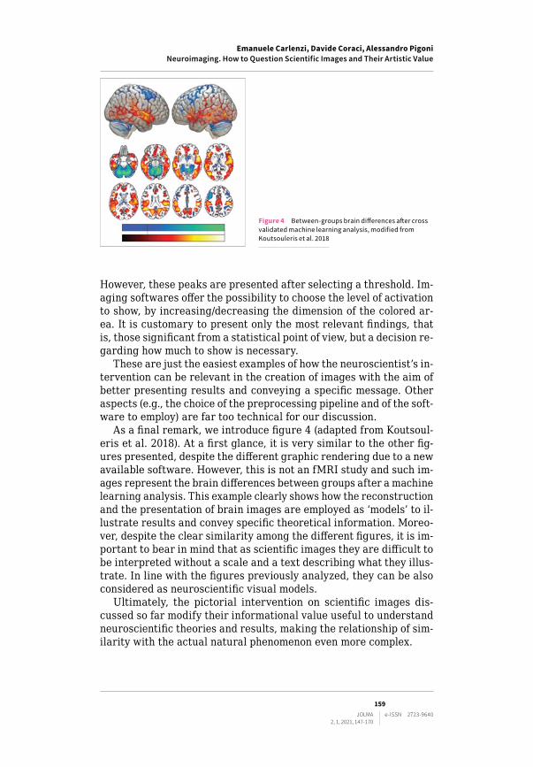
As a final remark, we introduce figure 4 (adapted from Koutsoul-eris et al. 2018). At a first glance, it is very similar to the other fig-ures presented, despite the different graphic rendering due to a new available software. However, this is not an fMRI study and such im-ages represent the brain differences between groups after a machine learning analysis. This example clearly shows how the reconstruction and the presentation of brain images are employed as ‘models’ to il-lustrate results and convey specific theoretical information. Moreo-ver, despite the clear similarity among the different figures, it is im-portant to bear in mind that as scientific images they are difficult to be interpreted without a scale and a text describing what they illus-trate. In line with the figures previously analyzed, they can be also considered as neuroscientific visual models.
Ultimately, the pictorial intervention on scientific images dis-cussed so far modify their informational value useful to understand neuroscientific theories and results, making the relationship of sim-ilarity with the actual natural phenomenon even more complex.
Figure 4 Between-groups brain differences after cross validated machine learning analysis, modified from Koutsouleris et al. 2018

160JOLMA e-ISSN 2723-9640
2, 1, 2021, 147-170
4 The Artistic Value of Scientific Images and the Contribution of Imagination
As already stated, the topic of representing reality has always been a challenging question for most people dealing with the production and reception of images. Even if this theme has more frequently been associated with the study of art history and the investigation of the artist as a pivotal figure in understanding how to connect the realm of reality with its depiction, the problem deeply influenced the sci-entific field as well.
What we will try to figure out with this last section can be sum-marized as it follows. First: we will take into account the central role played by images for their use as a symbolic metaphor and linguis-tic tool to convey information. Second: we will discuss how repre-sentation of reality cannot be considered objective neither for paint-ings and artworks nor for scientific images. They ‘re-present’ reality as an abstract ensemble of forms revealing a particular relationship between the observer and the outer world. Third: we will argue how the intervention of imagination and creativity in creating and mod-ifying images to make them more or less scientifically informative can be considered a crucial characteristic belonging both to the ar-tistic and the scientific field. In particular, with regard to science, we will take into account the role of the imagination not in bringing to light new discoveries (e.g., the famous Kekulé’s discovery of the ring structure of benzene, see footnote 12), but in conceiving scien-tific models and conveying information through images. According to this, fMRI images would not only be pictorial representations of neuroscientific theories, but scientific models which the researcherʼs imagination contributes to create. On the other hand, although crea-tivity can be treated as a synonym of imagination, it is instead a con-cept that we can connect to the active contribution brought by the scientist that reworks and interprets images to make them explica-tive and supportive of a given theory. As we will try to briefly outline later in the text, the debate regarding the connection between imag-ination and creativity has a lasting and rich literature (Salis, Frigg 2020; Stokes 2016). However, if imagination plays a central role in building theories, the subject of creativity – more generally associ-ated with art history – could be also applicable to the scientific field in order to explain the aesthetic contribution of the researcher on the production of fMRI images. Even though his or her contribution might appear merely visual, it also impacts on the informational con-tent lying on the image. The centrality of images and their use as a symbolic form to convey information and meanings is a point we can all agree upon, as already treated in the first chapter. Rudolph Arn-heim, for example, a German art and film theorist and a perceptu-al psychologist, writes “what makes language so valuable for think-
Emanuele Carlenzi, Davide Coraci, Alessandro PigoniNeuroimaging. How to Question Scientific Images and Their Artistic Value

Emanuele Carlenzi, Davide Coraci, Alessandro PigoniNeuroimaging. How to Question Scientific Images and Their Artistic Value
161JOLMA e-ISSN 2723-9640
2, 1, 2021, 147-170
ing cannot be thinking in words. It must be the help that words lend to thinking while it operates in a more appropriate medium such as visual imagery” (Arnheim 1971, 231).9 That statement can be easily applied to the scientific field in the same measure we tend to do with the artistic one. While on the one hand, we have paintings, sculp-tures, installations, drawings, on the other we can equally take into account charts, diagrams, histograms, and fMRI as images that try to represent phenomena happening in the external world.
The proximity between art and non-art images have been already investigated by Elkins who gives examples of artists that use scien-tific methods to create artworks over time and, vice versa, by dem-onstrating that fine-art conventions intervene preponderantly in the production of scientific images to make them more expressive and ‘prettier’, especially the medical ones. “In terms of the attention sci-entists lavish on creating, manipulating, and presenting images, the ‘two cultures’ are virtually indistinguishable” (Elkins 1995, 559).
The example he provides is about the diagrams used by the Ger-man immunologist Paul Ehrlich who presents some drawings regard-ing the function of antibodies in response to the presence of toxins attacking a cell. The Y-shaped diagrams he uses to represent the an-tibodies do not correspond at all to previous knowledge but can be fully-fledged considered as a pictorial way to give shape to a scientific process and represent something that cannot be seen. Nonetheless, from that moment on, those images become the theory from which modern immunology derives and consequently evolves.
Yet, even if imagination in the scientific field could be evident by keeping in mind Elkin’s example of Paul Ehrlich’s drawings, it could also not be necessary or sufficiently reliable when applied to other scientific images – like the aforementioned fMRI – that seem to ob-jectively represent reality throughout mechanic and sophisticated processes. But the question is: do they represent reality? Is objectiv-ity a pure and untouched value belonging to those images or there is something that goes beyond that?
As we saw in the previous section, fMRI images represent the neu-ral activity by detecting changes in the ratio of oxygenated and deoxy-genated blood. As Roskies states (2007, 864) the visual product deriving from this process lets the observer believe that neural firing – which is colored in the image – corresponds exactly to the brain activity. How-ever, as she notes, it is not entirely correct. In fact, what we see in ev-idence reflects an indirect measure of the neural activity that forces
9 The statement inevitably recalls the activity of scholars coming from the German context that is not appropriate to define as art historians but more as image historians such as Aby Warburg. His famous project titled Mnemosyne is in fact one of the clearest examples of the visual thinking practice, based on the association of various pictures.
162JOLMA e-ISSN 2723-9640
2, 1, 2021, 147-170
the image to be interpreted since it does not provide a clear explana-tion or an unquestionable representation of the phenomenon. Despite their mechanical and technical features, neuroimages establish a com-plex relationship with reality, since they make visible phenomena such as the brain activity that naturally lack any visual property. Due to this indirect relationship with reality (Roskies 2007), neuroimages seem to require some level of (aesthetic) intervention by the scientist. This is aimed at making visible information that otherwise would have been unreadable and would remain obscure. In other words, the attempt of describing the brain’s activity with an image could be decidedly unsat-isfactory without pictorial modifications (e.g., the use of colours) and an iconological interpretation (Panofsky 2019) which is – by necessi-ty – required to the observer. Hence, the contribution of the research-er is essential to let data emerge from the image and to make them in-formative about a specific theory. Imagination (involved in creating the scientific model) and creativity (useful for aesthetically intervening on the scientific model) can be considered as two sides of a unified process.
It is not coincidental that members of the scientific community do not always ‘read’ and convey information in the same way and that the understanding of the image is all but unique or univocal (although brain imaging remains a reliable tool). The absence of a process mak-ing those images alike and equally readable, leaves the neuroscien-tist the chance to deliberately and pictorially intervene.
Referring to a statement Paul Klee famously made “art does not reproduce the visible; rather it makes visible” (Klee [1920] 2013) he seems to declare that art does not reproduce what we see but rather it manufactures what we see. Under this interpretation, a painting is not a sort of mechanism that captures and displays existing visi-ble data, but an engine to create a way of looking and interpreting the world. In other words, the act of painting is an endeavor to make visible what commonly is not seen.
Scientific images can be related to that statement inasmuch as they are not simply a mechanical process of reproduction. On the con-trary, they create and interpret what is not visible, and even when they are produced through scanning technologies, like the magnetic resonance, they demand the participation of a ‘creator’ in any case. If we accept this vision, fMRI products could be closer to painting than photography (Roskies 2007, 2008), closer to interpretation than objectivity, closer to imagination than reproduction.
However, while the contribution of art conventions in the analysis of scientific or, more generally, informational images, have already been investigated, the figure of the neuroscientist as an active ob-server is still lacking. In this perspective, the topic of imagination requires more attention.
Art historians have always been inclined to consider artists in-volving scientific samples in their artworks as such. Contempo-
Emanuele Carlenzi, Davide Coraci, Alessandro PigoniNeuroimaging. How to Question Scientific Images and Their Artistic Value
Emanuele Carlenzi, Davide Coraci, Alessandro PigoniNeuroimaging. How to Question Scientific Images and Their Artistic Value
163JOLMA e-ISSN 2723-9640
2, 1, 2021, 147-170
rary art is full of examples in which medical images have been ‘re-mediated’(Bolter, Grusin 1999; Montani 2010) and turned into art.10 Strongly established artists employ scientific material in their works: Robert Rauschenberg in Shades (1964) and Booster (1967) and Meret Oppenheim in X-ray of M.O.’s Skull (1964)11 make use of x-ray photo-graphs to create sculptures and collage paintings; Joseph Beuys stud-ies accurately a various aspect of the human skull by creating new arrangements of elements according to his own creativity; Hermann Nitsch uses entrails for his performances; Pierre Huyghe in 2019 at the Serpentine Gallery in London presents Uumwelt,12 an installation in which fMRI technology is used to create surrealistic neuro-images in motion thanks to an artificial neural network software.
The terms ‘art’ and ‘imagination’, at least till post-Kantians thought, seem to trace some sort of parallel lines in which the latter is considered as a fundamental and unified human faculty which is essentially intertwined with conscious life and artistic genius. Our commonsense view is generally associated with the belief that art making, in a broader definition, is considered as the ability of acti-vating our inner faculties that “conjure new things, or at least, new ideas of things, into being” (Wiltsher, Meskin 2016, 180). However, even if we tend to think that imagination and creativity are widely distant from the scientific field – maybe because they appear not to lead to a systematic objective result – they play a significant role in scientific inquiry and discovery.13 Paying attention to the imagina-tion might help us not only to shed a light on scientific discoveries but also to better understand how scientific theories are conveyed and modelled. Given that, scientists may acquire a more relevant po-sition in the complex relationship between scientific information, fic-tion, and art.
In this regard, fMRI images can be considered as an example of scientific models, depending on the level of intervention of neurosci-entists. Not by chance, the contribution of the imagination in the de-velopment of scientific models have reached major importance in the
10 Plenty of other examples could have been discussed here, such as the famous brain studies conducted by Leonardo da Vinci, for instance. We decided to refer to Contem-porary Art because artists have more easy access and use more frequently techniques and materials belonging to the scientific field.11 Cf. Casini 2011; 2015; Stephens 2012.12 https://www.serpentinegalleries.org/whats-on/pierre-huyghe-uumwelt/.13 August Kekulé’s discovery of the ring structure of benzene after dreaming of a snake swallowing its own tail as well as Paul Elrich’s drawings regarding antibodies and toxins are two explanatory examples of how imagination impacts scientific think-ing (Elkins 1995). Cf. also Holton 1978.

164JOLMA e-ISSN 2723-9640
2, 1, 2021, 147-170
recent literature in philosophy of science.14 Imagination can be de-fined (even if not always) as a voluntary mental activity that involves a visual or other sensory mental state to subjectively describe non-present objects or circumstances (Stokes 2016). The term ‘imagina-tion’ is often used as a synonym of ‘creativity’ but, although being imaginative can be easily associated with originality or innovative-ness, not all imaginative actions rely on creativity. Indeed, creativi-ty is more connected to the creation of something new, of novelties which become substantial in and for a specific context. (Gaut 2003, 2010; Salis, Frigg 2020) Therefore, with respect to neuroimages, cre-ativity refers to the aesthetic intervention of the scientist that, by modifying some visual features, creates something new.
Neuroscientists study our brain via an abstract and simplified system that includes a pictorial and visual outcome. Therefore, neu-roimages do not represent phenomena until we make the effort that stands behind the creation process of modeling, that is imagination. Even though there is an intricate debate regarding the type of mod-els, that can be physical or theoretical, they are central to scientists’ attempt to understand and give shape to the world and its phenome-na (Toon 2016). In addition, a creative and aesthetic action from the scientist is required in order to express and communicate those nat-ural events. Thus, creativity becomes central to pictorially intervene on the outcome by creating a new product and favoring the compre-hension of some information.
Boundaries and interplays between art and neuroscience progres-sively appear blurred if creativity is taken into account not only for a better understanding of artistic production and artists, not only for investigating possible categories medical images could borrow from art, but also to start seeing neuroscientists through a different angle. As a matter of fact, due to the complex analysis they carry out and their creative contribution, they seem to engage in a peculiar rela-tionship with images and act like visual investigators.
In this regard, the 2017 exhibitions Reaching Beyond the Obvious, taking place in Montréal on the occasion of the Organization for Hu-man Brain Mapping (OHBM) Conference, a world-famous event on neuroscience, show how profoundly arts and sciences can collabo-rate symbiotically, combining creative thinking with scientific de-scriptions of the brain. The undeniable aesthetic values of those im-ages – characterized by charming colors and manneristic nuanced shapes – make the spectator doubtful if he or she is looking at scien-tific products or artworks and bring us to reconfigure the scientist persona appealing to his or her creativity to create images.
14 Godfrey-Smith 2006; Toon 2012; 2016; Weisberg 2013; Levy 2015; Frigg 2010; Frigg, Reiss 2010; Salis, Frigg 2020.
Emanuele Carlenzi, Davide Coraci, Alessandro PigoniNeuroimaging. How to Question Scientific Images and Their Artistic Value

Emanuele Carlenzi, Davide Coraci, Alessandro PigoniNeuroimaging. How to Question Scientific Images and Their Artistic Value
165JOLMA e-ISSN 2723-9640
2, 1, 2021, 147-170
“Beauty is inside” is also the motto of the neuroimaging artist duo DiMa – composed by Diana Roettger and Matthew Rowe – that trans-forms complex visualizations of the brain into artistic pictures. The two imaging scientists aim at questioning the fMRI technique by making 3D images representing the workings of the human brain, by pictorially intervening on the product that results incredibly articu-lated, eye-catching, and profoundly distant from a representation of objective reality (surprisingly enough, they are also sold as it is usu-ally done with paintings and artworks).15
The development of unique and creative techniques for mapping and visualizing such data has led more and more to abstract pictures that, albeit providing detailed information, become detached from the natural phenomenon it describes. As a consequence, they turn into artistic products that highlight the active contribution of their authors and end up counteracting the concept of mimesis itself by re-jecting reality as it roughly appears.
5 Conclusion
Images are crucial subjects that have been investigated in many areas of knowledge such as neuroscience and visual studies, research fields we discuss in this essay. As claimed by Elkins (1995) and Bredekamp (2003), the pictorial categories traditionally exploited in art history can also be applied in the study of scientific-oriented images. In this regard, brain images can be analyzed in a multidisciplinary way and provide the opportunity to reflect upon their theoretical nature.
As we tackled in section 2 and highlighted by aforementioned scholars, a conceptual independence between the notions of resem-blance, representation, and informativeness can be drawn. Images can be informational and explicative about scientific theories even if they lack a high degree of similarity with the studied phenomena. In other words, the quality of informativeness belonging to a scien-tific image does not depend on how it objectively adheres to reality.
Neuroimaging, i.e., fMRI, is a clear example of the deliberate picto-rial intervention brought by scientists that could alter the production of images for scientific purposes. Over the years, fMRI-based imag-es have undergone technical improvements, providing higher levels of detail and better resolutions. Undoubtedly, beside a more sophisti-cated reconstruction of the brain, technological advances increased the possibility to pictorially intervene on the images, making them
15 On the relationship between objectivity and scientific image-making see Jones, Gal-ison 1998; Daston, Galison 2007. Moreover, for a suggestive discussion about the per-formative capacity of MRI outcomes to function as a portrait see Casini 2011.
166JOLMA e-ISSN 2723-9640
2, 1, 2021, 147-170
more informative about the scientific findings. As seen in section 3, the choice of selecting 2D or 3D format, showing a distinctive slice of the brain to the observer, specifying colors, texts, and shades to de-note the presumed neural activity, are all elements at the neurosci-entist’s full discretion. Therefore, the graphic representation and its stylistic results widely depend on the intentional contribution of the researchers that work on the visual properties of the neuroimages.
Such pictorial interventions create products able to convey sci-entific knowledge as part of an active process that originates from the neuroscientist’s imagination and creativity. As seen in section 4, both play a significant role in scientific inquiry, even though it is of-ten regarded as distant from science. Paying attention to the imag-ination might help to better understand how scientific theories are conveyed and modelled.
The interpretation of neuroimages requires the effort of consid-ering the purpose of the neuroscientist’s imagination that aims at framing the scientific message in what can be considered a pictorial model of the theory itself. On the other hand, creativity also plays a fundamental role in the construction of images. Even though inter-twined with the imagination, creativity is more related to the aes-thetic and stylistic intervention coming from the deliberate choices of the scientists. Therefore, neuroimages which are determined by a theoretical framework, can acquire and increase their informational content due to this creative work. In this process, the neuroscientist turns into an active observer in the creation of images.
Furthermore, the fact that neuroimages have effectively become material for recent artworks such as Huyghe’s Uumwelt and DiMa creations and for related academic discussion (Jones, Galison 1998; Daston, Galison 2007; Casini 2011; 2015; 2020), suggest that such im-ages can talk even outside the scientific field, highlighting a range of potential manipulations and creative interventions.
In conclusion, by looking at images of the brain as a spectrum rang-ing from the mere reproduction of reality to the creation of pictorial models of neuroscientific theories till the most radical artistic inter-ventions, we are encouraged to further explore the concepts of mimet-ism, scientific objectivity, and intentional manipulation. This multidis-ciplinary perspective opens up the opportunity to consider the figure of the neuroscientist not only as an objective observer that merely doc-uments reality, but as a visual expert able to create images.
Emanuele Carlenzi, Davide Coraci, Alessandro PigoniNeuroimaging. How to Question Scientific Images and Their Artistic Value
Emanuele Carlenzi, Davide Coraci, Alessandro PigoniNeuroimaging. How to Question Scientific Images and Their Artistic Value
167JOLMA e-ISSN 2723-9640
2, 1, 2021, 147-170
List of abbreviations
CT Computed Tomography fMRI functional Magnetic Resonance Imaging MRI structural Magnetic Resonance ImagingOHBM Organization for Human Brain MappingPET Positron Emission Tomography
Bibliography
Arnheim, R. (1971). Visual thinking. Berkeley: University of California Press.Bandettini, P.A. (2012). “Twenty Years of Functional Mri: the Science and the
Stories”. Neuroimage, 62(2), 575-88. https://doi.org/10.1016/j.neu-roimage.2012.04.026.
Bilbokaitė, R. (2008). “Analysis of Visual Thinking Meaning in Science Educa-tion”. Problems of Education in the 21st Century, 4, 7-13.
Bolter, D.J.; Grusin, R. (1999). Remediation. Understanding New Media. Cam-bridge (MA); London: MIT Press.
Bredekamp, H. (2003). “A Neglected Tradition? Art History as Bildwissen-schaft”. Critical Inquiry 29(3), 418-28. https://www.jstor.org/sta-ble/10.1086/376303.
Casini, S. (2011). “Magnetic Resonance Imaging (MRI) as Mirror and Portrait: MRI Configurations between Science and the Arts”. Configurations, 19, 73-99. https://doi.org/10.1353/con.2011.0008.
Casini, S. (2015). “The Aesthetics of Magnetic Resonance Imaging: from the Sci-entific Laboratory to a Work of Art”. Berleant, A.; Saito, Y. (eds), Perspec-tives on Contemporary Aesthetics. Providence (RI): Rhode Island School of Design, 69-91.
Casini, S. (2020). “The Material Site of Abstraction: Grid-based Data Visuali-sation in Brain Scans”. Purgar, K. (ed.), The Iconology of Abstraction. New York: Routledge, 221-34.
Chen, W. et al. (1998). “Human Primary Visual Cortex and Lateral Geniculate Nu-cleus Activation During Visual Imagery”. Neuroreport, 9(16), 3669-74. htt-ps://doi.org/10.1097/00001756-199811160-00019.
Daston, L.; Galison, P. (2007). Objectivity. New York: Zone Books.Donnellan, K.S. (1970). “Proper Names and Identifying Descriptions”. Synthese,
21(3-4), 335-58.Dumit, J. (2014). “How (Not) to Do Things with Brain Images”. Coopman, C. et al.
(eds), Representation in scientific practice revisited. Cambridge (MA): MIT Press.Elkins, J. (1995). “Art History and Images that are not Art”. The Art Bulletin, 77(4),
553-71. https://www.jstor.org/stable/3046136?seq=1&cid=pdf-reference#references_tab_contents.
Frege, G. [1892] (1980). “On Sense and Reference”. Geach, P.; Black, M. (eds), Translations from the Philosophical Writings of Gottlob Frege. Oxford: Black-well, 56-78.
Frigg, R. (2010). “Fiction and Scientific Representation”. Frigg, R.; Hunter, M. (eds), Beyond Mimesis and Convention. Dordrecht: Springer, 97-138.
Frigg, R.; Reiss, J. (2010). “Fiction in Science”. Fictions and Models: New Essays. Munich: Philosophia Verlag, 247-87.

168JOLMA e-ISSN 2723-9640
2, 1, 2021, 147-170
Gaut, B. (2003). “Creativity and Imagination”. Gaut, B.; Livingston, P. (eds), The Creation of Art. Cambridge; New York; Melbourne; Madrid; Cape Town; Sin-gapore; São Paulo: Cambridge University Press, 148-73.
Gaut, B. (2010). “The Philosophy of Creativity”. Philosophy Compass, 5(12), 1034-46.Godfrey-Smith, P. (2006). “The Strategy of Model-Based Science”. Biology & Phi-
losophy, 21, 725-40. https://doi.org/10.1007/s10539-006-9054-6.Goodman, N. (1976). Languages of Art: An Approach to a Theory of Symbols. In-
dianapolis: Hackett.Hentschel, K. (2014). Visual Cultures in Science and Technology: A Comparative
History. Oxford: Oxford University Press.Holton, G. (1978). The Scientific Imagination: Case Studies. Cambridge: Cam-
bridge University Press.Jones, C.A.; Galison, P. (1998). Picturing Science, Producing Art. New York: Rout-
ledge.Klee, P. [1920] (2013). “Creative Confession”. Abrams, H.N. (ed.), Paul Klee. Cre-
ative Confession and Other Writings. London: Tate Publishing, 4.Kosslyn, S.M.; Thompson, W.L.; Ganis, G. (2006). The Case for Mental Imagery.
Oxford: Oxford University Press.Koutsouleris, N. et al. (2018). “PRONIA Consortium. Prediction Models of Func-
tional Outcomes for Individuals in the Clinical High-Risk State for Psychosis or With Recent-Onset Depression: A Multimodal, Multisite Machine Learning Analysis”. JAMA Psychiatry, 75(11), 1156-72. https://doi.org/10.1001/jamapsychiatry.2018.2165.
Kripke, S. (1972). Naming and Necessity. Cambridge, MA: Harvard University Press.
Kris, E.; Kurz, O. (1979). Legend, Myth, and Magic in the Image of the Artist: A His-torical Experiment. New Haven: Yale University Press.
Levy, A. (2015). “Modeling without Models”. Philosophical Studies, 172, 781-98.Menon, R.S. et al. (1992). “Functional Brain Mapping Using Magnetic Reso-
nance Imaging. Signal Changes Accompanying Visual Stimulation”. Invest Radiol., 27, Suppl. 2, S47-S53. https://doi.org/10.1097/00004424-199212002-00009.
Montani, P. (2010). L̓ immaginazione intermediale. Roma-Bari: Laterza.Ogawa, S. (2012). “Finding the BOLD Effect in Brain Images”. Neuroimage, 62(2),
608-9. https://doi.org/10.1016/j.neuroimage.2012.01.091.Ott, B.L.; Mack, R.L. (2020). Critical Media Studies: An Introduction. Hoboken:
John Wiley & Sons.Panosfky, E. (2019). Studies in Iconology: Humanistic Themes in the Art of the Re-
naissance. New York: Routledge.Prasad, A. (2005). “Making Images/Making Bodies: Visibilizing and Disciplining
through Magnetic Resonance Imaging (MRI)”. Science, Technology, & Human Values, 30(2), 291-316.
Pujol, J. et al. (2018). “Mapping the Sequence of Brain Events in Response to Disgusting Food”. Hum Brain Mapp., 39(1), 369-80. https://doi.org/10.1002/hbm.23848.
Putnam, H. (1974). “Meaning and Reference”. The Journal of Philosophy, 70(19), 699-711.
Putnam, H. (1975). “The Meaning of ‘Meaning’”. Language, Mind, and Knowl-edge. Vol. 7 of Minnesota Studies in the Philosophy of Science. Minneapolis: University of Minnesota Press, 131-93.
Richardson, A. (2013). Mental Imagery. New York: Springer.
Emanuele Carlenzi, Davide Coraci, Alessandro PigoniNeuroimaging. How to Question Scientific Images and Their Artistic Value

Emanuele Carlenzi, Davide Coraci, Alessandro PigoniNeuroimaging. How to Question Scientific Images and Their Artistic Value
169JOLMA e-ISSN 2723-9640
2, 1, 2021, 147-170
Ritchie, S.J. et al. (2018). “Sex Differences in the Adult Human Brain: Evidence from 5216 UK Biobank Participants”. Cereb Cortex, 28(8), 2959-75. https://doi.org/10.1093/cercor/bhy109.
Roskies, A.L. (2007). “Are Neuroimages Like Photographs of the Brain?”. Philos-ophy of Science, 74, 860-72.
Roskies, A.L. (2008). “Neuroimaging and Inferential Distance”. Neuroethics, 1, 19-30.
Russell, B. (1905). “On Denoting”. Mind, 14(56), 479-93.Russell, B. (1911). “Knowledge by Acquaintance and Knowledge by Descrip-
tion”. Proceedings of the Aristotelian Society, 11, 108-28.Salis, F.; Frigg, R. (2020). “Capturing the Scientific Imagination”. Levy, A.; God-
frey-Smith, P. (eds), The Scientific Imagination. New York: Oxford Universi-ty Press, 17-50.
Schweitzer, N.J. et al. (2011). “Neuroimages as Evidence in a Mens Rea Defense: No Impact”. Psychology, Public Policy, and Law, 17(3), 357-93. https://doi.org/10.1037/a0023581.
Seydell-Greenwald, A. et al. (2017). “Bilateral Parietal Activations for Com-plex Visual-spatial Functions: Evidence from a Visual-spatial Construction Task”. Neuropsychologia, 106, 194-206. https://doi.org/10.1016/j.neuropsychologia.2017.10.005.
Smith, C.U.M. (2008). “Visual Thinking and Neuroscience”. Jour-nal of the History of the Neurosciences, 17(3), 260-73. https://doi.org/10.1080/09647040701436475.
Specht, K. (2020). “Current Challenges in Translational and Clinical fMRI and Future Directions”. Frontiers in Psychiatry, 10(924). https://doi.org/10.3389/fpsyt.2019.00924.
Stephens, E. (2012). “Anatomical Imag(inari)es: The Cultural Impact of Medical Imaging Technologies”. Somatechnics, 2(2), 159-70.
Stokes, D. (2016). “Imagination and Creativity”. Kind, A. (ed.), The Routledge Handbook of Philosophy of Imagination. New York: Routledge, 247-61.
Toon, A. (2012). Models as Make- Believe: Imagination, Fiction and Scientific Rep-resentation. Basingstoke: Palgrave Macmillan.
Toon, A. (2016). “Imagination and Scientific Modeling”. Kind, A. (ed.), The Rout-ledge Handbook of Philosophy of Imagination. New York: Routledge, 451-62.
Tsubomi, H. et al. (2008). “Connectivity and Signal Intensity in the Parieto-oc-cipital Cortex Predicts Top-down Attentional Effect in Visual Masking: An fMRI Study Based on Individual Differences”. Neuroimage, 45(2), 587-97. https://doi.org/10.1016/j.neuroimage.2008.11.028.
Wagner, J. et al. (2008) “Mind the Bend: Cerebral Activations Associated with Mental Imagery of Walking Along a Curved Path”. Experimental Brain Re-search, 191(2), 247-55. https://doi.org/10.1007/s00221-008-1520-8.
Waller, J. et al. (2021). “Reviewing Applications of Structural and Functional MRI for Bipolar Disorder”. Japanese Journal of Radiology, 39, 414-23. htt-ps://doi.org/10.1007/s11604-020-01074-5.
Weisberg, M. (2013). Simulation and Similarity: Using Models to Understand the World. Oxford: Oxford University Press.
Wiltsher, N.; Meskin, A. (2016). “Art and Imagination”. Kind, A. (ed.), The Rout-ledge Handbook of Philosophy of Imagination. New York: Routledge, 179-91.