SPLtrak Abstracts - The American Society of Neuroimaging
36
1. Feasibility and validation of spinal cord vasculature imaging using high resolution ultrasound Foad Abd-Allah 1 , Shahram Majidi 2 , Masaki Watanabe 2 , Saqib A Chaudhry 2 , Adnan I Qureshi 2 1 Neurology Department Cairo University Hosoitals Cairo, Egypt, 2 Zeenat Qureshi Stroke Research Center, University of Minnesota Minneapolis, MN, USA BACKGROUND & PURPOSE: Background:Ultrasound is non invasive method for visualization of arterial vasculature Purpose: We assessed the feasibility of imaging and characterizing blood flow in the anterior spinal artery using ultrasound with concurrent validation using a cadaveric model. METHODS: We developed a protocol for ultrasonographic assessment of anterior spinal artery based on anatomic, morphologic, and physiologic characteristics of anterior spinal artery and determined the feasibility in 24 healthy volanteers using high frequency probe (3-9 MHz) through the left lateral paramedian approach in the area between T8 and T12. We ascertained the detection rate, depth of insonation, and flow parameters, including peak systolic velocity,end diastolic velocity, and resistivity indexes for both segmental arteries and anterior spinal artery . We validated the anatomical landmarks using simultaneous spinal angiography and simulated anterior spinal artery flow in a cadaveric set-up. RESULTS: We detected flow in all segmental arteries at different levels of our field of insonation with mean depth (± standard deviation) of insonation at 3.9± 0.7 cm identified by characteristic high resistance flow pattern. Anterior spinal artery was detected in 15 (62.5%) subjects at mean depth (± standard deviation) of 6.4 ± 1.2 cm identified by characteristic low resistance bidirectional flow. Age, gender, and body mass index were not correlated with either the detection rate or depth of insonation for anterior spinal artery. Simultaneous spinal angiography and simulated anterior spinal artery flow in a cadaveric set-up confirmed the validity of our technique. CONCLUSIONS: The current study describes a novel technique for noninvasive imaging of spinal vasculature using ultrasound which may enhance our diagnostic capabilities in emergent and intraoperative settings. 2. Not All “Successful” Angiographic Reperfusion Patients Are Equal: Validation of a modified TICI scoring system Mohammed A Almekhlafi 1,2 , Sachin Mishra 1 , Jamsheed A Desai 1 , Vivek Nambiar 1 , Ondrej Volny 1, 6 , Ankur Goel 1 , Muneer Eesa 1, 3 , Andrew M Demchuk 1, 3, 4 , Bijoy K Menon 1, 3, 4, 5 , Mayank Goyal 1, 3, 4 1 Calgary Stroke Program, Department of Clinical Neurosciences, University of Calgary Calgary, AB, Canada, 2 Department of Internal Medicine, King Abdulaziz University Jeddah, Saudi Arabia, 3 Department of Radiology, University of Calgary Calgary, AB, Canada, 4 Hotchkiss Brain Institute Calgary, AB, Canada, 5 Department of Community Health Sciences, University of Calgary Calgary, AB, Canada, 6 1st Neurological Clinic & International Clinical Research Centre, Brno, Czech Republic BACKGROUND & PURPOSE: Rapid reperfusion of the entire territory distal to vascular occlusions is the aim of stroke interventions. Recent studies defined successful reperfusion as establishing some perfusion with distal branch filling of <50% or more of territory visualized (TICI 2a). We investigate the importance of the quality of final reperfusion and whether a revision of successful reperfusion definition is warranted. METHODS: This is a single-center prospective database of anterior circulation strokes treated using stentrievers. The quality of final reperfusion was assessed using two scores: the traditional TICI score and a modified TICI score that includes an additional category (TICI 2c) for near complete perfusion except for slow flow or distal emboli in a few distal cortical vessels. We compared different cut-off definitions of reperfusion (TICI 2a-3 vs. TICI-2b-3 vs. TICI 2c-3) using Area under the curve to identify their correlation with favourable 90-day outcome (mRS<2). RESULTS: In our cohort of 110 patients, 90% achieved TICI 2a-3 reperfusion with 80% achieving TICI 2b-3 and 55.5% achieving TICI 2c-3. The proportion with favourable 90-day outcome was higher in the TICI 2c (46.9%) compared to TICI 2b (22.2%; p .04) but similar to the TICI 3 group (51.7%, p 0.7) [Figure 1]. A TICI 2c-3 definition of successful reperfusion had a better predictive value than TICI 2b-3 for 90-day mRS o-1 [Figure 2]. CONCLUSIONS: Defining successful reperfusion as TICI 2c-3 has merits. In this cohort, there was evidence toward faster recovery and better outcomes in patients with the TICI 2c vs. TICI 2b grades.
-
Upload
khangminh22 -
Category
Documents
-
view
0 -
download
0
Transcript of SPLtrak Abstracts - The American Society of Neuroimaging

1. Feasibility and validation of spinal cord vasculature imaging using high resolution ultrasound Foad Abd-Allah
1, Shahram Majidi
2, Masaki Watanabe
2, Saqib A Chaudhry
2, Adnan I Qureshi
2
1Neurology Department Cairo University Hosoitals Cairo, Egypt,
2Zeenat Qureshi Stroke Research Center, University of
Minnesota Minneapolis, MN, USA
BACKGROUND & PURPOSE: Background:Ultrasound is non invasive method for visualization of arterial vasculature
Purpose: We assessed the feasibility of imaging and characterizing blood flow in the anterior spinal artery using
ultrasound with concurrent validation using a cadaveric model.
METHODS: We developed a protocol for ultrasonographic assessment of anterior spinal artery based on anatomic,
morphologic, and physiologic characteristics of anterior spinal artery and determined the feasibility in 24 healthy volanteers
using high frequency probe (3-9 MHz) through the left lateral paramedian approach in the area between T8 and T12. We
ascertained the detection rate, depth of insonation, and flow parameters, including peak systolic velocity,end diastolic
velocity, and resistivity indexes for both segmental arteries and anterior spinal artery . We validated the anatomical
landmarks using simultaneous spinal angiography and simulated anterior spinal artery flow in a cadaveric set-up.
RESULTS: We detected flow in all segmental arteries at different levels of our field of insonation with mean depth (±
standard deviation) of insonation at 3.9± 0.7 cm identified by characteristic high resistance flow pattern. Anterior
spinal artery was detected in 15 (62.5%) subjects at mean depth (± standard deviation) of 6.4 ± 1.2 cm identified by
characteristic low resistance bidirectional flow. Age, gender, and body mass index were not correlated with either
the detection rate or depth of insonation for anterior spinal artery. Simultaneous spinal angiography and simulated
anterior spinal artery flow in a cadaveric set-up confirmed the validity of our technique.
CONCLUSIONS: The current study describes a novel technique for noninvasive imaging of spinal vasculature using
ultrasound
which may enhance our diagnostic capabilities in emergent and intraoperative settings.
2. Not All “Successful” Angiographic Reperfusion Patients Are Equal: Validation of a modified TICI scoring system Mohammed A Almekhlafi
1,2, Sachin Mishra
1, Jamsheed A Desai
1, Vivek Nambiar
1, Ondrej Volny
1, 6, Ankur Goel
1, Muneer
Eesa1, 3
, Andrew M Demchuk1, 3, 4
, Bijoy K Menon 1, 3, 4, 5
, Mayank Goyal1, 3, 4
1Calgary Stroke Program, Department of Clinical Neurosciences, University of Calgary Calgary, AB, Canada,
2Department
of Internal Medicine, King Abdulaziz University Jeddah, Saudi Arabia, 3Department of Radiology, University of Calgary
Calgary, AB, Canada, 4Hotchkiss Brain Institute Calgary, AB, Canada,
5Department of Community Health Sciences,
University of Calgary Calgary, AB, Canada, 61st Neurological Clinic & International Clinical Research Centre, Brno,
Czech Republic
BACKGROUND & PURPOSE: Rapid reperfusion of the entire territory distal to vascular occlusions is the aim of stroke
interventions. Recent studies defined successful reperfusion as establishing some perfusion with distal branch filling of
<50% or more of territory visualized (TICI 2a). We investigate the importance of the quality of final reperfusion and
whether a revision of successful reperfusion definition is warranted.
METHODS: This is a single-center prospective database of anterior circulation strokes treated using stentrievers. The
quality of final reperfusion was assessed using two scores: the traditional TICI score and a modified TICI score that
includes an additional category (TICI 2c) for near complete perfusion except for slow flow or distal emboli in a few distal
cortical vessels. We compared different cut-off definitions of reperfusion (TICI 2a-3 vs. TICI-2b-3 vs. TICI 2c-3) using
Area under the curve to identify their correlation with favourable 90-day outcome (mRS<2).
RESULTS: In our cohort of 110 patients, 90% achieved TICI 2a-3 reperfusion with 80% achieving TICI 2b-3 and 55.5%
achieving TICI 2c-3. The proportion with favourable 90-day outcome was higher in the TICI 2c (46.9%) compared to TICI
2b (22.2%; p .04) but similar to the TICI 3 group (51.7%, p 0.7) [Figure 1]. A TICI 2c-3 definition of successful
reperfusion had a better predictive value than TICI 2b-3 for 90-day mRS o-1 [Figure 2].
CONCLUSIONS: Defining successful reperfusion as TICI 2c-3 has merits. In this cohort, there was evidence toward
faster recovery and better outcomes in patients with the TICI 2c vs. TICI 2b grades.

�
�
3. WITHDRAWN
4. Unraveling the Brain Resting State in the Contexts of Gender, Education and Profession Rose Dawn Bharath
1, Rajanikant Panda
1, Rajakumari P. Reddy
2, Neeraj Upadhya
1, Lija George
1, Thamodharan
Arumugam1, Silpa Kanungo
1, Shobini L. Rao
2, Jamuna Rajeswaran
2, Arun Kumar Gupta
1
1Dept. of Neuroimaging & Interventional Radiology, National Institute of Mental Health and Neuro Science Bangalore,
India, 2Dept. of Clinical Psychology, National Institute of Mental Health and Neuro Science Bangalore, India

BACKGROUND & PURPOSE: Thirty years of brain imaging research has converged to define the brain&rsquos resting
networks and proposed the clinical utility. The alterations in brain networks associated with ageing were studied previously
but the underlying factors are still unclear. We aim to examine brain resting state networks in the context of gender,
education and profession using functional magnetic resonance imaging (fMRI).
METHODS: Two hundred right handed college educated healthy volunteers from both genders with mean age of
23.67±4.53 years and mean education of 17.58±4.8 years were recruited. Subjects were grouped 50 males and 50 females,
50 engineers and 50 doctors and 50 mental health care and 50 non medical professionals. All were instructed to lie down
with eye closed, awake and to be in relaxed-unfocused state. fMRI with EPI sequence (TR: 3000, 180 dynamics) and
MPRAGE were acquired in a 3T MRI. Independent components were computed using MELODIC and group difference
was computed using dual regression of FSL toolbox.
RESULTS: We noted that there was greater activation (higher intensity and voxel size) in sensory motor networks,
executive networks and in DMN in males compared to females. Doctors had greater activation in sensory motor networks,
parietotemporal lobe and lower in executive networks, DMN. Significant changes were also noted in resting state networks
for mental health and non medical professionals.
CONCLUSIONS: Alteration of resting state network along with DMN suggested that cognitive functions could differ with
respect to gender, education and profession. It is essential to consider these variables to minimize the variability in
cognitive studies.
5. Case of Occipital Epilepsia Partialis Continua Presenting As Status Migrainosus: Clinical, EEG, and MRI
Findings Weiwei Dai, Madeleine Grigg-Damberger
University of New Mexico School of Medicine, Department of Neurology Albuquerque, NM, USA
BACKGROUND & PURPOSE: Epilepsia partialis continua (EPC) and status epilepticus (SE) have been shown to cause
transient MRI changes which often correlate with the epileptic focus.
METHODS: This is a case report of a 42-year-old woman with acquired left occipital simple and complex partial epilepsy
following eclampsia in 2000. Her seizures were characterized by auras of flashing lights and scintillating scotomas
lateralizing to her right visual field. In April 2013, she was admitted for a 2-week history of status migrainosus. She
reported visual auras recurring every 5-10 minutes accompanied by near constant throbbing headache, nausea and repeated
vomiting. Her seizures continued despite rapid escalation of outpatient antiepileptic medications.
RESULTS: Extended video-EEG findings performed upon admission demonstrated: 1) frequent local spike or sharp and
slow wave discharges localized to the left occipital or left occipital/posterior temporal regions 2) recurring focal
electrographic seizures emanating from the left occipital or left occipital-posterior temporal region and 3) patient reported
visual auras consisting of flashing lights which correlated with prolonged episodes of focal left occipital electrographic
seizures (Figure 1). MRI imaging of the brain demonstrated cortically based gyriform T2 FLAIR hyperintensity causing
cortical thickening, with involvement of left parietal, temporal, and occipital lobes (Figure 2). Injection of contrast
demonstrated subtle asymmetric enhancement of the overlying dura (Figure 2).
CONCLUSIONS: Occipital EPC can mimic status migrainosus. EPC can cause transient MRI findings which can help
localize the epileptic focus. The medical literature on transient, often localizing, findings seen on brain MRI in EPC and
SE will be reviewed.

�
�
6. Severe Intracranial Hypotension in a Post-Partum Women Status-Post Epidural Anesthesia, who was
Misdiagnosed With Superior Sagittal Sinus Thrombosis. Case Report. Crystal Dixon, Yazan Suradi, Ali Bozorg
University of South Florida Tampa, FL, USA
BACKGROUND & PURPOSE: Intracranial hypotension (ICH) is becoming a more recognized cause of orthostatic-

headaches. ICH is associated with low-cerebrospinal-fluid-(CSF) pressure, which causes decreased brain buoyancy and
tension on the pain-sensitive structures within the cranium. We report the case of a post-partum woman with ICH, who was
misdiagnosed with superior sagittal sinus thrombosis (SSST) because of a persistent-daily headache and hypodensity over
the brain-convexity on CT-scan.
METHODS: An 18-year-old-woman presented with progressively-worsening daily headache, which was exacerbated by
sitting-up and improved when lying-supine. The onset began after an uncomplicated vaginal-delivery one-week prior, in
which she received epidural-anesthesia. The headache was accompanied by neck-pain and nausea. A CT-scan at an outside-
hospital revealed a hypodensity within the central and bilateral high-convexity of the brain-parenchyma that was
concerning for SSST. Therefore, she was transferred to our hospital for further-care. Patient had a normal neurological-
exam.
RESULTS: Further work-up for a SSST with brain-MRI/MRV, revealed significant pachymeningeal enhancement,
bilateral subdural-hygromas, parafalcine subdural-hematoma, patent but engorged dural-sinuses, pituitary hyperemia and
brain sagging. These MRI findings are consistent with ICH, which most likely occurred secondary to CSF-leak post-
epidural anesthesia. The patient improved significantly with conservative management, which included strict bed-rest,
aggressive oral/intravenous-hydration, high-dose caffeine and analgesics.
CONCLUSIONS: ICH can be spontaneous or secondary to any cause of dural-puncture. These patients frequently have
normal neurological-examination, therefore advancements in MRI-imaging has augmented our ability to accurately
diagnose ICH in those presenting with orthostatic-headache. Lumbar-puncture demonstrating low-opening-pressure is also
supportive. Conservative treatment is very effective. However, in refractory cases lumbar-epidural-blood patch, epidural-
fibrin-glue or surgical-repair may be necessary.
�
�
7. WITHDRAWN
8. Cerebral Amyloid Angiopathy: One Disease, Three Different Presentations Pravin George, Ken Uchino

Cleveland Clinic Cerebrovascular Center Cleveland, OH, USA
BACKGROUND & PURPOSE: Cerebral amyloid angiopathy (CAA) often manifests with intracerebral hemorrhage.
Cerebral ischemia and leukoencephalopathy are other manifestations of CAA. We present the case of a patient affected by
several manifestations of CAA in a one month period.
METHODS: Case Report
RESULTS: An 81-year-old female initially presented for evaluation of recurrent symptoms of left hand/arm and leg
weakness followed by right hand numbness and mumbled speech. MRI was completed which showed bilateral infarct-like
lesions suggestive of proximal embolic source or vasculitis. Initial embolic stroke workup was negative. About four weeks
later, repeat MRI was completed for workup of recurrent numbness, aphasia, rapid cognitive decline and visual
hallucinations. An acute/subacute right medial temporal 4x1.5cm hemorrhage with localized mass effect and innumerable
punctuate foci of susceptibility artifact were noted throughout the cortical-medullary junctions. Bilateral occipital
T2/FLAIR hyperintensities were also noted. Lumbar puncture was not suggestive of inflammatory disease, malignancy or
infection. EEG was negative for seizures. Follow-up one year later showed reversal of the T2/FLAIR changes in the
occipital lobes.
CONCLUSIONS: CAA has numerous presentations. It is generally characterized on MRI by numerous punctuate foci of
susceptibility artifact and intracerebral hemorrhage. Ischemic lesions are common as well. Edema with associated
encephalopathy and rapid cognitive decline is a rare complication of amyloid angiopathy related to inflammatory amyloid
angiitis.
�

�
9. cervico-cephalic arterial dissection (CCAD) diagnosis and management: 8 year experience at Loyola University
Medical Center Esteban E Golombievski, Miguel Situ, Sarkis Morales-Vidal, Michael Schneck
LUMC Maywood, IL, USA
BACKGROUND & PURPOSE: Dissection of the carotid and vertebral arteries are known causes of ischemic stroke in
otherwise healthy young and middle age adults. The term dissection applies to the separation of the different layers that
constitute the arterial wall resulting in hemorragic or ischemic stroke among different clinical presentations. Neuroimaging
plays a definitive role in diagnosis due to the wide array of symptoms than can present with an underlying CCAD.
METHODS: A retrospective chart review was conducted in order to identify how patients were diagnosed with CCAD
(including findings on neuroimaging) and treated between 2006 and 2013.
RESULTS: A total of 98 patients were identified. CTA was the most common method used for diagnosis (73 out of 98
pts). The second image modality was MRA (42 pts) followed by conventional angiogram (25 pts) and ultrasound (3 pts).
The most common feature suggestive for dissection was the 'string sign' demonstrated in CTA, MRA and Angiogram in
descending order. Intimal flap was seen with CTA (11 pts) and MRA (1 pt). Regarding treatment the majority of patients
were treated with antiplatelets/antiaggregants either alone or in combination with the remaining being treated with
anticoagulants alone (26 pts).
CONCLUSIONS: CTA was the most commonly used tool for diagnosis likely due to its accessibility and high diagnostic
yield. Ultrasound was less often used given its low specificity and sensitivity when comparing to other imaging
modalities. Despite no clear consensus for treatment and no evidence supporting one type of treatment over the other there
was a trend to use antiplatelets rather than anticoagulants.
10. Spontaneous Dissection of Bilateral Carotid and Vertebral Arteries with Successful Endovascular Treatment Nitin Goyal
1, Shailesh Male
1, Vinodh T Doss
3, Lucas Elijovich
1,2,3
1University of Tennessee Health Science Center, Department of Neurology Memphis, TN, USA,
2University of Tennessee
Health Science Center, Department of Neurosurgery Memphis, TN, USA, 3Semmes-Murphey Neurologic and Spine

Institute Memphis, TN, USA
BACKGROUND & PURPOSE: Spontaneous non-traumatic dissection of cerebral vasculature is a common cause of
stroke in the young. Usually dissection is limited to one or two cervical vessels. Non-traumatic four vessel dissection is rare
and only handful of cases have been reported. We present a case of spontaneous dissection of all four cervical vessels and
discuss the successful endovascular treatment.
METHODS: Case Report: A 28 year old female, two weeks postpartum after spontaneous vaginal delivery presented with
two weeks of headache. On admission she presented with acute right hemiparesis, gaze deviation and mixed aphasia -
NIHSS 6. MRI of the brain revealed left fronto-parietal and right parieto-occipital acute strokes. CTA demonstrate bilateral
internal carotid artery dissections. Digital subtraction angiography confirmed carotid occlusion and demonstrated non-flow
limiting bilateral cervical vertebral artery dissection. (Figure 1.)
RESULTS: Endovascular Treatment and Follow-up: Stenting of the bilateral cervical ICA was performed with multiple
overlapping carotid and coronary stents. (Figure 2) In our patient we decided to proceed with stenting and reconstruction of
her carotids given the dependence of the patient’s entire cerebral circulation on her vertebral arteries which were also
injured. Progression of the vertebral dissections or embolization could have therefore been catastrophic. She made an
excellent recovery and was discharged home. Follow-up NIHSS and MRS were both 0.
CONCLUSIONS: A high index of suspicion for cervical dissection is necessary in postpartum women with headache.
Prompt identification and appropriate endovascular management can be life saving. Multi-vessel flow limiting dissections
should be considered for endovascular treatment.

�

�

11. A rare case of giant AVM and pineal germinoma Ronak Jani, Thomas Pfiffner, Laszlo Metchler
DENT Neurologic Institute Amherst, NY, USA
BACKGROUND & PURPOSE: A 17 year old male without any significant past medical history presented with persistent
severe headaches associated with nausea and vomiting for about a week. MRI of brain with and without contrast was done
for further evaluation which revealed an enhancing pineal mass causing obstructive hydrocephalus, and also a giant AVM
in left frontal and temporal lobes. Endoscopic biopsy of the pineal mass and fenestration of the 3rd
ventricle
(Ventriculostomy) was performed. The diagnosis of pineal germinoma was made based on pathology. The patient
underwent radiotherapy for the pineal germinoma. Chemotherapy was deferred due to the risk of bleeding from the giant
AVM. Multiple embolizations (~10) and Stage III gamma knife surgery were performed for the giant AVM, which was
complicated by a left frontal stroke, and right hemiparesis. No known cases of combined AVM and pineal germinoma have
been reported in the literature. To our knowledge, we report a first case of combined giant AVM and germinoma.
METHODS:
RESULTS:
CONCLUSIONS:
�
�
12. Spectrum of Magnetic Resonance Imaging Findings in Sneddon's Syndrome Seby John
1, Shumei Man
1, Rula Hajj-ali
2, Ken Uchino
1
1Cerebrovascular Center, Cleveland Clinic Cleveland, OH, USA,
2Rheumatology, Cleveland Clinic Cleveland, OH, USA
BACKGROUND & PURPOSE: Sneddon's syndrome (SS), an uncommon cause of stroke in young is a non-inflammatory
arteriopathy characterized by livedo reticularis and cerebrovascular disease. The spectrum of neuroimaging findings in SS
is expanding. We describe our observations in patients with SS
METHODS: Three patients with SS at our institution were included. Images of MRI brain (with/without) contrast at initial
presentation and subsequent follow-up were examined.

RESULTS: Case 1: (51 years, male) Initial MRI demonstrated multiple bi-hemispheric cortical punctate foci of restricted
diffusion and symmetric T2/FLAIR hyperintensities extending along antero-posterior axis of the fronto-parieto-occipital
lobe, suggestive of chronic ischemia. Repeat MRI after 18 months showed extension of the T2/FLAIR hyperintensities.
Rituximab was started and imaging has remained stable a year later Case 2: (46 years, female) Initial MRI and follow-up
over 36 months showed the development of silent infarcts, mostly cortical involving bilateral fronto-parietal lobes. Case 3:
(31 years, female) Initial MRI showed multiple cortical and subcortical T2/FLAIR hyperintensities bilaterally. It also
showed innumerable cortical microhemorrhages bilaterally. Over 24 months, there was development of multiple acute-
subacute punctate areas of restricted diffusion, associated with contrast enhancement in addition to increasing
microhemorrhages despite being on immunosuppresion. Head and neck vessel imaging in all three cases was normal.
Thorough evaluation for alternate etiologies in these patients was negative.
CONCLUSIONS: Neuroimaging in our patients with SS showed a spectrum of findings, including rare features of
extensive cortical microhemorrhages with progression, and bi-hemispheric watershed infarcts. The pathological
mechanisms responsible for these findings are unclear.
13. A Case of Bickerstaff's Encephalitis Treated with IVIG Haris Kamal
1, M Khaliq Ahmed
1, Nicholas Silvestri
1, Bijal K Mehta
2
1SUNY Buffalo/ Department of Neurology Buffalo, NY, USA,
2UCLA/Department of Neurology, Harbor-UCLA Medical
Center Torrance, CA, USA
BACKGROUND & PURPOSE: A Case of Bickerstaff's Encephalitis treated with IVIG.
METHODS: 66 y/o female with a PMHx of GERD,hypothyroidism and rosacea presented with blurry vision that began 3
weeks prior to admission. She reported also developing dizziness, nausea , vomiting and diplopia around 4 days prior that
had been persistent. She also noted a left facial droop and inability to close her left eye for the past 2 days. She appeared
confused with a waxing and waning level of attention and concentration.
RESULTS: Cranial nerve exam showed her right eye deviated laterally with end kinetic nystagmus but the left eye did not
cross the midline and the same finding for left lateral gaze. Her left eyelid showed ptosis. She was complaining of double
vision in all directions of gaze.She also had a left sided facial weakness. The motor and sensory exam was normal but noted
to be ataxic. MRI brain reported an area of hyperintensity along the anterior aspect of the fourth ventricle on
FLAIR imaging. CSF was normal except for an elevated myelin basic protein level. She was initially started on IV
Methyprednisolone which was later changed to a 5 days of IV Immunoglobulin. By day 5 of IVIG, her symptoms had
resolved and the repeat MRI showed resolution of the lesion. Her GQ1-antibodies were positive consistent with a diagnosis
of Bickerstaff's Encephalitis.
CONCLUSIONS: BBE is a variant of MFS. The efficacy of IVIG administration or plasma exchange in treatment of GBS
is known, but there is no prior evidence in BBE. MRI after the treatment with IVIG showed significant resolution of
demyelination and complete resolution of symptoms.
�

14. Baseline Diffusion Weighted Imaging Lesion Volume Predicts Disappearance of Infarct on Fluid Attenuated
Inverse Recovery Sequences Within 30 Days of TIA/Minor Stroke Mahesh P Kate, Parnian Riaz, Leka Sivakumar, Ashfaq Shuaib, Thomas Jeerakathil, Derek Emery, Kenneth Butcher
University of Alberta Edmonton, AB, Canada
BACKGROUND & PURPOSE: Recent reports suggest that diffusion-weighted imaging(DWI) changes are not associated
with evidence of infarction on Fluid Attenuated Inverse Recovery(FLAIR) sequences 90 days later in 30% of TIA/minor
stroke patients. We completed a serial MRI study to determine the time course of infarct resolution. We tested the
hypothesis that acute DWI lesion volume predicts infarct disappearance by 30 days.
METHODS: Acute TIA/minor stroke patients (NIHSS<3) were prospectively imaged at baseline, 7 and 30 days after
symptom onset. Lesion volumes were measured planimetrically by a blinded rater at each time point on DWI and FLAIR
sequences. Mean and relative(to contralateral tissue) Apparent Diffusion Co-efficient (rADC) values were also calculated.
RESULTS: Acute DWI lesions were found in 94/172 patients.DWI lesions at 7 days pseudo-normalized (infarct visible on
FLAIR) in 3.4% and resolved without evidence of infarction in 8% of patients. Seventy-seven (82%) patients had persistent
lesions on FLAIR at 30 days(FLAIR+). Baseline rADC was similar in FLAIR+ (0.84(0.17)) and FLAIR- patients
(0.89(0.21),P=0.155). Baseline DWI volumes were larger in FLAIR+ (2.08 ml(6.9)) than FLAIR- patients (0.69
ml(0.95),P=0.007). ROC analysis indicated that a baseline DWI volume <2.04 ml predicted absence of FLAIR lesion at 30
days with a specificity of 100% and sensitivity of 50.6%.
CONCLUSIONS: Acute DWI lesions are transient and not always associated with visible infarction on FLAIR, even as
early as 7 days after symptom onset. Delaying MRI results in a failure to demonstrate evidence of infarction in the majority
of patients, particularly those with DWI lesion volumes below 2 ml.
15. Uncommon radiological findings of CNS diseases in HIV patients Sakshi Kaul
1, Ajani Mason
1, Mohan Kurukumbi
2, Kamyar Sartip
3, Annapurni Jayam-Trouth
2
1Howard University College of Medicine Washington, DC, USA,
2Department of Neurology, Howard University Hospital
Washington, DC, USA, 3Department of Radiology, Howard University Hospital Washington, DC, USA
BACKGROUND & PURPOSE: Opportunistic CNS lesions in HIV patients are a cause for morbidity and mortality. They
may be identified by neuroimaging but also have the capacity to present atypically and can be diagnostically challenging.
METHODS: In this series, we present four uncommon radiological presentations of CNS disease in HIV patients.
RESULTS: Case 1 had CSF-confirmed cryptococcosis with atypical location, shape (Fig 1a), and non-enhancing features
(Fig 1b). Case 2 had HIV-associated PRES (Fig 2a) with the characteristic normal DWI (Fig 2b), but without the common
preceding etiology of hypertension. Case 3 had a previously untreated biopsy-confirmed posterior fossa CNS Lymphoma
(Fig 3a) and new-onset treatment-responsive Toxoplasmosis (Fig 3b) in the same patient. Case 4 had CSF-confirmed PML
(Fig 4a) and the development of IRIS following HAART initiation (Fig 4b).
CONCLUSIONS: Cryptococcosis may present without enhancement in a patient with significantly low CD4 levels. PRES
may occur in HIV patients without hypertension. CNS Lymphoma can present concomitantly with Toxoplasmosis and the
two may be difficult to distinguish. IRIS development in a severely immunocompromised HIV patient can stem from a
previously overlooked PML. These findings call for increased radiological suspicion for early diagnosis of CNS disease in
HIV patients.

�
�
16. Locked- in Syndrome Secondary to Post- Infectious Encephalomyelitis Mohankumar Kurukumbi
1, Suzanne Al- Hamad
1, Kim Han
2, Annapurni Jayam-Trouth
1
1Howard University Hospital Department of Neurology Washington, DC, USA,
2Howard University Hospital Department
of Radiology Washington, DC, USA
BACKGROUND & PURPOSE: Locked- in syndrome (LIS) is a clinical condition characterized by quadriplegia and
anarthria with preserved consciousness and vertical eye movements. The etiologies of locked- in syndrome include
infarction, hemorrhage, brainstem trauma, demyelinating disease, tumours and encephalitis. Here we present a unique
case of a patient with post-infectious encephalomyelitis resulting in the rare outcome of LIS.

METHODS: The case is presented on a 35- year-old previously healthy male with post-infectious encephalomyelitis
resulting in LIS.
RESULTS: The patient initially presented with headache and flu-like symptoms. He soon began to experience weakness
that progressed to paraparesis followed by quadraparesis leading to respiratory failure and coma three weeks after initial
presentation. Extensive investigation, including brain biopsy, did not reveal the etiological diagnosis however, based on the
natural course of the disease post-infectious encephalitis was suspected. Initial MRI brain (Figure 1) revealed T2
hyperintensities in the pons, inferior midbrain and upper medulla. T2 signal abnormalities also involved the bilateral middle
cerebellar peduncles and to a lesser extent the superior cerebellar peduncles. He had a poor response to IV Ig, steroid
therapy and plasmaphoresis. Currently, seven months after the initial presentation, the patient's cognition has
improved, however, he remains quadriplegic and in a locked-in state. His current MRI (Figure 2) shows marked atrophy
and increased T2 hyperintensity of the pons, inferior midbrain and upper medulla. In addition, extensive worsening
T2 signal abnormalities involve the bilateral middle cerebellar peduncles and superior cerebellar peduncles.
CONCLUSIONS: Post-infectious encephalomyelitis resulting in LIS is extremely rare. We are reporting this particular
case to bring increased awareness to unique outcome of this disease.
�
�
17. WITHDRAWN
18. The difference of carotid duplex and neuroimaging between vertebral artery hypoplasia and vertebral artery
stenosis/occlusion. Ran Liu, Hai-ying Xing, Qing Peng, YU-hui Yin, Wei Sun
department of neurology, peking university first hospital beijing, China
BACKGROUND & PURPOSE: to investigate the value of carotid duplex in the diagnosis of vertebral artery stenosis and
hypoplasia.
METHODS: 88 patients were included whose carotid duplex ultrasonography showed vertebral artery velocity and/or

waveform abnormality on carotid duplex ultrasonography. 55 patients were diagnosed with vertebral artery stenosis or
occlusion, 33 patients were diagnosed with vertebral artery hypoplasia by MRA, CTA or DSA. The diameter, peak systolic
velocity (PSV), and resistance index(RI) of vertebral artery and the distribution of cerebral infarction were compared.
RESULTS: compareding with vertebral artery hdypoplasia patients, the mean age(66.82±9.76 vs 60.45±13.43, P=0.01)
and vertebral artery diameter(0.27±0.09 vs 0.23±0.07, P=0.048) were significantly greater than in vertebral artery
stenosis/occlusion patients. The pravelance of high resistive waveform was higher in vertebral artery stenosis/occlusion
patients than vertebral artery dypoplasia patients (50.9%vs.39.4%,P=0.049). The PSV and distribution of cerebral
infarction were not significantly different.
CONCLUSIONS: RI is a sensitive index in differentiating vertebral artery stenosis from hypoplasia, comparing with
diameter and PSV.
19. Serial Power Doppler Neurosonography during Transarterial Embolization of Vein of Galen Paul Maertens, Maude Crepault, Daniel Peterson, Steve Cordina
university of South Alabama/ department of Neurology Mobile, AL, USA
BACKGROUND & PURPOSE: It has been shown that power Doppler neurosonography (PDN) can identify feeding and
drainage vessels of vein of Galen malformation (VGAM). No previous report of the serial use of PDN during embolization
of VGAM is available in the literature.
METHODS: An infant with VGAM was followed prospectively from birth until 8 month of age with serial PDN.
Conventional Doppler spectrometry was utilized to assess flow intensity and direction.
RESULTS: First PDN was at birth confirming diagnosis of VGAM. Feeding vessels were identified based on anatomy and
flow spectrum (high time average mean velocities, high end diastolic velocities, low pulsatile and resistance indices). At
age 4 months he underwent first transarterial embolization (TAEM) with coil placement in the right PCA feeder. The
embolic material appears hyperechoic and produces shadowing. At age 7 months, TAEM of the left anterior choroidal
artery feeder branch was complicated by migration of a piece of coil into the left cavernous sinus. This resulted in no
impact on venous drainage. At age 8 months, TAEM of posterior and anterior choroidal artery and left SCA branches was
successful in decreasing flow in the VGAM, and eventually led to clot formation in the VGAM (figure 1).
CONCLUSIONS: Serial PDN during TAEM allows visualization of embolic material, and non invasive evaluation of the
status of the VGAM after each procedure.
�
20. Corpus Callosum lesion: stroke or glioblastoma? Konark Malhotra, Ramnath Santosh Ramanathan, Zain Gudduru, Nawal Shaikh, Sandeep Rana
Neurology, Allegeheny General Hospital, Drexel university College of Medicine, Allegheny Health Network Pittsburgh,
PA, USA
BACKGROUND & PURPOSE: Corpus callosal infarcts are rare due to dual blood supply from anterior and posterior
circulation. Branch from anterior communicating artery supplies rostrum and genu while splenium derives its vascular
supply from a branch of posterior cerebral artery. Differential diagnosis of lesions in corpus callosum includes neoplasm,
lymphoma, demyelinating conditions, contusion, and glioblastoma multiforme (GBM). GBM usually presents with
heterogeneous enhancement and central necrosis giving the butterfly appearance.
METHODS: We present a 58 year-old caucasian male with history of hypertension, obstructive sleep apnea and tobacco
abuse presented with confusion, gait instability and reportedly being "slow" for 2 weeks. On initial neurological exam,
patient was found to have left facial droop with no other neurological deficits.
RESULTS: Initial CT-head showed hypoattenuation within left frontal lobe and genu of corpus callosum. CT angiogram
head and neck showed occlusion of left internal carotid artery and proximal right M1 segment with extensive narrowing of
anterior cerebral arteries. MRI brain revealed an area of restricted diffusion in the rostrum, genu and body of corpus
callosum. CT-chest/abdomen/pelvis showed no evidence of primary neoplasm. Biopsy of the brain lesion showed

accumulation of macrophages and reactive vascular changes with partial loss of axons consistent with subacute infarct.
CONCLUSIONS: Corpus callosum stroke is a diagnostic challenge due to confusing clinical and imaging presentation.
Biopsy has been gold standard to diagnose corpus callosal lesions but can be avoided with detailed clinical and repeated
imaging studies.
�
21. The Association between Hyperdense Middle Cerebral Artery Sign and the Location of Vessel Occlusion Shumei Man, Shazam M. Hussain, Dolora Wisco, Esteban Cheng-ching, Ken Uchino
Cerebrovascular Center/Neurological Institute, Cleveland Clinic Cleveland, OH, USA
BACKGROUND & PURPOSE: Hyperdense middle cerebral artery sign (HMCAS) on nonenhanced CT (NECT) is a sign
of thrombus in the middle cerebral artery. HMCAS has been studied to predict thrombolytic therapy outcome. However, its
association with vessel occlusion location remains unclear. In community hospitals, CT angiography (CTA) is not readily
available. We aimed to investigate whether the location of HMCAS on pre-treatment NECT indicates the location of vessel
occlusion.
METHODS: Data were abstracted from our Hyperacute Ischemic Stroke database. Patients with occlusion in ICA, MCA
or MCA M2 branches who underwent IA therapy were included. CTA was performed immediately after NECT. Based on
the location of HMCAS on NECT, HMCAS was categorized into M1 HMCAS, hyperdense M2 and No HMCAS. M1
HMCAS was further grouped into: Proximal HMCAS, hyperdensity within the proximal half of M1 MCA Distal HMCAS,
hyperdensity at the distal half of M1 and Full length HMCAS.
RESULTS: Among 126 patients with acute large vessel occlusion undergoing endovascular treatment, 64 (51%) had M1
HMCAS, 11 (9%) had hyperdense M2, and 51 (40%) had no HMCAS (No HMCAS group). The location of HMCAS
represented a distinct pattern of vessel occlusion identified on CTA (Figure). ICA occlusion happened in 33% no HMCAS
group, 50% full length HMCAS and 31.3% proximal HMCAS group. Hyperdense M2 reflected M2 occlusion.
CONCLUSIONS: For acute ischemic stroke due to anterior circulation large vessel occlusion, the lack of HMCAS on
NECT harbors both ICA, proximal and distal MCA occlusion. Among those with hyperdense MCA sign, proximal
HMCAS predicts proximal vessel occlusion.

�
22. The location of Vessel Occlusion Predicts the Outcome of Intra-arterial Thrombectomy for Acute Stroke Shumei Man, Shazam M. Hussain, Dolora Wisco, Esteban Cheng-ching, Ken Uchino
Cerebrovascular Center/Neurolgical Institute, Cleveland Clinic Cleveland , OH, USA
BACKGROUND & PURPOSE: Intra-arterial (IA) mechanical thrombectomy has an excellent recanalization rate but does
not always correlate with good clinical outcomes. We aimed to investigate whether vessel occlusion location on pre-
intervention CT angiogram can predict IA therapy outcome for acute stroke.
METHODS: Data were abstracted from our Hyperacute Ischemic Stroke database. Patients with occlusion in ICA, MCA
M1 segment or M2 branches who underwent IA therapy were included. Based on the location of the vessel occlusion,
patients were divided into four groups: ICA, Proximal M1, distal M1 and M2 MCA group. Favorable outcome was defined
as modified Rankin score 0-2 at 30 days.
RESULTS: Among 126 patients, numbe of patients with occlusion at ICA, proximal M1, distal M1 and M2 was 34, 29, 54
and 9 respectively. There was no difference among groups in age, gender and time to tPA (p>0.05). ICA and proximal M1
group had significantly higher initial NIHSS than M2 and distal group (median 19, 15, 13, and 13 respectively); lower
initial CT ASPECT (median 8, 8, 9, and 9). After IA therapy, ICA and proximal M1 occlusion had larger stroke volume on
DWI than M2 and distal M1 occlusion (median 168cc, 51cc, 9cc and 36cc), and fewer favorable outcome: 6(17.6%),
4(13.8%), 21(38.9%), and 3 (33/3%) respectively, p=0.005 by Chi-square test comparing ICA and proximal M1 as a group
vs distal M1 and M2 group.
CONCLUSIONS: For acute ischemic stroke, ICA and proximal M1 occlusion was associated with lower rates of favorable
outcome after IA therapy.
23. The Bishop’s Crook Sign: A New MRI and Neurosonography Pareidolia in Joubert Syndrome Andrew T Manley, Paul M Maertens
University of South Alabama Mobile, AL, USA
BACKGROUND & PURPOSE: By pareidolically recognizing specific patterns indicative of particular diseases,
neuroimagers reinforce their mnemonic strategies and improve their neuroimaging diagnostic skills. Joubert syndrome (JS)
is an autosomal recessive disorder characterized clinically by mental retardation, episodes of abnormal deep and rapid
breathing, abnormal eye movements and ataxia. The pareidolical perceptions of a molar tooth and batwing have been used
as neuroimaging signs characteristic of JS. The discovery of a new pareidocal sign may strengthen our diagnostic skills of
JS.
METHODS: Retrospective case study. We evaluated a neonate who had a brother with JS with brain MRI and
neurosonography.
RESULTS: Both cranial ultrasound and MRI of the brain showed the characteristic molar tooth sign. There was no

batwing sign. Instead there was a Bishop’s crook in the parasagittal views of the posterior fossa where the shaft of the crook
is the thickened superior cerebellar peduncle and the arc is made by the section of the cerebellar hemisphere. By ultrasound
the Bishop’s crook sign was seen through the posterior fontanelle only.
CONCLUSIONS: Neuroimaging diagnosis of JS, which already involves the pareidolical recognition of specific patterns
indicative of the disease, can be improved by recognition of the Bishop’s sign on MRI and ultrasound.
�
�
24. Identifying spinal intradural arachnoid cysts on MR and CT imaging as a cause of progressive myelopathy and
radiculopathy: A case series. Belinda O Marquis, Patrick Capone
Winchester Neurological Consultants Winchester, VA, USA
BACKGROUND & PURPOSE: Spinal arachnoid cysts are relatively rare and subtle lesions that produce neurologic
deficits related to mass effect upon the spinal cord and nerve roots. These lesions can be an overlooked etiology of
myelopathy and radiculopathy. Spinal arachnoid cysts can occur on a congenital basis or as a result of trauma or
inflammation. Often occurring in the thoracic region, they can be associated with syringomelia development related to
abnormal CSF dynamics. The importance of a timely and accurate identification of these lesions may influence clinical
outcomes and surgical management. This case series illustrates the imaging characteristics of symptomatic intradural spinal
arachnoid cysts with MR and CT myelography.
METHODS: A case series of patients from our institution with a diagnosis of intradural arachnoid cyst were identified
over a five year period. Cases were retrospectively reviewed. Diagnostic imaging techniques, included CT myelography
and MRI, were reviewed in addition to clinical course and outcome.
RESULTS: Arachnoid cysts were identified primarily in the thoracic region. The presenting symptoms consisted of
(myelopathy, paresthesia, and neuropathic pain). The patients underwent laminectomy and cyst fenestration. Clinical course
and follow-up imaging were documented.
CONCLUSIONS: Spinal arachnoid cysts, though relatively rare, are an important treatable cause of myelopathy,
radiculopathy, and gait disturbances. These lesions are subtle and frequently overlooked on modern imaging techniques. An
adequate familiarity with the imaging characteristics of these lesions is warranted in order to optimize clinical decisions and
outcomes.
25. Activation by working memory in Parkinson's disease: Relationship to caudate dopamine levels Joseph C Masdeu, Daniel P Eisenberg, Catherine E Hegarty, Brett Cropp, Philip Kohn, Karen F Berman
National Institutes of Health Bethesda, MD, USA
BACKGROUND & PURPOSE: Working memory (WM) is impaired in Parkinson disease (PD) and there is some
evidence that the impairment may be associated with reduced striatal dopamine synthesis. To further clarify the relationship
between striatal dopamine synthesis and WM-related rCBF activation in PD, we used positron emission tomography (PET)
to assess both measures in PD patients and in controls well matched by age, sex and performance.
METHODS: The N-back WM task was performed by PD patients who had been off PD drugs for at least 12 hours (N=15;
mean age±SD=60±7; 25% women; average 2-back correct performance=65±18%) and by healthy controls (HC, N=24;

age=60±9; 26% women; 66±15% correct performance). Brain activation by the task was determined with 10mCi H215
O
rCBF PET scans, 7 for each condition. Presynaptic dopamine synthesis was also measured by PET, using 16 mCi of 18
F-
fluorodopa (FDOPA) and a voxel-wise Patlak method with a cerebellar reference region.
RESULTS: PD patients had decreased striatal Ki values (p<10-11
, FWE corrected). Some regions in the dorsolateral
prefrontal cortex (DLPFC) tended to have less activation in the PD group (p<10-3
, uncorrected). Moreover, the FDOPA-
rCBF activation correlation analysis revealed several regions in the DLPFC where a positive relationship in patients
contrasted with a negative relationship in controls, resulting in an interaction at p=5x10⁻⁴
, uncorrected.
CONCLUSIONS: The observed diagnosis-by-dopamine interaction effect on DLPFC WM activation affirms hypotheses
of a link between striatal dopamine depletion and WM dysfunction in PD and furthermore demonstrates the inverted U
pattern characteristically associated with dopamine D1 responses in the DLPFC.
26. Long term course of relapsing Reversible Cerebral Vasoconstriction Syndrome Hesham Masoud
1, Thanh Nguyen
1, Alexander Norbash
1, Meg Babikian
1, Ashkan Shoamanesh
2, Viken Babikian
1
1Boston Medical Center Boston, MA, USA,
2Massachusetts General Hospital Boston, MA, USA
BACKGROUND & PURPOSE: Reversible vasoconstriction syndrome (RCVS) describes reversible angiopathies
presenting with recurrent, acute, and severe headaches during the first week after presentation, and both hemorrhagic and
ischemic events during the first 2 weeks. Multifocal arterial vasoconstriction is detected on neuroimaging studies. Literature
on the long term course of RCVS is sparse. We present a patient with chronic marijuana use and recurrent RCVS with three
episodes occurring over 10 years of follow up.
METHODS: Case report.
RESULTS: A 28 year old woman with known history of migraines and daily marijuana use first presented to our
institution with sudden onset headache. She endorsed a similar history at age 21 limited details were available from that
admission, however, a head CT demonstrated subarachnoid hemorrhage (SAH), a cerebral angiogram was suggestive of
vasculitis but the leptomeningeal biopsy was negative for inflammation. On this presentation a CT head (CTH)
demonstrated right frontal intraparenchymal and SAH. A cerebral angiogram disclosed segmental arterial narrowing in
multiple vessels (Figure 1). She recovered completely back to her intact neurologic baseline. At age 32 she returned with
recurrent sudden onset headache, CTH disclosed bifrontal and right parietal SAH. Angiography again demonstrated
segmental arterial narrowing (Figure 2). She was managed with steroids and Nimodipine. At the time of her last visit she
was neurologically intact.
CONCLUSIONS: We report a rare case of relapsing recurrent RCVS with long term follow up. RCVS can have a
relapsing-remitting course in some patients.
�
�

27. Neuroimaging Detected Neurocysticercosis and Neuropsychiatric Symptoms in Refractory Epilepsy Aaron McMurtray
1,2,3, Erin Saito
3, Amanda Leon
4, Karandev Rai
1,2, Julia Morrow
1,2, Natalie Diaz
1,2,3, Bijal Mehta
1,2,3,
David Naylor1,2,3
1Harbor-UCLA Medical Center/Neurology Department Torrance, CA, USA,
2David Geffen School of Medicine at
UCLA/Neurology Department Los Angeles, CA, USA, 3Los Angeles Biomedical Research Institute/Neurology Department
Torrance, CA, USA, 4Pitzer College Claremont, CA, USA
BACKGROUND & PURPOSE: Recent American Academy of Neurology (AAN) guidelines suggest treating
parenchymal neurocysticercosis with a combination of albendazole and corticosteroids may reduce development of
refractory epilepsy. This study was designed to determine if presence of parenchymal neurocysticercosis detected by
conventional clinical neuroimaging studies was greater among refractory epilepsy patients compared to non-refractory
epilepsy patients.
METHODS: Retrospective case series of twenty patients referred to a community based neurology clinic for treatment of
refractory epilepsy during a one-year period and twenty controls with non-refractory epilepsy who presented during the
same time period. All patients underwent history and physical examinations, CT brain imaging and standard laboratory
tests. Presence of parenchymal neurocysticercosis was determined using standard radiologic criteria. Presence of
neuropsychiatric symptoms including delusions, hallucinations and paranoia were determined by review of chart notes.
RESULTS: No significant differences were determined for frequency of structural brain lesions or for non-
neurocysticercosis brain lesions. A trend was identified for increased likelihood of parenchymal neurocysticercosis among
refractory epilepsy patients (likelihood ratio 2.185, p = 0.139). Additionally, presence of parenchymal neurocysticercosis
was associated with increased likelihood for delusions and paranoia (likelihood ratio = 4.267, p = 0.039) and a trend was
identified for increased likelihood of hallucinations (likelihood ratio = 5.578, p = 0.061).
CONCLUSIONS: Neuroimaging detected neurocysticercosis is associated with increased likelihood of paranoia and
delusions among refractory epilepsy patients. Consequently, treatment according to the AAN guidelines may reduce the
frequency of neuropsychiatric symptoms among refractory epilepsy patients.
28. Case Report and Literature Review of Intramedullary Melanotic Schwannoma of the Thoracic Spinal Cord Laszlo L. Mechtler
1,2, Natalie M. Chapman
1,2
1Roswell Park Cancer Institute Buffalo, NY, USA,
2DENT Institute Buffalo, NY, USA
BACKGROUND & PURPOSE: Introduction: Discussed as one of the three variants to schwannomas, melanotic
schwannomas are benign tumors, which arise from a common precursor for Schwann cells and melanocytes. Diagnosis of
intramedullary melanotic schwannoma can be quite challenging due to the vague symptomatology and lack of current gold
standard treatment modality in practice. These lesions are infrequently presented in the clinical setting due to the melanotic-
producing component, location, and cause of spinal cord dysfunction symptoms. Correlational findings between advanced
diagnostic imaging and pathology readily distinguish melanotic schwannoma from classic schwannoma.Case History: A
68-year-old male former-smoker presented with 2 ½ years of progressively worsening symptoms of numbness, tingling,
and weakness in the lower left extremity accompanied by decreased reflexes. Both 3T MRI and STIR studies of the
thoracic spine were performed. MRI study revealed heterogenous areas of mixed T1 and T2 signal changes at the T11 and
T11-T12 levels (Figure 1). Diagnostic imaging characteristics for melanotic schwannomas reveal hyperintense signals on
T1-weighted sequences and hypointense signals on T2-weighted sequences. Immunological differentiation of melanotic
schwannoma from classic schwannoma includes nuclear expression for HMB-45 and Melan-A. Positive staining for S-100
is no longer pathogenic for differentiation, as both schwannomas express this protein. Post near total resection, the patient
receives follow-up imaging every 3 months, as melanotic schwannomas are the only variant of schwannomas with the
possibility to behave erratically and become metastatic.
METHODS:
RESULTS:
CONCLUSIONS:
�
29. Single Balloon Microcatheter technique for coiling wide necked aneurysms: A Case Series

Sonal Mehta, Connor J Einertson , Randall Edgell
St Louis University Hospital St Louis, MO, USA
BACKGROUND & PURPOSE: Coil embolization of wide necked cerebral aneurysms frequently requires the use of
stents and temporary occlusion using non-detachable compliant balloons. The traditional technique of balloon assisted
coiling involves the use of two microcatheters, which may be associated with greater thromboembolic complications. We
describe a series of coil embolizations performed using a single microcatheter balloon technique to treat wide-necked
aneurysms. In this technique the coils were delivered through a balloon microcatheter with a coaxial dual-lumen design
with the balloon inflated at the aneurysm neck.
METHODS: A retrospective chart review was performed to identify cases in which the Ascent balloon (Codman,
Raynham, MA) was used for aneurysm coil embolization as a single balloon microcatheter. Clinical, demographic
and angiographic data were obtained.
RESULTS: Five cerebral aneurysms were treated using the single balloon microcatheter technique. Four of these were
unruptured whereas one was ruptured. All aneurysms were large (maximum diameter 6 mm or greater), with an average
maximum diameter of 7.8 mm, an average neck diameter of 3.5 mm, and average volume of 154.7 mm3. Complete
occlusion with coil embolization (RROC I) was achieved in all cases. The average packing density was 42.44%. High PD
(>22%) was achieved in 4 cases whereas moderate PD (12-22%) was achieved in one case.
CONCLUSIONS: This initial experience demonstrates the feasibility and immediate outcomes of a single balloon
microcatheter technique in coil embolization of wide-neck cerebral aneurysms. This technique may be used to achieve high
packing density while avoiding permanent stent placement and potentially reducing thromboembolic complications.
30. WITHDRAWN
31. WITHDRAWN
32. Effect of Rajyoga Meditation: A Resting State Simultaneous EEG-fMRI Study Rajanikant Panda
1, Rose Dawn Bharath
1, Mangalore Sandhya
1, Neeraj Upadhyay
1, Silpa Kanungo
1, Arumugam
Thamodharan1, Shobini L Rao
2, Arun Kumar Gupta
1
1Dept. of Neuroimaging & Interventional Radiology, National Institute of Mental Health and Neuro Science Bangalore,
India, 2Clinical Psychology, National Institute of Mental Health and Neuro Science Bangalore, India
BACKGROUND & PURPOSE: Understanding the role of brain in bringing about awareness and consciousness has
always been a challenging area of neuroscience and an impressive debate on effect of meditation in various aspects. Our
study aims at understanding the effect of Rajyoga-Meditation. We hypothesized that effect of Rajyoga-Meditation may be
visualized using EEG-fMRI as changes in neuro-hemodynamic correlates.
METHODS: We studied twenty male long term (more than 10 years) Rajyoga Meditators (RMs) and twenty healthy
control (HCs) with the mean age of 30 (SD 10) year using EEG-fMRI .We used functional magnetic resonance
imaging(fMRI) for examining the default mode network(DMN) and Electroencephalography(EEG) derived quasi-stable
states known as EEG-microstates to examining the brain stability over the time. The EEG-microstates were convolved with
resting fMRI to understand the changes in meditators compared with healthy controls.
RESULTS: We noted that the long term meditators have lower intensity with lesser number of voxel in medial prefrontal
cortex(MPFC), posterior cingulate cortex(PCC) as compared to healthy people at rest and this activity was further reduced
during meditation where as increased in medial temporal cortex (MTC). The average duration of EEG-microstate
corresponding to DMN, in healthy control was 80ms (30ms to 240ms) and mediators was 90ms (50ms to 310ms). This
significantly increased to 120ms (70ms to 370ms during meditation.
CONCLUSIONS: EEG-fMRI is a valuable in understanding the effects of meditation. Decrease DMN and Increase in the
duration of EEG-microstates could suggest increased brain stability, less ruminative thought in meditators. Increased MTC
in DMN network is indicative of a better cognitive process.
33. Brain Functional Connectivity of Learning Memory: An fMRI Study Rajanikant Panda
1, Rajakumari P Reddy
2, Neeraj Upadhya
1, Thamodharan Arumugam
1, Silpa Kanungo
1, Shobini L Rao
2,
Jamuna Rajeswaran2, Arun Kumar Gupta
1, Rose Dawn Bharath
1
1Dept. of Neuroimaging & Interventional Radiology, National Institute of Mental Health and Neuro Science (NIMHANS)
Bangalore, India, 2Dept. Clinical Psychology, National Institute of Mental Health and Neuro Science (NIMHANS)
Bangalore, India

BACKGROUND & PURPOSE: Learning memory is a crucial cognitive function. We aimed to examine brain
connectivity for visual learning memory encoding and retrieval using functional magnetic resonance imaging(fMRI).
METHODS: Thirty right handed healthy normal volunteers in the mean age of 23.67±4.53 years and no of years of
education was 16.58±2.8 years were recruited. A block design paradigm consisting of four active blocks and four rest block
was presented. First active block had ten pictures representing visual learning in which the subject was instructed to
remember each of the pictures(encoding) in order to identify the similar picture in the next active block(retrieval). In
response to a repeating picture in the retrieval block the subject was instructed to give the response. fMRI with EPI
sequence(TR: 3000, 160 dynamics) and MPRAGE were acquired. Region of interest based(ROI) connectivity of fMRI was
processed using SPM8 & CONN toll box with p<0.05 False Discovery Rate(FDR) correction.
RESULTS: We noted that there was greater connectivity during retrieval than encoding. Posterior cingulate cortex(PCC)
ROI was connected with many regions especially dorso-lateral prefrontal region(DLPFR) and supramarginal gyrus as
evidenced by their positive correlation and negative correlation of PCC region and MPFC which associate with Default
mode network(DMN). Inhibition of DMN was more in encoding compare to retrieval in other hand The DLPFR and
supramarginal gyrus activations were found to be increased in retrieval.
CONCLUSIONS: Our results suggest that the associative brain areas for retrieval are greater than encoding. Higher level
of cognitive process such as retrieval will reduce negative inhibition of DMN.
34. A Case of Assimilation of the Atlas Thomas J Pfiffner, Ronak Jani, Laszlo Mechtler
DENT Neurologic Institute Amherst, NY, USA
BACKGROUND & PURPOSE: A 40-year-old male without any significant previous medical history presented with
three months of intermittent visual disturbances, followed by headaches, which had been becoming more frequent. He also
had one episode of right arm numbness during one of these spells. MRI of the brain was performed, which showed fusion
of the C1 arch to the skull base with enlargement of the left lateral mass and clivus, and narrowing of the foramen magnum
with left to right displacement and compression of the cervicomedullary junction. Imaging also revealed congenital block
between C2 and C3. CT of the skull base was then performed to better characterize the bone elements of this malformation,
and MRI of the cervical spine was ordered to evaluate for further cervical spine anomalies. Subsequently, CT angiogram
with 3D reconstruction was performed for evaluation of the surrounding vasculature, and for neurosurgical purposes, whom
the patient was referred to. Occipitalization of the atlas, also known as assimilation of the atlas, or atlanto-occipital fusion is
one of the most common osseous anomalies of the craniovertebral junction. The reported prevalence in the general
population ranges from 0.08% to 2.76%, with males and felmales equally affected. It may occur in isolation, or can be
associated with other anomalies, and/or syndromes. The patients with craniovertebral joint anomalies exhibit the first
neurological signs and symptoms usually no sooner than the second decade of life. In cases of neural impairment,
decompression (suboccipital craniectomy and upper cervical laminectomy) with occiput-to-axis arthrodesis is usually
required.
METHODS:
RESULTS:
CONCLUSIONS:
�
�
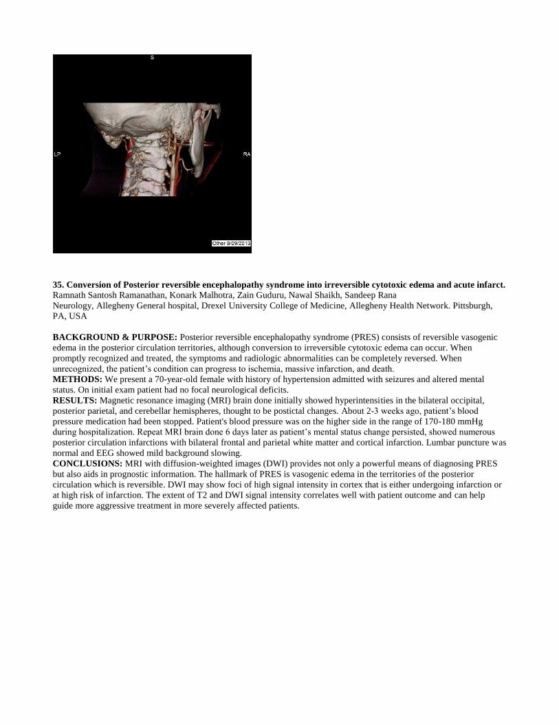
35. Conversion of Posterior reversible encephalopathy syndrome into irreversible cytotoxic edema and acute infarct. Ramnath Santosh Ramanathan, Konark Malhotra, Zain Guduru, Nawal Shaikh, Sandeep Rana
Neurology, Allegheny General hospital, Drexel University College of Medicine, Allegheny Health Network. Pittsburgh,
PA, USA
BACKGROUND & PURPOSE: Posterior reversible encephalopathy syndrome (PRES) consists of reversible vasogenic
edema in the posterior circulation territories, although conversion to irreversible cytotoxic edema can occur. When
promptly recognized and treated, the symptoms and radiologic abnormalities can be completely reversed. When
unrecognized, the patient’s condition can progress to ischemia, massive infarction, and death.
METHODS: We present a 70-year-old female with history of hypertension admitted with seizures and altered mental
status. On initial exam patient had no focal neurological deficits.
RESULTS: Magnetic resonance imaging (MRI) brain done initially showed hyperintensities in the bilateral occipital,
posterior parietal, and cerebellar hemispheres, thought to be postictal changes. About 2-3 weeks ago, patient’s blood
pressure medication had been stopped. Patient's blood pressure was on the higher side in the range of 170-180 mmHg
during hospitalization. Repeat MRI brain done 6 days later as patient’s mental status change persisted, showed numerous
posterior circulation infarctions with bilateral frontal and parietal white matter and cortical infarction. Lumbar puncture was
normal and EEG showed mild background slowing.
CONCLUSIONS: MRI with diffusion-weighted images (DWI) provides not only a powerful means of diagnosing PRES
but also aids in prognostic information. The hallmark of PRES is vasogenic edema in the territories of the posterior
circulation which is reversible. DWI may show foci of high signal intensity in cortex that is either undergoing infarction or
at high risk of infarction. The extent of T2 and DWI signal intensity correlates well with patient outcome and can help
guide more aggressive treatment in more severely affected patients.

�
�
36. Tumefactive Demyelination: Biopsy dilemma. Ramnath Santosh Ramanathan, Konark Malhotra, Zain Guduru, Nawal Shaikh, Sandeep Rana
Neurology, Allegheny General hospital, Drexel University College of Medicine, Allegheny Health Network. Pittsburgh,
PA, USA
BACKGROUND & PURPOSE: Tumefactive demyelinating lesion is a rare atypical form of multiple sclerosis (MS)
which presents as solitary lesions measuring about 2cm or larger on magnetic resonance imaging (MRI) brain. It can mimic
neoplasms, cerebral abscess or stroke. About 50% of tumefactive lesions show ring enhancement which is usually in the
form on an open ring and the incomplete portion of the ring is on the gray matter side of the lesion
METHODS: We present a 35-year-old female with history of hypothyroidism who came with a complaint of right facial
droop, blurred vision, and right upper extremity weakness for 10 days. She also had slurred speech and episodes of
expressive aphasia. On neurology examination patient had tight 7th
facial palsy and right sided weakness.
RESULTS: MRI brain revealed focal areas of abnormal signal intensity in the cerebral white matter with the larger
enhancing lesion at the left centrum semiovale measuring 2.8 x 2.5 cm. with additional 1.2 x 0.7 cm focal area of abnormal
T2 signal in the white matter of the left frontal lobe.
CONCLUSIONS: The term 'tumefactive MS' is applicable when the clinical presentation and MRI findings are
indistinguishable from those of a brain tumor. Tumefactive lesion can be diagnosed using spectroscopy and positron
emission tomography (PET). Biopsy should be avoided and careful follow-up by serial MRI is sufficient to establish the
diagnosis. In general, tumefactive demyelinating lesions respond well to steroids and no radiological evidence of new
lesions is identified after the treatment in most patients.

�
37. Acute necrotizing vasculitis mimicking glioblastoma multiforme in a patient with neuromyelitis optica. Ramnath Santosh Ramanathan, Konark Malhotra, Laxmi Shah, Edwards J Gettings, Nawal Shaikh, Sandeep Rana
Neurology, Allegheny General hospital, Drexel University College of Medicine, Allegheny Health Network. Pittsburgh,
PA, USA
BACKGROUND & PURPOSE: Neuromyelitis optica (NMO) is a demyelinating disease of the central nervous system
characterized by optic neuritis and myelitis. The association of NMO with other autoimmune disease like lupus has been
reported but the association with vasculitis is not described.
METHODS: We present a 51 year female with history of NMO who noticed new-onset left-sided weakness and sensory
deficits.
RESULTS: Magnetic resonance imaging (MRI) brain with contrast showed aggressive ring enhancing mass in the
posterior right frontoparietal region measuring 3.5x2.7x2.5 cm with mass effect, partial enhancement of left lateral
ventricle, medialization of the right uncus and extensive vasogenic edema suspicious for high-grade glioma such as
glioblastoma multiforme. Patient underwent brain biopsy which revealed acute necrotizing vasculitis with small vessel
transmural inflammation and fibrinoid necrosis. Patient was treated with rituximab with a plan to add on treatment with
cyclophosphamide.
CONCLUSIONS: Central nervous system (CNS) necrotizing vasculitis is a rare but potentially life threatening disease
characterized by inflammation of the vessels. It is not reported to be directly a component of NMO. Brain and meningeal
biopsy is the gold standard for the diagnosis of CNS vasculitis as it helps in identifying the characteristic pathology and
also exclude other disorders like lymphoproliferative diseases, infections or sarcoidosis. In our patient necrotizing vasculitis
appeared to be isolated angitis with tumor-like lesion and extensive necrosis, mimicking high-grade glioma on MRI.
Though primary CNS angitis is a devastating disease, the prognosis in small-vessel primary angitis of the CNS (angiogram
negative) is reported to be favorable.

�
�
38. WITHDRAWN
39. WITHDRAWN
40. Unilateral Subarachnoid Hemorrhage and Ipsilateral Retinal Hemorrhage in Infants Joseph M Scheller, Patrick Capone
Winchester Medical Center Winchester, VA, USA
BACKGROUND & PURPOSE: Retinal hemorrhage in the setting of subarachnoid hemorrhage in adults is known as
Terson Syndrome. The finding can be seen in traumatic, spontaneous, and aneurysmal subarachnoid hemorrhage. In young
children the association of retinal hemorrhage and subarachnoid hemorrhage is typically thought to be consistent with
abusive head injury. We reviewed our cases of infantile unilateral subarachnoid hemorrhage to determine if there were
associated ipsilateral retinal hemorrhages.
METHODS: Medical records and MRI scans were reviewed in medicolegal cases of infants who had intracranial bleeding
and were suspected of being victims of abuse. Infants who had skull fractures, bilateral subarachnoid hemorrhage, and
bilateral retinal hemorrhages were excluded.
RESULTS: Five infants were found to have unilateral subarachnoid hemorrhage with ipsilateral retinal hemorrhage. In all
cases abusive head injury was assumed by the clinicians and legal proceedings ensued.
CONCLUSIONS: Unilateral subarachnoid hemorrhage in infants without skull fracture can be associated with ipsilateral
retinal hemorrhage. This finding argues against a shaking mechanism for retinal hemorrhage and is more consistent with
that of Terson syndrome in adults.

41. Embolic stroke post ablation due to atrioesophageal fistula. Nawal Shaikh, Ramnath Santosh Ramanathan, Nnamdi Dike, Konark Malhotra, Sandeep Rana
Neurology, Allegeheny General Hospital, Drexel university College of Medicine, Allegheny Health Network Pittsburgh,
PA, USA
BACKGROUND & PURPOSE: Atrioesophageal fistula (AEF) is a rare cause of stroke that neurologists do not encounter
often since ablations are not done that routinely. Additionally, AEF is an uncommon complication of ablation. Treatment
for the resulting stroke is crucial but unfortunately convoluted because it is difficult to differentiate whether cause is purely
septic emboli, air emboli or combination.
METHODS: A 57 year old Caucasian male with history of previous ischemic stroke, hypertension, diabetes, coronary
artery disease status post bypass with mitral valve repair, patent foramen ovale closure, atrial fibrillation status post
pulmonary vein ablation on coumadin presented with sudden onset of fever, nausea, vomiting, diarrhea and altered mental
status 4 weeks after the ablation procedure. He had mild fluctuating left sided weakness and left facial droop on exam and
the next day developed hematemesis with lower GI bleed.
RESULTS: CT head showed no evidence of acute transcortical infarct. CT angiogram head and neck perfusion was
unremarkable. MRI brain showed numerous regions of restricted diffusion within the bilateral cerebral and cerebellar
hemispheres as well as within the brainstem, consistent with acute embolic infarcts. CT chest with contrast showed minimal
pneumomediastinum with indistinct fat planes between the left atrium and esophagus suggestive of AEF.
CONCLUSIONS: Perioperative period around ablations is critical as fatal complications like AEF can occur despite being
on anticoagulation causing strokes via multiple mechanisms. Intermittent transient ischemic attacks (TIAs) post ablative
procedures should be promptly evaluated by CT chest with contrast and if possible barium swallow for early identification
of AEF.
�
�

42. Rhombencephalosynopsis as an incidental finding in an adult with head trauma Lakshmi Shankar
1,2, Hongyan Li
2
1Cleveland Clinic Foundation Cleveland, OH, USA,
2University of Toledo Toledo, OH, USA
BACKGROUND & PURPOSE: Rhombencephalosynapsis is a rare congenital midline brain abnormality characterized by
dorsal fusion of cerebellar hemispheres, agenesis or hypogenesis of the vermis, fusion of dentate nuclei and superior
cerebellar peduncles with variable supratentorial involvement. Clinical presentation can be of varying severity. This has
been most commonly described in the pediatric age group.
METHODS: Case report
RESULTS: A 36 year old right handed man presented with headache and unsteady gait after head trauma due to assault.
History was positive for alcohol use. Clinical examination showed scanning dysarthria, saccadic eye movements,
spontaneous and gaze-evoked nystagmus, dysmetria and ataxia in both upper and lower extremities, and broad based ataxic
gait with tendency to fall to either side. Patient and his mother noted some minor problems with balance since
childhood.MRI Brain showed fusion of the cerebellar hemispheres, hypoplasia of posterior cerebellar incisures,
Hypoplastic nodulus, mild ventriculomegaly with abnormally shaped 4th
ventricle, residual anterior vermis at the level of
junction of aqueduct and 4th
ventricle, residual posterior vermis and unremarkable superior medullary velum, aqueduct and
pons. No forebrain abnormalities were noted.
CONCLUSIONS: Alcohol use likely exacerbated preexisting cerebellar findings in this adult patient with
Rhombencephalosynapsis.
�
�
43. Acute Paradoxical Embolic Ischemic Stroke Related to Intravenous Line Pressure Injection in a Patient with

Superficial Thrombophlebitis Lakshmi Shankar, Russell Cerejo, Mei Lu
Cleveland Clinic Foundation Cleveland, OH, USA
BACKGROUND & PURPOSE: Paradoxical embolic stroke can occur in right to left shunt, most commonly with deep
venous thrombosis. The incidence of acute ischemic stroke with superficial venous clots is relatively low, likely due to
presence of competent venous valves. Pressure injection through IV access distal to a thrombus could increase the chance
of embolic stroke in the presence of a right to left shunt.
METHODS: Case report
RESULTS: A 67-year-old-female admitted for chest pain, complained of seeing 'shimmering lights' for less than a minute
immediately after pressure injection of a radio-tracer through right arm IV access. Two weeks prior she had a similar
episode when another medication was pressure injected through an IV access in the same arm. She had no other
neurological complaints and neurological exam was normal. MRI Brain showed bilateral hemispheric sub-centimeter sub-
acute ischemic strokes. Work-up revealed acute superficial thrombophlebitis in right median cubital vein (proximal to IV
access) and great saphenous vein near the sapheno-femoral junction (site for previous vein graft for coronary artery bypass
graft surgery). Transthoracic echocardiogram and transcranial doppler were positive for a right to left circulatory shunt
through a patent foramen ovale. IV access was switched to the left arm and no further episodes were noted with use of the
new IV access. She was placed on anticoagulation therapy for acute superficial thrombophlebitis which was a possible
cause of the ischemic strokes.
CONCLUSIONS: This case illustrates a cause for paradoxical embolic stroke due to IV line related thrombosis in the
superficial venous system.
44. Brain Networks Underlying visual perception of ambiguous figures:A study with fMRI and dyanamic causal
modelling. Hariom Sharma
1, Rekha varrier
2, Shripad Kondra
3, Prasun K Roy
4
1NIMHANS Bangalore, India,
2NBRC Gurgaon, India,
3Mando Softtech India Pvt. Ltd. Bangalore, India,
4NBRC Gurgaon,
India
BACKGROUND & PURPOSE: How visual perception of ambiguous figures was processed in brain and which networks
were activated. To detect unusual perception at a subclinical level, the inkblot test has been used as a diagnostic and
research tool for the past 90 years (Rorschach, 1921 Ganellen, 1996 Jorgensen et al., 2000 Kircher et al., 2001 Exner, 2003
Jimura et al., 2009) .
METHODS: A total of 7 healthy volunteers (4 females and 3 males) ranging in age from 18 to 25 years participated in the
study.A total of ten ambiguous figures were shown to the subjects in two experimental runs. A block design paradigm was
used in fMRI experiment.The duration of stimuli exposure was constant at 15 seconds and 10 seconds for fixcation.
RESULTS: We were extracted two networks of effective connectivity. One of them was task positive and the other one
was task-negative network.Task positive network of effective connectivity informed, information of visual perception
diverge from visual (BA 19) to frontal area (BA 9), processed this visual information in two networks, frontal (BA
6,9,10,11,44) and fronto-parietal (BA 7,23,9,10).These Signals are presumably attributed for perceptual changes.Other
network of effective connectivity is task negative network, probably it is responsible for visual imagination.
CONCLUSIONS: We attempted to utilize the ICA and DCM techniques to portrait the effective connectivity showing
flow of information for the visual perception of ambiguous figures,succeed to find out the task positive and negative
networks.
�
�
45. Cerebral Vasospasm Following Transsphenoidal Pituitary Macroadenoma Resection. Case Report. Yazan M. Suradi
1, Crystal Dixon
2, David Rose
3
1University of South Florida Tampa, FL, USA,
2University of South Florida Tampa, FL, USA,
3University of South Florida
Tampa, FL, USA
BACKGROUND & PURPOSE: Cerebral vasospasm (CV) frequently occurs subsequent to aneurysmal-subarachnoid-
hemorrhage and is responsible for significant neurological deterioration and morbidity. However, CV following

transcranial pituitary adenoma resection has been extremely infrequent, with only few cases documented. Consequently,
CV and its management has remained a great challenge ever since its initial report in 1951.
METHODS: A 19 year-old female with a known history of pituitary-macroadenoma with secondary Cushing's disease,
status-post transsphenoidal resection at age eighteen, presented with difficulty speaking and and right arm weakness. The
symptoms began seven-days after left-sided fronto-temporal craniotomy with orbital osteotomy, carried-out for
supplementary resection of the cavernous-sinus adenoma. Neurological examination revealed decreased motor-strength in
right-upper-extremity and expressive aphasia. NIH-Stroke-Scale-Score was 11.
RESULTS: Brain-MRI confirmed acute stroke in the distribution of the left-middle-cerebral (MCA) and left anterior-
cerebral (ACA) arteries. Head-MRA showed narrowing of left proximal M1-MCA and A1- ACA, which was concerning
for vasospasm. Therefore, triple-HHH therapy was initiated immediately. Concurrently, the patient was transported to the
angiography-lab for intra-arterial (IA) verapamil and nicardipine treatment. Over the next few days, IA-treatment was
repeated nine times. Though each IA-treatment offered symptomatic and angiographic improvement, occasionally
symptoms would recur afterward. Still, the weakness and aphasia markedly improved by the time of discharge.
CONCLUSIONS: The pathogenesis of CV after tumor-surgery is not very well understood. Various etiologies associated
with CV status-post surgery include direct arterial wall damage, blood deposition in the basal cisterns, aseptic meningitis
and/or release of chemical substances during tumor resection. Management can be very challenging therefore the key to
success is a high-index-of suspicion and early intervention.
�
�
46. Utility of brain MRI in the diagnosis of Vogt–Koyanagi–Harada syndrome Reuben Mari Valenzuela, Jorge C Kattah
University of IL College of Medicine Peoria Peoria, IL, USA
BACKGROUND & PURPOSE: Vogt Koyanagi Harada syndrome (VKH syndrome) is a rare disease predominantly
affecting women with an incidence of 1 to 6 per million in the US. The diagnosis is made clinically, however fluorescein
angiography (FA) and optical coherence tomography (OCT) are used to support the diagnosis. Brain MRI is not a common
imaging modality, however in the absence of FA and OCT, it can be helpful in the diagnosis.
METHODS: This is a case report where extensive review of history, laboratory evaluation and imaging of VKH syndrome
is performed.
RESULTS: A 32 year old man with vitiligo presents with headache and progressive right vision (OD) loss to almost
counting finger (CF). Past medical history is unremarkable and he denies ocular trauma. Fluorescein angiography shows
focal areas of leakage in the retinal pigment epithelium while the brain MRI shows fluid collection in the choroid of the
right eye with some meningeal enhancement. Prednisone was initially started, however because of persistent
hyperglycemia, azathioprine was initiated while prednisone was tapered. Vision improved from CF to 20/20 OD while the
left eye remained unaffected.
CONCLUSIONS: Since VKH syndrome presents diversely, ancillary procedures like FA, ultrasound and OCT are
commonly employed to arrive at a diagnosis, however the unavailability of OCT and FA in the rural setting makes this
difficult. Brain MRI is useful especially in the absence of FA and OCT. The most distinguishing MRI feature of VKH
syndrome include choroidal and retrobulbar contrast enhancement in the orbits, white matter abnormalities on FLAIR and
meningeal enhancement.
�
�
47. Assessment of Brain Plasticity Induced by rTMS in Patient with Writer Cramp Using Resting State Connectivity Bhaskar M Venkateshappa
1, Rose D Bharath
1, Arun K Gupta
1, Pramod K Pal
2, Rajanikanth Panda
1, Ketan jhunjhunwala
2
1Department of NIIR, NIMHANS Bangalore, India,
2Department of Neurology, NIMHANS Bangalore, India
BACKGROUND & PURPOSE: Writer's cramp (WC) is a task-specific form of focal dystonia. The pathophysiology
related to abnormal intrinsic functional connections in the brain can be modulated by low frequency transcranial magnetic
stimulation (rTMS) to increase the connectivity and hence improve symptoms of WC.
METHODS: A prospective study of twenty subjects, ten WC patients and ten age and gender matched healthy subjects
using resting-state EEG-fMRI. 1Hz rTMS, (900 pulses for 15 minutes) given in the dominant sensory motor cortex. All
subjects were evaluated with pre and post rTMS using 3T MRI and 32-channel MR compatible EEG. All were instructed to
lie-down with eye closed, awake and relaxed state. fMRI with EPI sequence (TR: 3000, 180 dynamics), MPRAGE and
EEG were acquired. The resting state functional connectivity of the sensory motor networks (Broadmann Areas 3, 4, 6)
were computed. Region of interest based connectivity of fMRI was processed using SPM 8 & CONN toolbox with p<0.05
FDR correction and connectivity of EEG was processed using EEGLAB toolbox.
RESULTS: In resting state EEG-fMRI, connectivity of the sensory motor area decreased in all WC patients. Post rTMS,
increase in connectivity not only on dominant side but also the both hemisphere of sensory motor cortex, as seen in the
healthy controls.
CONCLUSIONS: EEG-fMRI is a valuable technique in understanding the effects of rTMS in patient with WC. 1 Hz

rTMS consistently improves the impaired connectivity of the sensory motor cortex as well the beta spectral power in the
EEG which is comparable to the normal subjects.
�

�
48. Recurrent spells of unresponsiveness secondary to bilateral carotid stenosis caused by giant cell arteritis

Mervat N Wahba
Neurology Memphis, TN, USA
BACKGROUND & PURPOSE: Occlusive lesions of the internal carotid artery (ICA) constitute the commonest cause of
cerebrovascular accidents. They are usually atherosclerotic in origin, although occasionally other causes such as dissection,
fibromuscular dysplasia, or vasculitic process may be etiogenic. Bilateral ICA stenosis is a particularly devastating
condition with an annual stroke rate of 7.6% per year and an annual mortality of 43%.
METHODS: A 63-year-old man presented with of recurrent spells of falling to the ground triggered by an upright position.
This may be associated with shaking of the extremities. No epileptogenic or cardiac etiology was found.. A CT angiogram
showed over 90% stenosis of the bilateral internal carotid arteries at the siphon. He was recently diagnosed with giant cell
arteritis . He was placed on prednisone.
RESULTS: The patient's constitutional symptoms and jaw claudication improved on prednisione but he continued to have
the episodes of altered consciouness with brief jerking of the limbs. A brief course of anticoagulation was tried with some
improvement but no resolution of his episodes. Bilateral angioplasty of his carotid arteries was performed with resolution of
his symptoms. Stroke risk factors were addressed.
CONCLUSIONS: The presence of bilateral carotid stenosis in patients with GCA is strongly associated with future risk of
neurologic deficits. It may be difficult to separate atherosclerotic lesions from vasculitic lesions related to GCA, however,
the presence of localized focal stenosis with relatively healthy arteries above and below the lesions may point to a vasculitic
process. Rigorous attention should be given to modification of atherosclerotic risk factors as well as recognizing and
treating the rare manifestation of GCA.
49. Lower ultrasonographic cut-off point of optic nerve sheath diameter for raised intracranial cerebral pressure in
Chinese Population lijuan wang
1, yingqi xing
1, yan yao
2, wenhua lin
3, jiachun feng
1
1Department of neurology, First Hospital of Jilin University changchun, China,
2Department of Epidemiology and
Biostatistics, School of Public Health, Jilin Unviersity changchun, China, 3Department of medicine and therapeutics, Prince
of Wales Hospital hongkong, China
BACKGROUND & PURPOSE: Ultrasonography of optic nerve sheath may help identify raised intracranial pressure
(ICP). As reported, an ONSD (optic nerve sheath diameter) above 5 mm on ultrasound was indicated for raised ICP in
Caucasian population. However, ONSD cut-off for raised ICP in Chinese patients has not been established .We aim to
identify the optimal ONSD for ICP > 20 mmHg in Chinese.
METHODS: This is a prospective observational study enrolling patients who were suspected raised ICP and performed
lumbar puncture to verify ICP. ICP > 20 mmHg was considered as abnormally high. We excluded patients with ophthalmic
diseases. ONSD was carried out before lumbar puncture. ONSD was measured bilaterally 3 mm behind the globe using a 9
MHz linear probe on the closed eyelids of supine patients. The average of two ONSD measurements was calculated. We
analysed clinical differences between abnormal and normal ICP groups. An ROC curve was constructed to determine the
optimal ONSD cut-off point for abnormal ICP.
RESULTS: Totally 234 patients were recruited. Eighty-two patients had raised ICP (152 normal ICP). Multivariate
analysis showed ONSD was the only independent predictor for raised ICP. ROC curve analysis revealed area under the
curve = 0.949 (95% CI 0.917-0.981). Optimal ONSD cut-off point for detection of ICP > 20 mmHg was &ge3.775 mm
with sensitivity of 90.2% and specificity of 96.1%.
CONCLUSIONS: The ONSD criterion for raised ICP in Chinese patients is remarkedly less than Caucasian. It
suggests that ethnic differences should be noted and applied corresponding ultrasonographic criteria when ONSD was used
to diagnose raised ICP.
50. Pediatric Nasopharyngeal Carcinoma: Imaging, Pathological and Clinical Evaluation Alfred Weber
1, Mary Beth Cunnane
2, Amy Juliano
3, M Larvie
4, Hugh Curtin
5
1Radiology MEEI Boston, MA, USA,
2Radiology MEEI Boston, MA, USA,
3Radiology MEEI Boston, MA, USA,
4Radiology MGH Boston, MA, USA,
5Radiology MEEI Boston, MA, USA
BACKGROUND & PURPOSE: The purpose of this exhibit is to evaluate the clinical, pathological and imaging findings
of nasopharyngeal carcinoma (NPC) in children and adolescents and compare the findings and differences with adult
nasopharyngeal carcinoma
METHODS: We retrieved cases from the files of Mass Eye and Ear Infirmary, Mass General Hospital and King Faisal
Specialist Hospital in Riyadh, Saudi Arabia over a period of 12 years. A total of 65 cases-up to the age of 20 years were

analysed with CT, MRI and in a few cases PET-CT
RESULTS: Carcinoma in children and adolescents manifested as a bulky large mass in the nasopharynx and in no cases as
an infiltrative flat lesion. Frequently the extension occurred to the oropharynx, oral cavity/paranasal sinuses, orbital cavity
and parapharyngeal space. Bony erosion in the skull base, principally clivus, was demonstrated in 50% of cases.
Lymphnode metastases to the neck were found in 90% of cases commonly manifesting as the initially finding. Noteworthy
was the complete clearing of the bulky tumor masses in the nasopharynx and neck following radiation therapy in
conjunction with multiagent chemotherapy. PET scanning was superior to CT and MRI in detecting recurrences and /or
residual tumor in the neck and nasopharynx. The histopathology in all cases was undifferentiated carcinoma
CONCLUSIONS: Carcinoma of the nasopharynx should be considered in children especially when older than 10 years of
age and when presenting with a large nasopharyngeal or a neck mass. Bone destruction in the base of skull is highly
suggestive of NPC. CT and MRI are the imaging modalities for assessment of the location and extent of the lesion while
PET-CT is useful for evaluating recurrence and regional and distant metastases. Undifferentiated carcinoma is the most
common histopatholgical type. NPC responds optimally to radiation therapy in conjunction with chemotherapy with
recurrent tumor and distant metastases
51. A case report on primary CNS vasculitis Xin Xin Yu
1, Cynthia Bamford
2
1Department of Neurology Cleveland, OH, USA,
2Neurological Center for Pain Cleveland, OH, USA
BACKGROUND & PURPOSE: Primary CNS vasculitis is a rare inflammatory condition that affects the CNS vessels
without systemic involvement.  It can be mimicked by a number of disorders due to its variable clinical presentations
and nonspecific radiographic findings. Brain tissue biopsy remains the gold standard for the diagnosis.
METHODS: Case report
RESULTS: A 55 year-old woman was admitted with 1-year fluctuating symptoms of headache, right side weakness and
numbness, blurry vision and dysarthria. Her initial MRI showed a mass lesion in the left parietal lobe with Gad
enhancement and vasogenic edema. Metabolic, infectious and autoimmune testing including repeat CSF studies was
negative. Brain biopsy showed focal acute and chronic inflammation with associated necrosis. Cerebral angiogram did not
find evidence of vasculitis. PET was negative for malignancy. Given the stability of her symptoms, she was monitored with
serial MRIs. One month later, she presented with worsening of symptoms, her repeat MRI showed mild progression of
signal abnormality including the nodular enhancement. Repeat brain biopsy showed focal angiocentric chronic
inflammation with necrosis and vascular sclerosis consistent with non-necrotizing vasculitis. In the absence of evidence of
systemic autoimmune disease, malignancy, and infection, she was treated with prednisone 60mg once daily for primary
CNS vasculitis with clinical improvement at 3-months follow up.
CONCLUSIONS: Primary CNS vasculitis is a difficult diagnosis to make and can be missed easily by clinicians due
to its clinical heterogeneity and lack of validated diagnostic criteria. Our case report adds to the existing literature on the
variable clinical presentations, radiographic manifestation that can be seen in primary CNS vasculitis.
�
�