Neurofibromatosis type 1
27
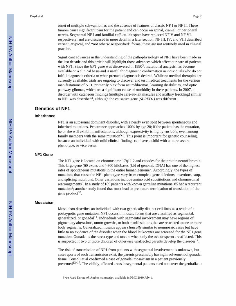
Neurofibromatosis type 1 Kevin P. Boyd, MD, Bruce R. Korf, MD, PhD, and Amy Theos, MD * University of Birmingham at Alabama, Department of Dermatology, EFH 414, 1530 3rd Ave S, Birmingham, AL 35294-0009, Business: 205-934-5188, Fax: 205-934-5766, E-mail: [email protected] Abstract Neurofibromatosis type 1 (NF1) is an autosomal dominant, multisystem disorder affecting approximately 1 in 3500 people. Significant advances in the understanding of the pathophysiology of NF1 have been made in the last decade. While no medical therapies are currently available, trials are ongoing to discover and test medical treatments for the various manifestations of NF1, primarily plexiform neurofibromas, learning disabilities, and optic pathway gliomas. Additionally, mutational analysis has become available on a clinical basis and is useful for diagnostic confirmation in individuals who do not fulfill diagnostic criteria or when prenatal diagnosis is desired. There are several disorders which may share overlapping features with NF1; in 2007, a disorder with cutaneous findings similar to NF1 was described. This paper addresses the dermatologist's role in diagnosis and management of NF1 and describes the variety of cutaneous and extracutaneous findings in NF1 to which the dermatologist may be exposed. Keywords neurofibromatosis type 1; cafe-au-lait macule; SPRED1; neurofibroma Introduction Neurofibromatosis type 1 (NF1) is an autosomal dominant, multisystem disorder affecting approximately 1 in 3500 people. The earliest historical evidence first appeared in the 13 th century but it wasn't until Friedrich Daniel von Recklinghausen published his landmark paper (in German) On the Multiple Fibromas of the Skin and Their Relationship to the Multiple Neuromas in 1882 that neurofibromatosis began gaining recognition as a distinct disorder. More recently, in 1956 Crowe, Schull, & Neel published a milestone manuscript detailing the numerous manifestations of this disorder 1 . In 1982, Riccardi classified the heterogeneous neurofibromatosis disorders into eight categories 2 . These have not been universally-accepted, although several persist. Neurofibromatosis type I and II have remained as originally classified. NF II results from a mutation in the NF2 gene and is characterized by bilateral vestibular schwannomas and various other tumors. NF VII is now referred to as schwannomatosis and is distinguished by the late- * To whom correspondence and reprint requests should be sent, Dr. Amy Theos, MD, University of Alabama at Birmingham. Conflict of Interest Disclosure: None declared CME Learning Objectives: Following this activity, the participant will be able to: 1) discuss the indications and limitations of genetic testing in NF1, 2) distinguish common and uncommon cutaneous findings, 3) and recognize the dermatologist's role in diagnosis and management. Capsule summary: Many advances have increased our ability to understand and diagnose neurofibromatosis type 1. This paper will review current thinking on the pathogenesis, discuss the visible manifestations, and describe a methodical approach to diagnosis. NIH Public Access Author Manuscript J Am Acad Dermatol. Author manuscript; available in PMC 2010 July 1. Published in final edited form as: J Am Acad Dermatol. 2009 July ; 61(1): 1–16. doi:10.1016/j.jaad.2008.12.051. NIH-PA Author Manuscript NIH-PA Author Manuscript NIH-PA Author Manuscript
-
Upload
independent -
Category
Documents
-
view
1 -
download
0
Transcript of Neurofibromatosis type 1
Neurofibromatosis type 1
Kevin P. Boyd, MD, Bruce R. Korf, MD, PhD, and Amy Theos, MD*University of Birmingham at Alabama, Department of Dermatology, EFH 414, 1530 3rd Ave S,Birmingham, AL 35294-0009, Business: 205-934-5188, Fax: 205-934-5766, E-mail:[email protected]
AbstractNeurofibromatosis type 1 (NF1) is an autosomal dominant, multisystem disorder affectingapproximately 1 in 3500 people. Significant advances in the understanding of the pathophysiologyof NF1 have been made in the last decade. While no medical therapies are currently available, trialsare ongoing to discover and test medical treatments for the various manifestations of NF1, primarilyplexiform neurofibromas, learning disabilities, and optic pathway gliomas. Additionally, mutationalanalysis has become available on a clinical basis and is useful for diagnostic confirmation inindividuals who do not fulfill diagnostic criteria or when prenatal diagnosis is desired. There areseveral disorders which may share overlapping features with NF1; in 2007, a disorder with cutaneousfindings similar to NF1 was described. This paper addresses the dermatologist's role in diagnosis andmanagement of NF1 and describes the variety of cutaneous and extracutaneous findings in NF1 towhich the dermatologist may be exposed.
Keywordsneurofibromatosis type 1; cafe-au-lait macule; SPRED1; neurofibroma
IntroductionNeurofibromatosis type 1 (NF1) is an autosomal dominant, multisystem disorder affectingapproximately 1 in 3500 people. The earliest historical evidence first appeared in the 13th
century but it wasn't until Friedrich Daniel von Recklinghausen published his landmark paper(in German) On the Multiple Fibromas of the Skin and Their Relationship to the MultipleNeuromas in 1882 that neurofibromatosis began gaining recognition as a distinct disorder.More recently, in 1956 Crowe, Schull, & Neel published a milestone manuscript detailing thenumerous manifestations of this disorder1.
In 1982, Riccardi classified the heterogeneous neurofibromatosis disorders into eightcategories2. These have not been universally-accepted, although several persist.Neurofibromatosis type I and II have remained as originally classified. NF II results from amutation in the NF2 gene and is characterized by bilateral vestibular schwannomas and variousother tumors. NF VII is now referred to as schwannomatosis and is distinguished by the late-
* To whom correspondence and reprint requests should be sent, Dr. Amy Theos, MD, University of Alabama at Birmingham.Conflict of Interest Disclosure: None declaredCME Learning Objectives: Following this activity, the participant will be able to: 1) discuss the indications and limitations of genetictesting in NF1, 2) distinguish common and uncommon cutaneous findings, 3) and recognize the dermatologist's role in diagnosis andmanagement.Capsule summary: Many advances have increased our ability to understand and diagnose neurofibromatosis type 1. This paper willreview current thinking on the pathogenesis, discuss the visible manifestations, and describe a methodical approach to diagnosis.
NIH Public AccessAuthor ManuscriptJ Am Acad Dermatol. Author manuscript; available in PMC 2010 July 1.
Published in final edited form as:J Am Acad Dermatol. 2009 July ; 61(1): 1–16. doi:10.1016/j.jaad.2008.12.051.
NIH
-PA Author Manuscript
NIH
-PA Author Manuscript
NIH
-PA Author Manuscript
onset of multiple schwannomas and the absence of features of classic NF I or NF II. Thesetumors cause significant pain for the patient and can occur on spinal, cranial, or peripheralnerves. Segmental NF I and familial café-au-lait spots have replaced NF V and NF VI,respectively, and are discussed in more detail in a later section. NF III, IV, and VIII describedvariant, atypical, and “not otherwise specified” forms; these are not routinely used in clinicalpractice.
Significant advances in the understanding of the pathophysiology of NF1 have been made inthe last decade and this article will highlight those advances which affect our care of patientswith NF1. Since the NF1 gene was discovered in 19903, mutational analysis has becomeavailable on a clinical basis and is useful for diagnostic confirmation in individuals who do notfulfill diagnostic criteria or when prenatal diagnosis is desired. While no medical therapies arecurrently available, trials are ongoing to discover and test medical treatments for the variousmanifestations of NF1, primarily plexiform neurofibromas, learning disabilities, and opticpathway gliomas, which are a significant cause of morbidity in these patients. In 2007, adisorder with cutaneous findings (multiple café-au-lait macules and axillary freckling) similarto NF1 was described4, although the causative gene (SPRED1) was different.
Genetics of NF1Inheritance
NF1 is an autosomal dominant disorder, with a nearly even split between spontaneous andinherited mutations. Penetrance approaches 100% by age 20; if the patient has the mutation,he or she will exhibit manifestations, although expressivity is highly variable, even amongfamily members with the same mutation5,6. This point is important for genetic counseling,because an individual with mild clinical findings can have a child with a more severephenotype, or vice versa.
NF1 GeneThe NF1 gene is located on chromosome 17q11.2 and encodes for the protein neurofibromin.This large gene (60 exons and >300 kilobases (kb) of genomic DNA) has one of the highestrates of spontaneous mutations in the entire human genome7. Accordingly, the types ofmutations that cause the NF1 phenotype vary from complete gene deletions, insertions, stop,and splicing mutations. Other variations include amino acid substitutions and chromosomalrearrangements8. In a study of 189 patients with known germline mutations, 85 had a recurrentmutation9; another study found that most lead to premature termination of translation of thegene product10.
MosaicismMosaicism describes an individual with two genetically distinct cell lines as a result of apostzygotic gene mutation. NF1 occurs in mosaic forms that are classified as segmental,generalized, or gonadal11. Individuals with segmental involvement may have regions ofpigmentary alterations, tumor growths, or both manifestations that are restricted to one or morebody segments. Generalized mosaics appear clinically similar to nonmosaic cases but havelittle to no evidence of the disorder when the blood leukocytes are screened for the NF1 genemutation. Gonadal is the rarest type and occurs when only the ova or sperm are affected. Thisis suspected if two or more children of otherwise unaffected parents develop the disorder12.
The risk of transmission of NF1 from patients with segmental involvement is unknown, butcase reports of such transmission exist; the parents presumably having involvement of gonadaltissue. Consoli et al confirmed a case of gonadal mosaicism in a patient previouslypresented13-17. The visibly-affected areas in segmental patients need not cover the genitalia to
Boyd et al. Page 2
J Am Acad Dermatol. Author manuscript; available in PMC 2010 July 1.
NIH
-PA Author Manuscript
NIH
-PA Author Manuscript
NIH
-PA Author Manuscript
confer risk of transmission, a fact which is important for counseling11. There are even somecases of segmental NF1 apparently being “transmitted” to offspring; the mechanism (if oneexists) behind this is unclear16,18. One case reports exists of a 29-year old man with clearevidence of segmental NF1 whose parents and siblings exhibited no evidence of NF1 but whosematernal aunt, cousin, and 2nd cousin had classic NF119. It is possible that this represents twoindependent NF1 mutations in the two branches of the family, though that was not tested inthe published data.
Pathogenesis of NF1Neurofibromin, the NF1 gene protein product, is a tumor suppressor expressed in many cells,primarily in neurons, glial, Schwann cells and early in melanocyte development20. This proteinis a regulator of ras guanosine triphosphatase activity (GTPase-activating protein, GAP) andas such serves as a regulator of signals for cell proliferation and differentiation5. (Figure 1).The loss of function of neurofibromin may therefore remove this regulation, and lead touncontrolled cell proliferation. Schwann cells in neurofibromas, and melanocytes in café-au-lait macules have a mutation in both NF1 alleles, including a germline and an acquired somaticmutation and are considered the primary tumor cell in their respective cutaneous manifestation.Based on these findings, it is likely that NF1 functions as a tumor suppressor gene21,22. Bothof these cell types are descendants of neural crest. The exact timing of the acquired mutationis unknown but is crucial in the development of the various manifestations; experiments haveshown that NF1 gene inactivation at the neural crest stage and mature Schwann cell stage donot lead to tumor formation. However, in a progenitor intermediate step between these twostages, tumor formation occurs with NF1 gene inactivation23.
DiagnosisDiagnostic Criteria
In 1987, seven cardinal diagnostic criteria for NF1 were established, two of which must be metin order to diagnose an individual with NF1. (Figure 2). These diagnostic criteria have beenshown to be both highly specific and sensitive for adults with NF124. Because some of thecriteria may not manifest until later in life, they are not as sensitive in children, especially thoseunder the age of 8. A 2000 retrospective review of nearly 1900 cases of NF1 found that 46%of sporadic cases did not meet criteria by age 1; however, 97% met criteria by age 8, and allfulfilled them by age 2025.
The diagnosis of NF1 is generally straightforward when one follows the NIH guidelines;however, patients with these classic findings may not first present to a dermatologist. Ofparticular interest to dermatologists is the approach to diagnosis of a patient who has somefeatures of NF1 but fails to fully meet diagnostic criteria. A diagnostic algorithm gearedtowards the dermatologist is provided in Figure 2. Further, it is important to recognize keyhistorical or physical findings that may require – beyond a referral to genetics – a timelyconsultation with varying specialties including orthopedics, ophthalmology, or neurology.
Genetic TestingRecently molecular testing for NF1 has become clinically available. Because of the large sizeof the NF1 gene and the lack of mutation hotspots, a multi-step detection protocol is preferred.A comprehensive screening approach to the NF1 gene found mutations in greater than 95% oftested subjects (including both spontaneous and inherited mutations) fulfilling NIH diagnosticcriteria8. This comprehensive approach begins with an optimized protein truncation test,followed by FISH analysis, direct sequencing, long-range RT-PCR with Southern blot analysis,and/or cytogenetic analysis, if necessary, and is utilized in only a select few laboratories(http://www.genetests.org; August 13, 2008). Molecular testing is not indicated for the routine
Boyd et al. Page 3
J Am Acad Dermatol. Author manuscript; available in PMC 2010 July 1.
NIH
-PA Author Manuscript
NIH
-PA Author Manuscript
NIH
-PA Author Manuscript
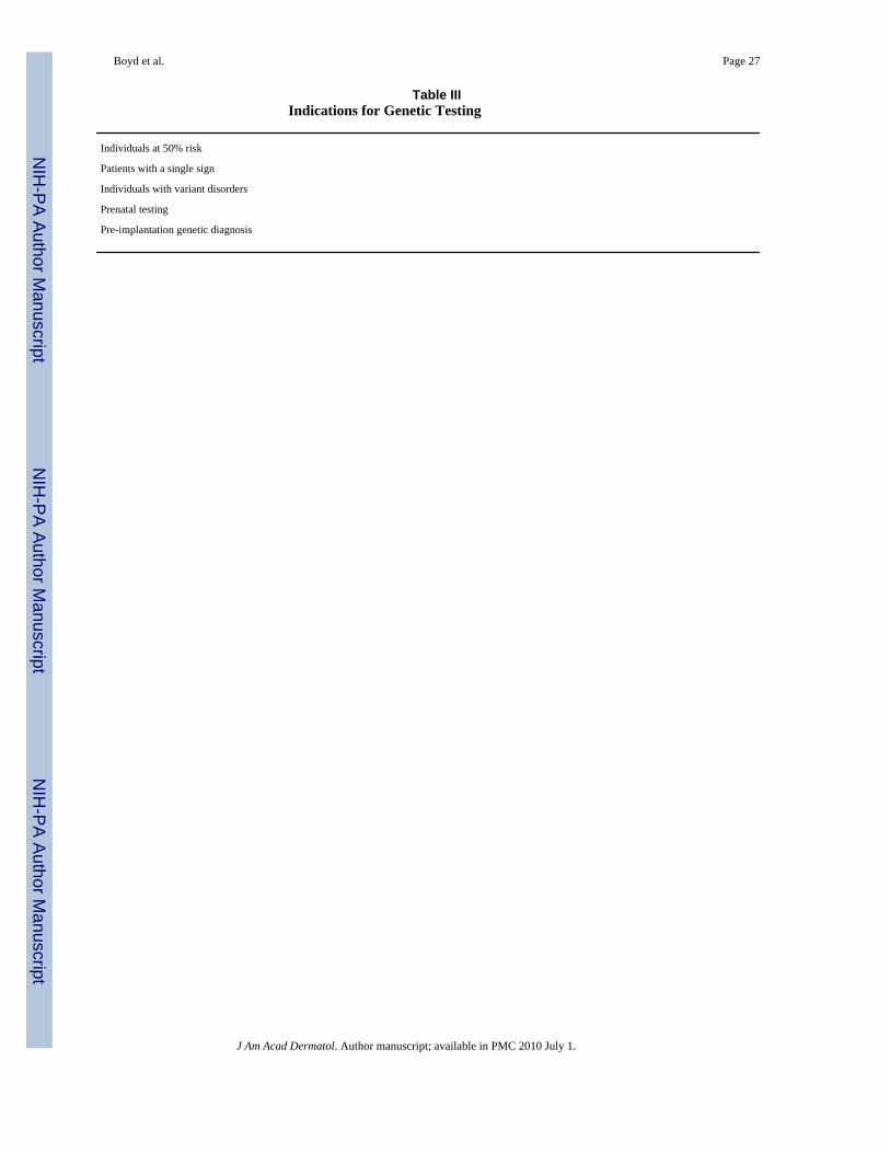
clinical care of patients with NF1, but can be helpful in individuals suspected of having NF1(e.g. a young child with multiple café-au-lait spots and unaffected parents or a patient with asingle criterion) or when prenatal or preimplantation genetic diagnosis is desired (Table III).Some experts are calling for a revision of the 1987 criteria to include a positive molecular testas a definitive, stand-alone diagnostic criterion.
A limitation of genetic testing is the lack of genotype-phenotype correlations. Therefore, whileuseful for diagnostic confirmation, a positive test will not predict disease severity or outcome.However, there are two exceptions. The first results from complete loss of the NF1 gene alongwith multiple contiguous genes and occurs in 4-5% of patients with NF126. Patients with wholegene deletion present with a very large neurofibroma burden, more severe cognitiveimpairment, large hands and feet, dysmorphic facial features, and have a higher lifetime riskof developing malignant peripheral nerve sheath tumors (MPNST)27,28. A 3-base pair inframedeletion in exon 17 of the NF1 gene is the second exception. Patients with this genetic mutationhave an absence of cutaneous neurofibromas and appear to have a lower incidence of seriouscomplications29.
Differential DiagnosisNF1-Like Syndrome: An important disorder to distinguish from NF1 is NF1-like syndrome.This disorder, first described in 2007, consists primarily of multiple café-au-lait macules,axillary freckling, and macrocephaly4. Comprehensive mutational analysis for the NF1 genewas performed on affected individuals from five families; all were negative. Through genomelinkage studies, they localized the disorder to chromosome 15 and, based on disease distributionwithin the families, determined that it was autosomal dominant. The responsible gene isSPRED1, which is involved in RAS-MAPK (mitogen-activated protein kinase) regulation,similar to neurofibromin (Figure 1). Lipomas, Noonan-like facies, learning and behavioralproblems were present in some of the families; however, it was uncertain how these findingswere related to the genetic alteration4. This disorder warrants further study to better characterizeits phenotype and distinguish it from NF1. At publication, clinical testing for this disorder wasavailable only at two laboratories in the United States. (http://www.genetests.org; October 20,2008). NF1-like syndrome should be suspected in families in which individuals present withmultiple café-au-lait macules, but without Lisch nodules, neurofibromas, and NF1 geneabnormalities.
Familial Café-au Lait Spots (FCALS): Familial café-au-lait spots is a disorder with anuncertain relationship to NF1. Riccardi described two families whose affected members hadonly multiple café-au-lait macules (CALMs) and felt that they represented a disorder that wasseparate from the classic form30. Affected individuals do not develop other manifestations ofNF1. It segregates as an autosomal dominant disorder and there are conflicting results onwhether FCALS is linked to NF131,32. Charrow et al contended that it was a separate entityand should be treated as such. It does not appear to confer any increased risk of developingclassical NF1.
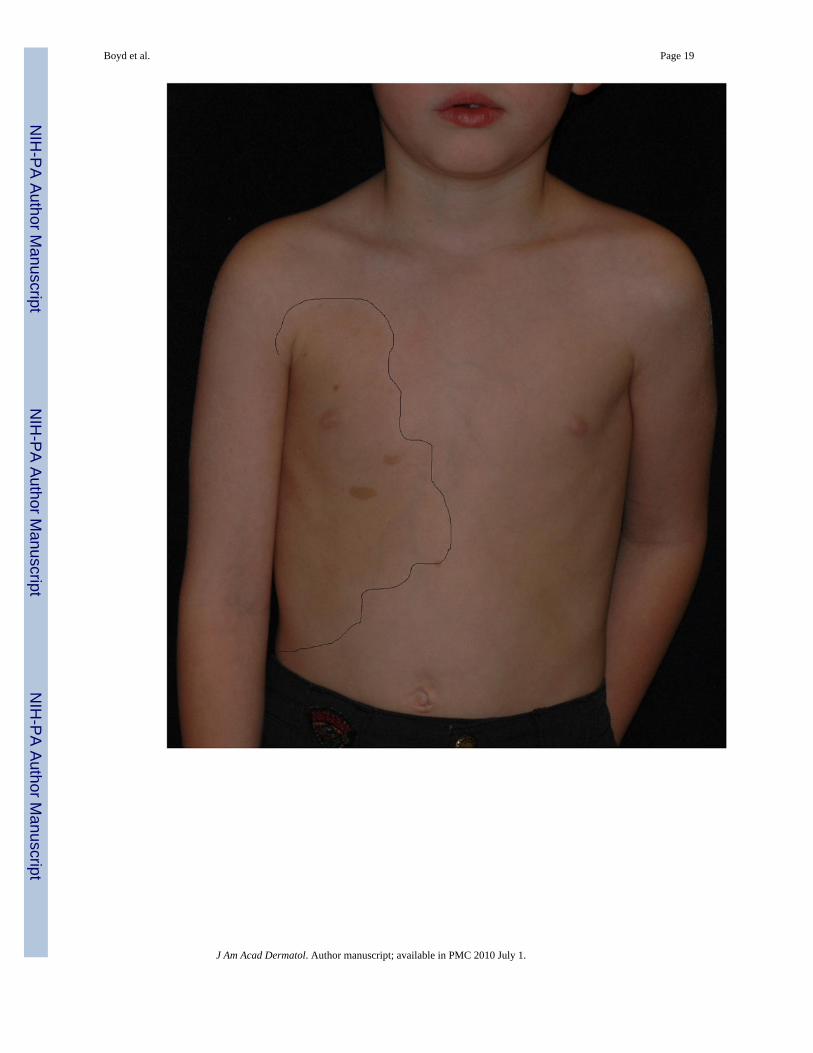
Segmental NF1: As mentioned previously, NF1 can manifest as a mosaic (segmental) disorderin both localized and generalized forms. Localized, segmental neurofibromatosis should beconsidered when skin findings are localized to a particular area of the skin and there is nofamily history (Figures 3a & b). This is in contrast to FCALS, wherein there is often a familyhistory of “multiple birthmarks” and the sole finding is that of CALMs. Testing of lesionaltissue (neurofibromas, café-au-lait macules) is available and is of particular use in thesepatients. Maertens et al. tested lesional tissue in three patients with segmental NF1, each ofwhich had different skin manifestations (pigmentary alone, tumor alone, and a combination ofboth). Through comprehensive testing, mutations were found in all affected cells; biallelic
Boyd et al. Page 4
J Am Acad Dermatol. Author manuscript; available in PMC 2010 July 1.
NIH
-PA Author Manuscript
NIH
-PA Author Manuscript
NIH
-PA Author Manuscript
inactivation was found in either the Schwann cells of neurofibromas or in the melanocytes ofCALMs21. These three cases illustrate not only the variability of the phenotypic expression ofNF1 but also the ability to reliably test lesional tissue for the NF1 mutation if the diagnosis isuncertain. In 2001, Ruggieri and Huson classified the various types of mosaic NF1 intopigmentary changes alone (background hyperpigmentation, CALMs, freckling),neurofibromas alone, both pigmentary and neurofibromas, and isolated plexiformneurofibromas11.
Other diagnostic considerations are given in Figure 2 and will not be discussed in detail here.
Cutaneous ManifestationsDermatologists should quickly recognize not only the salient skin features but also the lesscommon cutaneous findings, as these latter aspects may be the source of a referral. A listingof the common and uncommon cutaneous features is provided in Table I. The following is abrief explanation of these findings. Additional detail is provided on the more rare skinmanifestations as well as non-cutaneous findings that are easy to detect on physicalexamination or history.
PigmentaryCafé-au-lait Macules—The café-au-lait macule is one of the seven cardinal diagnosticcriteria of NF1 (Figure 4). The classic lesion is well-demarcated with smooth borders (“coastof California”) and homogeneous in appearance. Generally, the color is close to that of itsnamesake but can range from tan to dark brown. To fulfill this requirement, patients need sixor more >5 mm (pre-puberty) or >15 mm (post-puberty). Less than 1% of children under 5without NF1 have more than two; when multiple are present this is highly suggestive ofNF133. The prevalence of CALMs in the general population has varied between 3 to 36%depending on the population studied but the presence of multiple CALMs in an otherwisenormal population is generally less than 1%34. The CALM is frequently the first sign, occurringin 99% of NF1 patients within first year of life25. Patients continue to accrue them throughoutchildhood but they often fade in adulthood.
CALMs get their pigment from the melanocyte, which has been shown to have an increasedconcentration of melanin and giant melanosomes (macromelanosomes or melaninmacroglobules); however, the absolute number of melanocytes in a non-NF1 CALM relativeto the surrounding normal skin is not greater35. In NF1 patients, it has been shown that theirCALMs have an increased number of melanocytes, although there is still some debate on thisissue36. Others have demonstrated at the organelle level an even greater concentration ofmelanin and macromelanosomes in both CALM and non-CALM skin when compared to non-NF1 skin controls37. Recent investigations have found that the melanocyte in the CALM hasa mutation in both copies of the NF1 gene and that the melanocytes of non-CALM NF1 skinshow the germline mutation only22.
Skinfold Freckling—Skinfold freckling (Crowe's sign) is the most specific of the cardinalcriteria for NF1 (Figure 5). It is considered nearly pathognomonic. It is second only to CALMsin terms of age-related frequency24 and generally occurs between the ages of 3 and 5 in eitherthe axillae and/or groin38; a majority of adults have the freckling (∼90%)39. Other sites includeunder the neck and breasts, around the lips, and even the trunk of adults; however, none ofthese fulfill the NIH diagnostic criterion. Their appearance is similar to that of solar-inducedfreckling but, notably, these occur almost exclusively in areas with minimal to no sun exposure.Their size ranges from 1-3 mm, distinguishing them from CALMs. The cause of these lesionsis unknown but it has been suggested that they might be due to the increased friction,temperature, and/or moisture inherent to these areas40,41.
Boyd et al. Page 5
J Am Acad Dermatol. Author manuscript; available in PMC 2010 July 1.
NIH
-PA Author Manuscript
NIH
-PA Author Manuscript
NIH
-PA Author Manuscript
Generalized Hyperpigmentation—Also noted (although not extensively studied) is ageneralized, hyperpigmentation in NF1 patients when compared to their unaffected parents orsiblings. Interestingly, the involved body regions of patients with segmental NF1 often have abackground of hyperpigmentation that is sharply demarcated from the uninvolved skin (Figure3a). Melanocytes from the hyperpigmented skin have a one-hit mutation whereas those fromthe overlying CALMS have two-hits21. This strongly suggests that in the patient with non-segmental NF1, the observed globally-increased pigmentation is likely due to the one-hitmutation in the melanocytes.
TumorCutaneous Neurofibromas—The neurofibroma is considered another one of the hallmarksigns of NF1 (Figures 6a & b). Neurofibromas can occur anywhere on the body and there is awide variation in their shape and size. Terminology has varied and, at times, been somewhatconfusing. Generally, the “cutaneous” or “dermal” tumors are dome-shaped, soft, fleshy, andskin-color to slightly hyperpigmented and “subcutaneous” are a firm, nodular variety.Cutaneous neurofibromas usually do not become apparent until puberty and may continue toincrease in size and number throughout adulthood. Outside of puberty, pregnancy is the othermajor time associated with increased growth. The neurofibroma is a major source of morbidityin these patients due to the sheer number, visibility, and size of these tumors42.
The tumors themselves are comprised of Schwann cells, fibroblasts, mast cells, and perineuralcells. There is also an admixture of collagen and extracelullar matrix. Analogously tomelanocytes, the neurofibroma-derived Schwann cell – believed to be the primary tumor cellin neurofibromas – has been shown to have both a germline and second-hit mutation in theNF1 gene, the latter of these differing in each tumor in a patient43. Both Schwann cells andmelanocytes share a common heritage in the neural crest and may be derived from a bipotentglial-melanocytic precursor21.
Blue-Red and Pseudoatrophic Macules—The blue-red macule (BRM) andpseudoatrophic macule (PAM) may represent unusual variants of cutaneous neurofibromas. Inone case report, the BRM was described as soft to palpation, ill-defined, and appearingprimarily on the trunk44. They appeared prior to or during puberty and became slightly elevatedor dome shaped with time. Histopathologic examination reveals an increased number ofthickened blood vessel walls with widened lumina and tumorlike neurogenic tissue in thepapillary and reticular dermis. The source of their red color is clear; when hemostasis occursin these lesions they develop a bluish-tint. A more recent report noted the presence of BRMsin 44 out of 583 patients, primarily on the trunk. Histologically, the thickened vessel wallswere attributed to neurofibromatous tissue infiltration. They proposed that the blue-red maculemay be considered an unusual variety of neurofibroma and, thus, sufficient for meeting adiagnostic criterion of NF145.
In the same 1982 report44, pseudoatrophic macules were described as oval, slightly depressedlesions ranging in size from 5 – 10 cm. Skin texture and color were similar relative tosurrounding skin, although upon palpation the lesions were “softer” and showed a loss ofunderlying subcutaneous tissue. Histologic examination showed a reduction in collagen in thereticular dermis and increased perivascularly-situated neuroid tissue with Schwann cells andfibroblasts. Another case report from 1996 had similar clinical features; histologic examinationrevealed replacement of collagen bundles with neuroid tissue, consisting of neoplastic cellswith oval/spindle nuclei46. Norris et al. examined the ultrastructure of biopsies of these lesions(they referred to them as neurofibromatous dermal hypoplasia or NDH) as well as the responseto several vasodilators and a vasoconstrictor. Histologic examination showed loose whorls ofneurofibromatous tissue intermingled with collagen fibers surrounding both nerve trunks and
Boyd et al. Page 6
J Am Acad Dermatol. Author manuscript; available in PMC 2010 July 1.
NIH
-PA Author Manuscript
NIH
-PA Author Manuscript
NIH
-PA Author Manuscript
small blood vessels; the primary cell type was the perineurial cell. Further, the authorspostulated that the cuff of cells surrounding the vessels acted as both a physical splint anddiffusion barrier because the pseudo-atrophic macules did not respond with a wheal and flareresponse to vasodilators47. It is interesting to note that in both the blue-red and pseudo-atrophicmacules, there is a similarity in that neurofibromatous tissue surrounds (PAMs) or infiltrates(BRMs) vascular structures, leading to their unique clinical appearances.
Plexiform Neurofibroma—The plexiform neurofibroma (PN) is distinct from the cutaneousneurofibroma in that it is usually congenital. Superficial PNs are often associated withoverlying hyperpigmentation and/or hypertrichosis and may be easily confused with acongenital melanocytic nevus (Figure 7). The affected skin may also appear thickened. Thesetumors are diffuse, growing along the length of a nerve and feel like a “bag of worms”. PN canbe quite disfiguring and may interfere with growth and function of the affected area. Eight totwelve percent of NF1 patients will develop a malignant peripheral nerve sheath tumor, andusually these arise from pre-existing plexiform neurofibromas48,49. A warning sign is thedevelopment of persistent pain or rapid growth in an otherwise stable PN.
Other Cutaneous FindingsJuvenile Xanthogranuloma—The purported triple association of juvenilexanthogranuloma (JXG), NF1, and juvenile myelomonocytic leukemia (JMML) is oftenreported and the source of frequent debate. In 1990, Morier et al. reported on their own caseand reviewed the literature, finding an additional 23 cases50. An analysis in 1995 found thatthe risk of JMML in patients with NF1 and JXG was 20-32x higher than in other patients withNF1, although the methodology of this analysis has been called into question51,52. In theircommentary, Burgdorf et al. analyzed the literature and all available information pertaining tothe association and found that patients with NF1 are, indeed, at an increased risk of developingJMML and JXG but the triple association of these findings (assuming the worst odds) is lessthan 1% per year; however, regardless of the presence of JXGs, children with NF1 are at a 200to 500-fold greater risk of this hematologic malignancy53. Their conclusion was that physiciansshould be aware of presenting features of JMML (hepatosplenomegaly, lymphadenopathy,pallor, petechiae), but routine screening in patients with NF1 is of no benefit. JXGs in childrenwith JMML most often present as multiple papules or nodules no larger than 1 cm or asconfluent papular lesions.54.
Glomus Tumor—The glomus tumor is derived from the glomus body, which is a specializedvascular structure involved in thermoregulation. Clinically, the tumor appears as a small blue-red papule or nodule, is characterized by marked pain and cold intolerance, and is mostcommonly seen in the subungual region of the finger55. The finding of multiple glomus tumorsin one individual is rare in the general population but seven case reports exist in NF1patients56-58. De Smet et al postulate that the glomus cell is of neural crest origin and, muchlike the Schwann cell in neurofibromas, is the tumor cell in glomus tumors. A similar two-hitmechanism that has been described in both neurofibroma-derived Schwann cells and CALM-derived melanocytes could explain the increased frequency of these tumors in NF1patients22,43,56. It is important to recognize the increased, albeit low, risk of glomus tumors inNF1 patients because surgical removal of the tumor eliminates the pain.
Melanoma—The first report of concurrent NF1 and melanoma dates to the 1930s59. Itsrelationship to NF1 is controversial; a variety of reports have been published in cutaneous60,61 and extracutaneous62 melanoma, but no true incidence has been established. Frequenciesvary between 0.1% and 5.4%41. This association is not completely implausible, however, giventhe aberrations of the melanocyte in NF1. One review examined 11 cases of cutaneousmelanoma in NF1 patients and found that, when compared against controls, patients were
Boyd et al. Page 7
J Am Acad Dermatol. Author manuscript; available in PMC 2010 July 1.
NIH
-PA Author Manuscript
NIH
-PA Author Manuscript
NIH
-PA Author Manuscript
predominantly female, younger in age, and had a higher Breslow thickness of their lesions63.De Schepper et al noted that this is consistent with reports of ocular melanoma and hypothesizedthat the increased lesional thickness might be due to confusion with the multitude of otherpigmented lesions in NF141.
Nevus Anemicus—Nevus anemicus is congenital, hypopigmented lesion found most oftenon the trunk. The pallor of these lesions is due to increased sensitivity of blood vessels tocathecholamines64,65. Most reports of the association of this finding and NF1 appear anecdotalas there is scant literature exploring this relationship. In 1969, Fleisher and Zeligmannperformed experiments on nevus anemicae of patients with NF1 but the association was notexplored. In their paper on NDH, Norris et al. drew comparisons to the similarity of diminishedvascular reactivity of NDH and nevus anemicus but the underlying pathophysiologicmechanisms are distinct66. It is uncertain whether the finding of nevus anemicus in NF1 iscoincidental since no studies – to our knowledge – have attempted to establish a correlation.
Pruritus—Another common manifestation in NF1 is pruritus. Generally it is a widespreadcutaneous phenomenon but, anecdotally, some patients are able to relate that certain tumorsitch more than others. The pathogenesis of this finding is uncertain but may be related to theincreased number of mast cells that are found in neurofibromas. These mast cells undergo rapidgrowth in these tumors and, consequently, release histamine, a substance known to causeitching5. Localized pruritus can also be a clue to the presence of an underlying spinal cord orcentral nervous tumor67.
Non-Cutaneous ManifestationsNeurofibromatosis type 1 has the ability to affect nearly every organ system. While this paperis focused on the cutaneous manifestations, consideration must also be given to findings whichthe dermatologist may note on physical examination or gather from history so that appropriatesteps may be taken. A summary of these findings is provided in Table II.
OrthopedicBony abnormalities in NF1 are variable and include scoliosis, sphenoid wing or long bonedysplasia, and more recently noted osteopenia/osteoporosis68. The underlying pathogenicmechanisms are not fully understood but experimental evidence suggests that osteoblasts(mediators of bone construction) are deficient and their counterparts the osteoclasts haveincreased survival leading to increased degradation of bony tissue69.
Scoliosis—Scoliosis (lateral curvature of the spine) is the most common orthopedic findingin NF1, occurring in up to 10% of patients49, usually manifesting by age 1016. The pathogenesisof this finding in NF1 is unknown but may be related to osteopenia and subsequent dysplasticbony elements69. Management varies depending on the degree of curvature, location, rate ofprogression, and age5. The dermatologist's role is limited, but periodic screening of the youngNF1 patient is simple and painless. If evidence of scoliosis is found, referral to orthopedics iswarranted.
Dysplasia of a Long Bone—Dysplasia of a long bone is another common manifestationof NF1, occurring in nearly 14% and are usually evident within the first year of life25. Thisfinding is particularly relevant to dermatology, as young patients come to the clinic forevaluation of “birthmarks” prior to the age of 1 and can be easily screened for this manifestation.The most commonly affected bone is the tibia, which will bow in an anterolateral direction.Coupled with the appropriate number and size of CALMs, this orthopedic manifestation issufficient to make the diagnosis of NF1. Other findings may include overgrowth of a limb or
Boyd et al. Page 8
J Am Acad Dermatol. Author manuscript; available in PMC 2010 July 1.
NIH
-PA Author Manuscript
NIH
-PA Author Manuscript
NIH
-PA Author Manuscript
congenital pseudoarthrosis (usually of the tibia), in which a fracture heals abnormally.Recently, loss of heterozygosity of the NF1 gene was demonstrated in osseous tissue from twopatients with tibial pseudoarthrosis suggesting this mutation has a role in the pathogenesis ofthese skeletal dysplasias70. Again, the dermatologist's role is limited but recognition of any ofthese findings will lead to appropriate management by an orthopedic specialist.
Other—Other features include short stature, relative macrocephaly, and a prominent foreheadand brow. Anywhere from 29-45% of patients with NF1 have a head circumference greaterthan or equal to two standard deviations above the mean for sex and age 39,49,71.
Neurologic/PsychiatricLearning Disabilities & Attention-Deficit Hyperactivity Disorder (ADHD)—In thebroadest sense, learning disabilities occur in nearly half of all NF1 patients and are a chiefconcern of parents. No consistent profile of the specific deficiencies in NF1 exists but anextensive review in 2006 found that patients have academic deficiencies, particularly in mathand reading, slightly lower intelligence quotients (IQs), and a high preponderance ofADHD72. These issues are of particular interest to the pediatric practitioner, as earlyintervention to address these concerns may lead to improved outcomes later in life.
A relationship between unidentified bright objects (UBOs) and cognitive function has beenproposed. UBOs are seen in up to 80% of patients with NF173. These hyperintense areas notedon magnetic resonance imaging (MRI) are of uncertain clinical significance; studies attemptingto correlate them to cognition have had mixed results but there is some evidence that the specificlocation (i.e., the thalamus) correlates to measured cognitive performance74,75. Utilizing MRIto screen for the presence of UBOs is not recommended, since they are neither diagnostic norhelpful in management. Regardless, children should be assessed for developmental milestonedelays, learning disabilities, and school performance and appropriate resources such asneurology and/or neuropsychology should be utilized.
OphthalmologicThe two most common problems in this category are not readily apparent in clinic to thedermatologist but are worth mention.
Lisch Nodules—Lisch nodules are small, dome-shaped hyperpigmented macules of the iristhat cause no impairment of vision. They are a common finding (by age 6, 15-20% have them,95% of adults have them) and are included as one of the cardinal NIH diagnosticcriteria77-80. Visualization requires slit-lamp examination by experienced practitioners. Forpatients or family members in whom the diagnosis of NF1 is uncertain, referral for a completeeye examination is necessary.
Optic Glioma—The optic glioma is a tumor of the optic nerve and is present in 15-20% ofpatients with NF181,82. It is a slow-growing tumor and can present clinically with proptosis,decreased visual acuity, or precocious puberty (the latter most commonly after age 6);accelerated linear growth is evidence of early puberty, thus necessitating the use of growthcharts in NF1 patients83. Symptomatic optic gliomas typically present prior to age 6, with mostchildren being diagnosed with an optic glioma by age 325,81-82.
There is considerable controversy over the diagnosis and management and the pros and consof the various approaches will not be debated here. It is important to the dermatologist whomakes the diagnosis in a child to understand how an optic glioma may manifest and that childrenunder the age of 8 should undergo annual evaluation by a skilled ophthalmologist or neuro-ophthalmologist. Less strong (but still graded as “good”) evidence suggests that
Boyd et al. Page 9
J Am Acad Dermatol. Author manuscript; available in PMC 2010 July 1.
NIH
-PA Author Manuscript
NIH
-PA Author Manuscript
NIH
-PA Author Manuscript
ophthalmologic evaluation should be continued every 2 years until the age of 1883. Accordingto this same review there is no strong evidence that routine screening with neuroimaging (MRI)in the asymptomatic individual is of benefit. However, if a reliable eye examination cannot beperformed, imaging may be appropriate83.
Management & Future DirectionsThe dermatologist has a primary role in recognizing and differentiating NF1 from otherconditions based on careful skin examination, making appropriate referrals once the diagnosisis made, and managing symptomatic or disfiguring cutaneous neurofibromas. Currently,neurofibromas are amenable only to surgical removal. Symptomatic (i.e., painful, bleeding)neurofibromas are most commonly removed but, depending on the community, this may notbe handled by dermatology. Several studies have looked or are looking at testing various agentsspecifically directed at the plexiform neurofibroma84-86. Clinical trials with a variety ofpharmacological agents are ongoing for neurofibromas, primarily progressive or disablingplexiform neurofibromas, and an updated list of trials is available at www.clinicaltrials.gov.One of these agents, sirolimus, targets mTOR (a known regulator of cell growth in the nervoussystem) and is the focus of a multicenter trial for plexiform neurofibromas. Of note, this samedrug has shown promise in the treatment of several tumor manifestations of tuberoussclerosis87,88. Another pharmacologic agent, imatinib mesylate, was shown to reduce the sizeof a plexiform neurofibroma compromising the airway of a 3-year old girl; a Phase 2 clinicaltrial is currently underway. Imatinib inhibits stem cell factor's growth-potentiating effects byinterfering with c-kit receptor activity89.
Pigmentary disturbances are generally not treated beyond the recommendation that patientswear sunscreen. Café-au-lait macules (CALMs) will darken in response to sunlight and tendto fade with time and become less noticeable. While not often located on the face, CALMsmay be amenable to various cover-up products as well. A study with 8 NF1 patients used thecombination of intense pulsed-radio frequency (IPL-RF) and topical vitamin D3 to treat bothfreckling and CALMs. Some improvement was noted, primarily in freckling, and norepigmentation of the lesions occurred for at least 6 months90. In another study, the same groupfound that topical vitamin D3 analogues had measurable clinical and histological effects: therewas notable lightening of the lesions and an increase in melanin incontinence91. This was asmall study, however, and no large-scale, double-blinded studies have been performed to ourknowledge. Other studies have examined the effects of laser on pigmented lesions in the generalpopulation but will not be discussed in detail here.
A relationship of vitamin D and neurofibromatosis type 1 has been proposed. Nakayama et alexamined the response of xenograft neurofibroma tissue to a vitamin D3 analogue and founddecreased cell density when compared against those treated with growth promoting agents92.Lammert et al. investigated the relationship of serum 25-OH vitamin D concentrations inpatients with NF1 and found a statistically-significant inverse relationship between the numberof dermal neurofibromas and serum levels of this hormone93.
Recently, improved cognition in NF1 was reported in mice following treatment with lovastatin,an HMG-CoA reductase inhibitor that also inhibits Ras94. A large, randomized double-blindplacebo-controlled trial failed to show any significant improvement after 3 months of treatmentwith simvastatin (another HMG-CoA reductase inhibitor) in the cognitive abilities of childrenwith NF1, however. This is still an ongoing area of investigation95.
SummaryNeurofibromatosis type 1 is a multisystem disorder requiring management by multipledisciplines, often coordinated through a primary care physician or a geneticist. The
Boyd et al. Page 10
J Am Acad Dermatol. Author manuscript; available in PMC 2010 July 1.
NIH
-PA Author Manuscript
NIH
-PA Author Manuscript
NIH
-PA Author Manuscript
dermatologist has a role not only in the diagnosis of NF1 and differentiating it from othersimilar disorders but also in the recognition of rare but associated skin manifestations. Genetictesting has increased our ability to make the diagnosis in uncertain cases but has not allowedus to predict a particular patient's natural history based on the mutation. Further research intogenotype-phenotype correlations is needed before such predictions can be made. There is apaucity of available medical treatments but ongoing trials hold promise in treating both thecutaneous and non-cutaneous manifestations of NF1.
AcknowledgmentsFunding Sources: None
Abbreviations/Acronyms Used in this TextNF1
neurofibromatosis type 1
DNA deoxyribonucleic acid
GAP guanosine triphosphatase activate protein
NIH National Institutes of Health
FISH fluorescent in situ hybridization
RT-PCR reverse transcriptase polymerase chain reaction
MPNST malignant peripheral nerve sheath tumor
RAS-MAPK Ras mitogen-activated protein kinase
FCALS familial café-au-lait spots
CALM café-au-lait macule
BRM blue-red macule
PAM pseudoatrophic macule
PN plexiform neurofibroma
JXG juvenile xanthogranuloma
JMML juvenile myelomonocytic leukemia
Boyd et al. Page 11
J Am Acad Dermatol. Author manuscript; available in PMC 2010 July 1.
NIH
-PA Author Manuscript
NIH
-PA Author Manuscript
NIH
-PA Author Manuscript
ADHD attention deficit hyperactivity disorder
UBO unidentified bright object
MRI magnetic resonance imaging
IPL-RF intense pulsed-radio frequency
References1. Crowe, FW.; Schull, WJ.; Neel, JV. A Clinical, Pathologicala and Genetic Study of Multiple
Neurofibromatosis. Springfield, IL: Charles C Thomas; 1956.2. Riccardi VM. Neurofibromatosis: clinical heterogeneity. Curr Probl Cancer 1982;7:1–34. [PubMed:
6816509]3. Viskochil D, Buchberg AM, Xu G, Cawthon RM, Stevens J, Wolff RK, et al. Deletions and a
translocation interrupt a cloned gene at the neurofibromatosis type 1 locus. Cell 1990;62:187–92.[PubMed: 1694727]
4. Brems H, Chmara M, Sahbatou M, Denayer E, Taniguchi K, Kato R, et al. Germline loss-of-functionmutations in SPRED1 cause a neurofibromatosis 1-like phenotype. Nat Genet 2007;39:1120–6.[PubMed: 17704776]
5. Korf, BR.; Rubenstein, AE. Neurofibromatosis: A Handbook for Patients, Families, and Health CareProfessionals. Vol. 2nd. New York: Thieme; 2005.
6. Riccardi VM, Lewis RA. Penetrance of von Recklinghausen neurofibromatosis: a distinction betweenpredecessors and descendants. Am J Hum Genet 1998;42:284–9. [PubMed: 3124613]
7. Theos A, Korf BR. Pathophysiology of Neurofibromatosis Type 1. Ann Intern Med 2006;144:842–9.[PubMed: 16754926]
8. Messiaen LM, Callens T, Mortier G, Beysen D, Vandenbroucke I, Van Roy N, et al. Exhaustivemutation analysis of the NF1 gene allows identification of 95% of mutations and reveals a highfrequency of unusual splicing defects. Hum Mutat 2000;15:541–55. [PubMed: 10862084]
9. Ars E, Kruyer H, Morell M, Pros E, Serra E, Ravella A, et al. Recurrent mutations in the NF1 geneare common among neurofibromatosis type 1 patients. J Med Genet 2003;40:e82. [PubMed:12807981]
10. Heim RA, Kam-Morgan LNW, Binnie CG, Corns DD, Cayouette MC, Farber RA, et al. Distributionof 13 truncating mutation in the neurofibromatosis 1 gene. Hum Mol Genet 1995;4:975–81. [PubMed:7655472]
11. Ruggieri M, Huson SM. The clinical and diagnostic implications of mosaicism in theneurofibromatoses. Neurology 2001;56:1433–43. [PubMed: 11409413]
12. Lazaro C, Ravella A, Gaona A, Volpini V, Estivill X. Neurofibromatosis type 1 due to germ-linemosaicism in a clinically normal father. N Engl J Med 1994;331:1403–7. [PubMed: 7969279]
13. Consoli C, Moss C, Green S, Balderson D, Cooper DN, Upadhyaya M. Gonosomal mosaicism for anonsense mutation (R1947X) in the NF1 gene in segmental neurofibromatosis type 1. J InvestDermatol 2005;125:463–6. [PubMed: 16117786]
14. Boltshauser E, Stocker H, Machler M. Neurofibromatosis type 1 in a child of a parent with segmentalneurofibromatosis (NF-5). Neurofibromatosis 1989;2:244–5. [PubMed: 2517819]
15. Theiler R, Stocker H, Boltshauser E. Zur Klassierung atypishcer Neurofibromatose-Formen. SchweizMed Wochenschr 1991;121:446–55. [PubMed: 1903213]
16. Rubenstein, AE.; Korf, BR. Neurofibromatosis: A Handbook for Patients, Families, and Health CareProfessionals. Vol. 1st. New York: Thieme; 1990.
17. Moss C, Green SH. What is segmental neurofibromatosis? Br J Dermatol 1994;130:106–10.[PubMed: 8305298]
Boyd et al. Page 12
J Am Acad Dermatol. Author manuscript; available in PMC 2010 July 1.
NIH
-PA Author Manuscript
NIH
-PA Author Manuscript
NIH
-PA Author Manuscript
18. Segal R. Segmental neurofibromatosis of the sciatic nerve: case report. Neurosurgery 1993;33:948.[PubMed: 8264903]
19. Uhlin SR. Segmental neurofibromatosis. South Med J 1980;73:526–7. [PubMed: 6768137]20. Stocker KM, Baizer L, Coston T, Sherman L, Ciment G. Regulated Expression of Neurofibromin in
Migrating Neural Crest Cells of Avian Embryos. J Neurobiol 1995;27:535–52. [PubMed: 7561832]21. Maertens O, De Schepper S, Vandesompele J, Brehms H, Heyns I, Janssens S, et al. Molecular
Dissection of Isolated Disease Features in Mosaic Neurofibromatosis Type 1. Am J Hum Genet2007;81:243–51. [PubMed: 17668375]
22. De Schepper S, Maertens O, Callens T, Naeyaert JM, Lambert J, Messiaen L. Somatic MutationAnalysis in NF1 Café-au-lait Spots Reveals Two NF1 Hits in the Melanocytes. J Invest Dermatol2008;128:1050–3. [PubMed: 17914445]
23. Wu J, Williams JP, Rizvi TA, Kordich JJ, Witte D, Meijer D, et al. Plexiform and dermalneurofibromas and pigmentation are caused by Nf1 loss in desert hedgehog-expressing cells. CancerCell 2008;13:105–16. [PubMed: 18242511]
24. Gutmann DH, Aylsworth A, Carey JC, Korf B, Marks J, Pyeritz RE, et al. The diagnostic evaluationand multidisciplinary management of neurofibromatosis 1 and neurofibromatosis 2. JAMA1997;278:51–7. [PubMed: 9207339]
25. DeBella K, Szudek J, Friedman JM. Use of the National Institutes of Health criteria for diagnosis ofneurofibromatosis 1 in children. Pediatrics 2000;105:608–14. [PubMed: 10699117]
26. Kluwe L, Siebert R, Gesk S, Friedrich RE, Tinschert S, Kehrer-Sawatzki H, et al. Screening 500unselected neurofibromatosis 1 patients for deletions of the NF1 gene. Hum Mutat 2004;23:111–6.[PubMed: 14722914]
27. Leppig KA, Kaplan P, Viskochil DWeaver M, Ortenberg J, Stephens K. Familial neurofibromatosis1 microdeletions: cosegregation with distinct facial phenotype and early onset of cutaneousneurofibromata. Am J Med Genet 1997;73:197–204. [PubMed: 9409873]
28. De Raedt T, Brems H, Wolkenstein P, Vidaud D, Pilotti S, Perrone F, et al. Elevated risk for MPNSTin NF1 microdeletion patients. Am J Hum Genet 2003;72:1288–92. [PubMed: 12660952]
29. Upadhyaya M, Huson SM, Davies M, Thomas N, Chuzhanova N, Giovannini S, et al. An absence ofcutaneous neurofibromas associated with a 3-bp inframe deletion in exon 17 of the NF1 gene (c.2970-2972 delAAT): evidence of a clinically significant NF1 genotype-phenotype correlation. AmJ Hum Genet 2007;80:140–51. [PubMed: 17160901]
30. Riccardi VM. Pathophysiology of neurofibromatosis: IV. Dermatologic insights into heterogeneityand pathogenesis. J Am Acad Dermatol 1980;3:157–66. [PubMed: 6774000]
31. Abeliovich D, Gelman-Kohan Z, Silverstein S, Lerer I, Chemke J, Merin S, et al. Familial cafe-au-lait spots: a variant of neurofibromatosis type 1. J Med Genet 1995;32:985–6. [PubMed: 8825931]
32. Charrow J, Listernick R, Ward K. Austomal dominant multiple café-au-lait spots andneurofibromatosis-1: evidence of non-linkage. Am J Med Genet 1993;45:606–8. [PubMed: 8456833]
33. Whitehouse D. Diagnostic value of the café-au-lait spot in children. Arch Dis Child 1966;41:316–319. [PubMed: 4957366]
34. Landau M, Krafchik BR. The diagnostic value of café-au-lait macules. J Am Acad Dermatol1999;40:877–90. [PubMed: 10365918]
35. Ortonne JP, Brocard E, Floret D, Perrot H, Thivolet J. Valeur diagnostique des taches café-au-lait.Ann Dermatol Venereol 1980;107:313–27. [PubMed: 6770739]
36. De Schepper S, Boucneau J, Vander Haeghen YV, Messiaen L, Naeyaert JM, Lambert J. Café-au-lait spots in neurofibromatosis type 1 and in healthy control individuals: hyperpigmentation of adifferent kind? Arch Dermatol Res 2006;297:439–49. [PubMed: 16479403]
37. Kaufmann D, Krone W, Hochsattel R, Martin R. A cell culture study on melanocytes from patientswith neurofibromatosis-1. Arch Dermatol Res 1989;281:510–3. [PubMed: 2514626]
38. Obringer AC, Meadows AT, Zackai EH. The diagnosis of neurofibromatosis-1 in the child under theage of 6 years. Am J Dis Child 1989;143:717–9. [PubMed: 2499182]
39. Huson SM, Harper PS, Compston DA. Von Recklinghausen neurofibromatosis: a clinical andpopulation study in south-east Wales. Brain 1988;111:1355–81. [PubMed: 3145091]
Boyd et al. Page 13
J Am Acad Dermatol. Author manuscript; available in PMC 2010 July 1.
NIH
-PA Author Manuscript
NIH
-PA Author Manuscript
NIH
-PA Author Manuscript
40. Riccardi VM. Von Recklinghausen neurofibromatosis. N Engl J Med 1981;305:1617–27. [PubMed:6796886]
41. De Schepper S, Boucneau J, Lambert J, Messiaen L, Naeyaert JM. Pigment cell-related manifestationsin neurofibromatosis type 1: an overview. Pigment Cell Res 2005;18:13–24. [PubMed: 15649148]
42. Page PZ, Page GP, Ecosse E, Korf BR, Leplege A, Wolkenstein P. Impact of neurofibromatosis 1 onQuality of Life: a cross-sectional study of 176 American cases. Am J Med Genet A 2006;140:1893–8. [PubMed: 16906549]
43. Maertens O, Brems H, Vandesompele J, De Raedt T, Heyns I, Rosenbaum T, et al. ComprehensiveNF1 screening on cultured Schwann cells from neurofibromas. Hum Mutat 2006;27:1030–40.[PubMed: 16941471]
44. Westerhof W, Konrad K. Blue-red macules and pseudoatrophic macules: additional cutaneous signsin neurofibromatosis. Arch Dermatol 1982;118:577–81. [PubMed: 6808930]
45. Zeller J, Wechsler J, Revuz J, Wolkenstein P. Blue-red macules and pseudoatrophic macules inneurofibromatosis 1. Ann Dermatol Venerol 2002;129:180–1.
46. Piqué E, Olivares M, Fariña MC, Martin L, Sarasa JL, Campos JM, et al. Pseudoatrophic Macules:A Variant of Neurofibroma. Cutis 1996;57:100–2. [PubMed: 8646852]
47. Norris JF, Smith AG, Fletcher PJ, Marshall TL, Hand MJ. Neurofibromatous dermal hypoplasia: aclinical, pharmacological and ultrastructural study. Br J Dermatol 1985;112:435–41. [PubMed:3922394]
48. Evans DG, Baser ME, McGaughran J, Sharif S, Howard E, Moran A. Malignant peripheral nervesheath tumors in neurofibromatosis 1. J Med Genet 2002;39:311–314. [PubMed: 12011145]
49. Friedman, JM.; Gutmann, DH.; MacCollin, M.; Riccardi, VM., editors. Neurofibromatosis:Phenotype, Natural History, and Pathogenesis. Vol. 3rd. Baltimore, MD: Johns Hopkins UniversityPress; 1999.
50. Morier P, Merot Y, Paccaud D, Beck D, Frenk E. Juvenile chronic granulocytic leukemia, juvenilexanthogranulomas, and neurofibromatosis. Case report and review of the literature. J Am AcadDermatol 1990;22:962–5. [PubMed: 2110580]
51. Zvulunov A, Barak Y, Metzker A. Juvenile xanthogranuloma, neurofibromatosis, and juvenilechronic myelogenous leukemia. World statistical analysis. Arch Dermatol 1995;131:904–8.[PubMed: 7632061]
52. Gutmann DH, Gurney JG, Shannon KM. Juvenile xanthogranuloma, neurofibromatosis 1, andjuvenile chronic myeloid leukemia. Arch Dermatol 1996;132:1390. [PubMed: 8915325]
53. Burgdorf WHC, Zelger B. JXG, NF1, and JMML: Alphabet Soup or a Clinical Issue? Pediatr Dermatol2004;21:174–6. [PubMed: 15078363]
54. Cooper PH, Frierson HF, Kayne AL, Sabio H. Association of juvenile xanthogranuloma with juvenilemyeloid leukemia. Arch Dermatol 1984;120:371–5. [PubMed: 6422862]
55. North, PE.; Kincannon, J. Vascular Neoplasms and Neoplastic-like Proliferations. In: Bolognia, JL.;Jorizzo, JL.; Rapini, RP., editors. Dermatology. Mosby; 2008. p. 1790-2.
56. De Smet L, Sciot R, Legius E. Multifocal glomus tumours of the fingers in two patients withneurofibromatosis type 1. BMJ 2002;39:e45.
57. Okada O, Demitsu T, Manabe M, Yoneda K. A case of multiple subungual glomus tumors associatedwith neurofibromatosis type 1. J Dermatol 1999;26:418–9.
58. Sawada S, Honda M, Kamide R, Niimura Ml. Three cases of subungual glomus tumors with vonRecklinghausen neurofibromatosis. J Am Acad Dermatol 1995;32:277–8. [PubMed: 7829715]
59. Bjorneboe M. Melanosarcoma of brain, pigmented naevi of the skin and cutaneous neurofibromatosis.Frankf Z Pathol 1934;47:363–73.
60. Gallino G, Belli F, Tragnia G, Ferro F, Massone PP, Ditto A, et al. Association between cutaneousmelanoma and neurofibromatosis type 1: analysis of three clinical cases and review of the literature.Tumori 2000;86:70–4. [PubMed: 10778770]
61. Salvi PF, Lombardi A, Puzzovio A, Stagnitti F, Tisba M, Gaudinieri A, et al. Cutaneous melanomawith neurofibromatosis type 1: a rare association? A case report and review of the literature. Ann ItalChir 2004;75:91–5. [PubMed: 15283396]
Boyd et al. Page 14
J Am Acad Dermatol. Author manuscript; available in PMC 2010 July 1.
NIH
-PA Author Manuscript
NIH
-PA Author Manuscript
NIH
-PA Author Manuscript
62. Honavar SG, Singh AD, Shields CL, Shields JA, Eagle RC Jr. Iris melanoma in a patient withneurofibromatosis. Surv Ophthalmol 2000;45:231–6. [PubMed: 11094247]
63. Guillot B, Dalac S, Delaunay M, Baccard M, Chevrant-Breton J, Dereure O, et al. Cutaneousmalignant melanoma and neurofibromatosis type 1. Melanoma Res 2004;14:159–63. [PubMed:15057048]
64. Mountcastle EA, Diestelmeier MR, Lupton GP. Nevus anemicus. J Am Acad Dermatol 1986;14:628–32. [PubMed: 3007588]
65. Greaves MW, Birkett D, Johnson C. Nevus anemicus: a unique catechoamine-dependent nevus. ArchDermatol 1970;102:172–6. [PubMed: 5430312]
66. Fleisher TL, Zeligman I. Nevus anemicus. Arch Dermatol 1969;100(6):750–5. [PubMed: 4188989]67. Johnson RE, Kanigsberg ND, Jimenez CL. Localized pruritus: a presenting symptom of a spinal cord
tumor in a child with features of neurofibromatosis. J Am Acad Dermatol 2000;43:958–61. [PubMed:11044833]
68. Lammert M, Kappler M, Mautner VF, Lammert K, Storkel S, Friedman JM, et al. Decreased bonemineral density in patients with neurofibromatosis 1. Osteoporos Int 2005;16:1161–6. [PubMed:15988556]
69. Schindeler A, Little DG. Recent insights into bone development, homeostasis, and repair in type 1neurofibromatosis (NF1). Bone 2008;42:616–22. [PubMed: 18248783]
70. Stevenson DA, Zhou H, Ashrafi S, Messiaen LM, Carey JC, D'Astous JL, et al. Double inactivationof NF1 in tibial pseudoarthrosis. Am J Hum Genet 2006;79:143–8. [PubMed: 16773574]
71. Carey JC, Laub JM, Hall BD. Penetrance and variabilitiy in neurofibromatosis: a genetic study of 60families. Birth Defects Orig Artic Ser 1979;15:271–81. [PubMed: 118780]
72. Levine TM, Materek A, Abel J, O'Donnell M, Cutting LE. Cognitive profile of neurofibromatosistype 1. Semin Pediatr Neurol 2006;13:8–20. [PubMed: 16818171]
73. North K. Neurofibromatosis type 1. AmJ Med Genet 2000;97:119–27. [PubMed: 11180219]74. Goh WH, Khong PL, Leung CS, Wong VC. T2-weighted hyperintensities (unidentified bright objects)
in children with neurofibromatosis 1: their impact on cognitive function. J Child Neurol 2004;19:853–8. [PubMed: 15658789]
75. Moore BD, Slopis JM, Schomer D, Jackson EF, Levy BM. Neuropsychological significance of areasof high signal intensity on brain MRIs of children with neurofibromatosis. Neurology 1996;46:1660–8. [PubMed: 8649566]
76. Lewis RA, Riccardi VM. Von Recklinghausen neurofibromatosis: incidence of iris hamartomas.Ophthalmology 1981;88:348–54. [PubMed: 6789269]
77. Huson SM, Jones D, Beck L. Ophthalmologic manifestations of neurofibromatosis. Br J Ophthalmol71:235–8. [PubMed: 3103673]198
78. Zehavi C, Romano A, Goodman RM. Iris (Lisch) nodules in neurofibromatosis. Clin Genet1986;29:51–5. [PubMed: 3081287]
79. Flueler U, Boltshauser E, Kilchhofer A. Iris hamartomata as diagnostic criterion in neurofibromatosis.Neuropediatrics 1986;17:183–5. [PubMed: 3100979]
80. Riccardi, VM. Neurofibromatosis: Phenotype, Natural History, and Pathogenesis. Vol. 2nd.Baltimore, MD: Johns Hopkins University Press; 1992.
81. Listernick R, Charrow J, Greenwald MJ, Esterly NB. Optic gliomas in children withneurofibromatosis type 1. J Pediatr 1989;114:788–92. [PubMed: 2497236]
82. Listernick R, Charrow J, Greenwald M, Mets M. Natural history of optic pathway tumors in childrenwith neurofibromatosis type 1: a longitudinal study. J Pediatr 1994;125:63–6. [PubMed: 8021787]
83. Listernick R, Ferner RE, Liu GT, Gutmann DH. Optic Pathway Gliomas in Neurofibromatosis-1:Controversies and Recommendations. Ann Neurol 2007;61:189–98. [PubMed: 17387725]
84. Babovic-Vuksanovic D, Widemann BC, Dombi E, Gillespie A, Wolters PL, Toledo-Tamula MA, etal. Phase I trial of pirfenidone in children with neurofibromatosis 1 and plexiform neurofibromas.Pediatr Neurol 2007;36:293–300. [PubMed: 17509460]
85. Babovic-Vuksanovic D, Ballman K, Michels V, McGrann P, Lindor N, King B, et al. Phase II trialof pirfenidone in adults with neurofibromatosis type 1. Neurology 2006;67:1860–2. [PubMed:17035676]
Boyd et al. Page 15
J Am Acad Dermatol. Author manuscript; available in PMC 2010 July 1.
NIH
-PA Author Manuscript
NIH
-PA Author Manuscript
NIH
-PA Author Manuscript
86. Widemann BC, Salzer WL, Arceci RJ, Blaney SM, Fox E, End D, et al. Phase I trial andpharmacokinetic study of the farnesyltransferase inhibitor tipifarnib in children with refractory solidtumors or neurofibromatos type I and plexiform neurofibromas. J Clin Oncol 2006;24:507–16.[PubMed: 16421428]
87. Franz DN, Leonard J, Tudor C, Chuck G, Care M, Seturaman G, et al. Rapamycin causes regressionof astrocytomas in tuberous sclerosis complex. Ann Neurol 2006;59:490–8. [PubMed: 16453317]
88. Wienecke R, Fackler I, Lisenmaier U, Mayer K, Licht T, Kretzler M. Antitumoral activity ofrapamycin in renal angiomyolipoma associated with tuberous sclerosis complex. Am J Kidney Dis2006;48:e27–9. [PubMed: 16931204]
89. Yang FC, Ingram DA, Chen S, Zhu Y, Yuan J, Li X, et al. Nf1-dependent tumors require amicroenvironment containing Nf 1+/- - and c-kit-dependent Bone Marrow. Cell 2008;135:437–48.[PubMed: 18984156]
90. Yoshida Y, Sato N, Furumura M, Nakayama J. Treatment of pigmented lesions of neurofibromatosis1 with intense pulsed-radio frequency in combination with topical application of vitamin D3 ointment.J Dermatol 2007;34:227–30. [PubMed: 17352718]
91. Nakayama J, Kiryu H, Urabe K, Matsuo S, Shibata S, Koga T, et al. Vitamin D3 analogues improvecafé au lait spots in patients with von Recklinghausen's disease: experimental and clinical studies.Eur J Dermatol 1999;9:202–6. [PubMed: 10210785]
92. Nakayama J, Matsuo S, Rikihisa W, Hori Y. Inhibitory effect of a new vitamin D3 analogue, 22-oxacalcitriol, on the growth of neurofibroma cells xenografted into nude mouse skin in vivo. Eur JDermatol 1997;7:475–9.
93. Lammert M, Friedman JM, Roth HJ, Friedrich RE, Kluwe L, Atkins D, et al. Vitamin D deficiencyassociated with number of neurofibromas in neurofibromatosis 1. J Med Genet 2006;43:810–3.[PubMed: 16571643]
94. Li W, Cui Y, Kushner SA, Brown RA, Jentsch JD, Frankland PW, et al. The HMG-CoA reductaseinhibitor lovastatin reverses the learning and attention deficits in a mouse model of neurofibromatosistype 1. Curr Biol 2005;15:1961–7. [PubMed: 16271875]
95. Krab LC, de Goede-Bolder A, Aarsen FK, Pluijm SM, Bouman MJ, van der Geest JN, et al. Effectof simvastatin on cognitive functioning in children with neurofibromatosis type 1: a randomizedcontrolled trial. JAMA 2008;300:287–94. [PubMed: 18632543]
Boyd et al. Page 16
J Am Acad Dermatol. Author manuscript; available in PMC 2010 July 1.
NIH
-PA Author Manuscript
NIH
-PA Author Manuscript
NIH
-PA Author Manuscript
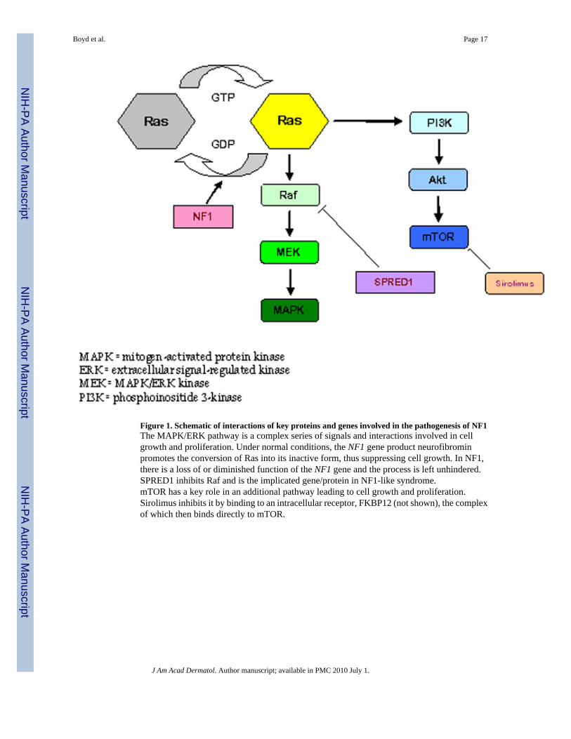
Figure 1. Schematic of interactions of key proteins and genes involved in the pathogenesis of NF1The MAPK/ERK pathway is a complex series of signals and interactions involved in cellgrowth and proliferation. Under normal conditions, the NF1 gene product neurofibrominpromotes the conversion of Ras into its inactive form, thus suppressing cell growth. In NF1,there is a loss of or diminished function of the NF1 gene and the process is left unhindered.SPRED1 inhibits Raf and is the implicated gene/protein in NF1-like syndrome.mTOR has a key role in an additional pathway leading to cell growth and proliferation.Sirolimus inhibits it by binding to an intracellular receptor, FKBP12 (not shown), the complexof which then binds directly to mTOR.
Boyd et al. Page 17
J Am Acad Dermatol. Author manuscript; available in PMC 2010 July 1.
NIH
-PA Author Manuscript
NIH
-PA Author Manuscript
NIH
-PA Author Manuscript
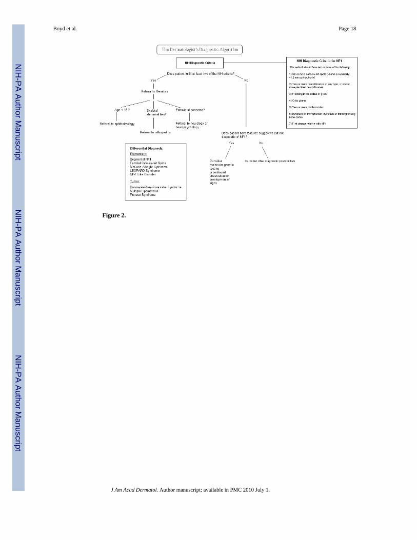
Figure 2.
Boyd et al. Page 18
J Am Acad Dermatol. Author manuscript; available in PMC 2010 July 1.
NIH
-PA Author Manuscript
NIH
-PA Author Manuscript
NIH
-PA Author Manuscript
Boyd et al. Page 19
J Am Acad Dermatol. Author manuscript; available in PMC 2010 July 1.
NIH
-PA Author Manuscript
NIH
-PA Author Manuscript
NIH
-PA Author Manuscript
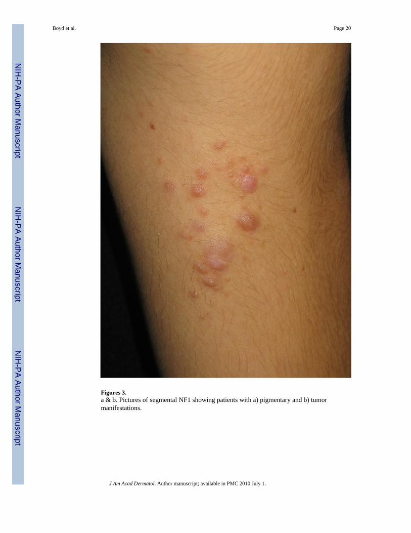
Figures 3.a & b. Pictures of segmental NF1 showing patients with a) pigmentary and b) tumormanifestations.
Boyd et al. Page 20
J Am Acad Dermatol. Author manuscript; available in PMC 2010 July 1.
NIH
-PA Author Manuscript
NIH
-PA Author Manuscript
NIH
-PA Author Manuscript
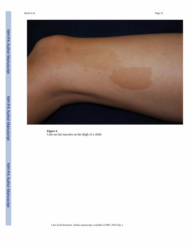
Figure 4.Cafe-au-lait macules on the thigh of a child.
Boyd et al. Page 21
J Am Acad Dermatol. Author manuscript; available in PMC 2010 July 1.
NIH
-PA Author Manuscript
NIH
-PA Author Manuscript
NIH
-PA Author Manuscript
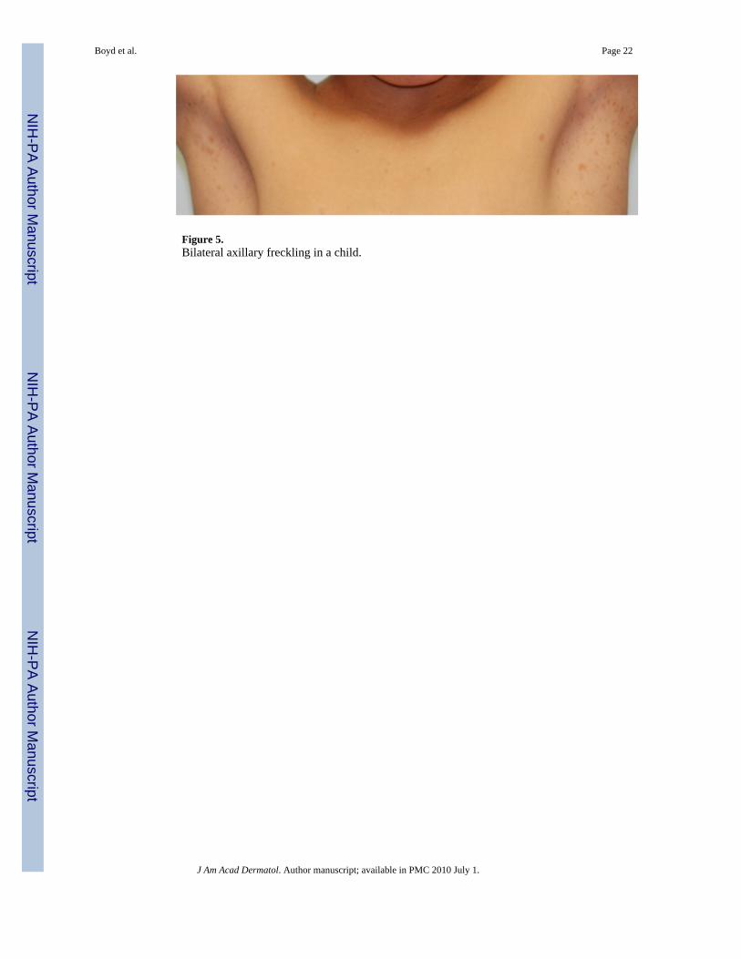
Figure 5.Bilateral axillary freckling in a child.
Boyd et al. Page 22
J Am Acad Dermatol. Author manuscript; available in PMC 2010 July 1.
NIH
-PA Author Manuscript
NIH
-PA Author Manuscript
NIH
-PA Author Manuscript
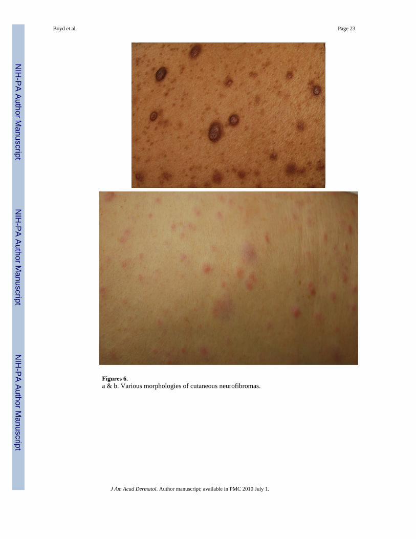
Figures 6.a & b. Various morphologies of cutaneous neurofibromas.
Boyd et al. Page 23
J Am Acad Dermatol. Author manuscript; available in PMC 2010 July 1.
NIH
-PA Author Manuscript
NIH
-PA Author Manuscript
NIH
-PA Author Manuscript
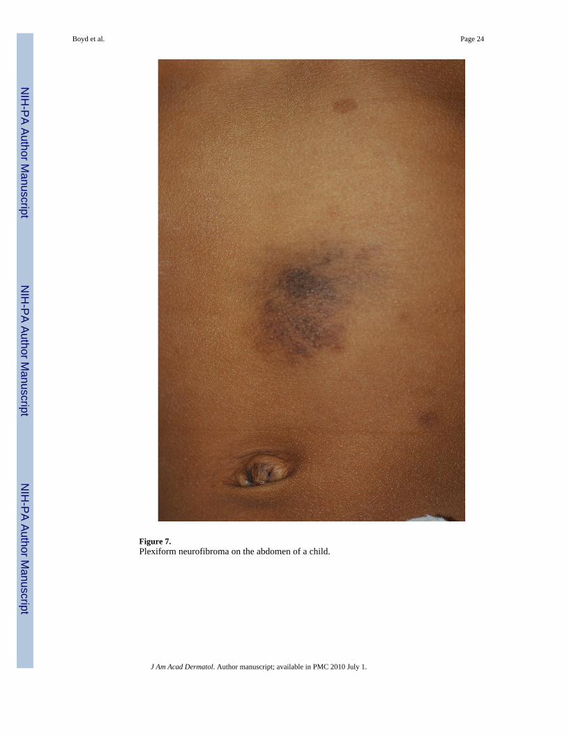
Figure 7.Plexiform neurofibroma on the abdomen of a child.
Boyd et al. Page 24
J Am Acad Dermatol. Author manuscript; available in PMC 2010 July 1.
NIH
-PA Author Manuscript
NIH
-PA Author Manuscript
NIH
-PA Author Manuscript
NIH
-PA Author Manuscript
NIH
-PA Author Manuscript
NIH
-PA Author Manuscript
Boyd et al. Page 25
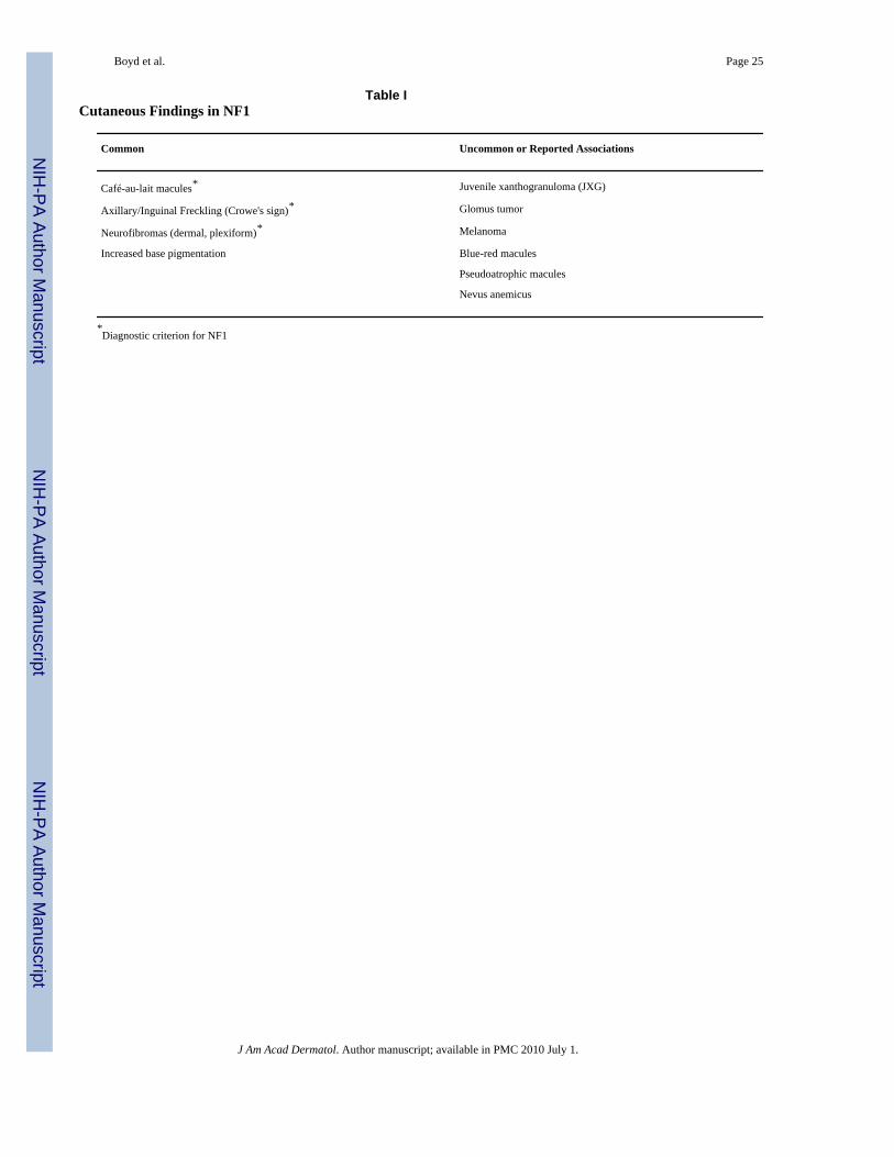
Table ICutaneous Findings in NF1
Common Uncommon or Reported Associations
Café-au-lait macules* Juvenile xanthogranuloma (JXG)
Axillary/Inguinal Freckling (Crowe's sign)* Glomus tumor
Neurofibromas (dermal, plexiform)* Melanoma
Increased base pigmentation Blue-red macules
Pseudoatrophic macules
Nevus anemicus
*Diagnostic criterion for NF1
J Am Acad Dermatol. Author manuscript; available in PMC 2010 July 1.
NIH
-PA Author Manuscript
NIH
-PA Author Manuscript
NIH
-PA Author Manuscript
Boyd et al. Page 26
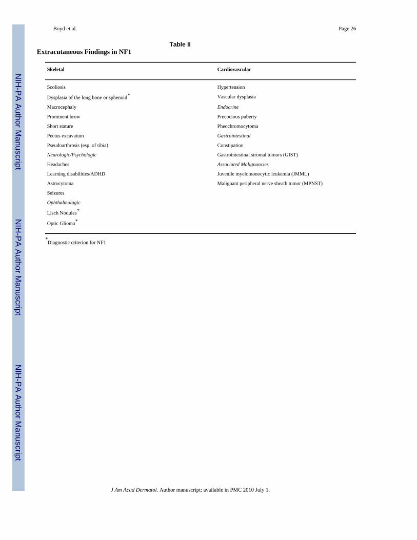
Table IIExtracutaneous Findings in NF1
Skeletal Cardiovascular
Scoliosis Hypertension
Dysplasia of the long bone or sphenoid* Vascular dysplasia
Macrocephaly Endocrine
Prominent brow Precocious puberty
Short stature Pheochromocytoma
Pectus excavatum Gastrointestinal
Pseudoarthrosis (esp. of tibia) Constipation
Neurologic/Psychologic Gastrointestinal stromal tumors (GIST)
Headaches Associated Malignancies
Learning disabilities/ADHD Juvenile myelomonocytic leukemia (JMML)
Astrocytoma Malignant peripheral nerve sheath tumor (MPNST)
Seizures
Ophthalmologic
Lisch Nodules*
Optic Glioma*
*Diagnostic criterion for NF1
J Am Acad Dermatol. Author manuscript; available in PMC 2010 July 1.
NIH
-PA Author Manuscript
NIH
-PA Author Manuscript
NIH
-PA Author Manuscript
Boyd et al. Page 27
Table IIIIndications for Genetic Testing
Individuals at 50% risk
Patients with a single sign
Individuals with variant disorders
Prenatal testing
Pre-implantation genetic diagnosis
J Am Acad Dermatol. Author manuscript; available in PMC 2010 July 1.