Lovastatin regulates brain spontaneous low-frequency brain activity in Neurofibromatosis type 1
12
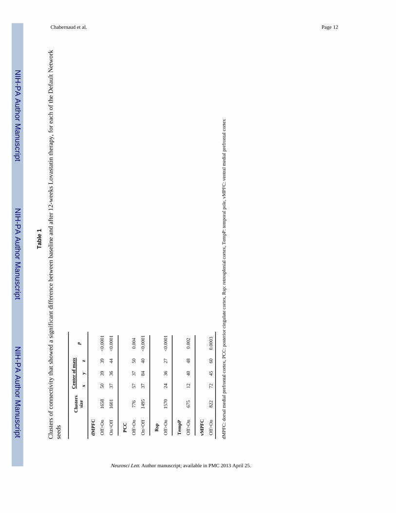
Lovastatin regulates brain spontaneous low-frequency brain activity in Neurofibromatosis type 1 Camille Chabernaud, PhD a , Maarten Mennes, PhD a , Peter G. Kardel, MA b , William D. Gaillard, MD c , M. Layne Kalbfleisch, PhD d , John W. VanMeter, PhD e , Roger J. Packer, MD b,c , Michael P. Milham, MD, PhD f,g , Francisco X. Castellanos, MD a,g , and Maria T. Acosta, MD b,c a Phyllis Green and Randolph Cowen Institute for Pediatric Neuroscience at the New York University Langone Medical Center, 215 Lexington Avenue, New York, NY 10016, USA b Jennifer and Daniel Gilbert Neurofibromatosis Institute, Children’s National Medical Center, 111 Michigan Avenue NW, Washington D.C., 20010, USA c The Center for Neuroscience and Behavioral Medicine, Children’s National Medical Center, 111 Michigan Avenue NW, Washington D.C., 20010, USA d KIDLAB, Krasnow Institute for Advanced Study and College of Education and Human Development, George Mason University, Mail Stop 2A1, Fairfax, VA 22030, USA e Center for Functional and Molecular Imaging, Georgetown University Medical Center, 3900 Reservoir Rd. NW, Washington, D.C., 20057, USA f Center for the Developing Brain, Child Mind Institute, 445 Park Avenue, New York, NY10022, USA g Nathan Kline Institute for Psychiatric Research, 140 Old Orangeburg Road, Orangeburg, NY 10962, USA Abstract In the Neurofibromatosis type 1 (NF1) mouse model, lovastatin, used clinically for hypercholesterolemia, improves cognitive dysfunction. While such impairment has been studied in NF1, the neural substrates remain unclear. The aim of this imaging add-on to a phase-1 open-label trial was to examine the effect of lovastatin on Default Network (DN) resting state functional connectivity (RSFC). Seven children with NF1 (aged 11.9±2.2; 1 female) were treated with lovastatin once daily for 12 weeks. A 7-minute 3-Tesla echo-planar-imaging scan was collected one day before beginning treatment (off-drug) and the last day of treatment (on-drug) while © 2012 Elsevier Ireland Ltd. All rights reserved. Corresponding author: Maria T. Acosta, MD, Jennifer and Daniel Gilbert Neurofibromatosis Institute, Children’s National Medical Center, Washington D.C., USA, 111 Michigan Av. NW, Washington DC, 20010, Phone: 202-476-2120, Fax: 202-476-5265, [email protected]. Publisher's Disclaimer: This is a PDF file of an unedited manuscript that has been accepted for publication. As a service to our customers we are providing this early version of the manuscript. The manuscript will undergo copyediting, typesetting, and review of the resulting proof before it is published in its final citable form. Please note that during the production process errors may be discovered which could affect the content, and all legal disclaimers that apply to the journal pertain. Contribution: MTA designed the study; MTA, PGK, JWV and MLK collected the data; CC and MM analyzed and interpreted the data; CC and FXC wrote the paper and MM, MTA, WDG, RJP, MLK and MPM critically revised the manuscript. Disclosures Authors C. Chabernaud, M. Mennes, P. G. Kardel, W. D. Gaillard, M. L. Kalbfleisch, J. W. VanMeter, R. J. Packer, M. P. Milham, F. X. Castellanos and M. T. Acosta declare no financial interests or potential conflicts of interest. All the authors have approved the final version of the manuscript. NIH Public Access Author Manuscript Neurosci Lett. Author manuscript; available in PMC 2013 April 25. Published in final edited form as: Neurosci Lett. 2012 April 25; 515(1): 28–33. doi:10.1016/j.neulet.2012.03.009. NIH-PA Author Manuscript NIH-PA Author Manuscript NIH-PA Author Manuscript
Transcript of Lovastatin regulates brain spontaneous low-frequency brain activity in Neurofibromatosis type 1
Lovastatin regulates brain spontaneous low-frequency brainactivity in Neurofibromatosis type 1
Camille Chabernaud, PhDa, Maarten Mennes, PhDa, Peter G. Kardel, MAb, William D.Gaillard, MDc, M. Layne Kalbfleisch, PhDd, John W. VanMeter, PhDe, Roger J. Packer,MDb,c, Michael P. Milham, MD, PhDf,g, Francisco X. Castellanos, MDa,g, and Maria T. Acosta,MDb,c
aPhyllis Green and Randolph Cowen Institute for Pediatric Neuroscience at the New YorkUniversity Langone Medical Center, 215 Lexington Avenue, New York, NY 10016, USAbJennifer and Daniel Gilbert Neurofibromatosis Institute, Children’s National Medical Center, 111Michigan Avenue NW, Washington D.C., 20010, USAcThe Center for Neuroscience and Behavioral Medicine, Children’s National Medical Center, 111Michigan Avenue NW, Washington D.C., 20010, USAdKIDLAB, Krasnow Institute for Advanced Study and College of Education and HumanDevelopment, George Mason University, Mail Stop 2A1, Fairfax, VA 22030, USAeCenter for Functional and Molecular Imaging, Georgetown University Medical Center, 3900Reservoir Rd. NW, Washington, D.C., 20057, USAfCenter for the Developing Brain, Child Mind Institute, 445 Park Avenue, New York, NY10022,USAgNathan Kline Institute for Psychiatric Research, 140 Old Orangeburg Road, Orangeburg, NY10962, USA
AbstractIn the Neurofibromatosis type 1 (NF1) mouse model, lovastatin, used clinically forhypercholesterolemia, improves cognitive dysfunction. While such impairment has been studied inNF1, the neural substrates remain unclear. The aim of this imaging add-on to a phase-1 open-labeltrial was to examine the effect of lovastatin on Default Network (DN) resting state functionalconnectivity (RSFC). Seven children with NF1 (aged 11.9±2.2; 1 female) were treated withlovastatin once daily for 12 weeks. A 7-minute 3-Tesla echo-planar-imaging scan was collectedone day before beginning treatment (off-drug) and the last day of treatment (on-drug) while
© 2012 Elsevier Ireland Ltd. All rights reserved.
Corresponding author: Maria T. Acosta, MD, Jennifer and Daniel Gilbert Neurofibromatosis Institute, Children’s National MedicalCenter, Washington D.C., USA, 111 Michigan Av. NW, Washington DC, 20010, Phone: 202-476-2120, Fax: 202-476-5265,[email protected].
Publisher's Disclaimer: This is a PDF file of an unedited manuscript that has been accepted for publication. As a service to ourcustomers we are providing this early version of the manuscript. The manuscript will undergo copyediting, typesetting, and review ofthe resulting proof before it is published in its final citable form. Please note that during the production process errors may bediscovered which could affect the content, and all legal disclaimers that apply to the journal pertain.
Contribution: MTA designed the study; MTA, PGK, JWV and MLK collected the data; CC and MM analyzed and interpreted thedata; CC and FXC wrote the paper and MM, MTA, WDG, RJP, MLK and MPM critically revised the manuscript.
DisclosuresAuthors C. Chabernaud, M. Mennes, P. G. Kardel, W. D. Gaillard, M. L. Kalbfleisch, J. W. VanMeter, R. J. Packer, M. P. Milham, F.X. Castellanos and M. T. Acosta declare no financial interests or potential conflicts of interest. All the authors have approved the finalversion of the manuscript.
NIH Public AccessAuthor ManuscriptNeurosci Lett. Author manuscript; available in PMC 2013 April 25.
Published in final edited form as:Neurosci Lett. 2012 April 25; 515(1): 28–33. doi:10.1016/j.neulet.2012.03.009.
NIH
-PA Author Manuscript
NIH
-PA Author Manuscript
NIH
-PA Author Manuscript
performing a Flanker task. After regressing-out task-associated variance, we used the residual timeseries as “continuous resting-state data” for RSFC analyses using 11 DN regions of interest. Forqualitative comparisons, we included a group of 19 typically developing children (TDC) collectedelsewhere. In the on-drug condition, lovastatin increased long-range positive RSFC within DNcore regions (i.e., anterior medial prefrontal cortex and posterior cingulate cortex, PCC). Inaddition, lovastatin produced less diffuse local RSFC in the dorsomedial prefrontal cortex andPCC. The pattern of RSFC observed in the NF1 participants when on-drug closely resembled theRSFC patterns exhibited by the TDC. Lovastatin administration in this open trial regulatedanterior-posterior long-range and local RSFC within the DN. These preliminary results areconsistent with a role for lovastatin in normalization of developmental processes and withapparent benefits in a mouse NF1 model.
KeywordsNeurofibromatosis type 1; lovastatin; resting-state fMRI; Default Network; children
IntroductionNeurofibromatosis type 1 (NF1) is one of the most frequent autosomal genetic disorders,affecting one in 3000 newborns [19]. Besides the typical diagnostic symptoms associatedwith neurofibromatosis, between 30 and 65% of children with NF1 exhibit learningdisabilities and cognitive deficits such as visuospatial or executive dysfunction [20]. Due totheir high incidence, neurocognitive impairments are considered the most functionallydisruptive chronic disabilities in children with NF1 [1].
The NF1 gene encodes neurofibromin, a protein underlying several functions includinginhibition of p21Ras GTPase-activating protein activity [5]. The increase in p21Ras activity,caused by mutations of the NF1 gene, has been related to the learning deficits observed inNF1 mice and in humans, leading to the hypothesis that inhibition of Ras activity mayreverse these cognitive deficits [10, 22]. Because posttranslational farnelysation is requiredfor p21Ras function, farnelysation has been suggested as a pharmacotherapy target for NF1cognitive deficits. Previous studies demonstrated that lovastatin, a 3-hydroxy-3-methylglutaryl coenzyme A reductase inhibitor, successfully inhibits p21Ras isoprenylationand activity [27]. Examining the effects of lovastatin on cognitive deficits, Li et al. reportedthat lovastatin effectively decreased p21Ras activity and reversed attention and learningdeficits observed in Nf1+/− mice [24]. While the results of statin therapy in mice models areencouraging, simvastatin used in humans did not yield similar results [23]. However, as partof a Phase 1 open-label safety trial, significant cognitive improvements were foundcomparing pre- and post-lovastatin treatment, suggesting that lovastatin might be effectivefor treating cognitive deficits in children with NF1 [2].
Cognitive dysfunction in children with NF1 has been extensively examined at the behaviorallevel [20]. However, the specific brain correlates of such deficits in NF1 are poorlyunderstood. Previous functional magnetic resonance imaging (fMRI) studies indicated thatchildren with NF1 recruit different brain areas during various cognitive tasks relative totypically developing children (TDC) [7, 8]. For example, Billingsley et al. reported greateractivation of posterior regions (i.e., temporal, parietal and occipital cortex) relative toanterior regions (i.e., inferior and lateral orbital frontal cortex) during visuo-spatialperformance in children with NF1 relative to controls [8]. Overall, findings from fMRIstudies in children with NF1 suggest that functional interactions between neural networks,rather than dysfunction in a specific brain area, might underlie the cognitive deficitsfrequently observed in this disease.
Chabernaud et al. Page 2
Neurosci Lett. Author manuscript; available in PMC 2013 April 25.
NIH
-PA Author Manuscript
NIH
-PA Author Manuscript
NIH
-PA Author Manuscript
Resting state functional connectivity (RSFC) approaches can successfully delineatefunctional brain networks without external stimulation [9]. A myriad of functional networkscan be revealed through patterns of synchrony in spontaneous, low-frequency brain activity,that are remarkably similar to functional networks observed in task-based fMRI approaches[29]. Here, we examined the effects of 12-week lovastatin treatment on the RSFC of thedefault network (DN), one of the main intrinsic functional brain networks, on seven childrenwith NF1 who were part of a Phase 1 open label trial [2]. Based on previous findings ofneuronal dysfunction in children with NF1, we expected that lovastatin would significantlyimprove and normalize DN organization.
MethodsParticipants
Seven children with NF1 (age 11.9±2.2 yrs; range: 10–15; 1 female) participated in thisstudy. The diagnosis was established according to National Institutes of Health internationalcriteria (Neurofibromatosis Conference Statement, 1987). Inclusion criteria required full-scale IQ above 80 and absence of any other chronic medical condition or symptomaticcomplication. The study was approved by the institutional review boards of GeorgetownUniversity, Children’s National Medical Center, New York University (NYU) and NYUSchool of Medicine. Prior to participation, written assent and consent were obtained fromchildren and their parents/legal guardian respectively, in accordance with the Declaration ofHelsinki.
In the absence of imaging data from TDC scanned at Georgetown University, we includeddata from 19 TDC scanned at the NYU Center for Brain Imaging for qualitativecomparisons (age 13.2±2.5 yrs; range: 9–16; 3 females). The TDC and patients were group-matched on age (t=−1.17, p=0.27), estimated IQ (95±10 and 88±6 respectively, t=−2.07,p=0.054) and sex (χ2
(1)=0.01, p=.92).
Lovastatin ProtocolEach patient was treated with lovastatin once daily for 12 weeks after breakfast (weeks 0–4,20 mg/d; weeks 5–8, 30 mg/d; and weeks 9–12, 40 mg/d). Pre-treatment laboratory testswere performed on day one of the study, including complete blood count, blood ureanitrogen, creatinine, glucose, alanine amino transferase, aspartate amino transferase,bilirubin, creatine phosphokinase, lipid profile (total cholesterol, HDL, vLDL, LDL,triglycerides), urinalysis, and a urine pregnancy test (females only). A monthly clinic visitand phone calls at weeks 1, 2, and 3 were completed to support and document complianceand side effects during the escalation dose period. As described elsewhere, participantsunderwent cognitive testing prior to treatment initiation (baseline) and post 12-week opentreatment [2]. Cognitive measures included the Test of Everyday Attention for Children(TEA-Ch), the California Verbal Learning Test for Children (CVLT) and the Wide RangeAssessment of Memory and Learning, Second Edition (WRAML-2) [2].
Imaging ProtocolImaging data from patients were acquired using a Siemens Magnetom Trio 3-Tesla scannerat the Center for Functional and Molecular Imaging at Georgetown University. For eachparticipant, a 7-minute echo-planar imaging scan was collected one day prior to beginningtreatment (off-drug) and on the last day of the 3-month trial (on-drug) (TR=3000 ms; TE=30ms; flip angle=90°, 39 slices, matrix=64×64; FOV=192 mm; acquisition voxel size= 3×3×3mm). All scans were collected in the early afternoon (~6 hours after daily dose of drugconsumed). The children were scanned while performing a version of the flanker task basedon the Naglieri Nonverbal Ability Test (NNAT) licensed to MLK. Each child performed two
Chabernaud et al. Page 3
Neurosci Lett. Author manuscript; available in PMC 2013 April 25.
NIH
-PA Author Manuscript
NIH
-PA Author Manuscript
NIH
-PA Author Manuscript
runs, each of which contained 4 blocks, 2 with congruent flankers (e.g., <<<<<) and 2 withincongruent flankers (e.g., >><>>) presented in ABBA order. Stimulus duration was1200ms with a 1200ms inter-trial interval. The resting epoch between blocks was 4800mslong. For the analyses presented here, we regressed out the task-induced BOLD variancefrom the event-related data, producing residuals that are considered comparable to“continuous resting-state” data [15]. Subsequent analyses were then performed using theseresiduals, which we considered a “pseudo-resting state” signal, parsed out/aggregated frommotion and physiological noise. For registration and analysis purposes, a T1-weightedanatomical image was also acquired using a magnetization prepared gradient echo sequence(TR/TE 9.7/4ms, 256×256 FOV, 160mm slab thickness, 256×256×160 matrix (effectiveresolution 1mm3), 1 excitation, 12° flip angle). Imaging data from TDC were acquired usinga Siemens Allegra 3-Tesla scanner at the NYU Center for Brain Imaging. For each TDCparticipant, a 6-min resting scan comprising 180 contiguous whole-brain functional volumeswas acquired using a multi-echo echo-planar imaging sequence (TR=2000ms; flipangle=90°; 33 slices; voxel size=3×3×4mm; effective TE=30ms, FOV=240×192mm). A T1-weighted anatomical image was also acquired using a magnetization prepared gradient echosequence (TR=2530ms; TE=3.25ms; TI=1100ms; flip angle=7°; 128 slices; FOV=256mm;acquisition voxel size=1.3×1×1.3mm).
Image preprocessingWe used a combination of AFNI (http://afni.nimh.nih.gov/afni/) and the FMRIB softwarelibrary tool (FSL, www.fmrib.ox.ac.uk) to preprocess the EPI scans. Details regarding thepreprocessing steps are provided in Supplementary S1.
Nuisance and task-induced BOLD signal regressionTo control for motion and physiological nuisance signals (e.g., cardiac and respiratoryfluctuations), we regressed the preprocessed data on nine nuisance covariates, removingvariance associated with non-neuronal signals derived from white matter, cerebrospinalfluid, the global signal and six motion parameters [21]. Subsequently we regressed theresultant 4-D residual time series on a covariate modeling all trials presented during theflanker task [15]. The residuals resulting from this analysis are considered a valid “pseudo-resting state” signal [15].
Selection of regions of interest (ROIs)We used 11 DN ROIs (“seeds”) as defined previously [4]. The seeds comprised: AnteriorMedial Prefrontal Cortex (aMPFC) and Posterior Cingulate Cortex (PCC) representing themidline core subsystem; Temporo-Parietal Junction (TPJ), Lateral Temporal Cortex (LTC),Temporal Pole (TempP), Dorsal Medial Prefrontal Cortex (dMPFC) and Ventral MedialPrefrontal Cortex (vMPFC), constituting the dMPFC subsystem; Posterior Inferior ParietalLobule (pIPL), Retrosplenial Cortex (Rsp), Parahippocampal Cortex (PHC) andHippocampal Formation (HF), constituting the Medial Temporal Lobe (MTL) subsystem.For each region, we created a spherical seed ROI (4mm radius), centered on publishedcoordinates [4].
Participant-level RSFC analysisFor each participant, the representative time series for each seed ROI was extracted fromtheir 4D residualized standard-space pseudo-resting state volume by averaging the timeseries across all voxels within the ROI. We then calculated the correlation between eachseed ROI time series and that of every other voxel in the brain. The resultant participant-level correlation maps were Fisher-z transformed to improve normal distribution andtransformed into MNI152 2mm standard space for group-level analyses.
Chabernaud et al. Page 4
Neurosci Lett. Author manuscript; available in PMC 2013 April 25.
NIH
-PA Author Manuscript
NIH
-PA Author Manuscript
NIH
-PA Author Manuscript
Group-level RSFC analysisFor each seed, group-level analyses were performed to compare off- and on-drug conditionsusing a within-participant repeated measures mixed-effects ordinary least-squares model asimplemented in FSL. This group-level analysis produced thresholded Z-score maps ofpositive and negative connectivity for each DN ROI and for each condition. Direct conditioncomparisons produced thresholded Z-score maps for each ROI of those voxels that showedsignificant lovastatin-related changes in functional connectivity with each ROI. Recentreports have pointed out the influence of small head movements on RSFC timecourses andhave suggested that standard regression analyses may not be sufficient to fully account forhead motion [26, 31]. To account for head motion micro-movements in our analyses, wecalculated the mean of frame displacement for each subject and used it as a covariate. For allanalyses, corrections for multiple comparisons were performed at the cluster level usingGaussian random field theory (min Z >2.3; cluster significance: p <0.0045, corrected, toaccount for the 11 seeds analyzed [0.05/11 = 0.0045]).
ResultsRegression analyses revealed that functional connectivity within the DN differedsignificantly between off- and on-drug conditions (Table 1). In particular, two main changeswere observed between conditions: increased long-range positive connectivity and decreasedlocal functional connectivity.
Lovastatin increases long-range functional connectivity within DN regionsWhen comparing the off- and on-drug conditions, we observed enhanced positive functionalconnectivity between core DN regions (i.e., aMPFC and PCC). In particular, during the off-drug condition, typical long-range RSFC between the PCC and aMPFC was absent (Figure1). In contrast, during the on-drug scan, a significant long-range positive relationshipemerged between these two regions (Figure 1). Importantly, the on-drug RSFC of childrenwith NF1 closely resembled the connectivity pattern exhibited by the TDC. The increase ofRSFC along the anterior-posterior axis was also highlighted for connectivity of the dMPFCseed with the PCC (see Supplementary S2) and between the Rsp seed and the medialprefrontal cortex (see Supplementary S3).
Lovastatin regulates focal functional connectivity within DN regionsDuring the off-drug condition, local functional connectivity (i.e., RSFC near the seedregion) was significantly more diffuse than during the on-drug condition. Specifically, forthe PCC seed, we observed significantly more diffuse and aberrant RSFC in the temporalcortex during the off-drug relative to the on-drug condition (Figure 1). A comparable patternwas observed in the TDC. We found similar results for the dMPFC seed, with significantlygreater positive RSFC in the rostral section of the supragenual anterior cingulate cortex(ACC) during the off-drug condition (See Supplementary S2), and for the Rsp seed, whichexhibited significantly greater aberrant short-range positive connectivity with the lingualgyrus (See Supplementary S3). This connectivity was not present during the on-drugcondition, or in the TDC. Finally, similar results were observed for connectivity between theTempP seed and the Supramarginal gyrus (See Supplementary S4), with decreasedconnectivity observed during the on-drug condition.
Lovastatin also regulated negative RSFC relationships within the DN. Specifically, weobserved a significantly greater negative relationship between the vMPFC and the superiorparietal lobule during off-drug condition, a pattern which was not present either in TDC orin the patients on-drug (See Supplementary S5).
Chabernaud et al. Page 5
Neurosci Lett. Author manuscript; available in PMC 2013 April 25.
NIH
-PA Author Manuscript
NIH
-PA Author Manuscript
NIH
-PA Author Manuscript
Although not the focus of this preliminary study, we note significant post-treatmentimprovements only on WRAML-2 Story Memory (t(7)=2.81, p=.03), WRAML-2 DesignMemory (t(7)=4.42, p=.005), and WRAML-2 Symbolic Working Memory (t(7)=2.76, p=.03).These did not correlate significantly with change in RSFC beyond chance expectations.
DiscussionUsing R-fMRI to investigate the effects of lovastatin on default network (DN) functionalconnectivity, we observed several significant abnormalities in the patterns of synchrony inspontaneous low frequency fluctuations in children affected with NF1 at baseline. Following12-week open treatment with lovastatin [2], we detected significant changes in within-DNlong-range RSFC consistent with normalization. In the on-drug condition, we also observedmore focal local (short-range) RSFC. Taken together, these observations mirror the patternsof RSFC reported in cross-sectional studies of typical development, that short-rangefunctional connectivity decreases and long-range functional connectivity increases acrossdevelopment [14, 21]. Such conclusions have also been supported by reports of a shift fromdiffuse to focal activation patterns in task-based functional imaging studies [13].
The DN is typically activated when participants are not engaged in attentionally demandingtasks and is deactivated during stimulus-driven activity [16, 30]. These observations led tothe idea that the DN underlies spontaneous cognitive processes including making predictionsabout the future or affective decision-making [3]. However, the PCC and the aMPFC havebeen reported to be significantly positively correlated during rest as well as during workingmemory processing [18]. In that study, the strength of the PCC-aMPFC correlation waspositively associated with better working memory performance leading to the suggestionthat the core regions of the DN may work together to facilitate cognitive processing.Accordingly, we speculate that alteration of DN functional connectivity may underlie someof the cognitive deficits observed in children with NF1 and that regulating its functionalconnectivity may contribute to improved cognitive performance. Studies with larger samplesizes will be needed to assess this hypothesis.
Lovastatin therapy significantly increased long-range RSFC in children with NF1,qualitatively approaching RSFC patterns observed in TDC. These changes paralleled but didnot correlate significantly with significant improvements in memory performance. Thediffusion tensor imaging apparent diffusion coefficient (ADC) is interpreted as an index ofaxon packing, internal axon structure and myelination [6]. Significantly higher ADC,particularly in frontal and parieto-occipital white matter, has been reported in children withNF1 relative to controls [32]. Such microstructural abnormalities may underlie the DNRSFC alterations we observed, as suggested by the finding of a close relationship betweentractography linking the aMPFC and PCC and their functional connectivity [17]. In NF1,deficient neurofibromin expression, the product of the NF1 gene, affects astrocyte growthand differentiation and impairs somatosensory cortical development [11, 25]. Together,these findings suggest that alteration of neurofibromin expression likely impacts braindevelopment, affecting both structural and functional connectivity.
Our results should be examined in light of limitations. Given the small sample size, whichreflected the nature of Phase I studies, our results are preliminary. The most importantlimitation is that order of scan acquisition was not counter-balanced, as all children werenaïve to lovastatin at baseline. While we cannot exclude the possibility that our findings ofDN RSFC modulation reflect maturation over the three-month period or habituation to thescanner environment, this possibility is not supported by the moderate to high test-retestreliability of RSFC in adults, nor by the cross-sectional data on RSFC maturation from 7 to30 years of age [12, 28]. Future studies should exclude the potential effect of order and re-
Chabernaud et al. Page 6
Neurosci Lett. Author manuscript; available in PMC 2013 April 25.
NIH
-PA Author Manuscript
NIH
-PA Author Manuscript
NIH
-PA Author Manuscript
scanning by counter-balancing on- and off-drug scans. Although event-related data residualsare qualitatively and quantitatively very similar to continuous resting-state data, somedifferences should be noted. Specifically, task engagement might reduce spontaneousfluctuations in brain activity, leading to stronger correlations in some regions duringcontinuous resting-state data [15]. In conclusion, although replication in larger samples withcounter-balancing of medication administration is needed, our results suggest the intriguingpossibility that lovastatin enhances maturation of DN brain functional connectivity inchildren with NF1 and that such changes may underlie improvements in cognitive function.
ConclusionsThis preliminary report of brain biomarkers in the context of a phase-1 open-label trialsuggests resting-state functional MRI will be a useful and powerful approach for assessingthe pharmacological responses of potential treatments such as lovastatin, even in limitedsamples.
Highlights
• We examined brain functional connectivity changes associated with lovastatintreatment
• Lovastatin regulated functional connectivity in Neurofibromatosis type 1
• Lovastatin produced increased long-range positive resting-state functionalconnectivity
• Lovastatin produced less diffuse local resting-state functional connectivity
Supplementary MaterialRefer to Web version on PubMed Central for supplementary material.
AcknowledgmentsWe would like to thank and acknowledge the patients and families who took part in this study and the support ofthe Jennifer and Daniel Gilbert Neurofibromatosis Institute and the Association Neurofibromatoses etRecklinghausen, France.
This work was supported by grants from Jennifer and Daniel Gilbert Neurofibromatosis Institute to MTA, NationalInstitute of Mental Health (R01MH08218 and R01HD065282) to FXC and a postdoctoral fellowship of theAssociation Neurofibromatoses et Recklinghausen (ANR), France, awarded to CC. These funding resources werenot involved in the study design, data collection, analysis or interpretation or in the preparation of the manuscript.
References1. Acosta MT, Gioia GA, Silva AJ. Neurofibromatosis type 1: new insights into neurocognitive issues.
Curr Neurol Neurosci Rep. 2006; 6:136–143. [PubMed: 16522267]
2. Acosta MT, Kardel PG, Walsh KS, Rosenbaum KN, Gioia GA, Packer RJ. Lovastatin as treatmentfor neurocognitive deficits in neurofibromatosis type 1: phase I study. Pediatr. Neurol. 2011;45:241–245. [PubMed: 21907886]
3. Andrews-Hanna JR, Reidler JS, Huang C, Buckner RL. Evidence for the default network's role inspontaneous cognition. J. Neurophysiol. 2010b; 104:322–335. [PubMed: 20463201]
4. Andrews-Hanna JR, Reidler JS, Sepulcre J, Poulin R, Buckner RL. Functional-AnatomicFractionation of the Brain's Default Network. Neuron. 2010a; 65:550–562. [PubMed: 20188659]
Chabernaud et al. Page 7
Neurosci Lett. Author manuscript; available in PMC 2013 April 25.
NIH
-PA Author Manuscript
NIH
-PA Author Manuscript
NIH
-PA Author Manuscript
5. Ballester R, Marchuk D, Boguski M, Saulino A, Letcher R, Wigler M, Collins F. The NF1 locusencodes a protein functionally related to mammalian GAP and yeast IRA proteins. Cell. 1990;63:851–859. [PubMed: 2121371]
6. Basser PJ, Jones DK. Diffusion-tensor MRI: theory, experimental design and data analysis - atechnical review. NMR Biomed. 2002; 15:456–467. [PubMed: 12489095]
7. Billingsley RL, Jackson EF, Slopis JM, Swank PR, Mahankali S, Moore BD 3rd. Functionalmagnetic resonance imaging of phonologic processing in neurofibromatosis 1. J. Child Neurol.2003; 18:731–740. [PubMed: 14696899]
8. Billingsley RL, Jackson EF, Slopis JM, Swank PR, Mahankali S, Moore BD III. Functional MRI ofvisual-spatial processing in neurofibromatosis, type 1. Neuropsychologia. 2004; 42:395–404.[PubMed: 14670578]
9. Cole DM, Smith SM, Beckmann CF. Advances and pitfalls in the analysis and interpretation ofresting-state FMRI data. Front Syst Neurosci. 2010; 4:8. [PubMed: 20407579]
10. Costa RM, Federov NB, Kogan JH, Murphy GG, Stern J, Ohno M, Kucherlapati R, Jacks T, SilvaAJ. Mechanism for the learning deficits in a mouse model of neurofibromatosis type 1. Nature.2002; 415:526–530. [PubMed: 11793011]
11. Dasgupta B, Gutmann DH. Neurofibromin regulates neural stem cell proliferation, survival, andastroglial differentiation in vitro and in vivo. J. Neurosci. 2005; 25:5584–5594. [PubMed:15944386]
12. Dosenbach NU, Nardos B, Cohen AL, Fair DA, Power JD, Church JA, Nelson SM, Wig GS,Vogel AC, Lessov-Schlaggar CN, Barnes KA, Dubis JW, Feczko E, Coalson RS, Pruett JR Jr,Barch DM, Petersen SE, Schlaggar BL. Prediction of individual brain maturity using fMRI.Science. 2010; 329:1358–1361. [PubMed: 20829489]
13. Durston S, Davidson MC, Tottenham N, Galvan A, Spicer J, Fossella JA, Casey BJ. A shift fromdiffuse to focal cortical activity with development. Dev Sci. 2006; 9:1–8. [PubMed: 16445387]
14. Fair DA, Cohen AL, Dosenbach NU, Church JA, Miezin FM, Barch DM, Raichle ME, PetersenSE, Schlaggar BL. The maturing architecture of the brain's default network. Proc. Natl. Acad. Sci.U. S. A. 2008; 105:4028–4032. [PubMed: 18322013]
15. Fair DA, Schlaggar BL, Cohen AL, Miezin FM, Dosenbach NU, Wenger KK, Fox MD, SnyderAZ, Raichle ME, Petersen SE. A method for using blocked and event-related fMRI data to study"resting state" functional connectivity. Neuroimage. 2007; 35:396–405. [PubMed: 17239622]
16. Fransson P. How default is the default mode of brain function? Further evidence from intrinsicBOLD signal fluctuations. Neuropsychologia. 2006; 44:2836–2845. [PubMed: 16879844]
17. Greicius MD, Supekar K, Menon V, Dougherty RF. Resting-state functional connectivity reflectsstructural connectivity in the default mode network. Cereb. Cortex. 2009; 19:72–78. [PubMed:18403396]
18. Hampson M, Driesen NR, Skudlarski P, Gore JC, Constable RT. Brain connectivity related toworking memory performance. J. Neurosci. 2006; 26:13338–13343. [PubMed: 17182784]
19. Huson SM, Compston DA, Clark P, Harper PS. A genetic study of von Recklinghausenneurofibromatosis in south east Wales. I. Prevalence, fitness, mutation rate, and effect of parentaltransmission on severity. J. Med. Genet. 1989; 26:704–711. [PubMed: 2511318]
20. Hyman SL, Shores A, North KN. The nature and frequency of cognitive deficits in children withneurofibromatosis type 1. Neurology. 2005; 65:1037–1044. [PubMed: 16217056]
21. Kelly AM, Di Martino A, Uddin LQ, Shehzad Z, Gee DG, Reiss PT, Margulies DS, CastellanosFX, Milham MP. Development of anterior cingulate functional connectivity from late childhood toearly adulthood. Cereb. Cortex. 2009; 19:640–657. [PubMed: 18653667]
22. Klose A, Ahmadian MR, Schuelke M, Scheffzek K, Hoffmeyer S, Gewies A, Schmitz F,Kaufmann D, Peters H, Wittinghofer A, Nurnberg P. Selective disactivation of neurofibrominGAP activity in neurofibromatosis type 1. Hum. Mol. Genet. 1998; 7:1261–1268. [PubMed:9668168]
23. Krab LC, de Goede-Bolder A, Aarsen FK, Pluijm SM, Bouman MJ, van der Geest JN, Lequin M,Catsman CE, Arts WF, Kushner SA, Silva AJ, de Zeeuw CI, Moll HA, Elgersma Y. Effect ofsimvastatin on cognitive functioning in children with neurofibromatosis type 1: a randomizedcontrolled trial. JAMA. 2008; 300:287–294. [PubMed: 18632543]
Chabernaud et al. Page 8
Neurosci Lett. Author manuscript; available in PMC 2013 April 25.
NIH
-PA Author Manuscript
NIH
-PA Author Manuscript
NIH
-PA Author Manuscript
24. Li W, Cui Y, Kushner SA, Brown RA, Jentsch JD, Frankland PW, Cannon TD, Silva AJ. TheHMG-CoA reductase inhibitor lovastatin reverses the learning and attention deficits in a mousemodel of neurofibromatosis type 1. Curr. Biol. 2005; 15:1961–1967. [PubMed: 16271875]
25. Lush ME, Li Y, Kwon CH, Chen J, Parada LF. Neurofibromin is required for barrel formation inthe mouse somatosensory cortex. J. Neurosci. 2008; 28:1580–1587. [PubMed: 18272679]
26. Power JD, Barnes KA, Snyder AZ, Schlaggar BL, Petersen SE. Spurious but systematiccorrelations in functional connectivity MRI networks arise from subject motion. Neuroimage.2011
27. Sebti SM, Tkalcevic GT, Jani JP. Lovastatin, a cholesterol biosynthesis inhibitor, inhibits thegrowth of human H-ras oncogene transformed cells in nude mice. Cancer Commun. 1991; 3:141–147. [PubMed: 2043425]
28. Shehzad Z, Kelly AM, Reiss PT, Gee DG, Gotimer K, Uddin LQ, Lee SH, Margulies DS, Roy AK,Biswal BB, Petkova E, Castellanos FX, Milham MP. The resting brain: unconstrained yet reliable.Cereb. Cortex. 2009; 19:2209–2229. [PubMed: 19221144]
29. Smith SM, Fox PT, Miller KL, Glahn DC, Fox PM, Mackay CE, Filippini N, Watkins KE, Toro R,Laird AR, Beckmann CF. Correspondence of the brain's functional architecture during activationand rest. Proc. Natl. Acad. Sci. U. S. A. 2009; 106:13040–13045. [PubMed: 19620724]
30. Tomasi D, Ernst T, Caparelli EC, Chang L. Common deactivation patterns during workingmemory and visual attention tasks: an intra-subject fMRI study at 4 Tesla. Hum. Brain Mapp.2006; 27:694–705. [PubMed: 16404736]
31. Van Dijk KRA, Sabuncu MR, Buckner RL. The influence of head motion on intrinsic functionalconnectivity MRI. Neuroimage. 2012; 59:431–438. [PubMed: 21810475]
32. van Engelen SJ, Krab LC, Moll HA, de Goede-Bolder A, Pluijm SM, Catsman-Berrevoets CE,Elgersma Y, Lequin MH. Quantitative differentiation between healthy and disordered brain matterin patients with neurofibromatosis type I using diffusion tensor imaging, AJNR. Am. J.Neuroradiol. 2008; 29:816–822. [PubMed: 18339726]
Chabernaud et al. Page 9
Neurosci Lett. Author manuscript; available in PMC 2013 April 25.
NIH
-PA Author Manuscript
NIH
-PA Author Manuscript
NIH
-PA Author Manuscript
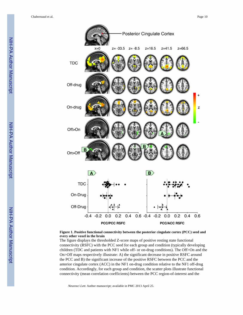
Figure 1. Positive functional connectivity between the posterior cingulate cortex (PCC) seed andevery other voxel in the brainThe figure displays the thresholded Z-score maps of positive resting state functionalconnectivity (RSFC) with the PCC seed for each group and condition (typically developingchildren (TDC and patients with NF1 while off- or on-drug conditions). The Off>On and theOn>Off maps respectively illustrate: A) the significant decrease in positive RSFC aroundthe PCC and B) the significant increase of the positive RSFC between the PCC and theanterior cingulate cortex (ACC) in the NF1 on-drug condition relative to the NF1 off-drugcondition. Accordingly, for each group and condition, the scatter plots illustrate functionalconnectivity (mean correlation coefficients) between the PCC region-of-interest and the
Chabernaud et al. Page 10
Neurosci Lett. Author manuscript; available in PMC 2013 April 25.
NIH
-PA Author Manuscript
NIH
-PA Author Manuscript
NIH
-PA Author Manuscript

clusters exhibiting the significant Off>On and On>Off drug effect. Cluster-level Gaussianrandom field theory was employed for multiple comparison correction (Z>2.3; p<0.0045,corrected).
Chabernaud et al. Page 11
Neurosci Lett. Author manuscript; available in PMC 2013 April 25.
NIH
-PA Author Manuscript
NIH
-PA Author Manuscript
NIH
-PA Author Manuscript
NIH
-PA Author Manuscript
NIH
-PA Author Manuscript
NIH
-PA Author Manuscript
Chabernaud et al. Page 12
Tabl
e 1
Clu
ster
s of
con
nect
ivity
that
sho
wed
a s
igni
fica
nt d
iffe
renc
e be
twee
n ba
selin
e an
d af
ter
12-w
eeks
Lov
asta
tin th
erap
y, f
or e
ach
of th
e D
efau
lt N
etw
ork
seed
s
Clu
ster
ssi
ze
Cen
ter
of m
ass
px
yz
dMP
FC
Off
>O
n16
5850
3939
<0.
0001
On>
Off
1601
3736
44<
0.00
01
PC
C
Off
>O
n77
657
3750
0.00
4
On>
Off
1495
3784
40<
0.00
01
Rsp
Off
>O
n15
7024
3627
<0.
0001
Tem
pP
Off
>O
n67
512
4048
0.00
2
vMP
FC
Off
>O
n82
272
4560
0.00
03
dMPF
C: d
orsa
l med
ial p
refr
onta
l cor
tex,
PC
C: p
oste
rior
cin
gula
te c
orte
x, R
sp: r
etro
sple
nial
cor
tex,
Tem
pP: t
empo
ral p
ole,
vM
PFC
: ven
tral
med
ial p
refr
onta
l cor
tex
Neurosci Lett. Author manuscript; available in PMC 2013 April 25.