
Brain Facts
58
Brain Facts A PRIMER ON THE BRAIN AND NERVOUS SYSTEM THE SOCIETY FOR NEUROSCIENCE
Transcript of Brain Facts

BrainFactsA P R I M E R O N T H E B R A I N A N D N E R V O U S S Y S T E M
T H E S O C I E T Y F O R N E U R O S C I E N C E

Brain FactsA P R I M E R O N T H E B R A I N A N D N E R V O U S S Y S T E M
T H E S O C I E T Y F O R N E U R O S C I E N C E

THE SOCIETY FOR NEUROSCIENCE
The Society for Neuroscience is the world’s largest organization of sci-entists and physicians dedicated to understanding the brain, spinal cordand peripheral nervous system.
Neuroscientists investigate the molecular and cellular levels of thenervous system; the neuronal systems responsible for sensory andmotor function; and the basis of higher order processes, such as cog-nition and emotion. This research provides the basis for understand-ing the medical fields that are concerned with treating nervous systemdisorders. These medical specialties include neurology, neurosurgery,psychiatry and ophthalmology.
Founded in 1970, the Society has grown from 500 charter membersto more than 29,000 members. Regular members are residents of Canada,Mexico and the United States—where more than 100 chapters organizelocal activities. The Society’s membership also includes many scientistsfrom throughout the world, particularly Europe and Asia.
The purposes of the Society are to:∫Advance the understanding of the nervous system by bringing togetherscientists from various backgrounds and by encouraging research in allaspects of neuroscience.∫ Promote education in the neurosciences.∫ Inform the public about the results and implications of new research.
The exchange of scientific information occurs at an annual fallmeeting that presents more than 14,000 reports of new scientificfindings and includes more than 25,000 participants. This meeting, thelargest of its kind in the world, is the arena for the presentation of newresults in neuroscience.
The Society’s bimonthly journal, The Journal of Neuroscience, con-tains articles spanning the entire range of neuroscience research andhas subscribers worldwide. A series of courses, workshops and sym-posia held at the annual meeting promote the education of Societymembers. The Neuroscience Newsletter informs members about Societyactivities.
A major mission of the Society is to inform the public about theprogress and benefits of neuroscience research. The Society providesinformation about neuroscience to school teachers and encourages itsmembers to speak to young people about the human brain and nervoussystem.
Brain Facts
INTRODUCTION . . . . . . . . . . . . . . . . . . . . . . . . . . . . . . . . . . . . . . . . . . . . . . . . 2
THE NEURON . . . . . . . . . . . . . . . . . . . . . . . . . . . . . . . . . . . . . . . . . . . . . . . . . 4
Neurotransmitters ∫ Second Messengers
BRAIN DEVELOPMENT . . . . . . . . . . . . . . . . . . . . . . . . . . . . . . . . . . . . . . . . . . . 8
Birth of Neurons and Brain Wiring ∫ Paring Back ∫ Critical Periods
SENSATION AND PERCEPTION . . . . . . . . . . . . . . . . . . . . . . . . . . . . . . . . . . . . 12
Vision ∫ Hearing ∫ Taste and Smell ∫ Touch and Pain
LEARNING AND MEMORY . . . . . . . . . . . . . . . . . . . . . . . . . . . . . . . . . . . . . . . . 18
MOVEMENT . . . . . . . . . . . . . . . . . . . . . . . . . . . . . . . . . . . . . . . . . . . . . . . . . . 20
SLEEP . . . . . . . . . . . . . . . . . . . . . . . . . . . . . . . . . . . . . . . . . . . . . . . . . . . . . . 22
The Stu∑ of Sleep ∫ Sleep Disorders ∫ How is Sleep Regulated?
STRESS . . . . . . . . . . . . . . . . . . . . . . . . . . . . . . . . . . . . . . . . . . . . . . . . . . . . . 25
The Immediate Response ∫ Chronic Stress
AGING . . . . . . . . . . . . . . . . . . . . . . . . . . . . . . . . . . . . . . . . . . . . . . . . . . . . . . 28
Aging Neurons ∫ Intellectual Capacity
ADVANCES . . . . . . . . . . . . . . . . . . . . . . . . . . . . . . . . . . . . . . . . . . . . . . . . . . . 30
Parkinson’s Disease ∫ Pain ∫ Epilepsy ∫Major DepressionManic-Depressive Illness
CHALLENGES . . . . . . . . . . . . . . . . . . . . . . . . . . . . . . . . . . . . . . . . . . . . . . . . . 33
Addiction ∫ Alzheimer’s Disease ∫ Learning DisordersStroke ∫ Neurological Trauma ∫ Anxiety DisordersSchizophrenia ∫ Neurological AIDS ∫Multiple SclerosisDown Syndrome ∫ Huntington’s Disease ∫ Tourette SyndromeBrain Tumors ∫ Amyotrophic Lateral Sclerosis
NEW DIAGNOSTIC METHODS . . . . . . . . . . . . . . . . . . . . . . . . . . . . . . . . . . . . . 43
Imaging Techniques ∫ Gene Diagnosis
POTENTIAL THERAPIES . . . . . . . . . . . . . . . . . . . . . . . . . . . . . . . . . . . . . . . . . 46
New Drugs ∫ Trophic Factors ∫ Cell and Gene Therapy
GLOSSARY . . . . . . . . . . . . . . . . . . . . . . . . . . . . . . . . . . . . . . . . . . . . . . . . . . 48
INDEX . . . . . . . . . . . . . . . . . . . . . . . . . . . . . . . . . . . . . . . . . . . . . . . . . . . . . . 53

2
t sets humans apart from all other species by allowing usto achieve the wonders of walking on the moon and com-posing masterpieces of literature, art and music. Through-out recorded time, the human brain—a spongy, three-pound mass of fatty tissue—has been compared to atelephone switchboard and a supercomputer.
But the brain is much more complicated than any of thesedevices, a fact scientists confirm almost daily with each new discovery. The extent of the brain’s capabilities is unknown, butit is the most complex living structure known in the universe.
This single organ controls all body activities, ranging fromheart rate and sexual function to emotion, learning and mem-ory. The brain is even thought to influence the response to dis-ease of the immune system and to determine, in part, how wellpeople respond to medical treatments. Ultimately, it shapes ourthoughts, hopes, dreams and imagination. In short, the brain iswhat makes us human.
Neuroscientists have the daunting task of deciphering themystery of this most complex of all machines: how as many asa trillion nerve cells are produced, grow and organize them-selves into e∑ective, functionally active systems that ordinarilyremain in working order throughout a person’s lifetime.
The motivation of researchers is twofold: to understandhuman behavior better—from how we learn to why peoplehave trouble getting along together—and to discover ways toprevent or cure many devastating brain disorders.
The more than 1,000 disorders of the brain and nervoussystem result in more hospitalizations than any other diseasegroup, including heart disease and cancer. Neurological illnessesa∑ect more than 50 million Americans annually at costs exceed-ing $400 billion. In addition, mental disorders, excluding drugand alcohol problems, strike 44 million adults a year at a costof some $148 billion.
However, during the congressionally designated Decade ofthe Brain, which ended in 2000, neuroscience made significantdiscoveries in these areas:∫Genetics. Key disease genes were identified that underlie sev-eral neurodegenerative disorders—including Alzheimer’s dis-ease, Huntington’s disease, Parkinson’s disease and amyotrophiclateral sclerosis. This has provided new insights into underlying
disease mechanisms and is beginning to suggest new treatments.With the mapping of the human genome, neuroscientists
will be able to make more rapid progress in identifying genes thateither contribute to human neurological disease or that directlycause disease. Mapping animal genomes will aid the search forgenes that regulate and control many complex behaviors.∫ Brain Plasticity. Scientists began to uncover the molecularbases of neural plasticity, revealing how learning and memoryoccur and how declines might be reversed. It also is leading tonew approaches to the treatment of chronic pain.∫New Drugs. Researchers gained new insights into the mech-anisms of molecular neuropharmacology, which provides a newunderstanding of the mechanisms of addiction. These advancesalso have led to new treatments for depression and obsessive-compulsive disorder.∫ Imaging. Revolutionary imaging techniques, including mag-netic resonance imaging and positron emission tomography,now reveal brain systems underlying attention, memory andemotions and indicate dynamic changes that occur in schizo-phrenia.∫ Cell Death. The discovery of how and why neurons die, aswell as the discovery of stem cells, which divide and form newneurons, has many clinical applications. This has dramaticallyimproved the outlook for reversing the e∑ects of injury both inthe brain and spinal cord. The first e∑ective treatments forstroke and spinal cord injury based on these advances have beenbrought to clinical practice.∫ Brain Development. New principles and molecules respon-sible for guiding nervous system development now give scien-tists a better understanding of certain disorders of childhood.Together with the discovery of stem cells, these advances arepointing to novel strategies for helping the brain or spinal cordregain functions lost to diseases.
Federal neuroscience research funding of more than $4 bil-lion annually and private support should vastly expand ourknowledge of the brain in the years ahead.
This book only provides a glimpse of what is known aboutthe nervous system, the disorders of the brain and some of theexciting avenues of research that promise new therapies formany neurological diseases.
Introduction
I

3
THE TOLL OF SELECTED BRAIN AND NERVOUS SYSTEM DISORDERS*
Condition Total Cases Costs Per Year
Hearing Loss 28 million $ 56 billion
All Depressive Disorders 18.8 million $ 44 billion
Alzheimer’s Disease 4 million $ 100 billion
Stroke 4 million $ 30 billion
Schizophrenia 3 million $ 32.5 billion
Parkinson’s Disease 1.5 million $ 15 billion
Traumatic Head Injury 1 million $ 48.3 billion
Multiple Sclerosis 350,000 $ 7 billion
Spinal Cord Injury 250,000 $ 10 billion
* Estimates provided by the National Institutes of Health and voluntary organizations.
THE BRAIN. Cerebral cortex
(above). This part of the brain is
divided into four sections: the
occipital lobe, the temporal
lobe, the parietal lobe and the
frontal lobe. Functions, such as
vision, hearing and speech, are
distributed in selected regions.
Some regions are associated
with more than one function.
Major internal structures
(below). The (1) forebrain is
credited with the highest intel-
lectual functions—thinking,
planning and problem-solving.
The hippocampus is involved in
memory. The thalamus serves as
a relay station for almost all of
the information coming into the
brain. Neurons in the hypothala-
mus serve as relay stations for
internal regulatory systems by
monitoring information coming
in from the autonomic nervous
system and commanding the
body through those nerves and
the pituitary gland. On the
upper surface of the (2) mid-
brain are two pairs of small
hills, colliculi, collections of
cells that relay specific sensory
information from sense organs
to the brain. The (3) hindbrain
consists of the pons and
medulla oblongata, which help
control respiration and heart
rhythms, and the cerebellum,
which helps control movement
as well as cognitive processes
that require precise timing.
Frontal lobe
Motor cortex Sensory cortex
Parietal lobe
Occipital lobe
Temporal lobe
Cerebrum
1 Forebrain
Amygdala
Hippocampus
Thalamus
Hypothalamus
2 Midbrain
3 Hindbrain
Pons
Cerebellum
Medullaoblongata
Spinal cord

4
Aspecialized cell designed to transmit infor-mation to other nerve cells, muscle or glandcells, the neuron is the basic working unit ofthe brain. The brain is what it is because ofthe structural and functional properties ofneurons. The brain contains between one bil-
lion and one trillion neurons.The neuron consists of a cell body containing the nucleus
and an electricity-conducting fiber, the axon, which also givesrise to many smaller axon branches before ending at nerve ter-
minals. Synapses, from the Greek words meaning to “clasptogether,” are the contact points where one neuron communi-cates with another. Other cell processes, dendrites, Greek forthe branches of a tree, extend from the neuron cell body andreceive messages from other neurons. The dendrites and cellbody are covered with synapses formed by the ends of axons ofother neurons.
Neurons signal by transmitting electrical impulses alongtheir axons that can range in length from a tiny fraction of aninch to three or more feet. Many axons are covered with a lay-ered insulating myelin sheath, made of specialized cells, thatspeeds the transmission of electrical signals along the axon.
Nerve impulses involve the opening and closing of ion chan-
nels, water-filled molecular tunnels that pass through the cellmembrane and allow ions—electrically charged atoms—orsmall molecules to enter or leave the cell. The flow of these ionscreates an electrical current that produces tiny voltage changesacross the membrane.
The ability of a neuron to fire depends on a small dif-ference in electrical charge between the inside and outside ofthe cell. When a nerve impulse begins, a dramatic reversaloccurs at one point on the cell’s membrane. The change, calledan action potential, then passes along the membrane of the axonat speeds up to several hundred miles an hour. In this way, aneuron may be able to fire impulses scores or even hundredsof times every second.
On reaching the ends of an axon, these voltage changestrigger the release of neurotransmitters, chemical messengers.Neurotransmitters are released at nerve ending terminals and
bind to receptors on the surface of the target neuron.These receptors act as on and o∑ switches for the next cell.
Each receptor has a distinctly shaped part that exactly matchesa particular chemical messenger. A neurotransmitter fits intothis region in much the same way as a key fits into an automo-bile ignition. And when it does, it alters the neuron’s outermembrane and triggers a change, such as the contraction of amuscle or increased activity of an enzyme in the cell.
Knowledge of neurotransmitters in the brain and the actionof drugs on these chemicals—gained largely through the studyof animals—is one of the largest fields in neuroscience. Armedwith this information, scientists hope to understand the circuitsresponsible for disorders such as Alzheimer’s disease and Parkin-son’s disease. Sorting out the various chemical circuits is vitalto understanding how the brain stores memories, why sex is sucha powerful motivation and what is the biological basis of men-tal illness.
Neurotransmitters
Acetylcholine The first neurotransmitter to be identified 70years ago, was acetylcholine (ACh). This chemical is releasedby neurons connected to voluntary muscles (causing them tocontract) and by neurons that control the heartbeat. ACh alsoserves as a transmitter in many regions of the brain.
ACh is formed at the axon terminals. When an actionpotential arrives at the terminal, the electrically charged cal-cium ion rushes in, and ACh is released into the synapse andattaches to ACh receptors. In voluntary muscles, this openssodium channels and causes the muscle to contract. ACh isthen broken down and re-synthesized in the nerve terminal.Antibodies that block the receptor for ACh cause myasthenia
gravis, a disease characterized by fatigue and muscle weakness.Much less is known about ACh in the brain. Recent dis-
coveries suggest, however, that it may be critical for normalattention, memory and sleep. Since ACh-releasing neurons diein Alzheimer’s patients, finding ways to restore this neuro-transmitter is one goal of current research.
Amino Acids Certain amino acids, widely distributedthroughout the body and the brain, serve as the building blocks
A
The Neuron
Nucleus
Myelin sheath
Dendrites
Direction of impulse
Axon terminals
Cell body Axon
Neurotransmitters
Receptor molecules
Synapse
Dendrite of receiving
neuron
Vesicle
Nerve impulse Axon
5
of proteins. However, it is now apparent that certain aminoacids can also serve as neurotransmitters in the brain.
The neurotransmitters glutamate and aspartate act as exci-tatory signals. Glycine and gamma-aminobutyric acid (GABA)inhibit the firing of neurons. The activity of GABA is increasedby benzodiazepine (Valium) and by anticonvulsant drugs. InHuntington’s disease, a hereditary disorder that begins duringmid-life, the GABA-producing neurons in the brain centerscoordinating movement degenerate, thereby causing incontrol-lable movements.
Glutamate or aspartate activate N-methyl-D-aspartate
(NMDA) receptors, which have been implicated in activitiesranging from learning and memory to development and speci-fication of nerve contacts in a developing animal. The stimula-tion of NMDA receptors may promote beneficial changes inthe brain, whereas overstimulation can cause nerve cell damageor cell death in trauma and stroke.
Key questions remain about this receptor’s precise structure,regulation, location and function. For example, developingdrugs to block or stimulate activity at NMDA receptors holds
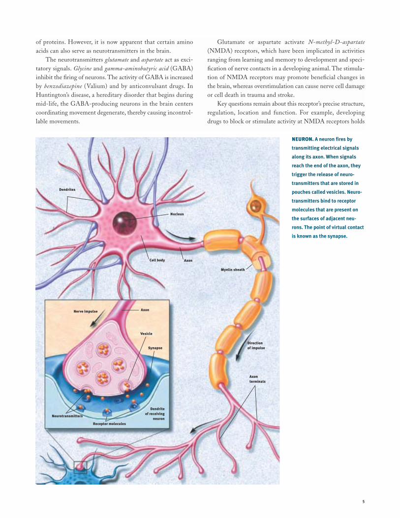
NEURON. A neuron fires by
transmitting electrical signals
along its axon. When signals
reach the end of the axon, they
trigger the release of neuro-
transmitters that are stored in
pouches called vesicles. Neuro-
transmitters bind to receptor
molecules that are present on
the surfaces of adjacent neu-
rons. The point of virtual contact
is known as the synapse.

6
promise for improving brain function and treating neurologi-cal disorders. But this work is still in the early stage.
Catecholamines Dopamine and norepinephrine are widelypresent in the brain and peripheral nervous system. Dopamine,which is present in three circuits in the brain, controls move-ment, causes psychiatric symptoms such as psychosis and reg-ulates hormonal responses.
The dopamine circuit that regulates movement has beendirectly related to disease. The brains of people with Parkinson’s
disease—with symptoms of muscle tremors, rigidity anddi≈culty in moving—have practically no dopamine. Thus,medical scientists found that the administration of levodopa, asubstance from which dopamine is synthesized, is an e∑ectivetreatment for Parkinson’s, allowing patients to walk and per-form skilled movements successfully.
Another dopamine circuit is thought to be important forcognition and emotion; abnormalities in this system have beenimplicated in schizophrenia. Because drugs that block dopaminereceptors in the brain are helpful in diminishing psychoticsymptoms, learning more about dopamine is important tounderstanding mental illness.
In a third circuit, dopamine regulates the endocrine sys-tem. It directs the hypothalamus to manufacture hormones andhold them in the pituitary gland for release into the blood-stream, or to trigger the release of hormones held within cellsin the pituitary.
Nerve fibers containing norepinephrine are present through-out the brain. Deficiencies in this transmitter occur in patientswith Alzheimer’s disease, Parkinson’s disease and those withKorsako∑’s syndrome, a cognitive disorder associated with chronicalcoholism. Thus, researchers believe norepinephrine may playa role in both learning and memory. Norepinephrine also issecreted by the sympathetic nervous system in the periphery toregulate heart rate and blood pressure. Acute stress increasesthe release of norepinephrine.
Serotonin This neurotransmitter is present in many tissues,particularly blood platelets and the lining of the digestive tractand the brain. Serotonin was first thought to be involved inhigh blood pressure because it is present in blood and inducesa very powerful contraction of smooth muscles. In the brain, ithas been implicated in sleep, mood, depression and anxiety.Because serotonin controls the di∑erent switches a∑ecting var-ious emotional states, scientists believe these switches can bemanipulated by analogs, chemicals with molecular structuressimilar to serotonin. Drugs that alter serotonin’s action, such asfluoxetine (Prozac), have relieved symptoms of depression andobsessive-compulsive disorder.
Peptides These chains of amino acids linked together, havebeen studied as neurotransmitters only in recent years. Brainpeptides called opioids act like opium to kill pain or cause sleepi-ness. (Peptides di∑er from proteins, which are much larger and
more complex combinations of amino acids.)In 1973, scientists discovered receptors for opiates on neu-
rons in several regions in the brain that suggested the brainmust make substances very similar to opium. Shortly thereafter,scientists made their first discovery of an opiate produced bythe brain that resembles morphine, an opium derivative usedmedically to kill pain. They named it enkephalin, literally mean-ing “in the head.” Subsequently, other opiates known as endor-
phins—from endogenous morphine—were discovered.The precise role of the opioids in the body is unclear. A
plausible guess is that enkephalins are released by brain neuronsin times of stress to minimize pain and enhance adaptive behav-ior. The presence of enkephalins may explain, for example, whyinjuries received during the stress of combat often are notnoticed until hours later.
Opioids and their receptors are closely associated with path-ways in the brain that are activated by painful or tissue-damag-ing stimuli. These signals are transmitted to the central nervous
system—the brain and spinal cord—by special sensory nerves,small myelinated fibers and tiny unmyelinated or C fibers.
Scientists have discovered that some C fibers contain a pep-tide called substance P that causes the sensation of burning pain.The active component of chili peppers, capsaicin, causes therelease of substance P.
Trophic factors Researchers have discovered several smallproteins in the brain that are necessary for the development,function and survival of specific groups of neurons. These smallproteins are made in brain cells, released locally in the brain,and bind to receptors expressed by specific neurons. Researchersalso have identified genes that code for receptors and areinvolved in the signaling mechanisms of trophic factors. Thesefindings are expected to result in a greater understanding ofhow trophic factors work in the brain. This information alsoshould prove useful for the design of new therapies for braindisorders of development and for degenerative diseases, includ-ing Alzheimer’s disease and Parkinson’s disease.
Hormones After the nervous system, the endocrine system
is the second great communication system of the body. Thepancreas, kidney, heart and adrenal gland are sources of hor-mones. The endocrine system works in large part through thepituitary that secretes hormones into the blood. Because endor-phins are released from the pituitary gland into the blood-stream, they might also function as endocrine hormones. Hor-mones activate specific receptors in target organs that releaseother hormones into the blood, which then act on other tissues,the pituitary itself and the brain. This system is very importantfor the activation and control of basic behavioral activities suchas sex, emotion, response to stress and the regulation of bodyfunctions, such as growth, energy use and metabolism. Actionsof hormones show the brain to be very malleable and capableof responding to environmental signals.

7
The brain contains receptors for both the thyroid hormoneand the six classes of steroid hormones—estrogens, androgens,
progestins, glucocorticoids, mineralocorticoids and vitamin D. Thereceptors are found in selected populations of neurons in thebrain and relevant organs in the body. Thyroid and steroid hor-mones bind to receptor proteins that in turn bind to the DNAgenetic material and regulate action of genes. This can result inlong-lasting changes in cellular structure and function.
In response to stress and changes in our biological clocks,such as day-and-night cycles and jet-lag, hormones enter theblood and travel to the brain and other organs. In the brain,they alter the production of gene products that participate insynaptic neurotransmission as well as the structure of braincells. As a result, the circuitry of the brain and its capacity forneurotransmission are changed over a course of hours to days.In this way, the brain adjusts its performance and control ofbehavior in response to a changing environment. Hormones areimportant agents of protection and adaptation, but stress andstress hormones also can alter brain function, including learn-ing. Severe and prolonged stress can cause permanent braindamage.
Reproduction is a good example of a regular, cyclic processdriven by circulating hormones: The hypothalamus producesgonadotropin-releasing hormone (GnRH), a peptide that acts oncells in the pituitary. In both males and females, this causes twohormones—the follicle-stimulating hormone (FSH) and theluteinizing hormone (LH)—to be released into the bloodstream.In males, these hormones are carried to receptors on cells in thetestes where they release the male hormone testosterone intothe bloodstream. In females, FSH and LH act on the ovariesand cause the release of the female hormones estrogen and prog-esterone. In turn, the increased levels of testosterone in malesand estrogen in females act back on the hypothalamus and pitu-itary to decrease the release of FSH and LH. The increased lev-els also induce changes in cell structure and chemistry that leadto an increased capacity to engage in sexual behavior.
Scientists have found statistically and biologically signi-ficant di∑erences between the brains of men and women thatare similar to sex di∑erences found in experimental animals.These include di∑erences in the size and shape of brain struc-tures in the hypothalamus and the arrangement of neurons inthe cortex and hippocampus. Some functions can be attributedto these sex di∑erences, but much more must be learned interms of perception, memory and cognitive ability. Althoughdi∑erences exist, the brains of men and women are more sim-ilar than they are di∑erent.
Recently, several teams of researchers have found anatom-ical di∑erences between the brains of heterosexual and homo-sexual men. Research suggests that hormones and genes actearly in life to shape the brain in terms of sex-related di∑erencesin structure and function, but scientists still do not have a firm
grip on all the pieces of this puzzle.Gases Very recently, scientists identified a new class of neu-
rotransmitters that are gases. These molecules—nitric oxide andcarbon monoxide—do not obey the “laws” governing neuro-transmitter behavior. Being gases, they cannot be stored in anystructure, certainly not in synaptic storage structures. Instead,they are made by enzymes as they are needed. They are releasedfrom neurons by di∑usion. And rather than acting at receptorsites, they simply di∑use into adjacent neurons and act uponchemical targets, which may be enzymes.
Though only recently characterized, nitric oxide hasalready been shown to play important roles. For example, nitricoxide neurotransmission governs erection in neurons of thepenis. In nerves of the intestine, it governs the relaxation thatcontributes to normal movements of digestion. In the brain,nitric oxide is the major regulator of the intracellular messen-ger molecule—cyclic GMP. In conditions of excess glutamaterelease, as occurs in stroke, neuronal damage following thestroke may be attributable in part to nitric oxide. Exact func-tions for carbon monoxide have not yet been shown.
Second messengers
Recently recognized substances that trigger biochemical com-munication within cells, second messengers may be responsi-ble for long-term changes in the nervous system. They conveythe chemical message of a neurotransmitter (the first messen-ger) from the cell membrane to the cell’s internal biochemicalmachinery. Second messengers take anywhere from a few milli-seconds to minutes to transmit a message.
An example of the initial step in the activation of a secondmessenger system involves adenosine triphosphate (ATP), thechemical source of energy in cells. ATP is present throughoutthe cell. For example, when norepinephrine binds to its recep-tors on the surface of the neuron, the activated receptor bindsG-proteins on the inside of the membrane. The activated G-protein causes the enzyme adenylyl cyclase to convert ATP tocyclic adenosine monophosphate (cAMP). The second messenger,cAMP, exerts a variety of influences on the cell, ranging fromchanges in the function of ion channels in the membrane tochanges in the expression of genes in the nucleus, rather thanacting as a messenger between one neuron and another. cAMPis called a second messenger because it acts after the first mes-senger, the transmitter chemical, has crossed the synaptic spaceand attached itself to a receptor.
Second messengers also are thought to play a role in themanufacture and release of neurotransmitters, intracellularmovements, carbohydrate metabolism in the cerebrum—thelargest part of the brain consisting of two hemispheres—andthe processes of growth and development. Direct e∑ects ofthese substances on the genetic material of cells may lead tolong-term alterations of behavior.

8
Three to four weeks after conception, one of thetwo cell layers of the gelatin-like human embryo,now about one-tenth of an inch long, starts tothicken and build up along the middle. As thisflat neural plate grows, parallel ridges, similar tothe creases in a paper airplane, rise across its
surface. Within a few days, the ridges fold in toward each otherand fuse to form the hollow neural tube. The top of the tubethickens into three bulges that form the hindbrain, midbrainand forebrain. The first signs of the eyes and then the hemi-spheres of the brain appear later.
How does all this happen? Although many of the mecha-nisms of human brain development remain secrets, neurosci-entists are beginning to uncover some of these complex stepsthrough studies of the roundworm, fruit fly, frog, zebrafish,mouse, rat, chicken, cat and monkey.
Many initial steps in brain development are similar acrossspecies, while later steps are different. By studying these simi-larities and differences, scientists can learn how the human braindevelops and how brain abnormalities, such as mental retarda-tion and other brain disorders, can be prevented or treated.
Neurons are initially produced along the central canal inthe neural tube. These neurons then migrate from their birth-
place to a final destination in the brain. They collect togetherto form each of the various brain structures and acquire specificways of transmitting nerve messages. Their processes, or axons,grow long distances to find and connect with appropriate part-ners, forming elaborate and specific circuits. Finally, sculptingaction eliminates redundant or improper connections, honingthe specificity of the circuits that remain. The result is the cre-ation of a precisely elaborated adult network of 100 billion neu-rons capable of a body movement, a perception, an emotion ora thought.
Knowing how the brain is put together is essential forunderstanding its ability to reorganize in response to externalinfluences or to injury. These studies also shed light on brainfunctions, such as learning and memory. Brain diseases, such asschizophrenia and mental retardation, are thought to resultfrom a failure to construct proper connections during develop-ment. Neuroscientists are beginning to discover some generalprinciples to understand the processes of development, manyof which overlap in time.
Birth of neurons and brain wiring
The embryo has three primary layers that undergo many inter-actions in order to evolve into organ, bone, muscle, skin or
Brain development
BRAIN DEVELOPMENT. The human brain and nervous system begin to develop at three weeks’ gestation as the closing neural tube (left).
By four weeks, major regions of the human brain can be recognized in primitive form, including the forebrain, midbrain, hindbrain, and optic vesicle
(from which the eye develops). Irregular ridges, or convolutions, are clearly seen by six months.
T
Future forebrain
Future spinal cord
Forebrain
Optic vesicle
Midbrain
Hindbrain
Hindbrain
Forebrain
Spinal cord
3 WEEKS 3 MONTHS
4 WEEKS 7 WEEKS
6 MONTHS 9 MONTHS
9
neural tissue. The skin and neural tissue arise from a singlelayer, known as the ectoderm, in response to signals providedby an adjacent layer, known as the mesoderm.
A number of molecules interact to determine whether theectoderm becomes neural tissue or develops in another way tobecome skin. Studies of spinal cord development in frogs showthat one major mechanism depends on specific molecules thatinhibit the activity of various proteins. If nothing interrupts theactivity of such proteins, the tissue becomes skin. If other mol-ecules, which are secreted from mesodermal tissue, block pro-tein signaling, then the tissue becomes neural.
Once the ectodermal tissue has acquired its neural fate,another series of signaling interactions determine the type ofneural cell to which it gives rise. The mature nervous systemcontains a vast array of cell types, which can be divided into twomain categories: the neurons, primarily responsible for signal-ing, and supporting cells called glial cells.
Researchers are finding that the destiny of neural tissuedepends on a number of factors, including position, that definethe environmental signals to which the cells are exposed. Forexample, a key factor in spinal cord development is a secretedprotein called sonic hedgehog that is similar to a signaling pro-tein found in flies. The protein, initially secreted from meso-dermal tissue lying beneath the developing spinal cord, marksyoung neural cells that are directly adjacent to become a spe-cialized class of glial cells. Cells further away are exposed tolower concentrations of sonic hedgehog protein, and they
become the motor neurons that control muscles. An even lowerconcentration promotes the formation of interneurons thatrelay messages to other neurons, not muscles.
A combination of signals also determines the type of chem-ical messages, or neurotransmitters, that a neuron will use tocommunicate with other cells. For some, such as motor neu-rons, the choice is invariant, but for others it is a matter ofchoice. Scientists found that when certain neurons are main-tained in a dish without any other cell type, they produce theneurotransmitter norepinephrine. In contrast, if the same neu-rons are maintained with other cells, such as cardiac or hearttissue cells, they produce the neurotransmitter acetylcholine.Since all neurons have genes containing the information for theproduction of these molecules, it is the turning on of a partic-ular set of genes that begins the production of specific neuro-transmitters. Many researchers believe that the signal to engagethe gene and, therefore, the final determination of the chemi-cal messengers that a neuron produces, is influenced by factorscoming from the targets themselves.
As neurons are produced, they move from the neural tube’sventricular zone, or inner surface, to near the border of the mar-ginal zone, or the outer surface. After neurons stop dividing,they form an intermediate zone where they gradually accumu-late as the brain develops.
The migration of neurons occurs in most structures of thebrain, but is particularly prominent in the formation of a largecerebral cortex in primates, including humans. In this structure,
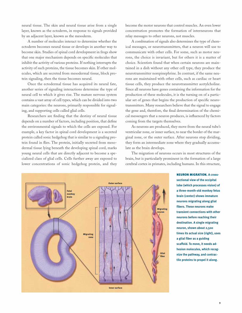
NEURON MIGRATION. A cross-
sectional view of the occipital
lobe (which processes vision) of
a three-month-old monkey fetus
brain (center) shows immature
neurons migrating along glial
fibers. These neurons make
transient connections with other
neurons before reaching their
destination. A single migrating
neuron, shown about 2,500
times its actual size (right), uses
a glial fiber as a guiding
sca≈old. To move, it needs ad-
hesion molecules, which recog-
nize the pathway, and contrac-
tile proteins to propel it along.
Fetalmonkeybrain
Migratingzone
Migratingneuron
Glialfiber
Outer surface
Inner surface
10
neurons slither from the place of origin near the ventricular sur-face along nonneuronal fibers that form a trail to their properdestination. Proper neuron migration requires multiple mech-anisms, including the recognition of the proper path and theability to move long distances. One such mechanism for longdistance migration is the movement of neurons along elongatedfibers that form transient scaffolding in the fetal brain. Manyexternal forces, such as alcohol, cocaine or radiation, preventproper neuronal migration and result in misplacement of cells,which may lead to mental retardation and epilepsy. Further-more, mutations in genes that regulate migration have recentlybeen shown to cause some rare genetic forms of retardation andepilepsy in humans.
Once the neurons reach their final location, they must makethe proper connections for a particular function, such as visionor hearing, to occur. They do this through their axons. Thesestalk-like appendages can stretch out a thousand times longerthan the cell body from which they arise. The journey of mostaxons ends when they meet the branching areas, called den-drites, on other neurons. These target neurons can be locatedat a considerable distance, sometimes at opposite sides of thebrain. In the case of a motor neuron, the axon may travel fromthe spinal cord all the way down to a foot muscle. The linkupsites, called synapses, are where messages are transferred fromone neuron in a circuit to the next.
Axon growth is spearheaded by growth cones. These enlarge-ments of the axon’s tip actively explore the environment as theyseek out their precise destinations. Researchers have discoveredthat many special molecules help guide growth cones. Somemolecules lie on the cells that growth cones contact, while oth-ers are released from sources found near the growth cone. Thegrowth cones, in turn, bear molecules that serve as receptors forthe environmental cues. The binding of particular signals withits receptors tells the growth cone whether to move forward,stop, recoil or change direction.
Recently researchers have identified some of the moleculesthat serve as cues and receptors. These molecules include pro-teins with names such as cadherin, netrin, semaphorin, ephrin,neuropilin and plexin. In most cases, these are families ofrelated molecules; for example there are at least 15 semapo-horins and at least 10 ephrins. Perhaps the most remarkableresult is that most of these are common to worms, insects andmammals, including humans. Each family is smaller in fliesor worms than in mice or people, but their functions are quitesimilar. It has therefore been possible to use the simpler ani-mals to gain knowledge that can be directly applied tohumans. For example, the first netrin was discovered in aworm and shown to guide neurons around the worm’s “nervering.” Later, vertebrate netrins were found to guide axonsaround the mammalian spinal cord. Worm receptors fornetrins were then found and proved invaluable in finding the
corresponding, and again related, human receptors.Once axons reach their targets, they form synapses, which
permit electric signals in the axon to jump to the next cell, wherethey can either provoke or prevent the generation of a new sig-nal. The regulation of this transmission at synapses, and the inte-gration of inputs from the thousands of synapses each neuronreceives, are responsible for the astounding information-processing capabilities of the brain. For processing to occur prop-erly, the connections must be highly specific. Some specificityarises from the mechanisms that guide each axon to its propertarget area. Additional molecules mediate “target recognition”whereby the axon chooses the proper neuron, and often theproper part of the target, once it arrives at its destination. Few ofthese molecules have been identified. There has been more suc-cess, however, in identifying the ways in which the synapse formsonce the contact has been made. The tiny portion of the axonthat contacts the dendrite becomes specialized for the release ofneurotransmitters, and the tiny portion of the dendrite thatreceives the contact becomes specialized to receive and respondto the signal. Special molecules pass between the sending andreceiving cell to ensure that the contact is formed properly.
Paring back
Following the period of growth, the network is pared back tocreate a more sturdy system. Only about one-half of the neu-rons generated during development survive to function in theadult. Entire populations of neurons are removed throughinternal suicide programs initiated in the cells. The programsare activated if a neuron loses its battle with other neurons toreceive life-sustaining nutrients called trophic factors. Thesefactors are produced in limited quantities by target tissues. Eachtype of trophic factor supports the survival of a distinct groupof neurons. For example, nerve growth factor is important forsensory neuron survival. It has recently become clear that theinternal suicide program is maintained into adulthood, andconstantly held in check. Based on this idea, researchers havefound that injuries and some neurodegenerative diseases killneurons not directly by the damage they inflict, but rather byactivating the death program. This discovery, and its implica-tion that death need not inevitably follow insult, have led tonew avenues for therapy.
Brain cells also form too many connections at first. Forexample, in primates, the projection from the two eyes to thebrain initially overlaps, and then sorts out to separate territo-ries devoted only to one or the other eye. Furthermore, in theyoung primate cerebral cortex, the connections between neu-rons are greater in number and twice as dense as an adult pri-mate. Communication between neurons with chemical andelectrical signals is necessary to weed out the connections. Theconnections that are active and generating electrical currentssurvive while those with little or no activity are lost.

Vertebrae
Peripheral nerves
11
SPINAL CORD AND NERVES. The
mature central nervous system
(CNS) consists of the brain and
spinal cord. The brain sends
nerve signals to specific parts of
the body through peripheral
nerves, known as the peripheral
nervous system (PNS). Peripheral
nerves in the cervical region
serve the neck and arms; those in
the thoracic region serve the
trunk; those in the lumbar region
serve the legs; and those in the
sacral region serve the bowels
and bladder. The PNS consists of
the somatic nervous system that
connects voluntary skeletal mus-
cles with cells specialized to re-
spond to sensations, such as
touch and pain. The autonomic
nervous system is made of neu-
rons connecting the CNS with
internal organs. It is divided into
the sympathetic nervous system,
which mobilizes energy and
resources during times of stress
and arousal, and the parasympa-
thetic nervous system, which
conserves energy and resources
during relaxed states.
Critical periods
The brain’s refining and building of the network in mammals,including humans, continues after birth. An organism’s interac-tions with its surroundings fine-tune connections.
Changes occur during critical periods. These are windows oftime during development when the nervous system must obtaincertain critical experiences, such as sensory, movement or emo-tional input, to develop properly. Following a critical period, con-nections become diminished in number and less subject tochange, but the ones that remain are stronger, more reliable andmore precise. Injury, sensory or social deprivation occurring at acertain stage of postnatal life may affect one aspect of develop-ment, while the same injury at a different period may affectanother aspect. In one example, a monkey is raised from birthup to six months of age with one eyelid closed. As a result of
diminished use, the animal permanently loses useful vision inthat eye. This gives cellular meaning to the saying “use it or loseit.” Loss of vision is caused by the actual loss of functional con-nections between that eye and neurons in the visual cortex. Thisfinding has led to earlier and better treatment of the eye disor-ders congenital cataracts and “crossed-eyes” in children.
Research also shows that enriched environments can bolsterbrain development during postnatal life. For example, studiesshow that animals brought up in toy-filled surroundings have morebranches on their neurons and more connections than isolated ani-mals. In one recent study, scientists found enriched environmentsresulted in more neurons in a brain area involved in memory.
Scientists hope that new insights on development will leadto treatments for those with learning disabilities, brain damageand even neurodegenerative disorders or aging.
CENTRAL NERVOUS SYSTEMBrain and spinal cord
PERIPHERAL NERVOUS SYSTEMNerves extending from spinal cord
Cervical region
Thoracic region
Lumbar region
Sacral region
Spinal cord
12
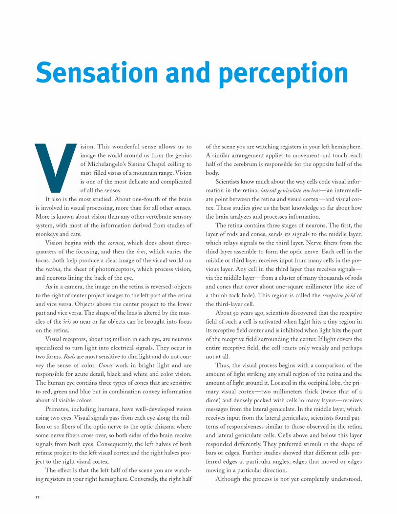
Vision. This wonderful sense allows us toimage the world around us from the geniusof Michelangelo’s Sistine Chapel ceiling tomist-filled vistas of a mountain range. Visionis one of the most delicate and complicatedof all the senses.
It also is the most studied. About one-fourth of the brainis involved in visual processing, more than for all other senses.More is known about vision than any other vertebrate sensorysystem, with most of the information derived from studies ofmonkeys and cats.
Vision begins with the cornea, which does about three-quarters of the focusing, and then the lens, which varies thefocus. Both help produce a clear image of the visual world onthe retina, the sheet of photoreceptors, which process vision,and neurons lining the back of the eye.
As in a camera, the image on the retina is reversed: objectsto the right of center project images to the left part of the retinaand vice versa. Objects above the center project to the lowerpart and vice versa. The shape of the lens is altered by the mus-cles of the iris so near or far objects can be brought into focuson the retina.
Visual receptors, about 125 million in each eye, are neuronsspecialized to turn light into electrical signals. They occur intwo forms. Rods are most sensitive to dim light and do not con-vey the sense of color. Cones work in bright light and areresponsible for acute detail, black and white and color vision.The human eye contains three types of cones that are sensitiveto red, green and blue but in combination convey informationabout all visible colors.
Primates, including humans, have well-developed visionusing two eyes. Visual signals pass from each eye along the mil-lion or so fibers of the optic nerve to the optic chiasma wheresome nerve fibers cross over, so both sides of the brain receivesignals from both eyes. Consequently, the left halves of bothretinae project to the left visual cortex and the right halves pro-ject to the right visual cortex.
The e∑ect is that the left half of the scene you are watch-ing registers in your right hemisphere. Conversely, the right half
of the scene you are watching registers in your left hemisphere.A similar arrangement applies to movement and touch: eachhalf of the cerebrum is responsible for the opposite half of thebody.
Scientists know much about the way cells code visual infor-mation in the retina, lateral geniculate nucleus—an intermedi-ate point between the retina and visual cortex—and visual cor-tex. These studies give us the best knowledge so far about howthe brain analyzes and processes information.
The retina contains three stages of neurons. The first, thelayer of rods and cones, sends its signals to the middle layer,which relays signals to the third layer. Nerve fibers from thethird layer assemble to form the optic nerve. Each cell in themiddle or third layer receives input from many cells in the pre-vious layer. Any cell in the third layer thus receives signals—via the middle layer—from a cluster of many thousands of rodsand cones that cover about one-square millimeter (the size ofa thumb tack hole). This region is called the receptive field ofthe third-layer cell.
About 50 years ago, scientists discovered that the receptivefield of such a cell is activated when light hits a tiny region inits receptive field center and is inhibited when light hits the partof the receptive field surrounding the center. If light covers theentire receptive field, the cell reacts only weakly and perhapsnot at all.
Thus, the visual process begins with a comparison of theamount of light striking any small region of the retina and theamount of light around it. Located in the occipital lobe, the pri-mary visual cortex—two millimeters thick (twice that of adime) and densely packed with cells in many layers—receivesmessages from the lateral geniculate. In the middle layer, whichreceives input from the lateral geniculate, scientists found pat-terns of responsiveness similar to those observed in the retinaand lateral geniculate cells. Cells above and below this layerresponded di∑erently. They preferred stimuli in the shape ofbars or edges. Further studies showed that di∑erent cells pre-ferred edges at particular angles, edges that moved or edgesmoving in a particular direction.
Although the process is not yet completely understood,
V
Sensation and perception

13
VISION. The cornea and lens help produce a clear image of the visual world on the retina, the sheet of photoreceptors and neurons lining the back
of the eye. As in a camera, the image on the retina is reversed: objects to the right of center project images to the left part of the retina and vice
versa. The eye’s 125 million visual receptors—composed of rods and cones—turn light into electrical signals. Rods are most sensitive to dim light
and do not convey the sense of color; cones work in bright light and are responsible for acute detail, black and white and color vision. The human
eye contains three types of cones that are sensitive to red, green and blue but, in combination, convey information about all visible colors. Rods and
cones connect with a middle cell layer and third cell layer (see inset, above). Light passes through these two layers before reaching the rods and
cones. The two layers then receive signals from rods and cones before transmitting the signals onto the optic nerve, optic chiasm, lateral geniculate
nucleus and, finally, the visual cortex.
Optic chiasm
Middle cell layer Rods and ConesThird cell layer
Pupil
Lens
Rods
Cones
Optic nerveRetina
IrisCornea
Visual cortex
Right visual field
Left visual field
Optic nerve
Lateral geniculate nucleus
Modified from Jane Hurd

14
recent findings suggest that visual signals are fed into at least three separate processing systems.One system appears to process information about shape; a second, color; and a third, movement,location and spatial organization. These findings of separate processing systems come from mon-key anatomical and physiological data. They are verified by human psychological studies showingthat the perception of movement, depth, perspective, the relative size of objects, the relative move-ment of objects and shading and gradations in texture all depend primarily on contrasts in lightintensity rather than in color.
Why movement and depth perception should be carried by only one processing system maybe explained by a school of thought called Gestalt psychology. Perception requires various ele-ments to be organized so that related ones are grouped together. This stems from the brain’s abil-ity to group the parts of an image together and also to separate images from one another and fromtheir individual backgrounds.
How do all these systems produce the solid images you see? By extracting biologically rele-vant information at each stage and associating firing patterns with past experience.
Vision studies also have led to better treatment for visual disorders. Information from researchin cats and monkeys has improved the therapy for strabismus, or squint, a term for “cross-eye” orwall-eye. Children with strabismus initially have good vision in each eye. But because they can-not fuse the images in the two eyes, they tend to favor using one eye and often lose useful visionin the other eye.
Vision can be restored but only during infancy or early childhood. Beyond the age of six orso, the blindness becomes permanent. But until a few decades ago, ophthalmologists waited until
HEARING. From the chirping of
crickets to the roar of a rocket
engine, almost all of the thou-
sands of single tones processed
by the human ear are heard by a
mechanism known as air con-
duction. In this process, sound
waves are first funneled
through the external ear—the
pinna and the external auditory
canal—to the middle ear—the
tympanic membrane (eardrum)
that vibrates at di≈erent
speeds. The malleus (hammer),
which is attached to the tym-
panic membrane, transmits the
vibrations to the incus (anvil).
The vibrations are then passed
onto the stapes (stirrup) and
oval window that, in turn, pass
them onto the inner ear. In the
inner ear, the fluid-filled spiral
passage of the cochlea contains
cells with microscopic, hairlike
projections that respond to the
vibrations produced by sound.
The hair cells, in turn, excite the
28,000 fibers of the auditory
nerve that end in the medulla in
the brain. Auditory information
flows via the thalamus to the
temporal gyrus, the part of the
cerebral cortex involved in
receiving and perceiving sound.
Auditory area
External auditory
canal
Pinna
Cochlea
Auditory nerve
External ear Middle ear Inner ear
Malleus Incus Stapes Oval window
To brain
BONES OF THE MIDDLE EAR
Releasedchemicalsexcite nerve and send impulses tobrain
Displacement of hair bundles
Tympanic membrane
Transmitters released
Hair cell of cochlea
Nucleus
Soundwaves

15
children reached the age of four before operating to align theeyes, or prescribe exercises or an eye patch. Now strabismus iscorrected very early in life—before age four—when normalvision can still be restored.
Hearing
Often considered the most important sense for humans, hear-ing allows us to communicate with each other by receivingsounds and interpreting speech. It also gives us informationvital to survival. For example, the sound of an oncoming traintells us to stay clear of the railroad track.
Like the visual system, our hearing system distinguishes sev-eral qualities in the signal it detects. However, our hearing systemdoes not blend di∑erent sounds, as the visual system does whentwo di∑erent wavelengths oflight are mixed to producecolor. We can follow the sep-arate melodic lines of severalinstruments as we listen to anorchestra or rock band.
From the chirping ofcrickets to the roar of a rocketengine, most of the soundsprocessed by the ear are heardby a mechanism known as air conduction. In this process, soundwaves are first funneled through the externally visible part of theear, the pinna (or external ear) and the external auditory canal tothe tympanic membrane (eardrum) that vibrates at di∑erentspeeds. The malleus (hammer), which is attached to the tym-panic membrane, transmits the vibrations to the incus (anvil).This structure passes them onto the stapes (stirrup) which deliv-ers them, through the oval window, to the inner ear.
The fluid-filled spiral passages of each cochlea contain16,000 hair cells whose microscopic, hairlike projectionsrespond to the vibrations produced by sound. The hair cells, inturn, excite the 28,000 fibers of the auditory nerve that termi-nate in the medulla of the brain. Auditory information flowsvia the thalamus to the temporal gyrus, the part of the cerebralcortex involved in receiving and perceiving sound.
The brain’s analysis of auditory information follows a pat-tern similar to that of the visual system. Adjacent neuronsrespond to tones of similar frequency. Some neurons respondto only a small range of frequencies, others react to a widerange; some react only to the beginning of a sound, others onlyrespond to the end.
Speech sounds, however, may be processed di∑erently thanothers. Our auditory system processes all the signals that itreceives in the same way until they reach the primary auditorycortex in the temporal lobe of the brain. When speech soundis perceived, the neural signal is funneled to the left hemispherefor processing in language centers.
Taste and smell
Although di∑erent, the two sensory experiences of taste andsmell are intimately entwined. They are separate senses withtheir own receptor organs. However, these two senses acttogether to allow us to distinguish thousands of di∑erentflavors. Alone, taste is a relatively focused sense concerned withdistinguishing among sweet, salty, sour and bitter. The interac-tion between taste and smell explains why loss of the sense ofsmell apparently causes a serious reduction in the overall tasteexperience, which we call flavor.
Tastes are detected by taste buds, special structures of whichevery human has some 5,000. Taste buds are embedded withinpapillae, or protuberances, located mainly on the tongue, withothers found in the back of the mouth and on the palate. Taste
substances stimulate hairs pro-jecting from the sensory cells.Each taste bud consists of 50 to100 sensory cells that respondto salts, acidity, sweet sub-stances and bitter compounds.Some researchers add a fifthcategory named umami, for thetaste of monosodium gluta-mate and related substances.
Taste signals in the sensory cells are transferred by synapsesto the ends of nerve fibers, which send impulses along cranialnerves to taste centers in the brain. From here, the impulses arerelayed to other brain stem centers responsible for the basicresponses of acceptance or rejection of the tastes, and to thethalamus and on to the cerebral cortex for conscious perceptionof taste.
Specialized smell receptor cells are located in a small patchof mucus membrane lining the roof of the nose. Axons of thesesensory cells pass through perforations in the overlying boneand enter two elongated olfactory bulbs lying on top of the bone.The portion of the sensory cell that is exposed to odors pos-sesses hair-like cilia. These cilia contain the receptor sites thatare stimulated by odors carried by airborne molecules. The odormolecules dissolve in the mucus lining in order to stimulatereceptor molecules in the cilia to start the smell response. Anodor molecule acts on many receptors to di∑erent degrees. Sim-ilarly, a receptor interacts with many di∑erent odor moleculesto di∑erent degrees.
Axons of the cells pass through perforations in the overly-ing bone and enter two elongated olfactory bulbs lying on topof the bone. The pattern of activity set up in the receptor cellsis projected to the olfactory bulb, where it forms a spatial imageof the odor. Impulses created by this stimulation pass to smellcenters, to give rise to conscious perceptions of odor in thefrontal lobe and emotional responses in the limbic system ofthe brain.
Taste and smell are two separate senses with
their own sets of receptor organs, but they act
together to distinguish an enormous number of
di≈erent flavors.
16
Touch and pain
Touch is the sense by which we determine the characteristics of objects: size, shape and texture.We do this through touch receptors in the skin. In hairy skin areas, some receptors consist of websof sensory nerve cell endings wrapped around the hair bulbs. They are remarkably sensitive, beingtriggered when the hairs are moved. Other receptors are more common in non-hairy areas, suchas lips and fingertips, and consist of nerve cell endings that may be free or surrounded by bulb-like structures.
Signals from touch receptors pass via sensory nerves to the spinal cord, then to the thalamusand sensory cortex. The transmission of this information is highly topographic, meaning that thebody is represented in an orderly fashion at di∑erent levels of the nervous system. Larger areas ofthe cortex are devoted to sensations from the hands and lips; much smaller cortical regions rep-resent less sensitive parts of the body.
Di∑erent parts of the body vary in their sensitivity to touch discrimination and painful stim-uli according to the number and distribution of receptors. The cornea is several hundred timesmore sensitive to painful stimuli than are the soles of the feet. The fingertips are good at touchdiscrimination but the chest and back are less sensitive.
Until recently, pain was thought to be a simple message by which neurons sent electricalimpulses from the site of injury directly to the brain.
Recent studies show that the process is more complicated. Nerve impulses from sites of injurythat persist for hours, days or longer lead to changes in the nervous system that result in anamplification and increased duration of the pain. These changes involve dozens of chemical mes-sengers and receptors.
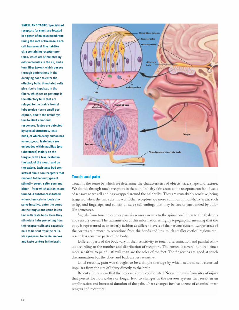
SMELL AND TASTE. Specialized
receptors for smell are located
in a patch of mucous membrane
lining the roof of the nose. Each
cell has several fine hairlike
cilia containing receptor pro-
teins, which are stimulated by
odor molecules in the air, and a
long fiber (axon), which passes
through perforations in the
overlying bone to enter the
olfactory bulb. Stimulated cells
give rise to impulses in the
fibers, which set up patterns in
the olfactory bulb that are
relayed to the brain’s frontal
lobe to give rise to smell per-
ception, and to the limbic sys-
tem to elicit emotional
responses. Tastes are detected
by special structures, taste
buds, of which every human has
some 10,000. Taste buds are
embedded within papillae (pro-
tuberances) mainly on the
tongue, with a few located in
the back of the mouth and on
the palate. Each taste bud con-
sists of about 100 receptors that
respond to the four types of
stimuli—sweet, salty, sour and
bitter—from which all tastes are
formed. A substance is tasted
when chemicals in foods dis-
solve in saliva, enter the pores
on the tongue and come in con-
tact with taste buds. Here they
stimulate hairs projecting from
the receptor cells and cause sig-
nals to be sent from the cells,
via synapses, to cranial nerves
and taste centers in the brain.
Olfactory tract
Olfactorybulb
Nerve fibers to brain
Receptor cells
Cilia
Airborne odors
Food chemicals
Taste bud pore
Synapse
Taste (gustatory) nerve to brain
Tongue

17
At the point of injury, nociceptors, special receptors, respondto tissue-damaging stimuli. Injury results in the release ofnumerous chemicals at the site of damage and inflammation.One such chemical, prostaglandin, enhances the sensitivity ofreceptors to tissue damage and ultimately can result in moreintense pain sensations. It also contributes to the clinical con-dition in which innocuous stimuli can produce pain (such as insunburned skin) because the threshold of the nociceptor issignificantly reduced.
Pain messages are transmitted to the spinal cord via smallmyelinated fibers and C fibers—very small unmyelinated fibers.Myelin is a covering around nerve fibers that helps them sendtheir messages more rapidly.
In the ascending system, the impulses are relayed from thespinal cord to several brain structures, including the thalamusand cerebral cortex, which are involved in the process by which“pain” messages become conscious experience.
Pain messages can also be suppressed by a system of neu-rons that originate within the gray matter in the brainstem ofthe midbrain. This descending system sends messages to the dor-sal horn of the spinal cord where it suppresses the transmissionof pain signals to the higher brain centers. Some of thesedescending systems use naturally occurring chemicals similar toopioids. The three major families of opioids—enkephalins,endorphins and dynorphins—identified in the brain originatefrom three precursor proteins coded by three di∑erent genes.They act at multiple opioid receptors in the brain and spinalcord. This knowledge has led to new treatments for pain: Opiate-like drugs injected into the space above the spinal cord providelong-lasting pain relief.
Scientists are now using modern tools for imaging brainstructures in humans to determine the role of the higher cen-ters of the brain in pain experience and how signals in thesestructures change with long-lasting pain.
PAIN. Messages about tissue
damage are picked up by recep-
tors and transmitted to the
spinal cord via small, myeli-
nated fibers and very small
unmyelinated fibers. From the
spinal cord, the impulses are
carried to the brainstem, thala-
mus and cerebral cortex and
ultimately perceived as pain.
These messages can be sup-
pressed by a system of neurons
that originates in the gray
matter of the midbrain. This
descending pathway sends mes-
sages to the spinal cord where it
suppresses the transmission of
tissue damage signals to the
higher brain centers. Some of
these descending pathways use
naturally occurring, opiate-like
chemicals called endorphins.
Message is received in the thalamus and cerebral cortex
Tissue-damaging stimulus activates nociceptors
Message carried to spinal cord
Descending pathway
Nociceptors
From brain To brain
Dorsal horn
Muscle fiber

18
The conscious memory of a patient known asH.M. is limited almost entirely to events thatoccurred years before his surgery, whichremoved part of the medial temporal lobe of hisbrain to relieve epilepsy. H.M. can rememberrecent events for only a few minutes. Talk with
him awhile and then leave the room. When you return, he hasno recollection of ever having seen you before.
The medial temporal lobe, which includes the hippocam-pus and adjacent brain areas, seems to play a role in convertingmemory from a short-term to a long-term, permanent form.The fact that H.M. retains memories for events that are remoteto his surgery is evidence that the medial temporal region is notthe site of permanent storage but that it plays a role in the for-mation of new memories. Other patients like H.M. have alsobeen described.
Additional evidence comes from patients undergoing elec-
troconvulsive therapy (ECT) for depression. ECT is thought totemporarily disrupt the function of the hippocampus andrelated structures. These patients typically su∑er di≈culty withnew learning and have amnesia for events that occurred duringthe several years before treatment. Memory of earlier events isunimpaired. As time passes after treatment, much of the lostpart of memory becomes available once again.
The hippocampus and the medial temporal region are con-nected with widespread areas of the cerebral cortex, especiallythe vast regions responsible for thinking and language. Whereasthe medial temporal region is important for forming and orga-nizing memory, cortical areas are important for the long-termstorage of knowledge about facts and events and for how theseare used in everyday situations.
Working memory, a type of transient memory that enablesus to retain what someone has said just long enough to reply,depends in part on the prefrontal cortex. Researchers discov-ered that certain neurons in this area are influenced by neuronsreleasing dopamine and other neurons releasing glutamate.
While much is unknown about learning and memory, scien-tists can recognize certain pieces of the process. For example, thebrain appears to process di∑erent kinds of information in sepa-
rate ways and then store it di∑erently. Procedural knowledge, theknowledge of how to do something, is expressed in skilled behav-ior and learned habits. Declarative knowledge provides an explicit,consciously accessible record of individual previous experiencesand a sense of familiarity about those experiences. Declarativeknowledge requires processing in the medial temporal region andparts of the thalamus, while procedural knowledge requires pro-cessing by the basal ganglia. Other kinds of memory depend onthe amygdala (emotional aspects of memory) and the cerebellum(motor learning where precise timing is involved).
An important factor that influences what is stored and howstrongly it is stored is whether the action is followed by reward-ing or punishing consequences. This is an important principlein determining what behaviors an organism will learn andremember. The amygdala appears to play an important role inthese memory events.
How exactly does memory occur? After years of study, thereis much support for the idea that memory involves a persistentchange in the relationship between neurons. In animal studies,scientists found that this occurs through biochemical events inthe short term that a∑ect the strength of the relevant synapses.The stability of long-term memory is conferred by structuralmodifications within neurons that change the strength andnumber of synapses. For example, researchers can correlatespecific chemical and structural changes in the relevant cellswith several simple forms of behavioral change exhibited by thesea slug Aplysia.
Another important model for the study of memory is thephenomenon of long-term potentiation (LTP), a long-lastingincrease in the strength of a synaptic response following stim-ulation. LTP occurs prominently in the hippocampus, as wellas in other brain areas. Studies of rats suggest LTP occurs bychanges in synaptic strength at contacts involving NMDAreceptors. It is now possible to study LTP and learning ingenetically modified mice that have abnormalities of specificgenes. Abnormal gene expression can be limited to particularbrain areas and time periods, such as during learning.
Scientists believe that no single brain center stores mem-ory. It most likely is stored in the same, distributed collection
Learning and memory
T

19
of cortical processing systems involved in the perception, pro-cessing and analysis of the material being learned. In short,each part of the brain most likely contributes di∑erently to per-manent memory storage.
One of the most prominent intellectual activities depen-dent on memory is language. While the neural basis of lan-guage is not fully understood, scientists have learned muchabout this feature of the brain from studies of patients who havelost speech and language abilities due to stroke, and from behav-ioral and functional neuroimaging studies of normal people.
A prominent and influential model, based on studies ofthese patients, proposes that the underlying structure of speechcomprehension arises in Wernicke’s area, a portion of the lefthemisphere of the brain. This temporal lobe region is connectedwith Broca’s area in the frontal lobe where a program for vocalexpression is created. This program is then transmitted to anearby area of the motor cortex that activates the mouth,tongue and larynx.
This same model proposes that, when we read a word, theinformation is transmitted from the primary visual cortex to theangular gyrus where the message is somehow matched with thesounds of the words when spoken. The auditory form of the
word is then processed for comprehension in Wernicke’s areaas if the word had been heard. Writing in response to an oralinstruction requires information to be passed along the samepathways in the opposite direction—from the auditory cortexto Wernicke’s area to the angular gyrus. This model accountsfor much of the data from patients, and is the most widely usedmodel for clinical diagnosis and prognosis. However, somerefinements to this model may be necessary due to both recentstudies with patients and functional neuroimaging studies innormal people.
For example, using an imaging technique called positron
emission tomography (PET), scientists have demonstrated thatsome reading tasks performed by normal people activated nei-ther Wernicke’s area nor the angular gyrus. These results sug-gest that there is a direct reading route that does not involvespeech sound recoding of the visual stimulus before the pro-cessing of either meaning or speaking. Other studies withpatients also have indicated that it is likely that familiar wordsneed not be recoded into sound before they can be understood.
Although the understanding of how language is imple-mented in the brain is far from complete, there are now severaltechniques that may be used to gain important insights.
LEARNING AND MEMORY,
SPEECH AND LANGUAGE.
Structures believed to be impor-
tant for various kinds of learning
and memory include the cere-
bral cortex, amygdala, hip-
pocampus, cerebellum and
basal ganglia. Areas of the left
hemisphere (inset) are known to
be active in speech and lan-
guage. The form and meaning of
an utterance is believed to arise
in Wernicke’s area and then
Broca’s area, which is related to
vocalization. Wernicke’s area is
also important for language
comprehension.
Cerebral cortex
Wernicke’s area
Broca’s area
AREAS OF SPEECH AND LANGUAGE
Amygdala Hippocampus
Cerebellum
BASAL GANGLIA
Caudate nucleus
Putamen
Globus pallidus
Amygdaloidnucleus
Angulargyrus

20
From the stands, we marvel at the perfectly placedserves of professional tennis players and lightning-fast double plays executed by big league infielders.But in fact, every one of us in our daily lives per-forms highly skilled movements, such as walkingupright, speaking and writing, that are no less
remarkable. A finely tuned and highly complex central nervoussystem controls the action of hundreds of muscles in accom-plishing these everyday marvels.
In order to understand how the nervous system performsthis trick, we have to start with muscles. Most muscles attachto points on the skeleton that cross one or more joints. Acti-vation of a given muscle, the agonist, can open or close thejoints that it spans or act to sti∑en them, depending on theforces acting on those joints from the environment or othermuscles that oppose the agonist, the antagonists. Relatively fewmuscles act on soft tissue. Examples include the muscles thatmove the eyes and tongue, and the muscles that control facialexpression.
A muscle is made up of thousands of individual musclefibers, each of which is controlled by one alpha motor neuron ineither the brain or spinal cord. On the other hand, a singlealpha neuron can control hundreds of muscle fibers, forming amotor unit. These motor neurons are the critical link betweenthe brain and muscles. When these neurons die, a person is nolonger able to move.
The simplest movements are reflexes—fixed muscleresponses to particular stimuli. Studies show sensory stretchreceptors—called muscle spindles, which include small, special-ized muscle fibers and are located in most muscles—send infor-mation about muscles directly to alpha motor neurons.
Sudden muscle stretch (such as when a doctor taps a mus-cle tendon to test your reflexes) sends a barrage of impulses intothe spinal cord along the muscle spindle sensory fibers. This,in turn, activates motor neurons in the stretched muscle, caus-ing a contraction which is called the stretch reflex. The samesensory stimulus causes inactivation, or inhibition, in the motorneurons of the antagonist muscles through connecting neurons,called inhibitory neurons, within the spinal cord.
The sensitivity of the muscle spindle organs is controlledby the brain through a separate set of gamma motor neurons thatcontrol the specialized spindle muscle fibers and allow the brainto fine-tune the system for di∑erent movement tasks. Othermuscle sense organs signal muscle force that a∑ects motor neu-rons through separate sets of spinal neurons. We now know thatthis complex system responds di∑erently for tasks that requireprecise control of position (holding a full teacup), as opposedto those that require rapid, strong movement (throwing a ball).You can experience such changes in motor strategy when youcompare walking down an illuminated staircase with the sametask done in the dark.
Another useful reflex is the flexion withdrawal that occursif your bare foot encounters a sharp object. Your leg is imme-diately lifted from the source of potential injury (flexion) butthe opposite leg responds with increased extension in order tomaintain your balance. The latter event is called the crossed
extension reflex. These responses occur very rapidly and withoutyour attention because they are built into systems of neuronslocated within the spinal cord itself.
It seems likely that the same systems of spinal neurons alsoparticipate in controlling the alternating action of the legs dur-ing normal walking. In fact, the basic patterns of muscle acti-vation that produce coordinated walking can be generated infour-footed animals within the spinal cord itself. It seems likelythat these spinal mechanisms, which evolved in primitive ver-tebrates, are probably still present in the human spinal cord.
The most complex movements that we perform, includingvoluntary ones that require conscious planning, involve controlof the spinal mechanisms by the brain. Scientists are onlybeginning to understand the complex interactions that takeplace between di∑erent brain regions during voluntary move-ments, mostly through careful experiments on animals. Oneimportant area is the motor cortex, which exerts powerful con-trol of the spinal cord neurons and has direct control of somemotor neurons in monkeys and humans. Some neurons in themotor cortex appear to specify the coordinated action of manymuscles, so as to produce organized movement of the limb toa particular place in space.
F
Movement
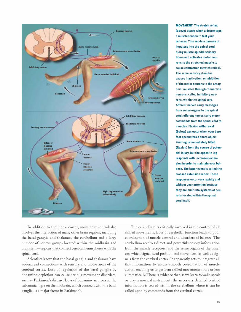
21
In addition to the motor cortex, movement control alsoinvolves the interaction of many other brain regions, includingthe basal ganglia and thalamus, the cerebellum and a largenumber of neuron groups located within the midbrain andbrainstem—regions that connect cerebral hemispheres with thespinal cord.
Scientists know that the basal ganglia and thalamus havewidespread connections with sensory and motor areas of thecerebral cortex. Loss of regulation of the basal ganglia bydopamine depletion can cause serious movement disorders,such as Parkinson’s disease. Loss of dopamine neurons in thesubstantia nigra on the midbrain, which connects with the basalganglia, is a major factor in Parkinson’s.
The cerebellum is critically involved in the control of allskilled movements. Loss of cerebellar function leads to poorcoordination of muscle control and disorders of balance. Thecerebellum receives direct and powerful sensory informationfrom the muscle receptors, and the sense organs of the innerear, which signal head position and movement, as well as sig-nals from the cerebral cortex. It apparently acts to integrate allthis information to ensure smooth coordination of muscleaction, enabling us to perform skilled movements more or lessautomatically. There is evidence that, as we learn to walk, speakor play a musical instrument, the necessary detailed controlinformation is stored within the cerebellum where it can becalled upon by commands from the cerebral cortex.
MOVEMENT. The stretch reflex
(above) occurs when a doctor taps
a muscle tendon to test your
reflexes. This sends a barrage of
impulses into the spinal cord
along muscle spindle sensory
fibers and activates motor neu-
rons to the stretched muscle to
cause contraction (stretch reflex).
The same sensory stimulus
causes inactivation, or inhibition,
of the motor neurons to the antag-
onist muscles through connection
neurons, called inhibitory neu-
rons, within the spinal cord.
A≈erent nerves carry messages
from sense organs to the spinal
cord; e≈erent nerves carry motor
commands from the spinal cord to
muscles. Flexion withdrawal
(below) can occur when your bare
foot encounters a sharp object.
Your leg is immediately lifted
(flexion) from the source of poten-
tial injury, but the opposite leg
responds with increased exten-
sion in order to maintain your bal-
ance. The latter event is called the
crossed extension reflex. These
responses occur very rapidly and
without your attention because
they are built into systems of neu-
rons located within the spinal
cord itself.
Inhibitory neuron
Alpha motor neuron
Sensory neuron
Extensor muscles activated
Flexor muscles inhibited
Response
Stimulus
Afferent nerves
Muscle spindle
Efferent nerves
Sensory neuron
Motor neurons
Motor neurons
Inhibitory neurons
Excitatory neurons
Extensor muscles activated
Flexor musclesinhibited
Flexor muscles activated
Stimulus
Right leg extends tobalance body
Extensor muscles inhibited -
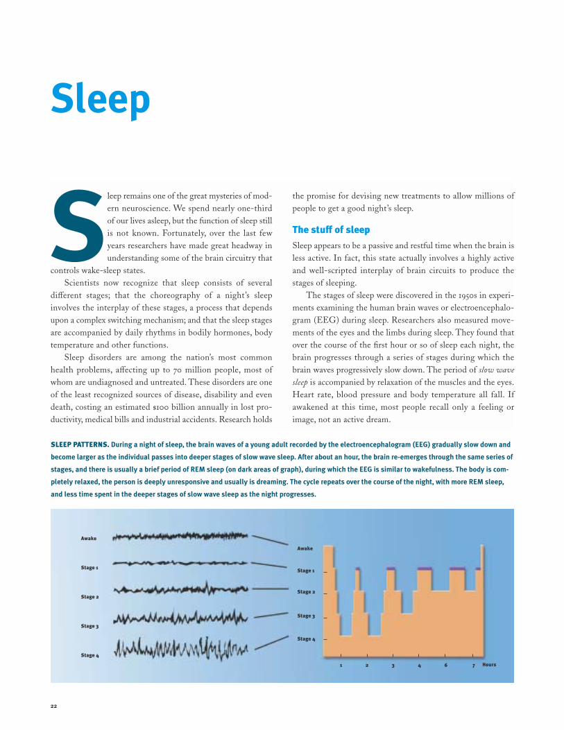
22
Sleep remains one of the great mysteries of mod-ern neuroscience. We spend nearly one-thirdof our lives asleep, but the function of sleep stillis not known. Fortunately, over the last fewyears researchers have made great headway inunderstanding some of the brain circuitry that
controls wake-sleep states.Scientists now recognize that sleep consists of several
di∑erent stages; that the choreography of a night’s sleepinvolves the interplay of these stages, a process that dependsupon a complex switching mechanism; and that the sleep stagesare accompanied by daily rhythms in bodily hormones, bodytemperature and other functions.
Sleep disorders are among the nation’s most commonhealth problems, a∑ecting up to 70 million people, most ofwhom are undiagnosed and untreated. These disorders are oneof the least recognized sources of disease, disability and evendeath, costing an estimated $100 billion annually in lost pro-ductivity, medical bills and industrial accidents. Research holds
the promise for devising new treatments to allow millions ofpeople to get a good night’s sleep.
The stu≈ of sleep
Sleep appears to be a passive and restful time when the brain isless active. In fact, this state actually involves a highly activeand well-scripted interplay of brain circuits to produce thestages of sleeping.
The stages of sleep were discovered in the 1950s in experi-ments examining the human brain waves or electroencephalo-gram (EEG) during sleep. Researchers also measured move-ments of the eyes and the limbs during sleep. They found thatover the course of the first hour or so of sleep each night, thebrain progresses through a series of stages during which thebrain waves progressively slow down. The period of slow wave
sleep is accompanied by relaxation of the muscles and the eyes.Heart rate, blood pressure and body temperature all fall. Ifawakened at this time, most people recall only a feeling orimage, not an active dream.
Sleep
SLEEP PATTERNS. During a night of sleep, the brain waves of a young adult recorded by the electroencephalogram (EEG) gradually slow down and
become larger as the individual passes into deeper stages of slow wave sleep. After about an hour, the brain re-emerges through the same series of
stages, and there is usually a brief period of REM sleep (on dark areas of graph), during which the EEG is similar to wakefulness. The body is com-
pletely relaxed, the person is deeply unresponsive and usually is dreaming. The cycle repeats over the course of the night, with more REM sleep,
and less time spent in the deeper stages of slow wave sleep as the night progresses.
S
Awake
Stage 1
Stage 2
Stage 3
Stage 4
Awake
Stage 1
Stage 2
Stage 3
Stage 4
1 2 3 4 6 7 Hours

23
THE WAKING AND SLEEPING
BRAIN. Wakefulness is main-
tained by activity in two systems
of brainstem neurons. Nerve
cells that make the neurotrans-
mitter acetylcholine stimulate
the thalamus, which activates
the cerebral cortex (red path-
way). Full wakefulness also
requires cortical activation by
other neurons that make
monoamine neurotransmitters
such as norepinephrine, sero-
tonin and histamine (blue path-
way). During slow wave sleep,
when the brain becomes less
active, neuron activity in both
pathways slows down. During
rapid eye movement sleep, in
which dreaming occurs, the neu-
rons using acetylcholine fire
rapidly, producing a dreaming
state, but the monoamine cells
stop firing altogether.
Over the next half hour or so, the brain emerges from thedeep slow wave sleep as the EEG waves become progressivelyfaster. Similar to during waking, rapid eye movements emerge,but the body’s muscles become almost completely paralyzed(only the muscles that allow breathing remain active). This stateis often called rapid eye movement (REM) sleep. During REMsleep, there is active dreaming. Heart rate, blood pressure andbody temperature become much more variable. Men often haveerections during this stage of sleep. The first REM period usu-ally lasts ten to 15 minutes.
Over the course of the night, these alternative cycles of slowwave and REM sleep alternate, with the slow wave sleepbecoming less deep, and the REM periods more prolonged,until waking occurs.
Over the course of a lifetime, the pattern of sleep cycleschanges. Infants sleep up to 18 hours per day, and they spendmuch more time in deep slow wave sleep. As children mature,they spend less time asleep, and less time in deep slow wavesleep. Older adults may sleep only six to seven hours per night,often complain of early wakening that they cannot avoid, andspend very little time in slow wave sleep.
Sleep disorders
The most common sleep disorder, and the one most people arefamiliar with, is insomnia. Some people have di≈culty fallingasleep initially, but other people fall asleep, and then awakenpart way through the night, and cannot fall asleep again.Although there are a variety of short-acting sedatives andsedating antidepressant drugs available to help, none of theseproduces a truly natural and restful sleep state because they tendto suppress the deeper stages of slow wave sleep.
Excessive daytime sleepiness may have many causes. Themost common are disorders that disrupt sleep and result ininadequate amounts of sleep, particularly the deeper stages.These are usually diagnosed in the sleep laboratory. Here, theEEG, eye movements and muscle tone are monitored electri-cally as the individual sleeps. In addition, the heart, breathing,and oxygen content of the blood can be monitored.
Obstructive sleep apnea causes the airway muscles in thethroat to collapse as sleep deepens. This prevents breathing,which causes arousal, and prevents the su∑erer from enteringthe deeper stages of slow wave sleep. This condition can alsocause high blood pressure and may increase the risk of heart
Cerebral cortex
Thalamus
Pons
Spinal cord

24
attack. There is also an increased risk of daytime accident, espe-cially automobile accidents, which may prevent driving. Treat-ment is complex and may include a variety of attempts to reduceairway collapse during sleep. While simple things like losingweight, avoiding alcohol and sedating drugs prior to sleep, andavoiding sleeping on one’s back can sometimes help, most peo-ple with sleep apnea require positive airway pressure to keep theairway open. This can be provided by fitting a small mask overthe nose that provides an air stream under pressure during sleep.In some cases, surgery is needed to correct the airway anatomy.
Periodic limb movements of sleep are intermittent jerks of thelegs or arms, which occur as the individual enters slow wavesleep, and can cause arousal from sleep. Other people haveepisodes in which their muscles fail to be paralyzed duringREM sleep, and they act out their dreams. This REM behavior
disorder can also be very disruptive to a normal nights’ sleep.Both disorders are more common in people with Parkinson’sdisease, and both can be treated with drugs that treat Parkin-son’s, or with an anti-epileptic drug called clonazepam.
Narcolepsy is a relatively uncommon condition (one case per2,500 people) in which the switching mechanism for REMsleep does not work properly. Narcoleptics have sleep attacksduring the day, in which they suddenly fall asleep. This issocially disruptive, as well as dangerous, for example, if they aredriving. They tend to enter REM sleep very quickly as well, andmay even enter a dreaming state while still awake, a conditionknown as hypnagogic hallucinations. They also have attacks dur-ing which they lose muscle tone, similar to what occurs duringREM sleep, but while they are awake. Often, this occurs whilethey are falling asleep or just waking up, but attacks of paraly-sis known as cataplexy can be triggered by an emotional expe-rience or even hearing a funny joke.
Recently, insights into the mechanism of narcolepsy havegiven major insights into the processes that control these mys-terious transitions between waking, slow wave and REM sleepstates.
How is sleep regulated?
During wakefulness, the brain is kept in an alert state by theinteractions of two major systems of nerve cells. Nerve cells inthe upper part of the pons and in the midbrain, which makeacetylcholine as their neurotransmitter, send inputs to the thal-amus, to activate it. When the thalamus is activated, it in turnactivates the cerebral cortex, and produces a waking EEG pat-tern. However, that is not all there is to wakefulness. As dur-ing REM sleep, the cholinergic nerve cells and the thalamusand cortex are in a condition similar to wakefulness, but thebrain is in REM sleep, and is not very responsive to externalstimuli.
The di∑erence is supplied by three sets of nerve cells in theupper part of the brainstem: nerve cells in the locus coeruleus
that contain the neurotransmitter norepinephrine; in the dor-sal and median raphe groups that contain serotonin; and in thetuberomammillary cell group that contains histamine. Thesemonoamine neurons fire most rapidly during wakefulness, butthey slow down during slow wave sleep, and they stop duringREM sleep.
The brainstem cell groups that control arousal are in turnregulated by two groups of nerve cells in the hypothalamus, partof the brain that controls basic body cycles. One group of nervecells, in the ventrolateral preoptic nucleus, contain inhibitoryneurotransmitters, galanin and GABA. When the ventrolateralpreoptic neurons fire, they are thought to turn o∑ the arousalsystems, causing sleep. Damage to the ventrolateral preopticnucleus produces irreversible insomnia.
A second group of nerve cells in the lateral hypothalamusact as an activating switch. They contain the neurotransmittersorexin and dynorphin, which provide an excitatory signal to thearousal system, particularly to the monoamine neurons. Inexperiments in which the gene for the neurotransmitter orexinwas experimentally removed in mice, the animals became nar-coleptic. Similarly, in two dog strains with naturally occurringnarcolepsy, an abnormality was discovered in the gene for thetype 2 orexin receptor. Recent studies show that in humans withnarcolepsy, the orexin levels in the brain and spinal fluid areabnormally low. Thus, orexin appears to play a critical role inactivating the monoamine system, and preventing abnormaltransitions, particularly into REM sleep.
Two main signals control this circuitry. First, there is home-ostasis, or the body’s need to seek a natural equilibrium. Thereis an intrinsic need for a certain amount of sleep each day. Themechanism for accumulating sleep need is not yet clear. Somepeople think that a chemical called adenosine may accumulatein the brain during prolonged wakefulness, and that it maydrive sleep homeostasis. Interestingly, the drug ca∑eine, whichis widely used to prevent sleepiness, acts as an adenosineblocker, to prevent its e∑ects.
If an individual does not get enough sleep, the sleep debtprogressively accumulates, and leads to a degradation of men-tal function. When the opportunity comes to sleep again, theindividual will sleep much more, to “repay” the debt, and theslow wave sleep debt is usually “paid o∑” first.
The other major influence on sleep cycles is the body’s cir-cadian clock, the suprachiasmatic nucleus. This small group ofnerve cells in the hypothalamus contains clock genes, which gothrough a biochemical cycle of almost exactly 24 hours, settingthe pace for daily cycles of activity, sleep, hormones and otherbodily functions. The suprachiasmatic nucleus also receives aninput directly from the retina, and the clock can be reset bylight, so that it remains linked to the outside world’s day-nightcycle. The suprachiasmatic nucleus provides a signal to the ven-trolateral preoptic nucleus and probably the orexin neurons.

25
The urge to act in the presence of stress has beenwith us since our ancient ancestors. In responseto impending danger, muscles are primed,attention is focused and nerves are readied foraction—fight or flight. But in today’s corpora-tion-dominated world, this response to stress is
simply inappropriate and may be a contributor to heart disease,accidents and aging.
Indeed, nearly two-thirds of ailments seen in doctors’o≈ces are commonly thought to be stress-induced or related tostress in some way. Surveys indicate that 60 percent of Amer-icans feel they are under a great deal of stress at least once aweek. Costs due to stress from absenteeism, medical expensesand lost productivity are estimated at $300 billion annually.
Only recently admitted into the medical vocabulary, stressis di≈cult to define because its e∑ects vary with each individ-ual. Dr. Hans Selye, a founder of stress research, called it “therate of wear and tear in the body.” Other specialists now definestress as any external stimulus that threatens homeostasis—thenormal equilibrium of body function. Among the most power-ful stressors are psychological and psychosocial stressors thatexist between members of the same species. Lack or loss of con-trol is a particularly important feature of severe psychologicalstress that can have physiological consequences.
During the last six decades, researchers using animals foundthat stress both helps and harms the body. When confrontedwith a crucial challenge, properly controlled stress responsescan provide the extra strength and energy needed to cope.Moreover, the acute physiological response to stress protectsthe body and brain and helps to re-establish or maintain home-ostasis. But stress that continues for prolonged periods of timecan repeatedly elevate the physiological stress responses or failto shut them o∑ when not needed. When this occurs, thesesame physiological mechanisims can badly upset the body’s bio-chemical balance and accelerate disease.
Scientists also believe that the individual variation inresponding to stress is somewhat dependent on a person’s per-ception of external events. This perception ultimately shapeshis or her internal physiological response. Thus, by controlling
your perception of events, you can do much to avoid the harm-ful consequences of stress.
The immediate response
A stressful situation activates three major communication sys-tems in the brain that regulate bodily functions. Scientists havecome to understand these complex systems through experi-ments primarily with rats, mice and nonhuman primates, suchas monkeys. Scientists then verified the action of these systemsin humans.
The first of these systems is the voluntary nervous system,which sends messages to muscles so that we may respond tosensory information. For example, the sight of a growling bearon a trail in Yellowstone National Park prompts you to run asquickly as possible.
The second communication system is the autonomic nervous
system. It combines the sympathetic or emergency branch,which gets us going in emergencies, and the parasympathetic orcalming branch, which keeps the body’s maintenance systems,such as digestion, in order and calms the body’s responses tothe emergency branch.
Each of these systems has a specific task. The emergencybranch causes arteries supplying blood to the muscles to relaxin order to deliver more blood, allowing greater capacity to act.At the same time, the emergency system reduces blood flow tothe skin, kidney and digestive tract and increases blood flow tothe muscles. In contrast, the calming branch helps to regulatebodily functions and soothe the body, preventing it fromremaining too long in a state of mobilization. Remaining mobi-lized and left unchecked, these body functions could lead todisease. Some actions of the calming branch appear to reducethe harmful e∑ects of the emergency branch’s response to stress.
The brain’s third major communication process is the neu-
roendocrine system, which also maintains the body’s internalfunctioning. Various “stress hormones” travel through the bloodand stimulate the release of other hormones, which a∑ect bod-ily processes, such as metabolic rate and sexual functions.
Major stress hormones are epinephrine (also known asadrenaline) and cortisol. When the body is exposed to stressors,
Stress
T

THE STRESS REACTION. When
stress occurs, the sympathetic
nervous system is triggered.
Norepinephrine is released by
nerves, and epinephrine is
secreted by the adrenal glands.
By activating receptors in blood
vessels and other structures,
these substances ready the
heart and working muscles for
action.
In the parasympathetic ner-
vous system, acetylcholine is
released, producing calming
e≈ects. The digestive tract is
stimulated to digest a meal, the
heart rate slows and the pupils
of the eye become smaller.
The neuroendocrine system
also maintains the body’s
normal internal function-
ing. Corticotrophin-releas-
ing factor (CRF), a peptide
formed by chains of amino acids,
is released from the hypothala-
mus, a collection of cells at the
base of the brain that acts as a
control center for the neuroen-
docrine system. CRF travels to
the pituitary gland where it trig-
gers the release of adrenocorti-
cotropic hormone (ACTH). ACTH
travels in the blood to the
adrenal glands where it stimu-
lates the release of cortisol.
26
NEUROENDOCRINE SYSTEM
AUTONOMIC NERVOUS SYSTEM
Intestines
Stomach
Heart
Thymus and Immune System
Muscle
Eyes
STRESS
Bloodvessels
Adrenal gland
Blood stream
Epinephrine
Cortisol
Prepares body for immediate
response
Re-establishes homeostatis
PituitaryHypothalamus
ACTH
CRF
STRESS
epinephrine is quickly released into the bloodstream to put thebody into a general state of arousal and enable it to cope witha challenge.
The secretion by the adrenal glands of cortisol—known asa glucocorticoid because it a∑ects the metabolism of glucose, asource of energy—starts about five minutes later. Some of itsactions help to mediate the stress-response, while some of itsother, slower ones, counteract the primary response to stressand help re-establish homeostasis. Over the short run, cortisolmobilizes energy and delivers it to muscles for the body’sresponse. With prolonged exposure, cortisol enhances feeding
and helps the body recover from energy mobilization.Acute stress also increases activity of the immune system
and helps protect the body from disease pathogens. The twomajor stress hormones, cortisol and adrenaline, facilitate themovement of immune cells from the bloodstream and storageorgans such as the spleen into tissue where they are needed todefend against an infection.
Glucocorticoids also a∑ect food intake during the sleep-wake cycle. Cortisol levels peak in the body in the early morn-ing hours just before waking. This hormone acts as a wake-upsignal and helps to turn on appetite and physical activity. This

27
e∑ect of glucocorticoids may help to explain disorders, such asjet-lag, which result when the light-dark cycle is altered by jettravel over long distances, causing the body’s biological clockto reset itself more slowly. Until that clock is reset, cortisolsecretion and hunger, as well as sleepiness and wakefulness,occur at inappropriate times of day in the new location.
Glucocorticoids do more than help the body respond tostress. In fact, they are an integral part of daily life and theadaptation to environmental change. The adrenal glands helpprotect us from stress and are essential for survival.
Chronic stress
When glucocorticoids or epinephrine are secreted in responseto the prolonged psychological stress commonly encounteredby humans, the results are notideal. Normally, bodily sys-tems gear up under stress, andhormones are released toenhance muscular activity andrestore homeostasis. If you arenot fighting or fleeing—butstanding frustrated in a super-market check-out line or sit-ting in a tra≈c jam—you arenot engaging in muscular exer-cise. Yet these systems continue to be stimulated.
Overexposure to cortisol also can lead to weakened mus-cles and the suppression of major bodily systems. Elevated epi-nephrine production increases blood pressure. Together, ele-vated cortisol and epinephrine can contribute to chronichypertension (high blood pressure), abdominal obesity and ath-erosclerosis (hardening of the arteries).
Scientists have identified a variety of stress-related disor-ders, including colitis, high blood pressure, clogged arteries,impotency and loss of sex drive in males, irregular menstrualcycles in females, adult-onset diabetes and possibly cancer.Aging rats show impairment of neuronal function in the hip-pocampus—an area of the brain important for learning, mem-ory and emotion—as a result of cortisol secretion throughouttheir lifetimes. Overexposure to glucocorticoids also increases thenumber of neurons damaged by stroke. Moreover, prolongedexposure before or immediately after birth can cause a decreasein the normal number of brain neurons and smaller brain size.
The immune system, which receives messages from the ner-vous system, also is sensitive to many of the circulating hor-mones of the body, including stress hormones. Moderate tohigh levels of glucocorticoids act to suppress immune function,although acute elevations of stress hormones actually facilitateimmune function.
Although acute stress-induced immunoenhancement canbe protective against disease pathogens, the glucocorticoid-
induced immunosuppression can also be beneficial. It reducesinflammation and counteracts allergic reactions and autoim-
mune responses that occur when the body’s defenses turn againstbody tissue. Synthetic glucocorticoids like hydrocortisone andprednisone are used all the time to decrease inflammation andautoimmunity. But glucocorticoids may be harmful in the caseof increased tumor growth associated with stress in experimentson animals—an area of intense research yet to yield any finalconclusions.
One important determinant of the immune system’s resis-tance or susceptibility to disease may be a person’s sense ofcontrol as opposed to a feeling of helplessness. This phenom-enon may help explain large individual variations in responseto disease. Scientists are trying to identify how the percep-
tion of control or helpless-ness influences physiologi-cal processes that regulateimmune function.
The cardiovascular system
receives many messages fromthe autonomic nervous sys-tem, and stressful experienceshave an immediate and directe∑ect on heart rate and bloodpressure. In the short run,
these changes help in response to stressors. But when stressorsare chronic and psychological, the e∑ect can be harmful andresult in accelerated atherosclerosis and increased risk for heartattack. Research supports the idea that people holding jobs thatcarry high demands and low control, such as telephone opera-tors, waiters and cashiers, have higher rates of heart diseasethan people who can dictate the pace and style of their work-ing lives.
Behavioral type a∑ects a person’s susceptibility to heartattack. People at greatest risk are hostile, irritated by trivialthings and exhibit signs of struggle against time and otherchallenges.
Researchers found that two groups of men—one with highhostility scores and the other with low hostility scores—exhibitedsimilar increases in blood pressure and blood muscle flow whenperforming a lab test. This confirmed that hostility scores donot predict the biological response to simple mental tasks.
Then the researchers added harassment to the test by lead-ing the subjects to believe that their performances were beingunfairly criticized. Men with high hostility scores showed muchlarger increases in muscle blood flow and blood pressure, andslower recovery than those with low hostility scores. Scientistsfound that harassed men with high hostility scores have largerincreases in levels of stress hormones. Thus, if you are a hostileperson, learning to reduce or avoid anger could be important toavoid cardiovascular damage.
Stressful experiences have a direct e≈ect on
heart rate and blood pressure. When stressors
are chronic and psychological, the e≈ect can be
extremely harmful and result in an increased
risk for heart attack.

28
Pablo Picasso, Georgia O’Keefe and GrandmaMoses, artists. Louise Nevelson, sculptor. AlbertEinstein, physicist. Giuseppe Verdi, musician.Robert Frost, poet. Each of these great mindsworked di∑erently, but they all shared an out-standing trait: They were creative and produc-
tive in old age. They defied the popular notion that aging alwaysleads to a pronounced decline and loss of cognitive ability.
Neuroscientists now believe that the brain can remain rel-atively healthy and fully functioning as it ages, and that diseasesare the causes of the most severe decline in memory, intelligence,verbal fluency and other tasks. Researchers are investigating thenormal changes that occur over time and the e∑ect that thesechanges have on reasoning and other intellectual activities.
It appears that the e∑ects of age on brain function varywidely. The vast majority of people get only a bit forgetful inold age, particularly in forming memories of recent events. Forexample, once you reach your 70s, you may start to forget namesor phone numbers, or respond more slowly to conflicting infor-mation. This is not disease. However, other individuals developsenile dementia, the progressive and severe impairment in men-tal function that interferes with daily living. The senile demen-tias include Alzheimer’s and cerebrovascular diseases and a∑ectabout one percent of people younger than age 65, with the inci-dence increasing to nearly 50 percent in those older than 85. Ina small, third group, that includes the Picassos, Nevelsons andothers, mental functioning seems una∑ected by age. Many peo-ple do well throughout life and continue to do well even whenthey are old. The oldest human, Jeanne Calment, was consid-ered to have all her wits during her 122-year lifespan.
It’s important to understand that scientific studies measuretrends and reflect what happens to the norm. They don’t tell whathappens to everybody. Some people in their 70s and 80s func-tion as well as those in their 30s and 40s. The wisdom and expe-rience of older people often make up for deficits in performance.
The belief that pronounced and progressive mental declineis inevitable was and still is popular for several reasons. For one,until the 20th century, few people lived to healthy old ages. In1900, when life expectancy was about 47 years, three million
people, or four percent of the population, were older than age65, and typically they were ill. In 1990, when life expectancy wasmore than 75 years, 30 million people, or 12 percent of the pop-ulation, were older than age 65. A generation ago, frailty wasseen among people in their 60s; today it is more typical amongthose in their 80s. Moreover, few people challenged the notionthat aging meant inevitable brain decline because scientistsknew little about the brain or the aging process. Today’s under-standing of how the normal brain ages comes from studies ofthe nervous system that began decades ago and are just nowbearing results. Modern technologies now make it possible toexplore the structure and functions of the brain in more depththan ever before and to ask questions about what actually hap-pens in its aging cells.
Thus, neuroscientists are increasingly able to distinguishbetween the processes of normal aging and disease. While somechanges do occur in normal aging, they are not as severe as sci-entists once thought.
All human behavior is determined by how well the brain’scommunication systems work. Often a failure in the cascade ofone of these systems results in a disturbance of normal func-tions. Such a failure may be caused by an abnormal biochemi-cal process or by a loss of neurons.
The cause of brain aging still remains a mystery. Dozens oftheories abound. One says that specific “aging genes” areswitched on at a certain time of life. Another points to geneticmutations or deletions. Other theories implicate hormonalinfluences, an immune system gone awry and the accumulationof damage caused by cell byproducts that destroy fats and pro-teins vital to normal cell function.
Aging neurons
The brain reaches its maximum weight near age 20 and slowlyloses about 10 percent of its weight over a lifetime. Subtlechanges in the chemistry and structure of the brain begin atmidlife in most people. During a lifetime, the brain is at riskfor losing some of its neurons, but neuron loss is not a normalprocess of aging. Brain tissue can respond to damage or loss ofneurons in Alzheimer’s disease or after stroke by expanding
Aging
P

29
dendrites and refining connections between neurons. A dam-aged brain neuron can readjust to damage only if its cell bodyremains intact. If it does, regrowth can occur in dendrites andaxons. When neurons are completely destroyed, nearby surviv-ing neurons can compensate, in part, by growing new dendritesand connections.
Intellectual capacity
In the first large studies to follow the same group of normalhealthy humans for many years, scientists have uncovered unex-pected results. They report declines in some mental functionsand improvements in others. In one study, the speed of carry-ing out certain tasks became slower, but vocabulary improved.Several studies found less severe declines in the type of intelli-gence relying on learned or stored information, compared withthe type that uses the ability to deal with new information.
This research is supported by animal studies in which sci-entists found that changes in mental function are subtle. Forexample, in rodents and primates in which only minor brainabnormalities can be detected, certain spatial tasks, such as nav-igating to find food, tend to become more di≈cult with age.
The aging brain is only as resilient as its circuitry. Scien-
tists debate whether this circuitry is changed only by neuronatrophy or whether some neuron loss over time also is in-evitable. In any event, when the circuitry begins to break down,remaining neurons can respond by expanding their roles.
Learning conditions may dictate what happens to braincells. Studies of rats shed light on some of the changes thatoccur in brain cells when the animals live in challenging andstimulating environments. In tests of middle-aged rats exposedto these environments, researchers found that dendrites in thecerebral cortex, which is responsible for all conscious activity,developed more and longer branches when compared with ratshoused in isolated conditions. Another study showed that braincells in rats given acrobatic training had greater numbers ofsynapses per cell than rats given only physical exercise or ratswho were inactive. The scientists concluded that motor learn-ing generates new synapses. Physical exercise, however, improvedblood circulation in the brain.
Other scientists report that rats reared in a stimulating envi-ronment made significantly fewer errors on a maze test thandid similar rats kept in an isolated environment. Moreover, thestimulated rats showed an increase in brain weight and corti-cal thickness when compared with the control animals.
Older rats tend to form new dendrites and synapses as doyounger animals in response to enriched environments. But theresponse is more sluggish and not as large. Compared to youngerrats, the older rats have less growth of the new blood vesselsthat nourish neurons.
While much has been learned about the aging brain, manyquestions remain to be answered. For instance, does the pro-duction of proteins decline with age in all brain neurons? In agiven neuron, does atrophy cause a higher likelihood of death?How does aging a∑ect gene expression in the brain—the organwith the greatest number of active genes? Are there genderdi∑erences in brain aging that may be due to hormonal changesat menopause?
Neuroscientists speculate that certain genes may be linked toevents leading to death in the nervous system. By understandingthe biology of the proteins produced by genes, scientists hope tobe able to influence the survival and degeneration of neurons.
THE AGING BRAIN. Studies of people who have died contradict the
popular belief that adults lose an enormous number of neurons every
day. In fact, many areas of the brain, primarily in the cortex, maintain
most of their neurons. Examples include the parietal cortex, which
plays a role in sensory processes and language, and the striate cortex,
which processes visual information. However, neurons in regions far
below the cortex—such as the nucleus basalis, the principal compo-
nent of the basal forebrain—decrease in number with age, and cell
processes, such as axons and synapses, also change. The nucleus
basalis sends connections to the cortex that produce acetylcholine,
a chemical important for memory. Thus, these abnormalities may con-
tribute to the mental declines that occur in some elderly individuals.
Parietal cortex
Frontal cortex
Basal forebrain
Striate cortex

30
arkinson’s disease. This neurologic disorder af-flicts one million individuals in the U.S., themajority of whom are older than 50. Parkinson’sis characterized by symptoms of slowness ofmovement, muscular rigidity and tremor.The discoveries in the late 1950s that the level of
dopamine was decreased in the brains of patients was followedin the 1960s by the successful treatment of this disorder byadministration of the drug levodopa. This drug is changed todopamine in the brain. The successful treatment of Parkinson’sby replacement therapy is one of the greatest success stories inall of neurology. Levodopa is now combined with another drug,carbidopa, that reduces the peripheral breakdown of levodopa,thus allowing greater levels to reach the brain and reducing sidee∑ects. Also playing an important role are newer drugs such aspergolide that act directly upon dopamine receptors and otherinhibitors of dopamine breakdown.
Genetic studies have demonstrated several inheritable geneabnormalities in certain families, but the vast majority of casesof Parkinson’s occur sporadically. It is believed that heredity fac-tors may render some individuals more vulnerable to environ-mental factors such as pesticides. The discovery in the late 1970sthat a chemical substance, MPTP, can cause parkinsonism indrug addicts stimulated intensive research on the causes of thedisorder. MPTP was accidently synthesized by illicit drugdesigners seeking to produce a heroin-like compound. MPTPwas found to be converted in the brain to a substance thatdestroys dopamine neurons. Parkinson’s is now being inten-sively studied in a primate MPTP model.
In the past several decades, scientists have shown in a pri-mate model of Parkinson’s that specific regions in the basal gan-glia, the collections of cell bodies deep in the brain, are abnor-mally overactive. Most importantly, they found that surgicaldestruction of these overactive nuclei—the pallidum and sub-thalamic nucleus—can greatly reduce symptoms. The pastdecade has witnessed a resurgence in this surgical procedure,pallidotomy, and more recently chronic deep brain stimulation.These techniques are highly successful for treating patients whohave experienced significant worsening of symptoms and are
troubled by the development of drug-related involuntary move-ments. The past decade has also seen further attempts to treatsuch patients with surgical implantation of cells, such as fetalcells, capable of producing dopamine. Replacement therapywith stem cells also is being explored.
Pain
If there is a universal experience, pain is it. Each year, more than97 million Americans su∑er chronic, debilitating headaches ora bout with a bad back or the pain of arthritis—all at a totalcost of some $100 billion. But it need not be that way. New dis-coveries about how chemicals in the body transmit and inter-cept pain have paved the way for new treatments for bothchronic and acute pain.
Until the middle of the 19th century, pain relief duringsurgery relied on natural substances, such as opium, alcohol andcannabis. All were inadequate and short-lived. Not until 1846did doctors discover the anesthetic properties of ether, first inanimals and then in humans. Soon afterwards, the usefulnessof chloroform and nitrous oxide became known and heralded anew era in surgery. The dozens of drugs used today duringsurgery abolish pain, relax muscles and induce unconsciousness.Other agents reverse these e∑ects.
Local anesthesia is used in a limited area of a person’s bodyto prevent pain during examinations, diagnostic procedures,treatments and surgical operations. The most famous of theseagents, which temporarily interrupt the action of pain-carryingnerve fibers, is Novocain. Until recently, Novocain was used asa local anesthetic by dentists; lidocaine is more popular today.
Analgesia produces loss of pain sensation without loss of sen-sitivity to touch. The two main types of analgesics are nonopioids(aspirin and related non-steroidal anti-inflammatory drugs suchas ibuprofen, naproxen and acetaminophen) and opioids (mor-phine, codeine). Nonopioid analgesics are useful for treatingmild or moderate pain, such as headache or toothache. Mod-erate pain also can be treated by combining a mild opioid, suchas codeine with aspirin. Opioids are the most potent pain-killers and are used for severe pain, such as that occurring aftermajor chest or abdominal surgery.
Advances
P

31
Insights into the body’s own pain-control system mediatedby naturally occurring opioids led to the use of injections ofmorphine and endorphins, and other opioids, into the cere-brospinal fluid in which the spinal cord is bathed without caus-ing paralysis, numbness or other severe side e∑ects. This tech-nique came about through experiments with animals that firstshowed that injecting opioids into the spinal cord could pro-duce profound pain control. This technique is now commonlyused in humans to treat pain after surgery.
New knowledge about other receptors and chemical medi-ators involved in the transmission of pain are leading to thedevelopment of new approaches to managing pain. Theseinclude drugs that intercept pain messages at receptors thatbind glutamate, the major excitatory neurotransmitter in painpathways. Other studies are using molecular biology techniquesto identify specialized receptors and ion channels in nerve end-ings that signal tissue damage of the skin, muscle or viscera.These studies have the promise of leading to new classes ofanalgesic agents in the future.
Epilepsy
A chronic neurological disorder characterized by sudden, dis-orderly discharge of brain cells, epilepsy is marked by recurrentseizures that temporarily alter one or more brain functions. Thedisorder a∑ects approximately one percent of the population.
Many di∑erent forms of epilepsy have been recognized.Epilepsy, which can start at any age, can result from inheritinga mutant gene. It also can result from a wide variety of diseasesor injuries (including head injury), birth trauma, brain infec-tion (such as meningitis), brain tumors, stroke, drug intoxica-tion, drug or alcohol withdrawal states and metabolic disorders.More than a dozen mutant genes that cause human epilepsyhave been identified during the past decade. In 70 percent ofcases, however, the cause is unknown.
Seizures are of two types. Generalized seizures, which resultin loss of consciousness, can cause several behavioral changesincluding convulsions or sudden changes in muscle tone andarise when there is excessive electrical activity over a wide areaof the brain. Partial seizures may occur in full consciousness orwith altered awareness, and can cause behavioral changes. Theycan range from visual, auditory and sensory disturbances touncontrolled movements, and arise from excessive electricalactivity in a limited area of the brain.
The drug phenytoin was a major advance in the treatmentof epilepsy because it illustrated that antiseizure medicationsneed not cause sedation (as does phenobarbital, an older drugfor epilepsy) and encouraged the search for other drugs. Todaymore than a dozen medications, approximately half of whichwere introduced in the last several years, are used to preventseizures. The principal targets of antiseizure drugs are voltage-
HOW PAIN KILLERS WORK.
At the site of injury, the body
produces prostaglandins that
increase pain sensitivity.
Aspirin, which acts primarily in
the periphery, prevents the pro-
duction of prostaglandins.
Acetaminophen is believed to
block pain impulses in the brain
itself. Local anesthetics inter-
cept pain signals traveling up
the nerve. Opiate drugs, which
act primarily in the central ner-
vous system, block the transfer
of pain signals from the spinal
cord to the brain.
Aspirin acts here
Spinal cord
Local anestheticsact here
Opiate drugsact here
Cerebral cortex
Thalamus
P A I N

32
gated ion channels permeable to sodium or calcium and synapsesusing the transmitter GABA, a naturally occurring substance in the brain that acts to inhibit electrical discharge. Identi-fication of the mutant genes underlying human epilepsy is pro-viding new targets for the next generation of antiseizure drugs.
In many instances, epilepsy can be controlled with a singleantiseizure drug that lessens the frequency of seizures. Some-times a combination of drugs is necessary. Complete control ofseizures can be achieved in more than 50 percent of patients, andanother 25 percent can be improved significantly. It is hopedthat the newly available antiseizure drugs will provide completecontrol in additional patients.
Surgery, considered for the patients who do not respond todrugs, should be performed only at specialized medical centersqualified to handle epilepsy surgery. One type of surgery requiresprecise location and removal of the area of the brain where theseizures originate. About 90 percent of properly selected patientsexperience striking improvement or complete remission ofseizures. Another type of surgery separates the left and righthemispheres of the brain to control a type of seizure that origi-nates in one hemisphere and spreads to involve the whole brain.
A new form of epilepsy treatment, electrical stimulationtherapy, was introduced during the mid-1990s as another optionfor hard-to-control seizures. The implantable pacemaker-likedevice delivers small bursts of electrical energy to the brain viathe vagus nerve on the side of the neck.
Major depression
This aΩiction, with its harrowing feelings of sadness, hope-lessness, pessimism, loss of interest in life and reduced emo-tional wellbeing, is one of the most common and debilitatingmental disorders. Depression is as disabling as coronary diseaseor arthritis. Depressed individuals are 18 times more likely toattempt suicide than people with no mental illness.
Annually, major depression a∑ects five percent of the pop-ulation or 9.8 million Americans aged 18 years and older. For-tunately, 80 percent of patients respond to drugs, psychother-apy or a combination of the two. Some severely depressedpatients can be helped with electroconvulsive therapy.
Depression arises from many causes: biological (includinggenetic), psychological, environmental or a combination ofthese. Stroke, hormonal disorders, antihypertensives and birthcontrol pills also can play a part.
Physical symptoms—disturbances of sleep, sex drive,appetite and digestion—are common. Some of these symptomsmay reflect the fact that the disorder a∑ects the delicate hor-monal feedback system linking the hypothalamus, the pituitarygland and the adrenal glands. For example, many depressedpatients secrete excess cortisol, a stress hormone, and do notrespond appropriately to a hormone that should counter corti-sol suppression. When tested in sleep laboratories, depressed
patients’ electroencephalograms (EEGs) often exhibit abnor-malities in their sleep patterns.
The modern era of drug treatment for depression began inthe late 1950s. Most antidepressants a∑ect norepinephrine andserotonin in the brain, apparently by correcting the abnormalexcess or inhibition of the signals that control mood, thoughts,pain and other sensations. The tricyclic antidepressants pri-marily block the reabsorption and inactivation of serotonin andnorepinephrine to varying degrees.
Another class of antidepressant medications is the mono-
amine oxidase inhibitors (MAOIs). MAOIs are thought to bemore complicated than tricyclics. These agents inhibit mon-
amine oxidase, an enzyme that breaks down serotonin, norepi-nephrine and dopamine, allowing these chemicals to remainactive. During the 1950s, the first of the MAOIs, iproniazid,was found to make experimental animals hyperalert and hyper-active. By 1957, scientists had proven iproniazid’s benefit inpatients. Later, other MAOIs were developed. Today, three areavailable for use: isocarboxazid, phenelzine and tranylcypromine.
The popular medication fluoxetine (Prozac) is the first of anew class of drugs, serotonin reuptake inhibitors. Fluoxetineblocks the reabsorption and inactivation of serotonin and keepsit active in certain brain circuits. This seems to restore overallserotonin activity to a more normal state and ease depression.
Manic-depressive illness
Patients with manic-depressive illness, also known as bipolardisorder, usually experience episodes of deep depression andmanic highs, with a return to relatively normal functioning inbetween. They also have an increased risk of suicide. Manicdepression a∑ects 1.2 percent of Americans age 18 or olderannually, or 2.2 million individuals. Approximately equal num-bers of men and women su∑er from this disorder.
Manic-depressive disorder tends to be chronic, and episodescan become more frequent without treatment. Because manicdepression runs in families, e∑orts are under way to identify theresponsible gene or genes.
However, manic-depressive patients also can benefit froma broad array of treatments. One of these is lithium. During the1940s, researchers showed that lithium injections into guineapigs made them placid, which implied mood-stabilizing e∑ects.When given to manic patients, lithium calmed them andenabled them to return to work and live relatively normal lives.Regarded as both safe and e∑ective, lithium is often used toprevent recurrent episodes.
Other useful medications include anticonvulsants, such asvalproate or carbamazepine, which can have mood-stabilizinge∑ects and may be especially useful for di≈cult-to-treat bipo-lar episodes. Newer anticonvulsant medications, are beingstudied to determine how well they work in stabilizing moodcycles.

33
Addiction. Drug abuse is one of the nation’smost serious health problems. Indeed, six per-cent of Americans, roughly 15 million people,abuse drugs on a regular basis. Recent esti-mates show that the abuse of drugs, includ-ing alcohol and nicotine from tobacco, costs
the nation more than $276 billion each year.If continued long enough, drug abuse—often defined as
harmful drug use—can eventually alter the very structure of thebrain, producing a true brain disorder. This disorder is calleddrug addiction or drug dependence. Drug addiction is defined ashaving lost much control over drug taking, even in the face ofadverse physical, personal or social consequences.
People abuse drugs for a simple reason: Drugs produce feel-ings of pleasure, or they remove feelings of stress and emotionalpain. Neuroscientists have found that almost all abused drugsproduce pleasure by activating a specific network of neuronscalled the brain reward system. The circuit is normally involvedin an important type of learning that helps us to stay alive. It isactivated when we fulfill survival functions, such as eating whenwe are hungry or drinking when we are thirsty. In turn, ourbrain rewards us with pleasurable feelings that teach us torepeat the task. Because drugs inappropriately turn on thisreward circuit, people want to repeat drug use.
Neuroscientists have also learned specifically how drugsa∑ect neurons to exert their actions. Neurons release specialchemicals, called neurotransmitters, to communicate with eachother. Drugs of abuse alter the ways in which neurotransmitterscarry their messages from neuron to neuron. Some drugs mimicneurotransmitters while others block them. Still others alter theway that the neurotransmitters are released or inactivated. Thebrain reward system is inappropriately activated because drugsalter the chemical messages sent among neurons in this circuit.
Finally, neuroscientists also have learned that addictionrequires more than the activation of the brain reward system.The process of becoming addicted appears to be influenced bymany factors. Motivation for drug use is an important one. Forexample, people who take drugs to get high may get addicted,but people who use them properly as medicine rarely do. Also
genetic susceptibility or environmental factors, like stress, mayalter the way that people respond to drugs. In addition, thedevelopment of tolerance—the progressive need that accompa-nies chronic use for a higher drug dose to achieve the samee∑ect—varies in di∑erent people. So does drug dependence—the adaptive physiological state that results in withdrawalsymptoms when drug use stops. While tolerance and depen-dence are standard responses of the brain and body to the pres-ence of drugs, addiction requires that these occur while a moti-
vational form of dependence—the feeling that a person can’t livewithout a drug, accompanied by negative a∑ective states—isalso developing. Together these insights on abuse and addic-tion are leading to new therapies.
Nicotine Some 57 million Americans were current smokersin 1999, and another 7.6 million used smokeless tobacco, mak-ing nicotine one of the most widely abused substances. Tobaccokills more than 430,000 U.S. citizens each year—more thanalcohol, cocaine, heroin, homicide, suicide, car accidents, fire,and AIDS combined. Tobacco use is the leading preventablecause of death in the United States. Smoking is responsible forapproximately seven percent of total U.S. health care costs, anestimated $80 billion each year. The direct and indirect costs ofsmoking are estimated at more than $138 billion per year.
Nicotine acts through the well known cholinergic nicotinicreceptor. This drug can act as both a stimulant and a sedative.Immediately after exposure to nicotine, there is a “kick” causedin part by the drug’s stimulation of the adrenal glands andresulting discharge of epinephrine. The rush of adrenaline stim-ulates the body and causes a sudden release of glucose as wellas an increase in blood pressure, respiration and heart rate.Nicotine also suppresses insulin output from the pancreas,which means that smokers are always slightly hyperglycemic.In addition, nicotine indirectly causes a release of dopamine inthe brain regions that control pleasure and motivation. This isthought to underlie the pleasurable sensations experienced bymany smokers.
Much better understanding of addiction, coupled with theidentification of nicotine as an addictive drug, has been instru-mental in developing treatments. Nicotine gum, the transder-
A
Challenges

34
mal patch, nasal spray and inhaler all appear to be equallye∑ective in successfully treating more than one million peopleaddicted to nicotine. These techniques are used to relieve with-drawal symptoms, produce less severe physiological alterationsthan tobacco-based systems and generally provide users withlower overall nicotine levels than they receive with tobacco. Thefirst non-nicotine prescription drug, bupropion, an antidepres-sant marketed as Zyban, has been approved for use as a phar-macological treatment for nicotine addiction. Behavioral treat-ments are important for helping an individual learn copingskills for both short- and long-term prevention of relapse.
Psychostimulants In 1997, 1.5 million Americans were cur-rent cocaine users. A popular, chemically altered form ofcocaine, crack, is smoked. It enters the brain in seconds, pro-ducing a rush of euphoria and feelings of power and self-confidence. The key biochemical factor that underlies the rein-forcing e∑ects of psychostimulants is the brain chemicaldopamine. We feel pleasure-like e∑ects when dopamine-containing neurons release dopamine into specific brain areasthat include a special portion of the nucleus accumbens.Cocaine and amphetamines produce their intense feelings ofeuphoria by increasing the amount of dopamine that is avail-able to send messages within the brain reward system.
Cocaine users often go on binges, consuming a largeamount of the drug in just a few days. A “crash” occurs afterthis period of intense drug-taking and includes symptoms ofemotional and physical exhaustion and depression. Thesesymptoms may result from an actual crash in dopamine func-tion and the activity of another brain chemical, serotonin, aswell as an increase in the response of the brain systems thatreact to stress. Vaccines to produce antibodies to cocaine in thebloodstream are in clinical trials.
Opiates Humans have used opiate drugs, such as mor-phine, for thousands of years. Monkeys and rats readily self-administer heroin or morphine and, like humans, will becometolerant and physically dependent with unlimited access. With-drawal symptoms range from mild flu-like discomfort to majorphysical ailments, including severe muscle pain, stomachcramps, diarrhea and unpleasant mood.
Opiates, like psychostimulants, increase the amount ofdopamine released in the brain reward system and mimic thee∑ects of endogenous opioids such as opioid peptides. Heroininjected into a vein reaches the brain in 15 to 20 seconds andbinds to opiate receptors found in many brain regions, includ-ing the reward system. Activation of the receptors in the rewardcircuits causes a brief rush of intense euphoria, followed by acouple of hours of a relaxed, contented state.
Opiates create e∑ects like those elicited by the naturallyoccurring opioid peptides. They relieve pain, depress breath-ing, cause nausea and vomiting, and stop diarrhea—importantmedical uses. In large doses, heroin can make breathing shal-
BRAIN DRUG REWARD SYSTEMS. Scientists are not certain about
all the structures involved in the human brain reward system. How-
ever, studies of rat and monkey brains, and brain imaging studies in
humans, have provided many clues. These illustrations show what
areas are most likely part of the reward systems in the human brain.
A central group of structures is common to the actions of all drugs.
These structures include a collection of dopamine-containing neurons
found in the ventral tegmental area. These neurons are connected to
the nucleus accumbens and other areas, such as the prefrontal cor-
tex. Cocaine exerts its e≈ects mainly through this system. Opiates act
in this system and many other brain regions, including the amygdala,
that normally use opioid peptides. Opioids are naturally occurring
brain chemicals that induce the same actions as drugs, such as heroin
and morphine. Alcohol activates the core reward system and addi-
tional structures throughout the brain because it acts where GABA
and glutamate are used as neurotransmitters. GABA and glutamate
are widely distributed in the brain, including the cortex, hippocam-
pus, amygdala and nucleus accumbens.
COCAINE AND AMPHETAMINES
OPIATES
ALCOHOL
Hippocampus
Amygdala
Prefrontalcortex
Nucleusaccumbens
Ventral tegmental area
Amygdala
Prefrontalcortex
Nucleusaccumbens
Ventral tegmental area
Prefrontalcortex
Nucleusaccumbens
Ventral tegmental area

Dopaminetransporter
35
low or stop altogether—the cause of death in thousands of people who have died of a heroinoverdose.
A standard treatment for opiate addiction involves methadone, a long-acting oral opiate thathelps keep craving, withdrawal and relapse under control. Methadone helps opiate addicts reha-bilitate themselves by preventing withdrawal symptoms that are powerful motivators of drug use.A synthetic opiate, known as LAAM, can block the e∑ects of heroin for up to 72 hours with min-imal side e∑ects when taken orally. In 1993 the Food and Drug Administration approved the useof LAAM for treating patients addicted to heroin. Its long duration of action permits dosing justthree times per week, thereby eliminating the need for daily dosing. LAAM will be increasinglyavailable in clinics that already dispense methadone. Naloxone and naltrexone are medicationsthat also block the e∑ects of morphine, heroin and other opiates. As antagonists, they are espe-cially useful as antidotes. Another medication to treat heroin addiction, buprenorphine, causesweaker opiate e∑ects and is less likely to cause overdose problems.
Alcohol Although legal, alcohol is highly addictive. Alcohol abuse and alcohol addiction(sometimes referred to as alcoholism or alcohol dependence) are the nation’s major drug problem,with some people being more susceptible to them than others. Nearly 14 million abuse alcohol orare alcoholic. Fetal alcohol syndrome is the leading preventable cause of mental retardation. It a∑ectsabout 0.5 to 3 of every 1,000 babies born in the United States. Chronic liver diseases, includingcirrhosis—the main chronic health problem associated with alcohol addiction—are responsiblefor more than 25,000 deaths each year. The annual cost of alcohol abuse and addiction is estimatedat $185 billion.
Genetic and environmental factors contribute to alcoholism, but no single factor or combi-nation of factors enables doctors to predict who will become an alcoholic.
Ethanol, the active ingredient in alcoholic beverages, reduces anxiety, tension and inhibitions.In low doses it may act as a stimulant, whereas at higher doses, it acts as a depressant. In bothcases, it significantly alters mood and behavior. It can also cause heat loss and dehydration.
The drug, which is easily absorbed into the bloodstream and the brain, a∑ects several neuro-transmitter systems. For example, alcohol’s interaction with the GABA receptor can calm anxi-ety, impair muscle control and delay reaction time. At higher doses, alcohol also decreases the
HOW CRACK COCAINE AFFECTS
THE BRAIN. Crack cocaine takes
the same route as nicotine by
entering the bloodstream
through the lungs. Within sec-
onds, it is carried by the blood
to the brain. The basis for
increased pleasure occurs at the
gap where the impulses that
represent neural messages are
passed from one neuron to
another. This gap is called a
synapse. Dopamine-containing
neurons normally relay their
signals by releasing dopamine
into many synapses. Dopamine
crosses the synapse and fits
into receptors on the surface of
the receiving cell. This triggers
an electrical signal that is
relayed through the receiver.
Then, to end the signal,
dopamine molecules break
away from the receptors and are
pumped back into the nerve ter-
minals that released them.
Cocaine molecules block the
pump or “transporter,” causing
more dopamine to accumulate in
the synapse. Pleasure circuits
are stimulated again and again,
producing euphoria.
NORMAL
COCAINE HIGH
Rightlung
Left lung
Dopaminerecycled
Dopaminetransporter
Dopamine
Dopaminereceptor site
Transmittercell
Receiver cell
Crack cocaine inhaled into lungs
Blood carries cocaineto brain in seconds
Heart
Cocaine blockstrans-porter
Dopaminenot
recycled

36
function of NMDA receptors that recognize the neurotrans-mitter glutamate. This interaction can cloud thinking and even-tually lead to a coma.
Researchers are developing treatments, which interfere withmolecules, such as the opioid peptides, and trigger alcohol’spositive reinforcing e∑ects. One such drug, naltrexone, recentlyhas been approved for treating alcoholism.
Marijuana This drug can distort perception, and alter thesense of time, space and self. In certain situations, marijuanacan produce intense anxiety.
In radioactive tracing studies, scientists found that tetrahy-
drocannabinol (THC), the active ingredient in marijuana, bindsto specific receptors, many of which coordinate movement. Thismay explain why people who drive after they smoke marijuanaare impaired. The hippocampus, a structure involved withmemory storage and learning, also contains many THC recep-tors. This may explain why heavy users or those intoxicated onmarijuana have poor short-term memory and problems pro-cessing complex information. Scientists recently discovered thatthese receptors normally bind to a natural internal chemicalcalled anandamide, and are now working to see how this nat-ural marijuana a∑ects brain function.
Club Drugs Ecstasy, Herbal Ecstasy, Rohypnol, GHB andketamine are among the drugs used by some teens and youngadults as part of rave and trance events which are generallynight-long dances, often held in warehouses. The drugs arerumored to increase stamina and to produce intoxicating highsthat are said to deepen the rave or trance experience. Recenthard science, however, is showing serious damage to severalparts of the brain from use of some of these drugs.
Many users tend to experiment with a variety of club drugsin combination. This creates a larger problem because combi-nations of any of these drugs, particularly with alcohol, can leadto unexpected adverse reactions and even death after highdoses. Physical exhaustion can enhance some toxicities andproblems.
MDMA, called “Adam,” “Ecstasy,” or “XTC,” on the street,is a synthetic, psychoactive drug with hallucinogenic andamphetamine-like properties. Users encounter problems similarto those found with the use of amphetamines and cocaine.Recent research also links Ecstasy use to long-term damage tothose parts of the brain critical to thought, memory and pleasure.
Rohypnol, GHB, and ketamine are predominantly centralnervous system depressants. Because they are often colorless,tasteless and odorless, they can be easily added to beverages andingested unknowingly. These drugs have emerged as the so-called “date rape” drugs. When mixed with alcohol, Rohypnolcan incapacitate a victim and prevent them from resisting sex-ual assault. Also, Rohypnol may be lethal when mixed withalcohol and other depressants. Since about 1990, GHB (gammahydroxy-butyrate) has been abused in the U.S. for euphoric, seda-
tive and anabolic (body building) e∑ects. It, too, has been asso-ciated with sexual assault. Ketamine is another central nervoussystem depressant abused as a “date rape” drug. Ketamine, or“Special K,” is a rapid-acting general anesthetic. It has sedative-hypnotic, analgesic and hallucinogenic properties. It is marketedin the U.S. and a number of foreign countries for use as a gen-eral anesthetic in both human and veterinary medical practice.
Alzheimer’s disease
One of the most frightening and devastating of all neurologi-cal disorders, is the dementia that occurs in the elderly. Themost common cause of this illness is Alzheimer’s disease (AD).Rare before age 60 but increasingly prevalent in each decadethereafter, AD a∑ects an estimated 4 to 5 million Americans.By the year, 2040, it is predicted to a∑ect approximately 14 mil-lion individuals in the U.S.
The earliest symptoms are forgetfulness and memory loss;disorientation to time or place; and di≈culty with concentra-tion, calculation, language and judgment. Some patients havesevere behavioral disturbances and may even become psychotic.The illness is progressive. In the final stages, the a∑ected indi-vidual is incapable of self-care. Unfortunately, no e∑ective treat-ments exist, and patients usually die from pneumonia or someother complication. AD, which kills 100,000 people a year, isone of the leading causes of death in the U.S.
In the earliest stages, the clinical diagnosis of possible orprobable AD can be made with greater than 80 percent accu-racy. As the course of the disease progresses, the accuracy ofdiagnosis at Alzheimer’s research centers exceeds 90 percent.The diagnosis depends on medical history, physical and neuro-logical examinations, psychological testing, laboratory tests andbrain imaging studies. At present, final confirmation of thediagnosis requires examination of brain tissue, usually obtainedat autopsy.
The causes and mechanisms of the brain abnormalities arenot yet fully understood, but great progress has been madethrough genetics, biochemistry, cell biology and experimentaltreatments. Microscopic examination of AD brain tissue showsabnormal accumulations of a small fibrillar peptide, termed abeta amyloid, in the spaces around synapses (neuritic plaques),and by abnormal accumulations of a modified form of the pro-tein tau in the cell bodies of neurons (neurofibrillary tangles).The plaques and tangles are mostly in brain regions importantfor memory and intellectual functions.
In cases of AD, there are reductions in levels of markers forseveral neurotransmitters, including acetylcholine, somato-statin, monoamine and glutamate, that allow cells to commu-nicate with one another. Damage to these neural systems,which are critical for attention, memory, learning and highercognitive abilities, is believed to cause the clinical symptoms.
Approximately five percent to 10 percent of individuals with
37
AD have an inherited form of the disease. These patients oftenhave early-onset illness. Recently, scientists have identifiedmutations in AD-linked genes on three chromosomes. The geneencoding the amyloid precursor protein gene is on chromosome21. In other families with early-onset AD, mutations have beenidentified in the presenilin 1 and 2 genes, which are on chro-mosomes 14 and 1, respectively. Apolipoprotein E (apoE), a chro-mosome 19 gene, which influences susceptibility in late life, existsin three forms, with apoE4 clearly associated with enhanced risk.
Treatments are available mostly only for some symptoms ofAD, such as agitation, anxiety, unpredictable behavior, sleepdisturbances and depression. Three drugs treat cognitive symp-toms in patients with mild to moderate Alzheimer’s. Theseagents improve memory deficits temporarily and modestly in20 percent to 30 percent of patients. Several other approaches,such as antioxidants, anti-inflammatories and estrogens, arebeing tested.
An exciting new area of research is the use of approachesin which genes are introduced in mice. These transgenic micecarrying mutant genes linked to inherited AD develop behav-ioral abnormalities and some of the cellular changes that occurin humans. It is anticipated that these mice models will provevery useful for studying the mechanisms of AD and testingnovel therapies.
Moreover, researchers have begun to knock out genes play-ing critical roles in the production of amyloid. These enzymes,termed beta and gamma secretase, which cleave the amyloidpeptide from the precursor, are clearly targets for developmentof drugs to block amyloid.
Learning disorders
An estimated 10 percent of the population, as many as 25 mil-lion Americans, have some form of learning disability involv-ing di≈culties in the acquisition and use of listening, speaking,reading, writing, reasoning or mathematical abilities. Theyoften occur in people with normal or high intelligence.
Dyslexia, or specific reading disability, is the most commonand most carefully studied of the learning disabilities. It a∑ects80 percent of all of those identified as learning-disabled.Dyslexia is characterized by an unexpected di≈culty in readingin children and adults who otherwise possess the intelligence,motivation and schooling considered necessary for accurate andfluent reading.
Previously, it was believed that dyslexia a∑ected boys pri-marily. However, more recent data indicate similar numbers ofboys and girls are affected. Studies indicate that dyslexia is apersistent, chronic condition. It does not represent a tran-sient “developmental lag.”
There is now a strong consensus that the central di≈cultyin dyslexia reflects a deficit within the language system, andmore specifically, in a component of the language system called
phonology. This is illustrated in di≈culty transforming the let-ters on the page to the sound structure of the language.
As children approach adolescence, a manifestation ofdyslexia may be a very slow reading rate. Children may learn toread words accurately but they will not be fluent or automatic,reflecting the lingering e∑ects of a phonologic deficit. Becausethey are able to read words accurately (albeit very slowly), dyslexicadolescents and young adults may mistakenly be assumed tohave “outgrown” their dyslexia. The ability to read aloud accu-rately, rapidly and with good expression as well as facility withspelling may be most useful clinically in distinguishing studentswho are average from those who are poor readers.
A range of investigations indicates that there are di∑erencesin the temporo-parieto-occipital brain regions between dyslexicand non-impaired readers. Recent data using functional brainimaging indicate that dyslexic readers demonstrate a functionaldisruption in an extensive system in the posterior portion of thebrain. The disruption occurs within the neural systems linkingvisual representations of the letters to the phonologic structuresthey represent. The specific cause of the disruption in neuronalsystems in dyslexia is not entirely understood. However, it isclear that dyslexia runs in families and can be inherited.
Interventions to help children with dyslexia focus on teach-ing the child that words can be segmented into smaller units ofsound and that these sounds are linked with specific letters andletter patterns. In addition, children with dyslexia require prac-tice in reading stories, both to allow them to apply their newlyacquired decoding skills to reading words in context and toexperience reading for meaning.
Stroke
Until recently, if you or a loved one had a stroke, your doctorwould tell your family there was no treatment. In all likelihood,the patient would live out the remaining months or years withsevere neurological impairment.
This dismal scenario has now been radically altered. Forone, use of the clot-dissolving bioengineered drug, tissue plas-minogen activator (tPA), is now a standard treatment in manyhospitals. This approach rapidly opens blocked vessels torestore circulation before oxygen loss causes permanent dam-age. Given within three hours of a stroke, it often can help inreturning patients to normal.
Also, attitudes about the nation’s third leading cause ofdeath are changing rapidly. Much of this has come from newand better understandings of the mechanisms that lead to thedeath of neurons following stroke and devising ways to protectthese neurons. A variety of chemicals appear to play a role,including calcium, potassium and zinc, and may be importantin devising new treatments.
Stroke a∑ects roughly 700,000 Americans a year—150,000of whom die; total annual costs are estimated at $43 billion.
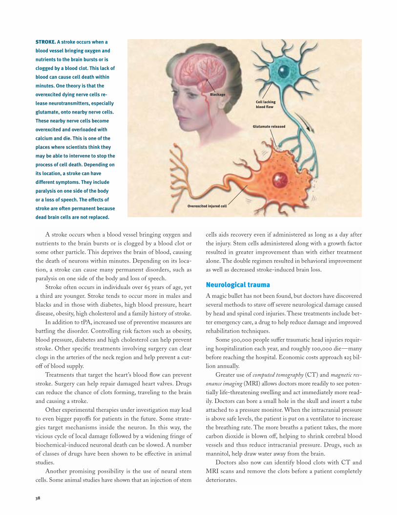
38
A stroke occurs when a blood vessel bringing oxygen andnutrients to the brain bursts or is clogged by a blood clot orsome other particle. This deprives the brain of blood, causingthe death of neurons within minutes. Depending on its loca-tion, a stroke can cause many permanent disorders, such asparalysis on one side of the body and loss of speech.
Stroke often occurs in individuals over 65 years of age, yeta third are younger. Stroke tends to occur more in males andblacks and in those with diabetes, high blood pressure, heartdisease, obesity, high cholesterol and a family history of stroke.
In addition to tPA, increased use of preventive measures arebattling the disorder. Controlling risk factors such as obesity,blood pressure, diabetes and high cholesterol can help preventstroke. Other specific treatments involving surgery can clearclogs in the arteries of the neck region and help prevent a cut-o∑ of blood supply.
Treatments that target the heart’s blood flow can preventstroke. Surgery can help repair damaged heart valves. Drugscan reduce the chance of clots forming, traveling to the brainand causing a stroke.
Other experimental therapies under investigation may leadto even bigger payo∑s for patients in the future. Some strate-gies target mechanisms inside the neuron. In this way, thevicious cycle of local damage followed by a widening fringe ofbiochemical-induced neuronal death can be slowed. A numberof classes of drugs have been shown to be e∑ective in animalstudies.
Another promising possibility is the use of neural stemcells. Some animal studies have shown that an injection of stem
cells aids recovery even if administered as long as a day afterthe injury. Stem cells administered along with a growth factorresulted in greater improvement than with either treatmentalone. The double regimen resulted in behavioral improvementas well as decreased stroke-induced brain loss.
Neurological trauma
A magic bullet has not been found, but doctors have discoveredseveral methods to stave o∑ severe neurological damage causedby head and spinal cord injuries. These treatments include bet-ter emergency care, a drug to help reduce damage and improvedrehabilitation techniques.
Some 500,000 people su∑er traumatic head injuries requir-ing hospitalization each year, and roughly 100,000 die—manybefore reaching the hospital. Economic costs approach $25 bil-lion annually.
Greater use of computed tomography (CT) and magnetic res-
onance imaging (MRI) allows doctors more readily to see poten-tially life-threatening swelling and act immediately more read-ily. Doctors can bore a small hole in the skull and insert a tubeattached to a pressure monitor. When the intracranial pressureis above safe levels, the patient is put on a ventilator to increasethe breathing rate. The more breaths a patient takes, the morecarbon dioxide is blown o∑, helping to shrink cerebral bloodvessels and thus reduce intracranial pressure. Drugs, such asmannitol, help draw water away from the brain.
Doctors also now can identify blood clots with CT andMRI scans and remove the clots before a patient completelydeteriorates.
STROKE. A stroke occurs when a
blood vessel bringing oxygen and
nutrients to the brain bursts or is
clogged by a blood clot. This lack of
blood can cause cell death within
minutes. One theory is that the
overexcited dying nerve cells re-
lease neurotransmitters, especially
glutamate, onto nearby nerve cells.
These nearby nerve cells become
overexcited and overloaded with
calcium and die. This is one of the
places where scientists think they
may be able to intervene to stop the
process of cell death. Depending on
its location, a stroke can have
di≈erent symptoms. They include
paralysis on one side of the body
or a loss of speech. The e≈ects of
stroke are often permanent because
dead brain cells are not replaced.
Glutamate released
Overexcited injured cell
Cell lackingblood flow
Blockage

39
An estimated 250,000 individuals are living with spinalcord injury in the U.S. Some 11,000 new injuries are reportedannually and are caused mostly by motor vehicle accidents, vio-lence and falls. Economic costs approach $10 billion a year.
Researchers have found that people who su∑er spinal cordinjuries become less severely paralyzed if they receive highintravenous doses of a commonly used steroid drug within eighthours after injury. The drug methylprednisolone appears to helpregardless of how severely the spinal cord is injured, and insome cases, makes the di∑erence between a patient beingconfined to a wheelchair and being able to walk. Building onthis knowledge, researchers hope to decipher the precise orderof chemical reactions that leads to damage.
Scientists have known that following a complete injury tothe spinal cord, animals can regain the ability to bear theirweight and walk at various speeds on a treadmill belt. Morerecently, scientists have recognized that the level of this recov-ery is dependent to a large degree on whether these tasks arepracticed—that is, trained—after injury. It appears that humanswith spinal cord injury also respond to training interventions.
Anxiety disorders
The most widespread mental illnesses, anxiety disorders annu-ally a∑ect an estimated 12.6 percent of the adult population, or24.8 million Americans. They include phobias, panic disorderand agoraphobia and obsessive-compulsive disorder (OCD).Some can keep people completely housebound or, as in the caseof panic disorder, contribute to suicide.
In OCD, people become trapped, often for many years, inrepetitive thoughts and behaviors, which they recognize asgroundless but cannot stop, such as repeatedly washing hands,or checking doors or stoves. The illness is estimated to a∑ect3.8 million Americans annually. Social learning and geneticsmay play a role in developing the disorder. But positron emis-sion tomography (PET) scans reveal abnormalities in both cor-tical and deep areas of the brain, suggesting a biological com-ponent as well.
Scientists recently discovered that certain breeds of largedogs that develop acral lick syndrome, severely sore paws throughcompulsive licking, respond to the serotonergic antidepressantclomipramine, which was the first e∑ective treatment developedfor OCD in people.
Serotonergic antidepressants, especially the tricyclics,clomipramine and serotonin reuptake inhibitors are e∑ective intreating OCD. A specialized type of behavioral intervention,exposure and response prevention, is also e∑ective in manypatients.
Panic disorder, which a∑ects 2.4 million Americans annu-ally, usually starts “out of the blue.” Patients experience an over-whelming sense of impending doom, accompanied by sweat-ing, weakness, dizziness and shortness of breath. With repeated
attacks, patients may develop anxiety in anticipation of anotherattack and avoid public settings where attacks might occur.Untreated, their lives may constrict until they develop agora-
phobia, or the fear of crowds.The recent discovery of brain receptors for the benzo-
diazepine anti-anxiety drugs has sparked research to identifythe brain’s own anti-anxiety chemical messengers. This findingmay lead to ways to regulate this brain system and correct itspossible defects in panic disorder. PET scans reveal that dur-ing such attacks, the tip of the brain’s temporal lobe is unusu-ally active compared with controls. When normal people expectto receive a shock to the finger, the same general area is acti-vated.
The serotonin reuptake inhibitors, cognitive behavior ther-apy, or a combination are now the first choice treatments ofpanic disorder. Tricyclic antidepressants, MAO inhibitors andhigh-potency benzodiazepines also are e∑ective.
Schizophrenia
Marked by disturbances in thinking, emotional reactions andsocial behavior, schizophrenia usually results in chronic illnessand personality change. Delusions, hallucinations and thoughtdisorder are common.
A∑ecting about one percent of the population or 2 millionAmericans a year, schizophrenia is disabling and costly. On agiven day, these patients occupy up to 100,000 hospital beds.Annual costs total about $32.5 billion.
Schizophrenia is thought to reflect changes in the brain,possibly caused by disease or injury at the time of birth, or agenetic disposition that may be exacerbated by environmentalstress. Brain systems using dopamine appear to be particularlyinvolved. Brain scans and postmortem studies show abnormal-ities in some people with schizophrenia, such as enlarged ven-tricles (fluid-filled spaces) and reduced size of certain brainregions. PET scans taken during intellectual tasks showabnormal functioning in specific brain areas of persons with thisillness.
The disorder usually begins in persons between the ages of15 and 25. Some patients fully recover following treatment, butmost continue to have moderate or severe symptoms, particu-larly in response to stress. About 15 percent of patients returnto normal life after a single episode; 60 percent will have inter-mittent episodes throughout their lives; another 25 percent willnot recover their ability to live as independent adults.
After a long search for an e∑ective antipsychotic medica-tion, scientists synthesized the drug chlorpromazine during thelate 1940s. By the 1950s, it was found useful for treating psy-chotic states and later became a mainstay of drug treatment.
Since then a large number of agents similar to chlorpro-mazine have been developed. When given as long-acting injec-tions, these drugs reduce some symptoms and aid patients’

40
readiness for adjustment back into the community. However,chronic use may cause abnormal muscle movements andtremors in some patients. Safer treatments are being sought.
Thus far, most drugs are successful in treating hallucina-tions and thought disorder. Clozapine, acts somewhat dif-ferently from other antipsychotics. It treats the approximately30 percent of patients who are not helped by conventional med-ications. However, the drug can induce a potentially fatal blooddisorder, agranulocytosis, in about one percent of patients. Toprevent this disorder, patients must take regular weekly tobiweekly blood tests, a precaution that makes the use of thedrug very costly. Several new antipsychotics—risperidone, olan-
zapine and sertindole—are now available. They do not involverisk of angranulocytosis but may have other side e∑ects.
Neurological AIDS
By the end of 2000, some 448,000 deaths and up to 774,000infections from acquired immune deficiency syndrome (AIDS)had occurred in the U.S. This is dwarfed by the more than 21.8million deaths and 58 million infections identified worldwide.
While the principal target of human immunodeficiency virus
(HIV) is the immune system, the nervous system also may beprofoundly a∑ected. Some 20 percent to 40 percent of patientswith full-blown AIDS also develop clinically significantdementia that includes movement impairment. Those a∑ectedhave mental problems ranging from mild di≈culty with con-centration or coordination to progressive, fatal dementia.
Despite advances in treating other aspects of the disease,AIDS dementia remains a mystery. Most current hypothesescenter on an indirect e∑ect of HIV infection related to secretedviral products or cell-coded signal molecules called cytokines.Nonetheless, HIV infection appears to be the prime mover inthis disorder since antiviral treatment may prevent or reversethis condition in some patients.
Experts believe that serious neurologic symptoms areuncommon early in AIDS infection. But later, patients developdi≈culty with concentration and memory and experience gen-eral slowing of their mental processes. At the same time, patientsmay develop leg weakness and a loss of balance. Imaging tech-niques, such as CT and MRI, show that the brains in thesepatients have undergone some shrinkage. The examination ofbrain cells under a microscope suggests that abnormalities arepresent principally in subcortical areas. Neurons in the cortexalso may be altered.
Recent studies indicate that highly active combination anti-retroviral treatment (‘cocktails’ of three or more drugs activeagainst HIV) is e∑ective in reducing the incidence of AIDSdementia. Such treatment also can e∑ectively reverse the cog-nitive abnormalities attributed to brain HIV infection.
Despite this remarkable progress, some patients developthese problems and fail to respond to treatment, thus requiring
additional approaches to prevention and treatment of thesesymptoms as well as the common peripheral neuropathy thatcan aΩict those with AIDS.
Multiple sclerosis
The most common central nervous system disease of youngadults after epilepsy, multiple sclerosis (MS) is a life-long ail-ment of unknown origin that a∑ects more than 300,000 Amer-icans. MS is diagnosed in individuals who are mainly betweenthe ages of 20 and 50, with two of three cases occurring inwomen. MS results in earning losses of about $2 billion annu-ally for families with MS.
Although a cause has yet to be found, MS is thought to bean autoimmune disease in which the body’s natural defenses actagainst the myelin in the central nervous system as though itwere foreign tissue. In MS, myelin is destroyed and replaced byscars of hardened “sclerotic” patches of tissue. Such lesions arecalled “plaques,” and appear in multiple places within the cen-tral nervous system. This can be compared to a loss of insulat-ing material around an electrical wire, which interferes with thetransmission of signals. Some nerve fibers are actually cut inassociation with the loss of myelin.
Siblings of people with MS are 10 to 15 times more likelythan others to be aΩicted by the disorder. In addition, the dis-ease is five times more prevalent in temperate zones, such as theNorthern United States and Northern Europe, than it is in thetropics. Thus, genetic and environmental factors are probablyinvolved in the cause. An infection acquired during the first15 years of life may be responsible for triggering the disease ina genetically susceptible individual.
The most common symptoms are blurred vision, awkwardgait, numbness and fatigue. These can occur singly or in com-bination, vary in intensity and last from several weeks to months.In some patients, symptoms include slurred speech, weakness,loss of coordination, uncontrollable tremors, loss of bladder con-trol, memory problems, depression and paralysis. Muscle spas-ticity can a∑ect balance and coordination, causing pain andinvoluntary jerking movement—and, if untreated, can createcontractures or the “freezing” of a joint that prevents movement.
MS cannot be cured at present, but several medicationscontrol relapsing forms of MS. A wide range of medicationsand therapies are available to control symptoms such as spas-ticity, pain, fatigue, mood swings and bladder, bowel or sexualdysfunctions. Steroids, which have been used in MS for threedecades, e∑ectively shorten attacks and speed recovery fromMS-related optic nerve inflammation. Promising new agents tocontrol MS or to alleviate its symptoms are in clinical trials.
Down syndrome
Down syndrome, the most frequently occurring chromosomalabnormality, appears in one out of every 800 to 1,000 babies

41
born. It occurs when an extra copy of chromosome 21 or part ofits long arm is present in the egg or, less commonly, the sperm,at the time of conception. It is not known why this error in celldivision occurs. It is not linked to any environmental or behav-ioral factors, either before or during pregnancy.
This disorder is associated with approximately 50 physicaland developmental characteristics. An individual with Downsyndrome is likely to possess, to varying degrees, some of thesecharacteristics. They include mild to moderate mental retarda-tion, low muscle tone, an upward slant to the eyes, a flat facialprofile, an enlarged tongue and an increased risk of congenitalheart defects, respiratory problems and obstructed digestivetracts.
The risk of having a child with this syndrome increases withthe age of the mother. At age 35,the risk is about one in 365births. At age 40, it is one in 110.However, it is important to notethat the average age of womenwho give birth to children withDown syndrome is 28. This isbecause younger women are giv-ing birth more often.
Prenatal screening tests, such as the Triple Screen andAlpha-fetaprotein Plus, can accurately detect about 60 percentof fetuses with Down syndrome.
Babies with Down syndrome will develop much like typi-cal children, but at a somewhat slower rate. They will learn tosit, walk, talk and toilet train, just like their peers. Early inter-vention programs can begin shortly after birth and can help fos-ter an infant’s development.
Down syndrome patients have been able to have longer andfuller lives, thanks to medical advances and a greater under-standing of the potential of those with this condition. Individ-uals with Down syndrome are being educated in their neigh-borhood schools, participating in community activities andfinding rewarding employment and relationships.
Although there is no cure or means of preventing Downsyndrome, scientists are moving closer to understanding the rolethat the genes on chromosome 21 play in a person’s develop-ment. Once this mystery is understood, they hope to decode thebiochemical processes that occur in Down syndrome and treator cure this disorder.
Huntington’s disease
A∑ecting some 30,000 Americans and placing another 150,000at risk, Huntington’s disease (HD) is now considered one of themost common hereditary brain disorders. The disease that killedfolk singer Woody Guthrie in 1967 progresses slowly over a tento 20-year period and eventually robs the a∑ected individual ofthe ability to walk, talk, think and reason. HD usually appears
between the ages of 30 and 50. It a∑ects both the basal gangliathat controls coordination and the brain cortex, which servesas the center for thought, perception and memory.
The most recognizable symptoms include involuntary jerk-ing movements of the limbs, torso and facial muscles. These areoften accompanied by mood swings, depression, irritability,slurred speech and clumsiness. As the disease progresses, com-mon symptoms include di≈culty swallowing, unsteady gait, lossof balance, impaired reasoning and memory problems. Even-tually, the individual becomes totally dependent on others forcare, with death often due to pneumonia, heart failure oranother complication.
Diagnosis consists of a detailed clinical examination andfamily history. Brain scans may be helpful. The identification
in 1993 of the gene that causesHD has simplified genetic test-ing, which can be used to helpconfirm a diagnosis. However,HD researchers and geneticcounselors have establishedspecific protocols for predictivetesting to ensure that the psy-
chological and social consequences of a positive or negativeresult are understood. Predictive testing is available only foradults, though children under 18 may be tested to confirm adiagnosis of juvenile onset HD. Prenatal testing may be per-formed. The ethical issues of testing must be considered andthe individual adequately informed, because there is no e∑ectivetreatment or cure.
The HD mutation is an expanded triplet repeat in the HDgene—a kind of molecular stutter in the DNA. This abnormalgene codes for an abnormal protein called huntingtin. Thehuntingtin protein, whose normal function is still obscure, iswidely distributed in the brain and appears to be associatedwith the intracellular machinery involved in the transport ofproteins. But the cause of HD probably involves a gain of a newand toxic function. Cell and transgenic animal models canreplicate many features of the disease and are now being usedto test new theories and therapies. Many researchers hope thattransplanted or resident stem cells may one day be able toreplace the neurons that have been lost to the disease.
Tourette syndrome
One of the most common and least understood neurobiologicaldisorders, Tourette syndrome (TS) is a genetic condition thata∑ects an estimated one in 500 Americans, roughly 200,000 peo-ple. Males are a∑ected three to four times as frequently asfemales.
Symptoms usually appear between the ages of four andeight, but in rare cases may emerge as late as age 18. The symp-toms include motor and vocal tics that are repetitive, involuntary
Scientists are moving closer to understand-
ing the role that the genes on chromosome
21 play in Down syndrome.
movements or utterances that are rapid and sudden. The typesof tics may change frequently, and increase or decrease in sever-ity over time. Generally, this disorder lasts a lifetime, but one-third of patients may experience a remission or decrease insymptoms as they get older. Most people with TS do notrequire medication; their symptoms are mild and do not a∑ectfunctioning.
The disorder seems to result from a hypersensitivity ofdopamine receptors. Another neurotransmitter, serotonin, alsohas been implicated. The most e∑ective drugs for control ofmovements, such as haloperidol, act by blocking the overactivesystem. Other symptoms, such as obsessive-compulsive traitsand attention deficit disorder, often require treatment withother classes of drugs that act on serotonin.
The neuroleptic drugs haloperidol and pimozide have beenthe mainstays of treatment. They are not perfect medications,however, because they can cause disturbing side e∑ects—abnormal involuntary movements, sti∑ness of the face andlimbs, or sedation and weight gain in some patients. Recently,newer medications have been found e∑ective in some patients.
Brain tumors
Although brain tumors are not always malignant—a conditionthat spreads and becomes potentially lethal—these growths arealways serious because they can cause pressure in the brain andcompression of nearby structures, interfering with normal brainactivity.
Primary brain tumors arise within the brain while sec-ondary brain tumors spread from other parts of the bodythrough the bloodstream. For tumors starting in the brain,about 60 percent of which are malignant, the cause is unknown.Tumors that begin as cancer elsewhere and spread to the brainare always malignant.
The incidence of primary brain tumors is about 12 per100,000 population. About 36,000 new cases occur in theUnited States annually. Because of di≈culties diagnosing andclassifying brain tumors, exact statistics on secondary tumorsare unknown.
Symptoms vary according to location and size. The com-pression of brain tissue or nerve tracts, as well as expansion ofthe tumor, can cause symptoms such as seizures, headaches,muscle weakness, loss of vision or other sensory problems andspeech di≈culties. An expanding tumor can increase pressurewithin the skull, causing headache, vomiting, visual distur-bances and impaired mental functioning. Brain tumors arediagnosed with MRI and CT scanning.
Surgery is a common treatment if the tumor is accessibleand vital structures will not be disturbed. Radiation is used tostop a tumor’s growth or cause it to shrink. Chemotherapydestroys tumor cells that may remain after surgery and radia-tion. Steroid drugs relieve swelling and other symptoms.
Immunotherapy uses the body’s own immune system against thetumor. Promising areas of research include bioengineered genes,monoclonal antibodies that attach to specifically targeted cells;growth factors; angiogenesis inhibitors and di∑erentiation thera-
pies; targeted toxins; and tumor vaccines.
Amyotrophic lateral sclerosis
This fatal disorder strikes 5,000 Americans annually with 50percent of patients dying within three to five years of diagno-sis. It is the most common disorder within a group of diseasesa∑ecting movement and costs Americans some $300 millionannually.
Commonly known as Lou Gehrig’s disease, amyotrophiclateral sclerosis (ALS) destroys neurons that control voluntarymuscle movements, such as walking. For reasons that are notunderstood, brain and spinal motor neurons in the spinal cordbegin to disintegrate. Because signals from the brain are notcarried by these damaged nerves to the body, the muscles beginto weaken and deteriorate from the lack of stimulation and use.
The first signs of progressive paralysis are usually seen inthe hands and feet. They include leg weakness, walkingdi≈culty, and clumsiness of the hands when washing and dress-ing. Eventually, almost all muscles under voluntary control,including those of the respiratory system, are a∑ected. Despitethe paralysis, the mind and the senses remain intact. Death isusually caused by respiratory failure or pneumonia.
No specific test identifies ALS; but muscle biopsies, bloodstudies, electrical tests of muscle activity, CT and MRI scansand X-rays of the spinal cord help identify the disease and ruleout other disorders. Still, diagnosis is often di≈cult because itscauses remain unknown. Potential causes include glutamatetoxicity, oxidative stress, factors in the environment and anautoimmune response in which the body’s defenses turn againstbody tissue.
In about 90 percent of cases, ALS is sporadic, arising inindividuals with no known family history of the disorder. In theother 10 percent of cases, it is familial—transmitted to familymembers because of a gene defect.
Scientists recently found a gene responsible for one form ofALS. Mutations in the gene that codes for super oxide dismu-
tase located on chromosome 21 were linked to the presence ofthis disorder. Scientists believe that whatever they learn fromstudying the gene will have relevance for understanding otherforms of motor neuron disease.
Once diagnosed, physical therapy and rehabilitation meth-ods help strengthen unused muscles. Various drugs can easespecific problems like twitching and muscle weakness, but thereis no cure. An antiglutamate drug modestly slows down the dis-ease. Additional drugs are now under study. Protecting orregenerating motor neurons using nerve growth factors andstem cells may someday provide significant hope for patients.
42

43
any of the recent advances in understand-ing the brain are due to the developmentof techniques that allow scientists to di-rectly monitor neurons throughout thebody.
Electrophysiological recordings tracebrain electrical activity in response to a specific external stim-ulus. In this method, electrodes placed in specific parts of thebrain—depending on which sensory system is being tested—make recordings that are then processed by a computer. Thecomputer makes an analysis based on the time lapse betweenstimulus and response. It then extracts this information frombackground activity.
Following the discovery that material is transported withinneurons, methods have been developed to visualize activity andprecisely track fiber connections within the nervous system.This can be done by injecting a radioactive amino acid into thebrain of an experimental animal; the animal is killed a fewhours later; and then the presence of radioactive cells is visual-ized on film. In another technique, the enzyme horseradish per-oxidase is injected and taken up by nerve fibers that can be lateridentified under a microscope.
These and other methods have resulted in many advancesin knowledge about the workings of the nervous system and arestill useful today. New methods, safely applicable to humans,promise to give even more precise information, particularlyabout the point of origin of disorders such as epilepsy.
Imaging techniques
Positron emission tomography (PET) This method of measur-ing brain function is based on the detection of radioactivityemitted when positrons, positively charged particles, undergoradioactive decay in the brain. Substances labeled withpositron-emitting radionuclides are used to produce three-dimensional PET images that reflect blood flow as well asmetabolic and chemical activity in the brain.
So far, PET studies have helped scientists understand moreabout how drugs a∑ect the brain and what happens duringlearning, language and certain brain disorders—such as stroke
and Parkinson’s disease. Within the next few years, PET couldenable scientists to identify the biochemical nature of neuro-logical and mental disorders and determine how well therapyis working in patients. For instance, depression produces verymarked changes in the brain as seen by PET. Knowing thelocation of these changes helps researchers understand betterthe causes of depression and monitor the e∑ectiveness ofspecific treatments.
Another technique, single photon emission computed tomog-
raphy (SPECT), is similar to PET but its pictures are not asdetailed. SPECT is much less expensive than PET because thetracers it uses have a longer half-life and do not require a nearbyaccelerator to produce them.
Magnetic resonance imaging (MRI) Providing a high-quality, three-dimensional image of organs and structuresinside the body without X-rays or other radiation, MRI imagesare unsurpassed in anatomical detail and may reveal minutechanges that occur with time.
MRI is expected to tell scientists when structural abnor-malities first appear in the course of a disease, how they a∑ectsubsequent development and precisely how their progressioncorrelates with mental and emotional aspects of a disorder.
During the 15-minute MRI imaging procedure, a patientlies inside a massive, hollow, cylindrical magnet and is exposedto a powerful, steady magnetic field. The protons of the body’shydrogen atoms, especially those in water and fat, normallypoint randomly in di∑erent directions, but in a very strongmagnetic field (many times the earth’s magnetic field) they lineup parallel to each other like rows of tiny bar magnets. If thehydrogen nuclei are then knocked out of alignment by a strongpulse of radio waves, they produce a detectable radio signal asthey fall back into alignment.
Magnetic coils in the machine detect these signals and acomputer changes them into an image based on di∑erent typesof body tissue. Tissue that contains a lot of water and fat pro-duces a bright image; tissue that contains little or no water, suchas bone, appears black. (The image is similar to that producedby CT scanning, but MRI generally gives much greater con-trast between normal and abnormal tissues.)
New diagnostic methods
M

44
MRI allows images to be constructed in any plane and isparticularly valuable in studying the brain and spinal cord. Itreveals tumors rapidly and vividly, indicating their precise extent.MRI provides early evidence of potential damage from stroke,thus allowing physicians to administer proper treatments early.
Magnetic resonance spectroscopy (MRS), a technique related toMRI which uses the same machinery but examines molecularcomposition and metabolic processes, rather than anatomy, alsoholds great promise to provide insights into how the brainworks. By measuring the molecular and metabolic changes thatoccur in the brain, MRS has already provided new informationon brain development and aging, Alzheimer’s disease, schizo-phrenia, autism and stroke. Because this method is noninvasive,it is ideally suited to study the natural course of a disease or itsresponse to treatment.
Functional magnetic resonance imaging (fMRI) Anotherexciting recent development in imaging is fMRI. This techniquemeasures brain activity under resting and activated conditions.It combines the high spatial resolution, noninvasive imaging ofbrain anatomy o∑ered by standard MRI with a strategy fordetecting changes in blood oxygenation levels driven by neuronalactivity. This technique allows for more detailed maps of brainareas underlying human mental activities in health and disease.To date, fMRI has been applied to the study of various functionsof the brain ranging from primary sensory responses to cognitiveactivities. While the exact origin of the signal changes found infMRI is still under debate, the success of fMRI in numerousstudies has clearly demonstrated its great potential.
Magnetoencephalography (MEG) One of the latest advancesin scanners, MEG reveals the source of weak magnetic fieldsemitted by neurons. An array of cylinder-shaped sensors mon-itor the magnetic field pattern near the patient’s head to deter-mine the positions and strengths of activity in various regions ofthe brain. In contrast with other imaging techniques, MEG cancharacterize rapidly changing patterns of neural activity withmillisecond resolution and provide a quantitative measure of itsstrength for individual subjects. Moreover, by presenting stimuliat various rates, it is possible to determine how long the neuralactivation is sustained in the diverse brain areas that respond.
One of the most exciting developments in imaging is thecombined use of information from fMRI and MEG. The for-mer provides detailed information about the areas of brain activ-ity in a particular task whereas MEG tells researcher and physi-cian when they become active. The combined use of thisinformation allows a much more precise understanding of howthe brain works in health and disease.
Gene diagnosis
The inherited blueprint for all human characteristics, genesconsist of short sections of deoxyribonucleic acid (DNA), thelong, spiraling, helix structure found on the 23 pairs of chromo-
somes in the nucleus of every human cell.New gene diagnosis techniques now make it possible to find
the chromosomal location of genes responsible for neurologicand psychiatric diseases and to identify structural changes inthese genes that are responsible for causing disease.
This information is useful for identifying individuals whocarry faulty genes and thereby improving diagnosis; for under-standing the precise cause of diseases in order to improve meth-ods of prevention and treatment; and for evaluating the malig-nancy and susceptibility of certain tumors.
So far, scientists have identified defective genes for morethan 50 neurological disorders and the chromosomal location ofthe defect in up to 100. Prenatal or carrier tests exist for manyof the most prevalent of these illnesses.
Scientists have tracked down the gene on chromosome 4that goes awry in Huntington’s patients. The defect is an expan-sion of a CAG repeat. CAG is the genetic code for the aminoacid glutamine, and the expanded repeat results in a long stringof glutamines within the protein. This expansion appears toalter the protein’s function. Scientists have found that the sizeof the expanded repeat in an individual is predictive of Hunt-ington’s disease. Other neurodegenerative disorders have beenidentified as due to expanded CAG repeats in other genes. Themechanisms by which these expansions caused adult onset neuro-degeneration is the focus of intense research.
Sometimes patients with single gene disorders are found tohave a chromosomal abnormality—a deletion or break in theDNA sequence of the gene—that can lead scientists to a moreaccurate position of the disease gene. This is the case with someabnormalities found on the X-chromosome in patients withDuchenne muscular dystrophy and on chromosome 13 in patientswith inherited retinoblastoma, a rare childhood eye tumor thatcan lead to blindness and other cancers.
Gene mapping has led to the localization on chromosome21 of the gene coding the beta amyloid precursor protein that isabnormally cut to form the smaller peptide, beta amyloid. It isthis peptide that accumulates in the senile plaques that clog thebrains of patients with Alzheimer’s disease. This discovery shedlight on the reason why individuals with Down syndrome (Tri-somy 21) invariably accumulate amyloid deposits: they make toomuch amyloid as a consequence of having three copies insteadof two copies of this gene. Mutations in this gene have recentlybeen shown to underlie Alzheimer’s in a distinct subset of thesepatients.
Several other genetic factors have been identified inAlzheimer’s disease, including genes for two proteins, presenilin1 and presenilin 2, located on chromosomes 14 and 1. A risk fac-tor for late onset Alzheimer’s is the gene for the apolipoproteinE protein located on chromosome 19.
Gene mapping has enabled doctors to diagnose Fragile X
mental retardation, the most common cause of inherited mental
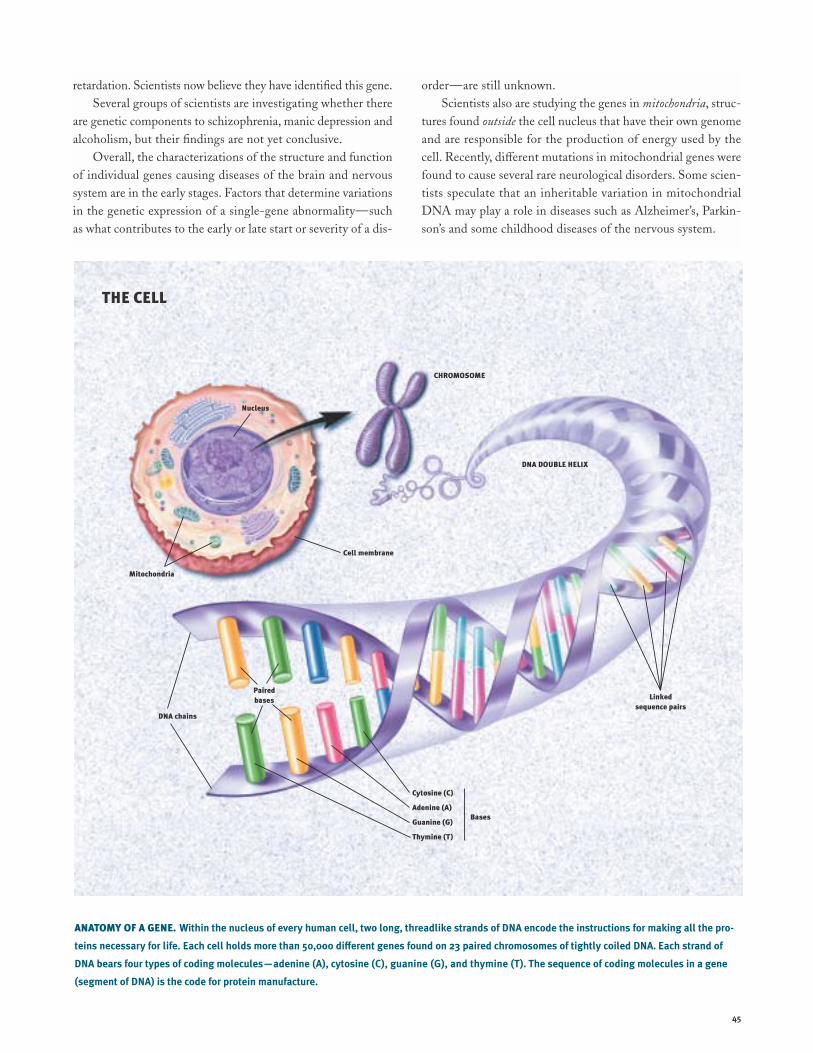
45
retardation. Scientists now believe they have identified this gene.Several groups of scientists are investigating whether there
are genetic components to schizophrenia, manic depression andalcoholism, but their findings are not yet conclusive.
Overall, the characterizations of the structure and functionof individual genes causing diseases of the brain and nervoussystem are in the early stages. Factors that determine variationsin the genetic expression of a single-gene abnormality—suchas what contributes to the early or late start or severity of a dis-
order—are still unknown.Scientists also are studying the genes in mitochondria, struc-
tures found outside the cell nucleus that have their own genomeand are responsible for the production of energy used by thecell. Recently, di∑erent mutations in mitochondrial genes werefound to cause several rare neurological disorders. Some scien-tists speculate that an inheritable variation in mitochondrialDNA may play a role in diseases such as Alzheimer’s, Parkin-son’s and some childhood diseases of the nervous system.
ANATOMY OF A GENE. Within the nucleus of every human cell, two long, threadlike strands of DNA encode the instructions for making all the pro-
teins necessary for life. Each cell holds more than 50,000 di≈erent genes found on 23 paired chromosomes of tightly coiled DNA. Each strand of
DNA bears four types of coding molecules—adenine (A), cytosine (C), guanine (G), and thymine (T). The sequence of coding molecules in a gene
(segment of DNA) is the code for protein manufacture.
CHROMOSOME
DNA DOUBLE HELIX
THE CELL
Cytosine (C)
Adenine (A)
Guanine (G)
Thymine (T)
DNA chains
Pairedbases
Bases
Linked sequence pairs
Cell membrane
Mitochondria
Nucleus

46
New drugs. Most drugs used today were devel-oped using trial-and-error techniques that oftendo not reveal why a drug produces a particulare∑ect. But the expanding knowledge gainedfrom the new methods of molecular biology—the ability to make a receptor gene and deter-
mine its structure—make it possible to design safer and moree∑ective drugs.
In a test tube, the potency of an agent can be determinedby how well it binds to a receptor. A scientist then can vary thedrug’s structure to enhance its action on the receptor. Thus,subsequent generations of drugs can be designed to interactwith the receptors more e≈ciently, producing higher potencyand fewer side e∑ects.
While this “rational drug design” holds promise for devel-oping drugs for conditions ranging from stroke and migraineheadaches to depression and anxiety, it will take considerablee∑ort to clarify the role of the di∑erent receptors in these dis-orders.
Trophic factors
One result of basic neuroscience research involves the discov-ery of numerous survival or trophic factors found in the brain thatcontrol the development and survival of specific groups of neu-rons. Once the specific actions of these molecules and theirreceptors are identified and their genes cloned, procedures canbe developed to modify trophic factor-regulated function in waysthat might be useful in the treatment of neurological disorders.
Already, researchers have demonstrated the possible valueof at least one of these factors, nerve growth factor (NGF).Infused into the brains of rats, NGF prevented cell death andstimulated the regeneration and sprouting of damaged neuronsthat are known to die in Alzheimer’s disease. When aged ani-mals with learning and memory impairments were treated withNGF, scientists found that these animals were able to remem-ber a maze task as well as healthy aged rats.
Recently, several new factors have been identified and arebeginning to be studied. They are potentially useful for ther-apy, but scientists must first understand how they may influence
neurons. Alzheimer’s, Parkinson’s and Lou Gehrig’s diseasesmay be treated in the future with trophic factors or their genes.
Because the destruction of neurons that use acetylcholineis one feature of Alzheimer’s disease, any substance that canprevent this destruction is an important topic of research. NGF,which can do this, also holds promise for slowing the memorydeficits associated with normal aging.
Once a trophic factor for a particular cell is found, copiesof the factor can be genetically targeted to the area of the brainwhere this type of cell has died. The treatment may not cure adisease but could improve symptoms and delay progression.
In an interesting twist on growth factor therapy, researchersfor the first time demonstrated that a neutralization ofinhibitory molecules can help repair damaged nerve fiber tractsin the spinal cord. Using antibodies to Nogo-A, a protein thatinhibits nerve regeneration, Swiss researchers succeeded in get-ting nerves of damaged spinal cords in rats to regrow. Treatedrats showed large improvements in their ability to walk afterspinal cord damage.
In these experiments, scientists cut one of the major groupsof nerve fiber tracts in the spinal cord that connect the spinalcord and the brain. When an antibody, directed against the fac-tor Nogo-A, was administered to the spinal cords or the brainsof adult rats, “massive sprouting” of nerve fibers occurred wherethe spinal cord had been cut. Within two to three weeks, neu-rons grew to the lower level of the spinal cord and in some ani-mals along its whole length. In untreated spinal cord-injuredrats, the maximum distance of nerve regrowth rarely exceededone-tenth of an inch. This research could eventually have clin-ical implications for spinal cord or brain-damaged people.
Cell and gene therapy
Researchers throughout the world are pursuing a variety of newways to repair or replace healthy neurons and other cells in thebrain. Most of the experimental approaches are still being workedout in animals and cannot be considered real therapies at thistime.
Scientists have identified an embryonic neuronal stem cell—an unspecialized cell that gives rise to cells with specific func-
N
Potential therapies
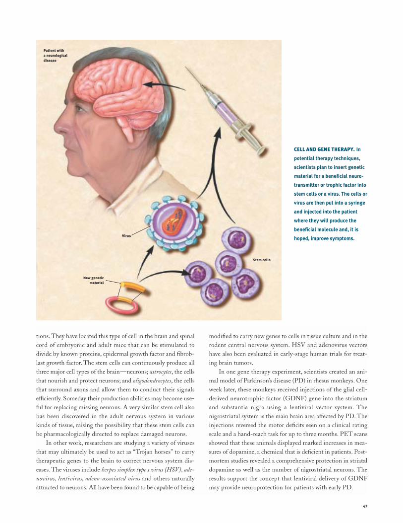
47
tions. They have located this type of cell in the brain and spinalcord of embryonic and adult mice that can be stimulated todivide by known proteins, epidermal growth factor and fibrob-last growth factor. The stem cells can continuously produce allthree major cell types of the brain—neurons; astrocytes, the cellsthat nourish and protect neurons; and oligodendrocytes, the cellsthat surround axons and allow them to conduct their signalse≈ciently. Someday their production abilities may become use-ful for replacing missing neurons. A very similar stem cell alsohas been discovered in the adult nervous system in variouskinds of tissue, raising the possibility that these stem cells canbe pharmacologically directed to replace damaged neurons.
In other work, researchers are studying a variety of virusesthat may ultimately be used to act as “Trojan horses” to carrytherapeutic genes to the brain to correct nervous system dis-eases. The viruses include herpes simplex type 1 virus (HSV), ade-
novirus, lentivirus, adeno-associated virus and others naturallyattracted to neurons. All have been found to be capable of being
modified to carry new genes to cells in tissue culture and in therodent central nervous system. HSV and adenovirus vectorshave also been evaluated in early-stage human trials for treat-ing brain tumors.
In one gene therapy experiment, scientists created an ani-mal model of Parkinson’s disease (PD) in rhesus monkeys. Oneweek later, these monkeys received injections of the glial cell-derived neurotrophic factor (GDNF) gene into the striatumand substantia nigra using a lentiviral vector system. Thenigrostriatal system is the main brain area a∑ected by PD. Theinjections reversed the motor deficits seen on a clinical ratingscale and a hand-reach task for up to three months. PET scansshowed that these animals displayed marked increases in mea-sures of dopamine, a chemical that is deficient in patients. Post-mortem studies revealed a comprehensive protection in striataldopamine as well as the number of nigrostriatal neurons. Theresults support the concept that lentiviral delivery of GDNFmay provide neuroprotection for patients with early PD.
CELL AND GENE THERAPY. In
potential therapy techniques,
scientists plan to insert genetic
material for a beneficial neuro-
transmitter or trophic factor into
stem cells or a virus. The cells or
virus are then put into a syringe
and injected into the patient
where they will produce the
beneficial molecule and, it is
hoped, improve symptoms.Virus
Stem cells
Patient with a neurological disease
New genetic material

48
ACETYLCHOLINE A neurotransmitter in both the brain, where itregulates memory, and in the peripheral nervous system,where it controls the actions of skeletal and smooth muscle.ACTION POTENTIAL This occurs when a neuron is activated andtemporarily reverses the electrical state of its interior mem-brane from negative to positive. This electrical charge travelsalong the axon to the neuron’s terminal where it triggers therelease of a neurotransmitter.ADRENAL CORTEX An endocrine organ that secretes corticos-teroids for metabolic functions; for example, in response tostress.ADRENAL MEDULLA An endocrine organ that secretes epineph-rine and norepinephrine in concert with the activation of thesympathetic nervous system; for example, in response to stress.AGONIST A neurotransmitter, a drug or other molecule thatstimulates receptors to produce a desired reaction.ALZHEIMER’S DISEASE The major cause of dementia mostprevalent in the elderly, it inflicts enormous human financialcost on society. The disease is characterized by death of neu-rons in the hippocampus, cerebral cortex and other brainregions.AMINO ACID TRANSMITTERS The most prevalent neurotrans-mitters in the brain, these include glutamate and aspartate,which have excitatory actions, and glycine and gamma-aminobutyric acid (GABA), which have inhibitory actions.AMYGDALA A structure in the forebrain that is an importantcomponent of the limbic system and plays a central role inemotional learning.ANDROGENS Sex steroid hormones, including testosterone,found in higher levels in males than females. They are respon-sible for male sexual maturation.ANTAGONIST A drug or other molecule that blocks receptors.Antagonists inhibit the e∑ects of agonists.APHASIA Disturbance in language comprehension or produc-tion, often as a result of a stroke.AUDITORY NERVE A bundle of nerve fibers extending from thecochlea of the ear to the brain, which contains two branches:the cochlear nerve that transmits sound information and thevestibular nerve that relays information related to balance.
Glossary
AUTONOMIC NERVOUS SYSTEM A part of the peripheral ner-vous system responsible for regulating the activity of internalorgans. It includes the sympathetic and parasympathetic ner-vous systems.AXON The fiberlike extension of a neuron by which the cellsends information to target cells.BASAL GANGLIA Clusters of neurons, which include the cau-date nucleus, putamen, globus pallidus and substantia nigra,located deep in the brain that play an important role in move-ment. Cell death in the substantia nigra contributes to Parkin-son’s disease.BRAINSTEM The major route by which the forebrain sendsinformation to and receives information from the spinal cordand peripheral nerves. The brainstem controls, among otherthings, respiration and regulation of heart rhythms.BROCA’S AREA The brain region located in the frontal lobe ofthe left hemisphere that is important for the production ofspeech.CATECHOLAMINES The neurotransmitters dopamine, epineph-rine and norepinephrine that are active both in the brain andthe peripheral sympathetic nervous system. These three mole-cules have certain structural similarities and are part of alarger class of neurotransmitters known as monoamines.CEREBELLUM A large structure located at the roof of the hind-brain that helps control movement by making connections tothe pons, medulla, spinal cord and thalamus. It also may beinvolved in aspects of motor learning.CEREBRAL CORTEX The outermost layer of the cerebral hemi-spheres of the brain. It is responsible for all forms of consciousexperience, including perception, emotion, thought and plan-ning.CEREBRAL HEMISPHERES The two specialized halves of thebrain. The left hemisphere is specialized for speech, writing,language and calculation; the right hemisphere is specializedfor spatial abilities, face recognition in vision and some aspectsof music perception and production.CEREBROSPINAL FLUID A liquid found within the ventricles ofthe brain and the central canal of the spinal cord.CHOLECYSTOKININ A hormone released from the lining of the

49
stomach during the early stages of digestion which acts as apowerful suppressant of normal eating. It also is found in thebrain.CIRCADIAN RHYTHM A cycle of behavior or physiologicalchange lasting approximately 24 hours.CLASSICAL CONDITIONING Learning in which a stimulus thatnaturally produces a specific response (unconditioned stimu-lus) is repeatedly paired with a neutral stimulus (conditionedstimulus). As a result, the conditioned stimulus can evoke aresponse similar to that of the unconditioned stimulus.COCHLEA A snail-shaped, fluid-filled organ of the inner earresponsible for transducing motion into neurotransmission toproduce an auditory sensation.COGNITION The process or processes by which an organismgains knowledge or becomes aware of events or objects in itsenvironment and uses that knowledge for comprehension andproblem-solving.CONE A primary receptor cell for vision located in the retina.The cone is sensitive to color and used primarily for daytimevision.CORPUS CALLOSUM The large bundle of nerve fibers linkingthe left and right cerebral hemispheres.CORTISOL A hormone manufactured by the adrenal cortex. Inhumans, cortisol is secreted in greatest quantities beforedawn, readying the body for the activities of the coming day.DEPRESSION A mental disorder characterized by depressedmood and abnormalities in sleep, appetite and energy level.DENDRITE A tree-like extension of the neuron cell body. Alongwith the cell body, it receives information from other neurons.DOPAMINE A catecholamine neurotransmitter known tohave multiple functions depending on where it acts.Dopamine-containing neurons in the substantia nigra of thebrainstem project to the caudate nucleus and are destroyedin Parkinson’s victims. Dopamine is thought to regulateemotional responses and play a role in schizophrenia anddrug abuse.DORSAL HORN An area of the spinal cord where many nervefibers from peripheral pain receptors meet other ascendingand descending nerve fibers.
DRUG ADDICTION Loss of control over drug intake or compul-sive seeking and taking of drugs, despite adverse consequences.ENDOCRINE ORGAN An organ that secretes a hormone directlyinto the bloodstream to regulate cellular activity of certainother organs.ENDORPHINS Neurotransmitters produced in the brain thatgenerate cellular and behavioral e∑ects like those of morphine.EPILEPSY A disorder characterized by repeated seizures,which are caused by abnormal excitation of large groups ofneurons in various brain regions. Epilepsy can be treated withmany types of anticonvulsant medications.EPINEPHRINE A hormone, released by the adrenal medullaand specialized sites in the brain, that acts with norepineph-rine to a∑ect the sympathetic division of the autonomic ner-vous system. Sometimes called adrenaline.ESTROGENS A group of sex hormones found more abun-dantly in females than males. They are responsible for femalesexual maturation and other functions.EVOKED POTENTIALS A measure of the brain’s electrical activ-ity in response to sensory stimuli. This is obtained by placingelectrodes on the surface of the scalp (or more rarely, insidethe head), repeatedly administering a stimulus and then usinga computer to average the results.EXCITATION A change in the electrical state of a neuron thatis associated with an enhanced probability of action potentials.FOLLICLE-STIMULATING HORMONE A hormone released by thepituitary gland that stimulates the production of sperm in themale and growth of the follicle (which produces the egg) inthe female.FOREBRAIN The largest division of the brain, which includesthe cerebral cortex and basal ganglia. The forebrain is cred-ited with the highest intellectual functions.FRONTAL LOBE One of the four divisions (parietal, temporal,occipital) of each hemisphere of the cerebral cortex. Thefrontal lobe has a role in controlling movement and in theplanning and coordinating of behavior.GAMMA-AMINO BUTYRIC ACID (GABA) An amino acid trans-mitter in the brain whose primary function is to inhibit thefiring of neurons.

50
GLIA Specialized cells that nourish and support neurons.GLUTAMATE An amino acid neurotransmitter that acts toexcite neurons. Glutamate stimulates N-methyl-D-aspartate(NMDA) receptors that have been implicated in activitiesranging from learning and memory to development andspecification of nerve contacts in a developing animal. Stimu-lation of NMDA receptors may promote beneficial changes,while overstimulation may be a cause of nerve cell damage ordeath in neurological trauma and stroke.GONAD Primary sex gland: testis in the male and ovary in thefemale.GROWTH CONE A distinctive structure at the growing end ofmost axons. It is the site where new material is added to theaxon.HIPPOCAMPUS A seahorse-shaped structure located withinthe brain and considered an important part of the limbic sys-tem. It functions in learning, memory and emotion.HORMONES Chemical messengers secreted by endocrineglands to regulate the activity of target cells. They play a rolein sexual development, calcium and bone metabolism, growthand many other activities.HUNTINGTON’S DISEASE A movement disorder caused bydeath of neurons in the basal ganglia and other brain regions.It is characterized by abnormal movements called chorea—sudden, jerky movements without purpose.HYPOTHALAMUS A complex brain structure composed ofmany nuclei with various functions. These include regulatingthe activities of internal organs, monitoring information fromthe autonomic nervous system, controlling the pituitary glandand regulating sleep and appetite.INHIBITION In reference to neurons, it is a synaptic messagethat prevents the recipient cell from firing.IONS Electrically charged atoms or molecules.LIMBIC SYSTEM A group of brain structures—including theamygdala, hippocampus, septum, basal ganglia and others—that help regulate the expression of emotion and emotionalmemory.LONG-TERM MEMORY The final phase of memory in whichinformation storage may last from hours to a lifetime.
MANIA A mental disorder characterized by excessive excite-ment, exalted feelings, elevated mood, psychomotor over-activity and overproduction of ideas. It may be associatedwith psychosis; for example, delusions of grandeur.MELATONIN Produced from serotonin, melatonin is releasedby the pineal gland into the bloodstream. Melatonin a∑ectsphysiological changes related to time and lighting cycles.MEMORY CONSOLIDATION The physical and psychologicalchanges that take place as the brain organizes and restruc-tures information in order to make it a permanent part ofmemory.METABOLISM The sum of all physical and chemical changesthat take place within an organism and all energy transforma-tions that occur within living cells.MIDBRAIN The most anterior segment of the brainstem.Along with the pons and medulla, the midbrain is involved inmany functions, including regulation of heart rate, respira-tion, pain perception and movement.MITOCHONDRIA Small cylindrical particles inside cells thatprovide energy for the cell by converting sugar and oxygeninto special energy molecules, called ATP.MONOAMINE OXIDASE (MAO) The brain and liver enzyme thatnormally breaks down the catecholamines norepinephrine,dopamine, and epinephrine and other monosomines such asserotonin.MOTOR NEURON A neuron that carries information from thecentral nervous system to muscle.MYASTHENIA GRAVIS A disease in which acetylcholine recep-tors on muscle cells are destroyed so that muscles can nolonger respond to the acetylcholine signal in order to con-tract. Symptoms include muscular weakness and progressivelymore common bouts of fatigue. The disease’s cause is unknownbut is more common in females than in males and usuallystrikes between the ages of 20 and 50.MYELIN Compact fatty material that surrounds and insulatesaxons of some neurons.NERVE GROWTH FACTOR A substance whose role is to guideneuronal growth during embryonic development, especially in the peripheral nervous system. Nerve growth factor also
51
probably helps sustain neurons in the adult.NEURON Nerve cell. It is specialized for the transmission ofinformation and characterized by long fibrous projectionscalled axons and shorter, branch-like projections called dendrites.NEUROTRANSMITTER A chemical released by neurons at asynapse for the purpose of relaying information to other neurons via receptors.NOCICEPTORS In animals, nerve endings that signal the sensa-tion of pain. In humans, they are called pain receptors.NOREPINEPHRINE A catecholamine neurotransmitter, pro-duced both in the brain and in the peripheral nervous system.It is involved in arousal, and regulation of sleep, mood andblood pressure.OCCIPITAL LOBE One of the four subdivisions of the cerebralcortex. The occipital lobe plays a role in processing visualinformation.ORGANELLES Small structures within a cell that maintain thecells and do the cells’ work.PARASYMPATHETIC NERVOUS SYSTEM A branch of the auto-nomic nervous system concerned with the conservation of thebody’s energy and resources during relaxed states.PARIETAL LOBE One of the four subdivisions of the cerebralcortex. The parietal lobe plays a role in sensory processes,attention and language.PARKINSON’S DISEASE A movement disorder caused by deathof dopamine neurons in the substantia nigra located in themidbrain. Symptoms include tremor, shuΩing gait and gen-eral paucity of movement.PEPTIDES Chains of amino acids that can function as neuro-transmitters or hormones.PERIPHERAL NERVOUS SYSTEM A division of the nervous sys-tem consisting of all nerves that are not part of the brain orspinal cord.PHOSPHORYLATION A process that modifies the properties ofneurons by acting on an ion channel, neurotransmitter receptoror other regulatory protein. During phosphorylation, a phos-phate molecule is placed on a protein and results in the activa-tion or inactivation of the protein. Phosphorylation is believed
to be a necessary step in allowing some neurotransmitters toact and is often the result of second messenger activity.PINEAL GLAND An endocrine organ found in the brain. Insome animals, the pineal gland serves as a light-influencedbiological clock.PITUITARY GLAND An endocrine organ closely linked with thehypothalamus. In humans, the gland is composed of two lobesand secretes a number of hormones that regulate the activityof other endocrine organs in the body.PONS A part of the hindbrain that, with other brain struc-tures, controls respiration and regulates heart rhythms. Thepons is a major route by which the forebrain sends informa-tion to and receives information from the spinal cord andperipheral nervous system.PSYCHOSIS A severe symptom of mental disorders character-ized by an inability to perceive reality. It can occur in manyconditions, including schizophrenia, mania, depression anddrug-induced states.RECEPTOR CELL A specialized sensory cell designed to pick upand transmit sensory information.RECEPTOR MOLECULE A specific protein on the surface orinside of a cell with a characteristic chemical and physicalstructure. Many neurotransmitters and hormones exert theire∑ects by binding to receptors on cells.REUPTAKE A process by which released neurotransmitters areabsorbed for subsequent reuse.ROD A sensory neuron located in the periphery of the retina.The rod is sensitive to light of low intensity and specializedfor nighttime vision.SCHIZOPHRENIA A chronic mental disorder characterized bypsychosis (e.g., hallucinations and delusions), flattened emo-tions and impaired cognitive function.SECOND MESSENGERS Substances that trigger communi-cations among di∑erent parts of a neuron. These chemicalsplay a role in the manufacture and release of neurotransmit-ters, intracellular movements, carbohydrate metabolism andprocesses of growth and development. The messengers directe∑ects on the genetic material of cells may lead to long-termalterations of behavior, such as memory and drug addiction.
52
SEROTONIN A monoamine neurotransmitter believed to playmany roles, including, but not limited to, temperature regula-tion, sensory perception and the onset of sleep. Neuronsusing serotonin as a transmitter are found in the brain and inthe gut. A number of antidepressant drugs are targeted tobrain serotonin systems.SHORT-TERM MEMORY A phase of memory in which a limitedamount of information may be held for several seconds tominutes.STIMULUS An environmental event capable of being detectedby sensory receptors.STROKE The third largest cause of death in America, strokeis an impeded blood supply to the brain. Stroke can be causedby a rupture of a blood vessel wall, an obstruction of bloodflow caused by a clot or other material or by pressure on ablood vessel (as by a tumor). Deprived of oxygen, which iscarried by blood, nerve cells in the a∑ected area cannot func-tion and die. Thus, the part of the body controlled by thosecells cannot function either. Stroke can result in loss of con-sciousness and death.
SYMPATHETIC NERVOUS SYSTEM A branch of the autonomicnervous system responsible for mobilizing the body’s energyand resources during times of stress and arousal.SYNAPSE A gap between two neurons that functions as thesite of information transfer from one neuron to another.TEMPORAL LOBE One of the four major subdivisions of eachhemisphere of the cerebral cortex. The temporal lobe func-tions in auditory perception, speech and complex visual per-ceptions.THALAMUS A structure consisting of two egg-shaped massesof nerve tissue, each about the size of a walnut, deep withinthe brain. The key relay station for sensory informationflowing into the brain, the thalamus filters out only informa-tion of particular importance from the mass of signals enter-ing the brain.VENTRICLES Of the four ventricles, comparatively large spacesfilled with cerebrospinal fluid, three are located in the fore-brain and one in the brainstem. The lateral ventricles, the twolargest, are symmetrically placed above the brainstem, one ineach hemisphere.WERNICKE’S AREA A brain region responsible for the compre-hension of language and the production of meaningfulspeech.

53
Acetylcholine 4Action potential 4Addiction 33–36Aging 28–29
and intellectual capacity 29AIDS 40Alcohol 34–36Alpha motor neurons 20Alzheimer’s disease 36–37Amino acid transmitters 4–5Amphetamines 34Amyloid protein 36–37Amyotrophic lateral sclerosis
(ALS) 42Analgesia 30Androgen 7Anxiety disorders 39Autoimmune response 27Autonomic nervous system
11, 25Axon 4–5Basal ganglia 19, 21, 30Biological clock 7, 27Brain
aging 28–29anatomical organization 3development 8–11diseases 2–3tumors 42
Broca’s area 19Catecholamines 6Central nervous system 6, 11Cerebellum 19, 21Cerebral cortex
3, 17, 19, 23, 31Club drugs 36Cocaine 34–35Cortisol 25–26Costs of brain diseases 2–3Crossed extension reflex 20–21Declarative knowledge 18Dementia 28, 36Dendrite 4–5Depression
major 32manic 32
Dopamine 6, 30, 34
Down syndrome 40–41Drug reward system 34Endocrine system 6–7, 25–27Endorphins 6, 17Epilepsy 31–32Epinephrine 25–26Estrogen 7Fetal alcohol syndrome 35Firing of neurons 4–5Flexion withdrawal 20–21Fluoxetine 32Forebrain 3Functional Magnetic Reso-
nance Imaging (fMRI) 44Gamma-amino butyric acid
(GABA) 5, 24, 32, 35Gamma motor neurons 20Gene 45
diagnosis 44–45therapy 46–47
Glucocorticoids 7, 26–27Glutamate 5, 36, 38Hearing 14–15Heroin 34Hippocampus 3, 18–19, 27Huntington’s disease 41Hypothalamus 3, 7, 24, 32Immune system 27Information processing,
and hearing 14–15and learning and memory
18–19and movement 20–21and pain 16–17and taste and smell 15–16and vision 12–13
Inhibitory neurons 20–21Ion channels 4Language 19Learning 18–19Learning disorders 37Levodopa 6, 30Limbic system 15Long-term potentiation 18Lou Gehrig’s disease 42Magnetic resonance imaging
(MRI) 43–44
Magnetoencephalography (MEG) 44
Marijuana 36Memory 18–19Methylprednisolone 39Midbrain 3, 8Mitochondria 45Monoamine oxidase inhibitors
(MAOIs) 32Morphine 6, 30, 31, 34Motor cortex 3, 20Motor neuron 20Motor unit 20Movement 20–21MPTP 30Multiple sclerosis 40Myasthenia gravis 4Myelin 4–5Narcolepsy 24Nerve growth factor (NGF) 46Nerve impulse 4, 5Neurofibrillary tangles 36Neurological trauma 38–39Neuron 4–5
birth 9–10migration 9–10pathfinding 10survival 10–11
Neurotransmitters 4–7Nicotine 33–34NMDA receptors 5, 18Norepinephrine 6Obsessive-compulsive
disorder 39Occipital lobe 3, 12Olfactory bulbs 15–16Opiates 34–35Pain 16–17, 30–31Panic disorder 39Parietal lobe 3Parkinson’s disease 30,
46–47Peptides 6Peripheral nervous system 11Phenytoin 31Phobias 39Pituitary gland 6, 7, 32
Pons 3, 23Positron emission tomography
(PET) 19, 43Primary visual cortex 12Procedural knowledge 18Prostaglandins 17, 31Psychostimulants 34–35Receptive field 12Receptors 4Reflex 20–21Regeneration 46Reproduction 7Schizophrenia 39–40Second messengers 7Selye, Hans 25Serotonin 6, 32Single photon emission
computed tomography(SPECT) 43
Sleep 22–24REM sleep 22–24stages 22disorders 23–24
Smell 15–16Spinal cord 6, 11, 17, 20–21,
38–39, 46Strabismus 14Stress 25–27
in arousal 25–26chronic 27and endocrine system 25–27and schizophrenia 39
Stroke 37–38Substance P 6Synapse 4, 5, 29Taste 15–16Temporal lobe 3, 18Testosterone 7Thalamus 3Touch 16–17Tourette syndrome 41–42Tricyclic antidepressants 32Trophic factors 6, 46Vision 12, 13 –15Wernicke’s area 19Working memory 18
IndexNumbers in bold refer to illustrations.

Copyright © 2002 The Society for Neuroscience
11 Dupont Circle, NW, Suite 500
Washington, DC 20036 USA
Telephone (202) 462-6688
www.sfn.org
All rights reserved. No portion of this publication may be reproduced,
stored in a retrieval system, or transmitted in any form or by any means,
electronic, mechanical, photocopying, recording, or otherwise without
permission of the The Society for Neuroscience.
To acquire additional copies of this book, please visit our Web site
www.sfn.org, go to Publications and click on Brain Facts.
The Society for Neuroscience
Editor: Joseph Carey, Senior Director, Communications & Public Affairs
Science writer: Leah Ariniello
Researcher: Mary McComb
Produced by Meadows Design Office Incorporated, Washington, DC
www.mdomedia.com
Creative Director and Designer: Marc Alain Meadows
Production Assistants: Ching Huang Ooi, Nancy Bratton
Illustrator: Lydia V. Kibiuk, Baltimore, Maryland
Printed and bound in China by Everbest Printing Company
Fourth edition
06 05 04 03 02 5 4 3 2 1
isbn 0-916110-00-1

ISBN 0-916110-00-1