
Classical and molecular cytogenetic analysis in head and neck squamous cell carcinomas
Upload
independentCategory
view
4download
0
Microarray and Biochemical Analysis of Lovastatin-InducedApoptosis of Squamous Cell Carcinomas
Jim Dimitroulakos*, Wilson H. Marhin*, Jason Tokunaga*, Jonathan Irishy, Patrick Gullaney, Linda Z. Penn* andSuzanne Kamel-Reid *,y
Departments of *Cellular and Molecular Biology, yOtolaryngology, The University Health Network, 610 UniversityAvenue, Toronto, Ontario, Canada M5G 2M9
Abstract
We recently identified 3 -hydroxy-3 -methylglutaryl
coenzyme A (HMG-CoA) reductase, the rate- limiting
enzyme of the mevalonate pathway, as a potential
therapeutic target of the head and neck squamous cell
carcinomas (HNSCC) and cervical carcinomas (CC).
The products of this complex biochemical pathway,
including de novo cholesterol, are vital for a variety of
key cellular functions affecting membrane integrity,
cell signaling, protein synthesis, and cell cycle pro-
gression. Lovastatin, a specific inhibitor of HMG-CoA
reductase, induces a pronounced apoptotic response
in a specific subset of tumor types, including HNSCC
and CC. The mediators of this response are not well
established. Identification of differentially expressed
genes represents a feasible approach to delineate
these mediators as lovastatin has the potential to
modulate transcription indirectly by perturbing levels
of sterols and other mevalonate metabolites. Expres-
sion analysis following treatment of the HNSCC cell
lines SCC9 or SCC25 with 10 mM lovastatin for 1 day
showed that less than 2% (9 cDNAs) of the 588
cDNAs on this microarray were affected in both cell
lines. These included diazepam-binding inhibitor /
acyl -CoA–binding protein, the activated transcription
factor 4 and rhoA. Because the biosynthesis of
mevalonate leads to its incorporation into more than
a dozen classes of end products, their role in
lovastatin - induced apoptosis was also evaluated.
Addition of the metabolites of all the major branches
of the mevalonate pathway indicated that only the
nonsterol moiety, geranylgeranyl pyrophosphate
(GGPP), significantly inhibited the apoptotic effects
of lovastatin in HNSCC and CC cells. Because rhoA
requires GGPP for its function, this links the micro-
array and biochemical data and identifies rhoA as a
potential mediator of the anticancer properties of
lovastatin. Our data suggest that the depletion of
nonsterol mevalonate metabolites, particularly GGPP,
can be potential mediators of lovastatin - induced
apoptosis of HNSCC and CC cells.
Neoplasia (2002) 4, 337–346 doi:10.1038/sj.neo.7900247
Keywords: HMG - CoA reductase, lovastatin, apoptosis, squamous cell carcinoma,
geranylgeranyl.
Introduction
Cellular processes that contribute to neoplastic transforma-
tion include deregulation of proliferation, differentiation, cell
survival signaling, and apoptosis [13,15,24]. Agents that can
target these pathways and induce an apoptotic or a
programmed cell death response of malignant cells are
potential anticancer therapeutic approaches [15,24,33]. The
clinical utility of such modulators, however, has been limited
due to toxicity and lack of specificity [27,33]. Refinements in
target identification and validation may uncover agents with
greater therapeutic potential. Using differential expression
methodologies, we recently identified the enzyme 3-
hydroxy-3-methylglutaryl coenzyme A (HMG-CoA) reduc-
tase, the rate- limiting enzyme of the mevalonate pathway, as
a potential anticancer therapeutic target [7,10]. Inhibition of
enzyme function elicited a potent apoptotic response in a
specific subset of human cancers [7,10].
Mevalonate is a critical component of a complex
biochemical pathway whose products are vital for a variety
of key cellular functions including membrane integrity, cell
signaling, protein synthesis, and cell cycle progression [17].
The diverse array of critical metabolic end products of this
pathway, which includes de novo cholesterol, strongly
suggests that physiological regulation of HMG-CoA reduc-
tase is essential for the maintenance of cellular homeostasis
[17]. Lovastatin is a specific, nonreversible competitive
inhibitor of HMG-CoA reductase [4,19,40], whose ability to
block this critical metabolic pathway has led to its extensive
clinical use as a treatment for hypercholesterolemia
[4,19,40]. Based on our original observations that targeting
HMG-CoA reductase induced a potent apoptotic response
in neuroblastoma and acute myeloid leukemia cells, we
evaluated the sensitivity of a variety of tumor cell lines to
lovastatin- induced apoptosis [6,9 ]. Included in this survey
were a variety of cancer types that had not yet been
adequately tested for their response to lovastatin exposure.
Neoplasia . Vol. 4, No. 4, 2002, pp. 337 – 346
www.nature.com/neo
337
Abbreviations: HMG - CoA, 3 - hydroxy - 3 - methylglutaryl coenzyme A; HNSCC, head and
neck squamous cell carcinomas; CC, cervical carcinomas; SREBP, sterol response
element - binding protein; DBI, diazepam - binding inhibitor; ATF, activated transcription
factor; GGPP, geranylgeranyl pyrophosphate; FPP, farnesyl pyrophosphate
Address all correspondence to: Dr. Suzanne Kamel - Reid, Division of Cellular and
Molecular Biology, Ontario Cancer Institute, University Health Network, 610 University
Avenue, Toronto, Ontario, Canada M5G 2M9. E - mail: [email protected]
Received 21 December 2001; Accepted 28 January 2002.
Copyright# 2002 Nature Publishing Group All rights reserved 1522-8002/02/$25.00
RESEARCH ARTICLE
Our data showed an increased sensitivity to lovastatin-
induced apoptosis in a number of tumor types, including
head and neck squamous cell carcinomas (HNSCC) and
cervical carcinomas (CC), at therapeutically achievable
levels of this drug [6,9 ]. Based on these data, we believe
that lovastatin may represent a novel therapeutic approach
in HNSCC and CC and a Phase I trial evaluating the
potential of lovastatin in the treatment of recurrent meta-
static HNSCC and CC patients is currently underway at this
Institute (Princess Margaret Hospital, Toronto, Ontario,
Canada).
The mechanism and mediators of lovastatin- induced
apoptosis of squamous cell carcinomas cells are currently
unknown. Identification of these mediators may have clinical
relevance by potentially uncovering predictors of response to
this agent or novel therapeutic targets. In this study, we
identified differentially expressed genes coincident with
lovastatin treatment in HNSCC cell lines in an attempt to
delineate the mechanism of its anticancer properties.
Previous work, including our own, has clearly demonstrated
that the identification of differentially expressed genes
represents a feasible approach for elucidating mediators of
the effects of various anticancer agents [3,5,7,10,20]. In this
study, we used the Clontech Atlas Human Cancer Mem-
brane Array spotted with 588 cancer- related cDNAs to
identify lovastatin- regulated genes in two HNSCC-derived
cell lines. We identified nine cDNAs that were modulated by
lovastatin in both HNSCC cell lines, representing less than
2% of the cDNAs tested.
Because the biosynthesis of mevalonate leads to its
incorporation into more than a dozen classes of end products
that are vital for a variety of critical cellular functions [17],
their role in lovastatin- induced apoptosis was also eval-
uated. Addition of the metabolites of all the major branches of
the mevalonate pathway to determine which can modulate
the apoptotic effects of lovastatin in HNSCC and CC cells
may elucidate lovastatin mechanisms of action. We used this
approach to identify potential mediators of the apoptotic
effects of lovastatin in HNSCC and CC.
Materials and Methods
Tissue Culture
The HNSCC cell lines SCC9 and SCC25, the CC cell line
SIHA, and the nontransformed monkey kidney cell line cos-7
were obtained from the American Tissue Culture Collection
(Rockville, MD). The cell lines SCC9 and SCC25 were
maintained in Dulbecco’s MEM, and the SIHA and cos-7 cell
lines in alpha-MEM (Princess Margaret Hospital Media
Services) supplemented with 10% fetal bovine serum
(Sigma, St. Louis, MO). The establishment of cos-7 cells
constitutively expressing activated sterol response element -
binding protein (SREBP) was performed by transfection
using Effectene (Qiagen, Mississauga, Ontario, Canada)
following the manufacturer’s protocol. The expression
plasmid containing the human SREBP-1a cDNA amino
acids 1 to 487 or an empty vector control were provided and
described by Dr. J. V. Swinnen (Catholic University of
Leuven, Leuven, Belgium) [37]. Cells were exposed to
solvent control or to 0 to 50 �M lovastatin (generously
provided by Merck Research Laboratories, Rahway, NJ and
diluted from a 10 mM stock in ethanol prepared as previously
described [10] ). The addback experiments utilized mevalo-
nate, cholesterol, squalene, ubiquinone, farnesyl pyrophos-
phate (FPP), and geranylgeranyl pyrophosphate (GGPP)
(all purchased from Sigma) as previously described [43].
cDNA Expression Microarray Blots
Total RNA was isolated from both SCC9 and SCC25 cell
lines treated with solvent control or 10 �M lovastatin for
24 hours using the RNeasy kit (Qiagen, Valencia, CA)
according to the manufacturer’s instructions. cDNA were
synthesized using the CDS primer mix (Clontech Laborato-
ries, Palo Alto, CA) in the presence of 32P dCTP (final probe
concentration of �0.5–1�106 cpm/ml) and hybridized to the
Atlas Human Cancer Array (cat no. 7742-1; Clontech
Laboratories) as per manufacturer’s instructions and pre-
viously published reports [31,34]. After the posthybridization
washes in a solution of SSC and SDS (2� SSC, 1% SDS)�3
(0.1� SSX, 0.5% SDS)�2, the blots were exposed to Kodak
Biomax MS X-ray film at �708C for varying lengths of time
to achieve optimal signal discrimination. The complete list of
genes present on the Atlas Cancer Array can be obtained
from Clontech Laboratories’ web site (http: / /www.clontech.
com/archive/APR98UPD/Atlaslist.html ). The autoradio-
grams were digitized with the Agfa Studiostar scanner (Agfa,
Toronto, Ontario, Canada). The Atlas Image software
(Clontech Laboratories) was used to normalize the TIFF
images of each expression array with respect to the levels of
nine housekeeping genes provided on the blot. The back-
ground value was determined by measuring the mean basal
signal intensity in regions of the autoradiogram away from
DNA targets. Relative expression levels of solvent control
versus lovastatin (10 �M for 24 hours)- treated cells were
obtained by comparing intensities for each gene in the array
and measuring the difference in signal intensity between
them. The following expression values in units �103 of signal
intensity were assigned for upregulated genes, as per Atlas
Image software guidelines: ‘‘+ ’’ ( range 14–20); ‘‘++ ’’ (21–
27); ‘‘+++’’ (28–34); ‘‘++++’’ (35 and above). Any signal
intensity close to background level was considered as no
change. Minus symbols ( ‘‘� ’’ ) denote downregulated
expression using the same criteria as above. Only genes
whose expression was altered in both the SCC9 and SCC25
cell lines were included as differentially expressed.
Luciferase Promoter Activity Assay
SCC9, SCC25, and cos-7 cells were seeded in 35-mm
dishes at a density of 5�105 cells. The cells were incubated
overnight to allow for cell attachment and recovery. The cell
cultures were then transfected using 0.5 �g of the indicated
luciferase plasmid construct and 0.5 �g of a � -galactosidase
reporter construct using Effectene (Qiagen, Mississauga,
Ontario, Canada) following the manufacturer’s method. The
diazepam-binding inhibitor (DBI), HMG-CoA reductase,
338 Lovastatin-Induced Apoptosis Dimitroulakos et al.
Neoplasia . Vol. 4, No. 4, 2002
and the HMG-CoA synthase promoter luciferase constructs
were kindly provided by Dr. J. V. Swinnen (Catholic Uni-
versity of Leuven) [36,37]. The activated transcription factor
(ATF) consensus luciferase construct was kindly provided
by Dr. T. Hai (Ohio State University, Columbus, OH)
[18,28]. The cells were then treated for 24 hours as indicated
and harvested in 200 �l of reporter lysis buffer (Promega,
Nepean, Ontario, Canada). Aliquots of 10 �l were assayed
for luciferase activity using the luciferase assay reporter kit
from Promega and a Turner 20/20 tube luminometer. The
activity of � -galactosidase was used to normalize the trans-
fection efficiencies.
MTT Assay
In a 96-well flat bottom plate (Nunc, Naperville, IL ),
approximately 5000 cells per 150 �l of cell suspension was
used to seed each well. The cells were incubated overnight
to allow for cell attachment and recovery. Following 2 days of
treatment, 50 �l of 5 mg/ml MTT tetrazolium substrate ( ICN,
Toronto, Ontario, Canada) solution in phosphate-buffered
saline was added and incubated for 6 hours at 378C. The
resulting violet formazan precipitate was solubilized by
addition of 100 �l of a 0.01 M HCl/10% SDS (Sigma)
solution shaking overnight at 378C. The plates were then
analyzed on an SLT Labinstruments 340 ATTC ELISA plate
reader at 450 nm to determine the optical density of the
samples.
Western Blot Analysis
Total cellular protein was extracted using a buffer that
consisted of 1% NP40 (Sigma), 0.5% sodium deoxycholate
(Sigma), 0.1% SDS (Sigma), 10 mg/ml leupeptin, and
2 trypsin inhibitory units /ml aprotinin (Sigma) in 2� PBS.
Approximately 200 �l of extraction buffer was used to treat
106 cells. Total protein was quantified with the Biorad Protein
Assay using bovine serum albumin (Sigma) as standard.
Protein extracts representing 20 �g of total protein from the
cell lines and their treatments were separated on a 10%
SDS-PAGE gel and electrophoretically transferred onto
PVDF membranes (Millipore, Toronto, Ontario, Canada).
Membranes were blocked in 5% skim milk powder in PBS
overnight at 408C. Primary antibody, diluted in 10% FBS in
PBS, was incubated with the membrane for 1 hour at room
temperature. The antibodies used were specific for rhoA
(Santa Cruz Biotechnology, Santa Cruz, CA) and actin
(Sigma). The secondary antibodies (Amersham) were
applied at a 1:5000 dilution in 3% BSA, 10% FBS in PBS,
and incubated for 1 hour at room temperature [washes
following antibody incubations are 3�5 minutes in PBS/
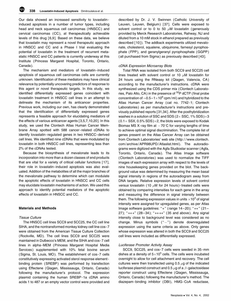
Figure 1. The mevalonate pathway.
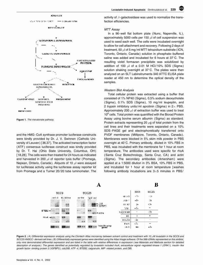
Figure 2. ( A ) Differential expression analysis using the Clontech Atlas microarray between solvent control and treatment with 10 �M lovastatin in the SCC9 and
SCC25 HNSCC - derived cell lines. ( B ) Differentially expressed genes were identified using the Atlas Image software. Of the 588 cDNAs represented on this slotblot,
only nine demonstrated differential expression and are listed in the table with relative differences in expression ( see Materials and Methods section for detailed
description of analysis ). The genes identified as potentially regulated by lovastatin included rhoA, extracellular signal - regulated kinase 1 ( ERK1 ), insulin - like
growth factor - binding protein 3 ( IGFBP3 ), cdc25B, ATF -4, BTEB2, calgranulin, MIF - related protein, and DBI.
Neoplasia . Vol. 4, No. 4, 2002
Lovastatin-Induced Apoptosis Dimitroulakos et al. 339
0.05% Tween 80 (Sigma) then processed for chemilumi-
nescent detection (Amersham)]. After the desired exposure
was obtained, the membrane was stained with Coomassie
Blue (Sigma) to ensure equal loading of the samples.
Phalloidin Staining
In a four -well Labtek Chamber slide (Nunc), approx-
imately 10,000 cells in a 1-ml suspension was used to seed
each well. The cells were incubated overnight to allow for cell
attachment and recovery. Following 24 hours of treatment,
the cells were washed twice with PBS and fixed for 5 minutes
in 1.5% neutral buffered formalin (Sigma) at room temper-
ature. The cells were then permeabilized using 0.1% Triton
X-100 (Sigma) in PBS for 15 minutes. After two washes in
PBS, the cells were incubated with FITC-conjugated
phalloidin (Sigma) (1 �g/ml in PBS) for 15 minutes at room
temperature. After two washes in PBS, the slides were
mounted with a DAPI containing immunofluorescent mount-
ing media (Vector Laboratories, San Diego, CA) and viewed
by immunofluorescent microscopy.
Results
Microarray Analysis of Lovastatin Treatment in HNSCC Cell
Lines
The cellular concentrations of mevalonate metabolites,
which include de novo cholesterol, dolichol, ubiquinone,
FPP, and GGPP (Figure 1 ), can affect the activity of various
transcription factors [17]. For example, lower cholesterol
levels can activate SREBP that binds sterol response
elements (SRE) located in the promoters of various genes
affecting their transcription [12,35]. The posttranslational
modifications of a number of signaling proteins through the
addition of the isoprenoid moieties farnesyl and geranylger-
anyl are critical to their function, including their effects on
transcriptional regulation [16,17,33]. Due to the potential of
lovastatin to modulate transcription through the depletion of
mevalonate products, we evaluated the effects of this drug
on the expression of a wide range of genes using microarray
analysis.
In this study, the HNSCC-derived cell lines SCC9 and
SCC25 were treated with solvent control and 10 �M
lovastatin for 24 hours. These conditions are prior to the
onset of overt apoptosis that is triggered by lovastatin in
these cell line models [9 ]. This response is dose- and time-
dependent and the above conditions were thus specifically
used to identify potential triggers of lovastatin- induced
apoptosis [9,10]. Total RNA was extracted from treated
cells and relative expression data of 588 genes were
generated using the Clontech Atlas array system [31,34]
(Figure 2A ). Of the 588 genes examined, only nine showed
significant differences between control and lovastatin-
treated cells (Figure 2B ). These differences included genes
involved in the ras signaling cascade [rhoA [2], extracellular
signal - regulated kinase 1 (ERK1) [30] ], growth regulation
( insulin- like growth factor -binding protein 3 [21], cdc25B
[11,26] ), transcription factors [ATF-4 [18], basic transcrip-
tional element -binding protein 2 (BTEB2) [42] ], calcium
regulation [calgranulin, migration inhibitory factor (MIF)–
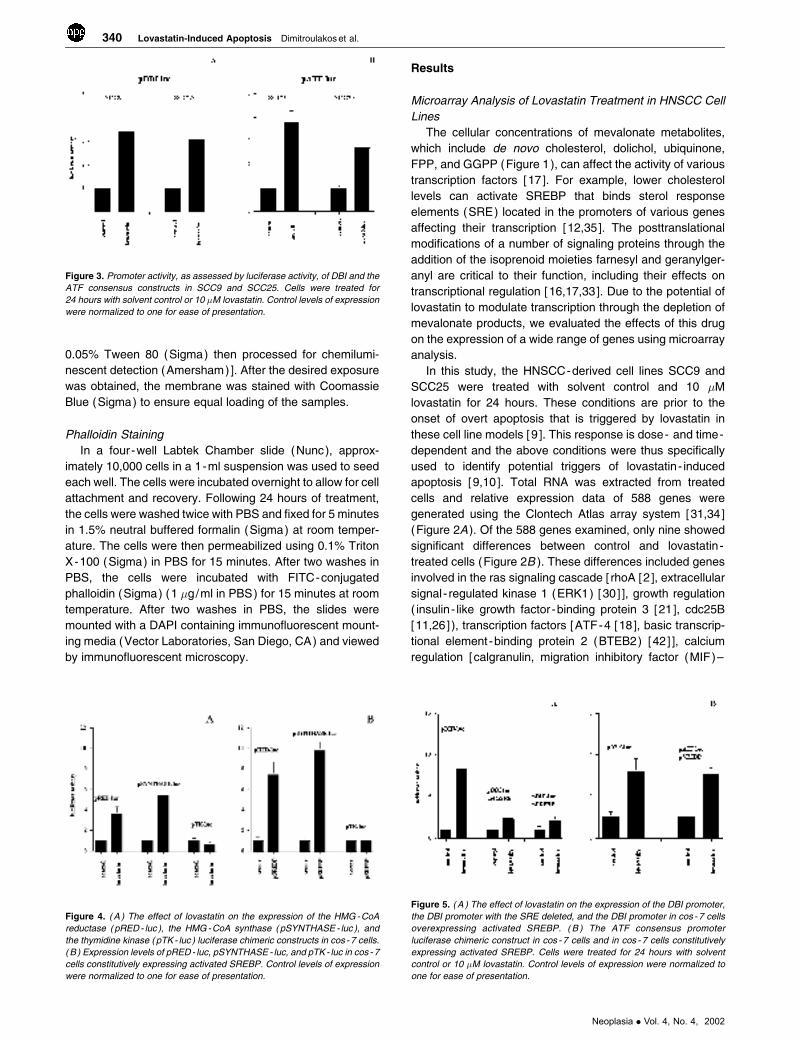
Figure 3. Promoter activity, as assessed by luciferase activity, of DBI and the
ATF consensus constructs in SCC9 and SCC25. Cells were treated for
24 hours with solvent control or 10 �M lovastatin. Control levels of expression
were normalized to one for ease of presentation.
Figure 4. ( A ) The effect of lovastatin on the expression of the HMG -CoA
reductase (pRED - luc ), the HMG -CoA synthase ( pSYNTHASE - luc ), and
the thymidine kinase ( pTK - luc ) luciferase chimeric constructs in cos -7 cells.
( B ) Expression levels of pRED - luc, pSYNTHASE - luc, and pTK - luc in cos - 7
cells constitutively expressing activated SREBP. Control levels of expression
were normalized to one for ease of presentation.
Figure 5. ( A ) The effect of lovastatin on the expression of the DBI promoter,
the DBI promoter with the SRE deleted, and the DBI promoter in cos - 7 cells
overexpressing activated SREBP. ( B ) The ATF consensus promoter
luciferase chimeric construct in cos - 7 cells and in cos - 7 cells constitutively
expressing activated SREBP. Cells were treated for 24 hours with solvent
control or 10 �M lovastatin. Control levels of expression were normalized to
one for ease of presentation.
340 Lovastatin-Induced Apoptosis Dimitroulakos et al.
Neoplasia . Vol. 4, No. 4, 2002
related protein [22,23] ] and metabolism (DBI [36,37] ). Of
interest, DBI is a known SREBP-regulated gene [36,37] and
the ATF family of transcription factors regulates the
expression of a large number of genes involved in cell
growth, differentiation, and homeostasis. Furthermore, a
member of this family is also released from the endoplasmic
reticulum by the same protease that cleaves SREBP [18].
This potential mechanistic link was further evaluated.
Promoter Analysis of DBI and the ATF Consensus
Sequence
To evaluate the effects of lovastatin on DBI expression
and the ability of lovastatin to regulate ATF target genes, we
used a luciferase-based promoter assay. The DBI promoter
[36,37] and a plasmid construct that contained the con-
sensus-binding site for the ATF family of transcription
factors were used [18,28]. Using both constructs, lovastatin
induced three- to four- fold increases in expression from
these promoters in both the SCC9 and SCC25 cell lines
(Figure 3 ). These results confirm DBI as a target of
lovastatin exposure, but also demonstrate that ATF target
genes may be regulated and play a role in the biological
effects of targeting HMG-CoA reductase in these cells.
To further evaluate the effect of lovastatin on the
expression of DBI and ATF target genes, we evaluated
lovastatin’s effects on these promoters in the nontrans-
formed monkey kidney cell line cos-7. In this model, we
confirmed that two mevalonate pathway enzymes that are
known to be regulated by lovastatin through SREBP, HMG-
CoA synthase, and HMG-CoA reductase [37] show three-
to six - fold induction with lovastatin, whereas the thymidine
kinase (TK) promoter was not affected (Figure 4A ). To
establish the role of SREBP in these responses and to
evaluate its potential role on the effects of lovastatin on the
DBI promoter and the ATF consensus promoter constructs,
we constitutively expressed the activated form of SREBP in
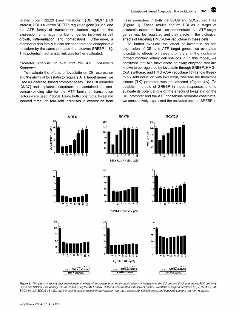
Figure 6. The effect of adding back mevalonate, cholesterol, or squalene on the cytotoxic effects of lovastatin in the CC cell line SIHA and the HNSCC cell lines
SCC9 and SCC25. Cell viability was assessed using the MTT assay. Cultures were treated with solvent control, lovastatin at its predetermined LD50 (SIHA 15 �M;
SCC9 50 �M; SCC25 35 �M ), and increasing concentrations of mevalonate ( top row ), cholesterol ( middle row ), and squalene ( bottom row ) for 48 hours.
Neoplasia . Vol. 4, No. 4, 2002
Lovastatin-Induced Apoptosis Dimitroulakos et al. 341

the in the cos-7 cell line. As expected, the expression of
activated SREBP resulted in elevated basal expression of
both HMG-CoA synthase and HMG-CoA reductase, but the
minimal TK promoter that lacks an SRE was unaffected
compared to empty vector transfection control (Figure 4B ).
These results demonstrated that activated SREBP
expressed in cos-7 cells was functional.
For the DBI promoter, we evaluated the effects of
lovastatin in cos-7 cells, with a DBI promoter construct that
lacked its SRE and in cos-7 cells constitutively expressing
active SREBP. The DBI promoter exhibited similar but more
pronounced effects to the HNSCC cell lines with a 5- to 12-
fold activation in response to treatment with 10 �M lovastatin
for 24 hours (Figure 5A ). Deletion of the SRE within this
promoter construct abrogated this induction (Figure 5A ).
Overexpression of activated SREBP muted the induction
elicited by lovastatin as well (Figure 5A ), and taken together,
these results indicate that SREBP was modulating the
response of the DBI promoter to lovastatin treatment.
For the ATF consensus promoter construct, we evaluated
the effects of lovastatin in cos-7 cells and in cos-7 cells
constitutively expressing active SREBP. The ATF consen-
sus promoter in cos-7 cells showed similar results to the
HNSCC cell lines with a three- to four- fold induction
following lovastatin exposure (Figure 5B ). The ATF con-
sensus promoter, in contrast to the DBI promoter, does not
possess an SRE and was not affected by SREBP over-
expression (Figure 5B ). These results indicate that the
potential effects of lovastatin on the expression of ATF target
genes are not dependent on SREBP activation but upon the
activation of the ATF family of transcription factors.
Biochemical Analysis of Lovastatin- Induced Apoptosis
The cytotoxic effects of lovastatin and the inhibition of this
cytotoxicity by the various products of the mevalonate
pathway were evaluated using the MTT assay. We used
the HNSCC cell lines SCC9 and SCC25 and the CC cell line
SIHA. SIHA was included in this study as an alternative SCC
Figure 7. The effect of adding back ubiquinone, FPP, and GPP on the cytotoxic effects of lovastatin in the CC cell line SIHA and the HNSCC cell lines SCC9 and
SCC25. Cell viability was assessed using the MTT assay. Cultures were treated with solvent control, lovastatin at its predetermined LD50 ( SIHA 15 �M; SCC9 50 �M;
SCC25 35 �M ), and increasing concentrations of ubiquinone ( top row ), FPP (middle row ), and GPP ( bottom row ) for 48 hours.
342 Lovastatin-Induced Apoptosis Dimitroulakos et al.
Neoplasia . Vol. 4, No. 4, 2002
derived from a different tissue site. The cell lines were
treated with solvent control, a concentration of lovastatin that
gave a 50% lethal dose [9], and a combination of lovastatin
with increasing concentrations of the mevalonate metabo-
lites used in this study. In all of the cell lines tested, the
cytotoxicity triggered by the 50% lethal dose of lovastatin
was inhibited following exposure to mevalonate in a dose-
dependent manner (Figure 6 ). The concentration range
tested was 2.5 to 100 �M and the inhibitory effect was
evident at 10 �M concentration.
There are a number of end products downstream of
mevalonate and it is, therefore, of interest to address which
of these components is /are essential for lovastatin- induced
cytotoxicity. In the cholesterol biosynthesis pathway, two
products, squalene or cholesterol, were added to the culture
medium at concentrations between 2.5 and 100 �M. These
compounds were ineffective in blocking the cytotoxicity
induced by lovastatin (Figure 6 ). The other mevalonate
metabolite tested that had no protective effects on lovasta-
tin - induced cytotoxicity was ubiquinone at concentrations of
1 to 100 �M (Figure 7 ).
To determine whether protein isoprenylation is critical to
the cytotoxic effects of lovastatin, the cell lines were also
exposed to solvent control or a broad concentration range of
GGPP or FPP. Coincubation with 0.5 to 20 �M GGPP
blocked the loss of viability caused by lovastatin exposure in
a dose-dependent manner. As shown in Figure 7, this
protective effect was detectable at a range of 1.0 to 5.0 �M
GGPP. Interestingly, at the same concentration range, FPP
had only partial protective effects.
RhoA as a Potential Target of Lovastatin- Induced
Apoptosis
In our microarray analysis, we identified rhoA as a gene
that was potentially regulated by lovastatin. RhoA is a
member of the Rho family of small GTPases that regulate
actin cytoskeletal reorganization, thereby regulating cell
shape and motility [2,14]. Rho proteins have been implicated
in cancer cell invasion, growth, and survival [25,27], and
require the isoprenoid moiety GGPP for their function. GGPP
facilitates their membrane localization and transduction of
upstream signals [27].
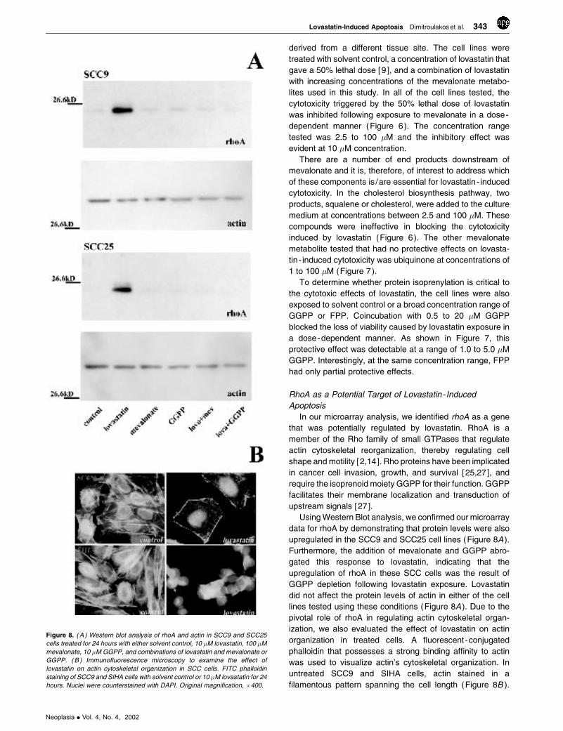
Using Western Blot analysis, we confirmed our microarray
data for rhoA by demonstrating that protein levels were also
upregulated in the SCC9 and SCC25 cell lines (Figure 8A ).
Furthermore, the addition of mevalonate and GGPP abro-
gated this response to lovastatin, indicating that the
upregulation of rhoA in these SCC cells was the result of
GGPP depletion following lovastatin exposure. Lovastatin
did not affect the protein levels of actin in either of the cell
lines tested using these conditions (Figure 8A ). Due to the
pivotal role of rhoA in regulating actin cytoskeletal organ-
ization, we also evaluated the effect of lovastatin on actin
organization in treated cells. A fluorescent-conjugated
phalloidin that possesses a strong binding affinity to actin
was used to visualize actin’s cytoskeletal organization. In
untreated SCC9 and SIHA cells, actin stained in a
filamentous pattern spanning the cell length (Figure 8B ).
Figure 8. ( A ) Western blot analysis of rhoA and actin in SCC9 and SCC25
cells treated for 24 hours with either solvent control, 10 �M lovastatin, 100 �M
mevalonate, 10 �M GGPP, and combinations of lovastatin and mevalonate or
GGPP. ( B ) Immunofluorescence microscopy to examine the effect of
lovastatin on actin cytoskeletal organization in SCC cells. FITC phalloidin
staining of SCC9 and SIHA cells with solvent control or 10 �M lovastatin for 24
hours. Nuclei were counterstained with DAPI. Original magnification, �400.
Neoplasia . Vol. 4, No. 4, 2002
Lovastatin-Induced Apoptosis Dimitroulakos et al. 343
Upon treatment with 10 �M lovastatin for 24 hours, this
staining pattern was dramatically altered with distinct stress
fibers forming on the periphery of the cells and disorganized
actin in the cytoplasm. This pattern was observed prior to the
induction of apoptosis as DAPI-counterstained nuclei did not
display nuclear condensation or fragmentation characteristic
of apoptosis (Figure 8B ). These data verify that lovastatin’s
effect on rhoA expression and function occurred prior to the
induction of apoptosis.
Discussion
The conversion of HMG-CoA to mevalonate is catalyzed by
HMG-CoA reductase, the rate- limiting enzyme of the
mevalonate pathway [17]. Mevalonate is the precursor of
isoprene units incorporated into both sterol and nonsterol
compounds such as cholesterol, dolichol, ubiquinone,
GGPP, and FPP [17]. Cholesterol is a component of cellular
membrane structure as well as a precursor for biosynthesis
of steroid hormones and bile acids [17]. Dolichol, in its
phosphorylated form, works as a carrier molecule of
oligosaccharides in N - linked protein glycosylation [17].
Ubiquinone functions as an electron acceptor in the
mitochondrial respiratory chain as well as an antioxidant
with an important function in the inhibition of lipid perox-
idation [17]. Farnesyl transferase and geranylgeranyl trans-
ferases utilize FPP and GGPP, respectively, for
posttranslational isoprenylation of proteins [16,33]. The
numbers of proteins that are modified by prenylation include
ras and many small G proteins such as members of the Rab,
Rac, and Rho families [16,32,33,38].
Inhibition of the mevalonate pathway by HMG-CoA
reductase inhibitors results in depletion of mevalonate, the
direct product of the enzyme reaction, and prevents the
biosynthesis of downstream products inhibiting sterol syn-
thesis, protein isoprenylation, and disruption of N -glycosy-
lation [4,19]. A number of studies, including our own, have
demonstrated that the targeting of HMG-CoA reductase
leads to a pronounced tumor-specific apoptotic response
that may represent a novel therapeutic approach in these
cancers [1,6,9,10,29]. The mechanism by which depletion of
these end products can result in a tumor-specific apoptotic
response remains unknown. Because the cellular concen-
tration of many of these products, in particular cholesterol,
can affect the transcription of a number of genes by
regulating the activity of various transcription factors
[12,16,17], the modulators of the anticancer effects of
lovastatin may be mediated by its gene targets. As
mevalonate metabolites are involved in respiration, glyco-
sylation, and posttranslational modification of proteins
[16,17], the end products of this pathway that may be critical
for the apoptotic effects of lovastatin may not involve direct
transcriptional regulation of lovastatin- regulated genes.
We used microarray analysis to identify gene expression
changes as potential mediators of lovastatin’s apoptotic
effects. We identified a number of genes that were regulated
by lovastatin and further characterized two promoters that
were regulated by sterol (DBI) and nonsterol (ATF
consensus) mechanisms and the GGPP-modified small
GTPase rhoA. The ATF family of transcription factors, which
includes ATF-4, binds to the ATF consensus motif
TGACGTCA that is present in many cellular promoters
[18]. This suggests that this family of transcription factors
may be involved in the regulation of the expression of
multiple genes. To our knowledge, this is the first demon-
stration that targeting HMG-CoA reductase can potentially
result in the regulation of ATF-driven promoters. The ability
of lovastatin to activate the ATF consensus promoter through
a nonsterol mechanism also suggests that its apoptotic
effects may be mediated by nonsterol mevalonate metabo-
lites. Our work indicates that this agent may activate a
number of transcriptional regulatory pathways and validates
this approach as a feasible method to identify mediators of
lovastatin’s anticancer effects.
We also evaluated the effect of various mevalonate
metabolites on lovastatin’s activity. Several studies have
shown that mevalonate can rescue lovastatin- induced cell
death in a variety of different tumor cells, including acute
myeloid leukemias and medulloblastoma cells [41,43]. We
found that mevalonate inhibited lovastatin- induced cell
death in HNSCC and CC cell lines. Taken together, these
results demonstrate that the cytotoxic effects of lovastatin
are due to its ability to inhibit mevalonate synthesis. Our data
clearly show that the depletion of mevalonate is responsible
for lovastatin- induced apoptosis in HNSCC and CC cells,
suggesting that blocking the production of specific mevalo-
nate derivatives must be involved in this process. Choles-
terol biosynthesis is the major metabolic branch in the
mevalonate pathway and certain aspects of cholesterol
metabolism appear to be relevant to cancer [17]. However,
no protective effects against lovastatin cytotoxicity could be
observed from the addition of squalene or cholesterol. These
results suggest that the inhibition of cholesterol production is
not critical to lovastatin- induced cytotoxicity in these cells.
In addition to cholesterol and squalene, we have also
shown that ubiquinone is ineffective in preventing lovastatin-
induced cytotoxicity in HNSCC and CC cells. It has been
reported that ubiquinone supplementation prevents lovasta-
tin - induced myopathy [39]; therefore, it may be a useful
supplement for those patients unable to tolerate this side
effect associated with lovastatin. The failure of these various
compounds to overcome the cytotoxicity of lovastatin
strongly suggests that other product(s) of mevalonate are
involved in the control of cell survival. Prenylated proteins are
posttranslationally modified at or near the carboxyl terminus
by formation of cysteine thioethers with the isoprenoid lipid
substrates FPP or GGPP [33]. These include small GTP-
binding proteins, such as Ras, Rho, Raf, Rab, Rac, and Rap,
that are involved in important cellular functions, such as the
regulation of proliferation, signal transduction, intracellular
transport, and cell death [32,38]. In this the study, we have
shown that GGPP inhibited lovastatin- induced cytotoxicity in
HNSCC and CC cells, whereas FPP only partially blocked
lovastatin’s effect.
The reversal of lovastatin- induced apoptosis in HNSCC
and CC cells by GGPP is likely due to the replenishment of
344 Lovastatin-Induced Apoptosis Dimitroulakos et al.
Neoplasia . Vol. 4, No. 4, 2002

the intracellular pool of GGPP that is depleted by lovastatin
treatment. By contrast, FPP leads only to a partial reversal of
lovastatin- induced apoptosis and is likely the result of partial
conversion of FPP to GGPP, as FPP is a substrate for other
downstream metabolites including GGPP [17]. Alternatively,
proteins that are normally geranylgeranylated may be
farnesylated under conditions of intracellular GGPP short-
age [27]. A number of studies have shown that GGPP can
reverse the apoptotic effects of lovastatin in various tumor-
derived cells including acute myeloid leukemias, colon
adenocarcinomas, and medulloblastomas [1,8,29,41,43].
Our results imply a common mechanism or targets within
these tumor types and HNSCC and CC. Furthermore, we
identified rhoA as a lovastatin - regulated gene in SCC that
requires GGPP modification for its membrane localization
and function [2,27]. RhoA plays an integral role in actin
cytoskeletal organization and regulates cell adhesion,
morphology, motility, and invasion [2,14,25,27]. As such,
rhoA has been implicated as a potential regulator of tumor
cell invasion, growth, and survival [2,14,25,27]. Thus, rhoA
may play a significant role in the anticancer properties of
lovastatin. The identification of nonsterol -mediated gene
targets of lovastatin and the GGPP reversal of its apoptotic
effects implicate the nonsterol metabolites of mevalonate as
mediators of lovastatin- induced apoptosis in HNSCC and
CC. Targeting the formation of these nonsterol metabolites
or their cellular targets may, therefore, lead to more refined
therapeutic approaches.
Acknowledgements
Support from the Charlie Conacher Research Fund (S.K.R. ),
The Princess Margaret Hospital Foundation (S.K.R. ), and
the Canadian Institute of Health Research ( fellowship; J.D.)
is gratefully acknowledged. We also thank J. V. Swinnen and
T. Hai for generously providing reagents used in this study.
References[1] Agarwal B, Rao CV, Bhendwal S, Ramey WR, Shirin H, Reddy BS, and
Holt PR (1999). Lovastatin augments sulindac - induced apoptosis in
colon cancer cells and potentiates chemopreventive effect of sulindac.
Proc Am Assoc Cancer Res 40, 57.
[2] Aznar S, and Lacal JC (2001). Rho signals to cell growth and apoptosis.
Cancer Lett 165, 1– 10.
[3] Cooper CS (2001). Applications of microarray technology in breast
cancer research. Breast Cancer Res 3, 158 – 75.
[4] Corsini A, Maggi FM, and Catapano AL (1995). Pharmacology of
competitive inhibitors of HMG -CoA reductase. Pharmacol Res 31, 9–
27.
[5] Cunningham MJ (2000). Genomics and proteomics: the new millen-
nium of drug discovery and development. J Pharmacol Toxicol Methods
44, 291 –300.
[6] Dimitroulakos J, Nohynek D, Backway KL, Hedley DW, Yeger H,
Freedman MH, Minden MD, and Penn LZ (1999). Increased sensitivity
of acute myeloid leukemias to lovastatin - induced apoptosis: a potential
therapeutic approach. Blood 93, 1308 – 18.
[7] Dimitroulakos J, Pienkowska M, Sun P, Farooq S, Zielenska M,
Squire JA, and Yeger H (1999). Identification of a novel zinc finger
gene, zf5 - 3, as a potential mediator of neuroblastoma differentiation.
Int J Cancer 81, 970 – 78.
[8] Dimitroulakos J, Thai S, Wasfy GH, Hedley DW, Minden MD, and Penn
LZ (2000). Lovastatin induces a pronounced differentiation response in
acute myeloid leukemias. Leuk Lymphoma Res 40, 167 –78.
[9] Dimitroulakos J, Ye LY, Benzaquen M, Moore MJ, Kamel - Reid S,
Freedman MH, Yeger H, and Penn LZ (2001). Differential sensitivity of
various pediatric cancers and squamous cell carcinomas to lovastatin -
induced apoptosis: therapeutic implications. Clin Cancer Res 7, 158 – 67.
[10] Dimitroulakos J, and Yeger H (1996). HMG - CoA reductase mediates
the biological effects of retinoic acid on human neuroblastoma cells:
lovastatin specifically targets P - glycoprotein – expressing cells. Nat
Med 2, 326 – 33.
[11] Eckstein JW (2000). Cdc25 as a potential target of anticancer agents.
Invest New Drugs 18, 149 –56.
[12] Edwards PA, Tabor D, Kast HR, and Venkateswaran A (2000).
Regulation of gene expression by SREBP and SCAP. Biochim Biophys
Acta 1529, 103 – 13.
[13] Evan GI, Wyllie AH, Gilbert CS, Littlewood TD, Land H, Brooks M,
Waters CM, Penn LZ, and Hancock DC (1992). Induction of apoptosis
in fibroblasts by c - myc protein. Cell 69, 119 – 28.
[14] Evers EE, Zondag GC, Malliri A, Price LS, ten Klooster JP, van der
Kammen RA, and Collard JG (2000). Rho family proteins in cell
adhesion and cell migration. Eur J Cancer 36, 1269 –74.
[15] Fisher DE (1994). Apoptosis in cancer therapy: crossing the threshold.
Cell 78, 539 – 42.
[16] Gibbs JB (1991). Ras C - terminal processing enzymes — new drug
targets? Cell 65, 1 – 4.
[17] Goldstein JL, and Brown MS (1990). Regulation of the mevalonate
pathway. Nature 343, 425 – 30.
[18] Hai T, and Hartman MG (2001). The molecular biology and
nomenclature of the activating transcription factor / cAMP responsive
element binding family of transcription factors: activating transcription
factor proteins and homeostasis. Gene 273, 1 – 11.
[19] Hunninghake DB (1992). HMG -CoA reductase inhibitors. Curr Opin
Lipidol 3, 22 –28.
[20] Kallioniemi OP (2001). Biochip technologies in cancer research. Ann
Med 33, 142 – 47.
[21] Kelley KM, Oh Y, Gargosky SE, Gucev Z, Matsumoto T, Hwa V, Ng L,
Simpson DM, and Rosenfeld RG (1996). Insulin - like growth factor –
binding proteins ( IGFBPs ) and their regulatory dynamics. Int J
Biochem Cell Biol 28, 619 –37.
[22] Kerkhoff C, Eue I, and Sorg C (1999). The regulatory role of MRP8
(S100A8 ) and MRP14 ( S100A9 ) in the transendothelial migration of
human leukocytes. Pathobiology 67, 230 – 32.
[23] Kerkhoff C, Klempt M, Kaever V, and Sorg C (1999). The two calcium -
binding proteins, S100A8 and S100A9, are involved in the metabolism
of arachidonic acid in human neutrophils. J Biol Chem 274, 32672 – 79.
[24] Kerr JF, Winterford CM, and Harmon BV (1994). Apoptosis. Its
significance in cancer and cancer therapy. Cancer 73, 2013 –26.
[25] Kusama T, Mukai M, Iwasaki T, Tatsuta M, Matsumoto Y, Akedo H, and
Nakamura H (2001). Inhibition of epidermal growth factor – induced
RhoA translocation and invasion of human pancreatic cancer cells by
3 - hydroxy - 3 -methylglutaryl - coenzyme a reductase inhibitors. Cancer
Res 61, 4885 –91.
[26] Lammer C, Wagerer S, Saffrich R, Mertens D, Ansorge W, and
Hoffmann I (1998). The cdc25B phosphatase is essential for the G2 / M
phase transition in human cells. J Cell Sci 111, 2445 – 53.
[27] Lebowitz PF, and Prendergast GC (1998). Non - Ras targets of
farnesyltransferase inhibitors: focus on Rho. Oncogene 17, 1439 – 45.
[28] Liang G, and Hai T (1997). Characterization of human activating
transcription factor 4, a transcriptional activator that interacts with
multiple domains of cAMP - responsive element - binding protein
(CREB ) -binding protein. J Biol Chem 272, 24088 –95.
[29] Macaulay RJ, Wang W, Dimitroulakos J, Becker LE, and Yeger H
(1999). Lovastatin - induced apoptosis of human medulloblastoma cell
lines in vitro. J Neuro -Oncol 42, 1– 11.
[30] Marais R, and Marshall CJ (1996). Control of the ERK MAP kinase
cascade by Ras and Raf. Cancer Surv 27, 101 – 25.
[31] Pandita A, Zielenska M, Thorner P, Bayani J, Godbout R, Greenberg M,
and Squire JA (1999). Application of comparative genomic hybridiza-
tion, spectral karyotyping, and microarray analysis in the identification
of subtype -specific patterns of genomic changes in rhabdomyosarco-
ma. Neoplasia 1, 262 – 75.
[32] Pruitt K, and Der CJ (2001). Ras and Rho regulation of the cell cycle
and oncogenesis. Cancer Lett 171, 1 –10.
[33] Sebti SM, and Hamilton AD (2000). Farnesyltransferase and geranyl-
geranyl transferase I inhibitors and cancer therapy: lessons from
mechanism and bench - to - bedside translational studies. Oncogene 19,
6584 – 93.
[34] Sehgal A, Boynton AL, Young RF, Vermeulen SS, Yonemura KS, Kohler
EP, Aldape HC, Simrell CR, and Murphy GP (1998). Application of the
Neoplasia . Vol. 4, No. 4, 2002
Lovastatin-Induced Apoptosis Dimitroulakos et al. 345

differential hybridization of Atlas Human expression arrays technique in
the identification of differentially expressed genes in human glioblasto-
ma multiforme tumor tissue. J Surg Oncol 67, 234 – 41.
[35] Shimano H, Yahagi N, Amemiya -Kudo M, Hasty AH, Osuga J, Tamura
Y, Shionoiri F, Iizuka Y, Ohashi K, Harada K, et al. (1999). Sterol
regulatory element - binding protein - 1 as a key transcription factor for
nutritional induction of lipogenic enzyme genes. J Biol Chem 274,
35832 – 39.
[36] Swinnen JV, Alen P, Heyns W, and Verhoeven G (1998). Identification
of diazepam - binding inhibitor / Acyl - CoA – binding protein as a sterol
regulatory element - binding protein - responsive gene. J Biol Chem 273,
19938 – 44.
[37] Swinnen JV, Ulrix W, Heyns W, and Verhoeven G (1997). Coordinate
regulation of lipogenic gene expression by androgens: evidence for a
cascade mechanism involving sterol regulatory element binding
proteins. Proc Natl Acad Sci USA 94, 12975 – 80.
[38] Takai Y, Sasaki T, and Matozaki T (2001). Small GTP - binding proteins.
Physiol Rev 81, 153 – 208.
[39] Thibault A, Samid D, Tompkins AC, Figg WD, Cooper MR, Hohl RJ,
Trepel J, Liang B, Patronas N, Venzon DJ, et al. (1996). Phase 1 study
of lovastatin, an inhibitor of the mevalonate pathway, in patients with
cancer. Clin Cancer Res 2, 483 – 91.
[40] Thompson GR, and Naoumova RP (2000). Novel lipid - regulating
drugs. Expert Opin Invest Drugs 9, 2619 – 28.
[41] Wang W, and Macaulay RJ (1999). Mevalonate prevents lovastatin -
induced apoptosis in medulloblastoma cell lines. Can J Neurol Sci 26,
305 – 10.
[42] Watanabe N, Kurabayashi M, Shimomura Y, Kawai - Kowase K,
Hoshino Y, Manabe I, Watanabe M, Aikawa M, Kuro -o M, Suzuki T,
et al. (1999). BTEB2, a Kruppel - like transcription factor, regulates
expression of the SMemb / nonmuscle myosin heavy chain B (SMemb /
NMHC -B ) gene. Circ Res 85, 182 – 91.
[43] Xia Z, Tan MM, Wong WW, Dimitroulakos J, Minden MD, and Penn LZ
(2001). Blocking protein geranylgeranylation is essential for lovastatin -
induced apoptosis of human acute myeloid leukemia cells. Leukemia
15, 1398 –407.
346 Lovastatin-Induced Apoptosis Dimitroulakos et al.
Neoplasia . Vol. 4, No. 4, 2002
Copyright © 2022 FDOKUMEN